Embed Size (px)
Citation preview
EAACI
European
Position Paper on Rhinosinusitis
and Nasal Polyps
Participants:
Wytske Fokkens, Chair Valerie Lund, Co-Chair Claus Bachert Peter Clement Peter Helllings Mats Holmstrom Nick Jones Livije Kalogjera, David Kennedy Marek Kowalski Henrik Malmberg Joaquim Mullol Desiderio Passali Heinz Stammberger Pontus Stierna
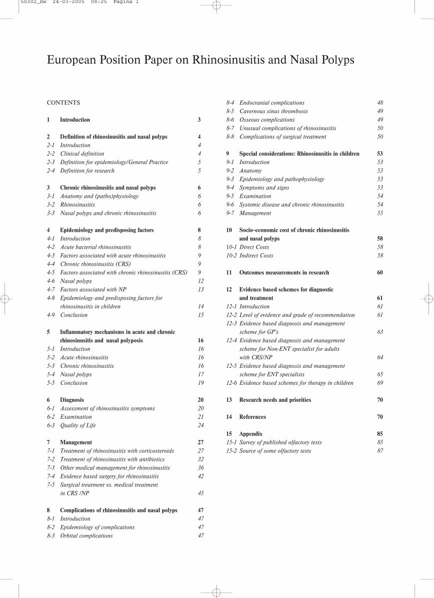
CONTENTS
1 Introduction 3
2 Definition of rhinosinusitis and nasal polyps 42-1 Introduction 4
2-2 Clinical definition 4
2-3 Definition for epidemiology/General Practice 5
2-4 Definition for research 5
3 Chronic rhinosinusitis and nasal polyps 63-1 Anatomy and (patho)physiology 6
3-2 Rhinosinusitis 6
3-3 Nasal polyps and chronic rhinosinusitis 6
4 Epidemiology and predisposing factors 84-1 Introduction 8
4-2 Acute bacterial rhinosinusitis 8
4-3 Factors associated with acute rhinosinusitis 9
4-4 Chronic rhinosinusitis (CRS) 9
4-5 Factors associated with chronic rhinosinusitis (CRS) 9
4-6 Nasal polyps 12
4-7 Factors associated with NP 13
4-8 Epidemiology and predisposing factors for
rhinosinusitis in children 14
4-9 Conclusion 15
5 Inflammatory mechanisms in acute and chronic rhinosinusitis and nasal polyposis 16
5-1 Introduction 16
5-2 Acute rhinosinusitis 16
5-3 Chronic rhinosinusitis 16
5-4 Nasal polyps 17
5-5 Conclusion 19
6 Diagnosis 206-1 Assessment of rhinosinusitis symptoms 20
6-2 Examination 21
6-3 Quality of Life 24
7 Management 277-1 Treatment of rhinosinusitis with corticosteroids 27
7-2 Treatment of rhinosinusitis with antibiotics 32
7-3 Other medical management for rhinosinusitis 36
7-4 Evidence based surgery for rhinosinusitis 42
7-5 Surgical treatment vs. medical treatment
in CRS /NP 45
8 Complications of rhinosinusitis and nasal polyps 478-1 Introduction 47
8-2 Epidemiology of complications 47
8-3 Orbital complications 47
8-4 Endocranial complications 48
8-5 Cavernous sinus thrombosis 49
8-6 Osseous complications 49
8-7 Unusual complications of rhinosinusitis 50
8-8 Complications of surgical treatment 50
9 Special considerations: Rhinosinusitis in children 539-1 Introduction 53
9-2 Anatomy 53
9-3 Epidemiology and pathophysiology 53
9-4 Symptoms and signs 53
9-5 Examination 54
9-6 Systemic disease and chronic rhinosinusitis 54
9-7 Management 55
10 Socio-economic cost of chronic rhinosinusitis and nasal polyps 58
10-1 Direct Costs 58
10-2 Indirect Costs 58
11 Outcomes measurements in research 60
12 Evidence based schemes for diagnostic and treatment 61
12-1 Introduction 61
12-2 Level of evidence and grade of recommendation 61
12-3 Evidence based diagnosis and management
scheme for GP’s 63
12-4 Evidence based diagnosis and management
scheme for Non-ENT specialist for adults
with CRS/NP 64
12-5 Evidence based diagnosis and management
scheme for ENT specialists 65
12-6 Evidence based schemes for therapy in children 69
13 Research needs and priorities 70
14 References 70
15 Appendix 8515-1 Survey of published olfactory tests 85
15-2 Source of some olfactory tests 87
European Position Paper on Rhinosinusitis and Nasal Polyps
50302_bw 24-03-2005 08:25 Pagina 1
European Position Paper on Rhinosinusitis and Nasal Polyps 3
Rhinosinusitis is a significant health problem which seems tomirror the increasing frequency of allergic rhinitis and whichresults in a large financial burden on society (1-3). The lastdecade has seen the development of a number of guidelines,consensus documents and position papers on the epidemiolo-gy, diagnosis and treatment of rhinosinusitis and nasal polypo-sis (4-6).
Data on (chronic) rhinosinusitis is limited and the disease enti-ty is badly defined. Therefore, the available data is difficult tointerpret and extrapolate. Although of considerable assistance,the available consensus documents on chronic rhinosinusitisand nasal polyps do not answer a number of relevant questionsthat would unify the information and current concepts thatexist in epidemiology, diagnosis, treatment and research. Toadd to this, none of these documents are evidence based.
There is considerable interest in guidelines as tools for imple-menting health care based on proof of effectiveness.Guidelines should be informative, simple and easy to use andin a form that can be widely disseminated within the medicalcommunity in order to improve patient care.
Evidence-based medicine is an important method of preparingguidelines (7, 8). Moreover, the implementation of guidelinesis equally important.
The European Academy of Allergology and ClinicalImmunology (EAACI) has created a Taskforce to considerwhat is known about rhinosinusitis and nasal polyps, to offerevidence based recommendations on diagnosis and treatment,and to consider how we can make progress with research inthis area. The EP3OS document is also approved by theEuropean Rhinologic Society (ERS).
The present document is intended to be state-of-the art for thespecialist as well as for the general practitioner:• to update their knowledge of rhinosinusitis and nasal poly-
posis;• to provide an evidence-based documented revision of the
diagnostic methods;• to provide an evidence-based revision of the available treat-
ments;• to propose a stepwise approach to the management of the
disease;• to propose guidance for definitions and outcome measure-
ments in research in different settings.
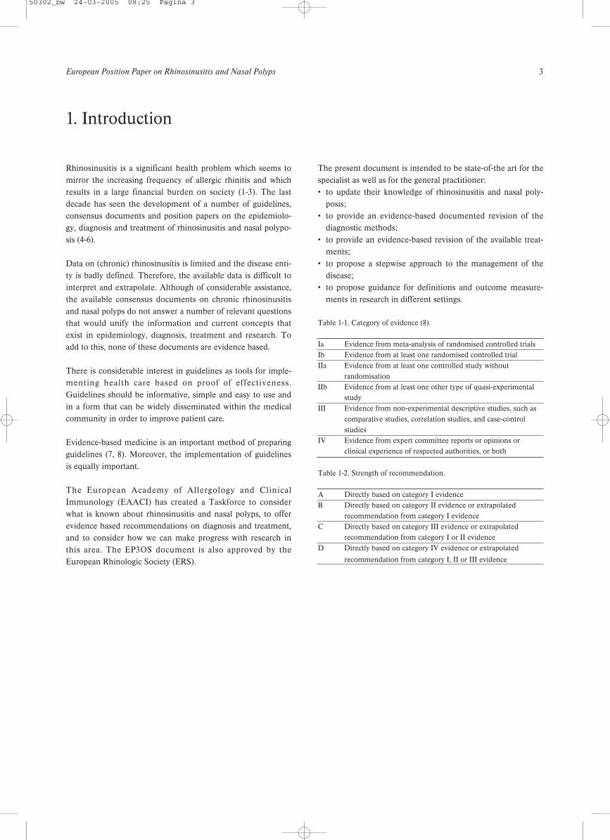
Table 1-1. Category of evidence (8).
Ia Evidence from meta-analysis of randomised controlled trialsIb Evidence from at least one randomised controlled trialIIa Evidence from at least one controlled study without
randomisationIIb Evidence from at least one other type of quasi-experimental
studyIII Evidence from non-experimental descriptive studies, such as
comparative studies, correlation studies, and case-controlstudies
IV Evidence from expert committee reports or opinions orclinical experience of respected authorities, or both
Table 1-2. Strength of recommendation.
A Directly based on category I evidenceB Directly based on category II evidence or extrapolated
recommendation from category I evidenceC Directly based on category III evidence or extrapolated
recommendation from category I or II evidenceD Directly based on category IV evidence or extrapolated
recommendation from category I, II or III evidence
1. Introduction
50302_bw 24-03-2005 08:25 Pagina 3
4 Supplement 18
2-1 IntroductionRhinitis and sinusitis usually coexist and are concurrent inmost individuals; thus, the correct terminology is now rhinosi-nusitis. The diagnosis of rhinosinusitis is made by a wide vari-ety of practitioners, including allergologists, otolaryngologists,pulmonologists, primary care physicians and many others.Therefore, an accurate, efficient, and accessible definition ofrhinosinusitis is required. A number of groups have publishedreports on rhinosinusitis and its definition. In most of thesereports definitions are based on symptomatology and durationof disease and one definition aims at all practitioners (4-6, 9).
In 2001 the WHO put together a working group on rhinitis andits impact on asthma (ARIA)(10). In this group rhinitis wasclassified according to duration and severity.
Table 2-1. Classification of allergic rhinitis (10).
1- “Intermittent” means that the symptoms are present:• Less than 4 days a week,• And for less than 4 weeks.
2- “Persistent” means that the symptoms are present:• More than 4 days a week,• Or for more than 4 weeks. (should it be “and”, not or?)
3- “Mild” means that there are none of the following items:• No sleep disturbance,• No impairment of daily activities, leisure and/or sport,• No impairment of school or work,• Symptoms are not troublesome.
4- “Moderate-severe” means that there are one or more of thefollowing items:• Sleep disturbance,• Impairment of daily activities, leisure and/or sport,• Impairment of school or work,
• Troublesome are symptoms.
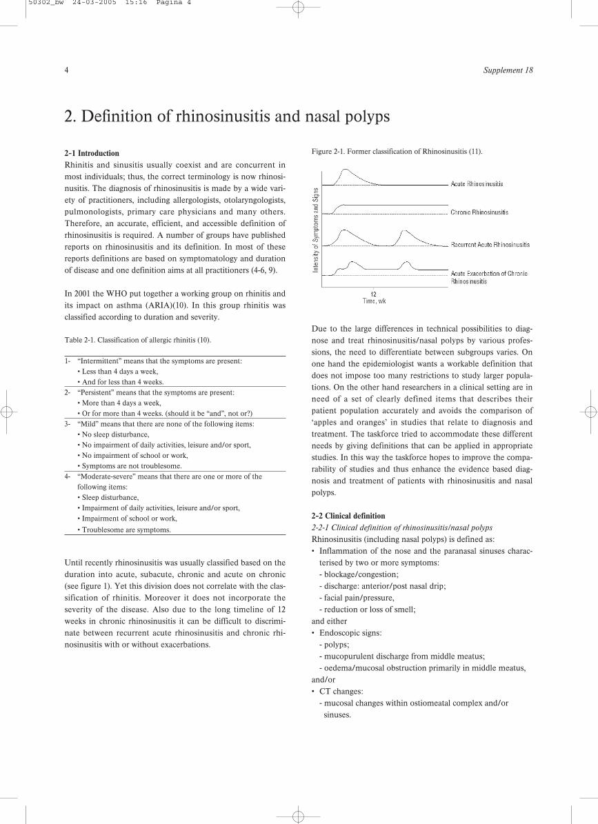
Until recently rhinosinusitis was usually classified based on theduration into acute, subacute, chronic and acute on chronic(see figure 1). Yet this division does not correlate with the clas-sification of rhinitis. Moreover it does not incorporate theseverity of the disease. Also due to the long timeline of 12weeks in chronic rhinosinusitis it can be difficult to discrimi-nate between recurrent acute rhinosinusitis and chronic rhi-nosinusitis with or without exacerbations.
Figure 2-1. Former classification of Rhinosinusitis (11).
Due to the large differences in technical possibilities to diag-nose and treat rhinosinusitis/nasal polyps by various profes-sions, the need to differentiate between subgroups varies. Onone hand the epidemiologist wants a workable definition thatdoes not impose too many restrictions to study larger popula-tions. On the other hand researchers in a clinical setting are inneed of a set of clearly defined items that describes theirpatient population accurately and avoids the comparison of‘apples and oranges’ in studies that relate to diagnosis andtreatment. The taskforce tried to accommodate these differentneeds by giving definitions that can be applied in appropriatestudies. In this way the taskforce hopes to improve the compa-rability of studies and thus enhance the evidence based diag-nosis and treatment of patients with rhinosinusitis and nasalpolyps.
2-2 Clinical definition2-2-1 Clinical definition of rhinosinusitis/nasal polyps
Rhinosinusitis (including nasal polyps) is defined as:• Inflammation of the nose and the paranasal sinuses charac-
terised by two or more symptoms:- blockage/congestion;- discharge: anterior/post nasal drip;- facial pain/pressure, - reduction or loss of smell;
and either• Endoscopic signs:
- polyps;- mucopurulent discharge from middle meatus;- oedema/mucosal obstruction primarily in middle meatus,
and/or• CT changes:
- mucosal changes within ostiomeatal complex and/or sinuses.
2. Definition of rhinosinusitis and nasal polyps
50302_bw 24-03-2005 15:16 Pagina 4
European Position Paper on Rhinosinusitis and Nasal Polyps 5
2-2-2 Severity of the disease
The disease can be divided into MILD and MODERATE/SEVERE based on total severity visual analogue scale (VAS)score (010 cm):
MILD = AS 0-4 MODERATE/SEVERE = VAS 5-10
To evaluate the total severity the patient is asked to indicateon a VAS the question:
How troublesome are your symptoms of rhinosinusitis?
2-2-3 Duration of the disease
Acute/Intermittent< 12 weeksComplete resolution of symptoms.
Chronic/Persistent>12 weeks symptomsNo complete resolution of symptoms.
2-3 Definition for epidemiology/General Practice
For epidemiological studies the definition is based on sympto-matology without ENT examination or radiology.
Acute/Intermittent Rhinosinusitis is defined assudden onset of two or more of the symptoms:
blockage/congestion;discharge anterior/post nasal drip;facial pain/pressure;reduction/loss of smell;
for <12 weeks,with symptom free intervals if the problem is intermittent,with validation by telephone or interview.Questions on allergic symptoms i.e. sneezing, watery rhinorhea, nasal itching and itchy watery eyes should be included.
Common cold/viral rhinosinusitis is defined as:duration of symptoms for less than 10 days.
Acute/Intermittent non-viral rhinosinusitis is defined as:increase of symptoms after 5 days or persistent symptoms after10 days with less than 12 weeks duration.
Persistent/Chronic Rhinosinusitis/nasal polyps is defined as:nasal congestion/obstruction/blockage with
facial pain/pressure, ordiscoloured discharge(anterior / posterior nasal drip), orreduction/loss of smell
for >12 weeks,with validation by telephone or interview.Questions on allergic symptoms i.e. sneezing, watery rhino
rhea, nasal itching and itchy watery eyes should be included.Also include questions on intermittent disease (see definitionabove).
2-4 Definition for research
For research purposes Chronic Rhinosinusitis (CRS) is themajor finding and Nasal Polyposis (NP) is considered a sub-group of this entity. For the purpose of a study, the differentia-tion between CRS and NP must be based on out-patientendoscopy.
The research definition is based on the presence of polyps andprior surgery.
2-4-1 Definitions when no earlier sinus surgery has been per-
formed
Polyposis: bilateral, endoscopically visualised inmiddle meatus
Chronic rhinosinusitis: bilateral, no visible polyps in middlemeatus, if necessary following decon-gestant
This definition accepts that there is a spectrum of disease inCRS which includes polypoid change in the sinuses and/ormiddle meatus but excludes those with polypoid disease pre-senting in the nasal cavity to avoid overlap.
2-4-2 Definitions when sinus surgery has been performed
Once surgery has altered the anatomy of the lateral wall, thepresence of polyps is defined as pedunculated lesions asopposed to cobblestoned mucosa > 6 months after surgery onendoscopic examination. Any mucosal disease without overtpolyps should be regarded as CRS .
2-4-3 Conditions for sub-analysis
The following conditions should be considered for sub-analysis:• aspirin sensitivity based on positive oral, bronchial or nasal
provocation or an obvious history;• asthma/bronchial hyper-reactivity /COPD based on symp-
toms, respiratory function tests;• allergy based on specific serum IgE or SPTs;• finding of purulent discharge/pus.
2-4-4 Exclusion from general studies
Patients with the following diseases should be excluded fromgeneral studies on chronic rhinosinusitis and/or nasal polypo-sis:• cystic fibrosis based on positive sweat test or DNA alleles;• gross immunodeficiency (congenital or acquired);• congenital mucociliary problems e.g. primary ciliary dyskine-
sia (PCD);• non-invasive fungal balls and invasive fungal disease;• systemic vasculitic and granulomatous diseases.
Not troublesome Most troublesome imaginable10 cm
50302_bw 24-03-2005 08:25 Pagina 5
6 Supplement 18
3-1 Anatomy and (patho)physiology
The nose and paranasal sinuses constitute a collection of air-filled spaces within the anterior skull. The paranasal sinusescommunicate with the nasal cavity through small apertures.The nasal cavity and its adjacent paranasal sinuses are lined bypseudostratified columnar ciliated epithelium. This containsgoblet cells and nasal glands, producers of nasal secretions thatkeep the nose moist and form a “tapis roulant” of mucus.Particles and bacteria can be caught in this mucus, renderedharmless by enzymes like lysozyme and lactoferrin, and betransported down towards the oesophagus. Cilia play animportant role in mucus transport. All paranasal sinuses arenormally cleared by this mucociliary transport, even thoughtransport from large areas of sinuses passes through smallopenings towards the nasal cavity.
A fundamental role in the pathogenesis of rhinosinusitis isplayed by the ostiomeatal complex, a functional unit that com-prises maxillary sinus ostia, anterior ethmoid cells and theirostia, ethmoid infundibulum, hiatus semilunaris and middlemeatus. The key element is the maintenance of optimal sinusventilation and clearance. Specifically, ostial patency signifi-cantly affects mucus composition and secretion; moreover, anopen ostium allows mucociliary clearance to easily remove par-ticulate matters and bacteria eventually come in contact withthe sinusal mucosa.Problems occur if the orifice is too small for the amount ofmucus, if mucus production is increased, for instance duringan upper respiratory tract infection (URI), or if ciliary functionis impaired. Stasis of secretions follows and bacterial exportceases, causing or exacerbating inflammation of the mucosawhilst aeration of the mucosa is decreased, causing even moreciliary dysfunction. This vicious cycle can be difficult to break,and if the condition persists, it can result as chronic rhinosi-nusitis. In chronic rhinosinusitis the role of ostium occlusionseems to be less pronounced than in acute rhinosinusitis.
3-2 Rhinosinusitis
Rhinosinusitis is an inflammatory process involving themucosa of the nose and one or more sinuses. The mucosa ofthe nose and sinuses form a continuum and thus more oftenthan not the mucous membranes of the sinus are involved indiseases which are primarily caused by an inflammation of thenasal mucosa. Chronic rhinosinusitis is a multifactorial disease(12). Factors contributing can be mucociliary impairment (13,14), (bacterial) infection (15), allergy (16), swelling of themucosa for another reason, but only rarely physical obstruc-tions caused by morphological/anatomical variations in the
nasal cavity or paranasal sinuses (17, 18). A role in the patho-genesis of rhinosinusitis is certainly played by the ostiomeatalcomplex, a functional unit that comprises maxillary sinus ostia,anterior ethmoid cells and their ostia, ethmoid infundibulum,hiatus semilunaris and middle meatus. The key element is themaintenance of the ostial patency. An in depth discussion onfactors contributing to chronic rhinosinusitis and nasal polypscan be found in chapter 4-4 and 4-6.
3-3 Nasal polyps and chronic rhinosinusitis
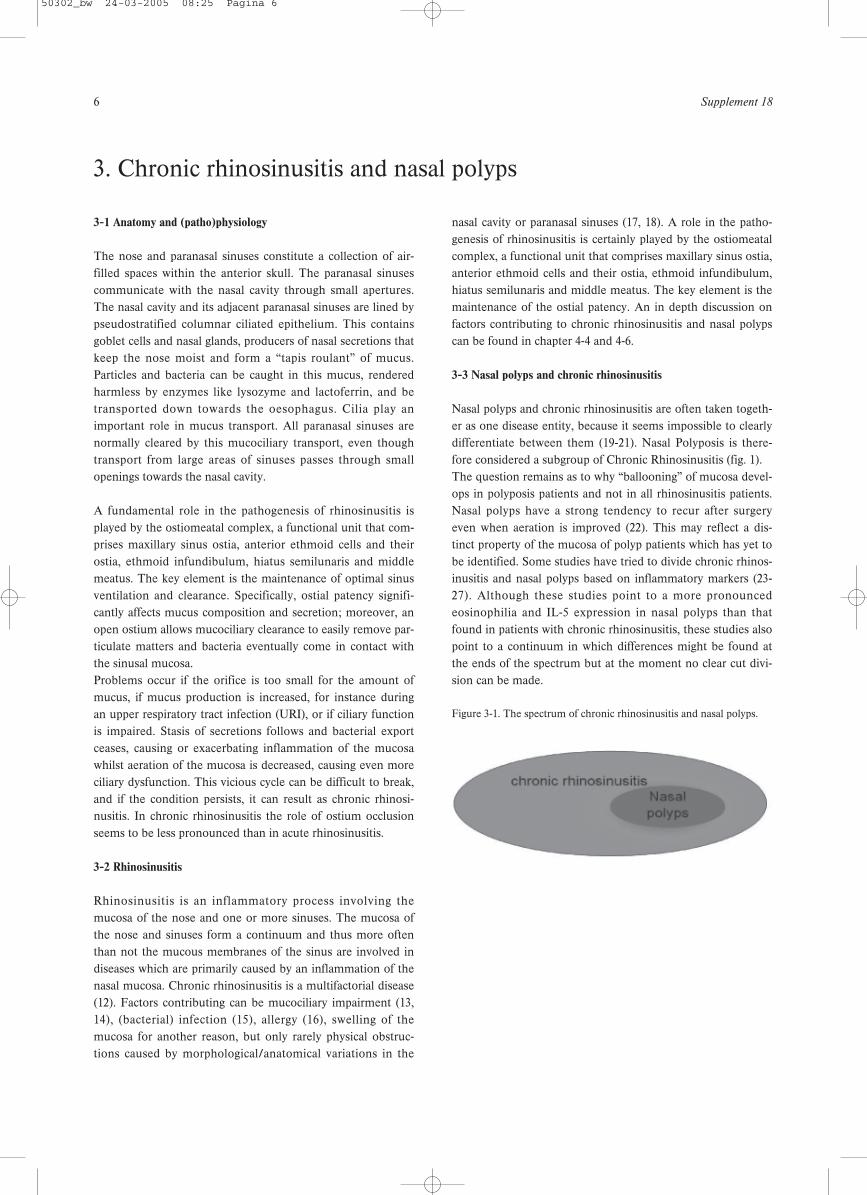
Nasal polyps and chronic rhinosinusitis are often taken togeth-er as one disease entity, because it seems impossible to clearlydifferentiate between them (19-21). Nasal Polyposis is there-fore considered a subgroup of Chronic Rhinosinusitis (fig. 1). The question remains as to why “ballooning” of mucosa devel-ops in polyposis patients and not in all rhinosinusitis patients.Nasal polyps have a strong tendency to recur after surgeryeven when aeration is improved (22). This may reflect a dis-tinct property of the mucosa of polyp patients which has yet tobe identified. Some studies have tried to divide chronic rhinos-inusitis and nasal polyps based on inflammatory markers (23-27). Although these studies point to a more pronouncedeosinophilia and IL-5 expression in nasal polyps than thatfound in patients with chronic rhinosinusitis, these studies alsopoint to a continuum in which differences might be found atthe ends of the spectrum but at the moment no clear cut divi-sion can be made.
Figure 3-1. The spectrum of chronic rhinosinusitis and nasal polyps.
3. Chronic rhinosinusitis and nasal polyps
50302_bw 24-03-2005 08:25 Pagina 6
European Position Paper on Rhinosinusitis and Nasal Polyps 7
Nasal polyps appear as grape-like structures in the upper nasalcavity, originating from within the ostiomeatal complex. Theyconsist of loose connective tissue, oedema, inflammatory cellsand some glands and capillaries, and are covered with varyingtypes of epithelium, mostly respiratory pseudostratified epithe-lium with ciliated cells and goblet cells. Eosinophils are themost common inflammatory cells in nasal polyps, but neu-trophils, mast cells, plasma cells, lymphocytes and monocytesare also present, as well as fibroblasts. IL-5 is the predominantcytokine in nasal polyposis, reflecting activation and prolongedsurvival of eosinophils (28).
The reason why polyps develop in some patients and not inothers remains unknown. There is a definite relationship in
patients with ‘Samter triad’: asthma, NSAID sensitivity andnasal polyps. However, not all patients with NSAID sensitivityhave nasal polyps, and vice-versa. In the general population,the prevalence of nasal polyps is 4% (29). In patients with asth-ma, a prevalence of 7 to 15% has been noted whereas, inNSAID sensitivity, nasal polyps are found in 36 to 60% ofpatients (30, 31). It had long been assumed that allergy predis-posed to nasal polyps because the symptoms of watery rhinor-rhoea and mucosal swelling are present in both diseases, andeosinophils are abundant. However, epidemiological data pro-vide no evidence for this relationship: polyps are found in 0.5to 1.5% of patients with positive skin prick tests for commonallergens (31, 32).
50302_bw 24-03-2005 08:25 Pagina 7
8 Supplement 18
4-1 Introduction
The incidence of acute viral rhinosinusitis (common cold) isvery high. It has been estimated that adults suffer 2 to 5 coldsper year, and school children may suffer 7 to 10 colds per year.The exact incidence is difficult to measure because mostpatients with common cold do not consult a doctor. More reli-able data are available on acute rhinosinusitis. As mentionedearlier acute non-viral rhinosinusitis is defined as an increase ofsymptoms after 5 days or persistent symptoms after 10 daysafter a sudden onset of two or more of the symptoms: block-age/congestion, discharge, anterior/post nasal drip, facialpain/pressure and/or reduction/loss of smell. It is estimatedthat only 0.5% to 2% of viral URTIs are complicated by bacteri-al infection; however, the exact incidence is unknown giventhe difficulty distinguishing viral from bacterial infection with-out invasive sinus-puncture studies. Bacterial culture results insuspected cases of acute community-acquired sinusitis are posi-tive in only 60% of cases (33). Signs and symptoms of bacterialinfection may be mild and often resolve spontaneously (34, 35).In spite of the high prevalence and significant morbidity ofchronic rhinosinusitis and nasal polyps, there is only limitedaccurate data on the epidemiology of these conditions. Thisobservation mainly relates to the lack of a uniformly accepteddefinition for CRS. In addition, patient selection criteria great-ly differ between epidemiologic studies complicating compari-son of studies. When interpreting epidemiologic data, one should be aware ofa significant selection bias of the different studies presentedbelow. The purpose of this section of the EPOS document isto give an overview of the currently available epidemiologicdata on rhinosinusitis and nasal polyps, and illustrate the fac-tors which are believed to predispose to the development.
4-2 Acute bacterial rhinosinusitis
When describing the incidence of acute bacterial rhinosinusitisthere has been a lot of debate about the definition of acutebacterial rhinosinusitis. For example in the Cochrane Reviewon antibiotics for acute sinusitis, studies were included ifsinusitis was proven by a consistent clinical history, and radi-ographic or aspiration evidence of acute sinusitis (36).However, most guidelines on the diagnosis of acute bacterialrhinosinusitis base the diagnosis on symptoms and clinicalexamination. However, if the diagnosis is based on clinicalexamination alone, the rate of false positive results is high. Inpatients with clinical diagnosis of acute rhinosinusitis less thanhalf have significant abnormalities at X-ray examination (37).Based on sinus puncture/aspiration (considered diagnosticallythe most accurate), 49-83% of symptomatic patients had acute
sinusitis (38). Compared with puncture/aspiration, radiographyoffered moderate ability to diagnose sinusitis Using sinus opac-ity or fluid as the criterion for sinusitis, radiography had sensi-tivity of 0.73 and specificity of 0.80 (38).An average of 8.4% of the Dutch population reported at leastone episode of acute rhinosinusitis per year in 1999 (39). Theincidence of visits to the general practioner for of acute sinusi-tis in the Netherlands in 2000 was 20.0 per 1,000 men and 33.8per 1,000 women (40). According to National AmbulatoryMedical Care Survey (NAMCS) data in the USA rhinosinusitisis the fifth most common diagnosis for which an antibiotic isprescribed. Rhinosinusitis accounted for 9% and 21% of allpaediatric and adult antibiotic prescriptions, respectively, writ-ten in 2002 (5).
4-3 Factors associated with acute rhinosinusitis
4-3-1 Pathogens
Superinfection of bacteria on mucosa damaged by viral infec-tion (common cold) is the most important cause of acute rhi-nosinusitis. The most common bacterial species isolated fromthe maxillary sinuses of patients with acute rhinosinusitis areStreptococcus pneumoniae, Haemophilus influenzae, andMoraxella catarrhalis, the latter being more common in chil-dren (41, 42). Other streptococcal species, anaerobic bacteriaand Staphylococcus aureus cause a small percentage of cases.Resistance patterns of the predominant pathogens vary consid-erably (43, 44). The prevalence and degree of antibacterialresistance in common respiratory pathogens are increasingworldwide. The association between antibiotic consumptionand the prevalence of resistance is widely assumed (45).
4-3-2 Ciliary impairment
Normal mucociliary flow is a significant defence mechanism inthe prevention of acute rhinosinusitis. Viral rhinosinusitisresults in the loss of cilia and ciliated cells, with a maximumaround one week after the infection. Three weeks after thebeginning of the infection the number of cilia and ciliated cellsincreases to nearly normal. However, as a sign of regeneration,immature short cilia (0.7 to 2.5 microns in length) were oftenseen (46). The impaired mucociliary function during viral rhi-nosinusitis results in an increased sensitivity to bacterial infec-tion.
Also in animal experimental work it was shown that early afterexposure to pathogenic bacteria, like Streptococcus pneumoni-ae, Hemophilus influenzae, Pseudomonas aeruginosa, a signif-icant loss of ciliated cells from sinus mucosa and a correspond-ing disruption of normal mucociliary flow was found (47).
4. Epidemiology and predisposing factors
50302_bw 24-03-2005 08:25 Pagina 8
European Position Paper on Rhinosinusitis and Nasal Polyps 9
4-3-3 Allergy
Review articles on sinusitis have suggested that atopy predis-poses to rhinosinusitis (48). This theory is attractive given thepopularity of the concept that disease in the ostiomeatal areacontributes to sinus disease in that the mucosa in an individualwith allergic rhinitis might be expected to be swollen and moreliable to obstruct sinus ostia, reduce ventilation, lead to mucusretention that might be more prone to become infected.Furthermore there has been an increase in the body of opinionthat regard the mucosa of the nasal airway as being in a contin-uum with the paranasal sinuses and hence the term rhinosi-nusitis (49). The number of studies determining the occur-rence of acute rhinosinusitis in patients with and without aller-gy is very limited. Savolainen studied the occurrence of allergy in 224 patientswith verified acute rhinosinusitis by means of an allergy ques-tionnaire, skin testing, and nasal smears. Allergy was found in25% of the patients and considered probable in another 6.5%.The corresponding percentages in the control group were 16.5and 3, respectively. There were no differences between allergicand non-allergic patients in the number of prior acute sinusitisepisodes or of previously performed sinus irrigations.Bacteriological and radiological findings did not differ signifi-cantly between the groups (50). Alho showed that subjectswith allergic IgE-mediated rhinitis had more severe paranasalsinus changes in CT scans than nonallergic subjects duringviral colds. These changes indicate impaired sinus functioningand may increase the risk of bacterial sinusitis (51).
In conclusions: although an attractive hypothesis we can repeatthe statement made a decade ago, there remain no publishedprospective reports on the incidence of infective rhinosinusitisin populations with and without clearly defined allergic rhinos-inusitis (52).
4-4 Chronic rhinosinusitis (CRS)
CRS is one of the most common health care problems, withsignificant direct medical costs and severe impact on lower air-way disease and general health outcomes (53, 54). The paucityof accurate epidemiologic data on CRS and nasal polyps con-trasts with the more abundant information on microbiology,diagnosis and treatment options for these conditions. Whenreviewing the current literature on CRS, it becomes clear thatgiving an accurate estimate of the prevalence of CRS remainsspeculative, because of the heterogeneity of the disorder andthe diagnostic imprecision often used in publications. In a sur-vey on the prevalence of chronic conditions, it was estimatedthat CRS, defined as having ‘sinus trouble’ for more than 3months in the year before the interview, affects 15.5% of thetotal population in the United States (55), ranking this condi-tion second in prevalence among all chronic conditions. Later,the high prevalence of CRS was confirmed by another surveysuggesting that 16% of the adult US population has CRS (56).
However the prevalence of doctor diagnosed CRS is muchlower; a prevalence of 2% was found using ICD-9 codes as anidentifier(57). Of note, the prevalence rate of CRS was substantially higher infemales with a female/male ratio of 6/4 (55). In Canada, preva-lence of CRS, defined as an affirmative answer to the question‘Has the patient had sinusitis diagnosed by a health profession-al lasting for more than 6 months?’ ranged from 3.4% in maleto 5.7% in female subjects (58). The prevalence increased withage, with a mean of 2.7% and 6.6% in the age groups of 20-29and 50-59 years respectively. After the age of 60 years, preva-lence levels of CRS levelled off to 4.7% (58). In a nationwidesurvey in Korea, the overall prevalence of chronic sinusitis,defined as the presence of at least 3 nasal symptoms lastingmore than 3 months along with the endoscopic finding of anasal polyp and/or mucopurulent discharge within the middlemeatus, was 1.01% (59), without differences neither in agegroups nor in sexes. By screening a non-ENT population,which may be considered representative of the general popula-tion in Belgium, Gordts et al. (60) reported that 6% of subjectssuffered from chronic nasal discharge and 40% had signs ofmucosal swelling of more than 3 mm on MRI .Notwithstanding the shortcomings of epidemiologic studies onCRS, it represents a common disorder of multifactorial origin.A list of factors will be discussed in the following chapterwhich are believed to be etiologically linked to CRS.
4-5 Factors associated with chronic rhinosinusitis (CRS)
4-5-1 Ciliary impairment
As may be concluded from the section on anatomy and patho-physiology, ciliary function plays an important role in theclearance of the sinuses and the prevention of chronic inflam-mation. Secondary ciliary dyskinesia is found in patients withchronic rhinosinusitis, and is probably reversible, althoughrestoration takes some time (61) It will be clear that in patientswith Kartagener’s syndrome and primary ciliary dyskinesia,chronic rhinosinusitis is a common problem. These patientsusually have a long history of respiratory infections. In patientswith cystic fibrosis (CF), the inability of the cilia to transportthe viscous mucus causes ciliary malfunction and consequentlychronic rhinosinusitis. Nasal polyps are present in about 40%of patients with CF (62). These polyps are generally more neu-trophilic than eosinophilic in nature but may respond tosteroids as well, as inhaled steroids in patients with CF reduceneutrophilic inflammation (63-65).
4-5-2 Allergy
Review articles on rhinosinusitis have suggested that atopypredisposes to its development (48, 66). It is tempting to spec-ulate that allergic inflammation in the nose predisposes theatopic individual to the development of CRS. Both conditionsshare the same trend of increasing prevalence (67, 68) and arefrequently associated.
50302_bw 24-03-2005 08:25 Pagina 9
10 Supplement 18
It has been postulated (69) that swelling of the nasal mucosa inallergic rhinitis at the site of the sinus ostia may compromiseventilation and even obstruct sinus ostia, leading to mucusretention and infection. Furthermore, there has been anincrease in the body of opinion that regard the mucosa of thenasal airway as being in a continuum with the paranasal sinus-es and hence the term ‘rhinosinusitis’ was introduced (49).However, critical analysis of the papers linking atopy as a riskfactor to infective rhinosinusitis (chronic or acute) reveal thatwhilst many of the studies suggest a higher prevalence of aller-gy in patients presenting with symptoms consistent withsinusitis than would be expected in the general population,there may well have been a significant selection process,because the doctors involved often had an interest in allergy(27, 70-74). A number of studies report that markers of atopyare more prevalent in populations with chronic rhinosinusitis.Benninger reported that 54% of outpatients with chronic rhi-nosinusitis had positive skin prick tests (75). Among CRSpatients undergoing sinus surgery, the prevalence of positiveskin prick tests ranges from 50 to 84% (50, 76, 77), of which themajority (60%) have multiple sensitivities (77). As far back as1975, Friedman reported an incidence of atopy in 94% ofpatients undergoing sphenoethmoidectomies (78).
However, the role of allergy in CRS is questioned by other epi-demiologic studies showing no increase in the incidence ofinfectious rhinosinusitis during the pollen season in pollen-sensitized patients (52). In a small prospective study, no differ-ence in prevalence of purulent rhinosinusitis was foundbetween patients with and without allergic rhinitis (79).Furthermore, allergy was found in 31.5% of patients with veri-fied acute maxillary sinusitis and there were no differencesbetween allergic and non-allergic patients in the number ofprior acute sinusitis episodes (50). Newman et al. reported thatwhilst 39% of patients with CRS had asthma, raised specificIgE or an eosinophilia, only 25% had true markers to showthey were atopic (80). Finally, Emanuel et al. (77)found rela-tively lower percentages of allergic patients in the group ofpatients with the most severe sinus disease on CT scan andIwens et al. (81) reported that the prevalence and extent ofsinus mucosa involvement on CT was not determined by theatopic state.
Taken together, epidemiologic data show an increased preva-lence of allergic rhinitis in patients with CRS, but the role ofallergy in CRS remains unclear.
Radiological studies are unhelpful in unravelling the correla-tion between allergy and rhinosinusitis. High percentages ofsinus mucosa abnormalities are found on radiological imagesof allergic patients, e.g. 60% incidence of abnormalities on CTscans among subjects with ragweed allergy during the season(82). However, one should interpret this data with caution inview of the fact that high percentages of incidental findings are
found on radiological images of the sinus mucosa in individu-als without nasal complaints, ranging from 24.7% to 49.2% (83-86), that the normal nasal cycle induces cyclical changes in thenasal mucosa volume (87), and that radiological abnormalitiescontribute minimally to the patient’s symptoms (82).
Notwithstanding the lack of hard epidemiologic evidence for aclear causal relationship between allergy and CRS, it is clearthat failure to address allergy as a contributing factor to CRSdiminishes the probability of success of a surgical intervention(88). Among allergy patients undergoing immunotherapy,those who felt most helped by immunotherapy were the sub-jects with a history of recurrent rhinosinusitis, and about halfof the patients, who had had sinus surgery before, believedthat the surgery alone was not sufficient to completely resolvethe recurrent episodes of infection (88).
4-5-3 Lower airway involvement
Recent evidence suggests that allergic inflammation in theupper and lower airways coexist and should be seen as a con-tinuum of inflammation, with inflammation in one part of theairway influencing its counterpart at a distance. The argumentsand consequences of this statement are summarized in theARIA document (10). Rhinosinusitis and lower airway involve-ment are also frequently associated in the same patients, buttheir interrelationship is poorly understood. The evidence thattreatment of rhinosinusitis improves asthma symptoms andhence reduces the need for medication to control asthmamainly results from research in children and will be discussedbelow (Chapter 7-6). In short, improvements in both asthmasymptoms and medication have been obtained after surgeryfor rhinosinusitis in children with both conditions (89-91).
Studies on radiographic abnormalities of the sinuses in asth-matic patients have shown high prevalences of abnormal sinusmucosa (92, 93). All patients with steroid dependant asthmahad abnormal mucosal changes on CT compared to 88% withmild to moderate asthma (94). Again caution should be exer-cised in the interpretation of these studies. Radiographicallydetected sinus abnormalities in sensitized patients may reflectinflammation related to the allergic state rather than to sinusinfection.
4-5-4 Immunocompromised state
Among conditions associated with dysfunction of the immunesystem, congenital immunodeficiencies manifest themselveswith symptoms early in life and will be dealt with in the paedi-atric CRS section (see Chapter 7-6). However, dysfunction ofthe immune system may occur later in life and present withCRS. In a retrospective review of refractory sinusitis patients,Chee et al. found an unexpectedly high incidence of immunedysfunction (95). Of the 60 patients with in vitro T-lymphocytefunction testing, 55% showed abnormal proliferation inresponse to recall antigens. Low immunoglobulin G, A and M
50302_bw 24-03-2005 08:25 Pagina 10
European Position Paper on Rhinosinusitis and Nasal Polyps 11
titres were found in respectively 18, 17 and 5% of patients withrefractory sinusitis. Common variable immunodeficiency wasdiagnosed in 10% and selective IgA deficiency in 6% ofpatients. Therefore, immunological testing should be an inte-gral part of the diagnostic pathway of patients with CRS notresponding to conservative treatment. In a cross-sectionalstudy to assess the overall prevalence of otolaryngologic dis-eases in patients with HIV-infection, Porter et al. (96) reportedthat sinusitis was present in more than half of the HIV-positivepopulation, ranking this condition one of the most prevalentdiseases in HIV-positive persons. However, the relevance ofthese data is questioned as there was no difference in sinonasalsymptom severity between HIV-positive and AIDS patientsnor was there a correlation between CD4+ cell counts andsymptom severity. In a more detailed study, Garcia-Rodrigueset al. (97) reported a lower incidences of rhinosinusitis (34%),but with a good correlation between low CD4+ cell count andthe probability of rhinosinusitis. It should also be mentionedhere that atypical organisms like Aspergillus spp,Pseudomonas aeruginosa and microsporidia are often isolatedfrom affected sinuses and that neoplasms such as non-Hodgkin lymphoma and Kaposi’s sarcoma, may account forsinonasal problems in patients with AIDS (98).
4-5-5 Genetic factors
Although chronic sinus disease has been observed in familymembers, no genetic abnormality has been identified linked toCRS. However, the role of genetic factors in CRS has beenimplicated in patients with cystic fibrosis (CF) and primary cil-iary dyskinesia (Kartagener’s syndrome). CF is one of the mostfrequent autosomal recessive disorders of the Caucasian popu-lation, caused by mutations of the CFTR gene on chromo-some 7 (99). The most common mutation, DF508, is found in70 to 80% of all CFTR genes in Northern Europe (100, 101).Upper airway manifestations of CF patients include chronicrhinosinusitis and nasal polyps, which are found in 25 to 40%of CF patients above the age of 5 (102-105). Interestingly,Jorissen et al. (106) reported that DF508 homozygosity repre-sents a risk factor for paranasal sinus disease in CF.
4-5-6 Pregnancy and endocrine state
During pregnancy, nasal congestion occurs in approximatelyone-fifth of women (107). The pathogenesis of this disorderremains unexplained, but there have been a number of pro-posed theories. Besides direct hormonal effects of oestrogen,progesterone and placental growth hormone on the nasalmucosa, indirect hormonal effects like vascular changes maybe involved. Whether pregnancy rhinitis predisposes to thedevelopment of sinusitis, is not clear. In a small prospectivestudy, Sobol et al. (108) report that 61% of pregnant womenhad nasal congestion during the first trimester, whereas only3% had sinusitis. In this study, a similar percentage of non-pregnant women in the control group developed sinusitis dur-ing the period of the study. Also in an earlier report, the inci-
dence of sinusitis in pregnancy was shown to be quite low, i.e.1.5% (109).
In addition, thyroid dysfunction has been implicated in CRS,but there is only limited data on the prevalence of CRS inpatients with hypothyroidism.
4-5-7 Local host factors
Certain anatomic variations such as concha bullosa, nasal sep-tal deviation and a displaced uncinate process, have been sug-gested as potential risk factors for developing CRS (110).However, Bolger et al. (111) found no correlation betweenCRS and bony anatomic variations in the nose. Also in thesurvey by Min et al. (112), no correlation was found betweenseptal deviation and the prevalence of CRS. However, oneshould mention here that no study has so far investigatedwhether a particular anatomic variation can impair drainage ofthe ostiomeatal complex per se. Whilst some authors havepostulated that anatomical variations of the paranasal sinusescan contribute to ostial obstruction (113) there are severalstudies that show the prevalence of anatomical variations is nomore common in patients with rhinosinusitis or polyposis thanin a control population (17, 18, 114, 115). One area where con-jecture remains is the effect of a deviated septum. Whilst thereis no recognised method of objectively defining the extent of adeviated septum, some studies have found a deviation of morethan 3mm from the midline to be more prevalent in rhinosi-nusitis (116, 117)whilst others have not (18, 118). Takentogether, there is no evidence for a causal correlation betweennasal anatomic variations in general and the incidence of CRS.In spite of the observation that sinonasal complaints oftenresolve after surgery, this does not necessarily imply thatanatomic variation is etiologically involved.
CRS of dental origin should not be overlooked when consider-ing the aetiology of CRS. Obtaining accurate epidemiologicdata on the incidence of CRS of dental origin is not possible asthe literature is limited to anecdotal reports.
4-5-8 Micro-organisms
4-5-8-1 BacteriaAlthough it is often hypothesized that CRS evolves from acuterhinosinusitis, the role of bacteria in CRS is far from clear. Anumber of authors have described the microbiology of themiddle meatus and sinuses. However if and which of thesepathogen are contributory to the disease remains a matter ofdebate.
Arouja isolated aerobes from 86% of the middle meatus sam-ples CRS patients, anaerobes were isolated in 8%. The mostfrequent microorganisms were Staphylococcus aureus (36%),coagulase-negative Staphylococcus (20%), and Streptococcuspneumoniae (17%). Middle meatus and maxillary sinus cul-tures presented the same pathogens in 80% of cases. In healthy
50302_bw 24-03-2005 08:25 Pagina 11
12 Supplement 18
individuals, coagulase-negative Staphylococcus (56%), S.aureus (39%), and S. pneumoniae (9%) were the most frequentisolates. (119).
Some authors suggest that as chronicity develops, the aerobicand facultative species are gradually replaced by anaerobes(120, 121). This change may result from the selective pressureof antimicrobial agents that enable resistant organisms to sur-vive and from the development of conditions appropriate foranaerobic growth, which include the reduction in oxygen ten-sion and an increase in acidity within the sinus. Often polymi-crobial colonisation is found; the contribution to the disease ofthe different pathogens remains unclear.
4-5-8-2 FungiFungi have been cultured from human sinuses with many dif-ferent ramifications (122). Their presence may be relativelybenign, colonizing normal sinuses or forming saprophyticcrusts. They also may cause a range of pathology, ranging fromnon-invasive fungus balls to invasive, debilitating disease (123).
There is an increasing interest in the concept that the mostcommon form of sinus disease induced by fungus may becaused by the inflammation stimulated by airborne fungal anti-gens. In 1999 it was proposed that most patients with CRSexhibit eosinophilic infiltration and the presence of fungi byhistology or culture (124). This assertion was based on findingpositive fungal culture by using a new culture technique in 202of 210 (96%) patients with CRS who prospectively were evalu-ated in a cohort study. No increase in type I sensitivity wasfound in patients as compared with controls. The term‘‘eosinophilic chronic rhinosinusitis’’ was proposed to replacepreviously used nomenclature. Using this new culture tech-nique, the same percentage of positive fungi cultures was alsofound in normal controls (125). A broad array of fungi has been identified in the sinus cavitiesof patients with sinusitis through varied staining and culturetechniques (124, 125). As with the isolation of bacteria in sinus cavities in thesepatients, the presence of fungi does not prove that thesepathogens directly create or perpetuate disease.
4-5-9 “Osteitis”—the role of bone
Areas of increased bone density and irregular bony thickeningare frequently seen on CT in areas of chronic inflammationand may be a marker of the chronic inflammatory process.However, the effect during the initial phases of a severe chron-ic rhinosinusitis frequently appears as rarefaction of the bonyethmoid partitions. Although to date bacterial organisms havenot been identified in the bone in either humans or animalmodels of chronic rhinosinusitis, it has been suggested thatthat this irregular bony thickening is sign of inflammation ofthe bone. This inflamed bone might maintain mucosal inflam-mation (126).
In rabbit studies it was demonstrated that not only the boneadjacent to the involved maxillary sinus become involved, butthat the inflammation typically spreads through the Haversiancanals and may result in bone changes consistent with somedegree of chronic osteomyelitis at a distance from the primaryinfection (127, 128). It is certainly possible that these changes,if further confirmed in patients, may at least in part, explainwhy chronic rhinosinusitis is relatively resistant to therapy.
4-5-10 Environmental factors
Cigarette smoking was associated with a higher prevalence ofrhinosinusitis in Canada (58), whereas this observation was notconfirmed in a nationwide survey in Korea (59). Otherlifestyle-related factors are undoubtedly involved in the chron-ic inflammatory processes of rhinosinusitis. For instance, lowincome was associated with higher prevalence of CRS (58). Inspite of in vitro data on the toxicity of pollutants on respiratoryepithelium, there exists no convincing evidence for the etiolog-ic role of pollutants and toxins such as ozone in CRS.
4-5-11 Iatrogenic factors
Among risk factors of CRS, iatrogenic factors should not beforgotten as they may be responsible for the failure of sinussurgery. The increasing number of sinus mucocoeles seems tocorrelate with the expansion of endoscopic sinus surgery pro-cedures. Among a group of 42 patients with mucocoeles, 11had prior surgery within 2 years before presentation (129).Another reason for failure after surgery can be the recircula-tion of nasal mucus out of the natural maxillary ostium andback through a separate surgically created antrostomy resultingin an increased risk of persistent sinus infection (130).
4-6 Nasal polyps
In the light of epidemiologic research, a distinction needs to bemade between clinically silent NP, or preclinical cases, andsymptomatic NP. Asymptomatic polyps may transiently bepresent or persist, and hence remain undiagnosed until theyare discovered by routine examination. On the other hand,polyps that become symptomatic may remain undiagnosed,either because the patient is not investigated properly orbecause they are missed on anterior rhinoscopy. Endoscopy ofthe nasal cavity makes it possible to visualize NP and to give areliable estimate of the prevalence of NP.
In a population-based study in Skövde, Sweden, Johansson etal. (131) reported a prevalence of nasal polyps of 2.7% of thetotal population. In this study, NP were diagnosed by nasalendoscopy and were more frequent in men (2.2 to 1), theelderly (5% at 60 years of age and older) and asthmatics. In anationwide survey in Korea, the overall prevalence of polypsdiagnosed by nasal endoscopy was 0.5% of the total population(112). Based on a postal questionnaire survey in Finland,Hedman et al. (29) found that 4.3% of the adult population
50302_bw 24-03-2005 08:25 Pagina 12
European Position Paper on Rhinosinusitis and Nasal Polyps 13
answered positively to the question as to whether polyps hadbeen found in their nose. However, nasal endoscopy appearsto be a prerequisite for an accurate estimate of the prevalenceof NP, as 1.4% of the sample population studied by Johanssonet al. (131) said to have NP, did not actually have any polypson nasal endoscopy. From autopsy studies, a prevalence of 2%has been found using anterior rhinoscopy (132). After remov-ing whole naso-ethmoidal blocks, nasal polyps were found in 5of 19 cadavers (133), and in 42% of 31 autopsy samples com-bining endoscopy with endoscopic sinus surgery (134). Themedian age of the cases in the 3 autopsy studies by Larsen andTos ranged from 70 to 79 years. From these cadaver studies,one may conclude that a significant number of patients withNP do not feel the need to seek medical attention or that thediagnosis of NP is often missed by doctors.
It has been stated that between 0.2 and 1% of people developnasal polyps at some stage (135). In a prospective study on theincidence of symptomatic NP, Larsen and Tos (136) found anestimated incidence of 0.86 and 0.39 patients per thousand peryear for males and females respectively. The incidenceincreased with age, reaching peaks of 1.68 and 0.82 patientsper thousand per year for males and females respectively in theage group of 50-59 years. When reviewing data from patientrecords of nearly 5000 patients from hospitals and allergy clin-ics in the US in 1977, the prevalence of NP was found to be4.2% (137), with a higher prevalence (6.7%) in the asthmaticpatients.
In general, NP occur in all races (138-141) and becomes morecommon with age. The average age of onset is approximately42 years, which is 7 years older than the average age of theonset of asthma (142-144). NP are uncommon under the age of20 (145) and are more frequently found in men than in women(29, 136, 146), except in the population studied by Settipane(137).
4-7 Factors associated with NP
4-7-1 Allergy
0.5-4.5% of subjects with allergic rhinitis have NP (31, 32, 147),which compares with the normal population (135). In childrenthe prevalence of NP has been reported to be 0.1% (31) andKern found NP in 25.6% of patients with allergy compared to3.9% in a control population (148). On the other hand, theprevalence of allergy in patients with nasal polyps has beenreported as varying from 10% (149), to 54% (150) and 64%(151). Contrary to reports that have implicated atopy as beingmore prevalent in patients with NP, others have failed to showthis (31, 147, 152-154). Recently, Bachert at al. (155) found anassociation between levels of both total and specific IgE andeosinophilic infiltration in nasal polyps. These findings wereunrelated to skin prick test results. Positive intradermal tests tofood allergies have been reported in 81% of polyp patients
compared to 11% of controls (156). Food and drug sensitivitieshave been reported in 31% of patients with nasal polyposis andthis was more common in men (43% vs. 24%) (140).
4-7-2 Asthma
In patients with asthma 7% have nasal polyps (31) with a preva-lence of 13% in non-atopic asthma (skin prick test and totaland specific IgE negative) and 5% in atopic asthma (145). Lateonset asthma is associated with the development of nasalpolyps in 10-15% (31). Asthma develops first in approximately69% of patients with both asthma and NP and NP takebetween 9 and 13 years to develop. Ten percent develop bothpolyps and asthma simultaneously and the remainder developpolyps first and asthma later that (between 2 and 12 years)(138). However, not all patients with nasal polyps have lowerrespiratory tract symptoms (157).
Generally NP are twice as prevalent in men although the pro-portion of those with polyps and asthma is twice that inwomen than men. Women that have nasal polyps are 1.6 timesmore likely to be asthmatic and 2.7 times to have allergic rhini-tis (141).
4-7-3 Aspirin sensitivity
In patients with aspirin sensitivity 36-96% have nasal polyps(32, 145, 158-163) and up to 96% have radiographic changesaffecting their paranasal sinuses (164). Patients with aspirinsensitivity, asthma and nasal polyposis are usually non-atopicand the prevalence increases over the age of 40 years.
The children of probands with asthma, nasal polyps andaspirin sensitivity had nasal polyps and rhinosinusitis moreoften than the children of controls (165). Concerning heredi-tary factors, HLA A1/B8 has been reported as having a higherincidence in patients with asthma and aspirin sensitivity (166).
4-7-4 Genetics
An interesting observation is that NP are frequently found torun in families, suggestive of an hereditary or shared environ-mental factor. In the study by Rugina et al. (140), more thanhalf of 224 NP patients (52%) had a positive family history ofNP. The presence of NP was considered when NP had beendiagnosed by an ENT practitioner or the patients had under-gone sinus surgery for NP. A lower percentage (14%) of famil-ial occurrence of NP was reported earlier by Greisner et al. insmaller group (n = 50) of adult patients with NP (59). Thus,these results strongly suggest the existence of a hereditary fac-tor in the pathogenesis of NP. In this regard, recent geneticstudies found a significant correlation between certain HLAalleles and NP. Luxenberger et al. (167) reported an associationbetween HLA-A74 and nasal polyps, whereas Molnar-Gabor etal. (168) report that subjects carrying HLA-DR7-DQA1*0201and HLA-DR7-DQB1*0202 haplotype had a 2 to 3 times oddsratio of developing NP.
50302_bw 24-03-2005 08:25 Pagina 13
14 Supplement 18
Of note, studies of monozygotic twins have not shown bothsiblings always develop polyps, indicating that there are likelyto be environmental factors influencing their development(169, 170). Nasal polyps have been described in identical twinsbut given the prevalence of nasal polyps it might be expectedthat there would be more than a rare report of this finding(171).
4-7-5 Environmental factors
The role of environmental factors in the development of NP isunclear. No difference in the prevalence of NP has been foundin the patient’s habitat or pollution at work (140). One studyfound that a significantly smaller proportion of the populationwith polyps were smokers compared to an unselected popula-tion (15% vs. 35%) (140) whilst another found an associationbetween the use of a woodstove as a primary source of heatingand the development of NP (172).
4-8 Epidemiology and predisposing factors for rhinosinusitis inchildren
4-8-1 Epidemiology
Since the introduction of CT scanning, it has become clearthat a runny nose in a child is not only due to limited rhinitisor adenoid hypertrophy, but that in the majority of the casesthe sinuses are involved as well. Van der Veken in a CT scanstudy showed that in children with a history of chronic puru-lent rhino rhea and a nasal obstruction 64% showed involve-ment of the sinuses (173). In a MRI study of a non-ENT paedi-atric population (60) it was shown that the overall prevalenceof sinusitis signs in children is 45%. This prevalence increasesin the presence of a history of nasal obstruction to 50%, to 80%when bilateral mucosal swelling is present on rhinoscopy, to81% after a recent upper respiratory tract infection (URI), andto 100% in the presence of purulent secretions. Kristo et al.found a similar overall percentage (50%) of abnormalities onMRI in 24 school children (174). They included, however, afollow-up after 6 to 7 months, and found that about half of theabnormal sinuses on MRI findings had resolved or improvedwithout any intervention.
Unfortunately, most studies in the paediatric ENT literaturedeal with patient populations (children with nasal complaints
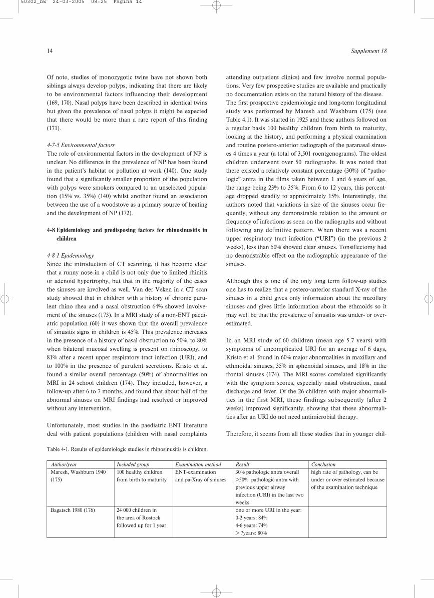
attending outpatient clinics) and few involve normal popula-tions. Very few prospective studies are available and practicallyno documentation exists on the natural history of the disease. The first prospective epidemiologic and long-term longitudinalstudy was performed by Maresh and Washburn (175) (seeTable 4.1). It was started in 1925 and these authors followed ona regular basis 100 healthy children from birth to maturity,looking at the history, and performing a physical examinationand routine postero-anterior radiograph of the paranasal sinus-es 4 times a year (a total of 3,501 roentgenograms). The oldestchildren underwent over 50 radiographs. It was noted thatthere existed a relatively constant percentage (30%) of “patho-logic” antra in the films taken between 1 and 6 years of age,the range being 23% to 35%. From 6 to 12 years, this percent-age dropped steadily to approximately 15%. Interestingly, theauthors noted that variations in size of the sinuses occur fre-quently, without any demonstrable relation to the amount orfrequency of infections as seen on the radiographs and withoutfollowing any definitive pattern. When there was a recentupper respiratory tract infection (“URI”) (in the previous 2weeks), less than 50% showed clear sinuses. Tonsillectomy hadno demonstrable effect on the radiographic appearance of thesinuses.
Although this is one of the only long term follow-up studiesone has to realize that a postero-anterior standard X-ray of thesinuses in a child gives only information about the maxillarysinuses and gives little information about the ethmoids so itmay well be that the prevalence of sinusitis was under- or over-estimated.
In an MRI study of 60 children (mean age 5.7 years) withsymptoms of uncomplicated URI for an average of 6 days,Kristo et al. found in 60% major abnormalities in maxillary andethmoidal sinuses, 35% in sphenoidal sinuses, and 18% in thefrontal sinuses (174). The MRI scores correlated significantlywith the symptom scores, especially nasal obstruction, nasaldischarge and fever. Of the 26 children with major abnormali-ties in the first MRI, these findings subsequently (after 2weeks) improved significantly, showing that these abnormali-ties after an URI do not need antimicrobial therapy.
Therefore, it seems from all these studies that in younger chil-
Table 4-1. Results of epidemiologic studies in rhinosinusitis is children.
Author/year Included group Examination method Result Conclusion
Maresh, Washburn 1940 100 healthy children ENT-examination 30% pathologic antra overall high rate of pathology, can be (175) from birth to maturity and pa-Xray of sinuses >50% pathologic antra with under or over estimated because
previous upper airway of the examination techniqueinfection (URI) in the last twoweeks
Bagatsch 1980 (176) 24 000 children in one or more URI in the year: the area of Rostock 0-2 years: 84% followed up for 1 year 4-6 years: 74%
> 7years: 80%
50302_bw 24-03-2005 08:25 Pagina 14
European Position Paper on Rhinosinusitis and Nasal Polyps 15
dren with chronic rhinosinusitis, there exists a spontaneoustendency towards recovery after the age of 6 to 8 years. Thisfinding of a decrease in prevalence of rhinosinusitis in olderchildren was also confirmed by other authors in patient popu-lations (177).
4-8-2 Predisposing factors
In an extensive prospective study Bagatsch et al. (see Table 1)saw an influence of day-care (176). If children stay in day carecentres, 72% in the group from 0 to 5 year develop one ormore episodes of upper airway infection per year compared to27% of the children staying at home.
Lind (178) and Bjuggren et al. (179) found a much higherprevalence of up to 100% of maxillary sinusitis in children stay-ing in day care centres compared with the same age group stay-ing at home or older children in schools.
The relationship between poor nasal patency and rhinosinusi-tis was confirmed by Van Cauwenberge (180) , who showed asignificant relationship between the results of passive anteriorrhinomanometry and pronounced oedema of the nasal mucosa(p=0.09 for the rights side, 0.03 for the left side) and the pres-ence of purulent rhinitis (p=0.006 for the right side and <0.05for the left side).
Breast feedings seems to have a beneficial influence on lowerrespiratory disease, but such an influence on sinusitis ininfants and young children has not yet been demonstrated(181-183).
Passive smoking is a putative risk factor, especially in allergicchildren (183). There is a clearly increased risk for recurrentcoryza (odds ratio 3.00) and sinus problems (odds ratio 4.73) inchildren with smoking mothers compared with children fromnon-smoking families (184).
4-9 Conclusion
The overview of the currently available literature illustrates thepaucity of accurate information on the epidemiology of CRSand NP, especially in European countries, and highlights theneed for large-scale epidemiologic research exploring theirprevalence and incidence. Only by the use of standardized def-initions for CRS and well-defined inclusion criteria for epi-demiologic research, will it be possible to obtain accurate epi-demiologic data on the natural evolution of CRS and NP, theinfluence of ethnic background and genetic factors on CRSand NP, and the factors associated with the disease manifesta-tion. Such studies need to be performed in order to make sig-nificant progress in the development of diagnostic and thera-peutic strategies for affected patients.
50302_bw 24-03-2005 08:25 Pagina 15
16 Supplement 18
5-1 Introduction
Rhinosinusitis is a heterogeneous group of diseases, with dif-ferent underlying aetiologies and pathomechanisms, and mayindeed represent an umbrella, covering different disease enti-ties. It is currently not understood whether acute recurrent rhi-nosinusitis necessarily develops into chronic rhinosinusitis,which then possibly gives rise to polyp growth, or whetherthese entities develop independently from each other. All ofthese items may be referred to as “rhinosinusitis”, meaning“inflammation of the nose and sinuses”; however, for didacticreasons and for future clinical and research purposes, a differ-entiation of these entities is preferred. For this purpose, we dif-ferentiate between acute rhinosinusitis (ARS), chronic rhinosi-nusitis (CRS) without polyps and chronic rhinosinusitis withnasal polyposis (NP), and omit an ill-defined group of “hyper-plastic chronic rhinosinusitis”, which might be included inCRS, or represent an overlap between CRS and NP.
5-2 Acute rhinosinusitis
Sinus mucosal tissue from subjects with acute bacterial rhinos-inusitis (ARS) is difficult to sample, with the exception ofacute complications of ARS, resulting in emergency sinussurgery. As a consequence, there is a relative lack of studies oncytokines and mediators in ARS. One of the first studiesreported in 10 subjects undergoing surgery for complications,with mucosal tissue sampled from the maxillary sinus, whichdemonstrated significantly elevated protein concentrations ofIL-8 compared to 7 controls (185) . Similar results, though notreaching significance, were obtained for IL-1ß and IL-6, where-as other cytokines such as GM-CSF, IL-5 and IL-4 were notupregulated. Recently, IL-8 and also TNF-alpha and total pro-tein content were increased in nasal lavage from subjects withARS compared to controls and allergic rhinitis subjects (186).
Proinflammatory cytokines such as IL-1ß, IL-6 and TNF play aprominent role in ongoing inflammatory reactions by activat-ing endothelial cells, T-lymphocytes and others, inducing theexpression of cell adhesion molecules and the release of othercytokines such as IL-8. IL-8 belongs to the CXC-chemokinegroup and is a potent neutrophil chemotactic protein, which isconstantly synthesized in the nasal mucosa (187). The cytokinepattern found in ARS resembles that in naturally acquired viralrhinitis lavage (188).
5-3 Chronic rhinosinusitis
5-3-1 Histopathology
In the sinus fluid of patients with chronic rhinosinusitis under-going surgery, the inflammatory cells are predominantly neu-trophils, as observed in acute rhinosinusitis, but a small num-ber of eosinophils, mast cells and basophils may also be found(189, 190). The mucosal lining in chronic rhinosinusitis is char-acterized by basement membrane thickening, goblet cellhyperplasia, subepithelial oedema, and mononuclear cell infil-tration. In a recent study evaluating the percentage ofeosinophils (out of 1000 inflammatory cells counted per visionfield), 31 patients with untreated chronic rhinosinusitis withoutnasal polyps all had less than 10% eosinophils (overall mean2%), whereas in 123 untreated nasal polyp specimen, 108 sam-ples showed more than 10% eosinophils (overall mean 50%)(191). These observations suggest that tissue eosinophilia isnot a hallmark of chronic rhinosinusitis without polyp forma-tion, and that there are major differences in the pathophysiolo-gy of both sinus diseases.
5-3-2 Pathomechanism: cytokines, chemokines and adhesion
molecules
A highly potent chemoattractant for neutrophils, IL-8 has beendemonstrated in chronic rhinosinusitis tissue (192) and IL-8protein concentrations in nasal discharge from chronic rhinosi-nusitis patients were significantly higher than in allergic rhini-tis patients in a study also involving immunohistochemistryand in situ hybridization (193). In a study measuring cytokineprotein concentrations including IL-3, IL-4, IL-5, IL-8 andGM-CSF in tissue homogenates, IL-8 was found to be signifi-cantly increased in acute rhinosinusitis, and IL-3 in chronicrhinosinusitis mucosa compared to inferior turbinate samples(194). IL-3 might be involved in the local defense and repair ofchronically inflamed sinus mucosa by supporting various cellpopulations and indirectly contributing to fibrosis and thicken-ing of the mucosa (195).
A range of mediators and cytokines has been described to beincreased in CRS versus control tissue, mostly inferiorturbinates, which comprises IL-1, IL-6, IL-8, TNF-a, IL-3, GM-CSF, ICAM-1, MPO and ECP (194, 196-198). Interestingly,VCAM-1, an adhesion molecule involved in selectiveeosinophil recruitment, and IL-5, a key cytokine for eosinophilsurvival and activity, have been shown not to be increased(194, 197). This cytokine and mediator profile resembles verymuch the profile found in viral rhinitis or acute rhinosinusitis,with the exception of a small though significant increase of
5. Inflammatory mechanisms in acute and chronic rhinosinusitisand nasal polyposis
50302_bw 24-03-2005 08:25 Pagina 16
European Position Paper on Rhinosinusitis and Nasal Polyps 17
ECP. This profile is different from the pattern in nasal polypo-sis (see below).
The expression of transforming growth factor beta 1 (TGF-b1)at protein and RNA level is significantly higher in CRS versusNP and linked to a fibrotic cross anatomy (199). In CRS,MMP-9 and TIMP-1, a natural antagonist, but not MMP-7 areincreased (200), probably resulting in a low MMP-9 activity.
5-4 Nasal polyps
5-4-1 Histopathology
Histomorphological characterisation of polyp tissue reveals fre-quent epithelial damage, a thickened basement membrane, andoedematous to sometimes fibrotic stromal tissue, with areduced number of vessels and glands, but virtually no neuralstructure (201-203). The stroma of mature polyps is mainlycharacterised by its oedematous nature and consists of support-ing fibroblasts and infiltrating inflammatory cells, localizedaround “empty” pseudocyst formations. Among the inflamma-tory cells, EG2+ (activated) eosinophils are a prominent andcharacteristic feature in about 80% of polyps (204), whereaslymphocytes and neutrophils are the predominant cells in cysticfibrosis and in CRS. Eosinophils are localised around the ves-sels, glands, and directly beneath the mucosal epithelium (202).
In small polyps, not larger than 5 mm, growing on normallooking mucosa of the middle turbinate in patients with bilat-eral polyposis, the early processes of polyp growth have beenstudied (205). Numerous subepithelial EG2+ eosinophils werepresent in the luminal compartment of the early stage polyp,forming a cap over the central pseudocyst area. In contrast,mast cells were scarce in the polyp tissue, but were normallydistributed in the pedicle and the adjacent mucosa, which hada normal appearance. This contrasts to mature polyps, wheredegranulated mast cells and eosinophils are often diffusely dis-tributed in the polyp tissue. Fibronectin deposition wasnoticed around the eosinophils in the luminal compartment ofthe early stage polyp, was accumulated subepithelially, andformed a network-like structure in the polyp centre and withinthe pseudocysts. The presence of myofibroblasts was limitedto the central pseudocyst area. Interestingly, albumin andprobably other plasma proteins were deposited within thepseudocysts, adjacent to the eosinophil infiltration. Theseobservations suggest a central deposition of plasma proteins,regulated by the subepithelial eosinophilic inflammation, as apathogenetic principle of polyp formation and growth.
5-4-2 Pathomechanism: cytokines, chemokines and adhesion
molecules
5-4-2-1 Eosinophilic inflammationA large body of studies has focussed on eosinophilic mediatorsin nasal polyp tissue, and demonstrated that different cell typesgenerate these mediators. Early studies by Denburg et al. (206,
207) demonstrated that conditioned medium, derived fromcultured nasal polyp epithelial cells, contained potenteosinophil colony-stimulating activities, as well as an inter-leukin-3-like activity. The authors suggested that accumulationof eosinophils in polyps may partly be a result of differentia-tion of progenitor cells stimulated by soluble haemopoietic fac-tors derived from mucosal cell populations. An increased syn-thesis of GM-CSF by epithelial cells, fibroblasts, monocytes,and eosinophils was suggested later (71, 208, 209). Accordingto Hamilos et al. (27), polyp tissue samples from patients withor without allergy contained different cytokine profiles. They found by in situ hybridization studies that patients with“allergic” polyps had higher tissue densities of GM-CSF, IL-3,IL-4, and IL-5 transcripts than controls, whereas patients withnon-allergic polyps had higher tissue densities of GM-CSF, IL-3, and IFN-gamma transcripts. From these results, distinctpathomechanisms for allergic versus non-allergic polyps weresuggested. Other studies involving protein measurements intissue homogenates could not support these findings (28, 194).
In contrast, IL-3 and GM-CSF protein were found in only asmall number of polyp and control turbinate samples.However, IL-5 was found to be significantly increased in nasalpolyps, compared to healthy controls, and the concentration ofIL-5 was independent of the atopic status of the patient.Indeed, the highest concentrations of IL-5 were found in sub-jects with non-allergic asthma and aspirin sensitivity.Furthermore, eosinophils were positively stained for IL-5, sug-gesting a possible autocrine role for this cytokine in the activa-tion of eosinophils, and a strong correlation between concen-trations of IL-5 protein and eosinophilic cationic protein (ECP)was demonstrated later (155). The key role of IL-5 was sup-ported by the finding that treatment of eosinophil-infiltratedpolyp tissue with neutralizing anti-IL-5 monoclonal antibody(mAB), but not anti-IL-3 or anti-GM-CSF mAbs in vitro,resulted in eosinophil apoptosis and decreased tissueeosinophilia (210).
Collectively, these studies suggest that increased production ofIL-5 is likely to influence the predominance and activation ofeosinophils in nasal polyps independent of atopy. The lack ofdifference in the amounts of cytokines detected in polyps fromallergic or non-allergic patients was meanwhile supported byseveral other studies (211, 212). Furthermore, Wagenmann etal. (213) demonstrated that both Th1 and Th2 type cytokineswere upregulated in eosinophilic NP, irrespective of allergenskin test results.
Recently, the regulation of the IL-5 receptor, which exists inthe soluble and transmembrane isoform, has been investigated(214). Whereas the probably antagonistic soluble isoform isupregulated, the signal transducing transmembrane isoform isdown-regulated in nasal polyps, especially if associated withasthma.
50302_bw 24-03-2005 08:25 Pagina 17
18 Supplement 18
Recent studies have also shown that nasal polyps also expresshigh levels of RANTES and eotaxin, the predominant recog-nised eosinophil chemoattractants. Bartels and colleagues (215)demonstrated that expression of eotaxin- and RANTESmRNA, but not MCP-3 mRNA, was elevated in non-atopicand atopic nasal polyps, when compared to normal nasalmucosa. Similarly, Jahnsen and colleagues (216) demonstratedan increased mRNA expression for eotaxin, eotaxin-2, andMCP-4. The expression of eotaxin-2, another CCR3-specificchemokine, was found to be the most prominent of the threechemokines investigated. According to other data (28, 155,205), it appears that eotaxin, rather than RANTES, in coopera-tion with IL-5, plays a key role in chemo-attraction and activa-tion of eosinophils in NP tissue. This is in accordance with thefindings of a recent extensive study of about 950 non-allergicand allergic polyp patients, which has also suggested that nasalpolyp eosinophilic infiltration and activation may correlatemainly with increased eotaxin gene expression, rather thanwith RANTES expression (217).
Studies of cell adhesion molecules are relatively few. Earlystudies by Symon and colleagues (218) demonstrated thatICAM-1, E-selectin and P-selectin were well expressed bynasal polyp endothelium, whereas VCAM-1 expression wasweak or absent. An elegant study by Jahnsen et al. (219),employing three-colour immunofluorescence staining, hashowever demonstrated that both the number of eosinophilsand the proportion of vessels positive for VCAM-1 were signif-icantly increased in nasal polyps compared with the turbinatemucosa of the same patients. Moreover, treatment with topicalglucocorticosteroids decreases the density of eosinophils andthe expression of VCAM-1 in polyps (220). The interactionbetween VLA-4 on eosinophils and VCAM-1 on endothelialcells may not only be of particular importance fortransendothelial migration of eosinophils, but may also modifytheir activation and effector functions (221).
5-4-2-2 Extracellular matrix regulationThe expression of TGF-β1 and TGF-β2, predominantly byeosinophils, and their putative effects on fibroblast activity andpathogenesis of nasal polyps have been suggested in severalstudies (222-224). These studies again compared protein levelsin tissue homogenates from patients with nasal polyps whowere either untreated or treated with oral corticosteroid, andcontrol subjects.
Patients with untreated polyp samples and controls showedsignificantly higher concentrations of IL-5, eotaxin, ECP andalbumin, and significantly lower concentrations of TGF-β1. Incontrast, corticosteroid treatment significantly reduced IL-5,ECP and albumin concentrations, whereas TGF-β1 wasincreased (205).
These observations suggest IL-5 and TGF-β1 representcytokines with counteracting activities, with a low TGF-β pro-tein concentration in IL-5 driven nasal polyps. Furthermore,they supported the deposition of albumin and other plasmaproteins as a possible pathogenic principle of polyp formation,regulated by the subepithelial eosinophilic inflammation.
TGF-β1 is a potent fibrogenic cytokine that stimulates extracel-lular matrix formation, acts as a chemoattractant for fibrob-lasts, but inhibits the synthesis of IL-5 and abrogates the sur-vival-prolonging effect of haematopoietins (IL-5 and GM-CSF)on eosinophils (225). Staining of nasal polyp tissue shows thatTGF-β1 is mainly bound to the extracellular matrix, where it isfound in its latent, inactive form.
Oedema and pseudocyst formation characterize NP, with onlya few areas of fibrosis. An imbalance of metallo-proteinaseswith an upregulation of MMP-7 and MMP-9 in nasal polypshas been recently demonstrated(200). This results in theenhancement of MMP-9 in NP, which may account for oede-ma formation with albumin retention.
5-4-2-3 Role of Staphylococcus aureus enterotoxins (SAEs)Early studies have shown that tissue IgE concentrations andthe number of IgE positive cells may be raised in nasal polyps,suggesting the possibility of local IgE production (226). Thelocal production of IgE is a characteristic feature of nasal poly-posis, with a more than tenfold increase of IgE producing plas-ma cells in NP versus controls. Analysis of specific IgErevealed a multiclonal IgE response in nasal polyp tissue andIgE antibodies to Staphylococcus aureus enterotoxins (SAEs)in about 30-50% of the patients and in about 60-80% of nasalpolyp subjects with asthma (155, 205, 227). A recent prospec-tive study revealed that colonization of the middle meatus withStaphylococcus aureus is significantly more frequent in NP(63.6%) compared to CRS (27.3%, p< 0.05), and is related tothe prevalence of IgE antibodies to classical enterotoxins (27.8vs 5.9%) (228). If aspirin sensitivity, including asthma, accom-panied nasal polyp disease, the Staph. aureus colonization ratewas as high as 87.5%, and IgE antibodies to enterotoxins werefound in 80% of cases.
Total and specific IgE in polyp homogenates is only partiallyreflected in the serum of these patients. In contrast, staining ofNP tissue revealed follicular structures characterised by B- andT-cells, and lymphoid agglomerates with diffuse plasma cellinfiltration, demonstrating the organization of secondary lym-phoid tissue with consecutive local IgE production in NP(229).
The classical SAEs, especially TSST-1 and Staphylococcus pro-tein A (SPA), are excellent candidates to induce multiclonalIgE synthesis by increasing the release of IL-4 as well as theexpression of CD40 ligand on T-cells and B7.2 on B-cells cells(230, 231).
50302_bw 24-03-2005 08:25 Pagina 18
European Position Paper on Rhinosinusitis and Nasal Polyps 19
SPA furthermore interacts with the VH3-family ofimmunoglobulin heavy chain variable gene products and thuspreferentially selects plasma cells presenting suchimmunoglobulins on their surface, which leads to a VH3 bias(232). In fact, follicle-like aggregates can be found in nasalpolyps, expressing CD20+ B-cells, CD3+ T-cells and IgE plas-ma cells, but largely lacking CD1a+ dendritic antigen present-ing cells, supporting the concept of a superantigen stimula-tion(229). SAEs furthermore stimulate T-cells by binding tothe variable beta-chain of the T-cell receptor, which inducescytokine production of IL-4 and IL-5, directly activateeosinophils and prolong their survival and also may directlyactivate epithelial cells to release chemokines (233). SAEs fur-thermore activate antigen presenting cells to increase antigenuptake. In fact, when comparing SAE-IgE positive nasal polypsto SAE-IgE negative, the number of IgE positive cells andeosinophils is significantly increased. The more severe inflam-mation is also reflected by significantly increased levels of IL-5,ECP and total IgE. In conclusion, SAEs are able to induce a
more severe eosinophilic inflammation as well as the synthesisof a multiclonal IgE response with high total IgE concentra-tions in the tissue, which would suggest that SAEs are at leastmodifiers of disease in nasal polyposis (233). Interestingly, sim-ilar findings have recently been reported in asthma, which isknown to be associated with NP (234). IgE antibody formationto SAE can be seen in nasal polyp tissue, but rarely in CRS.
5-5 Conclusion
Although far from being completely understood, pathomech-anisms in ARS, CRS and NP are better understood today andbegin to allow us to differentiate these diseases via theircytokine profile, their pattern of inflammation as well asremodelling processes. In NP, but not in CRS, staphylococcus-derived superantigens may at least modulate disease severityand expression. For these reasons, CRS and NP should proba-bly be considered as distinct diseases.
50302_bw 24-03-2005 08:25 Pagina 19
20 Supplement 18
6-1 Assessment of rhinosinusitis symptoms
6-1-1 Symptoms of rhinosinusitis
Subjective assessment of rhinosinusitis is based on symptoms:• nasal blockage, congestion or stuffiness;• nasal discharge or postnasal drip, often mucopurulent;• facial pain or pressure, headache, and • reduction/loss of smell.Besides these local symptoms, there are distant and generalsymptoms. Distant symptoms are pharyngeal, laryngeal andtracheal irritation causing sore throat, dysphonia and cough,whereas general symptoms include drowsiness, malaise andfever. Individual variations of these general symptom patternsare many (21, 235-239).
The symptoms are principally the same in intermittent andpersistent rhinosinusitis as well as in nasal polyposis, but thesymptom pattern and intensity may vary. Acute forms of infec-tions, both acute intermittent and acute exacerbations in per-sistent, have usually more distinct and often more severesymptoms.
Simple nasal polyps may cause constant non-periodic nasalblockage , which can have a valve-like sensation allowing bet-ter airflow in only one direction. Nasal polyps may cause nasalcongestion, which can be a feeling of pressure and fullness inthe nose and paranasal cavities. This is typical for ethmoidalpolyposis, which in severe cases can cause widening of thenasal and paranasal cavities demonstrated radiologically and inextreme cases, hyperteliorism. Disorders of smell are moreprevalent in patients with nasal polyps than in other chronicrhinosinusitis patients (22).
6-1-2 Subjective assessment of the symptoms
Subjective assessment of the symptoms should consider thestrength or degree of the symptoms, the duration of the symp-tom. During the last decade more attention has been paid notonly to symptoms but also to their effect on the patient’s quali-ty of life (QoL) (240, 241).
The assessment of subjective symptoms is done using ques-tionnaires or in clinical studies recorded in logbooks.Evaluation frequency depends on the aims of the study, usual-ly once or twice daily. Continuous recording devices are alsoavailable.
The degree or strength of the symptoms can be estimatedusing many different grading tools:• recorded as such: severe, moderate, slight and no symptom;
• recorded as numbers: from 4 to 0 or as many degrees asneeded;
• recorded as VAS score on a line giving a measurable contin-uum (0 – 10 cm).
Terms such as mild, moderate or severe may include bothsymptom severity estimation, but also an estimate of durationi.e. “moderate symptom severity” can mean an intense symp-tom but only for a short time in the recorded period or lesssevere symptom but lasting for most of the recording period.
The duration of the symptoms is evaluated as symptomatic orsymptom free moments in given time periods, i.e. as hoursduring the recording period or as day per week.
“No symptom” can be regarded as a consistent finding in moststudies. It provides the possibility to record time periods (e.g.days) without symptoms, which can be reliably comparedbetween testees (inter-patient) and from study to study.
These criteria are inconsistent and not always comparablewhen considering rhinosinusitis (239), where the symptomsmay fluctuate from time to time. Nevertheless in many ran-domised, controlled and prospective rhinosinusitis interven-tion studies, both allergic and infective, these methods ofrecording symptoms have given statistically significant results.
6-1-3 Validation of subjective symptoms assessment
Validation of the rhinosinusitis symptoms to show the rele-vance in distinguishing disease modalities and repeatabilitybetween ratings of the same patient (intrapatient) and betweendifferent patients (interpatient) have been done. Lately, morespecific and validated subjective symptom scoring tools havebecome available with the development of quality of life (QoL)evaluations. These are either assess general health evaluating(242, 243) or are disease specific (240, 241).
6-1-3-1 Nasal obstructionValidation of subjective assessment of nasal obstruction orstuffiness has been done by studying the relationship betweensubjective and objective evaluation methods for functionalnasal obstruction.
Generally the subjective sensation of nasal obstruction and rhi-nomanometric or nasal peak flow evaluations show a goodintra-individual correlation in a number of studies consideringnormal controls, patients with structural abnormalities, hyper-reactivity or infective rhinitis (244-248). However, there arealso some studies where this correlation is not seen (249) orthe correlation was poor (250, 251).
6. Diagnosis
50302_bw 24-03-2005 08:25 Pagina 20
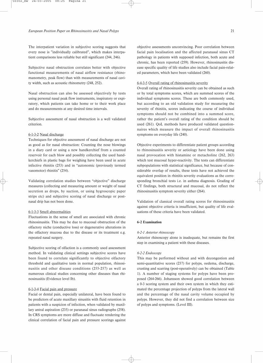
European Position Paper on Rhinosinusitis and Nasal Polyps 21
The interpatient variation in subjective scoring suggests thatevery nose is ”individually calibrated”, which makes interpa-tient comparisons less reliable but still significant (244, 246).
Subjective nasal obstruction correlates better with objectivefunctional measurements of nasal airflow resistance (rhino-manometry, peak flow) than with measurements of nasal cavi-ty width, such as acoustic rhinometry (248, 252).
Nasal obstruction can also be assessed objectively by testsusing personal nasal peak flow instruments, inspiratory or expi-ratory, which patients can take home or to their work placeand do measurements at any desired time intervals.
Subjective assessment of nasal obstruction is a well validatedcriterion.
6-1-3-2 Nasal dischargeTechniques for objective assessment of nasal discharge are notas good as for nasal obstruction: Counting the nose blowingsin a diary card or using a new handkerchief from a countedreservoir for each blow and possibly collecting the used hand-kerchiefs in plastic bags for weighing have been used in acuteinfective rhinitis (253) and in “autonomic (previously termedvasomotor) rhinitis” (254).
Validating correlation studies between “objective” dischargemeasures (collecting and measuring amount or weight of nasalsecretion as drops, by suction, or using hygroscopic paperstrips etc) and subjective scoring of nasal discharge or post-nasal drip has not been done.
6-1-3-3 Smell abnormalitiesFluctuations in the sense of smell are associated with chronicrhinosinusitis. This may be due to mucosal obstruction of theolfactory niche (conductive loss) or degenerative alterations inthe olfactory mucosa due to the disease or its treatment e.g.repeated nasal surgery.
Subjective scoring of olfaction is a commonly used assessmentmethod. In validating clinical settings subjective scores havebeen found to correlate significantly to objective olfactorythreshold and qualitative tests in normal population, rhinosi-nusitis and other disease conditions (255-257) as well asnumerous clinical studies concerning other diseases than rhi-nosinusitis (Evidence level Ib).
6-1-3-4 Facial pain and pressureFacial or dental pain, especially unilateral, have been found tobe predictors of acute maxillary sinusitis with fluid retention inpatients with a suspicion of infection, when validated by maxil-lary antral aspiration (235) or paranasal sinus radiographs (258).In CRS symptoms are more diffuse and fluctuate rendering theclinical correlation of facial pain and pressure scorings against
objective assessments unconvincing. Poor correlation betweenfacial pain localisation and the affected paranasal sinus CTpathology in patients with supposed infection, both acute andchronic, has been reported (259). However, rhinosinusitis dis-ease specific quality of life studies also include facial pain-relat-ed parameters, which have been validated (260).
6-4-3-5 Overall rating of rhinosinusitis severityOverall rating of rhinosinusitis severity can be obtained as suchor by total symptoms scores, which are summed scores of theindividual symptoms scores. These are both commonly used,but according to an old validation study for measuring theseverity of rhinitis, scores indicating the course of individualsymptoms should not be combined into a summed score,rather the patient’s overall rating of the condition should beused (261). QoL methods have produced validated question-naires which measure the impact of overall rhinosinusitissymptoms on everyday life (240).
Objective experiments to differentiate patient groups accordingto rhinosinusitis severity or aetiology have been done usingnasal provocation with histamine or metacholine (262, 263)which test mucosal hyper-reactivity. The tests can differentiatesubpopulations with statistical significance, but because of con-siderable overlap of results, these tests have not achieved theequivalent position in rhinitis severity evaluations as the corre-sponding bronchial tests i.e. in asthma diagnosis. Grading ofCT findings, both structural and mucosal, do not reflect therhinosinusitis symptom severity either (264).
Validation of classical overall rating scores for rhinosinusitisagainst objective criteria is insufficient, but quality of life eval-uations of these criteria have been validated.
6-2 Examination
6-2-1 Anterior rhinoscopy
Anterior rhinoscopy alone is inadequate, but remains the firststep in examining a patient with these diseases.
6-2-2 Endoscopy
This may be performed without and with decongestion andsemi-quantitative scores (237) for polyps, oedema, discharge,crusting and scarring (post-operatively) can be obtained (Table1). A number of staging systems for polyps have been pro-posed (264-266). Johansson showed good correlation betweena 0-3 scoring system and their own system in which they esti-mated the percentage projection of polyps from the lateral walland the percentage of the nasal cavity volume occupied bypolyps. However, they did not find a correlation between sizeof polyps and symptoms. (Level III).
50302_bw 24-03-2005 08:25 Pagina 21
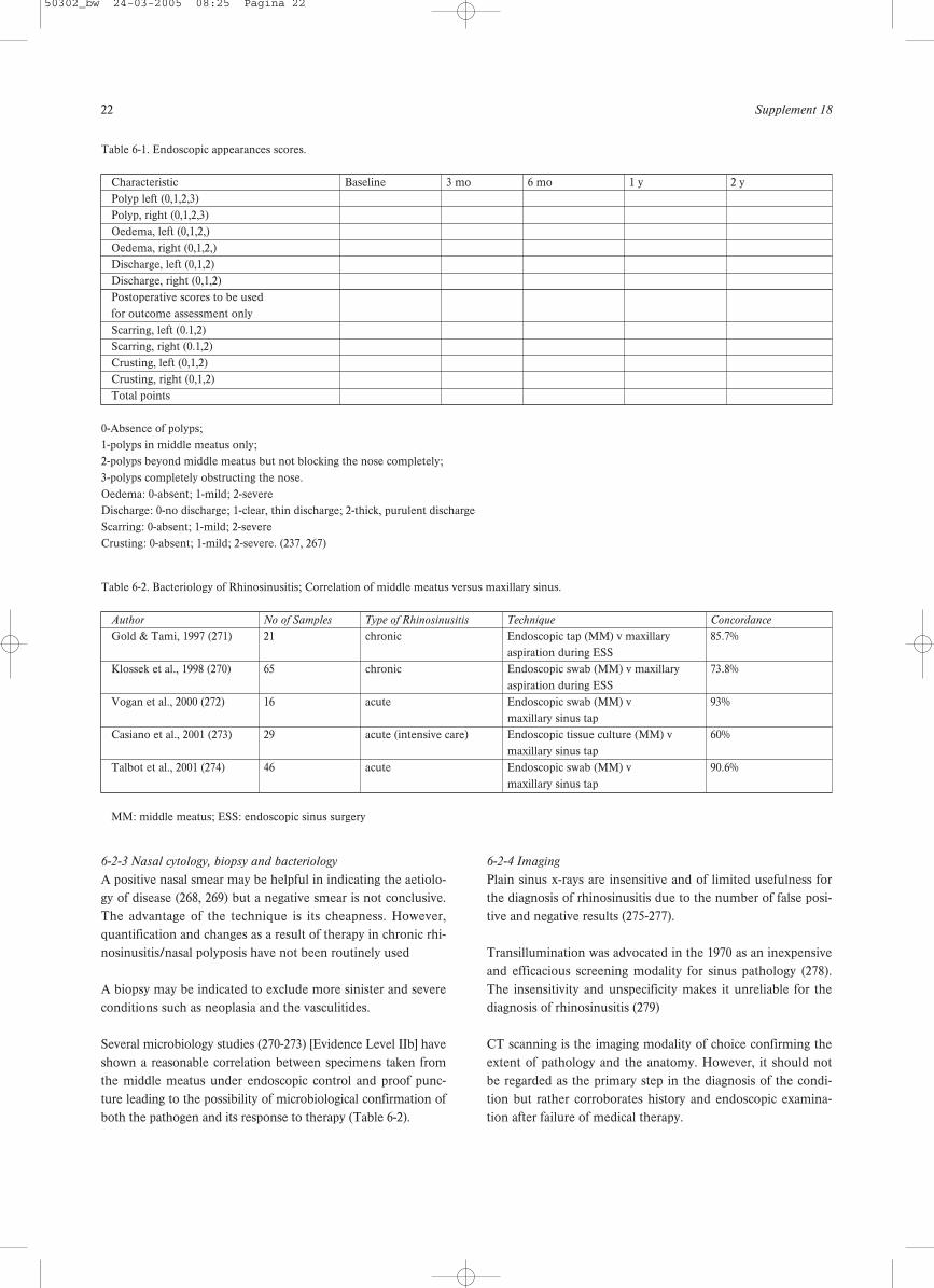
22 Supplement 18
6-2-3 Nasal cytology, biopsy and bacteriology
A positive nasal smear may be helpful in indicating the aetiolo-gy of disease (268, 269) but a negative smear is not conclusive.The advantage of the technique is its cheapness. However,quantification and changes as a result of therapy in chronic rhi-nosinusitis/nasal polyposis have not been routinely used
A biopsy may be indicated to exclude more sinister and severeconditions such as neoplasia and the vasculitides.
Several microbiology studies (270-273) [Evidence Level IIb] haveshown a reasonable correlation between specimens taken fromthe middle meatus under endoscopic control and proof punc-ture leading to the possibility of microbiological confirmation ofboth the pathogen and its response to therapy (Table 6-2).
6-2-4 Imaging
Plain sinus x-rays are insensitive and of limited usefulness forthe diagnosis of rhinosinusitis due to the number of false posi-tive and negative results (275-277).
Transillumination was advocated in the 1970 as an inexpensiveand efficacious screening modality for sinus pathology (278).The insensitivity and unspecificity makes it unreliable for thediagnosis of rhinosinusitis (279)
CT scanning is the imaging modality of choice confirming theextent of pathology and the anatomy. However, it should notbe regarded as the primary step in the diagnosis of the condi-tion but rather corroborates history and endoscopic examina-tion after failure of medical therapy.
Table 6-1. Endoscopic appearances scores.
Characteristic Baseline 3 mo 6 mo 1 y 2 yPolyp left (0,1,2,3)Polyp, right (0,1,2,3)Oedema, left (0,1,2,)Oedema, right (0,1,2,)Discharge, left (0,1,2)Discharge, right (0,1,2)Postoperative scores to be used for outcome assessment onlyScarring, left (0.1,2)Scarring, right (0.1,2)Crusting, left (0,1,2)Crusting, right (0,1,2)Total points
0-Absence of polyps; 1-polyps in middle meatus only;2-polyps beyond middle meatus but not blocking the nose completely;3-polyps completely obstructing the nose.Oedema: 0-absent; 1-mild; 2-severe Discharge: 0-no discharge; 1-clear, thin discharge; 2-thick, purulent dischargeScarring: 0-absent; 1-mild; 2-severeCrusting: 0-absent; 1-mild; 2-severe. (237, 267)
Table 6-2. Bacteriology of Rhinosinusitis; Correlation of middle meatus versus maxillary sinus.
Author No of Samples Type of Rhinosinusitis Technique Concordance
Gold & Tami, 1997 (271) 21 chronic Endoscopic tap (MM) v maxillary 85.7%aspiration during ESS
Klossek et al., 1998 (270) 65 chronic Endoscopic swab (MM) v maxillary 73.8%aspiration during ESS
Vogan et al., 2000 (272) 16 acute Endoscopic swab (MM) v 93%maxillary sinus tap
Casiano et al., 2001 (273) 29 acute (intensive care) Endoscopic tissue culture (MM) v 60%maxillary sinus tap
Talbot et al., 2001 (274) 46 acute Endoscopic swab (MM) v 90.6%maxillary sinus tap
MM: middle meatus; ESS: endoscopic sinus surgery
50302_bw 24-03-2005 08:25 Pagina 22
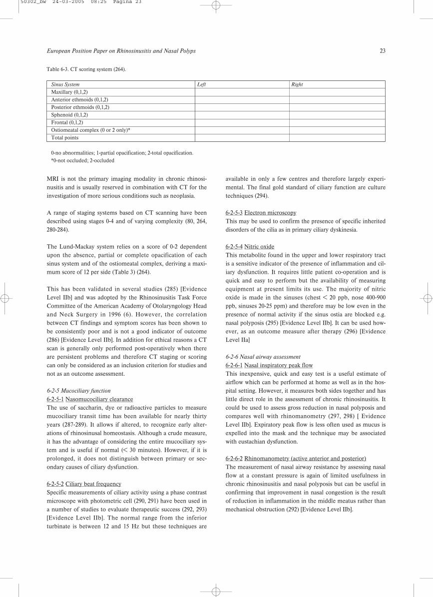
European Position Paper on Rhinosinusitis and Nasal Polyps 23
MRI is not the primary imaging modality in chronic rhinosi-nusitis and is usually reserved in combination with CT for theinvestigation of more serious conditions such as neoplasia.
A range of staging systems based on CT scanning have beendescribed using stages 0-4 and of varying complexity (80, 264,280-284).
The Lund-Mackay system relies on a score of 0-2 dependentupon the absence, partial or complete opacification of eachsinus system and of the ostiomeatal complex, deriving a maxi-mum score of 12 per side (Table 3) (264).
This has been validated in several studies (285) [EvidenceLevel IIb] and was adopted by the Rhinosinusitis Task ForceCommittee of the American Academy of Otolaryngology Headand Neck Surgery in 1996 (6). However, the correlationbetween CT findings and symptom scores has been shown tobe consistently poor and is not a good indicator of outcome(286) [Evidence Level IIb]. In addition for ethical reasons a CTscan is generally only performed post-operatively when thereare persistent problems and therefore CT staging or scoringcan only be considered as an inclusion criterion for studies andnot as an outcome assessment.
6-2-5 Mucociliary function
6-2-5-1 Nasomucociliary clearanceThe use of saccharin, dye or radioactive particles to measuremucociliary transit time has been available for nearly thirtyyears (287-289). It allows if altered, to recognize early alter-ations of rhinosinusal homeostasis. Although a crude measure,it has the advantage of considering the entire mucociliary sys-tem and is useful if normal (< 30 minutes). However, if it isprolonged, it does not distinguish between primary or sec-ondary causes of ciliary dysfunction.
6-2-5-2 Ciliary beat frequencySpecific measurements of ciliary activity using a phase contrastmicroscope with photometric cell (290, 291) have been used ina number of studies to evaluate therapeutic success (292, 293)[Evidence Level IIb]. The normal range from the inferiorturbinate is between 12 and 15 Hz but these techniques are
available in only a few centres and therefore largely experi-mental. The final gold standard of ciliary function are culturetechniques (294).
6-2-5-3 Electron microscopyThis may be used to confirm the presence of specific inheriteddisorders of the cilia as in primary ciliary dyskinesia.
6-2-5-4 Nitric oxideThis metabolite found in the upper and lower respiratory tractis a sensitive indicator of the presence of inflammation and cil-iary dysfunction. It requires little patient co-operation and isquick and easy to perform but the availability of measuringequipment at present limits its use. The majority of nitricoxide is made in the sinuses (chest < 20 ppb, nose 400-900ppb, sinuses 20-25 ppm) and therefore may be low even in thepresence of normal activity if the sinus ostia are blocked e.g.nasal polyposis (295) [Evidence Level IIb]. It can be used how-ever, as an outcome measure after therapy (296) [EvidenceLevel IIa]
6-2-6 Nasal airway assessment
6-2-6-1 Nasal inspiratory peak flowThis inexpensive, quick and easy test is a useful estimate ofairflow which can be performed at home as well as in the hos-pital setting. However, it measures both sides together and haslittle direct role in the assessment of chronic rhinosinusitis. Itcould be used to assess gross reduction in nasal polyposis andcompares well with rhinomanometry (297, 298) [ EvidenceLevel IIb]. Expiratory peak flow is less often used as mucus isexpelled into the mask and the technique may be associatedwith eustachian dysfunction.
6-2-6-2 Rhinomanometry (active anterior and posterior)The measurement of nasal airway resistance by assessing nasalflow at a constant pressure is again of limited usefulness inchronic rhinosinusitis and nasal polyposis but can be useful inconfirming that improvement in nasal congestion is the resultof reduction in inflammation in the middle meatus rather thanmechanical obstruction (292) [Evidence Level IIb].
Table 6-3. CT scoring system (264).
Sinus System Left Right
Maxillary (0,1,2)Anterior ethmoids (0,1,2)Posterior ethmoids (0,1,2)Sphenoid (0,1,2)Frontal (0,1,2)Ostiomeatal complex (0 or 2 only)*Total points
0-no abnormalities; 1-partial opacification; 2-total opacification. *0-not occluded; 2-occluded
50302_bw 24-03-2005 08:25 Pagina 23
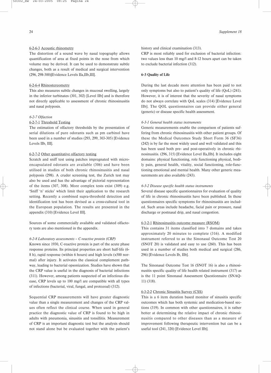
24 Supplement 18
6-2-6-3 Acoustic rhinometryThe distortion of a sound wave by nasal topography allowsquantification of area at fixed points in the nose from whichvolume may be derived. It can be used to demonstrate subtlechanges, both as a result of medical and surgical intervention(296, 298-300)[Evidence Levels IIa,IIb,III].
6-2-6-4 RhinostereometryThis also measures subtle changes in mucosal swelling, largelyin the inferior turbinates (301, 302) [Level IIb] and is thereforenot directly applicable to assessment of chronic rhinosinusitisand nasal polyposis.
6-2-7 Olfaction
6-2-7-1 Threshold TestingThe estimation of olfactory thresholds by the presentation ofserial dilutions of pure odorants such as pm carbinol havebeen used in a number of studies (293, 299, 303-305) [EvidenceLevels IIb, III].
6-2-7-2 Other quantitative olfactory testingScratch and sniff test using patches impregnated with micro-encapsulated odorants are available (306) and have beenutilised in studies of both chronic rhinosinusitis and nasalpolyposis (298). A cruder screening test, the Zurich test mayalso be used and has the advantage of pictorial representationof the items (307, 308). More complex tests exist (309) e.g.‘Sniff ‘n’ sticks’ which limit their application to the researchsetting. Recently a combined supra-threshold detection andidentification test has been devised as a cross-cultural tool inthe European population. The results are presented in theappendix (310) [Evidence Level III].
Sources of some commercially available and validated olfacto-ry tests are also mentioned in the appendix.
6-2-8 Laboratory assessments – C-reactive protein (CRP)
Known since 1930, C-reactive protein is part of the acute phaseresponse proteins. Its principal properties are short half-life (6-8 h), rapid response (within 6 hours) and high levels (x500 nor-mal) after injury. It activates the classical complement path-way, leading to bacterial opsonization. Studies have shown thatthe CRP value is useful in the diagnosis of bacterial infections(311). However, among patients suspected of an infectious dis-ease, CRP levels up to 100 mg/l are compatible with all typesof infections (bacterial, viral, fungal, and protozoal) (312).
Sequential CRP measurements will have greater diagnosticvalue than a single measurement and changes of the CRP val-ues often reflect the clinical course. When used in generalpractice the diagnostic value of CRP is found to be high inadults with pneumonia, sinusitis and tonsillitis. Measurementof CRP is an important diagnostic test but the analysis shouldnot stand alone but be evaluated together with the patient’s
history and clinical examination (313). CRP is most reliably used for exclusion of bacterial infection:two values less than 10 mg/l and 8-12 hours apart can be takento exclude bacterial infection (312).
6-3 Quality of Life
During the last decade more attention has been paid to notonly symptoms but also to patient’s quality of life (QoL) (241).However, it is of interest that the severity of nasal symptomsdo not always correlate with QoL scales (314) [Evidence LevelIIb]. The QOL questionnaires can provide either general(generic) or disease specific health assessment.
6-3-1 General health status instruments
Generic measurements enable the comparison of patients suf-fering from chronic rhinosinusitis with other patient groups. Ofthese the Medical Outcomes Study Short Form 36 (SF36)(242) is by far the most widely used and well validated and thishas been used both pre- and post-operatively in chronic rhi-nosinusitis. (296, 315) [Evidence Level IIa,IIb]. It includes eightdomains: physical functioning, role functioning physical, bodi-ly pain, general health, vitality, social functioning, role-func-tioning emotional and mental health. Many other generic mea-surements are also available (243).
6-3-2 Disease specific health status instruments
Several disease specific questionnaires for evaluation of qualityof life in chronic rhinosinusitis have been published. In thesequestionnaires specific symptoms for rhinosinusitis are includ-ed. Such areas include headache, facial pain or pressure, nasaldischarge or postnasal drip, and nasal congestion.
6-3-2-1 Rhinosinusitis outcome measure (RSOM)This contains 31 items classified into 7 domains and takesapproximately 20 minutes to complete (316). A modifiedinstrument referred to as the Sinonasal Outcome Test 20(SNOT 20) is validated and easy to use (260). This has beenused in a number of studies both medical and surgical (286,296) [Evidence Levels Ib, IIb].
The Sinonasal Outcome Test 16 (SNOT 16) is also a rhinosi-nusitis specific quality of life health related instrument (317) asis the 11 point Sinonasal Assessment Questionnaire (SNAQ-11) (318).
6-3-2-2 Chronic Sinusitis Survey (CSS)This is a 6 item duration based monitor of sinusitis specificoutcomes which has both systemic and medication-based sec-tions (319). In common with other questionnaires, it is ratherbetter at determining the relative impact of chronic rhinosi-nusitis compared to other diseases than as a measure ofimprovement following therapeutic intervention but can be auseful tool (241, 320) [Evidence Level IIb].
50302_bw 24-03-2005 08:25 Pagina 24
European Position Paper on Rhinosinusitis and Nasal Polyps 25
6-3-2-3 Rhinosinusitis Disability Index (RSDI)In this 30 item questionnaire the patient is asked to relate nasaland sinus symptoms to specific limitations on daily functioning(240, 321). It is similar to the RSOM 31 in the types of ques-tions it contains. It can be completed easily and quickly butdoes not allow the patient to indicate their most importantsymptoms. However, it does have some general questions sim-ilar to the SF-36.
6-3-2-4 The Chronic Rhinosinusitis Type Specific QuestionnaireThis test contains three forms. Form 1 collects data on nasaland sinus symptoms prior to treatment, Form 2 collects data onthe clinical classification of sinus disease and Form 3 data onnasal and sinus symptoms after sinus surgery. Hoffman etal.have used this in combination with an SF-36 to look at patientoutcomes after surgical management of chronic rhinosinusitisthough it is somewhat time consuming to complete (322).
6-3-2-5 Rhinoconjunctivitis quality of life questionnaire(RQLQ)This is a well-validated questionnaire but specifically focuseson allergy and is of less relevance in chronic rhinosinusitis andnasal polyposis (323).
6-3-2-6 Rhinitis Symptom Utility Index (RSUI) This consists of ten questions on the severity and frequency ofa stuffy or blocked nose, runny nose, sneezing, itching, wateryeyes and itching nose or throat. The two-week reproducibilityof the RSUI was weak, probably reflecting the day to day vari-ability of rhinitis (324).
6-3-2-7 GeneralMost questionnaires concentrate on the duration of the symp-toms and not on the severity of the symptoms. A QoL ques-tionnaire developed by Damm et al.includes the severity of thesymptom scale (239). The domains in the questionnaire are theoverall quality of life, nasal breathing obstruction, post-nasaldrip or discharge, dry mucosa, smell, headache and asthmaticcomplaints.
6-3-3 Results
6-3-3-1 GenericIn a generic SF-36 survey the scores of chronic rhinosinusitispatients were compared to those of a healthy population. Theresults showed statistically significant differences in seven ofeight domains (325). Gliklich and Metson (53) have reportedthat patients with chronic rhinosinusitis have more bodily painand worse social functioning than for example patients withchronic obstructive pulmonary disease, congestive heart fail-ure, or back pain.
Winstead and Barrett (315) confirmed a similar degree ofimpact on general quality of life in chronic rhinosinusitis with
the SF-36. Following endoscopic sinus surgery they demon-strated a return to normality in all eight domains six monthspost-operatively which was maintained at twelve months.
6-3-3-2 Disease specificIn a study by Gliklich and Metson the effect of sinus surgeryon QoL was studied (320). After the surgery significantimprovements were found in reduction of the symptoms andmedications needed. Significant improvements in generalhealth status were noted in six of eight categories, and mostattained near-normative levels. A disease-specific question-naire seems to be more sensitive than a general questionnairein following patients after ethmoid sinus surgery (319). 76%patients reported relief of the symptoms at least in two of thedomains studied after FESS surgery (239).
The Chronic Sinusitis Survey has been used in QoL outcomesafter osteoplastic frontal sinus obliteration (326). Most patientswere satisfied with the results and had significant improve-ments in their survey scores. The number of clinic visits andantibiotic use also declined.
Mean scores one year after endoscopic frontal sinus surgeryshowed a significant improvement in symptoms of pain, con-gestion, and drainage as measured by the Chronic SinusitisSurvey. Medication use was also significantly reduced (327).
Radenne et al.have studied the QoL of nasal polyposis patientsusing a generic SF-36 questionnaire (314). Polyposis impairedthe QoL more than for example perennial rhinitis. Treatmentsignificantly improved the symptoms and the QoL of the poly-posis patients. FESS surgery on asthmatic patients with mas-sive nasal polyposis improved nasal breathing and QoL, andalso the use of asthma medications was significantly reduced(328).
In a recent randomised study of patients with chronic rhinosi-nusitis/nasal polyposis, treatment was either endoscopic sinussurgery or three months of a macrolide antibiotic such as ery-thromycin (296). Patients were followed up at 3, 6, 9 and 12months with a variety of parameters including visual analoguescores of nasal symptoms, SNOT 20, SF-36, nitric oxide mea-surements of upper and lower respiratory tract expired air,acoustic rhinometry, saccharine clearance test and nasalendoscopy. Ninety patients were randomised, with 45 in eacharm and at the end of one year, 38 were available for analysisin the medical arm and 40 in the surgical arm. The studyshowed that there had been improvement in all subjective andobjective parameters (p <0.01) but there was no differencebetween the medical and surgical groups except that total nasalvolume as measured by acoustic rhinometry was greater in thesurgical group. This study shows the usefulness of objectivemeasurement in confirming subjective impressions (EvidenceLevel 1b).
50302_bw 24-03-2005 08:25 Pagina 25
26 Supplement 18
Quality of life measurement is quite a new tool evaluating theimpact of disease and the efficacy of treatment. In rhinosinusi-tis studies, when the effect of medical treatment or surgery hasbeen evaluated, QoL has been considered to be an importantoutcome measurement as distinct from classic rhinosinusitissymptom parameters. In a number of studies, chronic rhinosi-nusitis has been shown to significantly impair QoL [Level Ib](260, 329-332) and this has also been shown to improve signifi-cantly with treatment [Level IIb] (239, 320, 333, 334).
50302_bw 24-03-2005 08:25 Pagina 26
European Position Paper on Rhinosinusitis and Nasal Polyps 27
7-1 Treatment of rhinosinusitis with corticosteroids
The introduction of topically administered glucocorticoids hasimproved the treatment of upper (rhinitis, nasal polyps) andlower (asthma) airway inflammatory disease. The clinical effi-cacy of glucocorticoids may depend in part on their ability toreduce airway eosinophil infiltration by preventing theirincreased viability and activation. Both topical and systemicglucocorticoids may affect the eosinophil function by bothdirectly reducing eosinophil viability and activation (207, 335-337) or indirectly reducing the secretion of chemotacticcytokines by nasal mucosa and polyp epithelial cells (208, 338-340). The potency of these effects is lower in nasal polyps thanin nasal mucosa suggesting an induced inflammatory resis-tance to steroid treatment in chronic rhinosinusitis / nasalpolyposis (337, 338).
The biological action of glucocorticoids is mediated throughactivation of intracellular glucocorticoid receptors (GR) (341),expressed in many tissues and cells (342). Two human iso-forms of GR have been identified, GR? and GR?, which origi-nate from the same gene by alternative splicing of the GR pri-mary transcript (343). Upon hormone binding, GR? enhancesanti-inflammatory or represses pro-inflammatory gene tran-scription, and exerts most of the anti-inflammatory effects ofglucocorticoids through protein-protein interactions betweenGR and transcription factors, such as AP-1 and NF-?B. TheGR? isoform does not bind steroids but may interfere with theGRa function. There may be several mechanisms accountingfor the resistance to the anti-inflammatory effects of glucocor-ticoids, including an overexpression of GR? or a downexpres-sion of GR? . Increased expression of GR? has been reportedin patients with nasal polyps (344, 345) while downregulationof GRa levels after treatment with glucocorticoids (346, 347)has also been postulated to be one of the possible explanationsfor the secondary glucocorticoid resistance phenomenon.
The anti-inflammatory effect of corticosteroids could, theoreti-cally, be expected as well in non-allergic (i.e. infectious) as inallergic rhinosinusitis. Tissue eosinophilia is thus also seen inpersistent RS (348).
Potential indications for corticosteroids in rhinosinusitis:• Acute/Intermittent rhinosinusitis without nasal polyposis
(NP);• Persistent rhinosinusitis without NP;• Persistent rhinosinusitis with NP;• Postoperative treatment of persistent rhinosinusitis to pre-
vent recurrences of NP;• Prophylactic treatment of intermittent rhinosinusitis;
• Oral steroids in persistent rhinosinusitis with NP;• Oral steroids in acute intermittent rhinosinusitis.
7-1-1 Acute/Intermittent Rhinosinusitis without nasal polyps
Qvarnberg et al. (349) measured the clinical effect of budes-onide (BUD)/placebo as a complement to erythromycin andsinus wash out in a randomized, double-blind study on patientsreferred for sinus surgery due to persistent or intermittent max-illary sinusitis. Three months treatment was given to 20 sub-jects in 2 groups, all without NP. Treatment with BUD resultedin a significant improvement of nasal symptoms, facial painand sensitivity. No significant improvement was seen inmucosal thickening on x-ray. The final clinical outcome did notdiffer between the groups. No side effects of treatment werenoted. It is not possible in this study to distinguish intermittentfrom persistent rhinosinusitis but all cases were reported tohave intermittent “episodes of sinusitis for the last two years”.
Melzer et al. (350) gave mometasone furoate (MF) 400 ?g to200 patients and placebo to 207 patients with acute intermit-tent RS as adjunctive therapy to amoxicillin/clavulanate potas-sium for 21 days. Total symptom score and individual symp-tom scores as congestion, facial pain, headache and rhino rheaimproved significantly, but not postnasal drip in the MF group.The effect was most obvious after 16 days treatment.Improvement on CT was seen in MF group but not statistical-ly significant. No side effects of treatment were seen.
Nayak et al. (351) compared MF 200 and 400 ?g to placebo in325, 318 and 324 patients with intermittent RS (no NP) asadjunctive therapy to amoxicillin/clavulanate potassium for 21days treatment. Total symptom score (TSS) was improved fromday 4 and at the end of the study (21 days) in both MF groupscompared to placebo. Improvement compared to the situationbefore treatment was 50 and 51% for MF groups and 44% inplacebo group, p<0,017. Individual nasal symptom scores suchas nasal congestion, facial pain, rhino rhea and postnasal dripimproved in both MF-groups compared to placebo. CT wasimproved, but not statistically significant in MF groups com-pared to placebo. No side effects of treatment were seen.
In a study by Dolor et al. (352) 200 µg FP daily was used asadjunctive therapy for 3 weeks (to cefuroxime for 10 days andxylometazoline for 3 days) in a double blind placebo con-trolled multicentre trial (n=47 in FP group and 48 in controlgroup) in patients with acute intermittent rhinosinusitis. Timewas measured to clinical success. After two weeks, success wasseen in 73.9 and 93.5% in placebo and FP group respectively(p=0.009). Time to clinical success was 9.5 and 6.0 days respec-tively (p=0.01)
7. Management
50302_bw 24-03-2005 08:25 Pagina 27
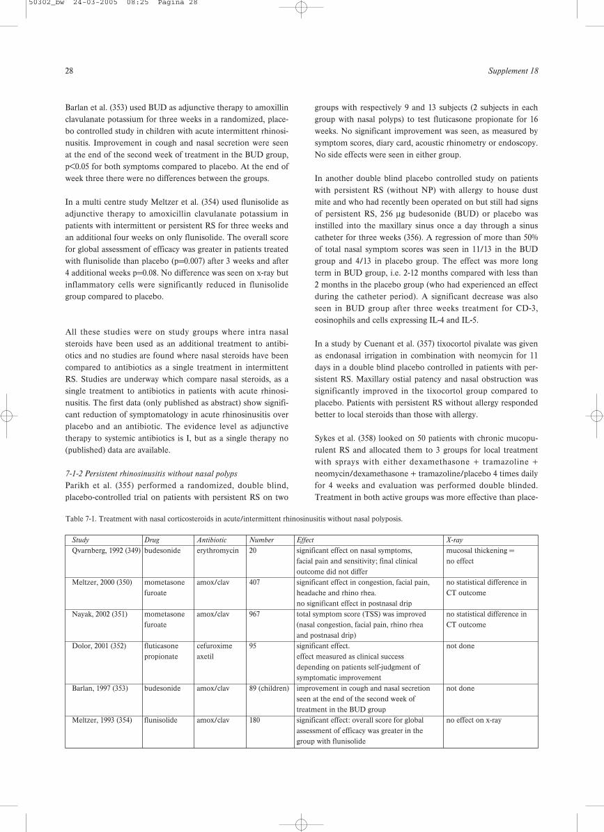
28 Supplement 18
Barlan et al. (353) used BUD as adjunctive therapy to amoxillinclavulanate potassium for three weeks in a randomized, place-bo controlled study in children with acute intermittent rhinosi-nusitis. Improvement in cough and nasal secretion were seenat the end of the second week of treatment in the BUD group,p<0.05 for both symptoms compared to placebo. At the end ofweek three there were no differences between the groups.
In a multi centre study Meltzer et al. (354) used flunisolide asadjunctive therapy to amoxicillin clavulanate potassium inpatients with intermittent or persistent RS for three weeks andan additional four weeks on only flunisolide. The overall scorefor global assessment of efficacy was greater in patients treatedwith flunisolide than placebo (p=0.007) after 3 weeks and after4 additional weeks p=0.08. No difference was seen on x-ray butinflammatory cells were significantly reduced in flunisolidegroup compared to placebo.
All these studies were on study groups where intra nasalsteroids have been used as an additional treatment to antibi-otics and no studies are found where nasal steroids have beencompared to antibiotics as a single treatment in intermittentRS. Studies are underway which compare nasal steroids, as asingle treatment to antibiotics in patients with acute rhinosi-nusitis. The first data (only published as abstract) show signifi-cant reduction of symptomatology in acute rhinosinusitis overplacebo and an antibiotic. The evidence level as adjunctivetherapy to systemic antibiotics is I, but as a single therapy no(published) data are available.
7-1-2 Persistent rhinosinusitis without nasal polyps
Parikh et al. (355) performed a randomized, double blind,placebo-controlled trial on patients with persistent RS on two
groups with respectively 9 and 13 subjects (2 subjects in eachgroup with nasal polyps) to test fluticasone propionate for 16weeks. No significant improvement was seen, as measured bysymptom scores, diary card, acoustic rhinometry or endoscopy.No side effects were seen in either group.
In another double blind placebo controlled study on patientswith persistent RS (without NP) with allergy to house dustmite and who had recently been operated on but still had signsof persistent RS, 256 µg budesonide (BUD) or placebo wasinstilled into the maxillary sinus once a day through a sinuscatheter for three weeks (356). A regression of more than 50%of total nasal symptom scores was seen in 11/13 in the BUDgroup and 4/13 in placebo group. The effect was more longterm in BUD group, i.e. 2-12 months compared with less than2 months in the placebo group (who had experienced an effectduring the catheter period). A significant decrease was alsoseen in BUD group after three weeks treatment for CD-3,eosinophils and cells expressing IL-4 and IL-5.
In a study by Cuenant et al. (357) tixocortol pivalate was givenas endonasal irrigation in combination with neomycin for 11days in a double blind placebo controlled in patients with per-sistent RS. Maxillary ostial patency and nasal obstruction wassignificantly improved in the tixocortol group compared toplacebo. Patients with persistent RS without allergy respondedbetter to local steroids than those with allergy.
Sykes et al. (358) looked on 50 patients with chronic mucopu-rulent RS and allocated them to 3 groups for local treatmentwith sprays with either dexamethasone + tramazoline +neomycin/dexamethasone + tramazoline/placebo 4 times dailyfor 4 weeks and evaluation was performed double blinded.Treatment in both active groups was more effective than place-
Table 7-1. Treatment with nasal corticosteroids in acute/intermittent rhinosinusitis without nasal polyposis.
Study Drug Antibiotic Number Effect X-ray
Qvarnberg, 1992 (349) budesonide erythromycin 20 significant effect on nasal symptoms, mucosal thickening = facial pain and sensitivity; final clinical no effectoutcome did not differ
Meltzer, 2000 (350) mometasone amox/clav 407 significant effect in congestion, facial pain, no statistical difference in furoate headache and rhino rhea. CT outcome
no significant effect in postnasal dripNayak, 2002 (351) mometasone amox/clav 967 total symptom score (TSS) was improved no statistical difference in
furoate (nasal congestion, facial pain, rhino rhea CT outcomeand postnasal drip)
Dolor, 2001 (352) fluticasone cefuroxime 95 significant effect. not donepropionate axetil effect measured as clinical success
depending on patients self-judgment of symptomatic improvement
Barlan, 1997 (353) budesonide amox/clav 89 (children) improvement in cough and nasal secretion not doneseen at the end of the second week of treatment in the BUD group
Meltzer, 1993 (354) flunisolide amox/clav 180 significant effect: overall score for global no effect on x-rayassessment of efficacy was greater in the group with flunisolide
50302_bw 24-03-2005 08:25 Pagina 28
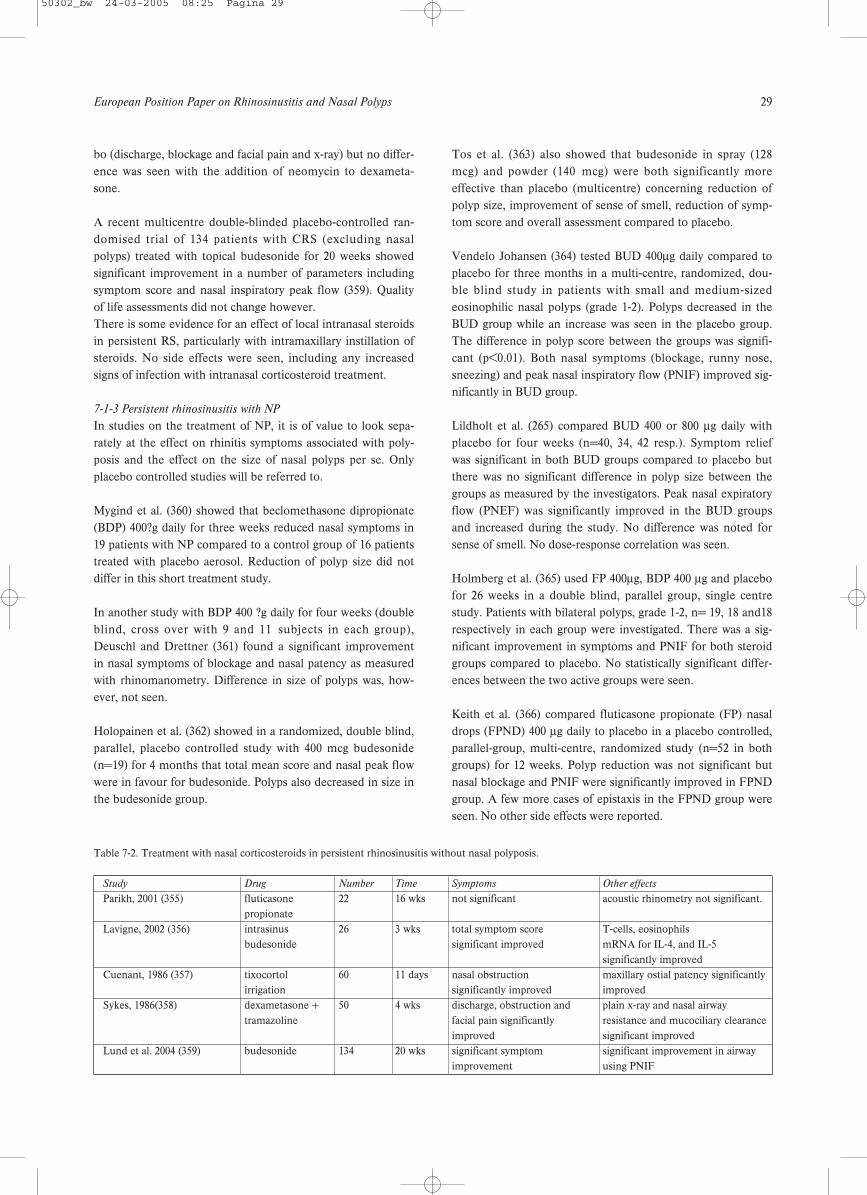
European Position Paper on Rhinosinusitis and Nasal Polyps 29
bo (discharge, blockage and facial pain and x-ray) but no differ-ence was seen with the addition of neomycin to dexameta-sone.
A recent multicentre double-blinded placebo-controlled ran-domised trial of 134 patients with CRS (excluding nasalpolyps) treated with topical budesonide for 20 weeks showedsignificant improvement in a number of parameters includingsymptom score and nasal inspiratory peak flow (359). Qualityof life assessments did not change however.There is some evidence for an effect of local intranasal steroidsin persistent RS, particularly with intramaxillary instillation ofsteroids. No side effects were seen, including any increasedsigns of infection with intranasal corticosteroid treatment.
7-1-3 Persistent rhinosinusitis with NP
In studies on the treatment of NP, it is of value to look sepa-rately at the effect on rhinitis symptoms associated with poly-posis and the effect on the size of nasal polyps per se. Onlyplacebo controlled studies will be referred to.
Mygind et al. (360) showed that beclomethasone dipropionate(BDP) 400?g daily for three weeks reduced nasal symptoms in19 patients with NP compared to a control group of 16 patientstreated with placebo aerosol. Reduction of polyp size did notdiffer in this short treatment study.
In another study with BDP 400 ?g daily for four weeks (doubleblind, cross over with 9 and 11 subjects in each group),Deuschl and Drettner (361) found a significant improvementin nasal symptoms of blockage and nasal patency as measuredwith rhinomanometry. Difference in size of polyps was, how-ever, not seen.
Holopainen et al. (362) showed in a randomized, double blind,parallel, placebo controlled study with 400 mcg budesonide(n=19) for 4 months that total mean score and nasal peak flowwere in favour for budesonide. Polyps also decreased in size inthe budesonide group.
Tos et al. (363) also showed that budesonide in spray (128mcg) and powder (140 mcg) were both significantly moreeffective than placebo (multicentre) concerning reduction ofpolyp size, improvement of sense of smell, reduction of symp-tom score and overall assessment compared to placebo.
Vendelo Johansen (364) tested BUD 400µg daily compared toplacebo for three months in a multi-centre, randomized, dou-ble blind study in patients with small and medium-sizedeosinophilic nasal polyps (grade 1-2). Polyps decreased in theBUD group while an increase was seen in the placebo group.The difference in polyp score between the groups was signifi-cant (p<0.01). Both nasal symptoms (blockage, runny nose,sneezing) and peak nasal inspiratory flow (PNIF) improved sig-nificantly in BUD group.
Lildholt et al. (265) compared BUD 400 or 800 µg daily withplacebo for four weeks (n=40, 34, 42 resp.). Symptom reliefwas significant in both BUD groups compared to placebo butthere was no significant difference in polyp size between thegroups as measured by the investigators. Peak nasal expiratoryflow (PNEF) was significantly improved in the BUD groupsand increased during the study. No difference was noted forsense of smell. No dose-response correlation was seen.
Holmberg et al. (365) used FP 400µg, BDP 400 µg and placebofor 26 weeks in a double blind, parallel group, single centrestudy. Patients with bilateral polyps, grade 1-2, n= 19, 18 and18respectively in each group were investigated. There was a sig-nificant improvement in symptoms and PNIF for both steroidgroups compared to placebo. No statistically significant differ-ences between the two active groups were seen.
Keith et al. (366) compared fluticasone propionate (FP) nasaldrops (FPND) 400 µg daily to placebo in a placebo controlled,parallel-group, multi-centre, randomized study (n=52 in bothgroups) for 12 weeks. Polyp reduction was not significant butnasal blockage and PNIF were significantly improved in FPNDgroup. A few more cases of epistaxis in the FPND group wereseen. No other side effects were reported.
Table 7-2. Treatment with nasal corticosteroids in persistent rhinosinusitis without nasal polyposis.
Study Drug Number Time Symptoms Other effects
Parikh, 2001 (355) fluticasone 22 16 wks not significant acoustic rhinometry not significant.propionate
Lavigne, 2002 (356) intrasinus 26 3 wks total symptom score T-cells, eosinophilsbudesonide significant improved mRNA for IL-4, and IL-5
significantly improvedCuenant, 1986 (357) tixocortol 60 11 days nasal obstruction maxillary ostial patency significantly
irrigation significantly improved improvedSykes, 1986(358) dexametasone + 50 4 wks discharge, obstruction and plain x-ray and nasal airway
tramazoline facial pain significantly resistance and mucociliary clearanceimproved significant improved
Lund et al. 2004 (359) budesonide 134 20 wks significant symptom significant improvement in airwayimprovement using PNIF
50302_bw 24-03-2005 08:25 Pagina 29
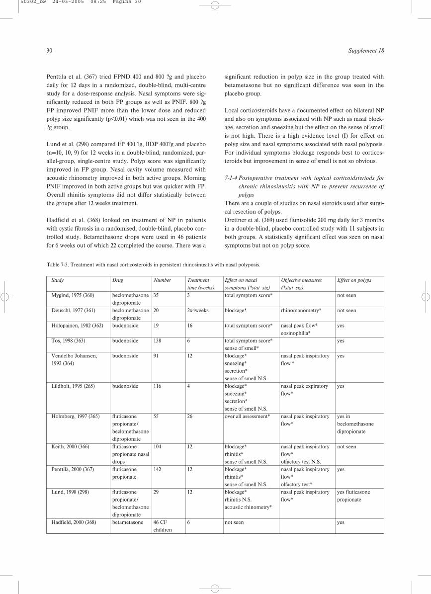
30 Supplement 18
Penttila et al. (367) tried FPND 400 and 800 ?g and placebodaily for 12 days in a randomized, double-blind, multi-centrestudy for a dose-response analysis. Nasal symptoms were sig-nificantly reduced in both FP groups as well as PNIF. 800 ?gFP improved PNIF more than the lower dose and reducedpolyp size significantly (p<0.01) which was not seen in the 400?g group.
Lund et al. (298) compared FP 400 ?g, BDP 400?g and placebo(n=10, 10, 9) for 12 weeks in a double-blind, randomized, par-allel-group, single-centre study. Polyp score was significantlyimproved in FP group. Nasal cavity volume measured withacoustic rhinometry improved in both active groups. MorningPNIF improved in both active groups but was quicker with FP.Overall rhinitis symptoms did not differ statistically betweenthe groups after 12 weeks treatment.
Hadfield et al. (368) looked on treatment of NP in patientswith cystic fibrosis in a randomised, double-blind, placebo con-trolled study. Betamethasone drops were used in 46 patientsfor 6 weeks out of which 22 completed the course. There was a
significant reduction in polyp size in the group treated withbetametasone but no significant difference was seen in theplacebo group.
Local corticosteroids have a documented effect on bilateral NPand also on symptoms associated with NP such as nasal block-age, secretion and sneezing but the effect on the sense of smellis not high. There is a high evidence level (I) for effect onpolyp size and nasal symptoms associated with nasal polyposis.For individual symptoms blockage responds best to corticos-teroids but improvement in sense of smell is not so obvious.
7-1-4 Postoperative treatment with topical corticoidsteriods for
chronic rhinosinusitis with NP to prevent recurrence of
polyps
There are a couple of studies on nasal steroids used after surgi-cal resection of polyps. Drettner et al. (369) used flunisolide 200 mg daily for 3 monthsin a double-blind, placebo controlled study with 11 subjects inboth groups. A statistically significant effect was seen on nasalsymptoms but not on polyp score.
Table 7-3. Treatment with nasal corticosteroids in persistent rhinosinusitis with nasal polyposis.
Study Drug Number Treatment Effect on nasal Objective measures Effect on polyps
time (weeks) symptoms (*stat sig) (*stat sig)
Mygind, 1975 (360) beclomethasone 35 3 total symptom score* not seendipropionate
Deuschl, 1977 (361) beclomethasone 20 2x4weeks blockage* rhinomanometry* not seendipropionate
Holopainen, 1982 (362) budenoside 19 16 total symptom score* nasal peak flow* yeseosinophilia*
Tos, 1998 (363) budenoside 138 6 total symptom score* yessense of smell*
Vendelbo Johansen, budenoside 91 12 blockage* nasal peak inspiratory yes1993 (364) sneezing* flow *
secretion*sense of smell N.S.
Lildholt, 1995 (265) budenoside 116 4 blockage* nasal peak expiratory yessneezing* flow*secretion*sense of smell N.S.
Holmberg, 1997 (365) fluticasone 55 26 over all assessment* nasal peak inspiratory yes in propionate/ flow* beclomethasone beclomethasone dipropionatedipropionate
Keith, 2000 (366) fluticasone 104 12 blockage* nasal peak inspiratory not seenpropionate nasal rhinitis* flow*drops sense of smell N.S. olfactory test N.S.
Penttilä, 2000 (367) fluticasone 142 12 blockage* nasal peak inspiratory yespropionate rhinitis* flow*
sense of smell N.S. olfactory test*Lund, 1998 (298) fluticasone 29 12 blockage* nasal peak inspiratory yes fluticasone
propionate/ rhinitis N.S. flow* propionatebeclomethasone acoustic rhinometry*dipropionate
Hadfield, 2000 (368) betametasone 46 CF 6 not seen yeschildren
50302_bw 24-03-2005 08:25 Pagina 30
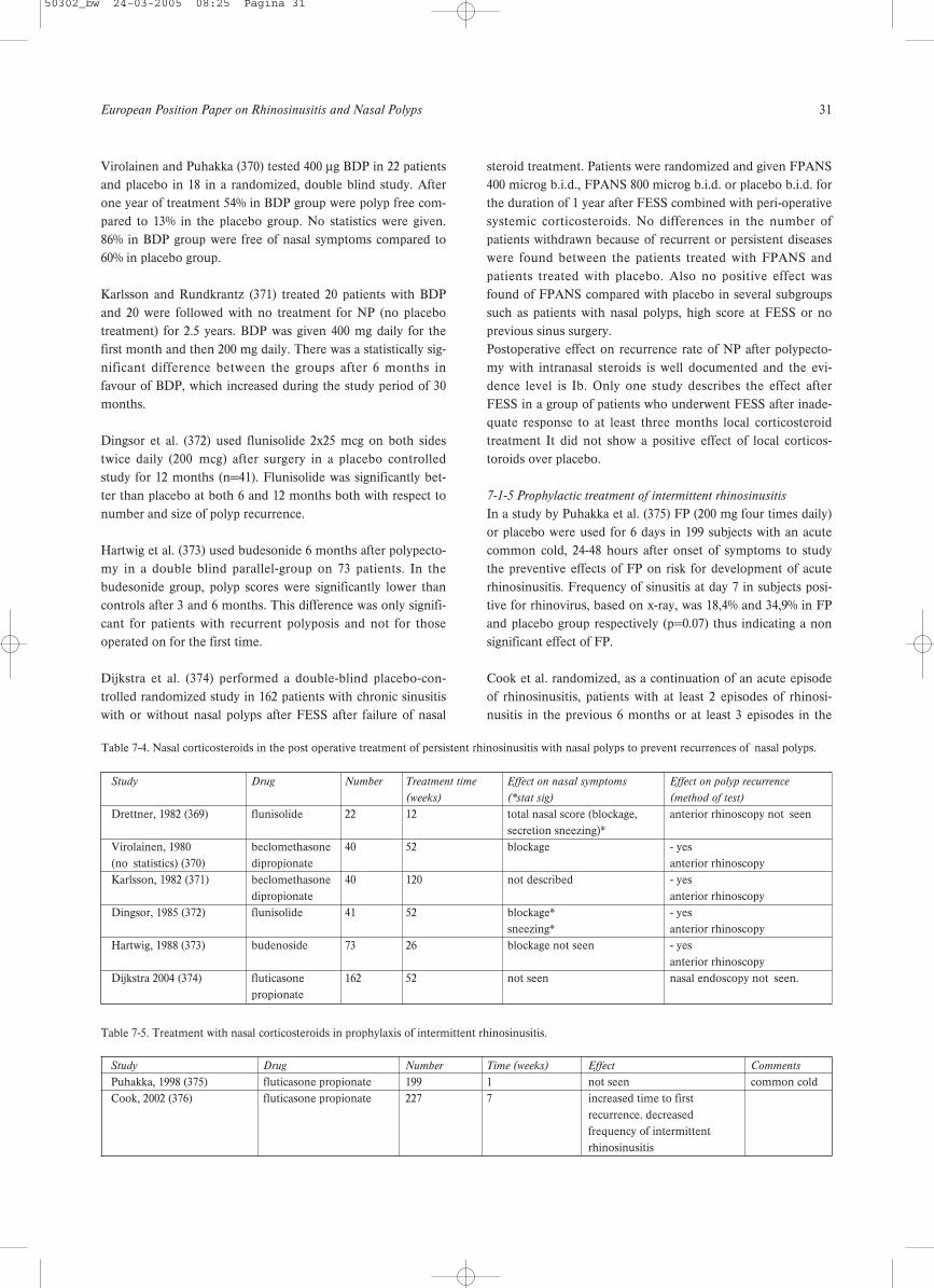
European Position Paper on Rhinosinusitis and Nasal Polyps 31
Virolainen and Puhakka (370) tested 400 µg BDP in 22 patientsand placebo in 18 in a randomized, double blind study. Afterone year of treatment 54% in BDP group were polyp free com-pared to 13% in the placebo group. No statistics were given.86% in BDP group were free of nasal symptoms compared to60% in placebo group.
Karlsson and Rundkrantz (371) treated 20 patients with BDPand 20 were followed with no treatment for NP (no placebotreatment) for 2.5 years. BDP was given 400 mg daily for thefirst month and then 200 mg daily. There was a statistically sig-nificant difference between the groups after 6 months infavour of BDP, which increased during the study period of 30months.
Dingsor et al. (372) used flunisolide 2x25 mcg on both sidestwice daily (200 mcg) after surgery in a placebo controlledstudy for 12 months (n=41). Flunisolide was significantly bet-ter than placebo at both 6 and 12 months both with respect tonumber and size of polyp recurrence.
Hartwig et al. (373) used budesonide 6 months after polypecto-my in a double blind parallel-group on 73 patients. In thebudesonide group, polyp scores were significantly lower thancontrols after 3 and 6 months. This difference was only signifi-cant for patients with recurrent polyposis and not for thoseoperated on for the first time.
Dijkstra et al. (374) performed a double-blind placebo-con-trolled randomized study in 162 patients with chronic sinusitiswith or without nasal polyps after FESS after failure of nasal
steroid treatment. Patients were randomized and given FPANS400 microg b.i.d., FPANS 800 microg b.i.d. or placebo b.i.d. forthe duration of 1 year after FESS combined with peri-operativesystemic corticosteroids. No differences in the number ofpatients withdrawn because of recurrent or persistent diseaseswere found between the patients treated with FPANS andpatients treated with placebo. Also no positive effect wasfound of FPANS compared with placebo in several subgroupssuch as patients with nasal polyps, high score at FESS or noprevious sinus surgery.Postoperative effect on recurrence rate of NP after polypecto-my with intranasal steroids is well documented and the evi-dence level is Ib. Only one study describes the effect afterFESS in a group of patients who underwent FESS after inade-quate response to at least three months local corticosteroidtreatment It did not show a positive effect of local corticos-toroids over placebo.
7-1-5 Prophylactic treatment of intermittent rhinosinusitis
In a study by Puhakka et al. (375) FP (200 mg four times daily)or placebo were used for 6 days in 199 subjects with an acutecommon cold, 24-48 hours after onset of symptoms to studythe preventive effects of FP on risk for development of acuterhinosinusitis. Frequency of sinusitis at day 7 in subjects posi-tive for rhinovirus, based on x-ray, was 18,4% and 34,9% in FPand placebo group respectively (p=0.07) thus indicating a nonsignificant effect of FP.
Cook et al. randomized, as a continuation of an acute episodeof rhinosinusitis, patients with at least 2 episodes of rhinosi-nusitis in the previous 6 months or at least 3 episodes in the
Table 7-4. Nasal corticosteroids in the post operative treatment of persistent rhinosinusitis with nasal polyps to prevent recurrences of nasal polyps.
Study Drug Number Treatment time Effect on nasal symptoms Effect on polyp recurrence
(weeks) (*stat sig) (method of test)
Drettner, 1982 (369) flunisolide 22 12 total nasal score (blockage, anterior rhinoscopy not seensecretion sneezing)*
Virolainen, 1980 beclomethasone 40 52 blockage - yes (no statistics) (370) dipropionate anterior rhinoscopy Karlsson, 1982 (371) beclomethasone 40 120 not described - yes
dipropionate anterior rhinoscopy Dingsor, 1985 (372) flunisolide 41 52 blockage* - yes
sneezing* anterior rhinoscopy Hartwig, 1988 (373) budenoside 73 26 blockage not seen - yes
anterior rhinoscopy Dijkstra 2004 (374) fluticasone 162 52 not seen nasal endoscopy not seen.
propionate
Table 7-5. Treatment with nasal corticosteroids in prophylaxis of intermittent rhinosinusitis.
Study Drug Number Time (weeks) Effect Comments
Puhakka, 1998 (375) fluticasone propionate 199 1 not seen common coldCook, 2002 (376) fluticasone propionate 227 7 increased time to first
recurrence. decreased frequency of intermittent rhinosinusitis
50302_bw 24-03-2005 08:25 Pagina 31
32 Supplement 18
last 12 months for a double blind, placebo-controlled studywith FP, 200 mcg QD. 227 subjects were included. Additionallycefuroxime axetil 250 mg BID was used for the first 20 days.39% had a recurrence in the placebo group and 25% in the FP-group (p=0.016) during the seven week-follow-up period.Mean number of days to first recurrence was 97.5 and 116.6respectively (p=0.011) (376).
There is very low evidence for a prophylactic effect of nasalcorticosteroids to prevent intermittent rhinosinusitis.
7-1-6 Systemic steroids in acute/intermittent rhinosinusitis
Gehanno et al. (377) tried 8 mg metylprednisolone three timesdaily for 5 days as adjunctive therapy to 10 days treatment withamoxicillin clavulanate potassium in patients with acute RS(criteria: symptoms < 10 days, craniofacial pain, purulent nasaldischarge with purulent drainage from the middle meatus,opacities of the sinuses in x-ray or CT scan) in a placebo con-trolled study. No difference was seen in therapeutic outcomeat day 14 between the groups (n=417) but at day 4 there was asignificant reduction of headache and facial pain in the steroidgroup. Evidence level: I b. Recently Klossek showed efficacy ofa short course of oral prednisone (3 days), versus a placebo, inthe treatment of the functional signs of acute maxillary rhinos-inusitis with severe pain in adults in addition to an appropriateantibiotic treatment (378).
7-1-7 Systemic steroids in persistent rhinosinusitis with nasal
polyps
There are no studies performed on single treatment with sys-temic steroids in patients with NP without concomitant treat-ment with topical steroids. Placebo-controlled studies anddose-effect studies are also lacking but there is a clinical accep-tance that systemic steroids have a significant effect on NPsupported by open studies where a single injection of 14 mgbetametasone have been compared with snare polypectomysurgery (267, 379). In these studies effects are seen on nasalpolyp size, nasal symptom score and nasal expiratory peak flowbut it is difficult to differentiate the effect of systemic steroidsfrom that of local treatment since both treatments were used atthe same time. The control groups underwent surgery duringthe study period.
In another open study oral prednisolone was given in doses of60 mg to 25 patients with severe polyposis for four days and foreach of the following 12 days the dose was reduced by 5 mgdaily. Antibiotics and antacids were also given. 72% experi-enced a clear improvement due to involution of polyps(380)and in 52% a clear improvement was seen on CT. In par-ticular nasal obstruction and the sense of smell were reportedto improve. Out of 22 subjects treated, 10 were polyp freebased on anterior rhinoscopy 2 weeks –2 months after therapy.
Damm et al. (381) showed a good effect with combined treat-
ment using local steroids (budesonide, unknown doses) andoral treatment with fluocortolone 560 mg or 715 mg in 2 differ-ent groups of patients with 20 severe cases of chronic RS withNP. This study was not controlled. A large improvement ofsymptoms was seen (80%) and improvement on MRI (>30%reduction of MRT-pathology) was observed in 50%.
Systemic steroids are less well documented than intranasalsteroids but open studies indicate that they are effective inpolyp reduction and nasal symptoms associated with NP, evenon sense of smell, in contrast to the effect of intranasal corti-costeroids. The effect is reversible. Evidence level :III.
There is also no study available on depot injection of corticos-teroids or local injection into polyps or the inferior turbinate.These types of treatment are actually obsolete, because of therisk of fat necrosis at the site of the injection or blindness fol-lowing endonasal injection.
7-2 Treatment of rhinosinusitis with antibiotics
7-2-1 Acute community acquired rhinosinusitis
Although more than 2000 studies on the antibiotic treatmentof acute sinusitis have already been published, only 49, involv-ing 13,660 participants, meet the Cochrane Board criteria forplacebo control, statistical analysis, sufficient sample sizes, andthe description of clinical improvements or success rates (36). Primary outcomes were:
a. clinical cure;b.clinical cure or improvement.
Secondary outcomes were: a. radiographic improvement;b. relapse rates;c. dropouts due to adverse effects.
Major comparisons were antibiotic versus control (n=3) (382-384); newer, non-penicillin antibiotic versus penicillin class(n=10); and amoxicillin-clavulanate versus other extendedspectrum antibiotics (n=17), where n is the number of trials.Most trials were conducted in otolaryngology settings. Only 8trials described adequate allocation and concealment proce-dures; 20 were double-blinded.
Compared to control, penicillin improved clinical cures [rela-tive risk (RR) 1.72; 95% confidence interval (CI) 1.00 to 2.96].For the outcome of cure or improvement, 77.2% of penicillin-treated participants and 61.5% of control participants wereresponders. Individuals treated with penicillin were more likelyto be cured [RR 1.72; 95% CI 1.00 to 2.96] or cured/improved[RR 1.24; 95% CI 1.00 to 1.53]. Rates for cure or improvementwere 82.3% for amoxicillin and 68.6% for placebo. Participantstreated with amoxicillin were not more likely to be cured thanwith placebo [RR 2.06; 95% CI 0.65 to 6.53] or cured/improved[RR 1.26; 95% CI 0.91 to 7.94] but there was significant vari-
50302_bw 24-03-2005 08:25 Pagina 32
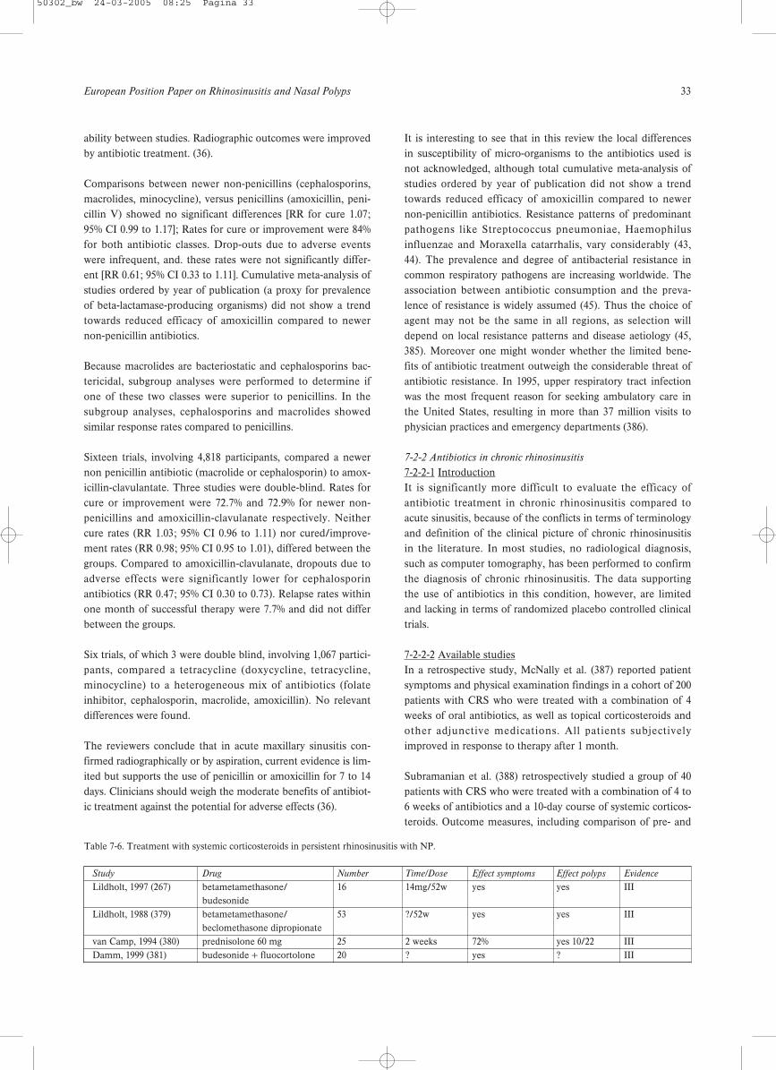
European Position Paper on Rhinosinusitis and Nasal Polyps 33
ability between studies. Radiographic outcomes were improvedby antibiotic treatment. (36).
Comparisons between newer non-penicillins (cephalosporins,macrolides, minocycline), versus penicillins (amoxicillin, peni-cillin V) showed no significant differences [RR for cure 1.07;95% CI 0.99 to 1.17]; Rates for cure or improvement were 84%for both antibiotic classes. Drop-outs due to adverse eventswere infrequent, and. these rates were not significantly differ-ent [RR 0.61; 95% CI 0.33 to 1.11]. Cumulative meta-analysis ofstudies ordered by year of publication (a proxy for prevalenceof beta-lactamase-producing organisms) did not show a trendtowards reduced efficacy of amoxicillin compared to newernon-penicillin antibiotics.
Because macrolides are bacteriostatic and cephalosporins bac-tericidal, subgroup analyses were performed to determine ifone of these two classes were superior to penicillins. In thesubgroup analyses, cephalosporins and macrolides showedsimilar response rates compared to penicillins.
Sixteen trials, involving 4,818 participants, compared a newernon penicillin antibiotic (macrolide or cephalosporin) to amox-icillin-clavulantate. Three studies were double-blind. Rates forcure or improvement were 72.7% and 72.9% for newer non-penicillins and amoxicillin-clavulanate respectively. Neithercure rates (RR 1.03; 95% CI 0.96 to 1.11) nor cured/improve-ment rates (RR 0.98; 95% CI 0.95 to 1.01), differed between thegroups. Compared to amoxicillin-clavulanate, dropouts due toadverse effects were significantly lower for cephalosporinantibiotics (RR 0.47; 95% CI 0.30 to 0.73). Relapse rates withinone month of successful therapy were 7.7% and did not differbetween the groups.
Six trials, of which 3 were double blind, involving 1,067 partici-pants, compared a tetracycline (doxycycline, tetracycline,minocycline) to a heterogeneous mix of antibiotics (folateinhibitor, cephalosporin, macrolide, amoxicillin). No relevantdifferences were found.
The reviewers conclude that in acute maxillary sinusitis con-firmed radiographically or by aspiration, current evidence is lim-ited but supports the use of penicillin or amoxicillin for 7 to 14days. Clinicians should weigh the moderate benefits of antibiot-ic treatment against the potential for adverse effects (36).
It is interesting to see that in this review the local differencesin susceptibility of micro-organisms to the antibiotics used isnot acknowledged, although total cumulative meta-analysis ofstudies ordered by year of publication did not show a trendtowards reduced efficacy of amoxicillin compared to newernon-penicillin antibiotics. Resistance patterns of predominantpathogens like Streptococcus pneumoniae, Haemophilusinfluenzae and Moraxella catarrhalis, vary considerably (43,44). The prevalence and degree of antibacterial resistance incommon respiratory pathogens are increasing worldwide. Theassociation between antibiotic consumption and the preva-lence of resistance is widely assumed (45). Thus the choice ofagent may not be the same in all regions, as selection willdepend on local resistance patterns and disease aetiology (45,385). Moreover one might wonder whether the limited bene-fits of antibiotic treatment outweigh the considerable threat ofantibiotic resistance. In 1995, upper respiratory tract infectionwas the most frequent reason for seeking ambulatory care inthe United States, resulting in more than 37 million visits tophysician practices and emergency departments (386).
7-2-2 Antibiotics in chronic rhinosinusitis
7-2-2-1 IntroductionIt is significantly more difficult to evaluate the efficacy ofantibiotic treatment in chronic rhinosinusitis compared toacute sinusitis, because of the conflicts in terms of terminologyand definition of the clinical picture of chronic rhinosinusitisin the literature. In most studies, no radiological diagnosis,such as computer tomography, has been performed to confirmthe diagnosis of chronic rhinosinusitis. The data supportingthe use of antibiotics in this condition, however, are limitedand lacking in terms of randomized placebo controlled clinicaltrials.
7-2-2-2 Available studiesIn a retrospective study, McNally et al. (387) reported patientsymptoms and physical examination findings in a cohort of 200patients with CRS who were treated with a combination of 4weeks of oral antibiotics, as well as topical corticosteroids andother adjunctive medications. All patients subjectivelyimproved in response to therapy after 1 month.
Subramanian et al. (388) retrospectively studied a group of 40patients with CRS who were treated with a combination of 4 to6 weeks of antibiotics and a 10-day course of systemic corticos-teroids. Outcome measures, including comparison of pre- and
Table 7-6. Treatment with systemic corticosteroids in persistent rhinosinusitis with NP.
Study Drug Number Time/Dose Effect symptoms Effect polyps Evidence
Lildholt, 1997 (267) betametamethasone/ 16 14mg/52w yes yes IIIbudesonide
Lildholt, 1988 (379) betametamethasone/ 53 ?/52w yes yes IIIbeclomethasone dipropionate
van Camp, 1994 (380) prednisolone 60 mg 25 2 weeks 72% yes 10/22 IIIDamm, 1999 (381) budesonide + fluocortolone 20 ? yes ? III
50302_bw 24-03-2005 08:25 Pagina 33
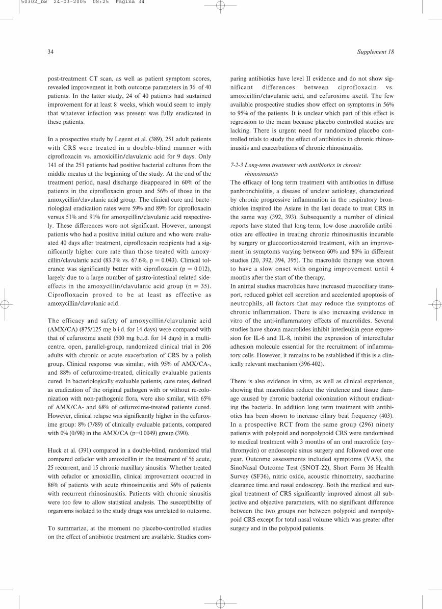
34 Supplement 18
post-treatment CT scan, as well as patient symptom scores,revealed improvement in both outcome parameters in 36 of 40patients. In the latter study, 24 of 40 patients had sustainedimprovement for at least 8 weeks, which would seem to implythat whatever infection was present was fully eradicated inthese patients.
In a prospective study by Legent et al. (389), 251 adult patientswith CRS were treated in a double-blind manner withciprofloxacin vs. amoxicillin/clavulanic acid for 9 days. Only141 of the 251 patients had positive bacterial cultures from themiddle meatus at the beginning of the study. At the end of thetreatment period, nasal discharge disappeared in 60% of thepatients in the ciprofloxacin group and 56% of those in theamoxycillin/clavulanic acid group. The clinical cure and bacte-riological eradication rates were 59% and 89% for ciprofloxacinversus 51% and 91% for amoxycillin/clavulanic acid respective-ly. These differences were not significant. However, amongstpatients who had a positive initial culture and who were evalu-ated 40 days after treatment, ciprofloxacin recipients had a sig-nificantly higher cure rate than those treated with amoxy-cillin/clavulanic acid (83.3% vs. 67.6%, p = 0.043). Clinical tol-erance was significantly better with ciprofloxacin (p = 0.012),largely due to a large number of gastro-intestinal related side-effects in the amoxycillin/clavulanic acid group (n = 35).Ciprofloxacin proved to be at least as effective asamoxycillin/clavulanic acid.
The efficacy and safety of amoxycillin/clavulanic acid(AMX/CA) (875/125 mg b.i.d. for 14 days) were compared withthat of cefuroxime axetil (500 mg b.i.d. for 14 days) in a multi-centre, open, parallel-group, randomized clinical trial in 206adults with chronic or acute exacerbation of CRS by a polishgroup. Clinical response was similar, with 95% of AMX/CA-,and 88% of cefuroxime-treated, clinically evaluable patientscured. In bacteriologically evaluable patients, cure rates, definedas eradication of the original pathogen with or without re-colo-nization with non-pathogenic flora, were also similar, with 65%of AMX/CA- and 68% of cefuroxime-treated patients cured.However, clinical relapse was significantly higher in the cefurox-ime group: 8% (7/89) of clinically evaluable patients, comparedwith 0% (0/98) in the AMX/CA (p=0.0049) group (390).
Huck et al. (391) compared in a double-blind, randomized trialcompared cefaclor with amoxicillin in the treatment of 56 acute,25 recurrent, and 15 chronic maxillary sinusitis: Whether treatedwith cefaclor or amoxicillin, clinical improvement occurred in86% of patients with acute rhinosinusitis and 56% of patientswith recurrent rhinosinusitis. Patients with chronic sinusitiswere too few to allow statistical analysis. The susceptibility oforganisms isolated to the study drugs was unrelated to outcome.
To summarize, at the moment no placebo-controlled studieson the effect of antibiotic treatment are available. Studies com-
paring antibiotics have level II evidence and do not show sig-nificant differences between ciprofloxacin vs.amoxicillin/clavulanic acid, and cefuroxime axetil. The fewavailable prospective studies show effect on symptoms in 56%to 95% of the patients. It is unclear which part of this effect isregression to the mean because placebo controlled studies arelacking. There is urgent need for randomized placebo con-trolled trials to study the effect of antibiotics in chronic rhinos-inusitis and exacerbations of chronic rhinosinusitis.
7-2-3 Long-term treatment with antibiotics in chronic
rhinosinusitis
The efficacy of long term treatment with antibiotics in diffusepanbronchiolitis, a disease of unclear aetiology, characterizedby chronic progressive inflammation in the respiratory bron-chioles inspired the Asians in the last decade to treat CRS inthe same way (392, 393). Subsequently a number of clinicalreports have stated that long-term, low-dose macrolide antibi-otics are effective in treating chronic rhinosinusitis incurableby surgery or glucocorticosteroid treatment, with an improve-ment in symptoms varying between 60% and 80% in differentstudies (20, 392, 394, 395). The macrolide therapy was shownto have a slow onset with ongoing improvement until 4months after the start of the therapy. In animal studies macrolides have increased mucociliary trans-port, reduced goblet cell secretion and accelerated apoptosis ofneutrophils, all factors that may reduce the symptoms ofchronic inflammation. There is also increasing evidence invitro of the anti-inflammatory effects of macrolides. Severalstudies have shown macrolides inhibit interleukin gene expres-sion for IL-6 and IL-8, inhibit the expression of intercellularadhesion molecule essential for the recruitment of inflamma-tory cells. However, it remains to be established if this is a clin-ically relevant mechanism (396-402).
There is also evidence in vitro, as well as clinical experience,showing that macrolides reduce the virulence and tissue dam-age caused by chronic bacterial colonization without eradicat-ing the bacteria. In addition long term treatment with antibi-otics has been shown to increase ciliary beat frequency (403).In a prospective RCT from the same group (296) ninetypatients with polypoid and nonpolypoid CRS were randomisedto medical treatment with 3 months of an oral macrolide (ery-thromycin) or endoscopic sinus surgery and followed over oneyear. Outcome assessments included symptoms (VAS), theSinoNasal Outcome Test (SNOT-22), Short Form 36 HealthSurvey (SF36), nitric oxide, acoustic rhinometry, saccharineclearance time and nasal endoscopy. Both the medical and sur-gical treatment of CRS significantly improved almost all sub-jective and objective parameters, with no significant differencebetween the two groups nor between polypoid and nonpoly-poid CRS except for total nasal volume which was greater aftersurgery and in the polypoid patients.
50302_bw 24-03-2005 08:25 Pagina 34
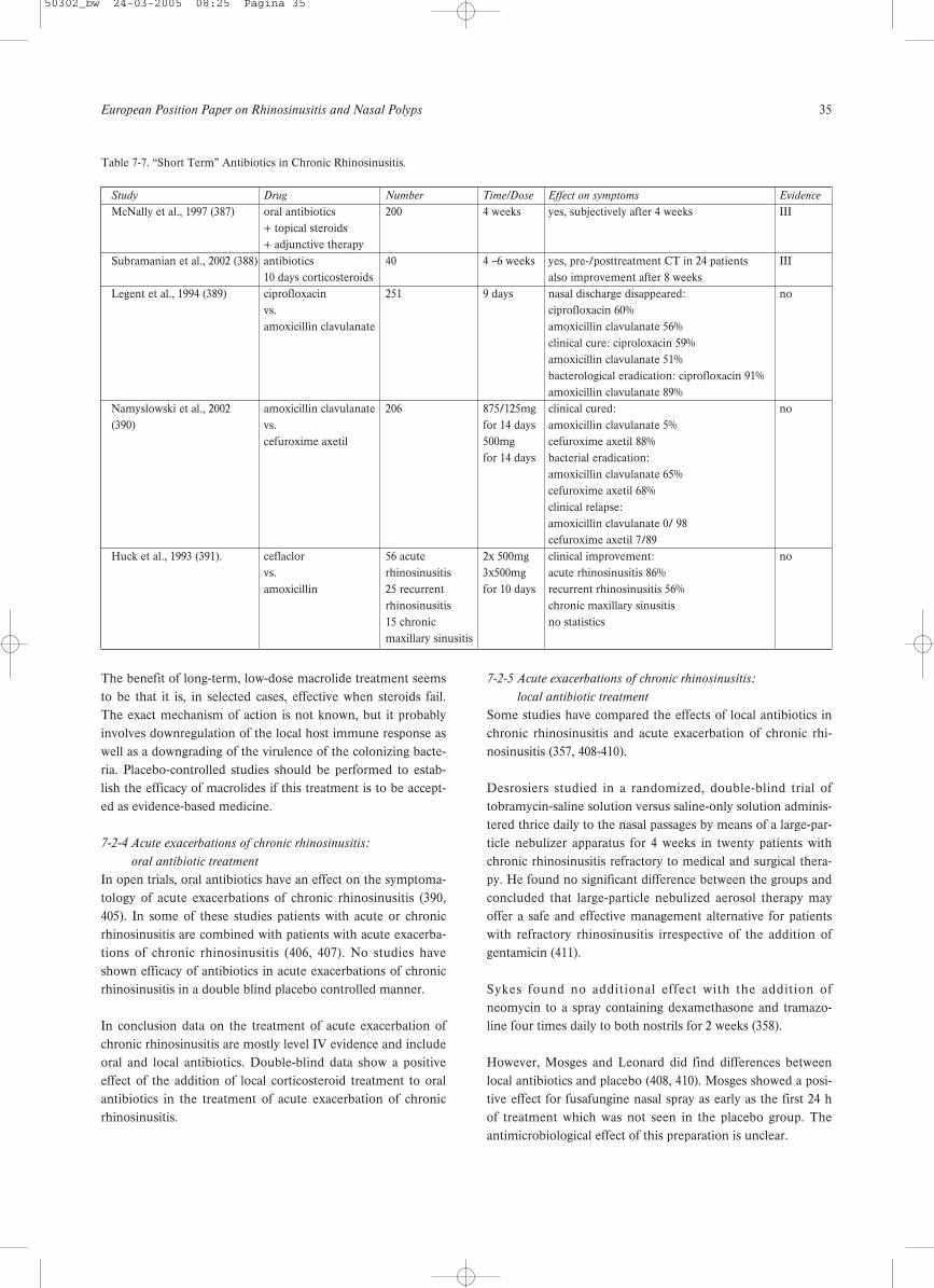
European Position Paper on Rhinosinusitis and Nasal Polyps 35
The benefit of long-term, low-dose macrolide treatment seemsto be that it is, in selected cases, effective when steroids fail.The exact mechanism of action is not known, but it probablyinvolves downregulation of the local host immune response aswell as a downgrading of the virulence of the colonizing bacte-ria. Placebo-controlled studies should be performed to estab-lish the efficacy of macrolides if this treatment is to be accept-ed as evidence-based medicine.
7-2-4 Acute exacerbations of chronic rhinosinusitis:
oral antibiotic treatment
In open trials, oral antibiotics have an effect on the symptoma-tology of acute exacerbations of chronic rhinosinusitis (390,405). In some of these studies patients with acute or chronicrhinosinusitis are combined with patients with acute exacerba-tions of chronic rhinosinusitis (406, 407). No studies haveshown efficacy of antibiotics in acute exacerbations of chronicrhinosinusitis in a double blind placebo controlled manner.
In conclusion data on the treatment of acute exacerbation ofchronic rhinosinusitis are mostly level IV evidence and includeoral and local antibiotics. Double-blind data show a positiveeffect of the addition of local corticosteroid treatment to oralantibiotics in the treatment of acute exacerbation of chronicrhinosinusitis.
7-2-5 Acute exacerbations of chronic rhinosinusitis:
local antibiotic treatment
Some studies have compared the effects of local antibiotics inchronic rhinosinusitis and acute exacerbation of chronic rhi-nosinusitis (357, 408-410).
Desrosiers studied in a randomized, double-blind trial oftobramycin-saline solution versus saline-only solution adminis-tered thrice daily to the nasal passages by means of a large-par-ticle nebulizer apparatus for 4 weeks in twenty patients withchronic rhinosinusitis refractory to medical and surgical thera-py. He found no significant difference between the groups andconcluded that large-particle nebulized aerosol therapy mayoffer a safe and effective management alternative for patientswith refractory rhinosinusitis irrespective of the addition ofgentamicin (411).
Sykes found no additional effect with the addition ofneomycin to a spray containing dexamethasone and tramazo-line four times daily to both nostrils for 2 weeks (358).
However, Mosges and Leonard did find differences betweenlocal antibiotics and placebo (408, 410). Mosges showed a posi-tive effect for fusafungine nasal spray as early as the first 24 hof treatment which was not seen in the placebo group. Theantimicrobiological effect of this preparation is unclear.
Table 7-7. “Short Term” Antibiotics in Chronic Rhinosinusitis.
Study Drug Number Time/Dose Effect on symptoms Evidence
McNally et al., 1997 (387) oral antibiotics 200 4 weeks yes, subjectively after 4 weeks III+ topical steroids+ adjunctive therapy
Subramanian et al., 2002 (388) antibiotics 40 4 –6 weeks yes, pre-/posttreatment CT in 24 patients III10 days corticosteroids also improvement after 8 weeks
Legent et al., 1994 (389) ciprofloxacin 251 9 days nasal discharge disappeared: novs. ciprofloxacin 60% amoxicillin clavulanate amoxicillin clavulanate 56%
clinical cure: ciproloxacin 59% amoxicillin clavulanate 51% bacterological eradication: ciprofloxacin 91% amoxicillin clavulanate 89%
Namyslowski et al., 2002 amoxicillin clavulanate 206 875/125mg clinical cured: no(390) vs. for 14 days amoxicillin clavulanate 5%
cefuroxime axetil 500mg cefuroxime axetil 88%for 14 days bacterial eradication:
amoxicillin clavulanate 65%cefuroxime axetil 68%clinical relapse:amoxicillin clavulanate 0/ 98cefuroxime axetil 7/89
Huck et al., 1993 (391). ceflaclor 56 acute 2x 500mg clinical improvement: novs. rhinosinusitis 3x500mg acute rhinosinusitis 86%amoxicillin 25 recurrent for 10 days recurrent rhinosinusitis 56%
rhinosinusitis chronic maxillary sinusitis15 chronic no statisticsmaxillary sinusitis
50302_bw 24-03-2005 08:25 Pagina 35
36 Supplement 18
Schienberg et al. studied the effectiveness of aerosol deliveryof antibiotics to the sinuses via a nebulizer in 41 patients whohad chronic, recurrent rhinosinusitis that had persisted despiteendoscopic sinus surgery and that had not responded to multi-ple courses of oral antibiotics. Following 3 to 6 weeks of treat-ment, 34 patients (82.9%) experienced either an excellent orgood response to treatment. Side effects were infrequent, mild,and transient. They concluded that nebulized antibioticsshould be considered for all patients with chronic rhinosinusi-tis who have undergone functional endoscopic sinus surgeryand who have failed to respond to oral antibiotics or who donot tolerate them (412).
Further studies with better characterized patient populationsare needed.
7-3 Other medical management for rhinosinusitis
Standard conservative treatment for intermittent and persistentrhinosinusitis is based on short or long-term antibiotics andtopical steroids with the addition of decongestants - mostly ina short term regimen and for the intermittent attack itself.Many other types of preparations have been investigated, butsubstantial evidence for their benefit is poor. These medica-tions include antral washings, isotonic/hypertonic saline asnasal douche, antihistamines, antimycotics, mucolyticagents/phytomedical preparations, immunomodulators/immunostimulants and bacterial lysate preparations. For
selected patients with persistent rhinosinusitis and gastroe-sophageal reflux, the impact of antireflux treatment on sinussymptom scores has been studied. Topical nasal application offurosemide and capsaicin have also been considered in thetreatment of nasal polyposis and prevention of recurrence.
7-3-1 Decongestants
7-3-1-1 Acute/Intermittent RhinosinusitisNasal decongestants are usually applied in the treatment ofacute/ intermittent rhinosinusitis, in order to achieve bettersinus ventilation and drainage. Experimental trials on theeffect of topical decongestants by CT (413) and MRI scans(414) on ostial and ostiomeatal complex patency have con-firmed marked effect on congestion of inferior and middleturbinates and infundibular mucosa, but no effect on eth-moidal and maxillary sinus mucosa. Experimental studies sug-gested beneficial anti-inflammatory effect of xylometazolineand oxymetazoline by decreasing nitric oxide synthetase (415)and anti-oxidant action (416). In contrast to previous in vitrotrials on the effect of decongestants on mucociliary transport, acontrolled clinical trial (II) by Inanli et al. suggested improve-ment in mucociliary clearance in vivo, after 2 weeks ofoxymetazoline application in acute bacterial rhinosinusitis,compared to fluticasone, hypertonic saline and saline, but itdid not show significant improvement compared to the groupwhere no topical nasal treatment was given, and the clinicalcourse of the disease between the groups was not significantlydifferent (417). This is in concordance with previous random-
Table 7-8. Long-term treatment with antibiotics in chronic rhinosinusitis.
Study Drug Number Time/Dose Effect symptoms Evidence
Hashiba et al., 1996 (392) clarithromycin 45 400mg /d for 8 to 12 weeks clinical improvement in 71% IIISuzuki et al., 1997 (393) roxithromycin 12 150mg /d CT scan pre- and post-therapy: III
improvement in the aeration of nasal sinuses
Nishi et al., 1995 (394) clarithromycin 32 400mg /d pre- and post-therapy assessment IIIof nasal clearance
Gahdhi et al., 1993 (395) prophylactic 26 not mentioned 19/26 decrease of acute exacerbation IIIantibiosis by 50% details not 7/26 decrease of acute exacerbation mentioned by less than 50%
Ichimura et al., 1996 (20) roxithromycin 20 150mg /d for at least clinical improve-ment and IIIroxithromycin 20 8 weeks polyp-shrinkage in 52% and azelastine 1mg /d clinical improve-ment and polyp
shrinkage in 68%Scadding et al., 1995 (403) oral antibiotic 10 3 month increased ciliary beating III
therapyCervin et al., (404) erythromicin or 17 1 year increase mucociliary III
clarithromycin clearance and endoscopic signsRagab et al., 2004 (296) erythromycin 45 in each arm 3 months improvement in upper & lower RT Ib
v ESS symptoms, SF36, SNOT-22, NO, Ac Rhin, SCT, nasal endoscopy at 6 & 12 months
RT: respiratory tract, SF 36: Short Form 36 QoL, SNOT-22: SinoNasal Outcome Test, NO:expired nitric oxide, Ac Rhin: acoustic rhinometry,
SCT: saccharine clearance time.
50302_bw 24-03-2005 08:25 Pagina 36
European Position Paper on Rhinosinusitis and Nasal Polyps 37
ized controlled trial in adult acute maxillary sinusitis (Ib),which did not prove significant impact of decongestant whenadded to antibiotic treatment in terms of daily symptomsscores on headache and obstruction and sinus x-ray scores,although decongestant and placebo were applied through abellow, which should have enabled better dispersion of thesolution in the nasal cavity (418). Decongestant treatment didnot prove superior to saline, when added to antibiotic and anti-histamine treatment in a randomized double-blind placebo-controlled trial for acute/intermittent paediatric rhinosinusitis(Ib) (419). Clinical experience, however, supports the use oftopical application of decongestants to the middle meatus inacute rhinosinusitis (evidence level IV).
7-3-1-2 Chronic/Persistent RhinosinusitisThe use of decongestants for adult chronic/persistent rhinosi-nusitis has not been evaluated in a randomized controlled trial.Decongestants and sinus drainage did not prove to be superiorto saline in chronic paediatric maxillary sinusitis in terms ofsubjective or x-ray scores (420).
7-3-1-3 Nasal polyps No controlled trials were used to test the effect of deconges-tant treatment in nasal polyposis. CT studies before and afterdecongestant application in patients with nasal polyposis didnot show any densitometric changes in the sinuses or polyps,only decongestion of the inferior turbinates (421).
7-3-2 Mucolytics
7-3-2-1 Acute/intermittent rhinosinusitis Mucolytics were used as adjuncts to antibiotic treatment anddecongestant treatment in acute/intermittent rhinosinusitis inorder to reduce the viscosity of sinus secretion. The benefit ofsuch treatment has not been evaluated in many trials. In paedi-atric rhinosinusitis, a RCT (Ib) did not prove bromhexinesuperior to saline in inhalation for children with chronic/per-sistent rhinosinusitis (422).A second RCT (Ib) suggestedbromhexine was superior to placebo (423).
7-3-2-2 Chronic/persistent rhinosinusitisA cohort study in a mixed group of 45 acute and chronic rhi-nosinusitis patients suggested beneficial effect of addingmucolytic to standard rhinosinusitis treatment in terms ofreducing treatment duration (424) (evidence level III).
7-3-2-3 Nasal polypsNo clinical trials have tested the effect of mucolytics in nasalpolyp treatment.
7-3-3 Antihistamines, cromones
7-3-3-1 Acute intermittent rhinosinusitisThe beneficial effect of loratadine in terms of symptom reduc-tion for the treatment of acute/intermittent sinusitis in patientswith allergic rhinitis was confirmed in a multicentre random-
ized double-blind, placebo controlled trial (Ib)(425). Patientsreceiving loratadine as an adjunct to antibiotic treatment suf-fered significantly less sneezing and obstruction on daily VASscores, and overall improvement was confirmed by their physi-cians. Cromolyn did not prove better than saline in a RCT (Ib)for treatment of acute intermittent hyperreactive sinusitis mea-sured by subjective scores and ultrasound scans, leading to50% improvement in both groups (426). A RCT (Ib) for inter-mittent paediatric rhinosinusitis did not confirm any benefit oforal antihistamine-nasal decongestant drops (419).
7-3-3-2 Chronic/persistent rhinosinusitisAlthough generally not recommended as rhinosinusitis treat-ment, an evaluation study of chronic rhinosinusits treatment inthe USA revealed antihistamines as rather often prescribedmedication in patients with chronic rhinosinusitis (an averageof 2.7 antibiotic courses; nasal steroids and prescription anti-histamines 18.3 and 16.3 weeks, respectively, in a 12-monthperiod) (427). However, no evidence of beneficial effects ofantihistamine treatment for persistent rhinosinusitis is found,as there are no controlled trials evaluating such treatment.
7-3-3-3 Nasal polypsCetirizine in a dose of 20 mg/day for three months, significant-ly reduced sneezing, rhinorrhoea and obstruction compared toplacebo in the postoperative treatment of recurrent polyposisbut with no effect on polyp size (Ib) (428).
7-3-4 Antimycotics
Antimycotics are used as topical and systemic treatment, as anadjunct to sinus surgery, in allergic fungal, and invasive fungalrhinosinusitis, especially in immunocompromized patients(429). Surgery is considered the first line treatment for allergicfungal (430) and invasive fungal rhinosinusitis (431). Althoughthe use of antimycotics in the treatment of allergic fungal rhi-nosinusitis has not been tested in controlled trials, high doseof postoperative itraconazole, combined with oral and topicalsteroids in a cohort of 139 patients with AFS reduced the needfor revision surgery rate to 20.5% (432). The state-of-art treat-ment for invasive fungal sinusitis is based on small series ofpatients and case reports, which do not meet the criteria formeta analysis and may be considered as level IV evidence.
7-3-4-1 Acute/intermittent sinusitisNo controlled trials for antimycotic treatment for acute rhinos-inusitis was found on the Medline search.
7-3-4-2 Chronic/persistent rhinosinusitisThe fungal hypothesis, based of the premise of an altered localimmune (non-allergic) response to fungal presence innasal/sinus secretions resulting in the generation of chroniceosinophilic rhinosinusitis and nasal polyposis (124), has led toidea of treating any persistent rhinosinusitis/nasal polyposiswith a topical antimycotic. Although the presence of fungus in
50302_bw 24-03-2005 08:25 Pagina 37
38 Supplement 18
sinus secretions was detected in a high proportion (< 90%) ofpatients with persistent rhinosinusitis, as well as in a controldisease-free population in a few study centres (124, 125), it can-not be taken as proof of aetiology. Until now a few case studies(level III) are conducted (433, 434). Ponikau, in a group of 51patient with chronic rhinosinusitis, including polyposispatients, treated with topical amphotericin B as nasal/sinuswashing, without placebo or other control treatment. The treat-ment resulted in 75% subjective improvement and 74% endo-scopic improvement (433). As the authors stated, antifungaltreatment should be evaluated in a controlled trial to be justi-fied. In a double blind randomized placebo controlled trial in60 patients with chronic rhinosinusitis, topical treatment withamphotericine B did not show superior to saline in CT scores(p 0.2) and subjective scores, which were (insignificantly) worsein active treatment group (435). In a recent small randomized,placebo-controlled, double-blind, trial using amphotericin B totreat 30 patients with CRS Ponikau was also not able to showsignificant effect on symptomatology although he did show areduced inflammatory mucosal thickening on both CT scanand nasal endoscopy and decreased levels of intranasal markersfor eosinophilic inflammation in patients with CRS (436).
7-3-4-3 Nasal polyposisAnother case study (as the previous trials also includedpatients with nasal polyposis) combined topical steroid treat-ment with amphotericin B in 74 patients with nasal polyposisfor 4 weeks (437) and found 48% disappearance of the polyps atendoscopy in previously endoscopically operated patients.
The effect of amphotericin B on sinus mucosa may beexplained by some other modes of action. In common withother polyene antibiotics and antimycotics, amphotericin Bacts on cellular membrane permeability, which may reduce thesize of nasal polyps by reducing oedema, leading to subjectiveimprovement (438). These studies were not placebo controlledand had short observation periods. Amphotericin B is a cyto-toxic drug and long-term topical application may have systemiceffect. On the other hand, nasal washings with hypertonic solu-tion (without antifungal medication) offer up to 60% improve-ment (see under chapter 7-4-7 Nasal and antral irrigation -saline, hypertonic saline).
7-3-5 Bacterial lysate preparations
Altered local (and systemic) immune response to bacterialinfection (antigens) may be responsible for frequent recurrenceof rhinosinusitis. Beneficial effect of antibiotic treatment isdeclining together with the increased microbial resistance afterrepeated treatments. Such patients are usually regarded as dif-ficult-to-treat, and usually unresponsive in the long-term tomedical and surgical treatment. As altered immune response isexpected to be responsible for frequent recurrence, differentimmunomodulators or immunostimulants have been tested insuch patients. The most common form of medications used
are bacterial lysates. Efficacy of bacterial lysate preparations(Enteroccocus faecalis autolysate (439), ribosomal fractions ofKlebsiella pneumoniae, Streptococcus pneumoniae,Streptococcus pyogenes, Haemophilus influenzae and themembrane fraction of Kp (440), and mixed bacterial lystate(441) in terms of the reduction of the number of acute relapsesin persistent rhinosinusitis, period between the relapses andneed for antibiotic treatment, have been tested in multicentre,placebo controlled RCTs (Ib) (439-441).
7-3-5-1 Acute/intermittent rhinosinusitisBacterial lysates were tested in the treatment of acute recurrentrhinosinusitis and the outcomes measured were the reducedrate of acute episodes and antibiotic treatment. Enteroccocusfaecalis autolysate treatment for 6 months in 78 patients (3x30drops daily) resulted in 50 relapses during 6 months treatmentand 8 months follow-up compared to 79 placebo treated groupwith 90 recurrences. The time interval to the first relapse wasclearly longer in the active arm (513 days) compared withplacebo (311 days) (439). A RCT of the effect of 6 monthstreatment with ribosomal fractions of Klebsiella pneumoniae,Streptococcus pneumoniae, Streptococcus pyogenes,Haemophilus influenzae and the membrane fraction of Kp wascompared to placebo in 327 adult patients (168 active and 159placebo treatment) with recurrent acute infectious rhinitis (thecriteria could meet recurrent rhinosinusitis based on symp-toms – 4.3 episodes per year) demonstrated 39% reduction ofantibiotic courses and 32% of days with antibiotics during the 6months treatment period (440).
7-3-5-2 Chronic/persistent rhinosinusitisSix months treatment with mixed bacterial lystate was testedin a multicentre randomized double-blind placebo-controlledtrial in 284 patients with CRS (diagnosed by persistent nasaldischarge, headache, and x-ray criteria). Reduction in symptomscores and over-all severity score, including cough and expec-toration were significant during the treatment period (441).
7-3-5-3 Nasal polyposisNo data could be found on treatment with bacterial lysates innasal polyposis.
7-3-6 Immunomodulators/immunostimulants
Treatment with filgrastim, recombinant human granulocytecolony stimulating factor, was tested in a RCT (Ib) in a groupof persistent rhinosinusitis patients refractory to conventionaltreatment, which did not confirm significantly improved out-comes after such expensive treatment (331). A pilot study (III)with interferon gamma suggested this treatment may be bene-ficial in treating resistant persistent rhinosinusitis, but thenumber of patients was not adequate to provide evidence tojustify such treatment (442). Certain groups of antibiotics maybe regarded as immunomodulators, like quinolones (443) andmacrolides (444).
50302_bw 24-03-2005 08:25 Pagina 38
European Position Paper on Rhinosinusitis and Nasal Polyps 39
7-3-7 Nasal and antral irrigation (saline, hypertonic saline)
A number of randomized controlled trials have tested nasaland antral irrigation with isotonic or hypertonic saline in thetreatment of acute/intermittent and chronic/persistent rhinosi-nusitis. Although saline is considered as a control treatmentitself, patients in these randomized trials were assigned to dif-ferent modalities of application of saline or hypertonic saline,or hypertonic compared to isotonic saline. The results betweenthe groups were compared. Most of them offer evidence thatnasal washouts or irrigations with isotonic or hypertonic salineare beneficial in terms of alleviation of symptoms, endoscopicfindings and HRQL improvement in patients with chronic per-sistent rhinosinusitis. Hypertonic saline is preferred to isotonictreatment for rhinosinusitis by some authors in the USA,mostly based on a paper indicating it significantly improvesnasal mucociliary clearance measured by saccharine test, inhealthy volunteers (445).
7-3-7-1 Acute/intermittent rhinosinusitis A randomized trial (Ib) by Adam et al. (446). with two con-trols, compared hypertonic nasal saline to isotonic saline andno treatment in 119 patients with common cold and acute rhi-nosinusitis (predominantly rhinosinusitis). Outcome measureswere subjective nasal symptoms scores (congestion, secretion,headache) at day-3, day-8-10 and the day of symptom resolu-tion. Rhinosinusitis patients (98%) were also treated withantibiotics. There was no difference between the groups andonly 44% of the patients would use the hypertonic saline sprayagain. Thirty-two percent noted burning, compared with 13%of the normal saline group.
Antral irrigation (Ib) did not offer significant benefit whenadded to standard 10-day antibiotic treatment in (4 antibiotics+decongestants vs. antral washouts; 50 patients per group)acute/intermittent rhinosinusitis, demonstrating approximately5% better cure rate in each group for washouts than for decon-gestants, which was not significant (447).
7-3-7-2 Chronic/persistent rhinosinusitisA randomised controlled trial (RCT) by Bachmann (Ib), com-paring isotonic saline and EMS solution (balneotherapeuticwater) in the treatment of persistent sinusitis in a double-blindfashion revealed improvement in both groups, with no differ-ence between them (448). In the 7-days follow-up, nasal airflow was not improved significantly. Subjective complaints,endonasal endoscopy, and radiology results revealed a signifi-cant improvement in both groups (P = 0.0001). A similar RCTby Taccariello et al. (Ib),with a longer follow-up confirmed thatnasal washing with sea water and alkaline nasal douche pro-duced benefit over standard treatments. Douching per seimproved endoscopic appearances (p = .009), and quality of lifescores (p = .008) (449). These measures did not change in acontrol group (n = 22) who received standard treatment forchronic rhinosinusitis, but no douche. There were significant
differences between the two douching preparations - the alka-line nasal douche improved endoscopic appearances but didnot enhance quality of life, whereas the opposite was true forthe spray. Rabago et al. (Ib) tested benefit from daily hyperton-ic saline washings compared to standard chronic rhinosinusitistreatment (control) for 6 months in a RCT using subjectivescores instruments: Medical Outcomes Survey Short Form(SF-12), the Rhinosinusitis Disability Index (RSDI), and aSingle-Item Sinus-Symptom Severity Assessment (SIA).Experimental subjects reported fewer 2-week periods withsinus-related symptoms (P <.05), used less antibiotics (P <.05),and used less nasal spray (P =.06) (450). On the exit question-naire 93% of study subjects reported overall improvement ofsinus-related quality of life, and none reported worsening (P<.001); on average, experimental subjects reported 57 -/+ 4.5%improvement measured by Medical Outcomes Survey ShortForm (SF-12), the Rhinosinusitis Disability Index (RSDI), anda Single-Item Sinus-Symptom Severity Assessment (SIA). Adouble blind RCT (Ib) compared the effect of nasal wash withhypertonic saline (3.5%) versus normal saline (NS) (0.9%) forthe 4 weeks in treatment of paediatric chronic/persistent rhi-nosinusitis using cough and nasal secretions/postnasal drip assubjective and a radiology score as objective outcome mea-sures (451). Hypertonic saline demonstrated significantimprovement for all the scores (13/15 for cough,13/15 post-nasal drip, 14/15 x-ray scores), while saline improved onlypostnasal drip.
Nebulised hypertonic saline improves mucociliary clearance inshort term clinical trials and appears to increase lung functioncompared to controls in cystic fibrosis patients (Wark,Cochrane Database Syst Rev. 2003;(1):CD001506 – aproposdoubt about general harm of hypertonic saline on lung func-tion. However, it does cause bronchocontriction in the asth-matics ).
Comparison of treatment with antral washouts in the treat-ment of persistent adult (452) and paediatric rhinosinusitis(453) did not prove benefit from such treatment. In a RCT byPang et al. patients received either antral washouts followed byantibiotics and topical nasal steroids or antibiotics and topicalnasal steroids alone. In each group 51.6 per cent and 50 percent of patients respectively improved with treatment (452).
7-3-7-3 Nasal polypsNasal saline has been used as a control treatment in trials onnasal polyposis with topical steroid, but there are no controlledtrials on saline/hypertonic saline treatment alone in nasal poly-posis.
7-3-8 Capsaicin
Capsaicin, the active substance from red hot chilli peppers, is aneurotoxin which depletes substance P with some other neu-rokinins and neuropeptides, leading to long lasting damage of
50302_bw 24-03-2005 08:25 Pagina 39
40 Supplement 18
unmyelinated axons and thinly myelinated axons when repeat-edly applied to the respiratory mucosa. Substance P was foundeffective in reducing nasal symptoms after cumulative topicalapplications in the treatment of non-allergic hyperreactiverhinitis, probably acting as desensitizer of nasal mucosa due todepletion of SP and neurokinins. The hypothesis that neuro-genic inflammation may play a role in the pathogenesis ofnasal polyps has lead to trials on capsaicin treatment of nasalpolyposis.
7-3-8-1 Acute/intermittent, chronic/persistent sinusitisNo trials of treatment of acute or chronic rhinosinusitis withcapsaicin could be found.
7-3-8-2 Nasal polypsA case study (III) by Filiaci et al. has demonstrated significantreduction of the size of nasal polyps after five (weekly) topicalapplications of capsaicin (30 mmol/L) solution in patients withnasal polyposis (454). The authors noted increased nasaleosinophilia after the treatment, which was not correlated tothe polyp size. A case study by Baudoin et al. has demonstrat-ed significant reduction of sinonasal polyposis after 5 consecu-tive days treatment with increasing doses (30-100 mmol/L) oftopical capsaicin in massive polyposis measured by CT scans atentry and after 4 weeks (III) (455). ECP in nasal lavage was notinfluenced by the treatment. Protection of polyp recurrencefollowing endonasal surgery by 5 topical applications of cap-saicin in 51 patient after surgery with a 9 months follow-up hasconfirmed significant recurrence protection and significantlybetter nasal patency in the active group in a randomized, dou-ble blind, placebo controlled trial (Ib) by Zheng et al. (456).The authors used 70% ethanol 3x10⁻⁶E ml capsaicin solution,which may explain the high rate of recurrence in the controlgroup after ESS, which received only 70% ethanol. They noted40% polyp stage 0 (Malm) and 45% stage 1 in the active treat-ment group, while controls demonstrated 45% stage 2 and 40%stage 3 polyposis following treatment at 9 months observation.The low cost of capsaicin treatment was noted as a certainadvantage compared to other postoperative treatments. As cap-saicin is NF kappa B antagonist in vitro, some other modes ofaction may be proposed (457).
7-3-9 Furosemide
The protection of hyperreactive response to different chal-lenges (propranolol (458); metabisulphite (459); exercise (460))in asthmatics was demonstrated after inhalation of furosemide,suggesting bronchoprotective effects, similar to the effect ofcromones. Histamine exocytosis from rat mast cells was pro-tected by furosemide in vitro (461). It exhibited an anti-inflam-matory effect through inhibition of production and release ofcytokines, interleukin (IL)-6, IL-8, and tumor necrosis factor-alpha from peripheral mononuclear cells in vitro (462).
7-3-9-1 Acute/intermittent, chronic/persistent sinusitisNo trials of treatment of acute or chronic rhinosinusitis withfurosemide have been found.
7-3-9-1 Nasal polypsProtection against nasal polyp recurrence following surgerywith 1-9 years follow-up, comparable to the effect of the topicalsteroid, was demonstrated after topical application offurosemide in 97 patients postoperatively vs. mometasonefuroate in 33 patients, in a prospective non-randomized con-trolled trial (IIa) by Passali et al. (463), previously reported bythe same group in a case study. Relapses were recorded in17.5% in the furosemide, 24.2% in the mometasone and 30% inthe no treatment group, suggesting that furosemide, as a muchcheaper medication than steroids, might be considered inpolyp recurrence protection treatment. Randomized trials how-ever are lacking.
7-3-10 Proton pump inhibitors
7-3-10-1 Acute/intermittent rhinosinusitisThere are no trials with proton pump inhibitors for acute rhi-nosinusitis
7-3-10-2 Chronic/persistent rhinosinusitisThere is no evidence for benefits in the general population suf-fering from rhinosinusitis following treatment with protonpump-inhibitors, while subjective improvement was noted inpatients with laryngopharyngeal reflux (proved by pH-metry)and rhinosinusitis. Grade C evidence for a positive associationbetween gastroesophageal reflux and rhinosinusitis was foundin a meta analysis of the literature for this co-morbidity (57articles screened, 14 articles included) (464, 465). A number ofcase trials of rhinosinusitis, especially paediatric (464), has test-ed the efficacy of antireflux treatment with proton pumpinhibitors on the clinical course and symptoms of rhinosinusi-tis. Increased rates of reflux were detected in persistent rhinos-inusitis in adults unresponsive to standard treatment (466). Abeneficial effect of proton pump inhibitors on sinusitis symp-toms in patients with resistant persistent sinusitis was demon-strated in an open label clinical trial (III) (467). Furtherresearch is expected in this field, and such treatment should bejustified by randomized controlled trials.
7-3-10-3 Nasal polypsThere are no data on proton-pump inhibitors in nasal polypo-sis.
7-3-11 Antileukotrienes
The role of leukotriens in the pathogenesis of bronchial asth-ma has been well documented, and increased levels of thesemediators have been detected in patients with rhinosinusitisand nasal polyps. Antileukotrienes have been evaluated in thetreatment of asthmatics, especially in those with ASA triad.The effect of leukotrienes was evaluated in a randomized con-
50302_bw 24-03-2005 08:25 Pagina 40
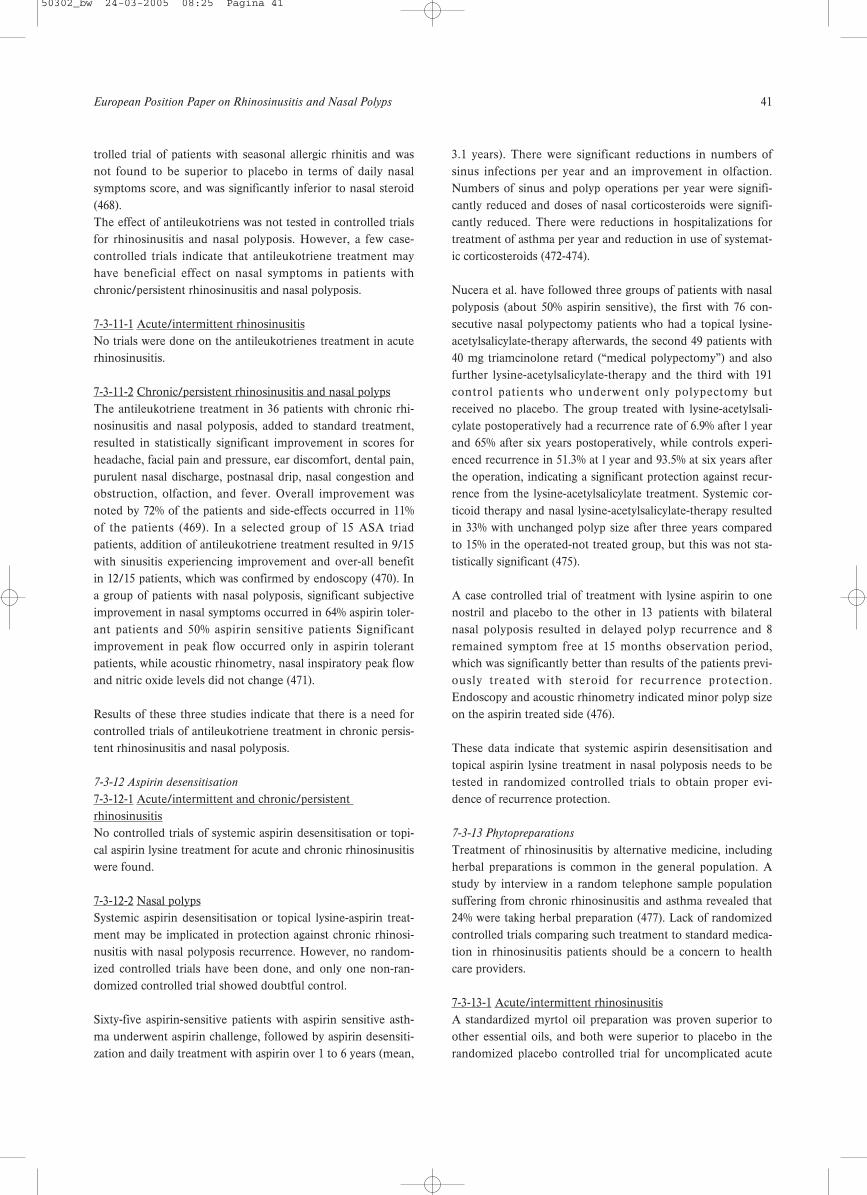
European Position Paper on Rhinosinusitis and Nasal Polyps 41
trolled trial of patients with seasonal allergic rhinitis and wasnot found to be superior to placebo in terms of daily nasalsymptoms score, and was significantly inferior to nasal steroid(468). The effect of antileukotriens was not tested in controlled trialsfor rhinosinusitis and nasal polyposis. However, a few case-controlled trials indicate that antileukotriene treatment mayhave beneficial effect on nasal symptoms in patients withchronic/persistent rhinosinusitis and nasal polyposis.
7-3-11-1 Acute/intermittent rhinosinusitisNo trials were done on the antileukotrienes treatment in acuterhinosinusitis.
7-3-11-2 Chronic/persistent rhinosinusitis and nasal polypsThe antileukotriene treatment in 36 patients with chronic rhi-nosinusitis and nasal polyposis, added to standard treatment,resulted in statistically significant improvement in scores forheadache, facial pain and pressure, ear discomfort, dental pain,purulent nasal discharge, postnasal drip, nasal congestion andobstruction, olfaction, and fever. Overall improvement wasnoted by 72% of the patients and side-effects occurred in 11%of the patients (469). In a selected group of 15 ASA triadpatients, addition of antileukotriene treatment resulted in 9/15with sinusitis experiencing improvement and over-all benefitin 12/15 patients, which was confirmed by endoscopy (470). Ina group of patients with nasal polyposis, significant subjectiveimprovement in nasal symptoms occurred in 64% aspirin toler-ant patients and 50% aspirin sensitive patients Significantimprovement in peak flow occurred only in aspirin tolerantpatients, while acoustic rhinometry, nasal inspiratory peak flowand nitric oxide levels did not change (471).
Results of these three studies indicate that there is a need forcontrolled trials of antileukotriene treatment in chronic persis-tent rhinosinusitis and nasal polyposis.
7-3-12 Aspirin desensitisation
7-3-12-1 Acute/intermittent and chronic/persistent rhinosinusitisNo controlled trials of systemic aspirin desensitisation or topi-cal aspirin lysine treatment for acute and chronic rhinosinusitiswere found.
7-3-12-2 Nasal polypsSystemic aspirin desensitisation or topical lysine-aspirin treat-ment may be implicated in protection against chronic rhinosi-nusitis with nasal polyposis recurrence. However, no random-ized controlled trials have been done, and only one non-ran-domized controlled trial showed doubtful control.
Sixty-five aspirin-sensitive patients with aspirin sensitive asth-ma underwent aspirin challenge, followed by aspirin desensiti-zation and daily treatment with aspirin over 1 to 6 years (mean,
3.1 years). There were significant reductions in numbers ofsinus infections per year and an improvement in olfaction.Numbers of sinus and polyp operations per year were signifi-cantly reduced and doses of nasal corticosteroids were signifi-cantly reduced. There were reductions in hospitalizations fortreatment of asthma per year and reduction in use of systemat-ic corticosteroids (472-474).
Nucera et al. have followed three groups of patients with nasalpolyposis (about 50% aspirin sensitive), the first with 76 con-secutive nasal polypectomy patients who had a topical lysine-acetylsalicylate-therapy afterwards, the second 49 patients with40 mg triamcinolone retard (“medical polypectomy”) and alsofurther lysine-acetylsalicylate-therapy and the third with 191control patients who underwent only polypectomy butreceived no placebo. The group treated with lysine-acetylsali-cylate postoperatively had a recurrence rate of 6.9% after l yearand 65% after six years postoperatively, while controls experi-enced recurrence in 51.3% at l year and 93.5% at six years afterthe operation, indicating a significant protection against recur-rence from the lysine-acetylsalicylate treatment. Systemic cor-ticoid therapy and nasal lysine-acetylsalicylate-therapy resultedin 33% with unchanged polyp size after three years comparedto 15% in the operated-not treated group, but this was not sta-tistically significant (475).
A case controlled trial of treatment with lysine aspirin to onenostril and placebo to the other in 13 patients with bilateralnasal polyposis resulted in delayed polyp recurrence and 8remained symptom free at 15 months observation period,which was significantly better than results of the patients previ-ously treated with steroid for recurrence protection.Endoscopy and acoustic rhinometry indicated minor polyp sizeon the aspirin treated side (476).
These data indicate that systemic aspirin desensitisation andtopical aspirin lysine treatment in nasal polyposis needs to betested in randomized controlled trials to obtain proper evi-dence of recurrence protection.
7-3-13 Phytopreparations
Treatment of rhinosinusitis by alternative medicine, includingherbal preparations is common in the general population. Astudy by interview in a random telephone sample populationsuffering from chronic rhinosinusitis and asthma revealed that24% were taking herbal preparation (477). Lack of randomizedcontrolled trials comparing such treatment to standard medica-tion in rhinosinusitis patients should be a concern to healthcare providers.
7-3-13-1 Acute/intermittent rhinosinusitisA standardized myrtol oil preparation was proven superior toother essential oils, and both were superior to placebo in therandomized placebo controlled trial for uncomplicated acute
50302_bw 24-03-2005 08:25 Pagina 41
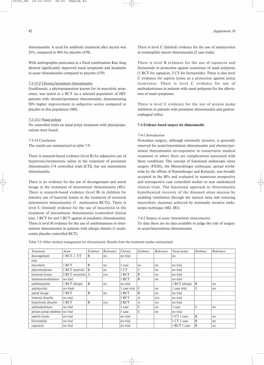
42 Supplement 18
rhinosinusitis. A need for antibiotic treatment after myrtol was23%, compared to 40% for placebo (478).
With andrographis paniculata in a fixed combination Kan Jangshowed significantly improved nasal symptoms and headachein acute rhinosinusitis compared to placebo (479).
7-3-13-2 Chronic/persistent rhinosinusitisGuaifenesin, a phytopreparation known for its mucolytic prop-erties, was tested in a RCT on a selected population of HIVpatients with chronic/persistent rhinosinusitis, demonstrating20% higher improvement in subjective scores compared toplacebo in this population (480).
7-3-13-3 Nasal polypsNo controlled trials on nasal polyp treatment with phytoprepa-rations were found.
7-3-14 Conclusion
The results are summarized in table 7-9.
There is research-based evidence (level B) for adjunctive use ofhypertonic/normotonic saline in the treatment of persistentrhinosinusitis (<4 controlled trials [CT]), but not intermittentrhinosinusitis.
There is no evidence for the use of decongestants and antrallavage in the treatment of intermittent rhinosinusitis (481).There is research-based evidence (level B) in children forselective use of bacterial lysates in the treatment of recurrentintermittent rhinosinusitis (3 multicentre RCTs). There islevel C (limited) evidence for the use of mucolytics in thetreatment of intermittent rhinosinusitis (controlled clinicaltrial, 1 RCT for and 1 RCT against in paediatric rhinosinusitis).There is level B evidence for the use of antihistamines in inter-mittent rhinosinusitis in patients with allergic rhinitis (1 multi-centre placebo controlled RCT).
There is level C (limited) evidence for the use of antimycoticsin eosinophilic mucin rhinosinusitis (2 case trials).
There is level B evidence for the use of capsaicin andfurosemide in protection against recurrence of nasal polyposis(1 RCT for capsaicin, 2 CT for furosemide). There is also levelC evidence for aspirin lysine as a protection against polyprecurrence. There is level C evidence for use ofantileukotrienes in patients with nasal polyposis for the allevia-tion of nasal symptoms.
There is level C evidence for the use of proton pumpinhibitors in patients with persistent rhinosinusitis and gastroe-sophageal reflux.
7-4 Evidence based surgery for rhinosinusitis
7-4-1 Introduction
Nowadays surgery, although minimally invasive, is generallyreserved for acute/intermittent rhinosinusitis and chronic/per-sistent rhinosinusitis un-responsive to conservative medicaltreatment or where there are complications associated withthese conditions. The concept of functional endoscopic sinussurgery (FESS), the Messerklinger technique, spread world-wide by the efforts of Stammberger and Kennedy, was broadlyaccepted in the 80’s and evaluated in numerous prospectiveand retrospective case controlled studies or non randomizedclinical trials. The functional approach to rhinosinusitishypothesized recovery of the diseased sinus mucosa byenabling ventilation through the natural ostia and restoringmucociliary clearance achieved by minimally invasive endo-scopic technique (482, 483).
7-4-2 Surgery in acute /intermittent rhinosinusitis.
To date there are no data available to judge the role of surgeryin acute/intermittent rhinosinusitis.
Table 7-9. Other medical management for rhinosinusitis. Results from the treatment studies summarised.
Treatment Acute Evidence Relevance Chronic Evidence Relevance Nasal polyp Evidence Relevance
decongestant 1 RCT, 1 CT B no. no trial notrialmucolytic 1 RCT B no 1 case no no no trialphytomedicine 1 RCT (myrtol) B no 1 CT C no no trialbacterial lysate 2 RCT recurrent A yes 1 RCT B no no trialimmunomodulation no trial 1 RCT B no no trialantihistamine 1 RCT allergic B no no trial 1 RCT allergic B noantimycotic no trials 1 case trial C no 1 case trial C noantral lavage 1 RCT B no 1 RCT B no no trialisotonic douche no trial 3 RCT A yes no trialhypertonic douche 1 RCT B yes 2 RCT A no no trialantileukotriene no trial 1 case C no 3 case C noproton pump inhibitor no trial 3 case C no no trialaspirin lysate no trial no trial 1 CT 1 case B nofurosemide no trial no trial 1 CT 1 case B nocapsaicin no trial no trial 1 RCT 1 case B no
50302_bw 24-03-2005 08:25 Pagina 42
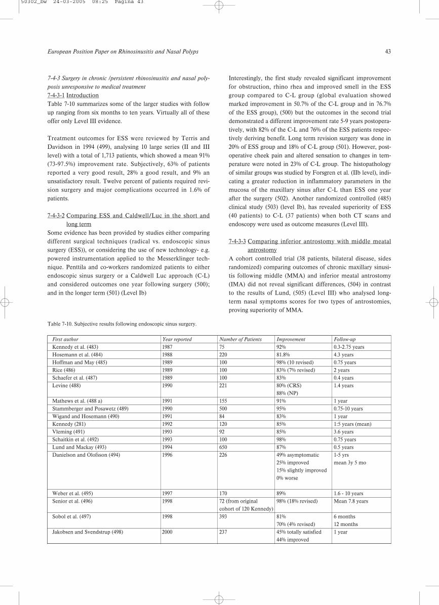
European Position Paper on Rhinosinusitis and Nasal Polyps 43
7-4-3 Surgery in chronic /persistent rhinosinusitis and nasal poly-
posis unresponsive to medical treatment
7-4-3-1 IntroductionTable 7-10 summarizes some of the larger studies with followup ranging from six months to ten years. Virtually all of theseoffer only Level III evidence.
Treatment outcomes for ESS were reviewed by Terris andDavidson in 1994 (499), analysing 10 large series (II and IIIlevel) with a total of 1,713 patients, which showed a mean 91%(73-97.5%) improvement rate. Subjectively, 63% of patientsreported a very good result, 28% a good result, and 9% anunsatisfactory result. Twelve percent of patients required revi-sion surgery and major complications occurred in 1.6% ofpatients.
7-4-3-2 Comparing ESS and Caldwell/Luc in the short andlong term
Some evidence has been provided by studies either comparingdifferent surgical techniques (radical vs. endoscopic sinussurgery (ESS)), or considering the use of new technology- e.g.powered instrumentation applied to the Messerklinger tech-nique. Penttila and co-workers randomized patients to eitherendoscopic sinus surgery or a Caldwell Luc approach (C-L)and considered outcomes one year following surgery (500);and in the longer term (501) (Level Ib)
Interestingly, the first study revealed significant improvementfor obstruction, rhino rhea and improved smell in the ESSgroup compared to C-L group (global evaluation showedmarked improvement in 50.7% of the C-L group and in 76.7%of the ESS group), (500) but the outcomes in the second trialdemonstrated a different improvement rate 5-9 years postopera-tively, with 82% of the C-L and 76% of the ESS patients respec-tively deriving benefit. Long term revision surgery was done in20% of ESS group and 18% of C-L group (501). However, post-operative cheek pain and altered sensation to changes in tem-perature were noted in 23% of C-L group. The histopathologyof similar groups was studied by Forsgren et al. (IIb level), indi-cating a greater reduction in inflammatory parameters in themucosa of the maxillary sinus after C-L than ESS one yearafter the surgery (502). Another randomized controlled (485)clinical study (503) (level Ib), has revealed superiority of ESS(40 patients) to C-L (37 patients) when both CT scans andendoscopy were used as outcome measures (Level III).
7-4-3-3 Comparing inferior antrostomy with middle meatalantrostomy
A cohort controlled trial (38 patients, bilateral disease, sidesrandomized) comparing outcomes of chronic maxillary sinusi-tis following middle (MMA) and inferior meatal antrostomy(IMA) did not reveal significant differences, (504) in contrastto the results of Lund, (505) (Level III) who analysed long-term nasal symptoms scores for two types of antrostomies,proving superiority of MMA.
Table 7-10. Subjective results following endoscopic sinus surgery.
First author Year reported Number of Patients Improvement Follow-up
Kennedy et al. (483) 1987 75 92% 0.3-2.75 yearsHosemann et al. (484) 1988 220 81.8% 4.3 yearsHoffman and May (485) 1989 100 98% (10 revised) 0.75 yearsRice (486) 1989 100 83% (7% revised) 2 yearsSchaefer et al. (487) 1989 100 83% 0.4 yearsLevine (488) 1990 221 80% (CRS) 1.4 years
88% (NP)Mathews et al. (488 a) 1991 155 91% 1 yearStammberger and Posawetz (489) 1990 500 95% 0.75-10 yearsWigand and Hosemann (490) 1991 84 83% 1 yearKennedy (281) 1992 120 85% 1:5 years (mean)Vleming (491) 1993 92 85% 3.6 yearsSchaitkin et al. (492) 1993 100 98% 0.75 yearsLund and Mackay (493) 1994 650 87% 0.5 yearsDanielson and Olofsson (494) 1996 226 49% asymptomatic 1-5 yrs
25% improved mean 3y 5 mo15% slightly improved0% worse
Weber et al. (495) 1997 170 89% 1.6 - 10 yearsSenior et al. (496) 1998 72 (from original 98% (18% revised) Mean 7.8 years
cohort of 120 Kennedy)Sobol et al. (497) 1998 393 81% 6 months
70% (4% revised) 12 monthsJakobsen and Svendstrup (498) 2000 237 45% totally satisfied 1 year
44% improved
50302_bw 24-03-2005 08:25 Pagina 43
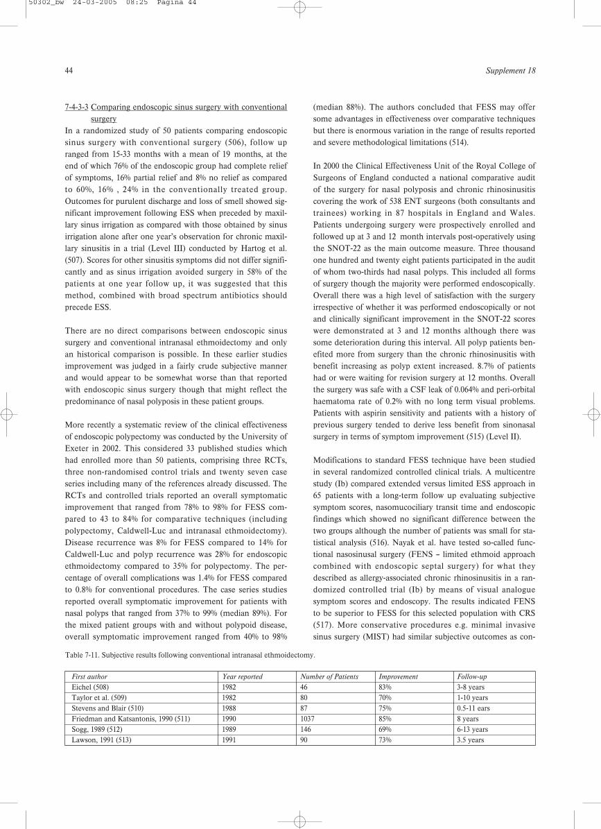
44 Supplement 18
7-4-3-3 Comparing endoscopic sinus surgery with conventionalsurgery
In a randomized study of 50 patients comparing endoscopicsinus surgery with conventional surgery (506), follow upranged from 15-33 months with a mean of 19 months, at theend of which 76% of the endoscopic group had complete reliefof symptoms, 16% partial relief and 8% no relief as comparedto 60%, 16% , 24% in the conventionally treated group.Outcomes for purulent discharge and loss of smell showed sig-nificant improvement following ESS when preceded by maxil-lary sinus irrigation as compared with those obtained by sinusirrigation alone after one year’s observation for chronic maxil-lary sinusitis in a trial (Level III) conducted by Hartog et al.(507). Scores for other sinusitis symptoms did not differ signifi-cantly and as sinus irrigation avoided surgery in 58% of thepatients at one year follow up, it was suggested that thismethod, combined with broad spectrum antibiotics shouldprecede ESS.
There are no direct comparisons between endoscopic sinussurgery and conventional intranasal ethmoidectomy and onlyan historical comparison is possible. In these earlier studiesimprovement was judged in a fairly crude subjective mannerand would appear to be somewhat worse than that reportedwith endoscopic sinus surgery though that might reflect thepredominance of nasal polyposis in these patient groups.
More recently a systematic review of the clinical effectivenessof endoscopic polypectomy was conducted by the University ofExeter in 2002. This considered 33 published studies whichhad enrolled more than 50 patients, comprising three RCTs,three non-randomised control trials and twenty seven caseseries including many of the references already discussed. TheRCTs and controlled trials reported an overall symptomaticimprovement that ranged from 78% to 98% for FESS com-pared to 43 to 84% for comparative techniques (includingpolypectomy, Caldwell-Luc and intranasal ethmoidectomy).Disease recurrence was 8% for FESS compared to 14% forCaldwell-Luc and polyp recurrence was 28% for endoscopicethmoidectomy compared to 35% for polypectomy. The per-centage of overall complications was 1.4% for FESS comparedto 0.8% for conventional procedures. The case series studiesreported overall symptomatic improvement for patients withnasal polyps that ranged from 37% to 99% (median 89%). Forthe mixed patient groups with and without polypoid disease,overall symptomatic improvement ranged from 40% to 98%
(median 88%). The authors concluded that FESS may offersome advantages in effectiveness over comparative techniquesbut there is enormous variation in the range of results reportedand severe methodological limitations (514).
In 2000 the Clinical Effectiveness Unit of the Royal College ofSurgeons of England conducted a national comparative auditof the surgery for nasal polyposis and chronic rhinosinusitiscovering the work of 538 ENT surgeons (both consultants andtrainees) working in 87 hospitals in England and Wales.Patients undergoing surgery were prospectively enrolled andfollowed up at 3 and 12 month intervals post-operatively usingthe SNOT-22 as the main outcome measure. Three thousandone hundred and twenty eight patients participated in the auditof whom two-thirds had nasal polyps. This included all formsof surgery though the majority were performed endoscopically.Overall there was a high level of satisfaction with the surgeryirrespective of whether it was performed endoscopically or notand clinically significant improvement in the SNOT-22 scoreswere demonstrated at 3 and 12 months although there wassome deterioration during this interval. All polyp patients ben-efited more from surgery than the chronic rhinosinusitis withbenefit increasing as polyp extent increased. 8.7% of patientshad or were waiting for revision surgery at 12 months. Overallthe surgery was safe with a CSF leak of 0.064% and peri-orbitalhaematoma rate of 0.2% with no long term visual problems.Patients with aspirin sensitivity and patients with a history ofprevious surgery tended to derive less benefit from sinonasalsurgery in terms of symptom improvement (515) (Level II).
Modifications to standard FESS technique have been studiedin several randomized controlled clinical trials. A multicentrestudy (Ib) compared extended versus limited ESS approach in65 patients with a long-term follow up evaluating subjectivesymptom scores, nasomucociliary transit time and endoscopicfindings which showed no significant difference between thetwo groups although the number of patients was small for sta-tistical analysis (516). Nayak et al. have tested so-called func-tional nasosinusal surgery (FENS – limited ethmoid approachcombined with endoscopic septal surgery) for what theydescribed as allergy-associated chronic rhinosinusitis in a ran-domized controlled trial (Ib) by means of visual analoguesymptom scores and endoscopy. The results indicated FENSto be superior to FESS for this selected population with CRS(517). More conservative procedures e.g. minimal invasivesinus surgery (MIST) had similar subjective outcomes as con-
Table 7-11. Subjective results following conventional intranasal ethmoidectomy.
First author Year reported Number of Patients Improvement Follow-up
Eichel (508) 1982 46 83% 3-8 yearsTaylor et al. (509) 1982 80 70% 1-10 yearsStevens and Blair (510) 1988 87 75% 0.5-11 earsFriedman and Katsantonis, 1990 (511) 1990 1037 85% 8 yearsSogg, 1989 (512) 1989 146 69% 6-13 yearsLawson, 1991 (513) 1991 90 73% 3.5 years
50302_bw 24-03-2005 08:25 Pagina 44
European Position Paper on Rhinosinusitis and Nasal Polyps 45
ventional ESS, in a prospective non-randomized study in 85patients with persistent rhinosinusitis (level III) but the resultsshould be validated by a RCT (518).
A randomized controlled trial (Ib) tested the outcomes forholmium-YAG laser in 32 patients with CRS undergoing ESS(randomization - one side conventional, contralateral laser)(519). The use of holmium-YAG laser in ESS resulted in sig-nificantly lower blood loss during surgery and less post-opera-tive crust formation than conventional ESS, but long term sub-jective outcomes did not show significant difference betweenthe methods. Similarly in a prospective randomized studySelivanova et al.were unable to demonstrate an advantage ofmechanical debriders over conventional instrumentation (520).
7-4-3-5 Endoscopic surgery in special situationsFrom a cohort of 650 patients undergoing ESS for CRS, 28patients suffered for cystic fibrosis and14 from immune defi-ciency (493) (Level III). Whilst overall subjective improvementwas less than in the cohort as a whole (91% improved), 54% ofthe cystics and 79% of those with immune deficiency derivedsignificant benefit at six month follow-up. No studies specifi-cally focusing on primary ciliary dyskinesia or congenitalimmune deficiency were found in the literature. However, anumber of authors have considered acquired immune deficien-cy, mainly related to HIV. These have been by definition a rel-atively small series (98) , (Level III). The bacterial profile maymirror that seen in conventional rhinosinusitis but can alsoincludes pseudomonas aeruginosa and toxoplasma. A range ofsurgical approaches have been used in this group with highrelapse rates reported of 76-81%.
The small number of papers concentrating on cystic fibrosishave mainly concerned the paediatric population. Halvorsen etal. (521) reported 16 adults with cystic fibrosis and chronic rhi-nosinusitis/nasal polyposis combined with pulmonary compli-cations. The study considered pulmonary function followingendoscopic sinus surgery and preliminary findings suggestedan improvement in both the symptoms of rhinosinusitis andexercise tolerance (Level III). However, again there was a highchance of relapse, 50% in the study by Rowe-Jones andMackay (522) (Level III) within two years of the procedure.
The relationship of asthma/aspirin-sensitivity on surgicalresults and the effects of surgery on the lower respiratory tractare debated. A prospective study of 120 patients maintainedthat when extent of disease was taken into account, asthma perse did not adversely affect outcome (281) (Level II). However,as a corollary of this, recurrence particularly in the aspirin-sen-sitive group is likely to be higher (492) (523) (Level III). Thismay be off-set to some extent in the short term by the extentof surgery (523, 524) The effect of sinonasal surgery on respira-tory function has generally been positive.
7-4-4 Conclusion
In conclusion, trials providing high level statements of evi-dence for efficacy of surgery for rhinosinusitis are lacking, asalready concluded by Lund in 2001 (539). Few sinonasal surgi-cal studies are designed as RCTs, and those that are should beof higher quality. Lack of consistency between the studies(inclusion-exclusion criteria, staging, scores, questionnairesetc.) and inadequate numbers for robust statistics are the maindrawbacks of these trials. In addition the experience of anendoscopic rhinosurgeon should be established before one cancompare results from different studies (540) though there havebeen some attempts to look at the ‘learning curve’ throughcomplication rates (Stankiewicz). We have a large amount oflow level evidence that ESS is a safe procedure that improvesrhinosinusitis symptom scores, HRQL and some objective cri-teria (see Chapter 7-5-3) in low-risk adult patients. However, atleast two studies have shown that aggressive medical therapyoffers similar results over a one year period (296, 541) (LevelIIb) underlining the need to reserve surgery for those who havefailed medical therapy. ‘High-risk’ patients should be treatedwith aggressive long-term medication pre- and postoperativelyand represent a different group when evaluating studies.
7-5 Surgical treatment vs. medical treatment in CRS /NP
7-5-1 Surgical treatment vs. steroids in NP
In the two open studies by Lildholt et al. (267, 379) singleinjections of 14 mg betametasone have been compared tointranasal polypectomy without any difference in outcome 12months after treatment with subsequent local steroids in bothgroups, as measured by mean nasal score or mean score ofsense of smell. In a study by Blomqvist et al. (542) 32 patientswere pre treated with systemic steroids (prednisolone for four-teen days) and budesonide for 4 weeks after which unilateralFESS was performed and intranasal steroids given for an addi-tional 12 months to both sides. The sense of smell improvedafter treatment with systemic and local steroids. Surgery hadan additional beneficial effect on nasal obstruction and secre-tion that persisted over the study period but no additionaleffect was observed on sense of smell. The authors concludethat surgical treatment is indicated after steroid treatment, ifnasal obstruction persists but not if hyposmia is the primarysymptom (Level III).To date there is too little data available to determine if there isany difference between surgery and steroid therapy in thelong-term outcome of patients with nasal polyposis.
7-5-2 Surgical vs. steroids in CRS
To our knowledge no studies have been published to datecomparing surgery and topical corticosteroids in the treatmentof CRS.
50302_bw 24-03-2005 08:25 Pagina 45
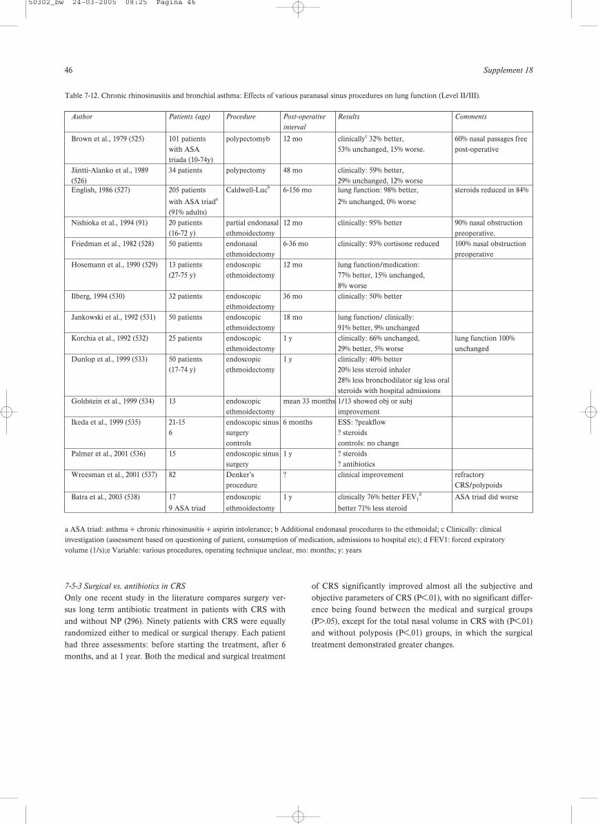
46 Supplement 18
7-5-3 Surgical vs. antibiotics in CRS
Only one recent study in the literature compares surgery ver-sus long term antibiotic treatment in patients with CRS withand without NP (296). Ninety patients with CRS were equallyrandomized either to medical or surgical therapy. Each patienthad three assessments: before starting the treatment, after 6months, and at 1 year. Both the medical and surgical treatment
of CRS significantly improved almost all the subjective andobjective parameters of CRS (P<.01), with no significant differ-ence being found between the medical and surgical groups(P>.05), except for the total nasal volume in CRS with (P<.01)and without polyposis (P<.01) groups, in which the surgicaltreatment demonstrated greater changes.
Table 7-12. Chronic rhinosinusitis and bronchial asthma: Effects of various paranasal sinus procedures on lung function (Level II/III).
Author Patients (age) Procedure Post-operative Results Comments
interval
Brown et al., 1979 (525) 101 patients polypectomyb 12 mo clinicallyc 32% better, 60% nasal passages free with ASA 53% unchanged, 15% worse. post-operativetriada (10-74y)
Jäntti-Alanko et al., 1989 34 patients polypectomy 48 mo clinically: 59% better, (526) 29% unchanged, 12% worseEnglish, 1986 (527) 205 patients Caldwell-Lucb 6-156 mo lung function: 98% better, steroids reduced in 84%
with ASA triada 2% unchanged, 0% worse(91% adults)
Nishioka et al., 1994 (91) 20 patients partial endonasal 12 mo clinically: 95% better 90% nasal obstruction (16-72 y) ethmoidectomy preoperative.
Friedman et al., 1982 (528) 50 patients endonasal 6-36 mo clinically: 93% cortisone reduced 100% nasal obstruction ethmoidectomy preoperative
Hosemann et al., 1990 (529) 13 patients endoscopic 12 mo lung function/medication: (27-75 y) ethmoidectomy 77% better, 15% unchanged,
8% worseIlberg, 1994 (530) 32 patients endoscopic 36 mo clinically: 50% better
ethmoidectomyJankowski et al., 1992 (531) 50 patients endoscopic 18 mo lung function/ clinically:
ethmoidectomy 91% better, 9% unchangedKorchia et al., 1992 (532) 25 patients endoscopic 1 y clinically: 66% unchanged, lung function 100%
ethmoidectomy 29% better, 5% worse unchangedDunlop et al., 1999 (533) 50 patients endoscopic 1 y clinically: 40% better
(17-74 y) ethmoidectomy 20% less steroid inhaler 28% less bronchodilator sig less oral steroids with hospital admissions
Goldstein et al., 1999 (534) 13 endoscopic mean 33 months 1/13 showed obj or subj ethmoidectomy improvement
Ikeda et al., 1999 (535) 21-15 endoscopic sinus 6 months ESS: ?peakflow6 surgery ? steroids
controls controls: no changePalmer et al., 2001 (536) 15 endoscopic sinus 1 y ? steroids
surgery ? antibioticsWreesman et al., 2001 (537) 82 Denker’s ? clinical improvement refractory
procedure CRS/polypoids
Batra et al., 2003 (538) 17 endoscopic 1 y clinically 76% better FEV1d ASA triad did worse
9 ASA triad ethmoidectomy better 71% less steroid
a ASA triad: asthma + chronic rhinosinusitis + aspirin intolerance; b Additional endonasal procedures to the ethmoidal; c Clinically: clinicalinvestigation (assessment based on questioning of patient, consumption of medication, admissions to hospital etc); d FEV1: forced expiratory volume (1/s);e Variable: various procedures, operating technique unclear, mo: months; y: years
50302_bw 24-03-2005 08:25 Pagina 46
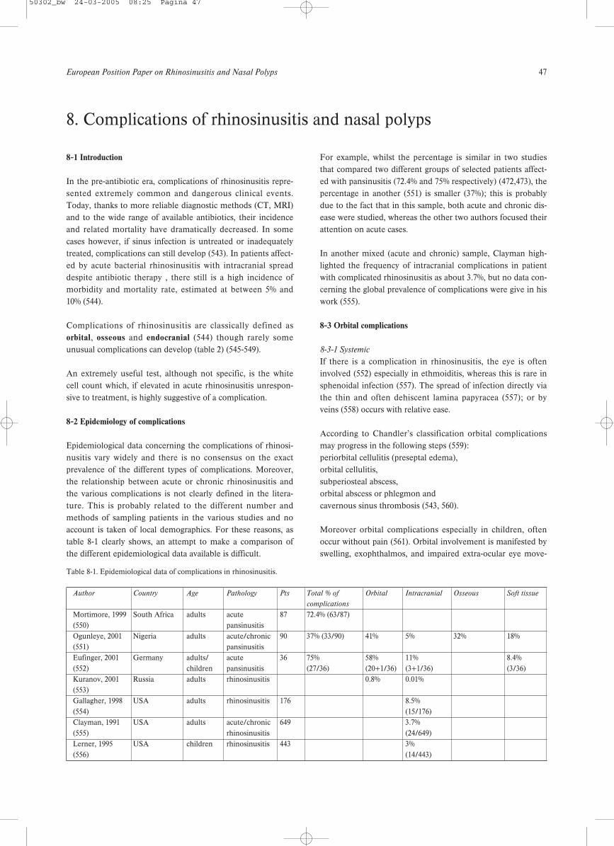
European Position Paper on Rhinosinusitis and Nasal Polyps 47
8-1 Introduction
In the pre-antibiotic era, complications of rhinosinusitis repre-sented extremely common and dangerous clinical events.Today, thanks to more reliable diagnostic methods (CT, MRI)and to the wide range of available antibiotics, their incidenceand related mortality have dramatically decreased. In somecases however, if sinus infection is untreated or inadequatelytreated, complications can still develop (543). In patients affect-ed by acute bacterial rhinosinusitis with intracranial spreaddespite antibiotic therapy , there still is a high incidence ofmorbidity and mortality rate, estimated at between 5% and10% (544).
Complications of rhinosinusitis are classically defined asorbital, osseous and endocranial (544) though rarely someunusual complications can develop (table 2) (545-549).
An extremely useful test, although not specific, is the whitecell count which, if elevated in acute rhinosinusitis unrespon-sive to treatment, is highly suggestive of a complication.
8-2 Epidemiology of complications
Epidemiological data concerning the complications of rhinosi-nusitis vary widely and there is no consensus on the exactprevalence of the different types of complications. Moreover,the relationship between acute or chronic rhinosinusitis andthe various complications is not clearly defined in the litera-ture. This is probably related to the different number andmethods of sampling patients in the various studies and noaccount is taken of local demographics. For these reasons, astable 8-1 clearly shows, an attempt to make a comparison ofthe different epidemiological data available is difficult.
For example, whilst the percentage is similar in two studiesthat compared two different groups of selected patients affect-ed with pansinusitis (72.4% and 75% respectively) (472,473), thepercentage in another (551) is smaller (37%); this is probablydue to the fact that in this sample, both acute and chronic dis-ease were studied, whereas the other two authors focused theirattention on acute cases.
In another mixed (acute and chronic) sample, Clayman high-lighted the frequency of intracranial complications in patientwith complicated rhinosinusitis as about 3.7%, but no data con-cerning the global prevalence of complications were give in hiswork (555).
8-3 Orbital complications
8-3-1 Systemic
If there is a complication in rhinosinusitis, the eye is ofteninvolved (552) especially in ethmoiditis, whereas this is rare insphenoidal infection (557). The spread of infection directly viathe thin and often dehiscent lamina papyracea (557); or byveins (558) occurs with relative ease.
According to Chandler’s classification orbital complicationsmay progress in the following steps (559): periorbital cellulitis (preseptal edema), orbital cellulitis, subperiosteal abscess, orbital abscess or phlegmon and cavernous sinus thrombosis (543, 560).
Moreover orbital complications especially in children, oftenoccur without pain (561). Orbital involvement is manifested byswelling, exophthalmos, and impaired extra-ocular eye move-
8. Complications of rhinosinusitis and nasal polyps
Table 8-1. Epidemiological data of complications in rhinosinusitis.
Author Country Age Pathology Pts Total % of Orbital Intracranial Osseous Soft tissue
complications
Mortimore, 1999 South Africa adults acute 87 72.4% (63/87)(550) pansinusitisOgunleye, 2001 Nigeria adults acute/chronic 90 37% (33/90) 41% 5% 32% 18%(551) pansinusitisEufinger, 2001 Germany adults/ acute 36 75% 58% 11% 8.4% (552) children pansinusitis (27/36) (20+1/36) (3+1/36) (3/36)Kuranov, 2001 Russia adults rhinosinusitis 0.8% 0.01%(553)Gallagher, 1998 USA adults rhinosinusitis 176 8.5% (554) (15/176)Clayman, 1991 USA adults acute/chronic 649 3.7% (555) rhinosinusitis (24/649)Lerner, 1995 USA children rhinosinusitis 443 3% (556) (14/443)
50302_bw 24-03-2005 08:25 Pagina 47
48 Supplement 18
ments (562). Periorbital or orbital cellulitis may result fromdirect or vascular spread of the sinus infection. As the spreadof sinus infection through the orbit follows a well-describedpattern, the initial manifestations are oedema and erythema ofthe medial aspects of the eyelid. Spread of infection from themaxillary or frontal sinus produces swelling of the lower orupper eyelid, respectively (560).
8-3-2 Periorbital cellulitis
Periorbital cellulitis (inflammation of the eyelid and conjuncti-va) (549) involves the tissue anterior to the orbital septum andis readily seen on CT scan as soft tissue swelling. It is the mostcommon complication of rhinosinusitis in children (563) and itmanifests itself as orbital pain, blepharal edema and high fever(564). Periorbital cellulitis usually responds to an oral antibioticappropriate to common sinus organisms but if not aggressivelytreated, may spread beyond the orbital septum (563).
8-3-3 Orbital cellulitis
As the inflammatory changes spread beyond the orbital sep-tum, proptosis develops together with some limitation of ocu-lar motion, indicating orbital cellulitis. Further signs are con-junctival oedema (chemosis), a protruding eyeball (proptosis),ocular pain and tenderness, and decreased movement of theextra ocular muscles (549, 565).
This complication requires aggressive treatment with intra-venous antibiotics.
Any children with rhinosinusitis and proptosis, ophthalmople-gia, or decreased visual acuity should have a CT scan of thesinuses with orbital detail to distinguish between an orbital andperiorbital (subperiosteal) abscess. Both conditions cause prop-tosis and limited ocular movement. Evidence of an abscess onthe CT scan or progressive orbital findings after initial i.v.antibiotic therapy are indications for orbital exploration anddrainage. Repeated ophthalmologic examination of visual acu-ity should take place and i.v. antibiotic therapy may be convert-ed into oral when the patient has been afebrile for 48 hours ifthe ophthalmological symptoms and signs are resolving (563).
8-3-4 Subperiosteal or orbital abscess
The clinical features of a subperiosteal abscess are oedema,erythema, chemosis and proptosis of the eyelid with limitationof ocular motility and as a consequence of extra-ocular muscleparalysis, the globe becomes fixed (ophthalmoplegia) and visu-al acuity diminishes.
An orbital abscess generally results from diagnostic delay or toimmunosuppression of the patient (564) with a frequency of9% and 8.3% (566, 567) in paediatric studies.A CT scan of the sinuses with orbital sequences to distinguishbetween orbital and periorbital (subperiosteal) abscess shouldbe performed. Evidence of an abscess on the CT scan or
absence of clinical improvement after 24-48 hours of i.v. antibi-otics are indications for orbital exploration and drainage. Anophthalmologist should check visual acuity from the earlystages of the illness and i.v. therapy should cover aerobic andanaerobic pathogens. It can be converted to an oral preparationwhen the patient has been afebrile for 48 hours (563).
Blindness may result from central retinal artery occlusion,optic neuritis, corneal ulceration, or pan-ophthalmitis. In sucha case the CT usually reveals oedema of the medial rectusmuscle, lateralization of the periorbita, and displacement ofthe globe downward and laterally. When the CT scan showsobliteration of the detail of the extraocular muscle and theoptic nerve by a confluent mass, the orbital cellulitis has pro-gressed to an abscess, in which there is sometimes air due toanaerobic bacteria. Sepsis not infrequently can spread intracra-nially as well as anteriorly into the orbit (568).
8-4 Endocranial complications
These include epidural or subdural abscesses, brain abscess,meningitis (most commonly), cerebritis, and cavernous sinusthrombosis (563, 569, 570).
The clinical presentation of all these complication is non-spe-cific, being characterized by high fever, frontal or retro-orbitalmigraine, generic signs of meningeal irritation and by variousdegrees of altered mental state (554) while intracranial abscess-es are often heralded by signs of increased intracranial pres-sure, meningeal irritation, and focal neurological deficits (562).Although an intracranial abscess is relatively asymptomatic,subtle affective and behavioural changes often occur showingaltered neurological function, altered consciousness, gait insta-bility, and severe, progressive headache.Endocranial complications are most often associated with eth-moidal or frontal rhinosinusitis. Infections can proceed fromthe paranasal cavities to the endocranial structures by two dif-ferent routes: pathogens, starting from the frontal most com-monly or ethmoid sinus, can pass through the diploic veins toreach the brain; alternatively, they can reach the intracranialstructures by eroding the sinus bones (554).
All endocranial complications start as cerebritis, but as necro-sis and liquefaction of brain tissue progresses, a capsule devel-ops resulting in brain abscess. Studies show a high incidenceof anaerobic organisms or mixed aerobic-anaerobic in patientswith CNS complications.
A CT scan is essential for diagnosis as it allows an extremelyaccurate definition of bone involvement, whereas MRI isessential when there are some degrees of soft tissues involve-ment such as in cavernous sinus thrombosis (554). Moreover,if meningitis is suspected, a lumbar puncture could be useful(554) once an abscess has been excluded.
50302_bw 24-03-2005 08:25 Pagina 48
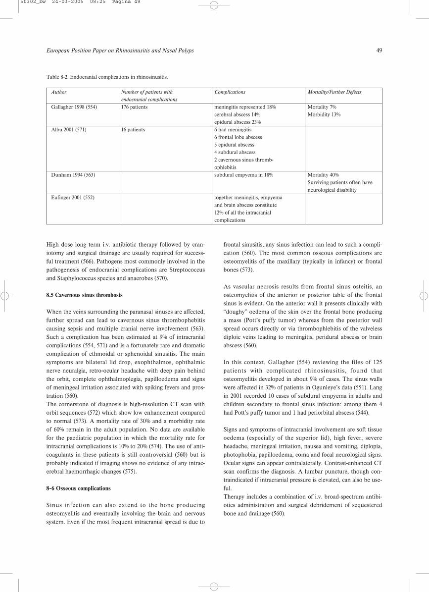
European Position Paper on Rhinosinusitis and Nasal Polyps 49
High dose long term i.v. antibiotic therapy followed by cran-iotomy and surgical drainage are usually required for success-ful treatment (566). Pathogens most commonly involved in thepathogenesis of endocranial complications are Streptococcusand Staphylococcus species and anaerobes (570).
8.5 Cavernous sinus thrombosis
When the veins surrounding the paranasal sinuses are affected,further spread can lead to cavernous sinus thrombophebitiscausing sepsis and multiple cranial nerve involvement (563).Such a complication has been estimated at 9% of intracranialcomplications (554, 571) and is a fortunately rare and dramaticcomplication of ethmoidal or sphenoidal sinusitis. The mainsymptoms are bilateral lid drop, exophthalmos, ophthalmicnerve neuralgia, retro-ocular headache with deep pain behindthe orbit, complete ophthalmoplegia, papilloedema and signsof meningeal irritation associated with spiking fevers and pros-tration (560). The cornerstone of diagnosis is high-resolution CT scan withorbit sequences (572) which show low enhancement comparedto normal (573). A mortality rate of 30% and a morbidity rateof 60% remain in the adult population. No data are availablefor the paediatric population in which the mortality rate forintracranial complications is 10% to 20% (574). The use of anti-coagulants in these patients is still controversial (560) but isprobably indicated if imaging shows no evidence of any intrac-erebral haemorrhagic changes (575).
8-6 Osseous complications
Sinus infection can also extend to the bone producingosteomyelitis and eventually involving the brain and nervoussystem. Even if the most frequent intracranial spread is due to
frontal sinusitis, any sinus infection can lead to such a compli-cation (560). The most common osseous complications areosteomyelitis of the maxillary (typically in infancy) or frontalbones (573).
As vascular necrosis results from frontal sinus osteitis, anosteomyelitis of the anterior or posterior table of the frontalsinus is evident. On the anterior wall it presents clinically with“doughy” oedema of the skin over the frontal bone producinga mass (Pott’s puffy tumor) whereas from the posterior wallspread occurs directly or via thrombophlebitis of the valvelessdiploic veins leading to meningitis, peridural abscess or brainabscess (560).
In this context, Gallagher (554) reviewing the files of 125patients with complicated rhinosinusitis, found thatosteomyelitis developed in about 9% of cases. The sinus wallswere affected in 32% of patients in Ogunleye’s data (551). Langin 2001 recorded 10 cases of subdural empyema in adults andchildren secondary to frontal sinus infection: among them 4had Pott’s puffy tumor and 1 had periorbital abscess (544).
Signs and symptoms of intracranial involvement are soft tissueoedema (especially of the superior lid), high fever, severeheadache, meningeal irritation, nausea and vomiting, diplopia,photophobia, papilloedema, coma and focal neurological signs.Ocular signs can appear contralaterally. Contrast-enhanced CTscan confirms the diagnosis. A lumbar puncture, though con-traindicated if intracranial pressure is elevated, can also be use-ful.Therapy includes a combination of i.v. broad-spectrum antibi-otics administration and surgical debridement of sequesteredbone and drainage (560).
Table 8-2. Endocranial complications in rhinosinusitis.
Author Number of patients with Complications Mortality/Further Defects
endocranial complications
Gallagher 1998 (554) 176 patients meningitis represented 18% Mortality 7%cerebral abscess 14% Morbidity 13%epidural abscess 23%
Albu 2001 (571) 16 patients 6 had meningitis6 frontal lobe abscess 5 epidural abscess4 subdural abscess2 cavernous sinus thromb-ophlebitis
Dunham 1994 (563) subdural empyema in 18% Mortality 40% Surviving patients often have neurological disability
Eufinger 2001 (552) together meningitis, empyema and brain abscess constitute 12% of all the intracranial complications
50302_bw 24-03-2005 08:25 Pagina 49
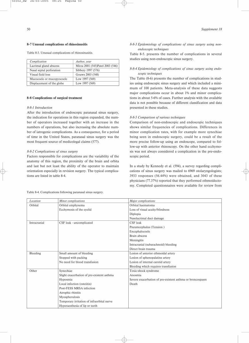
50 Supplement 18
8-7 Unusual complications of rhinosinusitis
8-8 Complications of surgical treatment
8-8-1 Introduction
After the introduction of endoscopic paranasal sinus surgery,the indication for operations in this region expanded, the num-ber of operators increased together with an increase in thenumbers of operations, but also increasing the absolute num-ber of iatrogenic complications. As a consequence, for a periodof time in the United States, paranasal sinus surgery was themost frequent source of medicolegal claims (577).
8-8-2 Complications of sinus surgery
Factors responsible for complications are the variability of theanatomy of this region, the proximity of the brain and orbitaand last but not least the ability of the operator to maintainorientation especially in revision surgery. The typical complica-tions are listed in table 8-4.
8-8-3 Epidemiology of complications of sinus surgery using non-
endoscopic techniques
Table 8-5. presents the number of complications in severalstudies using non-endoscopic sinus surgery.
8-8-4 Epidemiology of complications of sinus surgery using endo-
scopic techniques
The Table (8-6) presents the number of complications in stud-ies using endoscopic sinus surgery and which included a mini-mum of 100 patients. Meta-analysis of these data suggestsmajor complications occur in about 1% and minor complica-tions in about 5-6% of cases. Further analysis with the availabledata is not possible because of different classification and datapresented in these studies.
8-8-5 Comparison of various techniques
Comparison of non-endoscopic and endoscopic techniquesshows similar frequencies of complications. Differences inminor complication rates, with for example more synechiaebeing seen in endoscopic surgery, could be a result of themore precise follow-up using an endoscope, compared to fol-low-up with anterior rhinoscopy. On the other hand ecchymo-sis was not always considered a complication in the pre-endo-scopic period.
In a study by Kennedy et al. (594), a survey regarding compli-cations of sinus surgery was mailed to 6969 otolaryngologists;3933 responses (56.44%) were obtained, and 3043 of thesephysicians (77.37%) reported that they performed ethmoidecto-my. Completed questionnaires were available for review from
Table 8-3. Unusual complications of rhinosinusitis.
Complication Author, year
Lacrimal gland abscess Mirza 2001 (545)Patel 2003 (546)Nasal septal perforation Sibbery 1997 (576)Visual field loss Gouws 2003 (548)Mucocoele or mucopyocoele Low 1997 (569)Displacement of the globe Low 1997 (569)
Table 8-4. Complications following paranasal sinus surgery.
Location Minor complications Major complications
Orbital Orbital emphysema Orbital haematomaEcchymosis of the eyelid Loss of visual acuity/blindness
DiplopiaNasolacrimal duct damage
Intracranial CSF leak - uncomplicated CSF leakPneumcephalus (Tension )EncephalocoeleBrain abscessMeningitisIntracranial (subarachnoid) bleedingDirect brain trauma
Bleeding Small amount of bleeding Lesion of anterior ethmoidal arteryStopped with packing Lesion of sphenopalatine arteryNo need for blood transfusion Lesion of internal carotid artery
Bleeding which requires transfusionOther Synechiae Toxic-shock syndrome
Slight exacerbation of pre-existent asthma AnosmiaHyposmia Severe exacerbation of pre-existent asthma or broncospasmLocal infection (osteiitis) DeathPost-FESS MRSA infectionAtrophic rhinitisMyospherulosisTemporary irritation of infraorbital nerveHyperaesthesia of lip or teeth
50302_bw 24-03-2005 08:25 Pagina 50
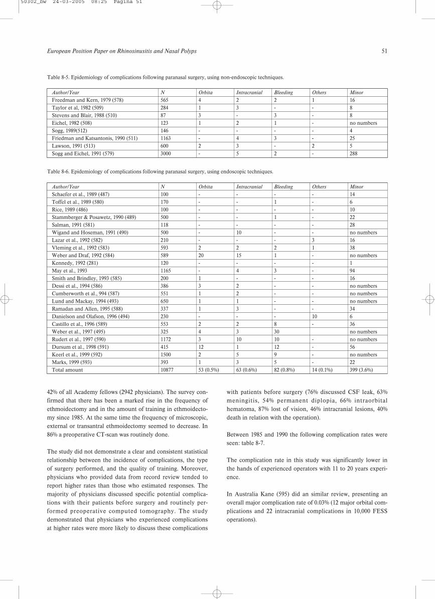
European Position Paper on Rhinosinusitis and Nasal Polyps 51
42% of all Academy fellows (2942 physicians). The survey con-firmed that there has been a marked rise in the frequency ofethmoidectomy and in the amount of training in ethmoidecto-my since 1985. At the same time the frequency of microscopic,external or transantral ethmoidectomy seemed to decrease. In86% a preoperative CT-scan was routinely done.
The study did not demonstrate a clear and consistent statisticalrelationship between the incidence of complications, the typeof surgery performed, and the quality of training. Moreover,physicians who provided data from record review tended toreport higher rates than those who estimated responses. Themajority of physicians discussed specific potential complica-tions with their patients before surgery and routinely per-formed preoperative computed tomography. The studydemonstrated that physicians who experienced complicationsat higher rates were more likely to discuss these complications
with patients before surgery (76% discussed CSF leak, 63%meningitis, 54% permanent diplopia, 66% intraorbitalhematoma, 87% lost of vision, 46% intracranial lesions, 40%death in relation with the operation).
Between 1985 and 1990 the following complication rates wereseen: table 8-7.
The complication rate in this study was significantly lower inthe hands of experienced operators with 11 to 20 years experi-ence.
In Australia Kane (595) did an similar review, presenting anoverall major complication rate of 0.03% (12 major orbital com-plications and 22 intracranial complications in 10,000 FESSoperations).
Table 8-5. Epidemiology of complications following paranasal surgery, using non-endoscopic techniques.
Author/Year N Orbita Intracranial Bleeding Others Minor
Freedman and Kern, 1979 (578) 565 4 2 2 1 16Taylor et al, 1982 (509) 284 1 3 - - 8Stevens and Blair, 1988 (510) 87 3 - 3 - 8Eichel, 1982 (508) 123 1 2 1 - no numbersSogg, 1989(512) 146 - - - - 4Friedman and Katsantonis, 1990 (511) 1163 - 4 3 - 25Lawson, 1991 (513) 600 2 3 - 2 5Sogg and Eichel, 1991 (579) 3000 - 5 2 - 288
Table 8-6. Epidemiology of complications following paranasal surgery, using endoscopic techniques.
Author/Year N Orbita Intracranial Bleeding Others Minor
Schaefer et al., 1989 (487) 100 - - - - 14Toffel et al., 1989 (580) 170 - - 1 - 6Rice, 1989 (486) 100 - - - - 10Stammberger & Posawetz, 1990 (489) 500 - - 1 - 22Salman, 1991 (581) 118 - - - - 28Wigand and Hoseman, 1991 (490) 500 - 10 - - no numbersLazar et al., 1992 (582) 210 - - - 3 16Vleming et al., 1992 (583) 593 2 2 2 1 38Weber and Draf, 1992 (584) 589 20 15 1 - no numbersKennedy, 1992 (281) 120 - - - - 1May et al., 1993 1165 - 4 3 - 94Smith and Brindley, 1993 (585) 200 1 - - - 16Dessi et al., 1994 (586) 386 3 2 - - no numbersCumberworth et al., 994 (587) 551 1 2 - - no numbersLund and Mackay, 1994 (493) 650 1 1 - - no numbersRamadan and Allen, 1995 (588) 337 1 3 - - 34Danielson and Olafson, 1996 (494) 230 - - - 10 6Castillo et al., 1996 (589) 553 2 2 8 - 36Weber et al., 1997 (495) 325 4 3 30 no numbersRudert et al., 1997 (590) 1172 3 10 10 - no numbersDursum et al., 1998 (591) 415 12 1 12 - 56Keerl et al., 1999 (592) 1500 2 5 9 - no numbersMarks, 1999 (593) 393 1 3 5 - 22Total amount 10877 53 (0.5%) 63 (0.6%) 82 (0.8%) 14 (0.1%) 399 (3.6%)
50302_bw 24-03-2005 08:25 Pagina 51
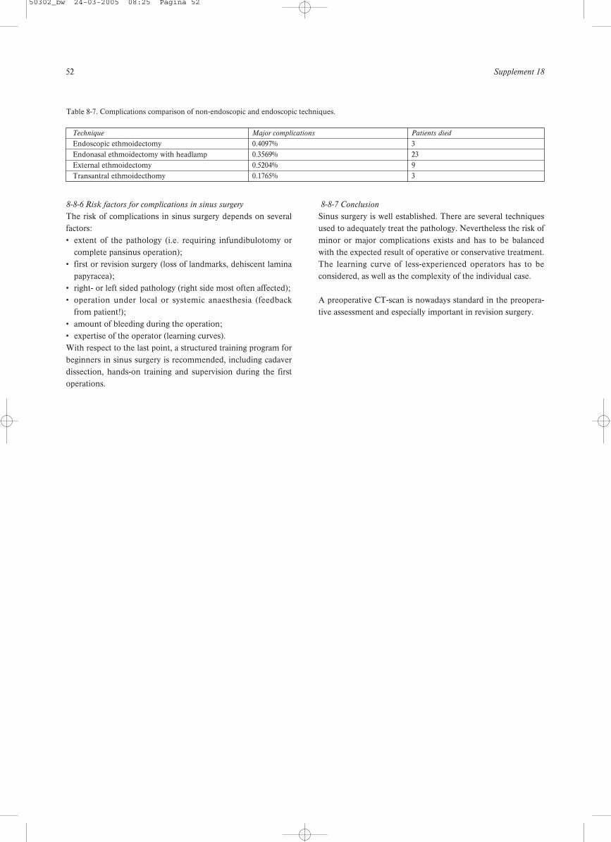
52 Supplement 18
8-8-6 Risk factors for complications in sinus surgery
The risk of complications in sinus surgery depends on severalfactors:• extent of the pathology (i.e. requiring infundibulotomy or
complete pansinus operation);• first or revision surgery (loss of landmarks, dehiscent lamina
papyracea);• right- or left sided pathology (right side most often affected);• operation under local or systemic anaesthesia (feedback
from patient!);• amount of bleeding during the operation;• expertise of the operator (learning curves).With respect to the last point, a structured training program forbeginners in sinus surgery is recommended, including cadaverdissection, hands-on training and supervision during the firstoperations.
8-8-7 Conclusion
Sinus surgery is well established. There are several techniquesused to adequately treat the pathology. Nevertheless the risk ofminor or major complications exists and has to be balancedwith the expected result of operative or conservative treatment.The learning curve of less-experienced operators has to beconsidered, as well as the complexity of the individual case.
A preoperative CT-scan is nowadays standard in the preopera-tive assessment and especially important in revision surgery.
Table 8-7. Complications comparison of non-endoscopic and endoscopic techniques.
Technique Major complications Patients died
Endoscopic ethmoidectomy 0.4097% 3Endonasal ethmoidectomy with headlamp 0.3569% 23External ethmoidectomy 0.5204% 9Transantral ethmoidecthomy 0.1765% 3
50302_bw 24-03-2005 08:25 Pagina 52
European Position Paper on Rhinosinusitis and Nasal Polyps 53
9-1 Introduction
Rhinosinusitis is a common problem in children that is oftenoverlooked. It is a multifactorial disease in which the impor-tance of several predisposing factors change with age. Themanagement of rhinosinusitis in children is a controversial anda rapidly evolving issue.
9-2 Anatomy
In the newborn, the maxillary sinus extends to a depth ofabout 7 mm, is 3 mm wide and 7 mm high (596). When a childreaches the age of 7-8 years the floor of the maxillary sinusalready occupies the same level as the nasal floor. In the new-born, two to three ethmoid cells are found bilaterally, and bythe age of four the ethmoid labyrinth has been formed. Thesphenoid sinuses are also present in the neonate. Each sphe-noid sinus is 4 mm wide and 2 mm high. At birth the frontalsinuses are not present, but they gradually develop from theanterior ethmoid cells into the cranium. When the upper edgeof the air cell (cupola) reaches the same level as the roof of theorbit, it can be termed a frontal sinus, a situation that appearsaround the age of five.
9-3 Epidemiology and pathophysiology
Since the introduction of CT-scanning, it has become clearthat a runny nose in a child is not only due to limited rhinitisor adenoid hypertrophy, but that in the majority of the casesthe sinuses are involved as well. Van der Veken (173) in a CTscan study showed that in children with a history of chronicpurulent rhino rhea and nasal obstruction 64% showedinvolvement of the sinuses. In a MRI study of a non-ENT pae-diatric population (597) it was shown that the overall preva-lence of sinusitis signs in children is 45%. This prevalenceincreases in the presence of a history of nasal obstruction to50%, to 80% when bilateral mucosal swelling is present onrhinoscopy, to 81% after a recent upper respiratory tract infec-tion (URI), and to 100% in the presence of purulent secretions.Also Kristo et al. (598) found a similar overall percentage(50%) of abnormalities on MRI in 24 school children. Theyincluded, however, a follow-up after 6 to 7 months, and foundthat about half of the abnormal sinuses on MRI findings hadresolved or improved without any intervention. Epidemiologic studies on rhinosinusitis in children are limitedbut reveal the following information on the pathophysiologyand clinically relevant factors influencing the prevalence of rhi-nosinusitis in children. There is a clear-cut decrease in the prevalence of rhinosinusitisafter 6 to 8 years of age. This is the natural history of the dis-
ease in children and is probably related to an immatureimmune system in the younger child (176, 177)In temperate climates there is a definite increase in the occur-rence of chronic rhinosinusitis in children during the fall andin the wintertime, so that the season seems to be anotherimportant factor (176). Younger children staying in day care centres show a dramaticincrease in the prevalence of chronic or recurrent rhinosinusi-tis compared to children staying at home.
Although viruses are uncommonly recovered from sinus aspi-rates (562, 599), most authors agree (600, 601) that viral infec-tions are the trigger to rhinosinusitis. Although CT scan abnor-mality can be seen up to several weeks after the onset of aURI, one can assume that only 5 to 10% of the URI in earlychildhood are complicated by acute rhinosinusitis (602). Thetime course (i.e. clinical symptoms) of viral to bacterial rhinosi-nusitis is the same as in adults.
The most common bacterial species isolated from the maxil-lary sinuses of patients with acute rhinosinusitis areStreptococcus pneumoniae, Haemophilus influenzae, andMoraxella catarrhalis, the latter being more common in chil-dren (41, 42).
Since antral punctures are not frequently performed in chil-dren anymore, it is interesting to know from the studies that inchildren there is a good correlation of bacteriology betweenthe maxillary sinus and the middle meatal specimen (83%),and a poor correlation between those of the nasopharynx andthe maxillary sinus (45%) (603).
9-4 Symptoms and signs
Several authors studied the presenting symptoms of rhinosi-nusitis in children (177, 604, 605). Children with acute rhinosi-nusitis frequently have less specific complaints than adults.Rhino rhea is the most frequent presenting symptom in allforms of rhinosinusitis (71% to 80%). Cough seems to be a fre-quent symptom (50% to 83%). In acute rhinosinusitis, however,nasal obstruction does not appear to be the most prominentsymptom, probably because it is masked by more severe symp-toms such as fever (50 to 63%) and pain (29% to 33%). On theother hand, in chronic sinusitis, nasal obstruction and mouthbreathing are very frequent (70% to 100%) and is often accom-panied by ear complaints (recurrent, purulent otitis media orchronic otitis media with effusion: 40% to 68%). This relation-ship between rhinosinusitis and otitis is not unexpected, as themiddle ear can be considered a kind of specialized paranasalspace.
9. Special considerations: Rhinosinusitis in children
50302_bw 24-03-2005 08:25 Pagina 53
54 Supplement 18
9-5 Examination
Physical examination of a child’s nose is often difficult, andonly limited anterior rhinoscopy is tolerated by these youngpatients (606). This examination may be accomplished in asimple way by tilting the tip of the nose upward (young chil-dren have wide noses with round nostrils and no vibrissae,allowing easy examination of the condition of the head of theinferior turbinate). Another convenient method is the use ofan otoscope (607, 608). Because of the difficulties of perform-ing anterior rhinoscopy in a young child, most studies providelimited information about the condition of the nasal cavity inrhinosinusitis in children. Mostly the nasal (boggy turbinate)and pharyngeal mucosa appears erythematous. Yellow togreenish purulent rhino rhea of varying viscosity can be seen.Lymphoid hyperplasia may be seen in the oropharynx. Theremay be adenoid and/or tonsillar hypertrophy, cervical lymphnodes may be moderately enlarged and slightly tender (606,607). The only quantitative data on rhinoscopy in young chil-dren is given by Clement et al. (605) showing a postnasal dripin 60% and presence of pus in the middle meatus in 50% andby Riding et al. showing turbinate mucosal swelling in 29%(604).
The value of transillumination and ultrasonography is certainlylimited, and these procedures are not recommended for diag-noses of adult rhinosinusitis. The increased thickness of boththe soft tissue and bony vault of the palate in children under10 years of age limits the clinical usefulness of transillumina-tion in the younger age group even more (279).
The value of plain sinus films in assessing the extent of thedisease, especially in young children, is questionable. Lusk etal. (607) studied 70 children who had symptoms compatiblewith chronic rhinosinusitis, and compared plain films withcoronal CT scans within a few hours of one another. When allsinuses were evaluated or considered, they found a lack of cor-relation between both methods in 74% of the patients. Fortyfive percent of the normal plain radiographs showed abnormal-ities on CT, and 34% of the abnormal plain radiographs wereactually normal on CT. These authors found that plain filmsboth over- and underestimated the amount of sinus disease.Thus, for evaluation of paediatric rhinosinusitis, CT remainsthe imaging modality of choice, because of its ability to resolveboth bone and soft tissue (608). Before defining which CT scanfindings are considered abnormal in children, it is interestingto discuss the incidental paranasal sinus abnormalities on CTscans of children and their clinical correlation. A number ofauthors have shown that radiographic opacification on the CTscan are found in considerable number of asymptomatic chil-dren (609).
Looking at the percentage of CT signs of sinus disease in asymptomatic paediatric population, the prevalence of CT
abnormalities increases from 64% to 81% (173, 610). Thus mostauthors are in agreement that an asymptomatic child with anincidental paranasal sinus finding on CT needs no furtherwork-up unless clinical symptoms and signs are elicited (611).A number of studies suggest that the growth of the maxillarysinus is not impaired by extensive or chronic disease, unlikethe temporal bone and it seems that the presence of ahypoplastic maxillary sinus per se is not an indication forsurgery (612).
9-6 Systemic disease and chronic rhinosinusitis
The role of atopy in chronic rhinosinusitis is unclear. Manyauthors attribute a great deal of importance to allergy (73, 604,608) although others (81, 173, 613) did not find an increasedprevalence of rhinosinusitis in allergic children.
All young children have a physiologic primary immune defi-ciency (608, 614). Defence against polysaccharides encapsulat-ed bacteria via immunoglobulin G subclasses 2 and 4 may notreach adult levels until the age of 10 years (95). IgG subclassdeficiency can lead to protracted or chronic rhinosinusitis(74,79,85,94,96). According to Polmar (615) recurrent andchronic rhinosinusitis is the most common clinical presenta-tion of common variable immunodeficiencies. Although notall patients who lack secretory IgA antibodies have anincreased number of more severe respiratory infections, thesubject who has IgA deficiency and chronic rhinosinusitis is adifficult management problem and replacement therapy can-not be provided (616). Patients with primary or acquiredimmune deficiencies (e.g. treatment for malignancies, organtransplants, maternally transmitted AIDS or blood-transmittedAIDS in hemophilics, drug induced conditions) are at risk fordeveloping a difficult-to-treat rhinosinusitis with resistant oruncommon micro-organisms and fungi. Also the initial signsand symptoms may be non-specific, such as thin rhino rhea,mild congestion, and chronic cough (608).
Cystic fibrosis is caused by a mutation of the gene FES1encoding the cystic fibrosis transmembrane conductance regu-lator (CFTR). This gene contains 27 exans encompassingapproximately 252 kb of DNA on chromosone 7q 31.2. Themost common mutation, deletion of phenylalanine at position508 (D F508) accounts for nearly 70% of mutuations inEuropean-derived Caucasian population (617).
In children with cystic fibrosis, sinusitis seems to be a com-mon problem. Although the prevalence of nasal polyposis inchildren with cystic fibrosis was previously estimated to bebetween 6 and 20% (618), Yung et al. (619) found it to be over50% and Brihaye et al. (620) reported that performing rigidendoscopy in 84 patients with cystic fibrosis, revealed inflam-matory polyps in 45% (mean age 15 years) and medial bulgingof the lateral nasal wall in 12% (mean age 5 years). In patients
50302_bw 24-03-2005 08:25 Pagina 54
European Position Paper on Rhinosinusitis and Nasal Polyps 55
with cystic fibrosis and chronic rhinosinusitis, CT showed in100% (620) opacification of the anterior complex (anterior eth-moid, maxillary and -if developed- frontal sinus) and 57%showed clouding of the posterior complex (posterior ethmoidand sphenoid). In all children with a medial displacement ofthe lateral nasal wall, there was a soft tissue mass in the maxil-lary antrum (large quantity of secretions surrounded by poly-poidal mucosa, representing a mucopurulent rhinosinusitus).In 80% of these children the displacement was so extreme thatthe lateral nasal wall touched the septum, resulting in totalnasal blockage. In the study by Brihaye et al. (620) massivepolyposis was never found before the age of 5 years.Mucopyosinusitis of the maxillary sinus occurs at a youngerage (3 months to 8 years) and the maxillary sinus seems to bethe first sinus affected by the disease. The youngest childreported with the disease was a 3 months old infant presentingsymptoms similar to those of children with bilateral choanalatresia (nasal blockade, stridor, and feeding problems), exceptthat the symptoms occurred only gradually following a symp-tom-free period after birth.
By definition, patients with Kartagener’s syndrome [a heredi-tary disease involving the classic triad of rhinosinusitis,bronchiectesis, and situs inversus] have chronic rhinosinusitis,but which develops later in life. The neonatal group withKartagener’s syndrome do not have rhinosinusitis andbronchiectesis, and therefore the term “immotile cilia syn-drome” was introduced in the seventies. As ultrastructuralchanges of cilia were also found after chronic infections orpolyposis, and situs inversus did not seem to be essential, thename of this autosomal recessive disease was changed to pri-mary ciliary dyskinesia (PCD) (621). One should always con-sider the diagnosis in any neonate with respiratory or ENTproblems of unknown origin. At least half of the PCD patientshave symptoms when first born and especially in a term babywith no risk factor for congenital infection showing signs ofrhinitis at birth, PCD should be excluded. The same goes foran infant or older child with atypical asthma, unresponsive totreatment, chronic wet cough, and sputum production in theolder child who is able to expectorate, very severe gastro-oesophagal reflux, bronchiectasis, rhinosinusitis (rarely withpolyposis), chronic and severe secretory otitis media, particu-larly with continuous, long lasting and diffuse discharge fromthe ears after grommet insertion. There are a number of ways to diagnose PCD. Clinically themost useful is the saccharine test, which is a cheap and easyprocedure to screen older children and adults. If the child istoo young for the test or the results are positive (transport timelonger than 60 minutes) or there exists a strong clinical suspi-cion, the ciliary beat frequency can be tested from a nasalepithelial biopsy.
If the direct inspection of the ciliary beat frequency is abnor-mal (less than 11-16 Hz) an ultrastructional study of cilia is
needed. The most common ciliary abnormalities in PCD are:dynein arm defects (absence or reduced number of inner,outer or both dynein arms), tubular defects (transposition andextra microtubules), radial spokes defects or absence, ciliarydysorientation (suspected if mean standard deviation of angleis larger than 20°), abnormal basal apparatus, ciliary aplasia,abnormally long cilia (622). Many of these abnormalities onTEM (transmission electron microscopy), however, can betransient or occur secondarily after infection. In cilia withpatients with PCD specific ultrastructural abnormalities arepresent, such as dynein and/or spoke deficiency, and absenceof the central pair of microtubules. Secondary ciliary dyskine-sia, the acquired form (infections, inflammatory or toxic) ismostly correlated with other anomalies, such as microtubularabnormalities and composed cilia. However, there exists agreat overlap of ultrastructural abnormalities between both(294). Therefore the study of cilia after sequential monolayer-suspension culture technique avoids the acquired form (623).
The parallel existence of upper airway inflammation withensuing problems of intractable rhinosinusitis, otitis, and gas-tro-oesophageal reflux (GER) has been observed and suggestsa causal relationship. Barbero found in a group of patients withupper airway disease and GER, that anti-reflux measures maypermit a greater well-being and that GER maybe among thevariables leading to refractory chronic upper airway disease(624). The otolaryngologist should be suspicious of GER inchildren complaining of chronic nasal discharge and obstruc-tion combined with chronic cough, hoarseness and stridulousrespiration. The endoscopic appearance of the laryngeal andtracheal areas are of considerable importance in conjunctionwith oesophageal examination, in determining potential rela-tionship between GER and otolaryngologic abnormalities. Theauthor found in 17 patients: 1 or more of these endoscopicsigns: cobblestoning of the mucosa of the laryngopharynx,inflammation of the upper airway, sinus involvement, rhinorhea, subglottic stenosis, velopharyngeal insufficiency, pharyn-geal tracheitis and tracheomalacia. In most of the patients theoesophagus on examination was erythematous. The diagnosisneeds to be confirmed by oesophageal 24 hours pH monitor-ing. In 30 children with chronic sinus disease found after 24hour pH monitoring in 63% oesophageal reflux and 32% hadnasopharyngeal reflux (466).
9-7 Management
9-7-1 Introduction
In 1994 Poole stated that chronic rhinosinusitis in the youngchild does not necessary have to be treated, as spontaneousresolution is the norm (625). With regard to the natural historyof the disease and the growing resistance and b-lactamase pro-duction of many microorganisms, one should refrain fromovertreating a runny nose in a young child. One should nottreat every common cold with antibiotics or smash any minor
50302_bw 24-03-2005 08:25 Pagina 55
56 Supplement 18
self-limiting infection of a common cold with the sledge ham-mer of broad spectrum antibiotics. Some physicians prescribeantibiotics for minor respiratory infections in the hope of pre-venting serious complications and/or avoid medical/legal liti-gations. Van Buchem et al. followed 169 children with a runnynose for 6 months, treating them only with decongestants orsaline nose drops. They did not find a single child who devel-oped a clinically serious disease with general symptoms suchas marked pain, pressure on sinuses, local swelling, or empye-ma, which proved that complications of rhinosinusitis in achild are uncommon (177).
9-7-2 Treatment of rhinosinusitis
9-7-2-1 Medical treatment of rhinosinusitis The data on specific treatment of children are very limited. In a short-term follow-up study Furukawa (626) showed asuperior result from erythromycin-sulfisoxazole plus topicaldecongestants compared with placebo + topical decongestants,and Rachelefsky et al. (85) studying 84 children, demonstratedon the basis of radiographs and clinical response a better resultin the group treated with amoxicillin, although trimethoprim-sulfamethoxazole was an adequate alternative, while ery-thromycin was not any better than an antihistamine-deconges-tant combination. The only long-term follow-up in the treat-ment of children with chronic maxillary sinusitis (n=141) com-paring oral amoxicillin combined with decongestant nosedrops, drainage of the maxillary sinus (antral lavage), a combi-nation of the two previous regimen, and placebo was per-formed by Otten et al. (627) showing that the therapeuticeffects of these four forms of treatment did not differ signifi-cantly or have a significant curative effect The usual durationof antimicrobial therapy is 10 to 14 days. This recommendationis based on experience in adults.
One study suggest that topical corticosteroids may be a usefulancillary treatment to antibiotics in childhood rhinosinusitis,effective in reducing the cough and nasal discharge earlier inthe course of acute sinusitis (353). There are a large number ofstudies showing that local corticosteroids are effective and safein children with rhinitis (628-632).
Additional therapy consists of topical or oral decongestants.Most authors prefer topical a2 agonists (xylo- and oxymetazo-line) in appropriate concentrations. Careful dosage is impor-tant when treating infants and young children, to prevent toxicmanifestations.
Saline nose drops or nasal douches are popular with paediatri-cians (606, 608, 616). As long as the saline is isotonic and atbody temperature, it can help in eliminating nasal secretionsand it can decrease nasal oedema.
In children with chronic rhinosinusitis and proven gastro-oesophageal reflux (GER) after 24 hours of pH monitoring
Phipps et al. (466) showed that most children showed improve-ment of sinus disease after GER treatment and Bothwell et al.(633) suggests that in 89% of the children (25 out of 28) surgerycould be avoided. These studies indicate that GER could beevaluated and treated in children with chronic sinus diseasebefore sinus surgical intervention.
9-7-2-2 Surgical treatment of rhinosinusitis The effectiveness of adenoidectomy in the management ofpaediatric rhinosinusitis is still a controversial issue. It is diffi-cult to differentiate between the symptoms typical for chronicrhinosinusitis and those of adenoid hypertrophy. Hibert (634)showed that nasal obstruction, snoring and speech defectsoccur more frequently in children with adenoid hypertrophywhile symptoms of rhino rhea, cough, headache, signs ofmouth breathing, and abnormalities on anterior rhinoscopyoccur as frequently in children with chronic rhinosinusitis as inchildren with adenoid hypertrophy. Wang et al. e.g. didn’t find any significant correlation betweenthe size of the adenoid and the presence of purulent secretionsin the middle meatus on fiberoptic examination in 420 chil-dren between the age of 1 and 7 years, while there was a verysignificant correlation between the size of the adenoid and thecomplaints of mouth breathing (p<0.001) and snoring(p<0.001) (635). The size of the adenoid and associated dis-eases seem to be factors for consideration. Adenoidectomywas included in the stepwise protocol for the treatment of pae-diatric rhinosinusitis proposed by Don et al. (636). RecentlyUngkanont et al. proved adenoidectomy to be effective in themanagement of paediatric rhinosinusitis. They suggest per-forming an adenoidectomy as a surgical option before endo-scopic sinus surgery (ESS), especially in younger children withobstructive symptoms (637).
Antral lavage: with the introduction of antroscopy at the endof the 1970s antral lavage in children became popular. As a tro-car of the endoscope has a 4 mm diameter, it was easy to leavea ventilation tube in position, making frequent irrigations ofthe maxillary sinus possible in children, without any need forrepetitive anaesthesia. It was shown, however, that in childrenwith chronic rhinosinusitis, irrigation of the maxillary sinusdoes not lead to a better cure after 3 weeks, compared with acontrol group (638) or is not statistically significantly more suc-cessful (607). Inferior antrostomy: as it had been demonstrated that theresults of antral lavage in children were not long lasting; thelogical consequence in children who required continuousantral lavage was to resort to a permanent antrostomy or nasalantral window in the inferior meatus. Lund (639), however,demonstrated that -especially in children under the age of 16years there is a higher rate of closure of these antral windows.She concluded that the inferior meatus in children is smallerthan in adults, making it impossible to create an adequatelysized antrostomy. As a consequence Lusk (607) was able to
50302_bw 24-03-2005 08:25 Pagina 56
European Position Paper on Rhinosinusitis and Nasal Polyps 57
show that in a six-month follow-up the success rate of thenasal antral window procedure dropped to 27%. All patientsremained symptomatic, and 28% needed further functionalendoscopic sinus surgery. So the only current indication for anaso-antral window in the inferior meatus is therefore mainlylimited to PCD where one hopes to achieve a kind of gravita-tional drainage.
Sinus surgery: the Caldwell-Luc operation is contra-indicatedin children as it can cause damage to the unerrupted teeth(608, 616). Most of the controversies seem to centre on theindications for functional endoscopic sinus surgery in children.(FESS or paediatric FESS=PESS). The “functional” in FESSstands for the restoration of the function of the ostiomeatalcomplex i.e. ventilation and drainage. In 1998 an internationalconsensus was reached concerning the indications of FESS inchildren (11):
a. absolute indications: 1. complete nasal obstruction in cystic fibrosis due to
massive polyposis or by medialization of the lateralnasal wall
2. orbital abscess 3. intracranial complications 4. antrochoanal polyp5. mucocoeles or mucopyocoeles 6. fungal rhinosinusitis
b. possible indications: in chronic rhinosinusitis with frequent exacerbations thatpersist despite optimal medical management and afterexclusion of any systemic disease, endoscopic sinussurgery is a reasonable alternative to continuous medicaltreatment. Optimal management includes a 2-6 weeks ofadequate antibiotics (IV or oral) with treatment of con-comitant disease.
Surgery for chronic rhinosinusitis with frequent exacerbationsthat persist despite optimal medical management is mostlylimited to a partial ethmoidectomy: removal of the uncinateprocess, with or without a maxillary antrostomy in the middlemeatus, and opening of the bulla is often sufficient. In othercases such as in cystic fibrosis with massive polyposis, exten-sive sphenoidectomy may be necessary.
Most results are judged on symptomatic relief and not includeendoscopic examination or CT scan. Lusk et al. (640) found asuccess rate of 88% in 24 children who had only one proce-dure. They saw a 24% improvement of the purulent rhino rhea(from 100% tot 64%), 33% improvement of fever, usually lowgrade (from 55% to 22%), and a 13% improvement of cough(from 48% to 35%).
A meta-analysis performed by Hebert et al. (641) focusing onthe number of patients per study, length of follow-up, prospec-tive versus retrospective, a separation or exclusion of patients
with significant underlying systemic disease, showed in 8 pub-lished articles (832 patients) positive outcome rates going from88 to 92%. The average combined follow-up was 3.7 years.Thus they concluded that FESS is a safe and effective treat-ment for chronic rhinosinusitis that is refractory to medicaltreatment.
Similar results were published in a more recent study by Jianget al. (642) and Fakhri et al. (643) showing a postoperativeimprovement in 84% of the FESS (n=121). For this indicationBothwell et al. (644) found no statistical significant differencein the outcome of facial growth between a retrospective age-matched cohort outcome study between 46 children whounderwent FESS surgery and 21 children who didn’t, usingqualitative antropomorphic analysis of 12 standard facial mea-surements after a 13.2 years follow-up.
As already mentioned before in a cystic fibrosis population of48 children, 12% showed on endosocpy a medialisation of thelateral nasal wall and 45% nasal polyps coming out of the mid-dle meatus (620). Initially polyps were removed as theyappeared. When nasal obstruction occurred polypectomy wasthe rule. Regrowth, however, was sometimes observed within3 weeks and many patients had multiple polypectomies rang-ing from 1 to 12 procedures per child. Crockett et al. (645) wasthe first to stress the importance of a long-term follow-up(average 5 years) and showed that when intranasal ethmoidec-tomy and Caldwell-Luc procedures were combined withpolypectomy, fewer recurrences and longer symptom-freeintervals resulted. Unfortunately Caldwell-Luc procedures canonly be performed in older children and adults. With the intro-duction of FESS, however, a new approach was possible toachieve radical surgery. Duplechain (646) reported for the firsttime the results of this kind of surgery in cystic fibrosis chil-dren which was soon followed by many more (522, 619, 647,648).
50302_bw 24-03-2005 08:25 Pagina 57
58 Supplement 18
10-1 Direct Costs
Chronic rhinosinusitis, which can be debilitating for patientsand imposes a major economic cost on society in terms ofboth direct costs as well as decreased productivity. To betterevaluate the socioeconomic impact of chronic rhinosinusitis,the current English literature has been reviewed. Data fromoutside the USA are very limited. In a 1999 publication, Ray etal. (3) estimated the total direct (medical and surgical) costs ofsinusitis to be a staggering $5.78 billion in the US. This figurewas extrapolated from governmental surveys such as thenational health care survey and medical expenditure data. Thecost of physician visits resulting in a primary diagnosis ofsinusitis was $3.39 billion, which does not reflect the completecost of radiographic studies, medication, or productivity losses.
Acknowledging that other airway disorders are closely tied torhinosinusitis, Ray et al. (3) used the Delphi method to quanti-fy how often rhinosinusitis is a secondary diagnosis contribut-ing to the primary diagnosis assigned by physicians. An expertpanel examined the co-incidence of rhinosinusitis in diseasessuch as asthma, otitis media, and allergic rhinitis, and deter-mined that 10-15% of the cost of these other diseases wasattributable to rhinosinusitis, increasing the economic burdenof rhinosinusitis to the often quoted $5.78 billion sum. Ray’spaper relied on data collected by the National Centre forHealth Statistics and did not attempt to distinguish acute rhi-nosinusitis from the chronic form of this disease.
In 2002, Murphy et al. (649) examined a single health mainte-nance organization to evaluate the cost of chronic rhinosinusi-tis. The authors compared the costs of healthcare for memberswith a diagnosis of CRS to the cost of those without the diag-nosis during 1994 and were able to determine the direct med-ical costs of the disease based on reimbursements paid ratherthan charges submitted. According to Murphy’s study, patientswith a diagnosis of CRS made 43% more outpatient and 25%more urgent care visits than the general population (p=0.001).CRS patients filled 43% more prescriptions, yet had fewer hos-pital stays than the general HMO adult population. In total, thecost of treating patients with CRS was $2,609 per year, 6%more than the average adult in the HMO. Because patientsreceived all healthcare services in one integrated system, thisfigure includes the costs of radiography, hospitalization, andmedication. Chronic rhinosinusitis care specifically cost $206per patient per year, thus contributing to a calculated nation-wide direct cost of $4.3 billion annually based on the 1994 sta-tistic of 20.9 million individuals seeking care for CRS. Usingthe more recent value of 32 million affected (56) the overallcost would increase to $6.39 billion annually.
Addressing the cost of pharmacologic management of chronicrhinosinusitis, Gliklich and Metson’s (650)1998 study reportedan annual expenditure of $1220. This figure is the sum of OTCmedications ($198), nasal sprays ($250), and antibiotics ($772).
Only one study in Europe has been found which considers thecosts of CRS. This study was done in patients with severechronic rhinosinusitis visiting a university hospital in theNetherlands (651). The direct cost of the CRS of these severepatients was €1,861,- per year. No data are available distinguishing costs of nasal polyps fromCRS.
In conclusion we can deduce from these limited data that theaverage direct costs of CRS per patient per year is between€200,- and €2,000,- depending on the severity of the disease.
10-2 Indirect Costs
The studies of direct medical costs demonstrate the social eco-nomic burden of the disorder. However, the total costs of CRSare greater. With 85% of patients with CRS of working age(between 18-65 years old) indirect costs such as missed work-days and decreased productivity at work significantly add tothe economic burden of disease(56).
Goetzel et al. (2) attempted to quantify the indirect costs ofrhinosinusitis. Their 2003 study resulted in rhinosinusitis beingnamed one of the top ten most costly health conditions to USemployers. A large multi-employer database was used to trackinsurance claims through employee health insurance, absenteedays, and short-term disability claims. Episodes of illness werelinked to missed workdays and disability claims, accurately cor-relating absenteeism to a given disease.
In a large sample size (375,000), total healthcare payments peremployee per year for rhinosinusitis (acute and chronic) werefound to be $60.17, 46% of which came from the cost of absen-teeism and disability. These figures approximate the cost toemployers, disregarding the cost incurred by other parties, andtherefore tremendously underestimate the entire economicburden of the disease. In his 2003 study, Bhattacharyya (427) used patient-completedsurveys to determine the direct and indirect costs of chronicrhinosinusitis. Patients completed a survey assessing symp-toms of disease, detailing medication use, and quantifyingmissed worked days attributable to CRS. According toBhattacharyya, the cost of treating CRS per patient totaled$1,539 per year. Forty percent of these costs were due to theindirect costs of missed work; the mean number of missed
10. Socio-economic cost of chronic rhinosinusitis and nasal polyps
50302_bw 24-03-2005 08:25 Pagina 58
European Position Paper on Rhinosinusitis and Nasal Polyps 59
workdays in this sample of 322 patients was 4.8 days (95% CI,3.4-6.1). Bhattacharyya’s study attempts to analyze both thedirect and indirect costs of CRS and the final figures are enor-mous. Assuming a cost of $1,500 per patient per year, andassuming CRS affects 32 million Americans, the overall cost ofthe disease would be $47 billion if the severity of disease wassimilar to that assessed in the study for all patients with thedisorder. However, this would appear to be an unlikelyassumption.
It should be noted that in this last study, the patient popula-tion evaluated were generated through visits to an otorhino-laryngologist. Therefore, this patient population had alreadyfailed initial therapy by primary care givers and possibly byother otolaryngologists. The therapeutic interventions by thespecialist are therefore likely to be biased toward more aggres-sive and thus more expensive therapy.
The cost burden of absenteeism is enormous, and yet it is onlythe beginning. The general health status of patients with CRSis poor relative to the normal US population (53). Thisdecreased quality of life not only leads to absenteeism, butalso contributes to the idea of “presenteeism” or decreasedproductivity when at work. Ray et al.estimated by the 1994National Health Interview Survey, that missed worked daysdue to rhinosinusitis was 12.5 million and restricted activitydays was 58.7 million days (3). Economic loss due to presen-teeism cannot be easily quantified, but surely increases thecost burden of the disease.
50302_bw 24-03-2005 08:25 Pagina 59
60 Supplement 18
For transparent and equal outcome results, the collection ofsome specific details is recommended by the Task Force on“Rhinosinusitis”.
For acute rhinosinusitis recommended information collection includes:
a. symptoms;b. endoscopic signs;c. fluid level or total opacification on a plain x-ray;d. medication used;e. dropouts.
For CRS and NP, the following minimum data set describingoutcome measures of research should include:
a. Symptoms as above (VAS) for Chronic Rhinosinusitis(CRS); and for Nasal polyps (NP);
b. QOL – general health (SF36) for CRS and NP;c. Endoscopy – polyps 0-4 (pictures & description) based on
the worst detected side for CRS and NP0 = no polyps1 = cobblestoned mucosa2 = pendunculated polyps only visible endoscopically
3 = pendunculated polyps not protruding under the mid-dle turbinate (equivalent to the back of the inferiorturbinate when the middle turbinate is (partially)resected or absent
4 = pendunculated polyps below the middle turbinate (see 3);
d. CT scan description – following the system of Lund/Mackay for CRS and NP;
e. Smell (validated) for NP;f. medication used for CRS and NP;g dropouts for CRS and NP;h. information about asthma and other lower airway disease
for CRS and NP.
Additional information for all types of rhinusinusitis isrequired on:
a. medication pre/post therapeutic intervention;b. smoking history;c. allergy (history and test);d. history of aspirin intolerance.
Additional tests may be done eg. cells, mediators, mucociliaryclearance, microbiology, haematology.
11. Outcomes measurements in research
50302_bw 24-03-2005 08:25 Pagina 60
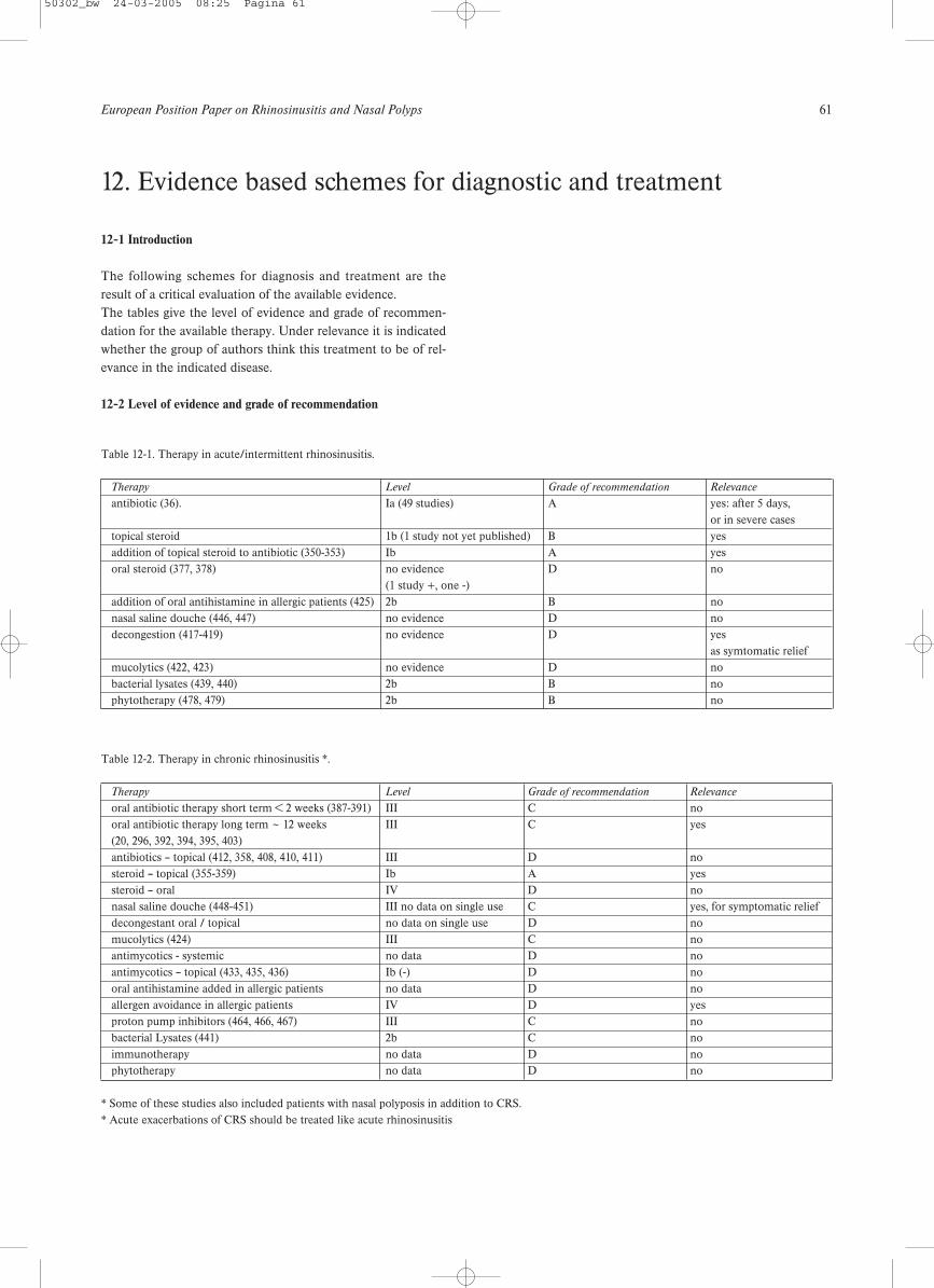
European Position Paper on Rhinosinusitis and Nasal Polyps 61
12-1 Introduction
The following schemes for diagnosis and treatment are theresult of a critical evaluation of the available evidence.The tables give the level of evidence and grade of recommen-dation for the available therapy. Under relevance it is indicatedwhether the group of authors think this treatment to be of rel-evance in the indicated disease.
12-2 Level of evidence and grade of recommendation
12. Evidence based schemes for diagnostic and treatment
Table 12-1. Therapy in acute/intermittent rhinosinusitis.
Therapy Level Grade of recommendation Relevance
antibiotic (36). Ia (49 studies) A yes: after 5 days,or in severe cases
topical steroid 1b (1 study not yet published) B yesaddition of topical steroid to antibiotic (350-353) Ib A yes oral steroid (377, 378) no evidence D no
(1 study +, one -)addition of oral antihistamine in allergic patients (425) 2b B nonasal saline douche (446, 447) no evidence D nodecongestion (417-419) no evidence D yes
as symtomatic reliefmucolytics (422, 423) no evidence D nobacterial lysates (439, 440) 2b B nophytotherapy (478, 479) 2b B no
Table 12-2. Therapy in chronic rhinosinusitis *.
Therapy Level Grade of recommendation Relevance
oral antibiotic therapy short term < 2 weeks (387-391) III C nooral antibiotic therapy long term ~ 12 weeks III C yes(20, 296, 392, 394, 395, 403)antibiotics – topical (412, 358, 408, 410, 411) III D nosteroid – topical (355-359) Ib A yessteroid – oral IV D nonasal saline douche (448-451) III no data on single use C yes, for symptomatic reliefdecongestant oral / topical no data on single use D nomucolytics (424) III C noantimycotics - systemic no data D noantimycotics – topical (433, 435, 436) Ib (-) D nooral antihistamine added in allergic patients no data D noallergen avoidance in allergic patients IV D yesproton pump inhibitors (464, 466, 467) III C nobacterial Lysates (441) 2b C noimmunotherapy no data D nophytotherapy no data D no
* Some of these studies also included patients with nasal polyposis in addition to CRS. * Acute exacerbations of CRS should be treated like acute rhinosinusitis
50302_bw 24-03-2005 08:25 Pagina 61
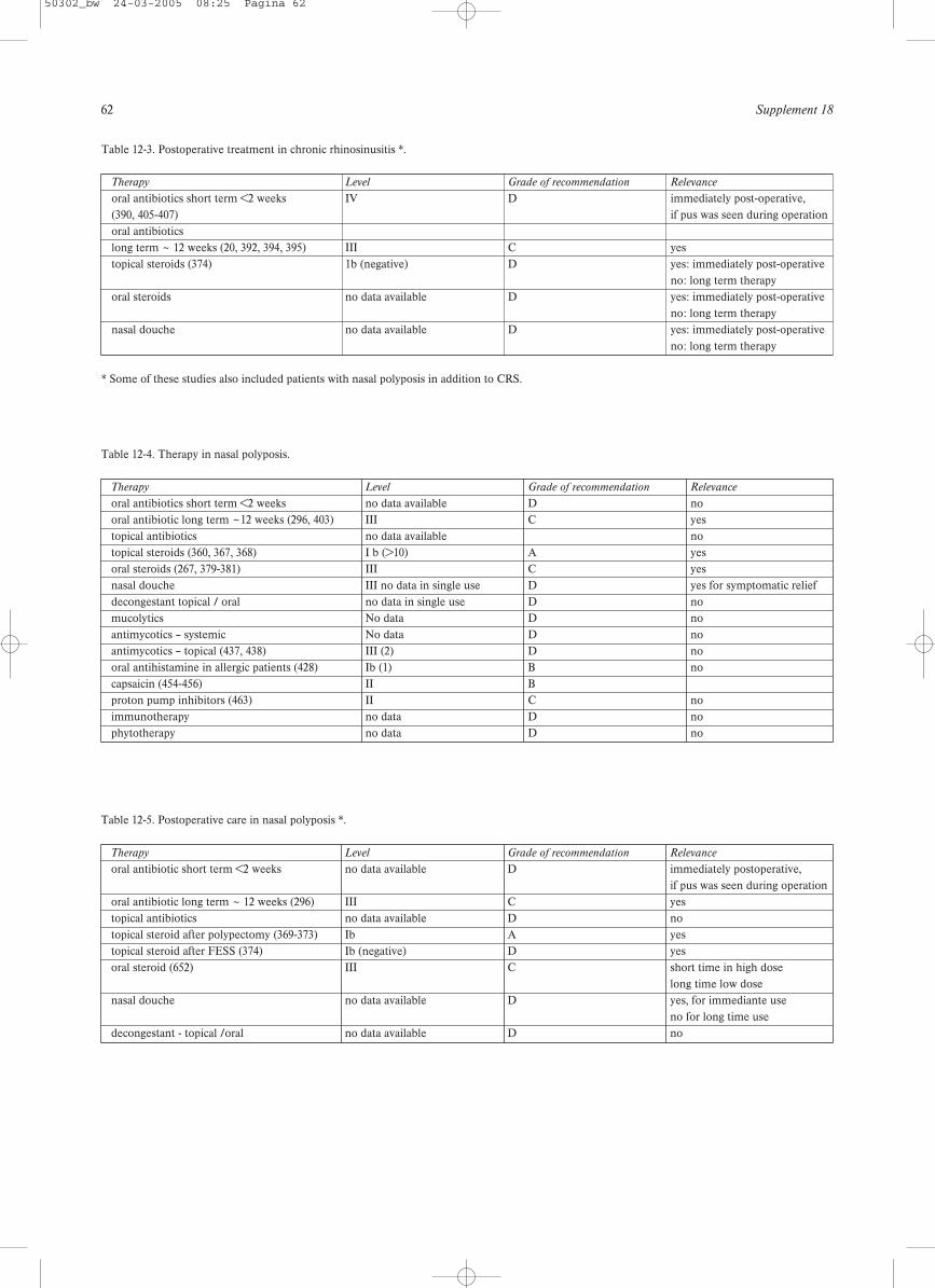
62 Supplement 18
Table 12-3. Postoperative treatment in chronic rhinosinusitis *.
Therapy Level Grade of recommendation Relevance
oral antibiotics short term <2 weeks IV D immediately post-operative, (390, 405-407) if pus was seen during operationoral antibioticslong term ~ 12 weeks (20, 392, 394, 395) III C yestopical steroids (374) 1b (negative) D yes: immediately post-operative
no: long term therapyoral steroids no data available D yes: immediately post-operative
no: long term therapynasal douche no data available D yes: immediately post-operative
no: long term therapy
* Some of these studies also included patients with nasal polyposis in addition to CRS.
Table 12-4. Therapy in nasal polyposis.
Therapy Level Grade of recommendation Relevance
oral antibiotics short term <2 weeks no data available D nooral antibiotic long term ~12 weeks (296, 403) III C yestopical antibiotics no data available notopical steroids (360, 367, 368) I b (>10) A yesoral steroids (267, 379-381) III C yesnasal douche III no data in single use D yes for symptomatic relief decongestant topical / oral no data in single use D nomucolytics No data D noantimycotics – systemic No data D noantimycotics – topical (437, 438) III (2) D nooral antihistamine in allergic patients (428) Ib (1) B nocapsaicin (454-456) II Bproton pump inhibitors (463) II C noimmunotherapy no data D nophytotherapy no data D no
Table 12-5. Postoperative care in nasal polyposis *.
Therapy Level Grade of recommendation Relevance
oral antibiotic short term <2 weeks no data available D immediately postoperative, if pus was seen during operation
oral antibiotic long term ~ 12 weeks (296) III C yestopical antibiotics no data available D notopical steroid after polypectomy (369-373) Ib A yestopical steroid after FESS (374) Ib (negative) D yesoral steroid (652) III C short time in high dose
long time low dosenasal douche no data available D yes, for immediante use
no for long time usedecongestant - topical /oral no data available D no
50302_bw 24-03-2005 08:25 Pagina 62
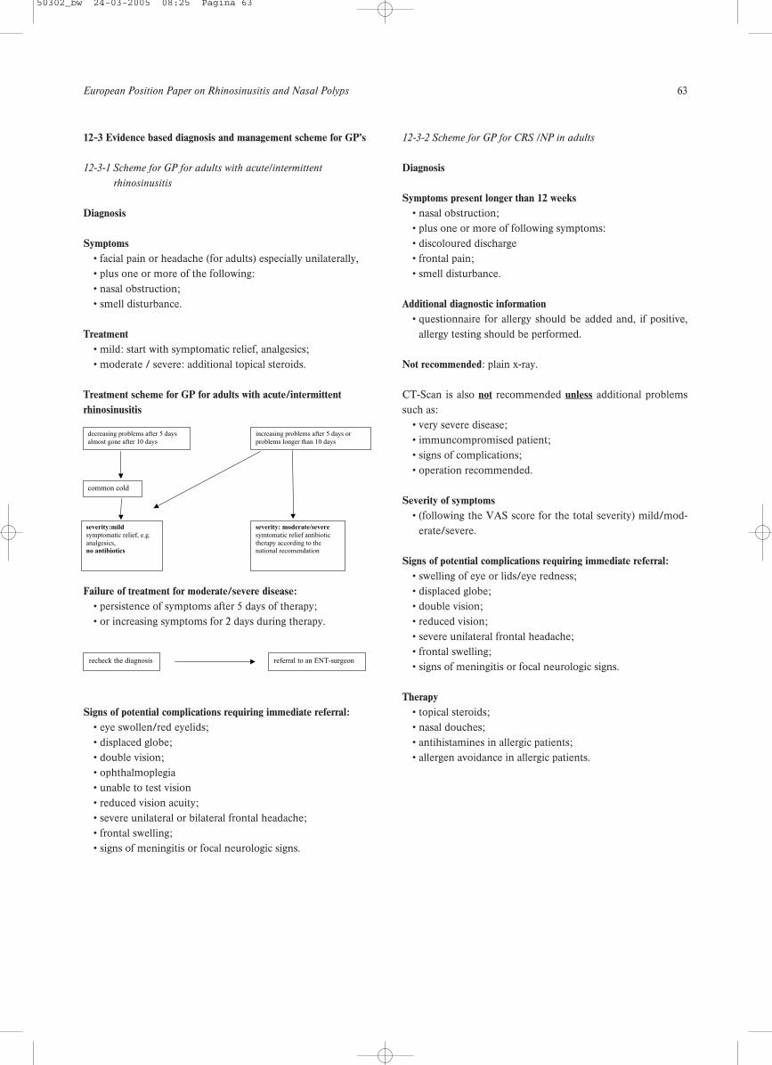
European Position Paper on Rhinosinusitis and Nasal Polyps 63
12-3 Evidence based diagnosis and management scheme for GP’s
12-3-1 Scheme for GP for adults with acute/intermittent
rhinosinusitis
Diagnosis
Symptoms• facial pain or headache (for adults) especially unilaterally,• plus one or more of the following: • nasal obstruction;• smell disturbance.
Treatment• mild: start with symptomatic relief, analgesics;• moderate / severe: additional topical steroids.
Treatment scheme for GP for adults with acute/intermittent rhinosinusitis
Failure of treatment for moderate/severe disease:• persistence of symptoms after 5 days of therapy;• or increasing symptoms for 2 days during therapy.
Signs of potential complications requiring immediate referral:• eye swollen/red eyelids;• displaced globe;• double vision;• ophthalmoplegia• unable to test vision• reduced vision acuity;• severe unilateral or bilateral frontal headache;• frontal swelling;• signs of meningitis or focal neurologic signs.
12-3-2 Scheme for GP for CRS /NP in adults
Diagnosis
Symptoms present longer than 12 weeks• nasal obstruction;• plus one or more of following symptoms:• discoloured discharge • frontal pain;• smell disturbance.
Additional diagnostic information• questionnaire for allergy should be added and, if positive,
allergy testing should be performed.
Not recommended: plain x-ray.
CT-Scan is also not recommended unless additional problemssuch as:
• very severe disease;• immuncompromised patient;• signs of complications;• operation recommended.
Severity of symptoms• (following the VAS score for the total severity) mild/mod-
erate/severe.
Signs of potential complications requiring immediate referral: • swelling of eye or lids/eye redness;• displaced globe;• double vision;• reduced vision;• severe unilateral frontal headache;• frontal swelling;• signs of meningitis or focal neurologic signs.
Therapy• topical steroids;• nasal douches;• antihistamines in allergic patients;• allergen avoidance in allergic patients.
decreasing problems after 5 days
almost gone after 10 days
increasing problems after 5 days or
problems longer than 10 days
common cold
severity:mild
symptomatic relief, e.g.
analgesics,no antibiotics
severity: moderate/severe
symtomatic relief antibiotic
therapy according to thenational recomendation
recheck the diagnosis referral to an ENT-surgeon
50302_bw 24-03-2005 08:25 Pagina 63
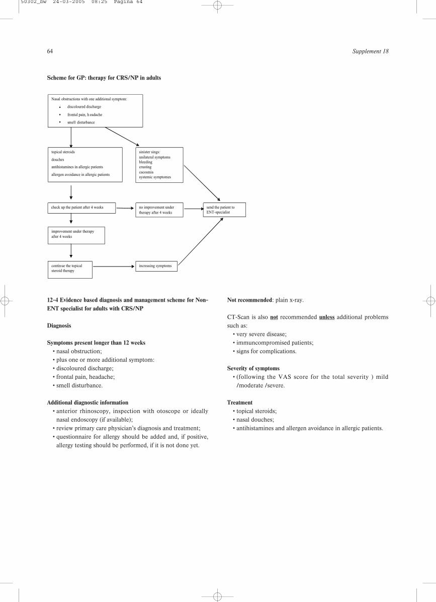
64 Supplement 18
Scheme for GP: therapy for CRS/NP in adults
12-4 Evidence based diagnosis and management scheme for Non-ENT specialist for adults with CRS/NP
Diagnosis
Symptoms present longer than 12 weeks• nasal obstruction;• plus one or more additional symptom:• discoloured discharge;• frontal pain, headache;• smell disturbance.
Additional diagnostic information• anterior rhinoscopy, inspection with otoscope or ideally
nasal endoscopy (if available);• review primary care physician’s diagnosis and treatment;• questionnaire for allergy should be added and, if positive,
allergy testing should be performed, if it is not done yet.
Not recommended: plain x-ray.
CT-Scan is also not recommended unless additional problemssuch as:
• very severe disease;• immuncompromised patients;• signs for complications.
Severity of symptoms • (following the VAS score for the total severity ) mild
/moderate /severe.
Treatment• topical steroids;• nasal douches;• antihistamines and allergen avoidance in allergic patients.
Nasal obstructions with one additional symptom:
? discoloured discharge
? frontal pain, h eadache
? smell disturbance
sinister sings:
unilateral symptoms bleeding crusting
cacosmia systemic symptomes
topical steroids
douches
antihistamines in allergic patients
allergen avoidance in allergic patients
no improvement under
therapy after 4 weeks
send the patient to ENT-specialist
check up the patient after 4 weeks
improvement under therapy
after 4 weeks
continue the topical steroid therapy
increasing symptoms
•••
50302_bw 24-03-2005 08:25 Pagina 64
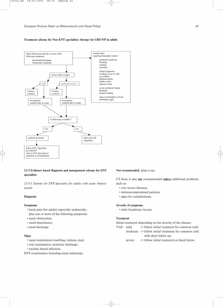
European Position Paper on Rhinosinusitis and Nasal Polyps 65
12-5 Evidence based diagnosis and management scheme for ENTspecialists
12-5-1 Scheme for ENT-Specialist for adults with acute rhinosi-
nusitis
Diagnosis
Symptoms• facial pain (for adults) especially unilaterally;• plus one or more of the following symptoms:• nasal obstruction;• smell disturbance;• nasal discharge.
Signs• nasal examination (swelling, redness, pus);• oral examination: posterior discharge;• exclude dental infection.
ENT-examination including nasal endoscopy.
Not recommended: plain x-ray.
CT-Scan is also not recommended unless additional problemssuch as:
• very severe diseases,• immuncompromised patients;• signs for complications.
Severity of symptoms• mild /moderate /severe.
TreatmentInitial treatment depending on the severity of the disease:VAS: mild -> follow initial treatment for common cold;
moderate -> follow initial treatment for common coldwith short follow up;
severe -> follow initial treatment as listed below.
Treatment scheme for Non-ENT specialists: therapy for CRS/NP in adults
Nasal obstruction and one or more of the
following symptoms:
discoloured discharge
frontal pain, headache
Is endoscopy available ?
NoYes
follow the GP-
Algorithm
medical treatment
follow ENT-Algorithm
for CRS/NP
refer to ENT-Specialist ifoperation is recommended
mild
review after 4 weeks
no progress:
recheck after 8 weeks
if better:continue
sinister signs:
requiring immediate referral
unilateral symptoms
bleeding
crusting
cacosmia
orbital symptoms:
swelling of eye or -lidseye redness
displaced globe
double vision
reduced vision
severe unilateral frontal
headache
frontal swelling
signs of meningitis or focal
neurologic signs
modereate/severe
if better:
continue
no progress:recheck after 4 weeks
50302_bw 24-03-2005 08:25 Pagina 65
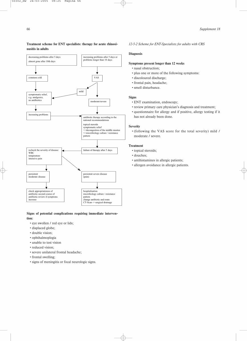
66 Supplement 18
Treatment scheme for ENT specialists: therapy for acute rhinosi-nusitis in adults
Signs of potential complications requiring immediate interven-tion:
• eye swollen / red eye or lids;• displaced globe;• double vision;• ophthalmoplegia• unable to test vision• reduced vision;• severe unilateral frontal headache;• frontal swelling;• signs of meningitis or focal neurologic signs.
12-5-2 Scheme for ENT-Specialists for adults with CRS
Diagnosis
Symptoms present longer than 12 weeks• nasal obstruction;• plus one or more of the following symptoms:• discoloured discharge;• frontal pain, headache;• smell disturbance.
Signs• ENT examination, endoscopy;• review primary care physician’s diagnosis and treatment;• questionnaire for allergy and if positive, allergy testing if it
has not already been done.
Severity• (following the VAS score for the total severity) mild /
moderate / severe.
Treatment• topical steroids;• douches;• antihistamines in allergic patients;• allergen avoidance in allergic patients.
common cold
symptomatic relief,
e.g. analgesics,
no antibiotics
increasing problems
persistentmoderate disease
check appropriateness of
antibiotic second course of
antibiotic review if symptomsincrease
recheck the severity of disease:WBC
temperature
intensive pain
decreasing problems after 5 days
almost gone after 10th days
mild
moderate/severe
antibiotic therapy according to the
national recommendations
topical steroids
symptomatic relief
+/-decongestion of the middle meatus
+/-microbiology culture / resistance
pattern
VAS
failure of therapy after 5 days
hospitalisation
microbiology culture / resistance
patternchange antibiotic and route
CT-Scan -> surgical drainage
persistent severe disease(pain)
increasing problems after 5 days or
problems longer than 10 days
50302_bw 24-03-2005 08:25 Pagina 66
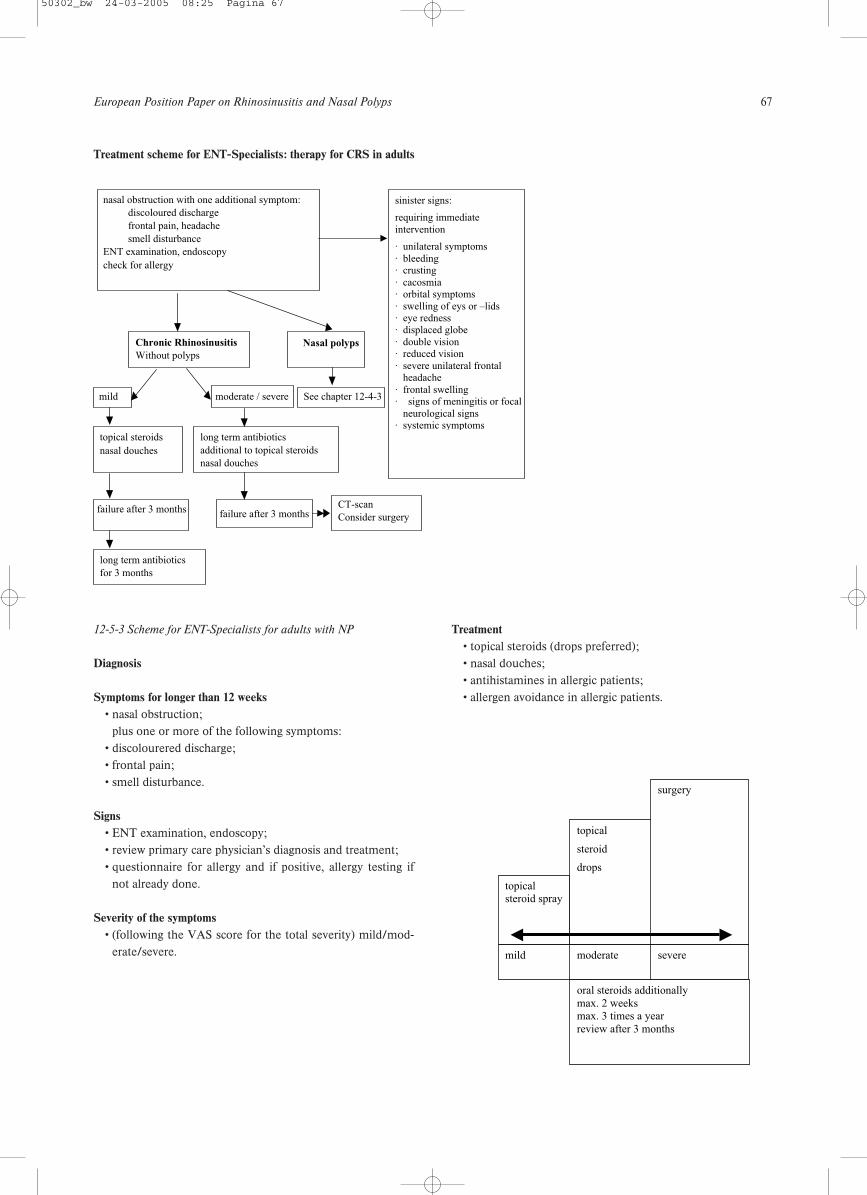
European Position Paper on Rhinosinusitis and Nasal Polyps 67
12-5-3 Scheme for ENT-Specialists for adults with NP
Diagnosis
Symptoms for longer than 12 weeks• nasal obstruction;• plus one or more of the following symptoms:• discolourered discharge;• frontal pain;• smell disturbance.
Signs• ENT examination, endoscopy;• review primary care physician’s diagnosis and treatment;• questionnaire for allergy and if positive, allergy testing if
not already done.
Severity of the symptoms• (following the VAS score for the total severity) mild/mod-
erate/severe.
Treatment• topical steroids (drops preferred);• nasal douches;• antihistamines in allergic patients;• allergen avoidance in allergic patients.
Treatment scheme for ENT-Specialists: therapy for CRS in adults
nasal obstruction with one additional symptom:
discoloured discharge
frontal pain, headache
smell disturbance
ENT examination, endoscopy
check for allergy
sinister signs:
requiring immediateintervention
· unilateral symptoms· bleeding· crusting· cacosmia· orbital symptoms· swelling of eys or –lids· eye redness· displaced globe· double vision· reduced vision· severe unilateral frontal
headache· frontal swelling· signs of meningitis or focal
neurological signs· systemic symptoms
CT-scan
Consider surgery
See chapter 12-4-3
Nasal polyps
mild
Chronic Rhinosinusitis
Without polyps
moderate / severe
topical steroids
nasal douches
failure after 3 months
long term antibiotics
additional to topical steroids
nasal douches
long term antibiotics
for 3 months
failure after 3 months
dance in allergic patients.
topicalsteroid spray
topical
steroid
drops
surgery
mild moderate severe
oral steroids additionallymax. 2 weeksmax. 3 times a yearreview after 3 months
50302_bw 24-03-2005 08:25 Pagina 67
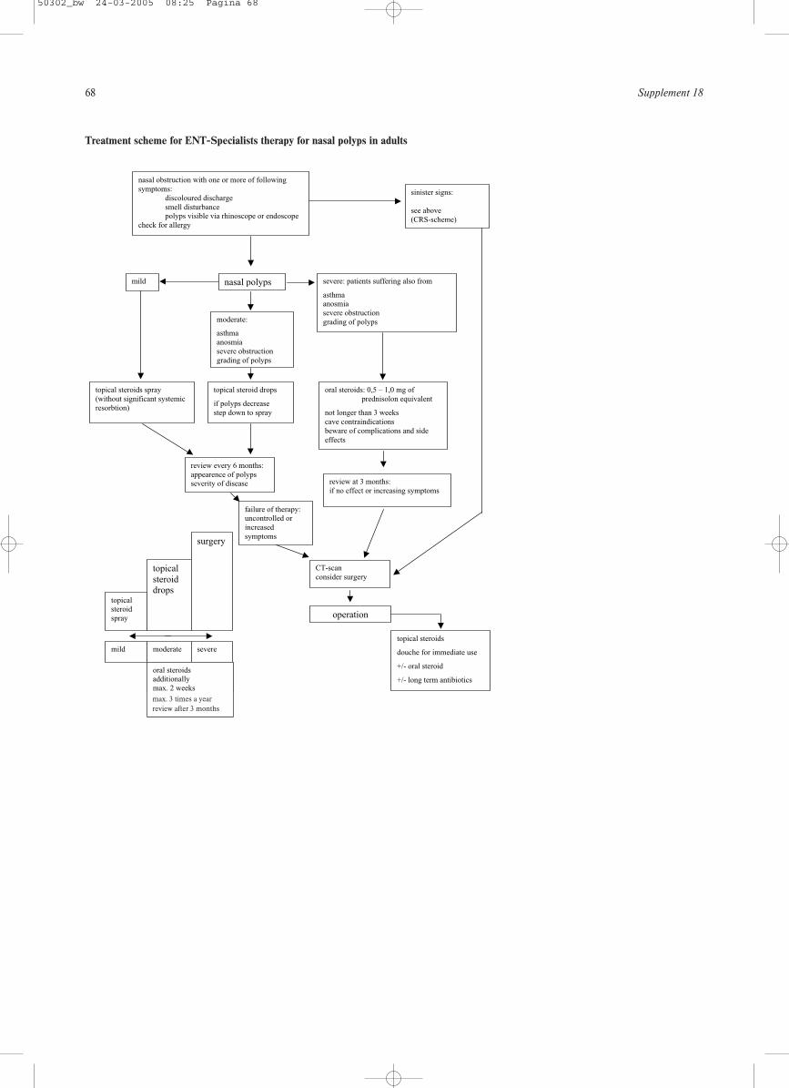
68 Supplement 18
Treatment scheme for ENT-Specialists therapy for nasal polyps in adults
113
sinister signs:
see above
(CRS-scheme)
nasal obstruction with one or more of following
symptoms:
discoloured discharge
smell disturbancepolyps visible via rhinoscope or endoscope
check for allergy
mild
topical steroids spray
(without significant systemic
resorbtion)
CT-scan
consider surgery
topicalsteroid
spray
topicalsteroiddrops
surgery
mild moderate severe
oral steroidsadditionally
max. 2 weeks
severe: patients suffering also from
asthmaanosmia
severe obstruction
grading of polyps
oral steroids: 0,5 – 1,0 mg of
prednisolon equivalent
not longer than 3 weeks
cave contraindications
beware of complications and side
effects
nasal polyps
moderate:
asthma
anosmia
severe obstruction
grading of polyps
review every 6 months:
appearence of polypsseverity of disease
topical steroid drops
if polyps decrease
step down to spray
failure of therapy:uncontrolled or
increased
symptoms
operation
topical steroids
douche for immediate use
+/- oral steroid
+/- long term antibiotics
review at 3 months:
if no effect or increasing symptoms
max. 3 times a yearreview after 3 months
50302_bw 24-03-2005 08:25 Pagina 68
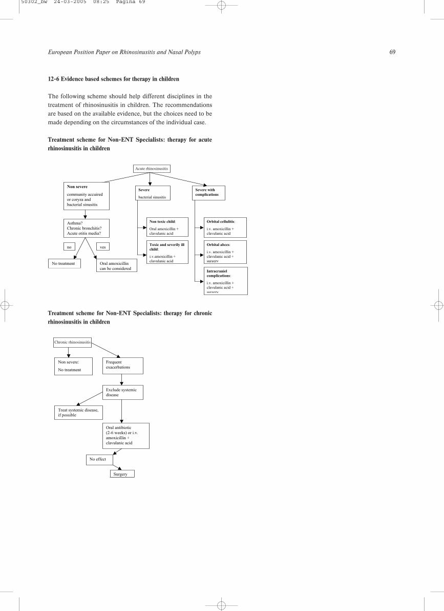
European Position Paper on Rhinosinusitis and Nasal Polyps 69
12-6 Evidence based schemes for therapy in children
The following scheme should help different disciplines in thetreatment of rhinosinusitis in children. The recommendationsare based on the available evidence, but the choices need to bemade depending on the circumstances of the individual case.
Treatment scheme for Non-ENT Specialists: therapy for acuterhinosinusitis in children
Treatment scheme for Non-ENT Specialists: therapy for chronicrhinosinusitis in children
Acute Sinusitis
Severe
bacterial sinusitis
Severe with
complications
Orbital cellulitis:
i.v. amoxicillin +clavulanic acid
Intracraniel
complications:
i.v. amoxicillin +clavulanic acid +
surgery
Orbital abces:
i.v. amoxicillin +clavulanic acid +surgery
Non toxic child:
Oral amoxicillin +clavulanic acid
Toxic and severily ill
child:
i.v.amoxicillin +clavulanic acid
Non severe
community accuired
or coryza and
bacterial sinusitis
No treatment
Chronic sinusitis
Asthma?
Chronic bronchitis?
Acute otitis media?
no yes
Oral amoxicillin
can be considered
Chronic sinusitis
Non severe:
No treatment
Frequent
exacerbations
Exclude systemicdisease
Treat systemic disease,if possible
Oral antibiotic
(2-6 weeks) or i.v.
amoxicillin +
clavulanic acid
No effect
Surgery
Acute rhinosinusitis
Chronic rhinosinusitis
50302_bw 24-03-2005 08:25 Pagina 69
70 Supplement 18
Although much work has been done on chronic rhinosinusitisand nasal polyps there are many questions still unanswered.
The following suggestions should highlight some areas ofinterest for further research.
A prospective population study of a group of age- and sex-matched controlled atopic and non-atopic individuals to con-sider the incidence of all upper respiratory tract symptomsincluding acute and chronic rhinosinusitis over a 5 year period.
A long-term follow-up of a cohort of patients with nasal poly-posis to study the natural history of the condition (a ran-domised medical and surgical arm could be done at the sametime).
A study of the benefit of long term macrolide therapy in nasalpolyposis and chronic rhinosinusitis (this needs repeating toverify the work already published on this).
Studies should be performed to compare nasal steroids as asingle modality of treatment with antibiotics in patients withintermittent or persistent rhinosinusitis.
There is an urgent need for randomized placebo controlled tri-als to study the effect of antibiotics in chronic rhinosinusitisand exacerbations of chronic rhinosinusitis.
To provide good evidence for the use of local antibiotic treat-ment in acute exacerbations of chronic rhinosinusitis, furtherstudies with better characterized patients are needed.
13. Research needs and priorities
1. Durr DG, Desrosiers MY, Dassa C. Impact of rhinosinusitis inhealth care delivery: the Quebec experience. J Otolaryngol2001;30(2):93-7.
2. Goetzel RZ, Hawkins K, Ozminkowski RJ, Wang S. The healthand productivity cost burden of the “top 10” physical and mentalhealth conditions affecting six large U.S. employers in 1999. JOccup Environ Med 2003;45(1):5-14.
3. Ray NF, Baraniuk JN, Thamer M, Rinehart CS, Gergen PJ,Kaliner M, et al. Healthcare expenditures for sinusitis in 1996:contributions of asthma, rhinitis, and other airway disorders. JAllergy Clin Immunol 1999;103(3 Pt 1):408-14.
4. New guidelines for sinusitis target prescribing practices. DisManag Advis 2004;10(3):27-30.
5. Anon JB, Jacobs MR, Poole MD, Ambrose PG, Benninger MS,Hadley JA, et al. Antimicrobial treatment guidelines for acute bac-terial rhinosinusitis. Otolaryngol Head Neck Surg 2004;130(1Suppl):1-45.
6. Report of the Rhinosinusitis Task Force Committee Meeting.Alexandria, Virginia, August 17, 1996. Otolaryngol Head NeckSurg 1997;117(3 Pt 2):S1-68.
7. Sackett DL, Rosenberg WM, Gray JA, Haynes RB, RichardsonWS. Evidence based medicine: what it is and what it isn’t. Bmj1996;312(7023):71-2.
8. Shekelle PG, Woolf SH, Eccles M, Grimshaw J. Clinical guide-lines: developing guidelines. Bmj 1999;318(7183):593-6.
9. Bachert C, Hormann K, Mosges R, Rasp G, Riechelmann H,Muller R, et al. An update on the diagnosis and treatment ofsinusitis and nasal polyposis. Allergy 2003;58(3):176-91.
10. Bousquet J, Van Cauwenberge P, Khaltaev N. Allergic rhinitis andits impact on asthma. J Allergy Clin Immunol 2001;108(5Suppl):S147-334.
11. Clement PA, Bluestone CD, Gordts F, Lusk RP, Otten FW,Goossens H, et al. Management of rhinosinusitis in children: con-sensus meeting, Brussels, Belgium, September 13, 1996. ArchOtolaryngol Head Neck Surg 1998;124(1):31-4.
12. Winstead W. Rhinosinusitis. Prim Care 2003;30(1):137-54.13. Slavin RG. Nasal polyps and sinusitis. Jama 1997;278(22):1849-54.14. Sturgess JM, Chao J, Wong J, Aspin N, Turner JA. Cilia with
defective radial spokes: a cause of human respiratory disease. NEngl J Med 1979;300(2):53-6.
15. Bhattacharyya Nl. The role of infection in chronic rhinosinusitis.Curr Allergy Asthma Rep 2002;2(6):500-6.
16. Zacharek MA, Krouse JH. The role of allergy in chronic rhinosi-nusitis. Curr Opin Otolaryngol Head Neck Surg 2003;11(3):196-200.
17. Jones NS. CT of the paranasal sinuses: a review of the correlationwith clinical, surgical and histopathological findings. ClinOtolaryngol 2002;27(1):11-7.
18. Jones NS, Strobl A, Holland I. A study of the CT findings in 100patients with rhinosinusitis and 100 controls. Clin Otolaryngol1997;22(1):47-51.
19. Steinke JW, Bradley D, Arango P, Crouse CD, Frierson H,Kountakis SE, et al. Cysteinyl leukotriene expression in chronichyperplastic sinusitis-nasal polyposis: importance to eosinophiliaand asthma. J Allergy Clin Immunol 2003;111(2):342-9.
20. Ichimura K, Shimazaki Y, Ishibashi T, Higo R. Effect of newmacrolide roxithromycin upon nasal polyps associated with chron-ic sinusitis. Auris Nasus Larynx 1996;23:48-56.
21. Dykewicz MS. 7. Rhinitis and sinusitis. J Allergy Clin Immunol2003;111(2 Suppl):S520-9.
22. Vento SI, Ertama LO, Hytonen ML, Wolff CH, Malmberg CH.Nasal polyposis: clinical course during 20 years. Ann AllergyAsthma Immunol 2000;85(3):209-14.
23. Bachert C, Van Cauwenberge PBl. Inflammatory mechanisms inchronic sinusitis. Acta Otorhinolaryngol Belg 1997;51(4):209-17.
24. Berger G, Kattan A, Bernheim J, Ophir Dl. Polypoid mucosa witheosinophilia and glandular hyperplasia in chronic sinusitis: ahistopathological and immunohistochemical study. Laryngoscope2002;112(4):738-45.
25. Rudack C, Stoll W, Bachert C. Cytokines in nasal polyposis, acuteand chronic sinusitis. Am J Rhinol 1998;12(6):383-8.
26. Hamilos DL, Leung DY, Wood R, Bean DK, Song YL, SchotmanE, et al. Eosinophil infiltration in nonallergic chronic hyperplasticsinusitis with nasal polyposis (CHS/NP) is associated withendothelial VCAM-1 upregulation and expression of TNF-alpha.Am J Respir Cell Mol Biol 1996;15(4):443-50.
27. Hamilos DL, Leung DY, Wood R, Cunningham L, Bean DK,
14. References
50302_bw 24-03-2005 08:25 Pagina 70
European Position Paper on Rhinosinusitis and Nasal Polyps 71
Yasruel Z, et al. Evidence for distinct cytokine expression in aller-gic versus nonallergic chronic sinusitis. J Allergy Clin Immunol1995;96(4):537-44.
28. Bachert C, Wagenmann M, Hauser U, Rudack Cl. IL-5 synthesis isupregulated in human nasal polyp tissue. J Allergy Clin Immunol1997;99(6 Pt 1):837-42.
29. Hedman J, Kaprio J, Poussa T, Nieminen MM. Prevalence of asth-ma, aspirin intolerance, nasal polyposis and chronic obstructivepulmonary disease in a population-based study. Int J Epidemiol1999;28(4):717-22.
30. Larsen K. The clinical relationship of nasal polyps to asthma.Allergy Asthma Proc 1996;17(5):243-9.
31. Settipane GA, Chafee FH. Nasal polyps in asthma and rhinitis. Areview of 6,037 patients. J Allergy Clin Immunol 1977;59(1):17-21.
32. Caplin I, Haynes JT, Spahn J. Are nasal polyps an allergic phe-nomenon? Ann Allergy 1971;29(12):631-4.
33. Gwaltney JM, Jr., Jones JG, Kennedy DW. Medical managementof sinusitis: educational goals and management guidelines. TheInternational Conference on sinus Disease. Ann Otol RhinolLaryngol Suppl 1995;167:22-30.
34. Leggett JE. Acute sinusitis. When—and when not—to prescribeantibiotics. Postgrad Med 2004;115(1):13-9.
35. Lindbaek M. Acute sinusitis: guide to selection of antibacterialtherapy. Drugs 2004;64(8):805-19.
36. Williams Jr JW, Aguilar C, Cornell J, Chiquette E. Dolor RJ,Makela M, Holleman DR, et al. Antibiotics for acute maxillarysinusitis (Cochrane Review). Cochrane Database Syst Rev 2003(4).
37. Varonen H, Savolainen S, Kunnamo I, Heikkinen R, Revonta M.Acute rhinosinusitis in primary care: a comparison of symptoms,signs, ultrasound, and radiography. Rhinology 2003;41(1):37-43.
38. Engels EA, Terrin N, Barza M, Lau J. Meta-analysis of diagnostictests for acute sinusitis. J Clin Epidemiol 2000;53(8):852-62.
39. Giesbers HrNo-IVTV, Nationale Atlas Volksgezondheid.Bilthoven: RIVM, < Gezondheid\Ziekten enaandoeningen\Ziekten van de ademhalingswegen, 27 september2002. Neusbijholte ontsteking 1995-1999. In: RIVM; 2002.
40. Gijsen R, Poos M. Nationaal Kompas. In: RIVM; 2003.41. Gwaltney JM, Jr. Acute community-acquired sinusitis. Clin Infect
Dis 1996;23(6):1209-23; quiz 1224-5.42. Berg O, Carenfelt C, Kronvall G. Bacteriology of maxillary sinusi-
tis in relation to character of inflammation and prior treatment.Scand J Infect Dis 1988;20(5):511-6.
43. Felmingham D, Feldman C, Hryniewicz W, Klugman K, KohnoS, Low DE, et al. Surveillance of resistance in bacteria causingcommunity-acquired respiratory tract infections. Clin MicrobiolInfect 2002;8(Suppl 2):12-42.
44. Hoban D, Felmingham D. The PROTEKT surveillance study:antimicrobial susceptibility of Haemophilus influenzae andMoraxella catarrhalis from community-acquired respiratory tractinfections. J Antimicrob Chemother 2002;50(Suppl S1):49-59.
45. Cars O, Molstad S, Melander A. Variation in antibiotic use in theEuropean Union. Lancet 2001;357(9271):1851-3.
46. Pedersen M, Sakakura Y, Winther B, Brofeldt S, Mygind N. Nasalmucociliary transport, number of ciliated cells, and beating patternin naturally acquired common colds. Eur J Respir Dis Suppl1983;128 (Pt 1):355-65.
47. Hinni ML, McCaffrey TV, Kasperbauer JL. Early mucosal changesin experimental sinusitis. Otolaryngol Head Neck Surg1992;107(4):537-48.
48. Kaliner M. Treatment of sinusitis in the next millennium. AllergyAsthma Proc 1998;19(4):181-4.
49. Lanza DC, Kennedy DW. Adult rhinosinusitis defined.Otolaryngol Head Neck Surg 1997;117(3 Pt 2):S1-7.
50. Savolainen S. Allergy in patients with acute maxillary sinusitis.Allergy 1989;44(2):116-22.
51. Alho OP, Karttunen TJ, Karttunen R, Tuokko H, Koskela M,Suramo I, et al. Subjects with allergic rhinitis show signs of moreseverely impaired paranasal sinus functioning during viral coldsthan nonallergic subjects. Allergy 2003;58(8):767-71.
52. Karlsson G, Holmberg K. Does allergic rhinitis predispose tosinusitis? Acta Otolaryngol Suppl 1994;515:26-8; discussion 29.
53. Gliklich RE, Metson R. The health impact of chronic sinusitis inpatients seeking otolaryngologic care. Otolaryngol Head Neck Surg1995;113(1):104-9.
54. Kaliner MA, Osguthorpe JD, Fireman P, Anon J, Georgitis J,Davis ML, et al. Sinusitis: bench to bedside. Current findings,future directions. Otolaryngol Head Neck Surg 1997;116(6 Pt2):S1-20.
55. Collins JG. Prevalence of selected chronic conditions: UnitedStates, 1990-1992. Vital Health Stat 10 1997(194):1-89.
56. Blackwell DCJ, Coles R. Summary health statistics for US adults:National Health Interview Survey 1997. National Center forHealth Statistics. Vital Health Stat 2002;10(205):15.
57. Shashy RG, Moore EJ, Weaver A. Prevalence of the chronicsinusitis diagnosis in Olmsted County, Minnesota. ArchOtolaryngol Head Neck Surg 2004;130(3):320-3.
58. Chen Y, Dales R, Lin M. The epidemiology of chronic rhinosi-nusitis in Canadians. Laryngoscope 2003;113(7):1199-205.
59. Greisner WA, 3rd, Settipane GA. Hereditary factor for nasalpolyps. Allergy Asthma Proc 1996;17(5):283-6.
60. Gordts F, Clement, P.A.R., Buisseret, T. Prevalence of sinusitis signsin a non-ENT population. Otorhinolaryngology 1996;58:315-319.
61. Al-Rawi MM, Edelstein DR, Erlandson RAl. Changes in nasalepithelium in patients with severe chronic sinusitis: a clinicopatho-logic and electron microscopic study. Laryngoscope1998;108(12):1816-23.
62. Hadfield PJ, Rowe-Jones JM, Mackay IS. The prevalence of nasalpolyps in adults with cystic fibrosis. Clin Otolaryngol2000;25(1):19-22.
63. Rowe-Jones JM, Shembekar M, Trendell-Smith N, Mackay IS.Polypoidal rhinosinusitis in cystic fibrosis: a clinical andhistopathological study. Clin Otolaryngol 1997;22(2):167-71.
64. Rowe-Jones JM, Trendell-Smith N, Shembekar M, Mackay IS.Polypoid rhinosinusitis in patients with host defence deficiencies:cellular infiltration and disease severity. Rhinology 1997;35(3):113-7.
65. Wojtczak HA, Kerby GS, Wagener JS, Copenhaver SC, GotlinRW, Riches DW, et al. Beclomethasone diproprionate reduced air-way inflammation without adrenal suppression in young childrenwith cystic fibrosis: a pilot study. Pediatr Pulmonol 2001;32(4):293-302.
66. Krause HF. Allergy and chronic rhinosinusitis. Otolaryngol HeadNeck Surg 2003;128(1):14-6.
67. Jones NS, Carney AS, Davis A. The prevalence of allergic rhinosi-nusitis: a review. J Laryngol Otol 1998;112(11):1019-30.
68. Bailey B. The impact of pollution on the upper alimentary and res-piratory tracts. Otolaryngol Head Neck Surg 1992;106:736-740.
69. Stammberger H. Functional endoscopic sinus surgery.Philadelphia: B.C. Decker; 1991.
70. Slavin RG. Sinusitis in adults and its relation to allergic rhinitis,asthma, and nasal polyps. J Allergy Clin Immunol 1988;82(5 Pt2):950-6.
71. Hamilos DL, Leung DY, Wood R, Meyers A, Stephens JK,Barkans J, et al. Chronic hyperplastic sinusitis: association of tis-sue eosinophilia with mRNA expression of granulocyte-macrophage colony-stimulating factor and interleukin-3. J AllergyClin Immunol 1993;92(1 Pt 1):39-48.
72. Rachelefsky GS, Goldberg M, Katz RM, Boris G, Gyepes MT,Shapiro MJ, et al. Sinus disease in children with respiratory aller-gy. J Allergy Clin Immunol 1978;61(5):310-4.
73. Shapiro GG. Role of allergy in sinusitis. Pediatr Infect Dis1985;4(6 Suppl):S55-9.
74. Shapiro GG, Virant FS, Furukawa CT, Pierson WE, Bierman CW.Immunologic defects in patients with refractory sinusitis.Pediatrics 1991;87(3):311-6.
75. Beninger M. Rhinitis, sinusitis and their relationship to allergies.Am J Rhinol 1992;6:37-43.
76. Grove R, Farrior, J. Chronic hyperplastic sinusitis in allergicpatients: a bacteriologic study of 200 operative cases. J AllergyClin Immunol 1990;11:271-276.
77. Emanuel IA, Shah SB. Chronic rhinosinusitis: allergy and sinuscomputed tomography relationships. Otolaryngol Head Neck Surg2000;123(6):687-91.
50302_bw 24-03-2005 08:25 Pagina 71
72 Supplement 18
78. Friedman WH. Surgery for chronic hyperplastic rhinosinusitis.Laryngoscope 1975;85(12 pt 1):1999-2011.
79. Hinriksdottir I, Melen I. Allergic rhinitis and upper respiratorytract infections. Acta Otolaryngol Suppl 1994;515:30-2.
80. Newman LJ, Platts-Mills TA, Phillips CD, Hazen KC, Gross CW.Chronic sinusitis. Relationship of computed tomographic findingsto allergy, asthma, and eosinophilia. Jama 1994;271(5):363-7.
81. Iwens P, Clement PA. Sinusitis in allergic patients. Rhinology1994;32(2):65-7.
82. Naclerio RM, deTineo ML, Baroody FM. Ragweed allergic rhinitisand the paranasal sinuses. A computed tomographic study. ArchOtolaryngol Head Neck Surg 1997;123(2):193-6.
83. Moser FG, Panush D, Rubin JS, Honigsberg RM, Sprayregen S,Eisig SB. Incidental paranasal sinus abnormalities on MRI of thebrain. Clin Radiol 1991;43(4):252-4.
84. Lloyd GA. CT of the paranasal sinuses: study of a control series inrelation to endoscopic sinus surgery. J Laryngol Otol1990;104(6):477-81.
85. Havas TE, Motbey JA, Gullane PJ. Prevalence of incidental abnor-malities on computed tomographic scans of the paranasal sinuses.Arch Otolaryngol Head Neck Surg 1988;114(8):856-9.
86. Patel K, Chavda SV, Violaris N, Pahor AL. Incidental paranasalsinus inflammatory changes in a British population. J LaryngolOtol 1996;110(7):649-51.
87. Zinreich SJ, Kennedy DW, Kumar AJ, Rosenbaum AE, ArringtonJA, Johns ME. MR imaging of normal nasal cycle: comparisonwith sinus pathology. J Comput Assist Tomogr 1988;12(6):1014-9.
88. Lane AP, Pine HS, Pillsbury HC, 3rd. Allergy testing andimmunotherapy in an academic otolaryngology practice: a 20-yearreview. Otolaryngol Head Neck Surg 2001;124(1):9-15.
89. Slavin RG. Relationship of nasal disease and sinusitis to bronchialasthma. Ann Allergy 1982;49(2):76-9.
90. Juntunen K, Tarkkanen J, Makinen J. Caldwell-Luc operation inthe treatment of childhood bronchial asthma. Laryngoscope1984;94(2 Pt 1):249-51.
91. Nisioka GJ, Cook, P.R., Davis, W.E., McKinsey, J.P. Functionalendoscopic sinus surgery in patients with chronic sinusitis andasthma. Otolaryngol Head Neck Surg 1994;110(6):494-500.
92. Salvin RG, Cannon RE, Friedman WH, Palitang E, Sundaram M.Sinusitis and bronchial asthma. J Allergy Clin Immunol1980;66(3):250-7.
93. Schwartz HJ, Thompson JS, Sher TH, Ross RJ. Occult sinusabnormalities in the asthmatic patient. Arch Intern Med1987;147(12):2194-6.
94. Bresciani M, Paradis L, Des Roches A, Vernhet H, Vachier I,Godard P, et al. Rhinosinusitis in severe asthma. J Allergy ClinImmunol 2001;107(1):73-80.
95. Chee L, Graham SM, Carothers DG, Ballas ZK. Immune dysfunc-tion in refractory sinusitis in a tertiary care setting. Laryngoscope2001;111(2):233-5.
96. Porter JP, Patel AA, Dewey CM, Stewart MG. Prevalence ofsinonasal symptoms in patients with HIV infection. Am J Rhinol1999;13(3):203-8.
97. Garcia-Rodriguez JF, Corominas M, Fernandez-Viladrich P,Monfort JL, Dicenta M. Rhinosinusitis and atopy in patientsinfected with HIV. Laryngoscope 1999;109(6):939-44.
98. Sabini P, Josephson GD, Reisacher WR, Pincus R. The role ofendoscopic sinus surgery in patients with acquired immune defi-ciency syndrome. Am J Otolaryngol 1998;19(6):351-6.
99. Riordan JR, Rommens JM, Kerem B, Alon N, Rozmahel R,Grzelczak Z, et al. Identification of the cystic fibrosis gene: cloningand characterization of complementary DNA. Science1989;245(4922):1066-73.
100.Kerem B, Rommens JM, Buchanan JA, Markiewicz D, Cox TK,Chakravarti A, et al. Identification of the cystic fibrosis gene:genetic analysis. Science 1989;245(4922):1073-80.
101.Cuppens H, Marynen P, De Boeck C, Cassiman JJ. Detection of98.5% of the mutations in 200 Belgian cystic fibrosis alleles byreverse dot-blot and sequencing of the complete coding regionand exon/intron junctions of the CFTR gene. Genomics1993;18(3):693-7.
102.De Gaudemar I, Contencin P, Van den Abbeele T, Munck A,Navarro J, Narcy P. Is nasal polyposis in cystic fibrosis a directmanifestation of genetic mutation or a complication of chronicinfection? Rhinology 1996;34(4):194-7.
103.Kerrebijn JD, Poublon RM, Overbeek SE. Nasal and paranasal dis-ease in adult cystic fibrosis patients. Eur Respir J 1992;5(10):1239-42.
104.Stern RC, Boat TF, Wood RE, Matthews LW, Doershuk CF.Treatment and prognosis of nasal polyps in cystic fibrosis. Am JDis Child 1982;136(12):1067-70.
105.Davidson TM, Stearns G. Extended indications for endoscopicsinus surgery. Ear Nose Throat J 1994;73(7):467-8, 473-4.
106.Jorissen MB, De Boeck K, Cuppens H. Genotype-phenotype cor-relations for the paranasal sinuses in cystic fibrosis. Am J RespirCrit Care Med 1999;159(5 Pt 1):1412-6.
107.Ellegard EK. The etiology and management of pregnancy rhinitis.Am J Respir Med 2003;2(6):469-75.
108.Sobol SE, Frenkiel S, Nachtigal D, Wiener D, Teblum C. Clinicalmanifestations of sinonasal pathology during pregnancy. JOtolaryngol 2001;30(1):24-8.
109.Sorri M, Hartikainen-Sorri AL, Karja J. Rhinitis during pregnancy.Rhinology 1980;18(2):83-6.
110.Zinreich SJ, Mattox, D.E., Kennedy, D.W., Chisholm, H.L.,Diffley, D.M., Rosenbaum, A.E. Concha bullosa: CT evaluation. JComput Assist Tomogr 1988;12:788-784.
111.Bolger WE, Butzin CA, Parsons DS. Paranasal sinus bony anatom-ic variations and mucosal abnormalities: CT analysis for endoscop-ic sinus surgery. Laryngoscope 1991;101(1 Pt 1):56-64.
112.Min YG, Jung HW, Kim HS, Park SK, Yoo KY. Prevalence andrisk factors of chronic sinusitis in Korea: results of a nationwidesurvey. Eur Arch Otorhinolaryngol 1996;253(7):435-9.
113.Wagenmann M, Naclerio RM. Complications of sinusitis. JAllergy Clin Immunol 1992;90(3 Pt 2):552-4.
114.Willner A, Choi SS, Vezina LG, Lazar RH. Intranasal anatomicvariations in pediatric sinusitis. Am J Rhinol 1997;11(5):355-60.
115.Harar RP, Chadha NK, Rogers G. The role of septal deviation inadult chronic rhinosinusitis: a study of 500 patients. Rhinology2004;42(3):126-30.
116.Calhoun KH, Waggenspack GA, Simpson CB, Hokanson JA,Bailey BJ. CT evaluation of the paranasal sinuses in symptomaticand asymptomatic populations. Otolaryngol Head Neck Surg1991;104(4):480-3.
117.Kayalioglu G, Oyar O, Govsa F. Nasal cavity and paranasal sinusbony variations: a computed tomographic study. Rhinology2000;38(3):108-13.
118.Perez P, Sabate J, Carmona A, Catalina-Herrera CJ, Jimenez-Castellanos J. Anatomical variations in the human paranasal sinusregion studied by CT. J Anat 2000;197(Pt 2):221-7.
119.Araujo E, Palombini BC, Cantarelli V, Pereira A, Mariante Al.Microbiology of middle meatus in chronic rhinosinusitis. Am JRhinol 2003;17(1):9-15.
120.Brook I. Microbiology and management of sinusitis. J Otolaryngol1996;25(4):249-56.
121.Brook I. Bacteriology of chronic maxillary sinusitis in adults. AnnOtol Rhinol Laryngol 1989;98(6):426-8.
122.Schubert MS. Fungal rhinosinusitis: diagnosis and therapy. CurrAllergy Asthma Rep 2001;1(3):268-76.
123.Schubert MS. Allergic fungal sinusitis. Otolaryngol Clin NorthAm 2004;37(2):301-26.
124.Ponikau JU, Sherris DA, Kern EB, Homburger HA, Frigas E,Gaffey TA, et al. The diagnosis and incidence of allergic fungalsinusitis. Mayo Clin Proc 1999;74(9):877-84.
125.Braun H, Buzina W, Freudenschuss K, Beham A, StammbergerH. ‘Eosinophilic fungal rhinosinusitis’: a common disorder inEurope? Laryngoscope 2003;113(2):264-9.
126.Kennedy DW, Senior BA, Gannon FH, Montone KT, Hwang P,Lanza DC. Histology and histomorphometry of ethmoid bone inchronic rhinosinusitis. Laryngoscope 1998;108(4 Pt 1):502-7.
127.Khalid AN, Hunt J, Perloff JR, Kennedy DW. The role of bone inchronic rhinosinusitis. Laryngoscope 2002;112(11):1951-7.
128.Perloff JR, Gannon FH, Bolger WE, Montone KT, Orlandi R,Kennedy DW. Bone involvement in sinusitis: an apparent pathway
50302_bw 24-03-2005 08:25 Pagina 72
European Position Paper on Rhinosinusitis and Nasal Polyps 73
for the spread of disease. Laryngoscope 2000;110(12):2095-9.129.Raynal M, Peynegre R, Beautru R, Coste A. [Sinus mucoceles and
surgery in iatrogenic diseases]. Ann Otolaryngol Chir Cervicofac1999;116(2):85-91.
130.Gutman M, Houser S. Iatrogenic maxillary sinus recirculation andbeyond. Ear Nose Throat J 2003;82(1):61-3.
131.Johansson L, Akerlund A, Holmberg K, Melen I, Bende M.Prevalence of nasal polyps in adults: the Skovde population-basedstudy. Ann Otol Rhinol Laryngol 2003;112(7):625-9.
132.Larsen PL, Tos M. Origin of nasal polyps. Laryngoscope1991;101(3):305-12.
133.Larsen PL, Tos M. Site of origin of nasal polyps. Transcraniallyremoved naso-ethmoidal blocks as a screening method for nasalpolyps in autopsy material. Rhinology 1995;33(4):185-8.
134.Larsen P, Tos M. Anatomic site of origin of nasal polyps: endo-scopic nasal and paranasal sinus surgery as a screening method fornasal polyps in autopsy material. Am J Rhinol 1996;10:211=216.
135.Drake-Lee A. Nasal polyps. In: Mygind N, Naclerio RM, editor.Allergic and non-allergic rhinitis. Copenhagen: Munksgaard; 1993.
136.Larsen K, Tos M. The estimated incidence of symptomatic nasalpolyps. Acta Otolaryngol 2002;122(2):179-82.
137.Settipane GA, Chafee, F.H. Nasal polyps in asthma and rhinitis. JAllergy Clin Immunol 1977;59:17-21.
138.Larsen K. The clinical relationship of nasal polyps to asthma. In:Settipane G, Lund VJ, Bernstein JM, Tos M, editor. Nasal polyps:epidemiology, pathogenesis and treatment. Rhode Island:Oceanside Publications; 1997. p. 97-104.
139.Larsen K, Tos M. A long-term follow-up study of nasal polyppatients after simple polypectomies. Eur Arch Otorhinolaryngol1997;254(Suppl 1):S85-8.
140.Rugina M, Serrano E, Klossek JM, Crampette L, Stoll D, BebearJP, et al. Epidemiological and clinical aspects of nasal polyposis inFrance; the ORLI group experience. Rhinology 2002;40(2):75-9.
141.Collins MM, Pang YT, Loughran S, Wilson JA. Environmentalrisk factors and gender in nasal polyposis. Clin Otolaryngol2002;27(5):314-7.
142.Drake-Lee AB, Lowe D, Swanston A, Grace A. Clinical profileand recurrence of nasal polyps. J Laryngol Otol 1984;98(8):783-93.
143.Moloney JR. Nasal polyps, nasal polypectomy, asthma, and aspirinsensitivity. Their association in 445 cases of nasal polyps. JLaryngol Otol 1977;91(10):837-46.
144.Larsen K, Tos M. Clinical course of patients with primary nasalpolyps. Acta Otolaryngol 1994;114(5):556-9.
145.Settipane G. Epidemiology of nasal polyps. In: Settipane G, LundVJ, Bernstein JM, Tos M., editor. Nasal polyps: epidemiology,pathogenesis and treatment. Rhode Island: OceansidePublications; 1997. p. 17-24.
146.Hosemann W, Gode U, Wagner W. Epidemiology, pathophysiolo-gy of nasal polyposis, and spectrum of endonasal sinus surgery.Am J Otolaryngol 1994;15(2):85-98.
147.Bunnag C, Pacharee P, Vipulakom P, Siriyananda C. A study ofallergic factor in nasal polyp patients. Ann Allergy 1983;50(2):126-32.
148.Kern R, Schenck H-P. Allergy: a constant factor in the etiology ofso-called mucous nasal polyps. J Allergy 1993;4:483.
149.Delaney JC. Aspirin idiosyncrasy in patients admitted for nasalpolypectomy. Clin Otolaryngol 1976;1(1):27-30.
150.Blumstein GI, Tuft Ll. Allergy treatment in recurrent nasal poly-posis: its importance and value. Am J Med Sci 1957;234(3):269-80.
151.English G. Nasal polyposis. In: GM E, editor. Otolaryngology.Philadelphia: Harper and Row; 1985. p. 1-30.
152.Pepys J, Duveen GE. Negative skin tests in allergic rhinitis andnasal polyposis. Int Arch Allergy Immunol 1951;2:147-160.
153.Drake-Lee AB. Histamine and its release from nasal polyps: pre-liminary communication. J R Soc Med 1984;77(2):120-4.
154.Liu CM, Shun CT, Hsu MM. Lymphocyte subsets and antigen-specific IgE antibody in nasal polyps. Ann Allergy 1994;72(1):19-24.
155.Bachert C, Gevaert P, Holtappels G, Johansson SG, vanCauwenberge Pl. Total and specific IgE in nasal polyps is relatedto local eosinophilic inflammation. J Allergy Clin Immunol2001;107(4):607-14.
156.Pang YT, Eskici O, Wilson JA. Nasal polyposis: role of subclinical
delayed food hypersensitivity. Otolaryngol Head Neck Surg2000;122(2):298-301.
157.Downing E, Braman S, Settipane GA. Bronchial reactivity inpatients with nasal polyposis before and after polypectomy. JAllergy Clin Immunol 1982;69(2):102.
158.Settipane GA, Chafee FH, Klein DE. Aspirin intolerance. II. Aprospective study in an atopic and normal population. J AllergyClin Immunol 1974;53(4):200-4.
159.Chafee F, Settipane GA. Aspirin intolerance. I. Frequency in anallergic population. J Allergy Clin Immunol 1974;53:193-199.
160.Weber RW, Hoffman M, Raine DA, Jr., Nelson HS. Incidence ofbronchoconstriction due to aspirin, azo dyes, non-azo dyes, andpreservatives in a population of perennial asthmatics. J AllergyClin Immunol 1979;64(1):32-7.
161.Szczeklik A, Gryglewski RJ, Czerniawska-Mysik G. Clinical pat-terns of hypersensitivity to nonsteroidal anti-inflammatory drugsand their pathogenesis. J Allergy Clin Immunol 1977;60(5):276-84.
162.Spector SL, Wangaard CH, Farr RS. Aspirin and concomitantidiosyncrasies in adult asthmatic patients. J Allergy Clin Immunol1979;64(6 Pt 1):500-6.
163.Ogino S, Harada T, Okawachi I, Irifune M, Matsunaga T, NaganoT. Aspirin-induced asthma and nasal polyps. Acta OtolaryngolSuppl 1986;430:21-7.
164.Szczeklik A, Stevenson DD. Aspirin-induced asthma: advances inpathogenesis and management. J Allergy Clin Immunol1999;104(1):5-13.
165.May A, Wagner D, Langenbeck U, Weber A. [Family study ofpatients with aspirin intolerance and rhinosinusitis]. Hno2000;48(9):650-4.
166.Moloney JR, Oliver RT. HLA antigens, nasal polyps and asthma.Clin Otolaryngol 1980;5(3):183-9.
167.Luxenberger W, Posch U, Berghold A, Hofmann T, Lang-LoidoltD. HLA patterns in patients with nasal polyposis. Eur ArchOtorhinolaryngol 2000;257(3):137-9.
168.Molnar-Gabor E, Endreffy E, Rozsasi A. HLA-DRB1, -DQA1, and-DQB1 genotypes in patients with nasal polyposis. Laryngoscope2000;110(3 Pt 1):422-5.
169.Lockey RF, Rucknagel DL, Vanselow NA. Familial occurrence ofasthma, nasal polyps and aspirin intolerance. Ann Intern Med1973;78(1):57-63.
170.Settipane GA. Benefit/risk ratio of aspirin. NES AllergyProceedings 1981;2:96-102.
171.Drake-Lee A. Nasal polyps in identical twins. J Laryngol Otol1992;106(12):1084-5.
172.Kim J, Hanley JA. The role of woodstoves in the etiology of nasalpolyposis. Arch Otolaryngol Head Neck Surg 2002;128(6):682-6.
173.van der Veken PJ, Clement PA, Buisseret T, Desprechins B,Kaufman L, Derde MP. CT-scan study of the incidence of sinusinvolvement and nasal anatomic variations in 196 children.Rhinology 1990;28(3):177-84.
174.Kristo A, Uhari M, Luotonen J, Koivunen P, Ilkko E, Tapiainen T,et al. Paranasal sinus findings in children during respiratory infec-tion evaluated with magnetic resonance imaging. Pediatrics2003;111(5 Pt 1):e586-9.
175.Maresh MM WA. Paranasal sinuses from birth to late adoles-cence. Clinical and roentgengraphic evidence of infection. Am JDis Child 1940(60):841-861.
176.Bagatsch K DK, Parthenheimer F, Ritter B. Morbidates analyseder unspezifisch-infektbedingten acute Erkrankungen derRespirationtraktes und der Mittelohrräume des Kindesalterns ineinem Ballungsgebiet mit modernen Wohnbedingungen. HNOPraxis 1980(5):1-8.
177.Van Buchem FL, Peeters MF, Knottnerus JA. Maxillary sinusitisin children. Clin Otolaryngol 1992;17(1):49-53.
178.Lind J. Über das vorkomen von Nebenhöhlenaffektion bei kinder.Arch Kinderheilk 1944(131):143-155.
179.Bjuggren G, Kraepelien S, Lind J, Tunevall G. Occult sinusitis inchildren. Acta Otolaryngol 1952;42(4-5):287-310.
180.Van Cauwenberge P. Secretoire otitis media. Een epidemiologis-che klinische en experimentele studie; 1988.
181.Myers MG, Fomon SJ, Koontz FP, McGuinness GA,
50302_bw 24-03-2005 08:25 Pagina 73
74 Supplement 18
Lachenbruch PA, Hollingshead R. Respiratory and gastrointestinalillnesses in breast- and formula-fed infants. Am J Dis Child1984;138(7):629-32.
182.Agre F. The relationship of mode of infant feeding and location ofcare to frequency of infection. Am J Dis Child 1985;139(8):809-11.
183.Cogswell JJ, Mitchell EB, Alexander J. Parental smoking, breastfeeding, and respiratory infection in development of allergic dis-eases. Arch Dis Child 1987;62(4):338-44.
184.Barr MB, Weiss ST, Segal MR, Tager IB, Speizer FE. The rela-tionship of nasal disorders to lower respiratory tract symptoms andillness in a random sample of children. Pediatr Pulmonol1992;14(2):91-4.
185.Rudack C, Hauser U, Wagenmann M, Bachert C, Ganzer U.[Cytokine pattern in various forms of sinusitis].Laryngorhinootologie 1998;77(1):34-7.
186.Repka-Ramirez S, Naranch K, Park YJ, Clauw D, Baraniuk JN.Cytokines in nasal lavage fluids from acute sinusitis, allergic rhini-tis, and chronic fatigue syndrome subjects. Allergy Asthma Proc2002;23(3):185-90.
187.Bachert C, Wagenmann M, Hauser U. Proinflammatory cytokines:measurement in nasal secretion and induction of adhesion recep-tor expression. Int Arch Allergy Immunol 1995;107(1-3):106-8.
188.Roseler S, Holtappels G, Wagenmann M, Bachert C. Elevated lev-els of interleukins IL-1 beta, IL-6 and IL-8 in naturally acquiredviral rhinitis. Eur Arch Otorhinolaryngol 1995;252 Suppl 1:S61-3.
189.Stierna P, Carlsoo B. Histopathological observations in chronicmaxillary sinusitis. Acta Otolaryngol 1990;110(5-6):450-8.
190.Georgitis JW, Matthews BL, Stone B. Chronic sinusitis: character-ization of cellular influx and inflammatory mediators in sinuslavage fluid. Int Arch Allergy Immunol 1995;106(4):416-21.
191.Jankowski R, Bouchoua F, Coffinet L, Vignaud JM. Clinical fac-tors influencing the eosinophil infiltration of nasal polyps.Rhinology 2002;40(4):173-8.
192.Takeuchi K, Yuta A, Sakakura Y. Interleukin-8 gene expression inchronic sinusitis. Am J Otolaryngol 1995;16(2):98-102.
193.Suzuki H, Takahashi Y, Wataya H, Ikeda K, Nakabayashi S,Shimomura A, et al. Mechanism of neutrophil recruitmentinduced by IL-8 in chronic sinusitis. J Allergy Clin Immunol1996;98(3):659-70.
194.Bachert C, Wagenmann M, Rudack C, Hopken K, Hillebrandt M,Wang D, et al. The role of cytokines in infectious sinusitis andnasal polyposis. Allergy 1998;53(1):2-13.
195.Persson CGA EJ, Andersson M, et al. Epithelium, microcircula-tion and eosinophils - new aspects of the allergic airway in vivo.Allergy 1997;52:241.
196.Rhyoo C, Sanders SP, Leopold DA, Proud D. Sinus mucosal IL-8gene expression in chronic rhinosinusitis. J Allergy Clin Immunol1999;103(3 Pt 1):395-400.
197.Nonoyama T, Harada T, Shinogi J, Yoshimura E, Sakakura Y.Immunohistochemical localization of cytokines and cell adhesionmolecules in maxillary sinus mucosa in chronic sinusitis. AurisNasus Larynx 2000;27(1):51-8.
198.Demoly P, Crampette L, Mondain M, Enander I, Jones I,Bousquet J. Myeloperoxidase and interleukin-8 levels in chronicsinusitis. Clin Exp Allergy 1997;27(6):672-5.
199.Watelet JB CC, Perez-Novo C, Gevaert P, van Cauwenberge P,Bachert C. TGF-beta 1 in remodeling of nasal tissue: differencesbetween chronic rhinosinusitis and nasal polyposis. Am J Rhinol2003;in press.
200.Watelet JB BC, Claeys C, van Cauwenberge P. MatrixMetalloproteinases MMP-7, MMP-9 and their tissue inhibitorTIM-1: expression in chronic sinusitis versus nasal polyposis.Allergy 2003;in press.
201.Taylor M. Histochemical studies on nasal polypi. J Laryngol Otol1963;77:326-41.
202.Kakoi H, Hiraide F. A histological study of formation and growthof nasal polyps. Acta Otolaryngol 1987;103(1-2):137-44.
203.Mygind N LT. Nasal Polyposis: an inflammatory disease and itstreatment. Copenhagen: Munksgaard; 1997.
204.Stoop AE, van der Heijden HA, Biewenga J, van der Baan S.Eosinophils in nasal polyps and nasal mucosa: an immunohisto-
chemical study. J Allergy Clin Immunol 1993;91(2):616-22.205.Bachert C, Gevaert P, Holtappels G, Cuvelier C, van
Cauwenberge Pl. Nasal polyposis: from cytokines to growth. Am JRhinol 2000;14(5):279-90.
206.Denburg JA, Otsuka H, Ohnisi M, Ruhno J, Bienenstock J,Dolovich J. Contribution of basophil/mast cell and eosinophilgrowth and differentiation to the allergic tissue inflammatoryresponse. Int Arch Allergy Appl Immunol 1987;82(3-4):321-6.
207.Xaubet A, Mullol J, Lopez E, Roca-Ferrer J, Rozman M, CarrionT, et al. Comparison of the role of nasal polyp and normal nasalmucosal epithelial cells on in vitro eosinophil survival. Mediationby GM-CSF and inhibition by dexamethasone. Clin Exp Allergy1994;24(4):307-17.
208.Mullol J, Xaubet A, Gaya A, Roca-Ferrer J, Lopez E, FernandezJC, et al. Cytokine gene expression and release from epithelialcells. A comparison study between healthy nasal mucosa and nasalpolyps. Clin Exp Allergy 1995;25(7):607-15.
209.Ohno I, Lea R, Finotto S, Marshall J, Denburg J, Dolovich J, et al.Granulocyte/macrophage colony-stimulating factor (GM-CSF)gene expression by eosinophils in nasal polyposis. Am J RespirCell Mol Biol 1991;5(6):505-10.
210.Simon HU, Yousefi S, Schranz C, Schapowal A, Bachert C, BlaserK. Direct demonstration of delayed eosinophil apoptosis as amechanism causing tissue eosinophilia. J Immunol1997;158(8):3902-8.
211.Ming YG LC, Rhee CS, et al. Inflammatory cytokine expressionon nasal polyps developed in allergic and infectious rhinitis. ActaOtolaryngol Suppl 1997;117:302.
212.Lee CH, Rhee CS, Min YG. Cytokine gene expression in nasalpolyps. Ann Otol Rhinol Laryngol 1998;107(8):665-70.
213.Wagenmann M G-AM, Helmig P. Increased production of type-2and type-1 cytokines in nasal polyps. J Allergy Clin Immunol2000;105(Supplement):S210.
214.Gevaert P, Bachert C, Holtappels G, Novo CP, Van Der Heyden J,Fransen L, et al. Enhanced soluble interleukin-5 receptor alphaexpression in nasal polyposis. Allergy 2003;58(5):371-9.
215.Bartels J, Maune S, Meyer JE, Kulke R, Schluter C, Rowert J, etal. Increased eotaxin-mRNA expression in non-atopic and atopicnasal polyps: comparison to RANTES and MCP-3 expression.Rhinology 1997;35(4):171-4.
216.Jahnsen FL, Haye R, Gran E, Brandtzaeg P, Johansen FE.Glucocorticosteroids inhibit mRNA expression for eotaxin, eotax-in-2, and monocyte-chemotactic protein-4 in human airwayinflammation with eosinophilia. J Immunol 1999;163(3):1545-51.
217.Shin SH, Park JY, Jeon CH, Choi JK, Lee SH. Quantitative analy-sis of eotaxin and RANTES messenger RNA in nasal polyps: asso-ciation of tissue and nasal eosinophils. Laryngoscope2000;110(8):1353-7.
218.Symon FA, Walsh GM, Watson SR, Wardlaw AJ. Eosinophiladhesion to nasal polyp endothelium is P-selectin-dependent. JExp Med 1994;180(1):371-6.
219.Jahnsen FL, Haraldsen G, Aanesen JP, Haye R, Brandtzaeg P.Eosinophil infiltration is related to increased expression of vascu-lar cell adhesion molecule-1 in nasal polyps. Am J Respir Cell MolBiol 1995;12(6):624-32.
220.Tingsgaard PK, Bock T, Larsen PL, Tos M. Topical budesonidetreatment reduces endothelial expression of intercellular adhesionmolecules (vascular cell adhesion molecule-1 and P-selectin) andeosinophil infiltration in nasal polyps. Acta Otolaryngol1999;119(3):362-8.
221.Palframan RT, Collins PD, Severs NJ, Rothery S, Williams TJ,Rankin SM. Mechanisms of acute eosinophil mobilization fromthe bone marrow stimulated by interleukin 5: the role of specificadhesion molecules and phosphatidylinositol 3-kinase. J Exp Med1998;188(9):1621-32.
222.Elovic A, Wong DT, Weller PF, Matossian K, Galli SJ. Expressionof transforming growth factors-alpha and beta 1 messenger RNAand product by eosinophils in nasal polyps. J Allergy ClinImmunol 1994;93(5):864-9.
223.Jordana M NK, Nakano A, et al. Nasal Polyposis: a model ofchronic airways inflammation. In: Busse WW HS, editor. Asthma
50302_bw 24-03-2005 08:25 Pagina 74
European Position Paper on Rhinosinusitis and Nasal Polyps 75
and Rhinitis. Oxford: Blackwell Science Ltd.; 2000. p. 223-231.224.Coste A, Lefaucheur JP, Wang QP, Lesprit E, Poron F, Peynegre
R, et al. Expression of the transforming growth factor beta iso-forms in inflammatory cells of nasal polyps. Arch OtolaryngolHead Neck Surg 1998;124(12):1361-6.
225.Alam R, Forsythe P, Stafford S, Fukuda Y. Transforming growthfactor beta abrogates the effects of hematopoietins on eosinophilsand induces their apoptosis. J Exp Med 1994;179(3):1041-5.
226.Donovan R, Johansson SG, Bennich H, Soothill JF.Immunoglobulins in nasal polyp fluid. Int Arch Allergy ApplImmunol 1970;37(2):154-66.
227.Perez-Novo CA, Kowalski ML, Kuna P, Ptasinska A, HoltappelsG, van Cauwenberge P, et al. Aspirin sensitivity and IgE antibodiesto Staphylococcus aureus enterotoxins in nasal polyposis: studieson the relationship. Int Arch Allergy Immunol 2004;133(3):255-60.
228.Van Zele T GT, Van Eekhoute M, Holtappels G, VanCauwenberge P, Bachert C. Staphylococcus aureus colonizationand IgE antibody formation to enterotoxins is increased in nasalpolyposis. JACI Submitted 2004.
229.Gevaert P, Holtappels G, Johansson S.G.O., Cuvelier C, vanCauwenberge P, Bachert C. Organisation of secondary lymphoidtissue and local IgE formation to Staphylococcus aureus entero-toxins in nasal polyps. Allergy Submitted 2004.
230.Hofer MF, Harbeck RJ, Schlievert PM, Leung DY. Staphylococcaltoxins augment specific IgE responses by atopic patients exposedto allergen. J Invest Dermatol 1999;112(2):171-6.
231.Jabara HH, Geha RS. The superantigen toxic shock syndrometoxin-1 induces CD40 ligand expression and modulates IgE iso-type switching. Int Immunol 1996;8(10):1503-10.
232.Roben PW, Salem AN, Silverman GJ. VH3 family antibodies binddomain D of staphylococcal protein A. J Immunol1995;154(12):6437-45.
233.Bachert C, Gevaert P, van Cauwenberge P. Staphylococcus aureusenterotoxins: a key in airway disease? Allergy 2002;57(6):480-7.
234.Bachert C, Gevaert P, Howarth P, Holtappels G, vanCauwenberge P, Johansson SG. IgE to Staphylococcus aureusenterotoxins in serum is related to severity of asthma. J AllergyClin Immunol 2003;111(5):1131-2.
235.Berg O, Carenfelt C. Analysis of symptoms and clinical signs in themaxillary sinus empyema. Acta Otolaryngol 1988;105(3-4):343-9.
236.Williams JW, Jr., Simel DL, Roberts L, Samsa GP. Clinical evalu-ation for sinusitis. Making the diagnosis by history and physicalexamination. Ann Intern Med 1992;117(9):705-10.
237.Lund VJ, Kennedy DW. Quantification for staging sinusitis. TheStaging and Therapy Group. Ann Otol Rhinol Laryngol Suppl1995;167:17-21.
238.Spector SL, Bernstein IL, Li JT, Berger WE, Kaliner MA, SchullerDE, et al. Parameters for the diagnosis and management of sinusi-tis. J Allergy Clin Immunol 1998;102(6 Pt 2):S107-44.
239.Damm M, Quante G, Jungehuelsing M, Stennert E. Impact offunctional endoscopic sinus surgery on symptoms and quality oflife in chronic rhinosinusitis. Laryngoscope 2002;112(2):310-5.
240.Benninger MS, Senior BA. The development of the RhinosinusitisDisability Index. Arch Otolaryngol Head Neck Surg1997;123(11):1175-9.
241.Metson RB, Gliklich RE. Clinical outcomes in patients withchronic sinusitis. Laryngoscope 2000;110(3 Pt 3):24-8.
242.Ware JE, Jr., Sherbourne CD. The MOS 36-item short-formhealth survey (SF-36). I. Conceptual framework and item selec-tion. Med Care 1992;30(6):473-83.
243.Anderson RT, Aaronson NK, Wilkin D. Critical review of theinternational assessments of health-related quality of life. QualLife Res 1993;2(6):369-95.
244.Fairley JW, Durham LH, Ell SR. Correlation of subjective sensa-tion of nasal patency with nasal inspiratory peak flow rate. ClinOtolaryngol 1993;18(1):19-22.
245.Sipila J, Suonpaa J, Laippala P. Sensation of nasal obstructioncompared to rhinomanometric results in patients referred for sep-toplasty. Rhinology 1994;32(3):141-4.
246.Simola M, Malmberg H. Sensation of nasal airflow compared withnasal airway resistance in patients with rhinitis. Clin Otolaryngol
1997;22(3):260-2.247.Hirschberg A, Rezek O. Correlation between objective and subjec-
tive assessments of nasal patency. ORL J Otorhinolaryngol RelatSpec 1998;60(4):206-11.
248.Numminen J, Ahtinen M, Huhtala H, Rautiainen M. Comparisonof rhinometric measurements methods in intranasal pathology.Rhinology 2003;41(2):65-8.
249.Jones AS, Willatt DJ, Durham LM. Nasal airflow: resistance andsensation. J Laryngol Otol 1989;103(10):909-11.
250.Eccles R, Jones AS. The effect of menthol on nasal resistance toair flow. J Laryngol Otol 1983;97(8):705-9.
251.Roithmann R, Cole P, Chapnik J, Barreto SM, Szalai JP, Zamel N.Acoustic rhinometry, rhinomanometry, and the sensation of nasalpatency: a correlative study. J Otolaryngol 1994;23(6):454-8.
252.Szucs E, Clement PA. Acoustic rhinometry and rhinomanometryin the evaluation of nasal patency of patients with nasal septaldeviation. Am J Rhinol 1998;12(5):345-52.
253.Ostberg B, Winther B, Borum P, Mygind N. Common cold andhigh-dose ipratropium bromide: use of anticholinergic medicationas an indicator of reflex-mediated hypersecretion. Rhinology1997;35(2):58-62.
254.Malmberg H, Grahne B, Holopainen E, Binder E. Ipratropium(Atrovent) in the treatment of vasomotor rhinitis of elderlypatients. Clin Otolaryngol 1983;8(4):273-6.
255.Amoore JE, Ollman BG. Practical test kits for quantitatively evalu-ating the sense of smell. Rhinology 1983;21(1):49-54.
256.Cain WS. Testing olfaction in a clinical setting. Ear Nose Throat J1989;68(4):316, 322-8.
257.Simola M. Allergic and non-allergic rhinitis: a long-term clinicalfollow-up study. Helsinki: Helsinki University; 2001.
258.Williams JW, Jr., Roberts L, Jr., Distell B, Simel DL. Diagnosingsinusitis by X-ray: is a single Waters view adequate? J Gen InternMed 1992;7(5):481-5.
259.Mudgil SP, Wise SW, Hopper KD, Kasales CJ, Mauger D,Fornadley JA. Correlation between presumed sinusitis-inducedpain and paranasal sinus computed tomographic findings. AnnAllergy Asthma Immunol 2002;88(2):223-6.
260.Piccirillo JF, Merritt MG, Jr., Richards ML. Psychometric andclinimetric validity of the 20-Item Sino-Nasal Outcome Test(SNOT-20). Otolaryngol Head Neck Surg 2002;126(1):41-7.
261.Linder A. Symptom scores as measures of the severity of rhinitis.Clin Allergy 1988;18(1):29-37.
262.Gerth van Wijk R, Dieges PH. Nasal hyper-responsiveness to hist-amine, methacholine and phentolamine in patients with perennialnon-allergic rhinitis and in patients with infectious rhinitis. ClinOtolaryngol 1991;16(2):133-7.
263.Simola M, Malmberg H. Sense of smell in allergic and nonallergicrhinitis. Allergy 1998;53(2):190-4.
264.Lund VJ, Mackay IS. Staging in rhinosinusitus. Rhinology1993;31(4):183-4.
265.Lildholdt T, Rundcrantz H, Lindqvist N. Efficacy of topical corti-costeroid powder for nasal polyps: a double-blind, placebo-con-trolled study of budesonide. Clin Otolaryngol 1995;20(1):26-30.
266.Johansson L, Akerlund A, Holmberg K, Melen I, Stierna P, BendeM. Evaluation of methods for endoscopic staging of nasal polypo-sis. Acta Otolaryngol 2000;120(1):72-6.
267.Lildholdt T, Rundcrantz H, Bende M, Larsen K. Glucocorticoidtreatment for nasal polyps. The use of topical budesonide powder,intramuscular betamethasone, and surgical treatment. ArchOtolaryngol Head Neck Surg 1997;123(6):595-600.
268.Godthelp T, Holm AF, Fokkens WJ, Doornenbal P, Mulder PG,Hoefsmit EC, et al. Dynamics of nasal eosinophils in response to anonnatural allergen challenge in patients with allergic rhinitis andcontrol subjects: a biopsy and brush study. J Allergy ClinImmunol 1996;97(3):800-11.
269.Meltzer EO, Orgel H, Jalowaski A. Nasal cytology. In: Nacleio RDS, Mygind N, editor. Rhinitis: mechanisms of management. NewYork: Marcel Dekker; 1999.
270.Klossek JM, Dubreuil L, Richet H, Richet B, Sedallian A, BeutterP. Bacteriology of the adult middle meatus. J Laryngol Otol1996;110(9):847-9.
50302_bw 24-03-2005 08:25 Pagina 75
76 Supplement 18
271.Gold SM, Tami TA. Role of middle meatus aspiration culture inthe diagnosis of chronic sinusitis. Laryngoscope 1997;107(12 Pt1):1586-9.
272.Vogan JC, Bolger WE, Keyes AS. Endoscopically guided sinonasalcultures: a direct comparison with maxillary sinus aspirate cul-tures. Otolaryngol Head Neck Surg 2000;122(3):370-3.
273.Casiano RR, Cohn S, Villasuso E, 3rd, Brown M, Memari F,Barquist E, et al. Comparison of antral tap with endoscopicallydirected nasal culture. Laryngoscope 2001;111(8):1333-7.
274.Talbot GH, Kennedy DW, Scheld WM, Granito K. Rigid nasalendoscopy versus sinus puncture and aspiration for microbiologicdocumentation of acute bacterial maxillary sinusitis. Clin InfectDis 2001;33(10):1668-75.
275.Jonas I, Mann W. [Misleading x-ray diagnosis due to maxillarysinus asymmetries (author’s transl)]. Laryngol Rhinol Otol (Stuttg)1976;55(11):905-13.
276.McAlister WH, Lusk R, Muntz HR. Comparison of plain radi-ographs and coronal CT scans in infants and children with recur-rent sinusitis. AJR Am J Roentgenol 1989;153(6):1259-64.
277.Iinuma T, Hirota Y, Kase Y. Radio-opacity of the paranasal sinus-es. Conventional views and CT. Rhinology 1994;32(3):134-6.
278.Landman MD. Ultrasound screening for sinus disease.Otolaryngol Head Neck Surg 1986;94(2):157-64.
279.Otten FW, Grote JJ. The diagnostic value of transillumination formaxillary sinusitis in children. Int J Pediatr Otorhinolaryngol1989;18(1):9-11.
280.Friedman WH, Katsantonis GP, Sivore M, Kay S. Computedtomography staging of the paranasal sinuses in chronic hyperplas-tic rhinosinusitis. Laryngoscope 1990;100(11):1161-5.
281.Kennedy DW. Prognostic factors, outcomes and staging in eth-moid sinus surgery. Laryngoscope 1992;102(12 Pt 2 Suppl 57):1-18.
282.Glicklich R, Metson R. A comparison of sinus computed tomogra-phy (CT) staging system for outcomes research. Am J Rhinol1994;8:291-297.
283.Jorgensen RA. Endoscopic and computed tomographic findings inostiomeatal sinus disease. Arch Otolaryngol Head Neck Surg1991;117(3):279-87.
284.Gaskins RE. A surgical staging system for chronic sinusitis. Am JRhinol 1992;6:5-12.
285.Oluwole M, Russell N, Tan L, Gardiner Q, White P. A compari-son of computerized tomographic staging systems in chronicsinusitis. Clin Otolaryngol 1996;21(1):91-5.
286.Browne J, Hopkins J, Hopkins C, Slack R, van der Meulen J, LundV, et al. The National Comparative Audit of Surgery for NasalPolyposis and Chronic Rhinosinusitis. Royal College of Surgeonsof England 2003 2003.
287.Andersen I, Camner P, Jensen PL, Philipson K, Proctor DF. Nasalclearance in monozygotic twins. Am Rev Respir Dis1974;110(3):301-5.
288.Puchelle E, Aug F, Pham QT, Bertrand A. Comparison of threemethods for measuring nasal mucociliary clearance in man. ActaOtolaryngol 1981;91(3-4):297-303.
289.Passali D, Bellussi L, Bianchini Ciampoli M, De Seta E.Experiences in the determination of nasal mucociliary transporttime. Acta Otolaryngol 1984;97(3-4):319-23.
290.Rutland J, Dewar A, Cox T, Cole P. Nasal brushing for the studyof ciliary ultrastructure. J Clin Pathol 1982;35(3):357-9.
291.Rautiainen M, Matsune S, Shima S, Sakamoto K, Hanamure Y,Ohyama M. Ciliary beat of cultured human respiratory cells stud-ied with differential interference microscope and high speed videosystem. Acta Otolaryngol 1992;112(5):845-51.
292.Lund VJ, Scadding GK. Objective assessment of endoscopic sinussurgery in the management of chronic rhinosinusitis: an update. JLaryngol Otol 1994;108(9):749-53.
293.Abdel-Hak B, Gunkel A, Kanonier G, Schrott-Fischer A, Ulmer H,Thumfart Wl. Ciliary beat frequency, olfaction and endoscopicsinus surgery. ORL J Otorhinolaryngol Relat Spec 1998;60(4):202-5.
294.Jorissen M, Van der Schueren B, Van den Berghe H, Cassiman JJ.Contribution of in vitro culture methods for respiratory epithelialcells to the study of the physiology of the respiratory tract. EurRespir J 1991;4(2):210-7.
295.Colantonio D, Brouillette L, Parikh A, Scadding GK. Paradoxicallow nasal nitric oxide in nasal polyposis. Clin Exp Allergy2002;32(5):698-701.
296.Ragab SM, Lund VJ, Scadding G. Evaluation of the medical andsurgical treatment of chronic rhinosinusitis: a prospective, ran-domised, controlled trial. Laryngoscope 2004;114(5):923-30.
297.Holmstrom M, Scadding GK, Lund VJ, Darby YC. Assessment ofnasal obstruction. A comparison between rhinomanometry andnasal inspiratory peak flow. Rhinology 1990;28(3):191-6.
298.Lund VJ, Flood J, Sykes AP, Richards DH. Effect of fluticasone insevere polyposis. Arch Otolaryngol Head Neck Surg1998;124(5):513-8.
299.Lund VJ, Holmstrom M, Scadding GK. Functional endoscopicsinus surgery in the management of chronic rhinosinusitis. Anobjective assessment. J Laryngol Otol 1991;105(10):832-5.
300.Numminen J, Dastidar P, Heinonen T, Karhuketo T, RautiainenM. Reliability of acoustic rhinometry. Respir Med 2003;97(4):421-7.
301.Juto JE, Lundberg C. An optical method for determining changesin mucosal congestion in the nose in man. Acta Otolaryngol1982;94(1-2):149-56.
302.Grudemo H, Juto JE. Intranasal histamine challenge in normalsubjects and allergic rhinitis before and after intranasal budesonidestudied with rhinostereometry and micromanipulator-guided laserDoppler flowmetry. ORL J Otorhinolaryngol Relat Spec2000;62(1):33-8.
303.Amoore JE. Odor standards in squeeze bottlekits for matchingquality and intensity. Wat Sci Tech 1992;25:1-9.
304.Rowe-Jones JM, Mackay IS. A prospective study of olfaction fol-lowing endoscopic sinus surgery with adjuvant medical treatment.Clin Otolaryngol 1997;22(4):377-81.
305.Delank KW, Stoll W. Olfactory function after functional endoscop-ic sinus surgery for chronic sinusitis. Rhinology 1998;36(1):15-9.
306.Doty RL, Shaman P, Dann M. Development of the University ofPennsylvania Smell Identification Test: a standardized microencap-sulated test of olfactory function. Physiol Behav 1984;32(3):489-502.
307.Simmen D. Screeningtest des Geruchssinnes mit Riechdisketten.Laryngorhinootologie 1998;77:1-6.
308.Briner HR, Simmen D, Jones N. Impaired sense of smell inpatients with nasal surgery. Clin Otolaryngol 2003;28(5):417-9.
309.Kobal G, Hummel T, Sekinger B, Barz S, Roscher S, Wolf S.“Sniffin’ sticks”: screening of olfactory performance. Rhinology1996;34(4):222-6.
310.Thomas-Danguin T, Rouby C, Sicard G, Vigouroux M, Farget V,Johanson A, et al. Development of the ETOC: a European test ofolfactory capabilities. Rhinology 2003;41(3):142-51.
311.Cambau E. [C-reactive protein: general review and role in thestudy of infections]. Pathol Biol (Paris) 1988;36(10):1232-6.
312.Ahlers AA, Schonheyder HC. [C-reactive protein in patients withinfection]. Ugeskr Laeger 1990;153(1):13-6.
313.Hansen JG, Dahler-Eriksen BS. [C-reactive protein and infectionsin general practice]. Ugeskr Laeger 2000;162(17):2457-60.
314.Radenne F, Lamblin C, Vandezande LM, Tillie-Leblond I, DarrasJ, Tonnel AB, et al. Quality of life in nasal polyposis. J AllergyClin Immunol 1999;104(1):79-84.
315.Winstead W, Barnett SN. Impact of endoscopic sinus surgery onglobal health perception: an outcomes study. Otolaryngol HeadNeck Surg 1998;119(5):486-91.
316.Piccirillo JF ED, Haiduk A et al. Psychometric and clinimetricvalidity of the 3-item rhinosinusitis outcome measure (RSOM-31).Am J Rhinol 1995;9:297-306.
317.Anderson ER, Murphy MP, Weymuller EA, Jr. Clinimetric evalu-ation of the Sinonasal Outcome Test-16. Student Research Award1998. Otolaryngol Head Neck Surg 1999;121(6):702-7.
318.Fahmy FF, McCombe A, McKiernan DC. Sino nasal assessmentquestionnaire, a patient focused, rhinosinusitis specific outcomemeasure. Rhinology 2002;40(4):195-7.
319.Gliklich RE, Metson R. Techniques for outcomes research inchronic sinusitis. Laryngoscope 1995;105(4 Pt 1):387-90.
320.Gliklich RE, Metson R. Effect of sinus surgery on quality of life.Otolaryngol Head Neck Surg 1997;117(1):12-7.
321.Senior BA, Glaze C, Benninger MS. Use of the Rhinosinusitis
50302_bw 24-03-2005 08:25 Pagina 76
European Position Paper on Rhinosinusitis and Nasal Polyps 77
Disability Index (RSDI) in rhinologic disease. Am J Rhinol2001;15(1):15-20.
322.Hoffman SR, Mahoney MC, Chmiel JF, Stinziano GD, HoffmanKN. Symptom relief after endoscopic sinus surgery: an outcomes-based study. Ear Nose Throat J 1993;72(6):413-4, 419-20.
323.Juniper EF, Guyatt GH. Development and testing of a new mea-sure of health status for clinical trials in rhinoconjunctivitis. ClinExp Allergy 1991;21(1):77-83.
324.Revicki DA, Leidy NK, Brennan-Diemer F, Thompson C, TogiasA. Development and preliminary validation of the multiattributeRhinitis Symptom Utility Index. Qual Life Res 1998;7(8):693-702.
325.Durr DG, Desrosiers MY, Dassa C. Quality of life in patients withrhinosinusitis. J Otolaryngol 1999;28(2):108-11.
326.Alsarraf R, Kriet J, Weymuller EA, Jr.level. Quality-of-life out-comes after osteoplastic frontal sinus obliteration. OtolaryngolHead Neck Surg 1999;121(4):435-40.
327.Metson R, Gliklich RE. Clinical outcome of endoscopic surgeryfor frontal sinusitis. Arch Otolaryngol Head Neck Surg1998;124(10):1090-6.
328.Uri N, Cohen-Kerem R, Barzilai G, Greenberg E, Doweck I,Weiler-Ravell D. Functional endoscopic sinus surgery in the treat-ment of massive polyposis in asthmatic patients. J Laryngol Otol2002;116(3):185-9.
329.Gliklich RE, Hilinski JM. Longitudinal sensitivity of generic andspecific health measures in chronic sinusitis. Qual Life Res1995;4(1):27-32.
330.Kay DJ, Rosenfeld RM. Quality of life for children with persistentsinonasal symptoms. Otolaryngol Head Neck Surg 2003;128(1):17-26.
331.van Agthoven M, Fokkens WJ, van de Merwe JP, Marijke vanBolhuis E, Uyl-de Groot CA, Busschbach JJ. Quality of life ofpatients with refractory chronic rhinosinusitis: effects of filgrastimtreatment. Am J Rhinol 2001;15(4):231-7.
332.Wang PC, Tai CJ, Lin MS, Chu CC, Liang SC. Quality of life inTaiwanese adults with chronic rhino-sinusitis. Qual Life Res2003;12(4):443-8.
333.Aukema AA, Fokkens WJ, Bhattacharyya N, Wallwork B, ComanW, Mackay-Sim A, et al. Chronic rhinosinusitis: management foroptimal outcomes. Treat Respir Med 2004;3(2):97-105.
334.Khalid AN, Quraishi SA, Kennedy DW. Long-term quality of lifemeasures after functional endoscopic sinus surgery. Am J Rhinol2004;18(3):131-6.
335.Mullol J, Xaubet A, Lopez E, Roca-Ferrer J, Picado C.Comparative study of the effects of different glucocorticosteroidson eosinophil survival primed by cultured epithelial cell super-natants obtained from nasal mucosa and nasal polyps. Thorax1995;50(3):270-4.
336.Mullol J, Xaubet A, Lopez E, Roca-Ferrer J, Carrion T, Rosello-Catafau J, et al. [Eosinophil activation by epithelial cells of the res-piratory mucosa. Comparative study of normal mucosa andinflammatory mucosa]. Med Clin (Barc) 1997;109(1):6-11.
337.Mullol J, Lopez E, Roca-Ferrer J, Xaubet A, Pujols L, Fernandez-Morata JC, et al. Effects of topical anti-inflammatory drugs oneosinophil survival primed by epithelial cells. Additive effect ofglucocorticoids and nedocromil sodium. Clin Exp Allergy1997;27(12):1432-41.
338.Mullol J, Roca-Ferrer J, Xaubet A, Raserra J, Picado C. Inhibitionof GM-CSF secretion by topical corticosteroids and nedocromilsodium. A comparison study using nasal polyp epithelial cells.Respir Med 2000;94(5):428-31.
339.Roca-Ferrer J, Mullol J, Lopez E, Xaubet A, Pujols L, FernandezJC, et al. Effect of topical anti-inflammatory drugs on epithelialcell-induced eosinophil survival and GM-CSF secretion. EurRespir J 1997;10(7):1489-95.
340.Xaubet A, Mullol J, Roca-Ferrer J, Pujols L, Fuentes M, Perez M,et al. Effect of budesonide and nedocromil sodium on IL-6 and IL-8 release from human nasal mucosa and polyp epithelial cells.Respir Med 2001;95(5):408-14.
341.Leung DY, Bloom JW. Update on glucocorticoid action and resis-tance. J Allergy Clin Immunol 2003;111(1):3-22; quiz 23.
342.Pujols L, Mullol J, Roca-Ferrer J, Torrego A, Xaubet A, Cidlowski
JA, et al. Expression of glucocorticoid receptor alpha- and beta-isoforms in human cells and tissues. Am J Physiol Cell Physiol2002;283(4):C1324-31.
343.Oakley RH, Sar M, Cidlowski JA. The human glucocorticoidreceptor beta isoform. Expression, biochemical properties, andputative function. J Biol Chem 1996;271(16):9550-9.
344.Pujols L, Mullol J, Benitez P, Torrego A, Xaubet A, de Haro J, etal. Expression of the glucocorticoid receptor alpha and beta iso-forms in human nasal mucosa and polyp epithelial cells. RespirMed 2003;97(1):90-6.
345.Hamilos DL, Leung DY, Muro S, Kahn AM, Hamilos SS,Thawley SE, et al. GRbeta expression in nasal polyp inflammatorycells and its relationship to the anti-inflammatory effects ofintranasal fluticasone. J Allergy Clin Immunol 2001;108(1):59-68.
346.Knutsson PU, Bronnegard M, Marcus C, Stierna P. Regulation ofglucocorticoid receptor mRNA in nasal mucosa by local adminis-tration of fluticasone and budesonide. J Allergy Clin Immunol1996;97(2):655-61.
347.Pujols L, Mullol J, Perez M, Roca-Ferrer J, Juan M, Xaubet A, etal. Expression of the human glucocorticoid receptor alpha andbeta isoforms in human respiratory epithelial cells and their regu-lation by dexamethasone. Am J Respir Cell Mol Biol2001;24(1):49-57.
348.Bhattacharyya N, Vyas DK, Fechner FP, Gliklich RE, Metson Rl.Tissue eosinophilia in chronic sinusitis: quantification techniques.Arch Otolaryngol Head Neck Surg 2001;127(9):1102-5.
349.Qvarnberg Y, Kantola O, Salo J, Toivanen M, Valtonen H, VuoriE. Influence of topical steroid treatment on maxillary sinusitis.Rhinology 1992;30(2):103-12.
350.Meltzer EO, Charous BL, Busse WW, Zinreich SJ, Lorber RR,Danzig MR. Added relief in the treatment of acute recurrentsinusitis with adjunctive mometasone furoate nasal spray. TheNasonex Sinusitis Group. J Allergy Clin Immunol2000;106(4):630-7.
351.Nayak AS, Settipane GA, Pedinoff A, Charous BL, Meltzer EO,Busse WW, et al. Effective dose range of mometasone furoatenasal spray in the treatment of acute rhinosinusitis. Ann AllergyAsthma Immunol 2002;89(3):271-8.
352.Dolor RJ, Witsell DL, Hellkamp AS, Williams JW, Jr., Califf RM,Simel DL. Comparison of cefuroxime with or without intranasalfluticasone for the treatment of rhinosinusitis. The CAFFS Trial: arandomized controlled trial. Jama 2001;286(24):3097-105.
353.Barlan IB, Erkan E, Bakir M, Berrak S, Basaran MM. Intranasalbudesonide spray as an adjunct to oral antibiotic therapy for acutesinusitis in children. Ann Allergy Asthma Immunol1997;78(6):598-601.
354.Meltzer EO, Orgel HA, Backhaus JW, Busse WW, Druce HM,Metzger WJ, et al. Intranasal flunisolide spray as an adjunct to oralantibiotic therapy for sinusitis. J Allergy Clin Immunol1993;92(6):812-23.
355.Parikh A, Scadding GK, Darby Y, Baker RC. Topical corticos-teroids in chronic rhinosinusitis: a randomized, double-blind,placebo-controlled trial using fluticasone propionate aqueous nasalspray. Rhinology 2001;39(2):75-9.
356.Lavigne F, Cameron L, Renzi PM, Planet JF, ChristodoulopoulosP, Lamkioued B, et al. Intrasinus administration of topical budes-onide to allergic patients with chronic rhinosinusitis followingsurgery. Laryngoscope 2002;112(5):858-64.
357.Cuenant G, Stipon JP, Plante-Longchamp G, Baudoin C, GuerrierY. Efficacy of endonasal neomycin-tixocortol pivalate irrigation inthe treatment of chronic allergic and bacterial sinusitis. ORL JOtorhinolaryngol Relat Spec 1986;48(4):226-32.
358.Sykes DA, Wilson R, Chan KL, Mackay IS, Cole PJ. Relativeimportance of antibiotic and improved clearance in topical treat-ment of chronic mucopurulent rhinosinusitis. A controlled study.Lancet 1986;2(8503):359-60.
359.Lund VJ, Black JH, Szabo LZ, Schrewelius C, Akerlund A.Efficacy and tolerability of budesonide aqueous nasal spray inchronic rhinosinusitis patients. Rhinology 2004;42(2):57-62.
360.Mygind N, Pedersen CB, Prytz S, Sorensen H. Treatment of nasalpolyps with intranasal beclomethasone dipropionate aerosol. Clin
50302_bw 24-03-2005 08:25 Pagina 77
78 Supplement 18
Allergy 1975;5(2):159-64.361.Deuschl H, Drettner B. Nasal polyps treated by beclomethasone
nasal aerosol. Rhinology 1977;15(1):17-23.362.Holopainen E, Grahne B, Malmberg H, Makinien J, Lindqvist N.
Budesonide in the treatment of nasal polyposis. Eur J Respir DisSuppl 1982;122:221-8.
363.Tos M, Svendstrup F, Arndal H, Orntoft S, Jakobsen J, Borum P,et al. Efficacy of an aqueous and a powder formulation of nasalbudesonide compared in patients with nasal polyps. Am J Rhinol1998;12(3):183-9.
364.Vendelo Johansen L, Illum P, Kristensen S, Winther L, VangPetersen S, Synnerstad B. The effect of budesonide (Rhinocort) inthe treatment of small and medium-sized nasal polyps. ClinOtolaryngol 1993;18(6):524-7.
365.Holmberg K, Juliusson S, Balder B, Smith DL, Richards DH,Karlsson G. Fluticasone propionate aqueous nasal spray in thetreatment of nasal polyposis. Ann Allergy Asthma Immunol1997;78(3):270-6.
366.Keith P, Nieminen J, Hollingworth K, Dolovich J. Efficacy andtolerability of fluticasone propionate nasal drops 400 microgramonce daily compared with placebo for the treatment of bilateralpolyposis in adults. Clin Exp Allergy 2000;30(10):1460-8.
367.Penttila M, Poulsen P, Hollingworth K, Holmstrom M. Dose-relat-ed efficacy and tolerability of fluticasone propionate nasal drops400 microg once daily and twice daily in the treatment of bilateralnasal polyposis: a placebo-controlled randomized study in adultpatients. Clin Exp Allergy 2000;30(1):94-102.
368.Hadfield PJ, Rowe-Jones JM, Mackay IS. A prospective treatmenttrial of nasal polyps in adults with cystic fibrosis. Rhinology2000;38(2):63-5.
369.Drettner B, Ebbesen A, Nilsson M. Prophylactive treatment withflunisolide after polypectomy. Rhinology 1982;20(3):149-58.
370.Virolainen E, Puhakka H. The effect of intranasal beclomethasonedipropionate on the recurrence of nasal polyps after ethmoidecto-my. Rhinology 1980;18(1):9-18.
371.Karlsson G, Rundcrantz H. A randomized trial of intranasalbeclomethasone dipropionate after polypectomy. Rhinology1982;20(3):144-8.
372.Dingsor G, Kramer J, Olsholt R, Soderstrom T. Flunisolide nasalspray 0.025% in the prophylactic treatment of nasal polyposis afterpolypectomy. A randomized, double blind, parallel, placebo con-trolled study. Rhinology 1985;23(1):49-58.
373.Hartwig S, Linden M, Laurent C, Vargo AK, Lindqvist N.Budesonide nasal spray as prophylactic treatment after polypectomy(a double blind clinical trial). J Laryngol Otol 1988;102(2):148-51.
374.Dijkstra MD, Ebbens FA, Poublon RM, Fokkens WJ. Fluticasonepropionate aqueous nasal spray does not influence the recurrencerate of chronic rhinosinusitis and nasal polyps 1 year after function-al endoscopic sinus surgery. Clin Exp Allergy 2004;34(9):1395-400.
375.Puhakka T, Makela MJ, Malmstrom K, Uhari M, Savolainen J,Terho EO, et al. The common cold: effects of intranasal fluticas-one propionate treatment. J Allergy Clin Immunol 1998;101(6 Pt1):726-31.
376.Cook PR. J Allergy Clin Immunol 2002(1):39-56.377.Gehanno P, Beauvillain C, Bobin S, Chobaut JC, Desaulty A,
Dubreuil C, et al. Short therapy with amoxicillin-clavulanate andcorticosteroids in acute sinusitis: results of a multicentre study inadults. Scand J Infect Dis 2000;32(6):679-84.
378.Klossek JM, Desmonts-Gohler C, Deslandes B, Coriat F, BordureP, Dubreuil C, et al. [Treatment of functional signs of acute maxil-lary rhinosinusitis in adults. Efficacy and tolerance of administra-tion of oral prednisone for 3 days]. Presse Med 2004;33(5):303-9.
379.Lildholdt T, Fogstrup J, Gammelgaard N, Kortholm B, Ulsoe C.Surgical versus medical treatment of nasal polyps. ActaOtolaryngol 1988;105(1-2):140-3.
380.van Camp C, Clement PA. Results of oral steroid treatment innasal polyposis. Rhinology 1994;32(1):5-9.
381.Damm M, Jungehulsing M, Eckel HE, Schmidt M, Theissen P.Effects of systemic steroid treatment in chronic polypoid rhinosi-nusitis evaluated with magnetic resonance imaging. OtolaryngolHead Neck Surg 1999;120(4):517-23.
382.Lindbaek M, Hjortdahl P, Johnsen UL. Randomised, doubleblind, placebo controlled trial of penicillin V and amoxycillin intreatment of acute sinus infections in adults. Bmj1996;313(7053):325-9.
383.van Buchem FL, Knottnerus JA, Schrijnemaekers VJ, Peeters MF.Primary-care-based randomised placebo-controlled trial of antibi-otic treatment in acute maxillary sinusitis. Lancet1997;349(9053):683-7.
384.Axelsson A, Chidekel N, Grebelius N, Jensen C. Treatment ofacute maxillary sinusitis. A comparison of four different methods.Acta Otolaryngol 1970;70(1):71-6.
385.hCars O. The hidden impact of antibacterial resistance in respira-tory tract infection. Steering an appropriate course: principles toguide antibiotic choice. Respir Med 2001;95(Suppl A):S20-5; dis-cussion S26-7.
386.McCaig LF, Hughes JM. Trends in antimicrobial drug prescribingamong office-based physicians in the United States. Jama1995;273(3):214-9.
387.McNally PA, White MV, Kaliner MA. Sinusitis in an allergist’soffice: analysis of 200 consecutive cases. Allergy Asthma Proc1997;18(3):169-75.
388.Subramanian HN, Schechtman KB, Hamilos DL. A retrospectiveanalysis of treatment outcomes and time to relapse after intensivemedical treatment for chronic sinusitis. Am J Rhinol2002;16(6):303-12.
389.Legent F, Bordure P, Beauvillain C, Berche P. A double-blind com-parison of ciprofloxacin and amoxycillin/clavulanic acid in the treat-ment of chronic sinusitis. Chemotherapy 1994;40(Suppl 1):8-15.
390.Namyslowski G, Misiolek M, Czecior E, Malafiej E, Orecka B,Namyslowski P, et al. Comparison of the efficacy and tolerabilityof amoxycillin/clavulanic acid 875 mg b.i.d. with cefuroxime 500mg b.i.d. in the treatment of chronic and acute exacerbation ofchronic sinusitis in adults. J Chemother 2002;14(5):508-17.
391.Huck W, Reed BD, Nielsen RW, Ferguson RT, Gray DW, LundGK, et al. Cefaclor vs amoxicillin in the treatment of acute, recur-rent, and chronic sinusitis. Arch Fam Med 1993;2(5):497-503.
392.Hashiba M, Baba S. Efficacy of long-term administration of clar-ithromycin in the treatment of intractable chronic sinusitis. ActaOtolaryngol Suppl 1996;525:73-8.
393.Suzuki H, Shimomura A, Ikeda K, Oshima T, Takasaka T. Effectsof long-term low-dose macrolide administration on neutrophilrecruitment and IL-8 in the nasal discharge of chronic sinusitispatients. Tohoku J Exp Med 1997;182(2):115-24.
394.Nishi K, Mizuguchi M, Tachibana H, Ooka T, Amemiya T, MyouS, et al. [Effect of clarithromycin on symptoms and mucociliarytransport in patients with sino-bronchial syndrome]. Nihon KyobuShikkan Gakkai Zasshi 1995;33(12):1392-1400.
395.Gandhi A, Brodsky L, Ballow M. Benefits of antibiotic prophylaxisin children with chronic sinusitis: assessment of outcome predic-tors. Allergy Proc 1993;14(1):37-43.
396.Kohyama T, Takizawa H, Kawasaki S, Akiyama N, Sato M, Ito K.Fourteen-member macrolides inhibit interleukin-8 release byhuman eosinophils from atopic donors. Antimicrob AgentsChemother 1999;43(4):907-11.
397.Khair OA, Andrews JM, Honeybourne D, Jevons G, Vacheron F,Wise R, et al. Lung concentrations of telithromycin after oral dosingBacterial-induced release of inflammatory mediators by bronchialepithelial cells. Effect of erythromycin on Haemophilus influenzaeendotoxin-induced release of IL-6, IL-8 and sICAM-1 by culturedhuman bronchial epithelial cells. J Antimicrob Chemother2001;47(6):837-40.
398.Khair OA, Devalia JL, Abdelaziz MM, Sapsford RJ, Davies RJ.Effect of erythromycin on Haemophilus influenzae endotoxin-induced release of IL-6, IL-8 and sICAM-1 by cultured humanbronchial epithelial cells. Eur Respir J 1995;8(9):1451-7.
399.Suzuki H, Shimomura A, Ikeda K, Furukawa M, Oshima T,Takasaka T. Inhibitory effect of macrolides on interleukin-8 secre-tion from cultured human nasal epithelial cells. Laryngoscope1997;107(12 Pt 1):1661-6.
400.Miyanohara T, Ushikai M, Matsune S, Ueno K, Katahira S,Kurono Y. Effects of clarithromycin on cultured human nasal
50302_bw 24-03-2005 08:25 Pagina 78
European Position Paper on Rhinosinusitis and Nasal Polyps 79
epithelial cells and fibroblasts. Laryngoscope 2000;110(1):126-31.401.Nonaka M, Pawankar R, Saji F, Yagi T. Effect of roxithromycin on
IL-8 synthesis and proliferation of nasal polyp fibroblasts. ActaOtolaryngol Suppl 1998;539:71-5.
402.Cervin A. The anti-inflammatory effect of erythromycin and itsderivatives, with special reference to nasal polyposis and chronicsinusitis. Acta Otolaryngol 2001;121(1):83-92.
403.Scadding GK, Lund VJ, Darby YC. The effect of long-term antibi-otic therapy upon ciliary beat frequency in chronic rhinosinusitis. JLaryngol Otol 1995;109(1):24-6.
404.Cervin A, Kalm O, Sandkull P, Lindberg S. One-year low-doseerythromycin treatment of persistent chronic sinusitis after sinussurgery: clinical outcome and effects on mucociliary parametersand nasal nitric oxide. Otolaryngol Head Neck Surg2002;126(5):481-9.
405.Fombeur JP, Barrault S, Koubbi G, Laurier JN, Ebbo D, LecomteF, et al. Study of the efficacy and safety of ciprofloxacin in the treat-ment of chronic sinusitis. Chemotherapy 1994;40(Suppl 1):24-8.
406.Matthews BL, Kohut RI, Edelstein DR, Rybak LP, Rapp M,McCaffrey TV, et al. Evaluation of cefixime in the treatment ofbacterial maxillary sinusitis. South Med J 1993;86(3):329-33.
407.Pakes GE, Graham JA, Rauch AM, Collins JJ. Cefuroxime axetilin the treatment of sinusitis. A review. Arch Fam Med1994;3(2):165-75.
408.Mosges R, Spaeth J, Berger K, Dubois F. Topical treatment of rhi-nosinusitis with fusafungine nasal spray. A double-blind, placebo-controlled, parallel-group study in 20 patients.Arzneimittelforschung 2002;52(12):877-83.
409.Wahl KJ, Otsuji A. New medical management techniques foracute exacerbations of chronic rhinosinusitis. Curr OpinOtolaryngol Head Neck Surg 2003;11(1):27-32.
410.Leonard DW, Bolger WE. Topical antibiotic therapy for recalci-trant sinusitis. Laryngoscope 1999;109(4):668-70.
411.Desrosiers MY, Salas-Prato M. Treatment of chronic rhinosinusi-tis refractory to other treatments with topical antibiotic therapydelivered by means of a large-particle nebulizer: results of a con-trolled trial. Otolaryngol Head Neck Surg 2001;125(3):265-9.
412.Scheinberg PA, Otsuji A. Nebulized antibiotics for the treatmentof acute exacerbations of chronic rhinosinusitis. Ear Nose Throat J2002;81(9):648-52.
413.Stringer SP, Mancuso AA, Avino AJ. Effect of a topical vasocon-strictor on computed tomography of paranasal sinus disease.Laryngoscope 1993;103(1 Pt 1):6-9.
414.Benammar-Englmaier M, Hallermeier JK, Englmaier B. [Alpha-mimetic effects on nasal mucosa in magnetic resonance tomogra-phy]. Digitale Bilddiagn 1990;10(2):46-50.
415.Westerveld GJ, Voss HP, van der Hee RM, de Haan-KoelewijnGJ, den Hartog GJ, Scheeren RA, et al. Inhibition of nitric oxidesynthase by nasal decongestants. Eur Respir J 2000;16(3):437-44.
416.Westerveld GJ, Scheeren RA, Dekker I, Griffioen DH, Voss HP,Bast A. Anti-oxidant actions of oxymethazoline and xylomethazo-line. Eur J Pharmacol 1995;291(1):27-31.
417.Inanli S, Ozturk O, Korkmaz M, Tutkun A, Batman C. The effectsof topical agents of fluticasone propionate, oxymetazoline, and 3%and 0.9% sodium chloride solutions on mucociliary clearance inthe therapy of acute bacterial rhinosinusitis in vivo. Laryngoscope2002;112(2):320-5.
418.Wiklund L, Stierna P, Berglund R, Westrin KM, Tonnesson M.The efficacy of oxymetazoline administered with a nasal bellowscontainer and combined with oral phenoxymethyl-penicillin in thetreatment of acute maxillary sinusitis. Acta Otolaryngol Suppl1994;515:57-64.
419.McCormick DP, John SD, Swischuk LE, Uchida T. A double-blind, placebo-controlled trial of decongestant-antihistamine forthe treatment of sinusitis in children. Clin Pediatr (Phila)1996;35(9):457-60.
420.Otten FW. Conservative treatment of chronic maxillary sinusitis inchildren. Long-term follow-up. Acta Otorhinolaryngol Belg1997;51(3):173-5.
421.Elbez M, De Pressigny M, Halimi P, Aidan D, Bonfils P, TrotouxJ. [Does the use of nasal vasoconstrictor agents change tomoden-
sitometric images of nasosinusal polyposis?]. Ann OtolaryngolChir Cervicofac 1993;110(5):277-80.
422.Van Bever HP, Bosmans J, Stevens WJ. Nebulization treatmentwith saline compared to bromhexine in treating chronic sinusitisin asthmatic children. Allergy 1987;42(1):33-6.
423.Tarantino V, Stura M, Marenco G, Leproux GB, Cremonesi G.[Advantages of treatment with bromhexine in acute sinus inflam-mation in children. Randomized double-blind study versus place-bo]. Minerva Pediatr 1988;40(11):649-52.
424.Szmeja Z, Golusinski W, Mielcarek-Kuchta D, Laczkowska-Przybylska J. [Use of mucolytic preparations (Mucosolvan) inselected diseases of the upper respiratory tract. Part II].Otolaryngol Pol 1997;51(5):480-6.
425.Braun JJ, Alabert JP, Michel FB, Quiniou M, Rat C, Cougnard J,et al. Adjunct effect of loratadine in the treatment of acute sinusi-tis in patients with allergic rhinitis. Allergy 1997;52(6):650-5.
426.Sederberg-Olsen JF, Sederberg-Olsen AE. Intranasal sodium cro-moglycate in post-catarrhal hyperreactive rhinosinusitis: a double-blind placebo controlled trial. Rhinology 1989;27(4):251-5.
427.Bhattacharyya Nl. The economic burden and symptom manifesta-tions of chronic rhinosinusitis. Am J Rhinol 2003;17(1):27-32.
428.Haye R, Aanesen JP, Burtin B, Donnelly F, Duby C. The effect ofcetirizine on symptoms and signs of nasal polyposis. J LaryngolOtol 1998;112(11):1042-6.
429.Kuhn FA, Javer AR. Allergic fungal rhinosinusitis: perioperativemanagement, prevention of recurrence, and role of steroids andantifungal agents. Otolaryngol Clin North Am 2000;33(2):419-33.
430.Schubert MS. Medical treatment of allergic fungal sinusitis. AnnAllergy Asthma Immunol 2000;85(2):90-7; quiz 97-101.
431.Rizk SS, Kraus DH, Gerresheim G, Mudan S. Aggressive combi-nation treatment for invasive fungal sinusitis in immunocompro-mised patients. Ear Nose Throat J 2000;79(4):278-80, 282, 284-5.
432.Rains BM, 3rd, Mineck CW. Treatment of allergic fungal sinusitiswith high-dose itraconazole. Am J Rhinol 2003;17(1):1-8.
433.Ponikau JU, Sherris DA, Kita H, Kern EB. Intranasal antifungaltreatment in 51 patients with chronic rhinosinusitis. J Allergy ClinImmunol 2002;110(6):862-6.
434.Jen A, Kacker A, Huang C, Anand V. Fluconazole nasal spray inthe treatment of allergic fungal sinusitis: a pilot study. Ear NoseThroat J 2004;83(10):692, 694-5.
435.Weschta M, Rimek D, Formanek M, Polzehl D, Podbielski A,Riechelmann H. Topical antifungal treatment of chronic rhinosi-nusitis with nasal polyps: a randomized, double-blind clinical trial.J Allergy Clin Immunol 2004;113(6):1122-8.
436.Ponikau JU, Sherris DA, Weaver A, Kita H. Treatment of chronicrhinosinusitis with intranasal amphotericin B: A randomized,placebo-controlled, double-blind pilot trial. J Allergy ClinImmunol 2005;115(1):125-31.
437.Ricchetti A, Landis BN, Maffioli A, Giger R, Zeng C, Lacroix JS.Effect of anti-fungal nasal lavage with amphotericin B on nasalpolyposis. J Laryngol Otol 2002;116(4):261-3.
438.Hartsel SC, Benz SK, Ayenew W, Bolard J. Na+, K+ and Cl-selectivity of the permeability pathways induced through sterol-containing membrane vesicles by amphotericin B and other poly-ene antibiotics. Eur Biophys J 1994;23(2):125-32.
439.Habermann W, Zimmermann K, Skarabis H, Kunze R, Rusch V.[Reduction of acute recurrence in patients with chronic recurrenthypertrophic sinusitis by treatment with a bacterial immunostimu-lant (Enterococcus faecalis Bacteriae of human origin].Arzneimittelforschung 2002;52(8):622-7.
440.Serrano E, Demanez JP, Morgon A, Chastang C, VanCauwenberge P. Effectiveness of ribosomal fractions of Klebsiellapneumoniae, Streptococcus pneumoniae, Streptococcus pyogenes,Haemophilus influenzae and the membrane fraction of Kp(Ribomunyl) in the prevention of clinical recurrences of infectiousrhinitis. Results of a multicenter double-blind placebo-controlledstudy. Eur Arch Otorhinolaryngol 1997;254(8):372-5.
441.Heintz B, Schlenter WW, Kirsten R, Nelson K. Clinical efficacy ofBroncho-Vaxom in adult patients with chronic purulent sinusitis—a multi-centric, placebo-controlled, double-blind study. Int J ClinPharmacol Ther Toxicol 1989;27(11):530-4.
50302_bw 24-03-2005 08:25 Pagina 79

80 Supplement 18
442.Jyonouchi H, Sun S, Kelly A, Rimell FL. Effects of exogenousinterferon gamma on patients with treatment-resistant chronic rhi-nosinusitis and dysregulated interferon gamma production: a pilotstudy. Arch Otolaryngol Head Neck Surg 2003;129(5):563-9.
443.Dalhoff A, Shalit I. Immunomodulatory effects of quinolones.Lancet Infect Dis 2003;3(6):359-71.
444.Davidson R, Peloquin L. Anti-inflammatory effects of themacrolides. J Otolaryngol 2002;31(Suppl 1):S38-40.
445.Talbot AR, Herr TM, Parsons DS. Mucociliary clearance andbuffered hypertonic saline solution. Laryngoscope 1997;107(4):500-3.
446.Adam P, Stiffman M, Blake RL, Jr. A clinical trial of hypertonicsaline nasal spray in subjects with the common cold or rhinosi-nusitis. Arch Fam Med 1998;7(1):39-43.
447.Axelsson A, Grebelius N, Jensen C, Melin O, Singer F. Treatmentof acute maxillary sinusitis. IV. Ampicillin, cephradine and ery-thromycinestolate with and without irrigation. Acta Otolaryngol1975;79(5-6):466-72.
448.Bachmann G, Hommel G, Michel Ol. Effect of irrigation of thenose with isotonic salt solution on adult patients with chronicparanasal sinus disease. Eur Arch Otorhinolaryngol2000;257(10):537-41.
449.Taccariello M, Parikh A, Darby Y, Scadding G. Nasal douching asa valuable adjunct in the management of chronic rhinosinusitis.Rhinology 1999;37(1):29-32.
450.Rabago D, Zgierska A, Mundt M, Barrett B, Bobula J, Maberry R.Efficacy of daily hypertonic saline nasal irrigation among patientswith sinusitis: a randomized controlled trial. J Fam Pract2002;51(12):1049-55.
451.Shoseyov D, Bibi H, Shai P, Shoseyov N, Shazberg G, Hurvitz H.Treatment with hypertonic saline versus normal saline nasal washof pediatric chronic sinusitis. J Allergy Clin Immunol1998;101(5):602-5.
452.Pang YT, Willatt DJ. Do antral washouts have a place in the cur-rent management of chronic sinusitis? J Laryngol Otol1996;110(10):926-8.
453.Maes JJ, Clement PA. The usefulness of irrigation of the maxillarysinus in children with maxillary sinusitis on the basis of theWater’s X-ray. Rhinology 1987;25(4):259-64.
454.Filiaci F, Zambetti G, Luce M, Ciofalo A. Local treatment of nasalpolyposis with capsaicin: preliminary findings. AllergolImmunopathol (Madr) 1996;24(1):13-8.
455.Baudoin T, Kalogjera L, Hat Jl. Capsaicin significantly reducessinonasal polyps. Acta Otolaryngol 2000;120(2):307-11.
456.Zheng C, Wang Z, Lacroix JS. Effect of intranasal treatment withcapsaicin on the recurrence of polyps after polypectomy and eth-moidectomy. Acta Otolaryngol 2000;120(1):62-6.
457.Sancho R, Lucena C, Macho A, Calzado MA, Blanco-Molina M,Minassi A, et al. Immunosuppressive activity of capsaicinoids: cap-siate derived from sweet peppers inhibits NF-kappaB activationand is a potent antiinflammatory compound in vivo. Eur JImmunol 2002;32(6):1753-63.
458.Myers JD, Higham MA, Shakur BH, Wickremasinghe M, Ind PW.Attenuation of propranolol-induced bronchoconstriction byfrusemide. Thorax 1997;52(10):861-5.
459.Yates DH, O’Connor BJ, Yilmaz G, Aikman S, Worsdell M,Barnes PJ, et al. Effect of acute and chronic inhaled furosemide onbronchial hyperresponsiveness in mild asthma. Am J Respir CritCare Med 1995;152(6 Pt 1):2173-5.
460.Munyard P, Chung KF, Bush A. Inhaled frusemide and exercise-induced bronchoconstriction in children with asthma. Thorax1995;50(6):677-9.
461.Stenton GR, Chow SM, Lau HY. Inhibition of rat peritoneal mastcell exocytosis by frusemide: a comparison with disodium cromo-glycate. Life Sci 1998;62(3):PL49-54.
462.Passali D, Mezzedimi C, Passali GC, Bellussi L. Efficacy of inhala-tion form of furosemide to prevent postsurgical relapses of rhinos-inusal polyposis. ORL J Otorhinolaryngol Relat Spec2000;62(6):307-10.
463.Passali D, Bernstein JM, Passali FM, Damiani V, Passali GC,Bellussi L. Treatment of recurrent chronic hyperplastic sinusitiswith nasal polyposis. Arch Otolaryngol Head Neck Surg
2003;129(6):656-9.464.Weaver EM. Association between gastroesophageal reflux and
sinusitis, otitis media, and laryngeal malignancy: a systematicreview of the evidence. Am J Med 2003;115(Suppl 3A):81S-89S.
465.Yuengsrigul A, Chin TW, Nussbaum E. Immunosuppressive andcytotoxic effects of furosemide on human peripheral bloodmononuclear cells. Ann Allergy Asthma Immunol 1999;83(6 Pt1):559-66.
466.Phipps CD, Wood WE, Gibson WS, Cochran WJ.Gastroesophageal reflux contributing to chronic sinus disease inchildren: a prospective analysis. Arch Otolaryngol Head Neck Surg2000;126(7):831-6.
467.Ulualp SO, Toohill RJ, Hoffmann R, Shaker R. Possible relation-ship of gastroesophagopharyngeal acid reflux with pathogenesis ofchronic sinusitis. Am J Rhinol 1999;13(3):197-202.
468.Pullerits T, Praks L, Skoogh BE, Ani R, Lotvall J. Randomizedplacebo-controlled study comparing a leukotriene receptor antago-nist and a nasal glucocorticoid in seasonal allergic rhinitis. Am JRespir Crit Care Med 1999;159(6):1814-8.
469.Parnes SM, Chuma AV. Acute effects of antileukotrienes onsinonasal polyposis and sinusitis. Ear Nose Throat J 2000;79(1):18-20, 24-5.
470.Ulualp SO, Sterman BM, Toohill RJ. Antileukotriene therapy forthe relief of sinus symptoms in aspirin triad disease. Ear NoseThroat J 1999;78(8):604-6, 608, 613, passim.
471.Ragab S, Parikh A, Darby YC, Scadding GK. An open audit ofmontelukast, a leukotriene receptor antagonist, in nasal polyposisassociated with asthma. Clin Exp Allergy 2001;31(9):1385-91.
472.Stevenson DD, Hankammer MA, Mathison DA, Christiansen SC,Simon RA. Aspirin desensitization treatment of aspirin-sensitivepatients with rhinosinusitis-asthma: long-term outcomes. J AllergyClin Immunol 1996;98(4):751-8.
473.Stevenson DD. Aspirin desensitization in patients with AERD.Clin Rev Allergy Immunol 2003;24(2):159-68.
474.Mardiney M, Borish L. Aspirin desensitization for chronic hyper-plastic sinusitis, nasal polyposis, and asthma triad. ArchOtolaryngol Head Neck Surg 2001;127(10):1287.
475.Nucera E, Schiavino D, Milani A, Del Ninno M, Misuraca C,Buonomo A, et al. Effects of lysine-acetylsalicylate (LAS) treat-ment in nasal polyposis: two controlled long term prospective fol-low up studies. Thorax 2000;55(Suppl 2):S75-8.
476.Scadding GK, Hassab M, Darby YC, Lund VJ, Freedman A.Intranasal lysine aspirin in recurrent nasal polyposis. ClinOtolaryngol 1995;20(6):561-3.
477.Blanc PD, Trupin L, Earnest G, Katz PP, Yelin EH, Eisner MDl.Alternative therapies among adults with a reported diagnosis ofasthma or rhinosinusitis: data from a population-based survey.Chest 2001;120(5):1461-7.
478.Federspil P, Wulkow R, Zimmermann T. [Effects of standardizedMyrtol in therapy of acute sinusitis—results of a double-blind, ran-domized multicenter study compared with placebo].Laryngorhinootologie 1997;76(1):23-7.
479.Gabrielian ES, Shukarian AK, Goukasova GI, Chandanian GL,Panossian AG, Wikman G, et al. A double blind, placebo-con-trolled study of Andrographis paniculata fixed combination KanJang in the treatment of acute upper respiratory tract infectionsincluding sinusitis. Phytomedicine 2002;9(7):589-97.
480.Wawrose SF, Tami TA, Amoils CP. The role of guaifenesin in thetreatment of sinonasal disease in patients infected with the humanimmunodeficiency virus (HIV). Laryngoscope 1992;102(11):1225-8.
481.DiBaise JK, Olusola BF, Huerter JV, Quigley EM. Role of GERDin chronic resistant sinusitis: a prospective, open label, pilot trial.Am J Gastroenterol 2002;97(4):843-50.
482.Stammberger H. Endoscopic endonasal surgery—concepts in treat-ment of recurring rhinosinusitis. Part II. Surgical technique.Otolaryngol Head Neck Surg 1986;94(2):147-56.
483.Kennedy DW, Zinreich SJ, Shaalan H, Kuhn F, Naclerio R, LochE. Endoscopic middle meatal antrostomy: theory, technique, andpatency. Laryngoscope 1987;97(8 Pt 3 Suppl 43):1-9.
484.Hosemann W, Wigand ME, Fehle R, Sebastian J, Diepgen DL.[Results of endonasal ethmoid bone operations in diffuse hyper-
50302_bw 24-03-2005 08:25 Pagina 80
European Position Paper on Rhinosinusitis and Nasal Polyps 81
plastic chronic paranasal sinusitis]. Hno 1988;36(2):54-9.485.Hoffmann D, May M. Endoscopic sinus surgery-experience with
the initial 100 patients. Trans Pa Acad Ophthalmol Otolaryngol1989;41:847-50.
486.Rice DH. Endoscopic sinus surgery: results at 2-year followup.Otolaryngol Head Neck Surg 1989;101(4):476-9.
487.Schaefer SD, Manning S, Close LG. Endoscopic paranasal sinussurgery: indications and considerations. Laryngoscope1989;99(1):1-5.
488.Levine HL. Functional endoscopic sinus surgery: evaluation,surgery, and follow-up of 250 patients. Laryngoscope1990;100(1):79-84.
488 a. Matthews BL, Smith LE, Jones R, Miller C, BrookschmidtJK. Endoscopic sinus surgery: outcome in 155 cases. OtolaryngolHead Neck Surg. 1991; 104(2):244-6.
489.Stammberger H, Posawetz W. Functional endoscopic sinussurgery. Concept, indications and results of the Messerklingertechnique. Eur Arch Otorhinolaryngol 1990;247(2):63-76.
490.Wigand ME, Hosemann WG. Results of endoscopic surgery of theparanasal sinuses and anterior skull base. J Otolaryngol1991;20(6):385-90.
491.Vleming M, Middelweerd MJ, de Vries N. [Good results of endo-scopic paranasal sinus surgery for chronic or recurrent sinusitisand for nasal polyps]. Ned Tijdschr Geneeskd 1993;137(29):1453-6.
492.Schaitkin B, May M, Shapiro A, Fucci M, Mester SJ. Endoscopicsinus surgery: 4-year follow-up on the first 100 patients.Laryngoscope 1993;103(10):1117-20.
493.Lund VJ, MacKay IS. Outcome assessment of endoscopic sinussurgery. J R Soc Med 1994;87(2):70-2.
494.Danielsen A, Olofsson J. Endoscopic endonasal sinus surgery. Along-term follow-up study. Acta Otolaryngol 1996;116(4):611-9.
495.Weber R, Draf W, Keerl R, Schick B, Saha A. Endonasal microen-doscopic pansinusoperation in chronic sinusitis. II. Results andcomplications. Am J Otolaryngol 1997;18(4):247-53.
496.Senior BA, Kennedy DW, Tanabodee J, Kroger H, Hassab M,Lanza D. Long-term results of functional endoscopic sinussurgery. Laryngoscope 1998;108(2):151-7.
497.Sobol SE, Wright ED, Frenkiel S. One-year outcome analysis offunctional endoscopic sinus surgery for chronic sinusitis. JOtolaryngol 1998;27(5):252-7.
498.Jakobsen J, Svendstrup F. Functional endoscopic sinus surgery inchronic sinusitis—a series of 237 consecutively operated patients.Acta Otolaryngol Suppl 2000;543:158-61.
499.Terris MH, Davidson TM. Review of published results for endo-scopic sinus surgery. Ear Nose Throat J 1994;73(8):574-80.
500.Penttila MA, Rautiainen ME, Pukander JS, Karma PH.Endoscopic versus Caldwell-Luc approach in chronic maxillarysinusitis: comparison of symptoms at one-year follow-up.Rhinology 1994;32(4):161-5.
501.Penttila M, Rautiainen M, Pukander J, Kataja M. Functional vs.radical maxillary surgery. Failures after functional endoscopicsinus surgery. Acta Otolaryngol Suppl 1997;529:173-6.
502.Forsgren K, Fukami M, Penttila M, Kumlien J, Stierna P.Endoscopic and Caldwell-Luc approaches in chronic maxillarysinusitis: a comparative histopathologic study on preoperative andpostoperative mucosal morphology. Ann Otol Rhinol Laryngol1995;104(5):350-7.
503.Unlu HH, Caylan R, Nalca Y, Akyar S. An endoscopic and tomo-graphic evaluation of patients with sinusitis after endoscopic sinussurgery and Caldwell-Luc operation: a comparative study. JOtolaryngol 1994;23(3):197-203.
504.Arnes E, Anke IM, Mair IWl. A comparison between middle andinferior meatal antrostomy in the treatment of chronic maxillarysinus infection. Rhinology 1985;23(1):65-9.
505.Lund VJ. The results of inferior and middle meatal antrostomyunder endoscopic control. Acta Otorhinolaryngol Belg1993;47(1):65-71.
506.Venkatachalam VP, Jain A. Comparative evaluation of functionalendoscopic sinus surgery and conventional surgery in the manage-ment of chronic sinusitis. J Indian Med Assoc 2002;100(2):78-9,82-3.
507.Hartog B, van Benthem PP, Prins LC, Hordijk GJ. Efficacy ofsinus irrigation versus sinus irrigation followed by functionalendoscopic sinus surgery. Ann Otol Rhinol Laryngol1997;106(9):759-66.
508.Eichel BS. The intranasal ethmoidectomy: a 12-year perspective.Otolaryngol Head Neck Surg 1982;90(5):540-3.
509.Taylor JS, Crocker PV, Keebler JS. Intranasal ethmoidectomy andconcurrent procedures. Laryngoscope 1982;92(7 Pt 1):739-43.
510.Stevens HE, Blair NJ. Intranasal sphenoethmoidectomy: 10-yearexperience and literature review. J Otolaryngol 1988;17(5):254-9.
511.Friedman WH, Katsantonis GP. Intranasal and transantral eth-moidectomy: a 20-year experience. Laryngoscope 1990;100(4):343-8.
512.Sogg A. Long-term results of ethmoid surgery. Ann Otol RhinolLaryngol 1989;98(9):699-701.
513.Lawson W. The intranasal ethmoidectomy: an experience with1,077 procedures. Laryngoscope 1991;101(4 Pt 1):367-71.
514.Dalziel K, Stein K, Round A, Garside R, Royle P. Systematicreview of endoscopic sinus surgery for nasal polyps. HealthTechnol Assess 2003;7(17):iii, 1-159.
515.Hopkins ea. Royal College of Surgeons Sinonasal Audit. in press2003.
516.Kuehnemund M, Lopatin A, Amedee RG, Mann WJ. Endonasalsinus surgery: extended versus limited approach. Am J Rhinol2002;16(4):187-92.
517.Nayak DR, Balakrishnan R, Murty KD. Endoscopic physiologicapproach to allergy-associated chronic rhinosinusitis: a preliminarystudy. Ear Nose Throat J 2001;80(6):390-2, 395-6, 398 passim.
518.Catalano P, Roffman E. Outcome in patients with chronic sinusitisafter the minimally invasive sinus technique. Am J Rhinol2003;17(1):17-22.
519.Metson R. Holmium:YAG laser endoscopic sinus surgery: a ran-domized, controlled study. Laryngoscope 1996;106(1 Pt 2 Suppl77):1-18.
520.Selivanova O, Kuehnemund M, Mann WJ, Amedee RG.Comparison of conventional instruments and mechanical debrid-ers for surgery of patients with chronic sinusitis. Am J Rhinol2003;17(4):197-202.
521.Halvorson DJ, Dupree JR, Porubsky ES. Management of chronicsinusitis in the adult cystic fibrosis patient. Ann Otol RhinolLaryngol 1998;107(11 Pt 1):946-52.
522.Rowe-Jones JM, Mackay IS. Endoscopic sinus surgery in the treat-ment of cystic fibrosis with nasal polyposis. Laryngoscope1996;106(12 Pt 1):1540-4.
523.Hosemann W. Surgical treatment of nasal polyposis in patientswith aspirin intolerance. Thorax 2000;55(Suppl 2):S87-90.
524.McFadden EA, Kany RJ, Fink JN, Toohill RJ. Surgery for sinusitisand aspirin triad. Laryngoscope 1990;100(10 Pt 1):1043-6.
525.Brown BL, Harner SG, Van Dellen RG. Nasal polypectomy inpatients with asthma and sensitivity to aspirin. Arch Otolaryngol1979;105(7):413-6.
526.Jantti-Alanko S, Holopainen E, Malmberg H. Recurrence of nasalpolyps after surgical treatment. Rhinol Suppl 1989;8:59-64.
527.English GM. Nasal polypectomy and sinus surgery in patients withasthma and aspirin idiosyncrasy. Laryngoscope 1986;96(4):374-80.
528.Friedman WH, Katsantonis GP, Slavin RG, Kannel P, Linford P.Sphenoethmoidectomy: its role in the asthmatic patient.Otolaryngol Head Neck Surg 1982;90(2):171-7.
529.Hosemann W, Michelson A, Weindler J, Mang H, Wigand ME.[The effect of endonasal paranasal sinus surgery on lung functionof patients with bronchial asthma]. Laryngorhinootologie1990;69(10):521-6.
530.Ilberg C, May A, Weber A. [Microsurgery of the nasal cavity andparanasal sinuses]. Laryngorhinootologie 1990;69(1):52-7.
531.Jankowski R, Moneret-Vautrin DA, Goetz R, Wayoff M.Incidence of medico-surgical treatment for nasal polyps on thedevelopment of associated asthma. Rhinology 1992;30(4):249-58.
532.Korchia D, Thomassin JM, Doris JM, Badier M. [Asthma andpolyposis. Efficacy and adverse effect of endonasal ethmoidecto-my. Results apropos of 70 patients]. Ann Otolaryngol ChirCervicofac 1992;109(7):359-63.
533.Dunlop G, Scadding GK, Lund VJ. The effect of endoscopic sinus
50302_bw 24-03-2005 08:25 Pagina 81
82 Supplement 18
surgery on asthma: management of patients with chronic rhinosi-nusitis, nasal polyposis, and asthma. Am J Rhinol 1999;13(4):261-5.
534.Goldstein MF, Grundfast SK, Dunsky EH, Dvorin DJ, Lesser R.Effect of functional endoscopic sinus surgery on bronchial asthmaoutcomes. Arch Otolaryngol Head Neck Surg 1999;125(3):314-9.
535.Ikeda K, Tanno N, Tamura G, Suzuki H, Oshima T, ShimomuraA, et al. Endoscopic sinus surgery improves pulmonary function inpatients with asthma associated with chronic sinusitis. Ann OtolRhinol Laryngol 1999;108(4):355-9.
536.Palmer JN, Conley DB, Dong RG, Ditto AM, Yarnold PR, KernRC. Efficacy of endoscopic sinus surgery in the management ofpatients with asthma and chronic sinusitis. Am J Rhinol2001;15(1):49-53.
537.Wreesmann VB, Fokkens WJ, Knegt PP. Refractory chronicsinusitis: evaluation of symptom improvement after Denker’s pro-cedure. Otolaryngol Head Neck Surg 2001;125(5):495-500.
538.Batra PS, Kern RC, Tripathi A, Conley DB, Ditto AM, HainesGK, 3rd, et al. Outcome analysis of endoscopic sinus surgery inpatients with nasal polyps and asthma. Laryngoscope2003;113(10):1703-6.
539.Lund VJ. Evidence-based surgery in chronic rhinosinusitis. ActaOtolaryngol 2001;121(1):5-9.
540.McCulloch P, Taylor I, Sasako M, Lovett B, Griffin D.Randomised trials in surgery: problems and possible solutions.Bmj 2002;324(7351):1448-51.
541.Blomqvist EH, Lundblad L, Anggard A, Haraldsson PO, Stjarne P.A randomized controlled study evaluating medical treatment ver-sus surgical treatment in addition to medical treatment of nasalpolyposis. J Allergy Clin Immunol 2001;107(2):224-8.
542.Blomqvist EH, Lundblad L, Anggard A, Haraldsson PO, StjarnePl. A randomized controlled study evaluating medical treatmentversus surgical treatment in addition to medical treatment of nasalpolyposis. J Allergy Clin Immunol 2001;107(2):224-8.
543.Healy GB. Chandler et al.: “The pathogenesis of orbital complica-tions in acute sinusitis.” (Laryngoscope 1970;80:1414-1428).Laryngoscope 1997;107(4):441-6.
544.Lang EE, Curran AJ, Patil N, Walsh RM, Rawluk D, Walsh MA.Intracranial complications of acute frontal sinusitis. ClinOtolaryngol 2001;26(6):452-7.
545.Mirza S, Lobo CJ, Counter P, Farrington WT. Lacrimal glandabscess: an unusual complication of rhinosinusitis. ORL JOtorhinolaryngol Relat Spec 2001;63(6):379-81.
546.Patel N, Khalil HM, Amirfeyz R, Kaddour HS. Lacrimal glandabscess complicating acute sinusitis. Int J Pediatr Otorhinolaryngol2003;67(8):917-9.
547.Kuo WT, Lee TJ, Chen YL, Huang CC. Nasal septal perforationcaused by invasive fungal sinusitis. Chang Gung Med J2002;25(11):769-73.
548.Gouws P. Visual-field loss caused by sinusitis: a case report. EarNose Throat J 2003;82(1):42-5.
549.Gungor A, Corey JP. Pediatric sinusitis: a literature review withemphasis on the role of allergy. Otolaryngol Head Neck Surg1997;116(1):4-15.
550.Mortimore S, Wormald PJ. Management of acute complicatedsinusitis: a 5-year review. Otolaryngol Head Neck Surg1999;121(5):639-42.
551.Ogunleye AO, Nwaorgu OG, Lasisi AO. Complications of sinusitisin Ibadan, Nigeria. West Afr J Med 2001;20(2):98-101.
552.Eufinger H, Machtens E. Purulent pansinusitis, orbital cellulitisand rhinogenic intracranial complications. J Craniomaxillofac Surg2001;29(2):111-7.
553.Kuranov NI. [Orbital and intracranial complications of rhinosinusi-tis]. Vestn Otorinolaringol 2001(4):46-7.
554.Gallagher RM, Gross CW, Phillips CD. Suppurative intracranialcomplications of sinusitis. Laryngoscope 1998;108(11 Pt 1):1635-42.
555.Clayman GL, Adams GL, Paugh DR, Koopmann CF, Jr.Intracranial complications of paranasal sinusitis: a combined insti-tutional review. Laryngoscope 1991;101(3):234-9.
556.Lerner DN, Choi SS, Zalzal GH, Johnson DL. Intracranial compli-cations of sinusitis in childhood. Ann Otol Rhinol Laryngol1995;104(4 Pt 1):288-93.
557.Rumelt S, Rubin PA. Potential sources for orbital cellulitis. IntOphthalmol Clin 1996;36(3):207-21.
558.Eustis HS, Mafee MF, Walton C, Mondonca J. MR imaging andCT of orbital infections and complications in acute rhinosinusitis.Radiol Clin North Am 1998;36(6):1165-83, xi.
559.Chandler JR, Langenbrunner DJ, Stevens ER. The pathogenesisof orbital complications in acute sinusitis. Laryngoscope1970;80(9):1414-28.
560.Josephson JS, Rosenberg SI. Sinusitis. Clin Symp 1994;46(2):1-32.561.Gordts F, Herzeel R. Orbital involvement in sinus pathology: often
without ocular pain. Bull Soc Belge Ophtalmol 2002(285):9-14.562.Wald ER. Sinusitis in children. N Engl J Med 1992;326(5):319-23.563.Dunham ME. New light on sinusitis. Contemp Pediatr
1994;11(10):102-6, 108, 110 passim.564.Berenholz L, Kessler A, Shlomkovitz N, Sarfati S, Segal S.
Superior ophthalmic vein thrombosis: complication of ethmoidalrhinosinusitis. Arch Otolaryngol Head Neck Surg 1998;124(1):95-7.
565.Bergin DJ, Wright JE. Orbital cellulitis. Br J Ophthalmol1986;70(3):174-8.
566.Sobol SE, Christodoulopoulos P, Manoukian JJ, Hauber HP,Frenkiel S, Desrosiers M, et al. Cytokine profile of chronic sinusi-tis in patients with cystic fibrosis. Arch Otolaryngol Head NeckSurg 2002;128(11):1295-8.
567.Mitchell R, Kelly J, Wagner J. Bilateral orbital complications ofpediatric rhinosinusitis. Arch Otolaryngol Head Neck Surg2002;128(8):971-4.
568.Jones NS, Walker JL, Bassi S, Jones T, Punt J. The intracranialcomplications of rhinosinusitis: can they be prevented?Laryngoscope 2002;112(1):59-63.
569.Low DE, Desrosiers M, McSherry J, Garber G, Williams JW, Jr.,Remy H, et al. A practical guide for the diagnosis and treatment ofacute sinusitis. Cmaj 1997;156(Suppl 6):S1-14.
570.Giannoni CM, Stewart MG, Alford EL. Intracranial complicationsof sinusitis. Laryngoscope 1997;107(7):863-7.
571.Albu S, Tomescu E, Bassam S, Merca Z. Intracranial complica-tions of sinusitis. Acta Otorhinolaryngol Belg 2001;55(4):265-72.
572.Gungor A, Adusumilli V, Corey JP. Fungal sinusitis: progressionof disease in immunosuppression—a case report. Ear Nose ThroatJ 1998;77(3):207-10, 215.
573.Skouteris CA, Velegrakis G, Christodoulou P, Helidonis E.Infantile osteomyelitis of the maxilla with concomitant subpe-riosteal orbital abscess: a case report. J Oral Maxillofac Surg1995;53(1):67-70.
574.Bromberg Tea. Devastating complications of acute pediatric bacte-rial sinusitis. Otolaryngol Head Neck Surg 1999;120:575-579.
575.Bhatia K, Jones NS. Septic cavernous sinus thrombosis secondaryto sinusitis: are anticoagulants indicated? A review of the litera-ture. J Laryngol Otol 2002;116(9):667-76.
576.Siberry GK, Costarangos C, Cohen BA. Destruction of the nasalseptum by aspergillus infection after autologous bone marrowtransplantation. N Engl J Med 1997;337(4):275-6.
577.Rice DH. Endoscopic sinus surgery. Otolaryngol Clin North Am1993;26(4):613-8.
578.Freedman HM, Kern EB. Complications of intranasal ethmoidec-tomy: a review of 1,000 consecutive operations. Laryngoscope1979;89(3):421-34.
579.Sogg A, Eichel B. Ethmoid surgery complications and their avoid-ance. Ann Otol Rhinol Laryngol 1991;100(9 Pt 1):722-4.
580.Toffel PH, Aroesty DJ, Weinmann RHt. Secure endoscopic sinussurgery as an adjunct to functional nasal surgery. Arch OtolaryngolHead Neck Surg 1989;115(7):822-5.
581.Salman SD. Complications of endoscopic sinus surgery. Am JOtolaryngol 1991;12(6):326-8.
582.Lazar RH, Younis RT, Gross CW. Pediatric functional endonasalsinus surgery: review of 210 cases. Head Neck 1992;14(2):92-8.
583.Vleming M, Middelweerd RJ, de Vries N. Complications of endo-scopic sinus surgery. Arch Otolaryngol Head Neck Surg1992;118(6):617-23.
584.Weber R, Draf W. [Complications of endonasal micro-endoscopicethmoid bone operation]. Hno 1992;40(5):170-5.
585.Smith LF, Brindley PC. Indications, evaluation, complications,
50302_bw 24-03-2005 08:25 Pagina 82
European Position Paper on Rhinosinusitis and Nasal Polyps 83
and results of functional endoscopic sinus surgery in 200 patients.Otolaryngol Head Neck Surg 1993;108(6):688-96.
586.Dessi P, Castro F, Triglia JM, Zanaret M, Cannoni M. Major com-plications of sinus surgery: a review of 1192 procedures. J LaryngolOtol 1994;108(3):212-5.
587.Cumberworth VL, Sudderick RM, Mackay IS. Major complica-tions of functional endoscopic sinus surgery. Clin Otolaryngol1994;19(3):248-53.
588.Ramadan HH, Allen GC. Complications of endoscopic sinussurgery in a residency training program. Laryngoscope 1995;105(4Pt 1):376-9.
589.Castillo L, Verschuur HP, Poissonnet G, Vaille G, Santini J.Complications of endoscopically guided sinus surgery. Rhinology1996;34(4):215-8.
590.Rudert H, Maune S, Mahnke CG. [Complications of endonasalsurgery of the paranasal sinuses. Incidence and strategies for pre-vention]. Laryngorhinootologie 1997;76(4):200-15.
591.Dursun E, Bayiz U, Korkmaz H, Akmansu H, Uygur K. Follow-upresults of 415 patients after endoscopic sinus surgery. Eur ArchOtorhinolaryngol 1998;255(10):504-10.
592.Keerl R, Stankiewicz J, Weber R, Hosemann W, Draf W. Surgicalexperience and complications during endonasal sinus surgery.Laryngoscope 1999;109(4):546-50.
593.Marks SC. Learning curve in endoscopic sinus surgery.Otolaryngol Head Neck Surg 1999;120(2):215-8.
594.Kennedy DW, Shaman P, Han W, Selman H, Deems DA, LanzaDC. Complications of ethmoidectomy: a survey of fellows of theAmerican Academy of Otolaryngology-Head and Neck Surgery.Otolaryngol Head Neck Surg 1994;111(5):589-99.
595.Kane K. Australian experience with functional endoscopic sinussurgery and its complications. Ann Otol Rhinol Laryngol1993;102(8 Pt 1):613-5.
596.Birrel J. Pediatric Otolaryngology. The nose and sinuses. Chapter9 Embryology and developmental anatomy. Bristol: ohn Wrightand Sons Ltd; 1978.
597.Gordts F, Clement PA, Destryker A, Desprechins B, Kaufman L.Prevalence of sinusitis signs on MRI in a non-ENT paediatric pop-ulation. Rhinology 1997;35(4):154-7.
598.Kristo A, Alho OP, Luotonen J, Koivunen P, Tervonen O, UhariM. Cross-sectional survey of paranasal sinus magnetic resonanceimaging findings in schoolchildren. Acta Paediatr 2003;92(1):34-6.
599.Wald ER, Milmoe GJ, Bowen A, Ledesma-Medina J, Salamon N,Bluestone CD. Acute maxillary sinusitis in children. N Engl J Med1981;304(13):749-54.
600.Gwaltney JM, Jr., Phillips CD, Miller RD, Riker DK. Computedtomographic study of the common cold. N Engl J Med1994;330(1):25-30.
601.Anon JB. Acute bacterial rhinosinusitis in pediatric medicine: cur-rent issues in diagnosis and management. Paediatr Drugs 2003;5Suppl 1:25-33.
602.Aitken M, Taylor JA. Prevalence of clinical sinusitis in young chil-dren followed up by primary care pediatricians. Arch PediatrAdolesc Med 1998;152(3):244-8.
603.Orobello PW, Jr., Park RI, Belcher LJ, Eggleston P, LedermanHM, Banks JR, et al. Microbiology of chronic sinusitis in children.Arch Otolaryngol Head Neck Surg 1991;117(9):980-3.
604.Riding KH, Irvine R. Sinusitis in children. J Otolaryngol1987;16(4):239-43.
605.Clement PA, Bijloos J, Kaufman L, Lauwers L, Maes JJ, Van derVeken P, et al. Incidence and etiology of rhinosinusitis in children.Acta Otorhinolaryngol Belg 1989;43(5):523-43.
606.Willner A, Lazar RH, Younis RT, Beckford NS. Sinusitis in chil-dren: current management. Ear Nose Throat J 1994;73(7):485-91.
607.Lusk RP, Lazar RH, Muntz HR. The diagnosis and treatment ofrecurrent and chronic sinusitis in children. Pediatr Clin North Am1989;36(6):1411-21.
608.Manning SC. Pediatric sinusitis. Otolaryngol Clin North Am1993;26(4):623-38.
609.Glasier CM, Ascher DP, Williams KD. Incidental paranasal sinusabnormalities on CT of children: clinical correlation. AJNR Am JNeuroradiol 1986;7(5):861-4.
610.Lazar RH, Younis RT, Parvey LS. Comparison of plain radi-ographs, coronal CT, and intraoperative findings in children withchronic sinusitis. Otolaryngol Head Neck Surg 1992;107(1):29-34.
611.Lesserson JA, Kieserman SP, Finn DG. The radiographic inci-dence of chronic sinus disease in the pediatric population.Laryngoscope 1994;104(2):159-66.
612.April MM, Zinreich SJ, Baroody FM, Naclerio RMl. Coronal CTscan abnormalities in children with chronic sinusitis.Laryngoscope 1993;103(9):985-90.
613.Iwens P, Clement PA. [Sinusitis in atopic children]. ActaOtorhinolaryngol Belg 1994;48(4):383-6.
614.Albegger Kl. [ENT aspects of rhino-sinusitis in children (author’stransl)]. Hno 1980;28(10):321-8.
615.Polmar SH. The role of the immunologist in sinus disease. JAllergy Clin Immunol 1992;90(3 Pt 2):511-4; discussion 514-5.
616.Rachelefsky GS, Katz RM, Siegel SC. Chronic sinusitis in theallergic child. Pediatr Clin North Am 1988;35(5):1091-101.
617.Mickle JE, Cutting GR. Genotype-phenotype relationships in cys-tic fibrosis. Med Clin North Am 2000;84(3):597-607.
618.Neely JG, Harrison GM, Jerger JF, Greenberg SD, Presberg H.The otolaryngologic aspects of cystic fibrosis. Trans Am AcadOphthalmol Otolaryngol 1972;76(2):313-24.
619.Yung MW, Gould J, Upton GJ. Nasal polyposis in children withcystic fibrosis: a long-term follow-up study. Ann Otol RhinolLaryngol 2002;111(12 Pt 1):1081-6.
620.Brihaye P, Clement PA, Dab I, Desprechin B. Pathological changesof the lateral nasal wall in patients with cystic fibrosis (mucovisci-dosis). Int J Pediatr Otorhinolaryngol 1994;28(2-3):141-7.
621.Sleigh MA. Primary ciliary dyskinesia. Lancet 1981;2(8244):476.622.Jorissen M. Differential diagnosis of local defense mechanism dis-
eases in ENT. Acta Otorhinolaryngol Belg 2000;54(3):413-5.623.Jorissen M, Willems T. Success rates of respiratory epithelial cell
culture techniques with ciliogenesis for diagnosing primary ciliarydyskinesia. Acta Otorhinolaryngol Belg 2000;54(3):357-65.
624.Barbero GJl. Gastroesophageal reflux and upper airway disease.Otolaryngol Clin North Am 1996;29(1):27-38.
625.Poole MD. Pediatric endoscopic sinus surgery: the conservativeview. Ear Nose Throat J 1994;73(4):221-7.
626.Furukawa CT, Shapiro GG, Rachelefsky GS. Children with sinusi-tis. Pediatrics 1983;71(1):133-4.
627.Otten FW, Grote JJ. Treatment of chronic maxillary sinusitis inchildren. Int J Pediatr Otorhinolaryngol 1988;15(3):269-78.
628.Passalacqua G, Albano M, Canonica GW, Bachert C, VanCauwenberge P, Davies RJ, et al. Inhaled and nasal corticos-teroids: safety aspects. Allergy 2000;55(1):16-33.
629.Scadding GK. Corticosteroids in the treatment of pediatric allergicrhinitis. J Allergy Clin Immunol 2001;108(1 Suppl):S59-64.
630.Fokkens WJ, Cserhati E, dos Santos JM, Praca F, van Zanten M,Schade A, et al. Budesonide aqueous nasal spray is an effectivetreatment in children with perennial allergic rhinitis, with an onsetof action within 12 hours. Ann Allergy Asthma Immunol2002;89(3):279-84.
631.Fokkens WJ, Scadding GK. Perennial rhinitis in the under 4s: adifficult problem to treat safely and effectively? A comparison ofintranasal fluticasone propionate and ketotifen in the treatment of2-4-year-old children with perennial rhinitis. Pediatr AllergyImmunol 2004;15(3):261-6.
632.Baena-Cagnani CE. Safety and tolerability of treatments for aller-gic rhinitis in children. Drug Saf 2004;27(12):883-98.
633.Bothwell MR, Parsons DS, Talbot A, Barbero GJ, Wilder Bl.Outcome of reflux therapy on pediatric chronic sinusitis.Otolaryngol Head Neck Surg 1999;121(3):255-62.
634.Hibbert J. The occurrence of adenoidal signs and symptoms innormal children. Clin Otolaryngol 1981;6(2):97-100.
635.Wang D, Clement P, Kaufman L, Derde MP. Fiberoptic evalua-tion of the nasal and nasopharyngeal anatomy in children withsnoring. J Otolaryngol 1994;23(1):57-60.
636.Don DM, Yellon RF, Casselbrant ML, Bluestone CD. Efficacy ofa stepwise protocol that includes intravenous antibiotic therapy forthe management of chronic sinusitis in children and adolescents.Arch Otolaryngol Head Neck Surg 2001;127(9):1093-8.
50302_bw 24-03-2005 08:25 Pagina 83
84 Supplement 18
637.Ungkanont K, Damrongsak S. Effect of adenoidectomy in childrenwith complex problems of rhinosinusitis and associated diseases.Int J Pediatr Otorhinolaryngol 2004;68(4):447-51.
638.Maes JJ, Clement PA. [The value of maxillary sinus irrigation inchildren with maxillary sinusitis using the Waters film]. ActaOtorhinolaryngol Belg 1986;40(4):570-81.
639.Lund VJ. Inferior meatal antrostomy. Fundamental considerationsof design and function. J Laryngol Otol Suppl 1988;15:1-18.
640.Lusk RP, Muntz HR. Endoscopic sinus surgery in children withchronic sinusitis: a pilot study. Laryngoscope 1990;100(6):654-8.
641.Hebert RL, 2nd, Bent JP, 3rd. Meta-analysis of outcomes of pedi-atric functional endoscopic sinus surgery. Laryngoscope1998;108(6):796-9.
642.Jiang RS, Hsu CY. Functional endoscopic sinus surgery in childrenand adults. Ann Otol Rhinol Laryngol 2000;109(12 Pt 1):1113-6.
643.Fakhri S, Manoukian JJ, Souaid JP. Functional endoscopic sinussurgery in the paediatric population: outcome of a conservativeapproach to postoperative care. J Otolaryngol 2001;30(1):15-8.
644.Bothwell MR, Piccirillo JF, Lusk RP, Ridenour BDl. Long-termoutcome of facial growth after functional endoscopic sinussurgery. Otolaryngol Head Neck Surg 2002;126(6):628-34.
645.Crockett DM, McGill TJ, Healy GB, Friedman EM, Salkeld LJ.Nasal and paranasal sinus surgery in children with cystic fibrosis.Ann Otol Rhinol Laryngol 1987;96(4):367-72.
646.Duplechain JK, White JA, Miller RH. Pediatric sinusitis. The roleof endoscopic sinus surgery in cystic fibrosis and other forms ofsinonasal disease. Arch Otolaryngol Head Neck Surg1991;117(4):422-6.
647.Jones JW, Parsons DS, Cuyler JP. The results of functional endo-scopic sinus (FES) surgery on the symptoms of patients with cysticfibrosis. Int J Pediatr Otorhinolaryngol 1993;28(1):25-32.
648.Rosbe KW, Jones DT, Rahbar R, Lahiri T, Auerbach AD.Endoscopic sinus surgery in cystic fibrosis: do patients benefitfrom surgery? Int J Pediatr Otorhinolaryngol 2001;61(2):113-9.
649.Murphy MP, Fishman P, Short SO, Sullivan SD, Yueh B,Weymuller EA, Jr. Health care utilization and cost among adultswith chronic rhinosinusitis enrolled in a health maintenance orga-nization. Otolaryngol Head Neck Surg 2002;127(5):367-76.
650.Gliklich RE, Metson R. Economic implications of chronic sinusi-tis. Otolaryngol Head Neck Surg 1998;118(3 Pt 1):344-9.
651.van Agthoven M, Uyl-de Groot CA, Fokkens WJ, van de MerweJP, Busschbach JJ. Cost analysis of regular and filgrastim treat-ment in patients with refractory chronic rhinosinusitis. Rhinology2002;40(2):69-74.
652.Stevens MH. Steroid-dependent anosmia. Laryngoscope2001;111(2):200-3.
50302_bw 24-03-2005 08:25 Pagina 84
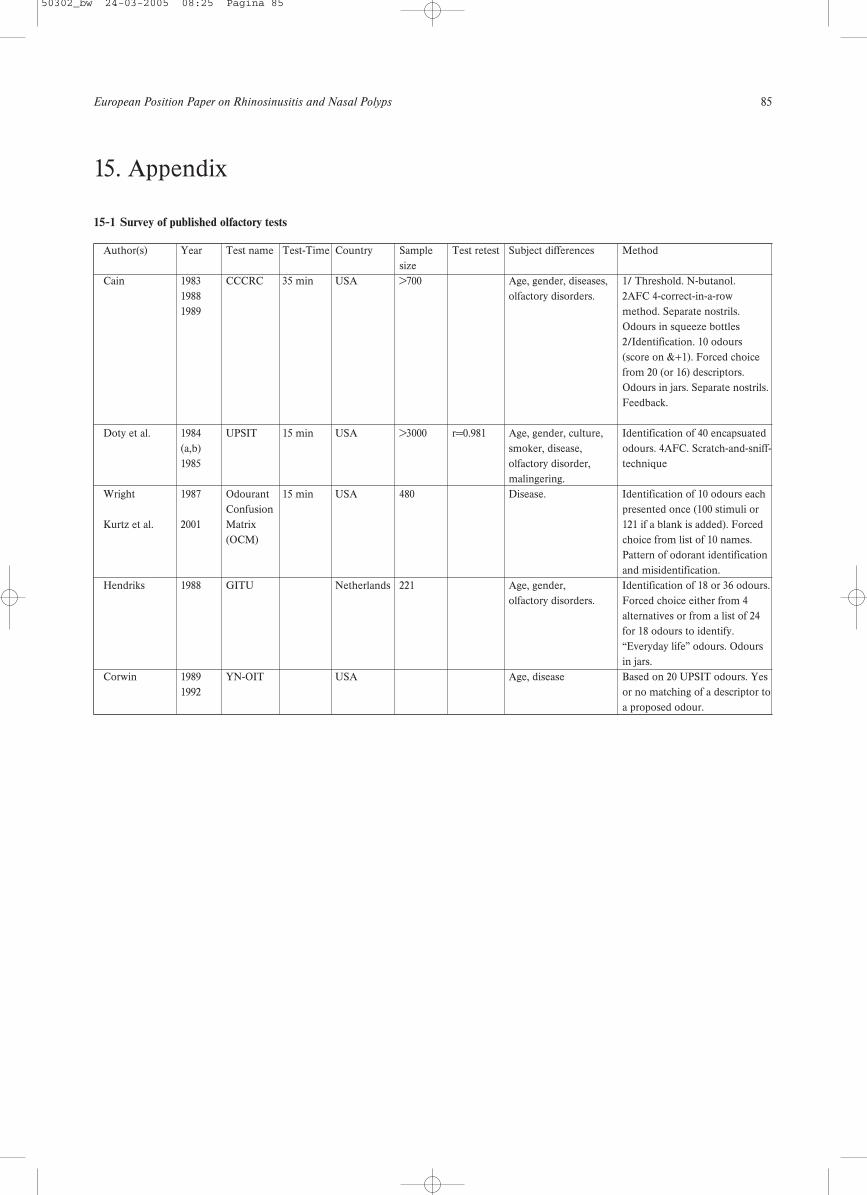
European Position Paper on Rhinosinusitis and Nasal Polyps 85
15-1 Survey of published olfactory tests
15. Appendix
Author(s) Year Test name Test-Time Country Sample Test retest Subject differences Methodsize
Cain 1983 CCCRC 35 min USA >700 Age, gender, diseases, 1/ Threshold. N-butanol.1988 olfactory disorders. 2AFC 4-correct-in-a-row 1989 method. Separate nostrils.
Odours in squeeze bottles2/Identification. 10 odours (score on &+1). Forced choice from 20 (or 16) descriptors. Odours in jars. Separate nostrils. Feedback.
Doty et al. 1984 UPSIT 15 min USA >3000 r=0.981 Age, gender, culture, Identification of 40 encapsuated (a,b) smoker, disease, odours. 4AFC. Scratch-and-sniff- 1985 olfactory disorder, technique
malingering.Wright 1987 Odourant 15 min USA 480 Disease. Identification of 10 odours each
Confusion presented once (100 stimuli or Kurtz et al. 2001 Matrix 121 if a blank is added). Forced
(OCM) choice from list of 10 names. Pattern of odorant identification and misidentification.
Hendriks 1988 GITU Netherlands 221 Age, gender, Identification of 18 or 36 odours.olfactory disorders. Forced choice either from 4
alternatives or from a list of 24 for 18 odours to identify.“Everyday life” odours. Odours in jars.
Corwin 1989 YN-OIT USA Age, disease Based on 20 UPSIT odours. Yes1992 or no matching of a descriptor to
a proposed odour.
50302_bw 24-03-2005 08:25 Pagina 85
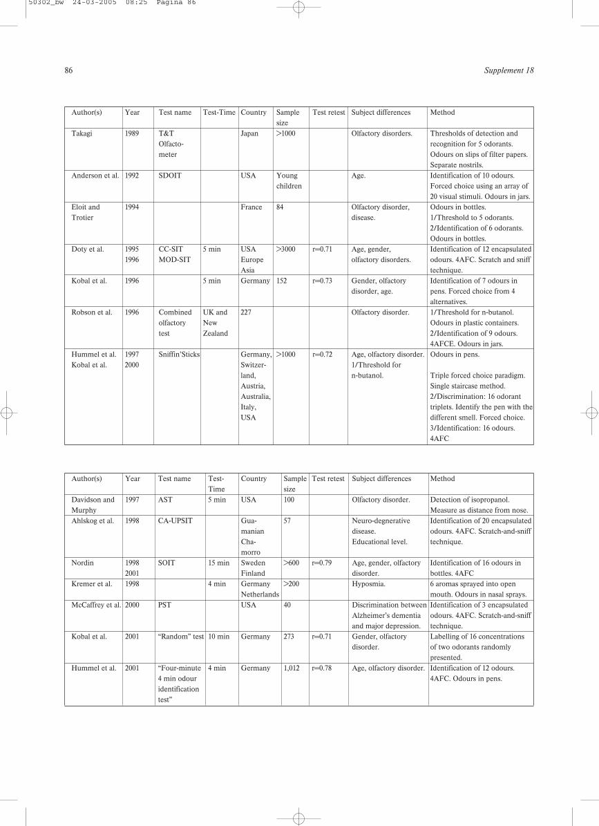
86 Supplement 18
Author(s) Year Test name Test-Time Country Sample Test retest Subject differences Methodsize
Takagi 1989 T&T Japan >1000 Olfactory disorders. Thresholds of detection and Olfacto- recognition for 5 odorants. meter Odours on slips of filter papers.
Separate nostrils.Anderson et al. 1992 SDOIT USA Young Age. Identification of 10 odours.
children Forced choice using an array of20 visual stimuli. Odours in jars.
Eloit and 1994 France 84 Olfactory disorder, Odours in bottles.Trotier disease. 1/Threshold to 5 odorants.
2/Identification of 6 odorants.Odours in bottles.
Doty et al. 1995 CC-SIT 5 min USA >3000 r=0.71 Age, gender, Identification of 12 encapsulated 1996 MOD-SIT Europe olfactory disorders. odours. 4AFC. Scratch and sniff
Asia technique.Kobal et al. 1996 5 min Germany 152 r=0.73 Gender, olfactory Identification of 7 odours in
disorder, age. pens. Forced choice from 4 alternatives.
Robson et al. 1996 Combined UK and 227 Olfactory disorder. 1/Threshold for n-butanol. olfactory New Odours in plastic containers.test Zealand 2/Identification of 9 odours.
4AFCE. Odours in jars.Hummel et al. 1997 Sniffin’Sticks Germany, >1000 r=0.72 Age, olfactory disorder. Odours in pens.Kobal et al. 2000 Switzer- 1/Threshold for
land, n-butanol. Triple forced choice paradigm. Austria, Single staircase method.Australia, 2/Discrimination: 16 odorant Italy, triplets. Identify the pen with the USA different smell. Forced choice.
3/Identification: 16 odours. 4AFC
Author(s) Year Test name Test- Country Sample Test retest Subject differences MethodTime size
Davidson and 1997 AST 5 min USA 100 Olfactory disorder. Detection of isopropanol. Murphy Measure as distance from nose.Ahlskog et al. 1998 CA-UPSIT Gua- 57 Neuro-degnerative Identification of 20 encapsulated
manian disease. odours. 4AFC. Scratch-and-sniff Cha- Educational level. technique.morro
Nordin 1998 SOIT 15 min Sweden >600 r=0.79 Age, gender, olfactory Identification of 16 odours in 2001 Finland disorder. bottles. 4AFC
Kremer et al. 1998 4 min Germany >200 Hyposmia. 6 aromas sprayed into open Netherlands mouth. Odours in nasal sprays.
McCaffrey et al. 2000 PST USA 40 Discrimination between Identification of 3 encapsulated Alzheimer’s dementia odours. 4AFC. Scratch-and-sniffand major depression. technique.
Kobal et al. 2001 “Random” test 10 min Germany 273 r=0.71 Gender, olfactory Labelling of 16 concentrations disorder. of two odorants randomly
presented.Hummel et al. 2001 “Four-minute 4 min Germany 1,012 r=0.78 Age, olfactory disorder. Identification of 12 odours.
4 min odour 4AFC. Odours in pens.identification test”
50302_bw 24-03-2005 08:25 Pagina 86
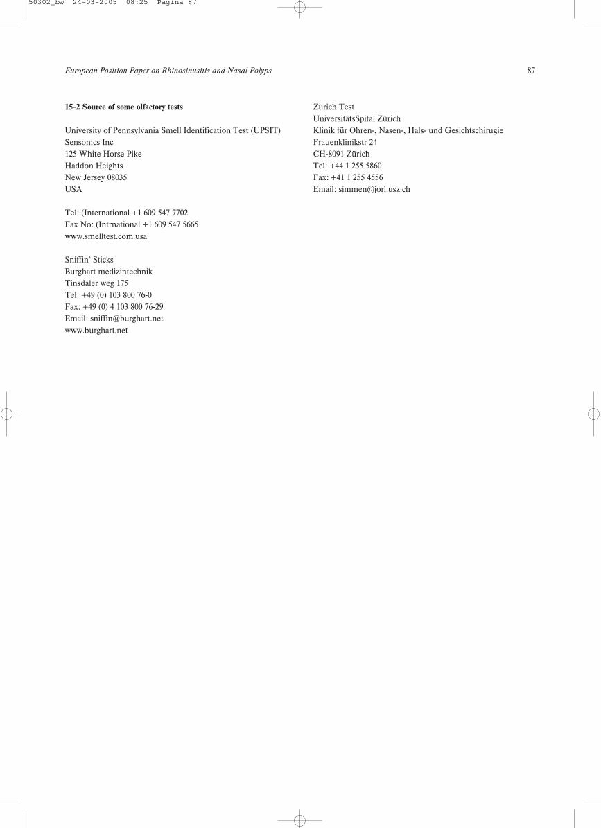
European Position Paper on Rhinosinusitis and Nasal Polyps 87
15-2 Source of some olfactory tests
University of Pennsylvania Smell Identification Test (UPSIT)Sensonics Inc125 White Horse PikeHaddon HeightsNew Jersey 08035USA
Tel: (International +1 609 547 7702Fax No: (Intrnational +1 609 547 5665www.smelltest.com.usa
Sniffin’ SticksBurghart medizintechnikTinsdaler weg 175Tel: +49 (0) 103 800 76-0Fax: +49 (0) 4 103 800 76-29Email: [email protected]
Zurich TestUniversitätsSpital ZürichKlinik für Ohren-, Nasen-, Hals- und GesichtschirugieFrauenklinikstr 24CH-8091 ZürichTel: +44 1 255 5860Fax: +41 1 255 4556Email: [email protected]
50302_bw 24-03-2005 08:25 Pagina 87

88 Supplement 18
50302_bw 24-03-2005 08:25 Pagina 88