Embed Size (px)
Citation preview
CASE REPORT
Right Hepatectomy in Icteric Type HepatocellularCarcinoma—Report of a Case and Literature Review
Neel Shetty & Jayanth Reddy & G. Srikanth
Received: 13 November 2013 /Accepted: 15 January 2014# Indian Association of Surgical Oncology 2014
Introduction
Obstructive jaundice caused by tumor invasion in Hepatocel-lular carcinoma (HCC) in cirrhotics was first reported in 1947by Mallory [1]. In 1975 Lin classified these as ‘Icteric typehepatoma’ [2]. We herein report one such patient whounderwent a major liver resection.
Case Presentation
A 54 year old male patient presented with weight loss of twomonths duration and jaundice of one week duration. He hadno prior history of gastrointestinal bleed, ascites or hepaticencephalopathy. He is a diabetic on oral hypoglycaemicagents.
His laboratory tests showed a total bilirubin of 3.5 mg/dl,serum albumin 3.6 gm/dl, alkaline phosphatase 208 IU/ml,GGT 480 IU/ml and INR of 1.1.
An ultrasound of the abdomen revealed coarse liver echotexture and a tumor in the right lobe (segment 5) of liver withmild splenomegaly and no ascites.
Endoscopy showed grade II varices, Alpha feto protein was5.2 ng/ml and his markers for hepatitis B and Cwere negative.
Triphasic multidetector computerised tomography(MDCT) of the abdomen showed chronic liver disease, rightlobe atrophy, a tumor in Segments 5, 6 and 8 of liver withenhancement pattern suggestive of HCC and no portal venousor hepatic venous involvement (Fig. 1).
CT volumetry showed left lobe with inclusion of middlehepatic vein to be 55 %.
Magnetic resonance cholangiopancreatography (MRCP)revealed intrabiliary extension of the tumor into right hepaticduct with associated intrahepatic biliary dilatation.
Although by Child-Pugh score he was in category B, thiswas due to high score associated with elevated value ofbilirubin, which in his case was presumably secondary tobiliary obstruction and not primary liver dysfunction.
He subsequently underwent a right hepatectomy. Per oper-atively there was a cirrhotic liver with atrophic right lobe andhypertrophic segment IV and left lateral segment. The Rightduct and common hepatic duct was filled with tumor. On cutsection the tumor in the liver showed a contiguous extensioninto the right and common hepatic ducts.
Histopathology showed a moderately differentiating hepato-cellular carcinoma infiltrating the right bile duct with intrabiliaryextension of tumor and negative margins on the left hepatic ductand the distal bile duct. The adjacent liver showed cirrhosis.
He made an uneventful recovery and he remains asymp-tomatic at 33 months follow-up with normal AFP values andno recurrence on CT imaging.
Discussion
Obstructive jaundice in cirrhosis caused by tumour invasionwas first reported by Mallory et al. in 1947 [1]. It was laterclassified by Lin et al. in 1975 as ‘Icteric type hepatoma’ [2].Bile duct invasion by HCC occurs in 0.8–12.9 % [3–5].Patients with cirrhosis and icteric type HCC have a poorerprognosis when compared to HCC patients without bile ductinvasion.
Lau et al. classified icteric type of HCC into the extrahe-patic and the intrahepatic types [6]. Patients with extrahepaticbiliary involvement secondary to HCC have a higher curativeresection rate, and improved survival rate compared withthose with intrahepatic biliary obstruction.
N. Shetty : J. Reddy :G. Srikanth (*)Department of Surgical Gastroenterology MIS and Bariatric Surgery,BGS Global Hospital, Bangalore 560060, Karnataka, Indiae-mail: [email protected]
Indian J Surg OncolDOI 10.1007/s13193-014-0287-6
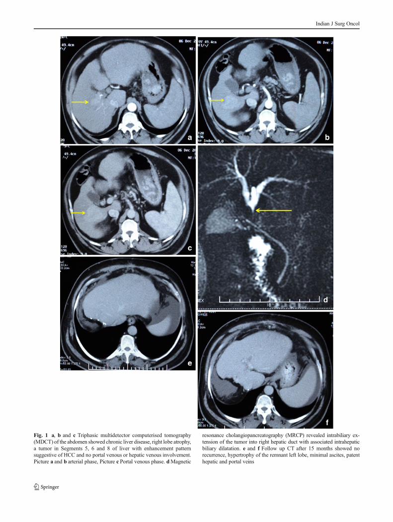
Fig. 1 a, b and c Triphasic multidetector computerised tomography(MDCT) of the abdomen showed chronic liver disease, right lobe atrophy,a tumor in Segments 5, 6 and 8 of liver with enhancement patternsuggestive of HCC and no portal venous or hepatic venous involvement.Picture a and b arterial phase, Picture c Portal venous phase. dMagnetic
resonance cholangiopancreatography (MRCP) revealed intrabiliary ex-tension of the tumor into right hepatic duct with associated intrahepaticbiliary dilatation. e and f Follow up CT after 15 months showed norecurrence, hypertrophy of the remnant left lobe, minimal ascites, patenthepatic and portal veins
Indian J Surg Oncol

Patients with cirrhosis and jaundice in HCC can beclassified into two types: hepatocellular type and icterictype. The hepatocellular type is secondary to extensiveparenchymal infiltration by tumour, which then leads tohepatic parenchymal insufficiency, accounting for 90 %of patients.
Imaging of the biliary tract is important to establishthe cause of jaundice in cirrhotics with HCC. Ultrasound(USG) may show a hypervascular biliary tumor thrombuswhich can be differentiated from portal vein thrombosis.MRCP may show the presence of enhancing intraluminalsoft tissue in the bile duct in the arterial phase which aretypical features of HCC with biliary tumour thrombi.Endoscopic Retrograde Cholangiopancreatogram (ERCP)or Percutaneous Transhepatic Cholangiography (PTC)can further delineate the level of block and afford theability to drain obstructed segments while stabilising thepatient [7].
Management consists of stabilisation of the patient, drain-age of obstructed segments, control of hemobilia and assess-ment for resectability. In the presence of high bilirubin orcholangitis biliary drainage should be preferentially per-formed intero-externally.
Curative resection includes complete extirpation of thetumor and in case of involvement of the extrahepatic biliarysystem the involved ducts are resected along with the tumorand biliary enteric anastomosis is performed. A controversialissue is whether tumor thrombi in the extrahepatic system /contralateral ducts be removed via a choledochotomy versusen bloc resection. The risk being tumor seedling at thecholedochotomy site / peritoneal seedling [8].
Summary of Literature
The prognosis for cirrhotics with HCC and bile duct invasionis poorer when compared to those without invasion withmedian survival of 11.4 versus 56.1 months [3–5, 9, 11].Curative resection in those with bile duct invasion have bettersurvival than those who without resection ( 25.3 vs2.1 months) [5].The incidence of portal vein invasion and bileduct invasion was higher, however incidence hepatic veininvasion was similar to regular HCC [10, 13]. Pre-operativedetection rates of 53–74 % were achieved [12]. Bilirubin,Alkaline Phosphatase and CA 19–9 levels were higher amongicteric type HCC than non icteric type [10]. Gross confluentmultinodular type (40 %) and infiltrative type were predom-inant histological patterns seen [10]. Intrahepatic recurrenceswere more common and early intrahepatic recurrences (within1 year) were significantly higher in those with bile ductinvasion [11].
Surgical resection with chemotherapy has been reported tobe beneficial in advanced HCC with intra-biliary tumorthrombus, however there is no confirmatory evidence [14].
Conclusions
With the availability of better diagnostic tools, strategies todeal with obstructive jaundice in cirrhotics with HCC andaggressive hepatic resection when feasible occasional longterm survival is possible as in the case reported by us.
Conflict of Interest There are no disclosures, disclaimers and no sup-port was received for this article.
Ethical Standards and Patient Consent The case report was ap-proved by the Hospital ethics committee and has been performed inaccordance with the ethical standards laid down in the 1964 Declarationof Helsinki and its later amendments.
The patient has given informed consent prior to his inclusion in the report.
References
1. Mallory TB, Castleman B, Parris EE (1947) Case records of theMassachusetts General Hospital case. N Engl J Med 237:673–676
2. Lin TY, Chen KM et al (1975) Icteric type hepatoma. Med Chir Dig4:267–270
3. Satoh S, Ikai I et al (2000) Clinicopathologic evaluation of hepato-cellular carcinoma with bile duct thrombi. Surgery 128:779–783
4. Yeh CN, Jan YY et al (2004) Hepatic resection for hepatocellularcarcinoma with obstructive jaundice due to biliary tumor thrombi.World J Surg 28:471–475
5. Lau W, Leung K et al (1997) A logical approach to hepatocellularcarcinoma presenting with jaundice. Ann Surg 225:281–285
6. Lau WY, Leung KL, Leung TW et al (1995) Obstructive jaundicesecondary to hepatocellular carcinoma. Surg Oncol 4:303–308
7. Chen MF (2002) Icteric type hepatocellular carcinoma: clinical fea-tures, diagnosis and treatment. Chang Gung Med J 25(8):496–501
8. Mok KT, Chang HT et al (1996) Surgical treatment of hepatocellularcarcinoma with biliary tumour thrombi. Int Surg 81:284–288
9. Shiomi M, Kamiya J, Nagino M et al (2001) Hepatocellular carcino-ma with biliary tumour thrombi: aggressive operative approach afterappropriate preoperative management. Surgery 129:692–698
10. Kojiro M, Kawabata K et al (1982) Hepatocellular carcinoma pre-senting as intrabile duct tumour growth. A clinicopathologic study of24 cases. Cancer 49:2144–2147
11. Shao W, Sui C et al (2011) Surgical outcome of hepatocellular carci-noma patients with biliary tumor thrombi. World J Surg Oncol 9:2
12. Ikenaga N, Kazuo C et al (2009) Clinicopathologic characteristics ofhepatocellular carcinoma with bile duct invasion. J Gastrointest Surg13:492–497
13. Huang JF, Wang LY, Lin ZY et al (2002) Incidence and clinicaloutcome of icteric type hepatocellular carcinoma. J GastroenterolHepatol 17:190–195
14. Fukuda S, Okuda K et al (2002) Surgical resection combined withchemotherapy for advancedhepatocellular carcinoma with tumorthrombus: report of 19 cases. Surgery 131:300–310
Indian J Surg Oncol