Embed Size (px)
Citation preview
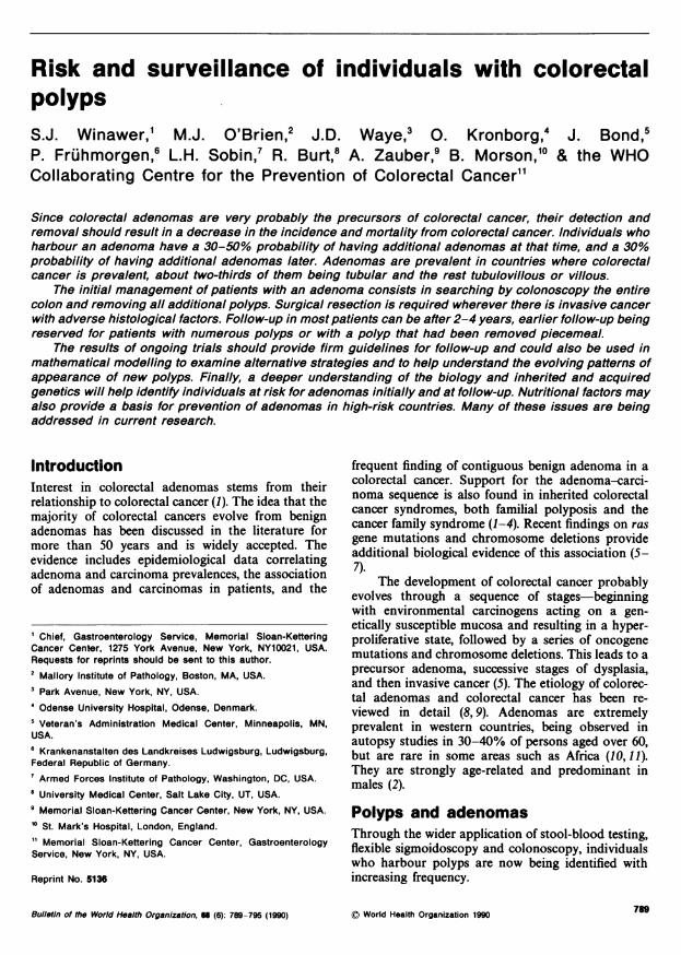
Risk and surveillance of individuals with colorectalpolypsS.J. Winawer,' M.J. O'Brien,2 J.D. Waye,3 0. Kronborg,4 J. Bond,5P. Fruhmorgen,6 L.H. Sobin,7 R. Burt,8 A. Zauber,9 B. Morson,10 & the WHOCollaborating Centre for the Prevention of Colorectal Cancer"1
Since colorectal adenomas are very probably the precursors of colorectal cancer, their detection andremoval should result in a decrease in the incidence and mortality from colorectal cancer. Individuals whoharbour an adenoma have a 30-50% probability of having additional adenomas at that time, and a 30%probability of having additional adenomas later. Adenomas are prevalent in countries where colorectalcancer is prevalent, about two-thirds of them being tubular and the rest tubulovillous or villous.
The initial management of patients with an adenoma consists in searching by colonoscopy the entirecolon and removing all additional polyps. Surgical resection is required wherever there is invasive cancerwith adverse histological factors. Follow-up in most patients can be after 2-4 years, earlier follow-up beingreserved for patients with numerous polyps or with a polyp that had been removed piecemeal.
The results of ongoing trials should provide firm guidelines for follow-up and could also be used inmathematical modelling to examine alternative strategies and to help understand the evolving pafferns ofappearance of new polyps. Finally, a deeper understanding of the biology and inherited and acquiredgenetics will help identify individuals at risk for adenomas initially and at follow-up. Nutritional factors mayalso provide a basis for prevention of adenomas in high-risk countries. Many of these issues are beingaddressed in current research.
IntroductionInterest in colorectal adenomas stems from theirrelationship to colorectal cancer (1). The idea that themajority of colorectal cancers evolve from benignadenomas has been discussed in the literature formore than 50 years and is widely accepted. Theevidence includes epidemiological data correlatingadenoma and carcinoma prevalences, the associationof adenomas and carcinomas in patients, and the
I Chief, Gastroenterology Service, Memorial Sloan-KetteringCancer Center, 1275 York Avenue, New York, NY10021, USA.Requests for reprints should be sent to this author.2 Mallory Institute of Pathology, Boston, MA, USA.
3 Park Avenue, New York, NY, USA.4 Odense University Hospital, Odense, Denmark.5 Veteran's Administration Medical Center, Minneapolis, MN,USA.6 Krankenanstalten des Landkreises Ludwigsburg, Ludwigsburg,Federal Republic of Germany.' Armed Forces Institute of Pathology, Washington, DC, USA.8 University Medical Center, Salt Lake City, UT, USA.9 Memorial Sloan-Kettering Cancer Center, New York, NY, USA.
'° St. Mark's Hospital, London, England.11 Memorial Sloan-Kettering Cancer Center, GastroenterologyService, New York, NY, USA.
Reprint No. 5136
frequent finding of contiguous benign adenoma in acolorectal cancer. Support for the adenoma-carci-noma sequence is also found in inherited colorectalcancer syndromes, both familial polyposis and thecancer family syndrome (1-4). Recent findings on rasgene mutations and chromosome deletions provideadditional biological evidence of this association (5-7).
The development of colorectal cancer probablyevolves through a sequence of stages-beginningwith environmental carcinogens acting on a gen-etically susceptible mucosa and resulting in a hyper-proliferative state, followed by a series of oncogenemutations and chromosome deletions. This leads to aprecursor adenoma, successive stages of dysplasia,and then invasive cancer (5). The etiology of colorec-tal adenomas and colorectal cancer has been re-viewed in detail (8,9). Adenomas are extremelyprevalent in western countries, being observed inautopsy studies in 30-40% of persons aged over 60,but are rare in some areas such as Africa (10, 11).They are strongly age-related and predominant inmales (2).
Polyps and adenomasThrough the wider application of stool-blood testing,flexible sigmoidoscopy and colonoscopy, individualswho harbour polyps are now being identified withincreasing frequency.
Bulletin of the World Health Organization, 66 (6): 789-795 (1990) © World Health Organization 1990
S.J. Winawer et al.
Polyps are growths into the lumen of the boweland may be classified as neoplastic or non-neoplastic(10). Non-neoplastic polyps have no malignantpotential and include hyperplastic polyps, hamar-tomas, benign lymphoid polyps, inflammatorypolyps, and normal mucosa. Neoplastic polyps areadenomas and are potentially malignant. These areclassified into three types: tubular, tubulovillous, andvillous adenomas having varying degrees of villousfeatures.
* Tubular adenomas contain a normal laminapropria and straight or branched tubules of dysplas-tic epithelium.* Villous adenomas contain elongated non-branch-ing folia or crypts of dysplastic epithelium.
Only a portion of colonic polyps that are detec-ted are true adenomas. In the United States NationalPolyp Study, adenomas accounted for 68% of thepolyps removed by colonoscopy in the initial examin-ation (12). The remaining polyps were overgrowths ofnormal mucosa and other miscellaneous non-neo-plastic polyps (22%) as well as hyperplastic polyps(11%). Adenomas discovered in autopsy studies aresmall (<1 cm), mostly tubular, and uniformly dis-tributed throughout the colon (11, 12). In clinicalstudies based on symptomatic patients such as'the St.Marks Study and the National Polyp Study, theadenomas tend to be larger, have a more variedhistology, and 2 out of 3 are distal to the splenicflexure (3,12). All adenomas have at least mild dys-plasia, by definition, and a proportion have moderateor severe dysplasia, carcinoma-in-situ, or invasivecancer (3,12). The terms low-grade and high-gradedysplasia are used by some investigators instead ofmoderate and severe dysplasia. Carcinoma-in-situhas no clinical significance and has been included insevere or high-grade dysplasia in order to avoidclinical overreaction to the diagnosis of carcinoma-in-situ in an adenoma (13).
The most common type of adenoma is tubular(68%), which has less premalignant potential thanthose with villous features. Villous features are morecommon with increasing size. Approximately 5% ofthe adenomas have high-grade dysplasia or carci-noma-in-situ, and 2.5% have invasive cancer at thetime of presentation. Carcinoma-in-situ and high-grade dysplasia are not influenced by sex but arerelated to age and also to multiplicity of the aden-oma. Individuals presenting with an adenoma have40-50% likelihood of having additional adenomas atthe same time (synchronous) (10, 12, 14).
Patients who have had an adenoma removedfrom their colon have an increased risk of developinga subsequent adenoma. Prior to fibreoptic colonos-copy, the reported rate of recurrence ranged from
20% to 50%. Although patients enrolled in theoriginal studies did not have an examination of theentire colonic mucosa to exclude synchronouslesions, investigations performed with colonoscopyhave confirmed the previous observations. In the pre-endoscopy era, Henry et al. (15) and Kirsner et al.(16) reported recurrent adenomas in 30-41% ofpatients over the 5-9 years of follow-up. Since theadvent of colonoscopy, Macrae & Williams followedup 330 patients after polypectomy for an average of3.6 years and found adenomas in 37% (17); Aubert etal. reported an identical incidence in a 10-yearfollow-up of 123 patients (18). Waye & Braunfeld (19)reported that 56% of 227 patients had adenomas attheir first annual colonoscopy following removal ofthe index adenomas. Fowler & Hedberg (20) reportedthat adenomas recurred in 60% of 383 polypectomypatients followed for four years. Matek and co-work-ers presented similar follow-up data and reviewedmany of the studies reported in the literature, all ofwhich had significant recurrence rates (21).
The National Polyp Study has also generateddata on adenomas after complete clearing of allsynchronous adenomas in a cohort that had not hadany prior intervention. In this population, it wasnoted that adenomas recurred at a rate of 29-35%,depending on the interval from their initial colonos-copy. Adenomas tended to be small, mostly tubular,with only mild dysplasia, and uniformly distributedthroughout the colon as compared to the distaldistribution of larger adenomas with varied histologyseen at initial diagnosis (12). This agrees with otherreports (17,19-22).
ManagementInitial managementThe management of patients with colorectal polypscan be divided into initial management and follow-up(10, 13). After detection, the index polyp should beremoved completely to eliminate all neoplastic tissueand the entire specimen should be submitted formicroscopic examination to detect the foci of malig-nancy and adequately classify the lesions histo-logically. Polypectomy of larger polyps can beaccomplished with the cautery snare, while smallsessile polyps may be biopsied and ablated with the"hot-biopsy" forceps. Pedunculated polyps and ses-sile polyps with a small attachment to the colon wallcan be removed completely with one application ofthe cautery snare.
If a sessile polyp with a wide-based attachmentis not completely removed at the initial polypectomy,additional endoscopy may be required to remove therest of the tumour. Endoscopic excision of a polypmay not be possible when it is located in an inacces-sible site, or if the polyp is larger than 2 cm in
WHO Bulletin OMS. Vol. 68, 1990.790
Risk and surveillance of individuals with colorectal polyps
diameter and sessile, especially with a broad area ofimplantation into the colonic wall. If complete endos-copic resection cannot be performed, surgical resec-tion may be required. This, however, is necessary inonly a very small proportion of cases. Since thefrequency of additional (synchronous) adenomas inpatients with a demonstrated adenoma at the time ofdiagnosis is approximately 40-50%, the initial man-agement of a patient with an identified adenomashould include total colonoscopy with removal of allpolyps. This policy may be modified, however, in thepresence of significant medical problems.
Classification of the removed polyp. After endoscopicresection, every effort must be made to retrieve theentire specimen for examination and classification. Itis important to record both clinical and anatomicalfeatures such as the number of polyps and their size,gross morphology (pedunculated or sessile), and theirlocations. An attempt should be made to identify thebase of the polyp. Contraction of the muscularismucosae may cause a specimen to curl into a ball,making subsequent identification of the resection siteextremely difficult. To avoid this, sessile polypsshould be placed flat on a piece of cardboard, thickpaper, Gelfoam, or a frosted glass slide before inser-tion into the fixative. Polyps that are pedunculated orhave a small site of attachment to the colon wall maybe marked with indian ink at the line of resection(10,11,14).
Histological classification of polyps is madeaccording to WHO criteria. Multiple histologicalsections are examined from stepwise slides of theentire polyp. In each adenoma, the degree of dys-plasia should be recorded as mild, moderate, andsevere or, alternatively, as low grade or high grade.The diagnosis of carcinoma-in-situ should beincluded in the category of severe or high-gradedysplasia for clinical reporting, the term "carcinoma-in-situ" being used for research studies only. Intra-mucosal or focal carcinoma are terms best not used,or used with caution, because of the same potentialfor clinical misinterpretation. Invasive cancer in anadenoma should be reported in terms of depth ofinvasion, involvement of stalk or cautery line, lym-phatic or vascular space involvement, degree of dif-ferentiation, and volume of adenoma replaced.Pseudo-invasion with adenoma misplaced into thestalk or submucosa should not be interpreted as trueinvasive carcinoma. The report should includewhether the excision appears to be complete(10,11,14).
Patients with carcinoma In an adenoma
The occurrence of carcinoma within an adenoma isnot unusual. Most cancers within adenomas are in
the category of high-grade dysplasia. Their presenceincreases with age and when there are multipleadenomas, as well as with increasing size and increas-ing villous histology. This finding has no clinicalsignificance (2,3,12,23). Invasive cancer has beenreported in less than 1% to more than 8% ofadenomas, but more often where there is villouschange and increasing size. Lymph-node metastasesand invasion of the bowel wall have been reported inup to 25% of the patients with invasive cancer,although most studies report the frequency of suchfindings as less than 10%. However, when the his-tology is favourable and there has been completeexcision of the polyp, the probability of havingresidual or metastatic cancer is considerably less than1%. This is equal to or less than the risk for surgerywith bowel resection in average-risk patients (24-31).The following guidelines can be used for patientswith invasive cancer within an adenoma. Adenomaswith severe or high-grade dysplasia or carcinoma-in-situ, i.e., histological features of carcinoma limited tothe mucosa and not penetrating the muscularismucosae, are not considered to have metastaticpotential. Surveillance of this group of patients, whohave had the adenoma completely resected, shouldfollow the protocol for other adenomas.
An adenoma is considered to have invasivecarcinoma when malignant cells have penetrated themuscularis mucosae. When invasive carcinomaoccurs in an adenoma, further clinical decisions arebased on the presence or absence of "favourablecriteria" which are: well-differentiated or moderatelywell-differentiated carcinoma, absence of malignantcells at the resection margin and absence of vascularor lymphatic invasion. If the adenoma with invasivecarcinoma is sessile, a surgical resection, with dissec-tion of regional lymph nodes, is recommended. Thismay be avoided if the cautery line is definitely clear.Patients with pedunculated adenomas with invasivecarcinoma should undergo similar surgery if thecancer extends to the line of cautery, the carcinoma ispoorly differentiated, or lymphatic or vascular in-vasion is demonstrated on histologic sectioning. Thealmost total replacement of an adenoma with in-vasive cancer may also require surgery.
There is considerable controversy about thenecessity for additional surgery following endoscopicremoval of a sessile adenoma with a small focus ofwell-differentiated or moderately differentiated car-cinoma that has invaded the muscularis mucosae, butdoes not involve vascular or lymphatic spaces or theline of resection. At present, there are insufficient dataon the metastatic potential of such lesions, and nogeneral guidelines can be given. However, there isgrowing conservative opinion for a non-operativeapproach to these patients. Thus, there is agreementthat a patient with invasive carcinoma in an adeno-
WHO Bulletin OMS. Vol. 68.1990. 791
S.J. Winawer et al.
ma who meets the usual criteria for surgical resectionmay be spared surgery when there are medicalproblems that make the patient a poor surgical risk.If the lesion is low in the rectum and surgery isindicated, a local deep excision is usually adequate.Abdominal perineal resection is generally not donefor malignant adenomas (10,24,31).
There has been interest in recent years in certaintypes of polyps such as the small polyps and hyper-plastic polyps. Small polyps are so classified whenthey are approximately 5-6 mm in sizeor less. In recent years, studies by Waye (32) andTedesco (33) have demonstrated that these areadenomas in about 60% of the cases. The NationalPolyp Study has demonstrated that small polyps thatare adenomas have all the features of the largeradenomas but to a lesser degree quantitatively.Hyperplastic polyps have no malignant potential butseem to arise in the colon of individuals who harbourtrue adenomas. These polyps are primarily located inthe rectosigmoid, but whether their presence in therectosigmoid implies the presence of adenomas moreproximally is as yet unsettled (34). The data indicat-ing the predictive value of the hyperplastic polyp formore proximal adenomas are based on small samplesizes and without true controls; larger studies withgood controls have not confirmed this finding (35).
Recommendations and guidelinesFollow-up recommendationsColonoscopy is the preferred method of follow-upexamination after removal of an initial adenoma.Sigmoidoscopy with a high-quality double-contrastbarium enema, however, is a possible acceptablealternative in the absence of good colonoscopy.Annual fecal occult-blood tests have been used in thefollow-up period when the surveillance intervals werelonger than one year (3), but this is of questionablevalue.
The objective of a surveillance programme is toprevent the development of colorectal cancer. Therecurrence rate of adenomas in patients after initialpolypectomy is high enough to justify periodicfollow-up. Ideally, all synchronous adenomas areremoved at the time of the initial polypectomy. Thefrequency of missed synchronous lesions, however,has been suggested to be 5-10%. A proper sur-veillance scheme should, therefore, offer the oppor-tunity of finding these missed lesions and new meta-chronous adenomas, but must be designed to protectthe patient from the risk and cost of unnecessaryexaminations and an overloading of medical re-sources. Several studies have been investigatingfollow-up strategies in these patients.
The endoscopist must be confident that a "clean
colon", free of adenomas, should be establishedbefore instituting long-term follow-up. Frequently,repeated examinations may be indicated after in-complete or piecemeal removal of some large orsessile lesions, for patients with numerous polyps,or after a technically unsatisfactory examination.Following apparently complete removal of a pedun-culated malignant polyp, judged on combined endos-copic and histological grounds, most endoscopistsperform repeat examination at 3-6 months and 1year before reverting to general follow-up.
Data on which to base general follow-upintervals are incomplete except that six-monthlyexaminations are too frequent. Current informationsuggests that after establishing a clean colon, therecan usually be an interval of 1-3 years before repeatexamination. Some centres present predictiveevidence of an increased risk in patients with multipleadenomas and recommend a follow-up examinationevery two years in those with two or more adenomasbut every four years in those with a single adenoma(21). The likelihood of prolonging life expectancy bycontinued colonic surveillance becomes small in oldage, but individual considerations such as ill-healthor predictive factors (such as very numerous polyps)will affect the age at which follow-up is discontinued,usually around 75-80 years (13).
Finally, the approach to patients with adenomaswill change dramatically over the next few years aswe begin better to understand the biology of theadenomas. Progress in inherited genetics may pro-vide a basis for identifying those individuals whoharbour adenomas with significant pathology andwhose adenomas are likely to progress and recur.Oncogene and surface antigen expression as well asother characteristics of adenomas will assume a moreimportant role as a basis for the management of thesehigh-risk patients (5,36-38).
Research recommendations(1) There is a need for demonstration of a reduction
in incidence and mortality from colorectal can-cer by periodic intervention using colonoscopyto remove adenomas.
(2) The most cost-effective intervals for follow-upsurveillance in post-polypectomy patients needto be demonstrated by completion of currentongoing trials.
(3) The relative value of colonoscopy, bariumenema, and stool-blood testing needs to be com-pared in the follow-up of patients after poly-pectomy.
(4) There should be improved methods for iden-tification of individuals harbouring adenomas,or at risk for developing adenomas.
WHO Bulletin OMS. Vol. 68.1990.792
Risk and surveillance of individuals with colorectal polyps
(5) Predictive factors for recurrent adenomas (bio-chemical, biological, pathological, and familyhistory) would be of importance in separatingrisk groups of individuals for varying follow-upstrategies after polyps have been removed.
(6) A standardized nomenclature for reporting ofclinical, endoscopic and pathological studiesrelated to polyps should be developed.
(7) Studies need to be done on the malignant polyp,to ascertain the need for surgical resection follow-ing polypectomy.
(8) Dietary assessment and family history should beobtained in well characterized cohorts ofpatients with polyps in order to understandtheir interrelationships in terms of etiology.
(9) The patient with polyps should be used moreextensively to study the biology of carcinoma ofthe colon. Blood and tissue phenotypic abnor-malities should be clearly examined in thesepatients and correlated with the occurrence,progression and recurrence of disease.
(10) Patients with polyps should be used more exten-sively to study effects of nutritional intervention.Parallel studies with phenotypic markers shouldbe done and familial factors should be con-trolled for.
(11) The benefit of polypectomy and follow-upsurveillance should be correlated with age, path-ology, family history and diet.
(12) The use of mathematical modelling based ondata from polypectomy patient groups may helpto answer many of the currently unsolved ques-tions.
(13) Adenoma tissue should be used for study ofchromosome, oncogene and other cellular abnor-malities in order to help elucidate the geneticbasis of "sporadic" adenomas and hence"sporadic" colorectal cancer.
Practical guidelines(1) When a polyp has been identified it should be
removed for histological examination. Smallpolyps found in the rectosigmoid on flexiblesigmoidoscopy can be biopsied.
(2) Histology of the polyp should be assessed toclassify the polyp according to the criteria of theWorld Health Organization.
(3) Patients with an adenoma should have the entirecolon examined for additional polyps by colonos-copy and all polyps should be removed andstudied histologically. If colonoscopy is unavail-able, flexible sigmoidoscopy and double-contrastbarium enema can be performed as an alter-native.
(4) Family history should be obtained in all patients
with adenomas to determine if screening of thefamily is indicated.
(5) Adenomas with severe (high-grade) dysplasianeed no additional surgery. The term carcinoma-in-situ should be dropped for routine clinical usesince it can be misleading. Adenomas that aresessile with invasive cancer to the cautery lineusually need surgical resection. Adenomas thatare pedunculated with cancer that is poorly dif-ferentiated, involves lymphatic or vascularspaces, or extends to the cautery line may requiresurgery, but each case must be judged indi-vidually.
(6) Rectal malignant adenomas requiring surgerycan often be managed by local excision.
(7) All patients with adenomas should have completeexcision initially.
(8) All patients with adenomas need a follow-upprogramme. In most patients, this can be withcolonoscopy every 1-3 years. Some patients willrequire alternative individual follow-up, e.g.,those with invasive cancer, large sessile adeno-mas, or a large number of adenomas.
ResumeLes polypes recto-coliques: risque etsurveillanceL'interet porte aux polypes recto-coliques vient dece qu'ils prec6dent souvent un cancer du colon etdu rectum. Ces polyad4nomes sont extrdmementfrequents dans les pays oiu ce type de cancer estcourant. L'evolution de ces cancers se fait pro-bablement par une serie d'etapes, commen9antpar I'action d'agents de l'environnement sur unemuqueuse genetiquement sensible au stade hy-perprolif&eratif et passant par un stade adeno-mateux avant d'atteindre le stade du cancer in-vasif. C'est un processus lent qui prend enmoyenne au moins 10 ans.
Les polyadenomes sont constitues pour en-viron deux tiers de polypes recto-coliques, le resteetant principalement forme d'excroissancesmuqueuses normales et de polypes hyper-plasiques. Ces derniers n'ont aucun potentiel neo-plasique et ne sont pas consideres comme can-cereux. On classe ces polyadenomes en formestubulaires, tubulo-villeuses, ou villeuses, selonl'importance des vegetations. Tous les poly-adenomes presentent au moins une legere dys-plasie et certains d'entre eux (5%) une forte dys-plasie. Ces derniers comprennent les cancers in-situ, terme qu'il faut eviter d'employer en cliniquecar sans aucune signification pour le malade. Les
WHO Bulletin OMS. Vol. 68. 1990. 793
S.J. Winawer et al.
polyadenomes avec cancer invasif (3%) sont ap-peles polyadenomes malins.
La probabilite que le malade chez qui l'ondecele un polyadenome en presente plusieursautres au meme moment (synchrones) est de 30 a50% et la probabilite pour que l'on en decouvred'autres (metachrones) a la colonoscopie estd'environ 30%. On n'a pas encore defini la propor-tion respective des polyadenomes metachronesrecents et de ceux passes inaper,us.
La prise en charge des sujets presentant despolypes se fait en deux temps: prise en chargeinitiale et suivi. Dans une premiere etape, ond6barrassera le colon de tous les autres polypestrouves. Plusieurs colonoscopies peuvent etrenecessaires pour eliminer un important polypesessile, verifier que l'exerese est complete ets'assurer de I'absence de toute recurrence. L'exa-men histologique apres orientation, fixation etcoupe convenable est indispensable. Les indica-tions de la resection chirurgicale apres ablationd'un polype pr6sentant une degenerescence ma-ligne sont les suivantes: cancer invasif dans unpolyadenome sessile ou extension a la based'implantation, accompagnee d'une faible differen-ciation cellulaire et de 1'extension aux espaceslymphatiques ou vasculaires dans un polype pedi-cule. Si l'exerese chirurgicale est indiquee pour unpolype rectal, 1'excision locale est generalementsuffisante.
Une fois le colon debarrasse de tous les poly-pes, la colonoscopie est la meilleure methode desuivi. Si l'on ne peut disposer d'une colonoscopiede bonne qualite, on pourra employer lasigmoidoscopie souple et le lavement baryte endouble contraste. L'objectif d'un programme desurveillance est d'eviter les deces par cancer ducolon. Des essais sont en cours pour evaluerl'intervalle optimal entre deux contr6les. A l'heureactuelle, on recommande des examens de controleprecoces apres exerese de nombreux polypes ouexerese d'un polype important en plusieurs fois.Chez la plupart des sujets, un examen 2 a 4 ansapres r6section de tous les polypes rectocoliquesest semble-t-il suffisant.
11 reste de nombreuses questions a resoudre,notamment celles de l'intervalle entre deux con-tr6les, de la necessite d'une resection chirur-gicale, de son cout/efficacite et de l'observancedes malades vis-a-vis des possibilites offertes. 11est probable que dans les quelques annees avenir, nous aurons une meilleure comprehensionde la biologie de la sequence polyadenome-adeno-carcinome, ce qui nous permettra d'identifier desla prise en charge initiale et au cours du suivi lessujets presentant le plus grand risque de canceri-
sation. Les progres dans le domaine de l'he'rditeseront particuli6rement importants a cet egard. Enoutre, la connaissance des interactions existantentre les facteurs nutritionnels et les facteursgenetiques pourrait nous fournir des methodesde prevention applicables des la prise en chargeinitiale des polyadenomes et lors du suivi. Onpense que la prevention des polypes et leur elimi-nation pr6coce devraient permettre de diminuerl'incidence et la mortalite liee au cancer du c6lon,mais cela reste a demontrer.
References1. Schottenfeld, D. & Winawer, S.J. Large intestine. In:
Schottenfeld, D. & Fraumeni, J. Jr., ed. Cancerepidemiology and prevention. Philadelphia, W.B.Saunders, 1982, pp. 703-727.
2. Gillespie, P.E. et al. Colonic adenomas: a colonos-copy survey. Gut, 20: 240-245 (1979).
3. Konlshi, F. & Morson, B.C. Pathology of colorectaladenomas: a colonoscopic survey. J. clin. pathol., 35:830-841 (1982).
4. Shinya, H. & Wolff, W.l. Morphology, anatomic dis-tribution and cancer potential of colonic polyps: ananalysis of 7000 polyps endoscopically removed.Ann. surg., 190: 679-683 (1979).
5. Vogelsteln, B. et al. Genetic alterations duringcolorectal tumor development. New England j.med.,319: 525-532 (1988).
6. Bodmer, W.F. et al. Localization of the gene forfamilial adenomatous polyposis on chromosome 5.Nature, 328: 614-616 (1987).
7. Solomon, E. et al. Chomosome 5 allele loss in humancolorectal carcinomas. Nature, 328: 616-619 (1987).
8. Shike, M. et al. Primary prevention of colorectalcancer. Bull. Wld Hlth Org., 68: 377-385 (1990).
9. Burt, R.W. et al. Risk and surveillance of individualswith heritable factors for colorectal cancer. Bull. WldHlth Org., 68: 655-665 (1990).
10. Lambert, R. et al. The management of patients withcolorectal adenomas. In: Holleb, A., ed. Third Inter-national Symposium on Colorectal Cancer. NewYork, American Cancer Society, 1984, pp. 43-52.
11. Morson, B.C. & Sobin, L.H. Histological typing ofintestinal tumours. In: International histological clas-sification of tumours, No. 15, Geneva, World HealthOrganization, 1976.
12. Winawer, S.J. et al. The national polyp study:overview of program and preliminary report ofpatient and polyp characteristics. In: Steele, G. et al.,ed. Basic and Clinical Perspectives of ColorectalPolyps and Cancer. Proceedings of a Meeting held inBoston, Massachuseffs, 20-21 November 1986. NewYork, Alan R. Liss, 1988, pp. 35-49.
13. Winawer, S.J. et al. Colorectal adenoma patients:risk of cancer and results of follow-up. ROMA 88Working Team Report - No. 11 (Summary). Rome,4-11 September 1988.
14. Morson, B.C. & Konoshi, F. Contribution of the path-ologist to the radiology and management of colorec-
794 WHO Bulletin OMS. Vol. 68.1990.
Risk and surveillance of Individuals with colorectal polyps
tal polyps. Gastrointest. radiol., 7: 275-281 (1982).15. Henry, L.G. et al. Risk of recurrence of colon polyps.
Ann. surg., 182: 511-515 (1975).16. Klrsner, J.B. et al. Polyps of the colon and rectum:
statistical analysis of a long-term follow-up study.Gastroenterology, 39: 178-182 (1960).
17. Macrae, F.A. & Williams, C.B. A prospective colonos-copic follow-up study of 500 adenoma patients withmultivariate analysis to predict risk of subsequentcolorectal tumors. Gastrointest. endosc., 28: 139(1982).
18. Aubert, H. et al. lnterdt de la surveillance desmalades polypectomises dans la prevention du can-cer rectocolique. A propos de 123 cas. Gastro-enterol. clin. biol., 6: 183-187 (1982).
19. Waye, J.D. & Braunfeld, S.F. Surveillance intervalsafter colonoscopic polypectomy. Endoscopy, 14: 79-81 (1982).
20. Fowler, D.L. & Hedberg, S.E. Follow-up colonoscopyafter polypectomy. Gastrointest. endosc., 26: 67(1980).
21. Matek, W. et al. Follow-up of patients with colorectaladenomas. Endoscopy, 17: 175-181 (1985).
22. Kronborg, 0. & Fenger, C. Prognostic evaluation ofplanned follow-up in patients with colorectal aden-omas. Int. j. colorect. dis., 2: 203-207 (1987).
23. O'Brien, M.J. et al. The National Polyp Study: patientand polyp characteristics with high-grade dysplasiain colorectal adenomas. Gastroenterology, 98: 371-379 (1990).
24. Wolf, W.I. & Shinya, H. Definitive treatment of'malignant" polyps of the colon. Ann. surg., 182:516-524 (1975).
25. Shatney, C.H. et al. Management of focally malignantpedunculated adenomatous colorectal polyps. Dis.col. & rect., 19: 334-340 (1976).
26. Couteeftides, T. et al. Colonoscopy and the manage-ment of polyps containing invasive carcinoma. Ann.surg., 188: 638-641 (1978).
27. Colacchlo, T.A. et al. Endoscopic polypectomy:inadequate treatment for invasive colorectal car-cinoma. Ann. surg., 194: 704-707 (1981).
28. Morson, B.C. et al. Histopathology and prognosis ofmalignant colorectal polyps treated by endoscopicpolypectomy. Gut, 25: 437-444 (1984).
29. Wilcox, G.M. & Beck, J.R. Early invasive cancer inadenomatous colonic polyps ("malignant polyps"):evaluation of the therapeutic options by decisionanalysis. Gastroenterology, 92: 1159-1168 (1987).
30. Haggitt, R.C. et al. Prognostic factors in colorectalcarcinomas arising in adenomas: implications forlesions removed by endoscopic polypectomy. Gas-troenterology, 89: 328-336 (1985).
31. Wlnawer, S.J. & Witt, T.R. Cancer in a colonic polyp,or malignant colonic adenomas: is polypectomy suf-ficient? Gastroenterology, 81: 625-626 (1981).
32. Waye, J.D. et al. Small colon polyps. Am. j. gastroen-terol., 83: 120-122 (1988).
33. Tedesco, F.J. et al. Diminutive polyps: histopatho-logy, spatial distribution and clinical significance.Gastrointest. endosc., 28: 1-5 (1982).
34. Achkar, E. & Carey, W. Small polyps found duringfiberoptic sigmoidoscopy in asymptomatic patients.Annals of internal medicine, 109: 880-883 (1988).
35. Wlnawer, S.J. et al. The national polyp study:colorectal adenomas and hyperplastic polyps. Gas-troenterology, 94: A499 (1988) (Abstract).
36. Kussin, S.Z. et al. State of the art-Inherited coloncancer: clinical implications. Am. j. gastroenterol.,72: 448-457 (1979).
37. Burt, R.W. et al. Dominant inheritance of adeno-matous colonic polyps and colorectal cancer. NewEngland j. med., 312: 1540-1544 (1985).
38. Cannon-Albright, L.A., et al. Common inheritance ofsusceptibility to colonic adenomatous polyps andassociated colorectal cancers. New England j. med.319: 533-537 (1988).
WHO Bulletin OMS. Vol. 68. 1990.