Embed Size (px)
Citation preview
NeuroMolecular Medicine 21 Volume 4, 2003
Role of Protein Aggregation in Mitochondrial Dysfunctionand Neurodegeneration in Alzheimer’sand Parkinson’s Diseases
Makoto Hashimoto,1 Edward Rockenstein,1 Leslie Crews,1and Eliezer Masliah*,1,2
Departments of 1Neurosciences and 2Pathology, University of California,San Diego, La Jolla, CA 92093-0624
Received July 15, 2003; Accepted July 18, 2003
Abstract
Abnormal interactions and misfolding of synaptic proteins in the nervous system are beingextensively explored as important pathogenic events resulting in neurodegeneration in vari-ous neurological disorders. These include Alzheimer’s disease (AD), Parkinson’s disease (PD),and dementia with Lewy bodies (DLB). In AD, misfolded amyloid β peptide 1–42 (Aβ), a pro-teolytic product of amyloid precursor protein metabolism, accumulates in the neuronal endo-plasmic reticulum and extracellularly as plaques. In contrast, in PD and DLB cases there isabnormal accumulation of α-synuclein in neuronal cell bodies, axons, and synapses. Further-more, in DLB, Aβ 1–42 may promote α-synuclein accumulation and neurodegeneration. Thecentral event leading to synaptic and neuronal loss in these diseases is not completely clear yet;however, recent advances in the field suggest that nerve damage might result from the con-version of nontoxic monomers to toxic oligomers and protofibrils. The mechanisms by whichmisfolded Aβ peptide and α-synuclein might lead to synapse loss are currently under investi-gation. Several lines of evidence support the possibility that Aβ peptide and α-synuclein mightinteract to cause mitochondrial and plasma membrane damage upon translocation of protofib-rils to the membranes. Accumulation of Aβ and α-synuclein oligomers in the mitochondrialmembrane might result in the release of cytochrome C with the subsequent activation of theapoptosis cascade. Conversely, the oxidative stress and mitochondrial dysfunction associatedwith AD and PD may also lead to increased membrane permeability and cytochrome C release,which promotes Aβ and α-synuclein oligomerization and neurodegeneration. Together, thesestudies suggest that the translocation of misfolded proteins to the mitochondrial membranemight play an important role in either triggering or perpetuating neurodegeneration. The insights
NeuroMolecular MedicineCopyright © 2003 Humana Press Inc.All rights of any nature whatsoever reserved.ISSN 1535-1084/03/04:21–35/$25.00
*Author to whom all correspondence and reprint requests should be addressed. E-mail: [email protected]
22 Hashimoto et al.
NeuroMolecular Medicine Volume 4, 2003
obtained from the characterization of this process may be applied to the role of mitochondrialdysfunction in other neurodegenerative disorders, including AD. New evidence may also pro-vide a rationale for the mitochondrial membrane as a target for therapy in a variety of neu-rodegenerative diseases.
Index Entries:Alzheimer’s disease; dementia with Lewy bodies; α-synuclein; synapse damage;mitochondrial dysfunction; protein misfolding; aggregation; oligomers; plasma membrane.
Introduction
Extensive research is currently underway toexplore the possibility that progressive accumula-tion and misfolding (toxic conversion) of synapticproteins in the nervous system could be a criticalpathogenic event leading to neurodegeneration inAlzheimer’s disease (AD), Parkinson’s disease (PD),dementia with Lewy bodies (DLB), and other neu-rological disorders (Koo et al., 1999; Ramassamy etal., 1999; Ferrigno and Silver, 2000).
In AD, misfolded amyloid β peptide 1–42 (Aβ),a proteolytic product of amyloid precursor protein(APP) metabolism, accumulates intracellularly inthe neuronal endoplasmic reticulum (ER) and extra-cellularly as plaques (Selkoe et al., 1996; Trojanowskiand Lee, 2000; Walsh et al., 2000). In PD and relatedconditions, such as DLB, abnormal accumulation ofα-synuclein occurs in neuronal cell bodies, axons,and synapses (Spillantini et al., 1997; Irizarry et al.,1998; Takeda et al., 1998a). In disorders caused bytrinucleotide repeats , such as Huntington’s disease(HD), the accumulation of misfolded proteins occursin the neuronal cytosol and nuclei (Cummings andZoghbi, 2000; Muchowski, 2002). The central path-ogenic event triggering synaptic loss and selectiveneuronal loss in these disorders is not yet completelyclear (Masliah, 2000, 2001). However, recentadvances in the field suggest that nerve damagemight result from the conversion of normally non-toxic monomers (and small oligomers) to toxicoligomers and protofibrils (Volles et al., 2001; Vollesand Lansbury, 2002). In familial forms of these dis-eases mutations accelerate protein aggregation(Conway et al., 1998), whereas in sporadic formsoxidative stress, toxins, and other yet-unknown fac-tors might play a role (Hashimoto et al., 1999b; Betar-bet et al., 2000; Giasson et al., 2000). Oligomerizationcould occur in several stages, including the forma-tion of protofibrils, nucleation (Wood et al., 1999),
and fibril formation (Hashimoto et al., 1998; Serpellet al., 2000). In contrast, it is still a matter of debatewhether larger polymers and fibers that often con-stitute the intracellular inclusions (e.g., Lewy bodiesand tangles) and extracellular lesions (e.g., plaques)might not be as toxic (Lansbury, 1999).
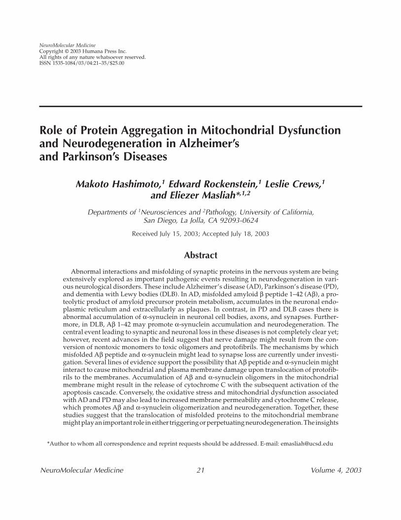
The mechanisms by which misfolded Aβ peptideand α-synuclein might lead to synapse loss and aber-rant sprouting are currently under investigation.Several possibilities are being considered, includ-ing mitochondrial damage, lysosomal leakage, frag-mentation of the Golgi apparatus, interference withsynaptic vesicle transport and interaction, and inter-ference with gene transcription and signaling(Fig. 1). Recent lines of work support the possibil-ity that misfolded Aβ peptide and α-synucleinmight interact to cause mitochondrial and plasmamembrane damage (Askanas et al., 1996; Hsu et al.,2000). Accumulation of Aβ and α-synuclein oligo-mers in the mitochondrial membrane might resultin the release of cytochrome C, with the subsequentactivation of the apoptosis cascade (Hashimotoet al., 1999a). Conversely, the oxidative stress andmitochondrial dysfunction associated with ADand PD might also promote increased membranepermeability and cytochrome C release, whichresults in Aβ and α-synuclein oligomerization andneurodegeneration.
Mitochondria are ubiquitous intracellularorganelles that are responsible for the productionof adenosine triphosphate (ATP) by aerobic metab-olism. In addition to ATP production, mitochondriaserve a variety of essential functions. These includeregulation of apoptosis, cellular calcium buffering,and provision of free radicals (Orth and Schapira,2001). Dysfunction of mitochondria can then leadto a number of deleterious consequences, such asimpaired calcium balance, oxidative stress, activa-tion of the mitochondrial permeability transitionpore, and secondary excitotoxicity (Shigenaga et al.,
Protein Aggregation in Mitochondrial Dysfunction, Neurodegeneration in AD and PD 23
NeuroMolecular Medicine Volume 4, 2003
1994). Thus, progressive impairment of mitochon-drial activity has been suggested to play a criticalrole in the pathogenesis of a wide range of neu-rodegenerative disorders, including PD, AD, andHuntington’s disease (Orth and Schapira, 2001).
In this context, the main objective of this work isto provide a perspective as to the role of Aβ andα-synuclein misfolding in mitochondrial pathologyand neurodegeneration. Conversely, mitochon-drial alterations and oxidative stress might also stim-ulate Aβ and α-synuclein aggregation in AD, PD,and DLB, suggesting that Aβ and α-synuclein mayhave roles as both cause and effect in the pathologyof neurodegenerative disorders.
Mitochondrial Dysfunction and AβProtein Accumulation in AD
AD is characterized clinically by progressive cog-nitive decline and neuropathologically by the earlyloss of synapses, formation of neurofibrillary tan-gles, and neuritic plaques composed of aggregated
Aβ in the neocortex and limbic system (Terry et al.,1994; Gearing et al., 1995; Masliah et al., 2001).Although several studies support the contentionthat early accumulation of Aβ oligomers plays amajor role in the pathogenesis of AD (Klein et al.,2002; Liu et al., 2002); others have suggested thataxonal transport and cytoskeletal alterations asso-ciated with tau phosphorylation might be impor-tant factors (Avila, 2002). Both scenarios involvealterations in common and divergent signaling path-ways that otherwise are critical mediators of synap-tic functioning, neuronal survival, and cell death.In familial forms of AD, mutations that increase ordisturb the production of Aβ have been shown tocause AD (Chartier-Harlin et al., 1991; Goate et al.,1991; Duff et al., 1996; Selkoe et al., 1996; Younkin,1997). However, in sporadic AD, it is less clear whyAβ accumulates. It has been proposed that althoughin some patients (e.g., those with apolipoprotein E4)a deficiency in Aβ clearance might be responsible,in others a shift in APP processing resulting fromincreased activity of proteolytic enzymes might bean important contributor (Haas et al., 1995).
Fig. 1. Misfolded synaptic proteins promote neurodegeneration by disturbing the functioning of a variety of sub-cellular organelles, such as the mitochondria.
24 Hashimoto et al.
NeuroMolecular Medicine Volume 4, 2003
The role of mitochondrial dysfunction in thepathogenesis of AD has been rather unclear. This isbecause it is still not fully understood how muta-tions in AD-related genes, such as APP, presenilin,and apolipoprotein E, are linked to mitochondrialdamage. Nonetheless, there are a limited numberof studies suggesting that mitochondrial dysfunc-tion might play a role in the pathogenesis of AD.For example, alterations in oxygen and glucosemetabolism have been documented in the AD brain.More specifically, cytochrome oxidase (COX) activ-ity and COX mRNA were found to be decreased inthe AD brain (Kish et al., 1992). Moreover, COX defi-ciency was originally described in AD platelets(Parker et al., 1990) and in cybrids generated fromneuroblastoma cells and AD platelet mtDNA, sub-normal COX activity and increased free radical gen-eration was reported (Swerdlow et al., 1997).However, the interpretation of these findingsremains uncertain because the underlying cause ofthe decreased COX activity in AD is not clear.
Taking into account these observations, it is yetto be determined whether mitochondrial dysfunc-tion may play a central role or may be a secondaryeffect in the pathogenesis of AD. In this review, wewill refer to recent studies linking Aβ protein aggre-gation with mitochondrial dysfunction in AD. Thesestudies suggest that 1) Aβ oligomers mightdamage the mitochondrial membrane, 2) mito-chondrial damage might also promote Aβ proteinaggregation, and 3) Aβ oligomers and α-synucleinmight interact in disrupting mitochondrial function.
Mitochondrial Dysfunction and α-Synuclein Accumulation in PD and DLB
PD is characterized by selective degeneration ofdopaminergic neurons in the nigrostriatal areasof the midbrain and accumulation of misfolded ofα-synuclein in the neuronal cell body and axons.Moreover, several studies have indicated thatmitochondrial dysfunction may play a central rolein the pathogenesis of PD (Jenner, 1998). Becausedopaminergic neurons specialize in catecholaminesynthesis, it has been thought that dopamine oxi-dation during catecholamine synthesis may cause
the accumulation of excess free radicals and oxida-tive stress, leading to dysfunction of mitochondria(Jenner, 1998). Thus, dopamine oxidation and mito-chondrial dysfunction are pathologically linkedthrough oxidative stress. Other evidence support-ing a role for mitochondrial dysfunction in thepathogenesis of PD includes the discovery that aneurotoxin, 1-methyl-4-phenyl-1,2,3,6-tetra-hydropyridine (MPTP), causes a Parkinson-likesyndrome in humans (Langston et al., 1984a). Thisneurotoxin causes nigral degeneration via the inhi-bition of complex I, the initial component of themitochondrial respiratory chain system (Langstonet al., 1984b). Consistent with this finding, a decreasein the complex I activity has been described in a rel-atively large group of PD patients (Mizuno et al.,1995). Thus, there is a high likelihood that envi-ronmental factors, such as mitochondrial neuro-toxins, may play a crucial role in the pathogenesisof PD.
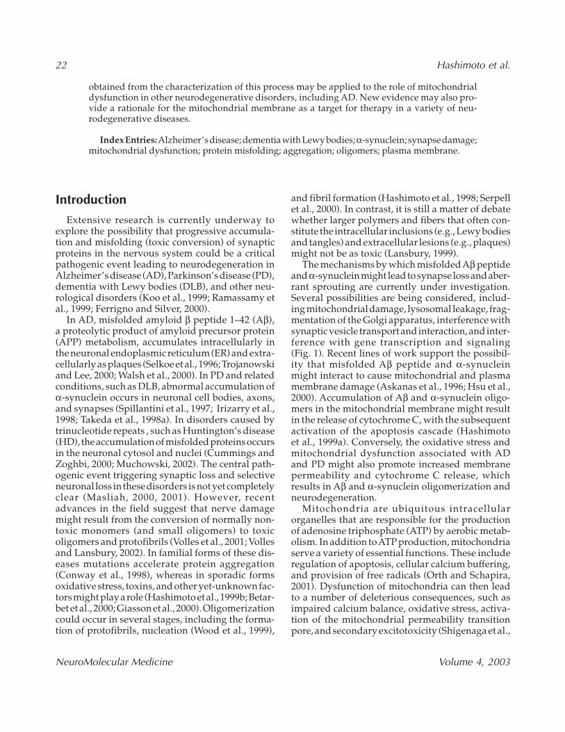
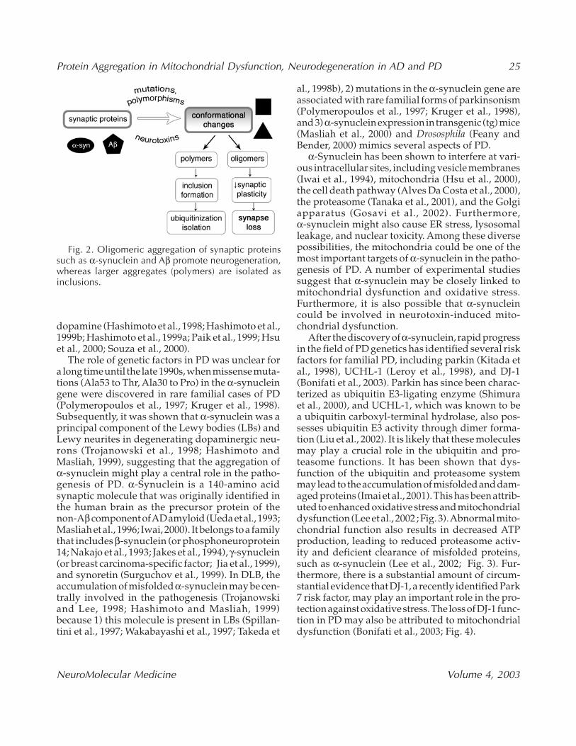
Mitochondrial alterations associated with oxida-tive stress can trigger α-synuclein accumulation;however, the relationship between mitochondrialdysfunction and α-synuclein accumulation isunclear. α-Synuclein is capable of self-aggregatingto form both oligomers and fibrillar polymers withamyloid-like characteristics (Hashimoto et al., 1998).Most recent evidence suggests that there might beboth low-molecular-weight (MW) nontoxicoligomers as well as higher MW toxic oligomers(protofibrils) that associate with the cell membrane(Hashimoto et al., 1998; Conway et al., 2000; Rochetet al., 2000). The association of nontoxic oligomerswith components of the plasma membrane, such aspolyunsaturated fatty acids, might play a role insynaptic plasticity (Perrin et al., 2000). In contrast,higher MW toxic oligomers form protofibrils thatcan potentially damage the cell membrane(Narayanan and Scarlata, 2001; Volles et al., 2001).The role of fibril formation in PD and related dis-orders is more controversial, but a number of stud-ies suggest that fibrils might represent less toxicbyproducts or even a cellular strategy to inactivateor isolate more toxic oligomers (Hashimoto et al.,1998; Conway et al., 2000; Rochet et al., 2000;Fig. 2). Additionally, mitochondrial neurotoxinsand components have been shown to enhance(α-synuclein accumulation. These include MPTP,paraquat, iron, cytochrome C, copper (II) and
Protein Aggregation in Mitochondrial Dysfunction, Neurodegeneration in AD and PD 25
NeuroMolecular Medicine Volume 4, 2003
dopamine (Hashimoto et al., 1998; Hashimoto et al.,1999b; Hashimoto et al., 1999a; Paik et al., 1999; Hsuet al., 2000; Souza et al., 2000).
The role of genetic factors in PD was unclear fora long time until the late 1990s, when missense muta-tions (Ala53 to Thr, Ala30 to Pro) in the α-synucleingene were discovered in rare familial cases of PD(Polymeropoulos et al., 1997; Kruger et al., 1998).Subsequently, it was shown that α-synuclein was aprincipal component of the Lewy bodies (LBs) andLewy neurites in degenerating dopaminergic neu-rons (Trojanowski et al., 1998; Hashimoto andMasliah, 1999), suggesting that the aggregation ofα-synuclein might play a central role in the patho-genesis of PD. α-Synuclein is a 140-amino acidsynaptic molecule that was originally identified inthe human brain as the precursor protein of thenon-Aβ component of AD amyloid (Ueda et al., 1993;Masliah et al., 1996; Iwai, 2000). It belongs to a familythat includes β-synuclein (or phosphoneuroprotein14; Nakajo et al., 1993; Jakes et al., 1994), γ-synuclein(or breast carcinoma-specific factor; Jia et al., 1999),and synoretin (Surguchov et al., 1999). In DLB, theaccumulation of misfolded α-synuclein may be cen-trally involved in the pathogenesis (Trojanowskiand Lee, 1998; Hashimoto and Masliah, 1999)because 1) this molecule is present in LBs (Spillan-tini et al., 1997; Wakabayashi et al., 1997; Takeda et
al., 1998b), 2) mutations in the α-synuclein gene areassociated with rare familial forms of parkinsonism(Polymeropoulos et al., 1997; Kruger et al., 1998),and 3) α-synuclein expression in transgenic (tg) mice(Masliah et al., 2000) and Drososphila (Feany andBender, 2000) mimics several aspects of PD.
α-Synuclein has been shown to interfere at vari-ous intracellular sites, including vesicle membranes(Iwai et al., 1994), mitochondria (Hsu et al., 2000),the cell death pathway (Alves Da Costa et al., 2000),the proteasome (Tanaka et al., 2001), and the Golgiapparatus (Gosavi et al., 2002). Furthermore,α-synuclein might also cause ER stress, lysosomalleakage, and nuclear toxicity. Among these diversepossibilities, the mitochondria could be one of themost important targets of α-synuclein in the patho-genesis of PD. A number of experimental studiessuggest that α-synuclein may be closely linked tomitochondrial dysfunction and oxidative stress.Furthermore, it is also possible that α-synucleincould be involved in neurotoxin-induced mito-chondrial dysfunction.
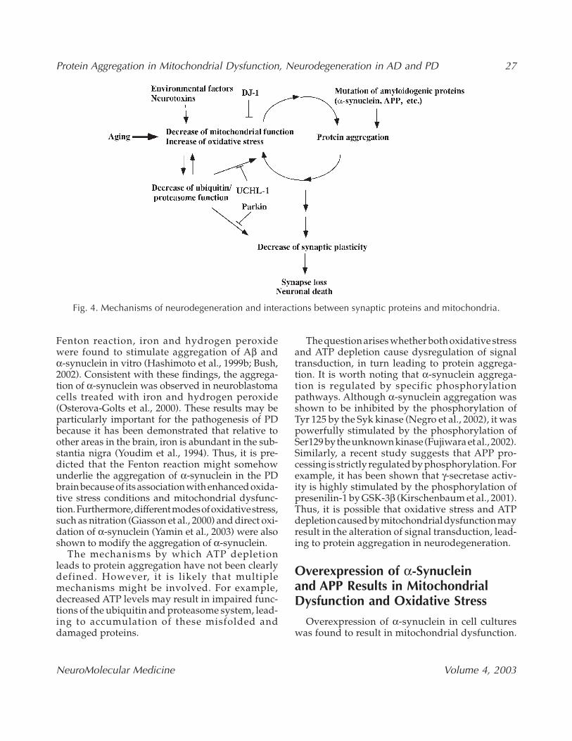
After the discovery of α-synuclein, rapid progressin the field of PD genetics has identified several riskfactors for familial PD, including parkin (Kitada etal., 1998), UCHL-1 (Leroy et al., 1998), and DJ-1(Bonifati et al., 2003). Parkin has since been charac-terized as ubiquitin E3-ligating enzyme (Shimuraet al., 2000), and UCHL-1, which was known to bea ubiquitin carboxyl-terminal hydrolase, also pos-sesses ubiquitin E3 activity through dimer forma-tion (Liu et al., 2002). It is likely that these moleculesmay play a crucial role in the ubiquitin and pro-teasome functions. It has been shown that dys-function of the ubiquitin and proteasome systemmay lead to the accumulation of misfolded and dam-aged proteins (Imai et al., 2001). This has been attrib-uted to enhanced oxidative stress and mitochondrialdysfunction (Lee et al., 2002 ; Fig. 3). Abnormal mito-chondrial function also results in decreased ATPproduction, leading to reduced proteasome activ-ity and deficient clearance of misfolded proteins,such as α-synuclein (Lee et al., 2002; Fig. 3). Fur-thermore, there is a substantial amount of circum-stantial evidence that DJ-1, a recently identified Park7 risk factor, may play an important role in the pro-tection against oxidative stress. The loss of DJ-1 func-tion in PD may also be attributed to mitochondrialdysfunction (Bonifati et al., 2003; Fig. 4).
Fig. 2. Oligomeric aggregation of synaptic proteinssuch as α-synuclein and Aβ promote neurogeneration,whereas larger aggregates (polymers) are isolated asinclusions.
26 Hashimoto et al.
NeuroMolecular Medicine Volume 4, 2003
Mitochondrial Dysfunction and Oxidative Stress Causes α-Synuclein and Aβ Aggregation
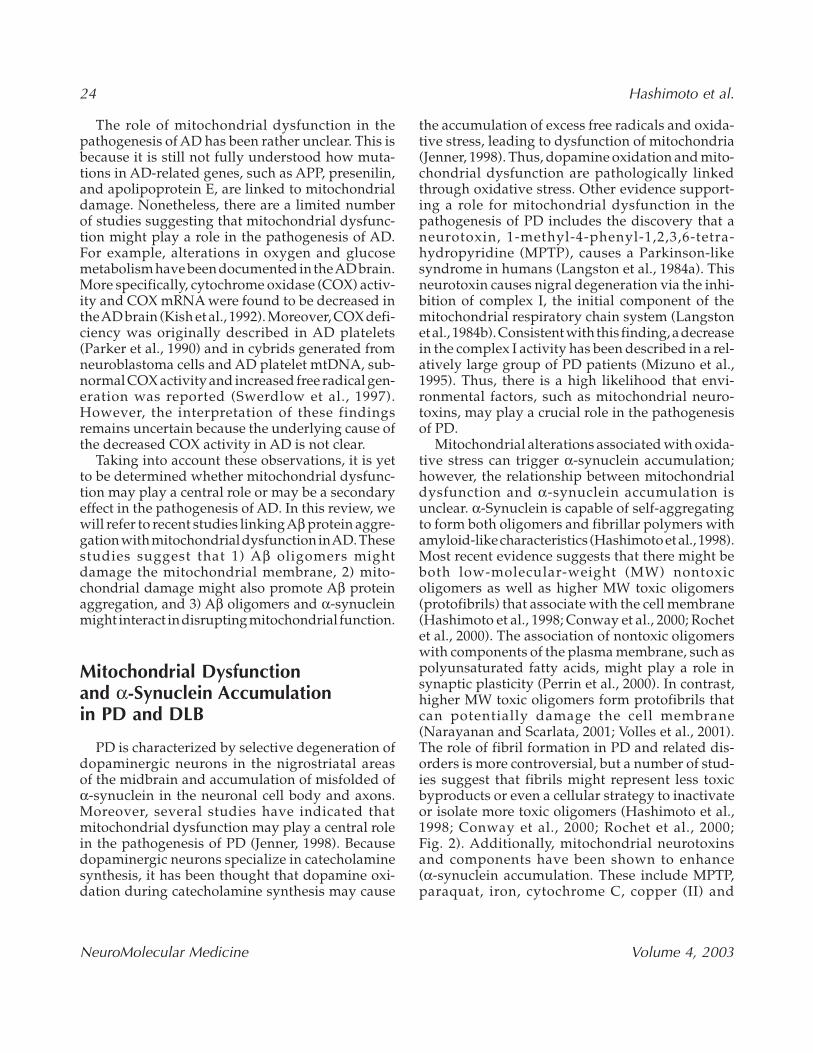
Mitochondrial dysfunction and enhanced oxida-tive stress conditions have been shown to stimulateaggregation of amyloidogenic proteins, such as α-synuclein and Aβ. However, it has been shown thatthe aggregation of amyloidogenic proteins maystimulate mitochondrial dysfunction and oxidativestress. Together, protein aggregation and mito-chondrial dysfunction can act as both cause and con-sequence of each other, forming a vicious cycle thatperpetuates neurodegeneration (Fig. 4). Recentstudies have used various inhibitors of the mito-chondrial electron-transport chain system to eval-uate the effect of mitochondrial dysfunction onprotein aggregation. For example, treatment of var-
ious cell cultures with rotenone and oligomycineresulted in increased accumulation of insoluble α-synuclein and formation of α-synuclein immunore-active inclusion bodies (Lee et al., 2002). Theseinclusion bodies disappeared upon withdrawal ofthe inhibitors, raising the intriguing possibility thatthe aggregation of α-synuclein might be reversedby the restoration of mitochondrial functions (Leeet al., 2002). These results were confirmed in a rodentexperimental system where the treatment of ratswith rotenone was shown to cause the formation ofα-synuclein immunoreactive inclusions (Sherer etal., 2003). Similar results were recently describedregarding the causative role of mitochondrial dys-function in Aβ aggregation. Astrocytes derived fromDown syndrome brains were shown to exhibit dys-regulation of APP metabolism and intracellularaccumulation of insoluble Aβ, associated withmitochondrial dysfunction (Busciglio et al., 2002).Furthermore, treatment of astrocytes with themitochondrial electron chain uncoupler carbonyl-cyanide-chloro-phenylhydrazone resulted inupregulation of Aβ production. These results sug-gest that mitochondrial dysfunction might lead toAβ aggregation in astrocytes (Busciglio et al., 2002).
Similarly, we have recently shown that the mito-chondrial and dopaminergic toxin MPTP promotesα-synuclein aggregation and abnormal mitochon-dria organization in α-synuclein transgenic mice(Song et al., 2003). In contrast, α-synuclein–deficientmice are resistant to MPTP (Dauer et al., 2002). Theresults obtained from different experimental sys-tems, including in vitro, cell cultures, and trans-genic mice, strongly suggest that mitochondrialdysfunction may cause protein aggregation ofspecies such as α-synuclein and Aβ.
Potential Mechanisms Through Which Mitochondrial DysfunctionMight Promote α-Synuclein and Aβ Aggregation
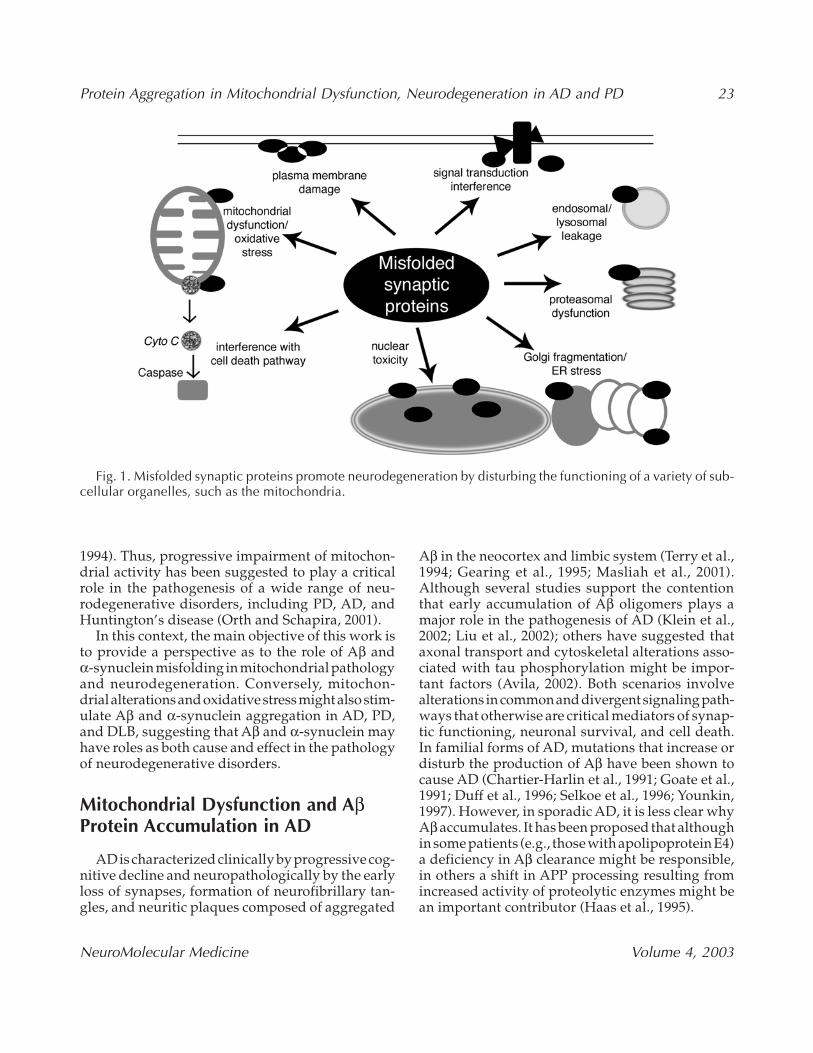
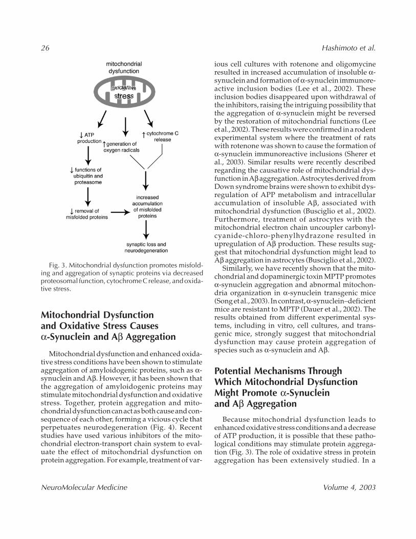
Because mitochondrial dysfunction leads toenhanced oxidative stress conditions and a decreaseof ATP production, it is possible that these patho-logical conditions may stimulate protein aggrega-tion (Fig. 3). The role of oxidative stress in proteinaggregation has been extensively studied. In a
Fig. 3. Mitochondrial dysfunction promotes misfold-ing and aggregation of synaptic proteins via decreasedproteosomal function, cytochrome C release, and oxida-tive stress.
Protein Aggregation in Mitochondrial Dysfunction, Neurodegeneration in AD and PD 27
NeuroMolecular Medicine Volume 4, 2003
Fenton reaction, iron and hydrogen peroxidewere found to stimulate aggregation of Aβ andα-synuclein in vitro (Hashimoto et al., 1999b; Bush,2002). Consistent with these findings, the aggrega-tion of α-synuclein was observed in neuroblastomacells treated with iron and hydrogen peroxide(Osterova-Golts et al., 2000). These results may beparticularly important for the pathogenesis of PDbecause it has been demonstrated that relative toother areas in the brain, iron is abundant in the sub-stantia nigra (Youdim et al., 1994). Thus, it is pre-dicted that the Fenton reaction might somehowunderlie the aggregation of α-synuclein in the PDbrain because of its association with enhanced oxida-tive stress conditions and mitochondrial dysfunc-tion. Furthermore, different modes of oxidative stress,such as nitration (Giasson et al., 2000) and direct oxi-dation of α-synuclein (Yamin et al., 2003) were alsoshown to modify the aggregation of α-synuclein.
The mechanisms by which ATP depletionleads to protein aggregation have not been clearlydefined. However, it is likely that multiplemechanisms might be involved. For example,decreased ATP levels may result in impaired func-tions of the ubiquitin and proteasome system, lead-ing to accumulation of these misfolded anddamaged proteins.
The question arises whether both oxidative stressand ATP depletion cause dysregulation of signaltransduction, in turn leading to protein aggrega-tion. It is worth noting that α-synuclein aggrega-tion is regulated by specific phosphorylationpathways. Although α-synuclein aggregation wasshown to be inhibited by the phosphorylation ofTyr 125 by the Syk kinase (Negro et al., 2002), it waspowerfully stimulated by the phosphorylation ofSer129 by the unknown kinase (Fujiwara et al., 2002).Similarly, a recent study suggests that APP pro-cessing is strictly regulated by phosphorylation. Forexample, it has been shown that γ-secretase activ-ity is highly stimulated by the phosphorylation ofpresenilin-1 by GSK-3β (Kirschenbaum et al., 2001).Thus, it is possible that oxidative stress and ATPdepletion caused by mitochondrial dysfunction mayresult in the alteration of signal transduction, lead-ing to protein aggregation in neurodegeneration.
Overexpression of α-Synuclein and APP Results in MitochondrialDysfunction and Oxidative Stress
Overexpression of α-synuclein in cell cultureswas found to result in mitochondrial dysfunction.
Fig. 4. Mechanisms of neurodegeneration and interactions between synaptic proteins and mitochondria.
28 Hashimoto et al.
NeuroMolecular Medicine Volume 4, 2003
For example, we have observed that mouse GT1-7hypothalamic neuronal cells transfected withα-synuclein undergo mitochondrial alterations thatare accompanied by free radical production. Thiswas demonstrated by 2′-7′-dichlorofluoresceinstaining, reduction of 3-(4,5-dimethylthiazol-2-yl)-2,5-diphenyltetrazolium bromide levels, increasedlevels of the enzyme glutathione that scavenges freeradicals, and the secretion of gonadotropin-releasing hormone. These cells also exhibited char-acteristic morphological changes, including forma-tion of α-synuclein-immunoreactive inclusion-likestructures and enlarged and distorted mitochon-dria (Hsu et al., 2000). Importantly, the neu-ropathological features were ameliorated by vitaminE treatment, suggesting that oxidative stress causedby α-synuclein may contribute to mitochondrialdysfunction. The introduction of the APP gene intonormal myoblasts was shown to cause histochem-ical and morphological changes in mitochondriasimilar to those observed in inclusion body myosi-tis, a disorder characterized by vacuoles containingamyloid-APP and 15- to 21-nm paired helical fila-ments positive for hyperphosporylated tau(Askanas et al., 1996). In conjunction with evidencefor oxidative damage in AD (Good et al., 1996 ; Smithet al., 1996), this suggests the possibility of mito-chondrial involvement that is secondary to abnor-mal APP metabolism.
In tg mice, we observed that enhanced oxidativestress conditions, characterized by enlarged and dis-torted mitochondria, became evident only whenThy-1 promoter-driven α-synuclein A53T mice(Rockenstein et al., 2002) were treated with MPTP(Song et al., 2003). Furthermore, bigenic mice over-expressing α-synuclein and APPexhibited increasednitrotyrosine immunoreactivity. These results sug-gest that there might be some protective mechanismagainst α-synuclein in α-synuclein tg mice becauseoxidative stress in these mice became apparent onlywhen they were further burdened with additionalstresses, such as exposure to the neurotoxin MPTPand other amyloidogenic proteins like Aβ. Consis-tent with this notion, it was recently suggested thatthe upregulation of heat shock protein (Hsp) 70expression in α-synuclein tg mice may function asa protective mechanism (Manning-Bog et al., 2003).
Potential Mechanisms Through Whichα-Synuclein Might PromoteMitochondrial Dysfunction
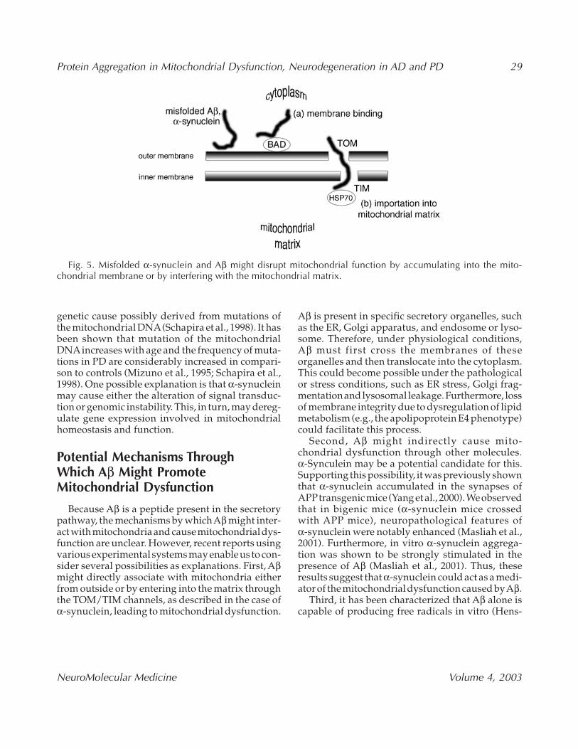
Although the precise mechanisms by whichα-synuclein causes mitochondrial dysfunction are yetto be confirmed, there are at least three possibilitiesthat are not mutually exclusive. First, α-synucleinmight cause mitochondrial dysfunction from outsidethe mitochondria (Fig. 5). Because α-synuclein bindsto specific lipids, such as acidic phospholipids (Jo etal., 2000), it is possible that accumulated α-synucleinmay directly interfere with the mitochondrial mem-brane. This, in turn, interferes with the mitochondr-ial respiratory chain system, which is situated betweenthe outer and inner membranes. Furthermore, it isalso possible that α-synuclein may interact with spe-cific molecules associated with mitochondria, suchas BAD and cytochrome C, both of which play a cru-cial role in apoptosis (Hashimoto et al., 1999a;Ostrerova et al., 1999). One may speculate that theinterference of α-synuclein with these moleculesmight be augmented through the formation of neu-rotoxic protofibrils of α-synuclein.
Second, α-synuclein might cause mitochondrialdysfunction from inside the mitochondria (Fig. 5).Recent studies have established that certain mito-chondrial proteins may enter into the mitochondr-ial matrix through the translocase channel ofouter/inner membranes (TOM/TIM; Pfanner andMeijer, 1997). These proteins are unfolded at theentrance of TOM and then refolded in the matrixwith the aid of mitochondrial Hsp 70 (Pfanner andMeijer, 1997). Because α-synuclein is unfolded in itsnative state (Weinreb et al., 1996) and consideringthe recent observation that Hsp 70 is upregulatedin the α-synuclein tg mice (Manning-Bog et al., 2003),it is probable that unfolded α-synuclein might passthrough TOM/TIM, reaching the matrix of mito-chondria. If this speculation is really the case, sim-ilar mechanisms could be applicable to thetranslocation of α-synuclein into various organelles.
Third, α-synuclein might indirectly cause mito-chondrial dysfunction through dysregulation ofgene expression. Recent studies using cybrid cellssuggest that the complex I defect in PD may have a
Protein Aggregation in Mitochondrial Dysfunction, Neurodegeneration in AD and PD 29
NeuroMolecular Medicine Volume 4, 2003
genetic cause possibly derived from mutations ofthe mitochondrial DNA(Schapira et al., 1998). It hasbeen shown that mutation of the mitochondrialDNAincreases with age and the frequency of muta-tions in PD are considerably increased in compari-son to controls (Mizuno et al., 1995; Schapira et al.,1998). One possible explanation is that α-synucleinmay cause either the alteration of signal transduc-tion or genomic instability. This, in turn, may dereg-ulate gene expression involved in mitochondrialhomeostasis and function.
Potential Mechanisms Through Which Aβ Might PromoteMitochondrial Dysfunction
Because Aβ is a peptide present in the secretorypathway, the mechanisms by which Aβ might inter-act with mitochondria and cause mitochondrial dys-function are unclear. However, recent reports usingvarious experimental systems may enable us to con-sider several possibilities as explanations. First, Aβmight directly associate with mitochondria eitherfrom outside or by entering into the matrix throughthe TOM/TIM channels, as described in the case ofα-synuclein, leading to mitochondrial dysfunction.
Aβ is present in specific secretory organelles, suchas the ER, Golgi apparatus, and endosome or lyso-some. Therefore, under physiological conditions,Aβ must first cross the membranes of theseorganelles and then translocate into the cytoplasm.This could become possible under the pathologicalor stress conditions, such as ER stress, Golgi frag-mentation and lysosomal leakage. Furthermore, lossof membrane integrity due to dysregulation of lipidmetabolism (e.g., the apolipoprotein E4 phenotype)could facilitate this process.
Second, Aβ might indirectly cause mito-chondrial dysfunction through other molecules.α-Synculein may be a potential candidate for this.Supporting this possibility, it was previously shownthat α-synuclein accumulated in the synapses ofAPPtransgenic mice (Yang et al., 2000). We observedthat in bigenic mice (α-synuclein mice crossedwith APP mice), neuropathological features ofα-synuclein were notably enhanced (Masliah et al.,2001). Furthermore, in vitro α-synuclein aggrega-tion was shown to be strongly stimulated in thepresence of Aβ (Masliah et al., 2001). Thus, theseresults suggest that α-synuclein could act as a medi-ator of the mitochondrial dysfunction caused by Aβ.
Third, it has been characterized that Aβ alone iscapable of producing free radicals in vitro (Hens-
Fig. 5. Misfolded α-synuclein and Aβ might disrupt mitochondrial function by accumulating into the mito-chondrial membrane or by interfering with the mitochondrial matrix.
30 Hashimoto et al.
NeuroMolecular Medicine Volume 4, 2003
ley et al., 1994). If this phenomenon is really applic-able in vivo as well, it is naturally predicted that theaccumulation of Aβ could enhance oxidative stressand disrupt cellular calcium homeostasis leading tomitochondrial dysfunction (Keller et al., 1997, 1998;Chan et al., 2002). Further investigations are clearlyrequired to examine these possibilities, but thesethree alternatives provide initial possible explana-tions based on various experimental evidence.
Possible Involvement of Other RiskFactors in Familial PD
Because the discovery of parkin (Kitada et al.,1998) and UCHL-1 (Leroy et al., 1998) as risk fac-tors for familial PD, subsequent characterizationsof these molecules have focused on the role ofimpaired functions of the ubiquitin and proteasomesystem. However, recent identification of DJ-1 asthe Park 7 risk factor for familial PD may promptreconsideration of the importance of the role of mito-chondrial dysfunction in the pathogenesis of PD(Bonifati et al., 2003).
Previous studies have shown that DJ-1 may beinvolved in diverse biological functions. DJ-1 wasprimarily identified as an oncogene product thatcooperates with Ras to transform mouse NIH3T3cells (Nagakubo et al., 1997). Independently, DJ-1was recognized as a molecule related to spermato-genesis, and dysfunction of this molecule mightcause male infertility (Wagenfeld et al., 1998). Fur-thermore, it was shown that DJ-1 acts as a regula-tory component of an RNA-binding protein complex(Hod et al., 1999). More importantly for the field ofneurodegenerative disorders, expression of DJ-1was stimulated by the treatment of cells withparaquat and endotoxin where the pI value ofDJ-1 was shown to change from pI6.2 to pI5.8, sug-gesting that DJ-1 may be involved in the protectionagainst oxidative stress (Mitsumoto et al., 2001).This is consistent with the fact that the loss of DJ-1function by mutation may cause an autosomalrecessive form of familial PD.
In light of the effects of α-synuclein on mito-chondrial dysfunction and oxidative stress, it is rea-sonable to speculate that DJ-1 may play a protectiverole in α-synuclein-induced mitochondrial dys-function. Such a relationship is reminiscent of accu-
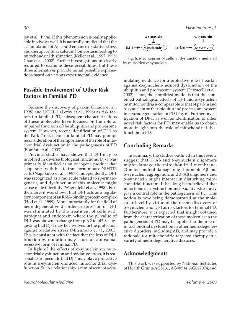
mulating evidence for a protective role of parkinagainst α-synuclein-induced dysfunction of theubiquitin and proteasome system (Petrucelli et al.,2002). Thus, the simplified model is that the com-bined pathological effects of DJ-1 and α-synucleinon mitochondria is comparable to that of parkin andα-synuclein on the ubiquitin and proteasome systemin neurodegeneration in PD (Fig. 6). Further inves-tigation of DJ-1, as well as identification of othernovel risk factors for PD, may enable us to obtainmore insight into the role of mitochondrial dys-function in PD.
Concluding Remarks
In summary, the studies outlined in this reviewsuggest that 1) Aβ and α-synuclein oligomersmight damage the mitochondrial membrane,2) mitochondrial damage might promote Aβ andα-synuclein aggregation, and 3) Aβ oligomers andα-synuclein might interact in disturbing mito-chondrial function. It has long been believed thatmitochondrial dysfunction and oxidative stress mayplay a central role in the pathogenesis of PD. Thisnotion is now being demonstrated at the mole-cular level by virtue of the recent discovery ofα-synuclein and DJ-1 as risk factors for familial PD.Furthermore, it is expected that insight obtainedfrom the characterization of these molecules in thepathogenesis of PD may be applied to the role ofmitochondrial dysfunction in other neurodegener-ative disorders, including AD, and may provide arationale for mitochondria-targeted therapy in avariety of neurodegenerative diseases.
Acknowledgments
This work was supported by National Institutesof Health Grants AG5131, AG08514, AG022074, and
Fig. 6. Mechanisms of cellular dysfunction mediatedby misfolded α-synuclein.
Protein Aggregation in Mitochondrial Dysfunction, Neurodegeneration in AD and PD 31
NeuroMolecular Medicine Volume 4, 2003
AG18440 and by a grant from the M. J. Fox Foun-dation for Parkinson’s Research.
ReferencesAlves Da Costa C., Ancolio K., and Checler F. (2000)
Wild-type but not Parkinson’s disease-related ala-53→Thr mutant alpha synuclein protects neuronalcells from apoptotic stimuli. J. Biol. Chem. 275,24065–24069.
Askanas V., McFerrin J., Baque S., et al. (1996) Trans-fer of β-amyloid precursor protein gene using ade-novirus vector causes mitochondrial abnormalitiesin cultures of normal human muscle. Proc. Natl.Acad. Sci. USA 93, 1314–1319.
Avila J., Lim, F., Moreno F. et al. (2002) Tau func-tion and dysfunction in neurons: its role in neuro-degenerative disorders. Mol. Neurobiol. 25,213–231.
Betarbet R., Sherer T. B., MacKenzie G., et al. (2000)Chronic systemic pesticide exposure reproducesfeatures of Parkinson’s disease. Nat. Neurosci. 3,1301–1306.
Bonifati V., Rizzu P., van Baren M. J., et al. (2003) Muta-tions in the DJ-1 gene associated with autosomalrecessive early-onset parkinsonism. Science 299,256–259.
Busciglio J., Pelsman A., Wong C., et al. (2002) Alteredmetabolism of the amyloid beta precursor proteinis associated with mitochondrial dysfunction inDown’s syndrome. Neuron 33, 677–688.
Bush A. I. (2002) Metal complexing agents as thera-pies for Alzheimer’s disease. Neurobiol. Aging 23,1031–1038.
Chan S. L., Furukawa K., and Mattson M. P. (2002) Pre-senilins and APP in neuritic and synaptic plastic-ity: implications for the pathogenesis ofAlzheimer’s disease. Neuromol. Med. 2, 167–196.
Chartier-Harlin M.-C., Crawford F., Houlden H., et al.(1991) Early-onset Alzheimer’s disease caused bymutations at codon 717 of the β-amyloid precur-sor protein gene. Nature 353, 844–846.
Conway K., Harper J., and Lansbury P. (1998) Accel-erated in vitro fibril formation by a mutant alpha-synuclein linked to early-onset Parkinson disease.Nat. Med. 4, 1318–1320.
Conway K. A., Lee S. J., Rochet J. C., et al. (2000) Accel-eration of oligomerization, not fibrillization, is ashared property of both alpha-synuclein muta-tions linked to early-onset Parkinson’s disease:implications for pathogenesis and therapy. Proc.Natl. Acad. Sci. USA 97, 571–576.
Cummings C. J. and Zoghbi H. Y. (2000) Trinucleotiderepeats: mechanisms and pathophysiology. Annu.Rev. Genomics Hum. Genet. 1, 281–328.
Dauer W., Kholodilov N., Vila M., et al. (2002) Resistanceof alpha-synuclein null mice to the parkinsonianneurotoxin MPTP. Proc. Natl. Acad. Sci. USA 99,14524–14529.
Duff K., Eckman C., Zehr C., et al. (1996) Increasedamyloid-beta42(43) in brains of mice expressingmutant presenilin 1. Nature 383, 710–713.
Feany M. and Bender W. (2000) A Drosophila model ofParkinson’s disease. Nature 404, 394–398.
Ferrigno P. and Silver P. (2000) Polyglutamine expan-sions: proteolysis, chaperones, and the dangers ofpromiscuity. Neuron 26, 9–12.
Fujiwara H., Hasegawa M., Dohmae N., et al. (2002)alpha-Synuclein is phosphorylated in synucle-inopathy lesions. Nat. Cell Biol. 4, 160–164.
Gearing M., Mirra S., Hedreen J., et al. (1995) Neu-ropathology confirmation of the clinical diagno-sis of Alzheimer ’s disease: CERAD. Part X.Neurology 45, 461–466.
Giasson B. I., Duda J. E., Murray I. V., et al. (2000)Oxidative damage linked to neurodegenerationby selective alpha-synuclein nitration in synucle-inopathy lesions. Science 290, 985–989.
Goate A., Chartier-Harlin M.-C., Mullan M., et al. (1991)Segregation of a missense mutation in the amy-loid precursor protein gene with familialAlzheimer’s disease. Nature 349, 704.
Good P. F., Werner P., Hsu A., Olanow C. W., andPerl D. P. (1996) Evidence of neuronal oxidativedamage in Alzheimer’s disease. Am. J. Pathol. 149,21–28.
Gosavi N., Lee H. J., Lee J. S., Patel S., and Lee S. J.(2002) Golgi fragmentation occurs in the cells withprefibrillar alpha-synuclein aggregates and pre-cedes the formation of fibrillar inclusion. J. Biol.Chem. 277, 48984–48992.
Haas C., Hung A. Y., Citron M., Teplow D. B., andSelkoe D. J. (1995) beta-Amyloid, protein pro-cessing and Alzheimer’s disease. Arzneimittel-forschung 45, 398–402.
Hashimoto M., and Masliah E. (1999) Alpha-synucleinin Lewy body disease and Alzheimer’s disease.Brain Pathol. 9, 707–720.
Hashimoto M., Takeda A., Hsu L. J., Takenouchi T.,and Masliah E. (1999a) Role of cytochrome c as astimulator of α-synuclein aggregation in Lewybody disease. J. Biol. Chem. 274, 28849–28852.
Hashimoto M., Hsu L., Xia Y., et al. (1999b) Oxidativestress induces amyloid-like aggregate formation
32 Hashimoto et al.
NeuroMolecular Medicine Volume 4, 2003
of NACP/α-synuclein in vitro. Neuroreport 10,717–721.
Hashimoto M., Hernandez-Ruiz S., Hsu L., et al. (1998)Human recombinant NACP/a-synuclein is aggre-gated and fibrillated in vitro: Relevance for Lewybody disease. Brain Res 799, 301–306
Hensley K., Carney J. M., Mattson M. P., et al. (1994)A model for beta-amyloid aggregation and neu-rotoxicity based on free radical generation by thepeptide: relevance to Alzheimer disease. Proc. Natl.Acad. Sci. USA 91, 3270–3274.
Hod Y., Pentyala S. N., Whyard T. C., and El-MaghrabiM. R. (1999) Identification and characterization ofa novel protein that regulates RNA-protein inter-action. J. Cell Biochem. 72, 435–444.
Hsu L. J., Sagara Y., Arroyo A., et al. (2000)α-Synuclein promotes mitochondrial deficienciesand oxidative stress. Am. J. Pathol. 157, 401–410.
Imai Y., Soda M., Inoue H., et al. (2001) An unfoldedputative transmembrane polypeptide, which canlead to endoplasmic reticulum stress, is a substrateof Parkin. Cell 105, 891–902.
Irizarry M., Growdon W., Gomez-Isla T., et al. (1998)Nigral and cortical Lewy bodies and dystrophicnigral neurites in Parkinson’s disease and corticalLewy body disease contain alpha-synucleinimmunoreactivity. J. Neuropathol. Exp. Neurol. 57,334–337.
Iwai A. (2000) Properties of NACP/alpha-synucleinand its role in Alzheimer’s disease. Biochim. Bio-phys. Acta 1502, 95–109.
Iwai A., Masliah E., Yoshimoto M., et al. (1994) Theprecursor protein of non-Ab component ofAlzheimer’s disease amyloid (NACP) is a presy-naptic protein of the central nervous system.Neuron 14, 467–475.
Jakes R., Spillantini M., and Goedert M. (1994) Iden-tification of two distinct synucleins from humanbrain. FEBS Lett. 345, 27–32.
Jenner P. (1998) Oxidative mechanisms in nigral celldeath in Parkinson’s disease. Mov. Disord. 13,24–34.
Jia T., Liu Y. E., Liu J., and Shi Y. E. (1999) Stimulationof breast cancer invasion and metastasis by synu-clein gamma. Cancer Res. 59, 742–747.
Jo E., McLaurin J., Yip C., St. George-Hyslop P., andGraser P. (2000) Alpha-synuclein membrane iter-actions and lipid specificity. J. Biol. Chem. 275,34328–34334.
Keller J. N., Kindy M. S., Holtsberg F. W., et al. (1998)Mitochondrial manganese superoxide dismutaseprevents neural apoptosis and reduces ischemic
brain injury: suppression of peroxynitrite pro-duction, lipid peroxidation, and mitochondrialdysfunction. J. Neurosci. 18, 687–697
Keller J. N., Pang Z., Geddes J. W., et al. (1997) Impair-ment of glucose and glutamate transport andinduction of mitochondrial oxidative stress anddysfunction in synaptosomes by amyloid beta-peptide: role of the lipid peroxidation product4-hydroxynonenal. J. Neurochem.
Kirschenbaum F., Hsu S. C., Cordell B., and McCarthyJ. V. (2001) Glycogen synthase kinase-3beta regu-lates presenilin 1 C-terminal fragment levels.J. Biol. Chem. 276, 30701–30707.
Kish S. J., Bergeron C., Rajput A., et al. (1992) Braincytochrome oxidase in Alzheimer’s disease. J. Neu-rochem. 59, 776–779.
Kitada T., Asakawa S., Hattori N., et al. (1998) Muta-tions in the parkin gene cause autosomal reces-sive juvenile parkinsonism. Nature 392, 605–608.
Klein R. L., King M. A., Hamby M. E., and MeyerE. M. (2002) Dopaminergic cell loss induced byhuman A30P alpha-synuclein gene transfer to therat substantia nigra. Hum. Gene Ther. 13, 605–612.
Koo E., Lansbury P. J., and Kelly J. (1999) Amyloiddiseases: abnormal protein aggregation in neu-rodegeneration. Proc. Natl. Acad. Sci. USA 96,9989–9990.
Kruger R., Kuhn W., Muller T., et al. (1998) Ala30Promutation in the gene encoding α-synuclein inParkinson’s disease. Nat. Genet. 18, 106–108.
Langston J. W., Langston E. B., and Irwin I. (1984a)MPTP-induced parkinsonism in human and non-human primates—clinical and experimentalaspects. Acta. Neurol. Scand. Suppl. 100, 49–54.
Langston J. W., Forno L. S., Rebert C. S., and Irwin I.(1984b) Selective nigral toxicity after systemicadministration of 1-methyl-4-phenyl-1,2,5,6-tetrahydropyrine (MPTP) in the squirrel monkey.Brain Res. 292, 390–394.
Lansbury P. T. J. (1999) Evolution of amyloid: whatnormal protein folding may tell us about fibrillo-genesis and disease. Proc. Natl. Acad. Sci. USA 96,3342–3344.
Lee H. J., Shin S. Y., Choi C., Lee Y. H., and Lee S. J.(2002) Formation and removal of alpha-synucleinaggregates in cells exposed to mitochondrialinhibitors. J. Biol. Chem. 277, 5411–5417.
Leroy E., Boyer R., Auburger G., et al. (1998) The ubiq-uitin pathway in Parkinsons’s disease. Nature 395,451–452.
Liu Y., Fallon L., Lashuel H. A., Liu Z., and LansburyP. T. J. (2002) The UCH-L1 gene encodes two oppos-
Protein Aggregation in Mitochondrial Dysfunction, Neurodegeneration in AD and PD 33
NeuroMolecular Medicine Volume 4, 2003
ing enzymatic activities that affect alpha-synuclein degradation and Parkinson’s diseasesusceptibility. Cell 111, 209–218.
Manning-Bog A. B., McCormack A. L., Purisai M. G.,Bolin L. M., and Di Monte D. A. (2003) Alpha-synuclein overexpression protects againstparaquat-induced neurodegeneration. J. Neurosci.23, 3095–3099.
Masliah E. (2001) Recent advances in the under-standing of the role of synaptic proteins inAlzheimer’s disease and other neurodegenerativedisorders. IJ. Alz. Dis. 3, 1–9.
Masliah E. (2000) The role of synaptic proteins inAlzheimer’s disease. Ann. N Y Acad. Sci. 924, 68–75.
Masliah E., Rockenstein E., Veinbergs I., et al. (2000)Dopaminergic loss and inclusion body formationin alpha-synuclein mice: Implications for neu-rodegenerative disorders. Science 287, 1265–1269.
Masliah E., Iwai A., Mallory M., Ueda K., Saitoh T(1996) Altered presynaptic protein NACP is asso-ciated with plaque formation and neurodegener-ation in Alzheimer’s disease. Am. J. Pathol. 148,201–210.
Masliah E., Rockenstein E., Veinbergs I.,et al. (2001)β amyloid peptides enhance α-synuclein accu-mulation and neuronal deficits in a transgenicmouse model linking Alzheimer’s and Parkinson’sdisease. Proc. Natl. Acad. Sci. USA 98, 12245–12250.
Mitsumoto A., Nakagawa Y., Takeuchi A., et al. (2001)Oxidized forms of peroxiredoxins and DJ-1 on two-dimensional gels increased in response to sub-lethal levels of paraquat. Free Radic. Res. 35,301–310.
Mizuno Y., Ikebe S., Hattori N., et al. (1995) Role ofmitochondria in the etiology and pathogenesis ofParkinson’s disease. Biochim. Biophys. Acta. 1271,265–274.
Muchowski P. J. (2002) Protein misfolding, amyloidformation, and neurodegeneration: a critical rolefor molecular chaperones? Neuron 35, 9–12.
Nagakubo D., Taira T., Kitaura H., et al. (1997) DJ-1, anovel oncogene which transforms mouse NIH3T3cells in cooperation with ras. Biochem. Biophys. Res.Commun. 231, 509–513.
Nakajo S., Tsukada K., Omata K., Nakamura Y., andNakaya K. (1993) Anew brain-specific 14-kDa pro-tein is a phosphoprotein. Its complete amino acidsequence and evidence for phosphorylation. Eur.J. Biochem. 217, 1057–1063.
Narayanan V. and Scarlata S. (2001) Membrane bind-ing and self-association of alpha-synucleins. Bio-chemistry 40, 9927–9934.
Negro A., Brunati A. M., Donella-Deana A., MassiminoM. L., and Pinna L. A. (2002) Multiple phospho-rylation of alpha-synuclein by protein tyrosinekinase Syk prevents eosin-induced aggregation.FASEB J. 16, 210–212.
Orth M. and Schapira A. H. (2001) Mitochondria anddegenerative disorders. Am. J. Med. Genet.106,27–36.
Osterova-Golts N., Petrucelli L., Hardy J., et al. (2000)The A53T alpha-synuclein mutation increases iron-dependent aggregation and toxicity. J. Neurosci.20, 6048–6054.
Ostrerova N., Petrucelli L., Farrer M., et al. (1999) alpha-synuclein shares physical and functional homol-ogy with 14–3-3 proteins. J. Neurosci. 19, 5782–5791.
Paik S. R., Shin H. J., Lee J. H., Chang C. S., and Kim J.(1999) Copper(II)-induced self-oligomerization ofalpha-synuclein. Biochem. J. 340, 821–828.
Parker W. D. J., Filley C. M., and Parks J. K. (1990)Cytochrome oxidase deficiency in Alzheimer’sdisease. Neurology 40, 1302–1303.
Perrin R., Woods W., Clayton D., and George J. (2000)Interaction of human alpha-synuclein and Parkin-son’s disease variants with phospholipids: struc-tural analysis using site-directed mutagenesis.J. Biol. Chem. 275, 34393–34398.
Petrucelli L., O’Farrell C., Lockhart P. J., et al. (2002)Parkin protects against the toxicity associated withmutant alpha-synuclein: proteasome dysfunctionselectively affects catecholaminergic neurons.Neuron 36, 1007–1019.
Pfanner N. and Meijer M (1997) The Tom and Timmachine. Curr. Biol. 7, 100–103.
Polymeropoulos M., Lavedan C., Leroy E., et al. (1997)Mutation in the α-synuclein gene identified in fam-ilies with Parkinson’s disease. Science276,2045–2047.
Ramassamy C., Averill D., Beffert U., et al. (1999) Oxi-dative damage and protection by antioxidants inthe frontal cortex of Alzheimer’s disease is relatedto the apolipoprotein E genotype. Free Radic. Biol.Med. 27, 544–553.
Rochet J., Conway K., and Lansbury P. J. (2000) Inhi-bition of fibrillization and accumulation of pre-fibrillar oligomers in mixtures of human andmouse α-synuclein. Biochemistry 39, 10619–10626.
Rockenstein E., Mallory M., Hashimoto M., et al. (2002)Differential neuropathological alterations in trans-genic mice expressing α-synuclein from theplatelet-derived growth factor and Thy-1 pro-moters. J. Neurosci. Res. 68, 568–578.
Schapira A. H., Gu M., Taanman J. W., et al. (1998)Mitochondria in the etiology and pathogenesis ofParkinson’s disease. Ann. Neurol. 44, S89–98.
34 Hashimoto et al.
NeuroMolecular Medicine Volume 4, 2003
Selkoe D. J., Yamazaki T., Citron M., et al. (1996) Therole of APP processing and trafficking pathwaysin the formation of amyloid beta-protein. Ann.NY Acad. Sci. 777, 57–64.
Serpell L., Berriman J., Jakes R., Goedert M., andCrowther R. (2000) Fiber diffraction of syntheticα-synuclein filaments shows amyloid-like cross-β conformation. Proc. Natl. Acad. Sci. USA 97,4897–4902.
Sherer T. B., Kim J. H., Betarbet R., and GreenamyreJ. T. (2003) Subcutaneous rotenone exposure causeshighly selective dopaminergic degeneration andalpha-synuclein aggregation. Exp. Neurol. 179,9–16.
Shigenaga M., Hagen T., and Ames B. (1994) Oxida-tive damage and mitochondrial decay in aging.Proc. Natl. Acad. Sci. USA 91, 10771–10778.
Shimura H., Hattori N., Kubo S.-I., et al. (2000) Famil-ialr Parkinson disease gene product, parkin, is aubiquitin-protein ligase. Nat. Genet. 25, 302–305.
Smith M. A., Perry G., Richey P. L., et al. (1996) Oxida-tive damage in Alzheimer’s. Nature 382, 120–121.
Song D., Shults C., Sisk A., Rockenstein E., and MasliahE. (2003) Enhanced Sustantia Nigra Pathology inhuman α-synuclein Transgenic Mice after treat-ment with MPTP. Exp. Neurol., in press.
Souza J., Giasson B., Chen Q., Lee V.-Y., and Ischi-ropoulos H. (2000) Dityrosine cross-linking pro-motes formation of stable alpha-synucleinpolymers. J. Biol. Chem. 275, 18344–18349.
Spillantini M., Schmidt M., Lee V.-Y., et al. (1997)α-Synuclein in Lewy bodies. Nature 388, 839–840.
Surguchov A., Surgucheva I., Solessio E., and BaehrW. (1999) Synoretin—A new protein belonging tothe synuclein family. Mol. Cell Neurosci. 13, 95–103.
Swerdlow R. H., Parks J. K., Cassarino D. S., et al. (1997)Cybrids in Alzheimer’s disease: a cellular modelof the disease? Neurology 49, 918–925.
Takeda A., Mallory M., Sundsmo M., et al. (1998a)Abnormal accumulation of NACP/α-synuclein inneurodegenerative disorders. Am. J. Pathol. 152,367–372.
Takeda A., Hashimoto M., Mallory M., et al. (1998b)Abnormal distribution of the non-Ab componentof Alzheimer ’s disease amyloid precursor/α-synuclein in Lewy body disease as revealed byproteinase K and formic acid pretreatment. Lab.Invest. 78, 1169–1177.
Tanaka Y., Engelender S., Igarashi S., et al. (2001)Inducible expression of mutant alpha-synuclein
decreases proteasome activity and increases sen-sitivity to mitochondria-dependent apoptosis.Hum. Mol. Genet. 10, 919–926.
Terry R., Hansen L., and Masliah E. (1994) Structuralbasis of the cognitive alterations in Alzheimer dis-ease. In: Alzheimer Disease (Terry R., Katzman R.,eds.), Raven Press, New York, pp. 179–196.
Trojanowski J. and Lee V. (1998) Aggregation of neu-rofilament and alpha-synuclein proteins in Lewybodies: implications for pathogenesis of Parkin-son disease and Lewy body dementia. Arch. Neurol.55, 151–152.
Trojanowski J., Goedert M., Iwatsubo T., and Lee V.(1998) Fatal attractions: abnormal protein aggre-gation and neuron death in Parkinson’s diseaseand lewy body dementia. Cell Death Differ. 5,832–837.
Trojanowski J. Q. and Lee V. M. (2000) “Fatal attrac-tions” of proteins. A comprehensive hypotheticalmechanism underlying Alzheimer’s disease andother neurodegenerative disorders. Ann. N Y Acad.Sci. 924, 62–67.
Ueda K., Masliah E., Xia Y., et al. (1993) Novel amy-loid component (NAC) differentiates Alzheimer’sdisease from normal aging plaques. Soc. Neurosci.Abstr. 19, 1254.
Volles M. J. and Lansbury P. T., Jr. (2002) Vesicle per-meabilization by protofibrillar alpha-synuclein issensitive to Parkinson’s disease-linked mutationsand occurs by a pore-like mechanism. Biochemistry41, 4595–4602.
Volles M. J., Lee S. J., Rochet J. C., et al. (2001)Vesicle permeabilization by protofibrillar alpha-synuclein: implications for the pathogenesis andtreatment of Parkinson’s disease. Biochemistry 40,7812–7819.
Wagenfeld A., Gromoll J., and Cooper T. G. (1998) Mol-ecular cloning and expression of rat contraceptionassociated protein 1 (CAP1), a protein putativelyinvolved in fertilization. Biochem. Biophys. Res.Commun. 251, 545–549.
Wakabayashi K., Hansen L., Vincent I., Mallory M.,and Masliah E. (1997) Neurofibrillary tangles inthe dentate granule cells in Alzheimer’s disease,Lewy body disease and progressive supranuclearpalsy. Acta. Neuropathol. 93, 7–12.
Walsh D., Tseng B., Rydel R., Podlisny M., and SelkoeD. (2000) The oligomerization of amyloid beta-protein begins intracellularly in cells derived fromhuman brain. Biochemistry 39, 10831–10839.
Protein Aggregation in Mitochondrial Dysfunction, Neurodegeneration in AD and PD 35
NeuroMolecular Medicine Volume 4, 2003
Weinreb P., Zhen W., Poon A., Conway K., and Lans-bury P. J. (1996) NACP, a protein implicated inAlzheimer’s disease and learning, is nativelyunfolded. Biochemistry 35, 13709–13715.
Wood S. J., Wypych J., Steavenson S., et al. (1999)α-Synuclein fibrillogenesis is nucleation depen-dent. Implications for the pathogenesis of Parkin-son’s disease. J. Biol. Chem. 274, 19509–19512.
Yamin G., Glaser C. B., Uversky V. N., and Fink A. L.(2003) Certain metals trigger fibrillation of methio-nine-oxidized alpha-synuclein. J. Biol. Chem. 278,27630–27635. Epub 2003 May 16.
Yang F., Ueda K., Chen P., Ashe K. H., and Cole G. M.(2000) Plaque-associated alpha-synuclein (NACP)pathology in aged transgenic mice express-ing amyloid precursor protein. Brain Res. 853,381–383.
Youdim M. B., Ben-Shachar D., Riederer P. (1994) Theenigma of neuromelanin in Parkinson’s disease sub-stantia nigra. J. Neural. Transm. Suppl. 43, 113–122.
Younkin S. G. (1997) The AAP and PS1/2 mutationslinked to early onset familial Alzheimer’s diseaseincrease the extracellular concentration and Abeta1–42 (43). Rinsho. Shinkeigaku. 37, 1099.


![Mitochondrial dysfunction and the role of …...various neurodegenerative diseases such as Parkinson’s disease, AD and aging [10-12]. Therefore, mitophagy serves as a pivotal role](https://img.pdfslide.net/doc/110x75/5f0efea27e708231d441f67c/mitochondrial-dysfunction-and-the-role-of-various-neurodegenerative-diseases.jpg)