Embed Size (px)
Citation preview
Room Decontamination Using an Ultraviolet-C Device with Short Ultraviolet Exposure TimeAuthor(s): William A. Rutala, PhD, MPH; Maria F. Gergen, MT (ASCP); Brian M. Tande, PhD;David J. Weber, MD, MPHSource: Infection Control and Hospital Epidemiology, Vol. 35, No. 8 (August 2014), pp. 1070-1072Published by: The University of Chicago Press on behalf of The Society for Healthcare Epidemiologyof AmericaStable URL: http://www.jstor.org/stable/10.1086/677149 .
Accessed: 01/08/2014 14:44
Your use of the JSTOR archive indicates your acceptance of the Terms & Conditions of Use, available at .http://www.jstor.org/page/info/about/policies/terms.jsp
.JSTOR is a not-for-profit service that helps scholars, researchers, and students discover, use, and build upon a wide range ofcontent in a trusted digital archive. We use information technology and tools to increase productivity and facilitate new formsof scholarship. For more information about JSTOR, please contact [email protected].
.
The University of Chicago Press and The Society for Healthcare Epidemiology of America are collaboratingwith JSTOR to digitize, preserve and extend access to Infection Control and Hospital Epidemiology.
http://www.jstor.org
This content downloaded from 78.61.87.11 on Fri, 1 Aug 2014 14:44:01 PMAll use subject to JSTOR Terms and Conditions
1070 infection control and hospital epidemiology august 2014, vol. 35, no. 8
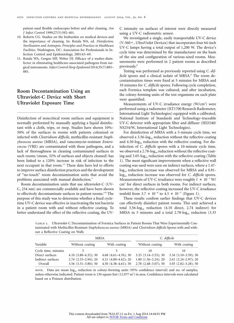
table 1. Ultraviolet-C Decontamination of Formica Surfaces in Patient Rooms That Were Experimentally Con-taminated with Methicillin-Resistant Staphylococcus aureus (MRSA) and Clostridium difficile Spores with and with-out a Reflective Coating on Walls
MRSA C. difficile
Variable Without coating With coating Without coating With coating
Cycle time, minutes 5 5 10 10Direct surfaces 4.10 (3.88–4.32); 30 4.68 (4.61–4.76); 30 3.35 (3.14–3.55); 30 3.34 (3.10–3.59); 30Indirect surfaces 2.74 (2.53–2.94); 20 4.21 (4.00–4.42); 20 1.80 (1.36–2.24); 20 2.61 (2.24–2.97); 20
Overall 3.56 (3.31–3.80); 50 4.50 (4.38–4.61); 50 2.78 (2.48–3.07); 50 3.05 (2.82–3.28); 50
note. Data are mean log10 reduction in colony-forming units (95% confidence interval) and no. of samples,unless otherwise indicated. Patient room is 130 square feet (12.077 m2) in area. Confidence intervals were calculatedbased on a Poisson distribution.
patient-used flexible endoscopes before and after cleaning. AmJ Infect Control 1999;27(5):392–401.
10. Roberts CG. Studies on the bioburden on medical devices andthe importance of cleaning. In: Rutala WA, ed. Disinfection,Sterilization and Antisepsis: Principles and Practices in HealthcareFacilities. Washington, DC: Association for Professionals in In-fection Control and Epidemiology, 2001:63–69.
11. Rutala WA, Gergen MF, Weber DJ. Efficacy of a washer-disin-fector in eliminating healthcare-associated pathogens from sur-gical instruments. Infect Control Hosp Epidemiol 2014;35(7):883–885.
Room Decontamination Using anUltraviolet-C Device with ShortUltraviolet Exposure Time
Disinfection of noncritical room surfaces and equipment isnormally performed by manually applying a liquid disinfec-tant with a cloth, wipe, or mop. Studies have shown 10%–50% of the surfaces in rooms with patients colonized orinfected with Clostridium difficile, methicillin-resistant Staph-ylococcus aureus (MRSA), and vancomycin-resistant Entero-coccus (VRE) are contaminated with these pathogens, and alack of thoroughness in cleaning contaminated surfaces insuch rooms (mean, 32% of surfaces and objects cleaned) hasbeen linked to a 120% increase in risk of infection to thenext occupant in that room.1,2 These data have led to effortsto improve surface disinfection practices and the developmentof “no-touch” room decontamination units that avoid theproblems associated with manual disinfection.3
Room decontamination units that use ultraviolet-C (UV-C, 254 nm) are commercially available and have been shownto effectively decontaminate surfaces in patient rooms.4-8 Thepurpose of this study was to determine whether a fixed cycle-time UV-C device was effective in inactivating the test bacteriain a patient room with and without reflective coating. Tobetter understand the effect of the reflective coating, the UV-
C intensity on surfaces of interest were directly measuredusing a UV-C radiometric sensor.
We investigated a single, easily transportable UV-C device(V-360�, UltraViolet Devices) that incorporates four 64-inchUV-C lamps having a total output of 1,200 W. The device’scycle time was determined by the manufacturer on the basisof the size and configuration of various-sized rooms. Mea-surements were performed in 2 patient rooms as describedpreviously.8
Testing was performed as previously reported using C. dif-ficile spores and a clinical isolate of MRSA.8 The room de-contamination times were fixed at 5 minutes for MRSA and10 minutes for C. difficile spores. Following cycle completion,each Formica template was cultured, and after incubation,the colony-forming units of the test organisms on each platewere quantified.
Measurements of UV-C irradiance energy (W/cm2) wereperformed using a radiometer (ILT1700 Research Radiometer,International Light Technologies) equipped with a calibrated,National Institute of Standards and Technology–traceableUV-C detector with appropriate filter and diffuser (SED240/NS254/W, International Light Technologies).
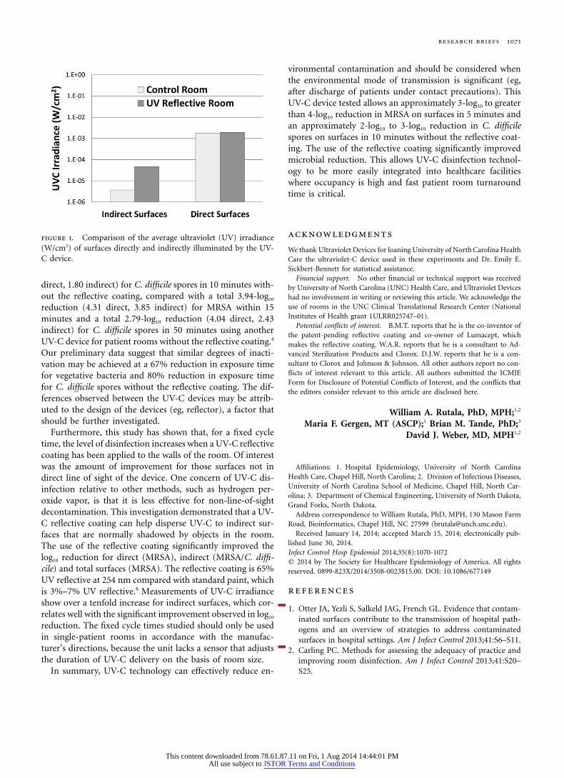
For disinfection of MRSA with a 5-minute cycle time, weobserved a 3.56-log10 reduction without the reflective coatingand 4.50-log10 reduction with the reflective coating. For dis-infection of C. difficile spores with a 10-minute cycle time,we observed a 2.78-log10 reduction without the reflective coat-ing and 3.05-log10 reduction with the reflective coating (Table1). The most significant improvements when a reflective wallcoating was used were seen on indirect surfaces, where a 1.47-log10 reduction increase was observed for MRSA and a 0.81-log10 reduction increase was observed for C. difficile spores.Measurements of UV-C irradiance were roughly W/�31 # 10cm2 for direct surfaces in both rooms. For indirect surfaces,however, the reflective coating increased the UV-C irradiancetenfold from to (Figure 1).�6 �53.7 # 10 4.5 # 10
These results confirm earlier findings that UV-C devicescan effectively disinfect patient rooms. This unit achieved atotal 3.56-log10 reduction (4.10 direct, 2.74 indirect) forMRSA in 5 minutes and a total 2.78-log10 reduction (3.35
This content downloaded from 78.61.87.11 on Fri, 1 Aug 2014 14:44:01 PMAll use subject to JSTOR Terms and Conditions
research briefs 1071
figure 1. Comparison of the average ultraviolet (UV) irradiance(W/cm2) of surfaces directly and indirectly illuminated by the UV-C device.
direct, 1.80 indirect) for C. difficile spores in 10 minutes with-out the reflective coating, compared with a total 3.94-log10
reduction (4.31 direct, 3.85 indirect) for MRSA within 15minutes and a total 2.79-log10 reduction (4.04 direct, 2.43indirect) for C. difficile spores in 50 minutes using anotherUV-C device for patient rooms without the reflective coating.4
Our preliminary data suggest that similar degrees of inacti-vation may be achieved at a 67% reduction in exposure timefor vegetative bacteria and 80% reduction in exposure timefor C. difficile spores without the reflective coating. The dif-ferences observed between the UV-C devices may be attrib-uted to the design of the devices (eg, reflector), a factor thatshould be further investigated.
Furthermore, this study has shown that, for a fixed cycletime, the level of disinfection increases when a UV-C reflectivecoating has been applied to the walls of the room. Of interestwas the amount of improvement for those surfaces not indirect line of sight of the device. One concern of UV-C dis-infection relative to other methods, such as hydrogen per-oxide vapor, is that it is less effective for non-line-of-sightdecontamination. This investigation demonstrated that a UV-C reflective coating can help disperse UV-C to indirect sur-faces that are normally shadowed by objects in the room.The use of the reflective coating significantly improved thelog10 reduction for direct (MRSA), indirect (MRSA/C. diffi-cile) and total surfaces (MRSA). The reflective coating is 65%UV reflective at 254 nm compared with standard paint, whichis 3%–7% UV reflective.8 Measurements of UV-C irradianceshow over a tenfold increase for indirect surfaces, which cor-relates well with the significant improvement observed in log10
reduction. The fixed cycle times studied should only be usedin single-patient rooms in accordance with the manufac-turer’s directions, because the unit lacks a sensor that adjuststhe duration of UV-C delivery on the basis of room size.
In summary, UV-C technology can effectively reduce en-
vironmental contamination and should be considered whenthe environmental mode of transmission is significant (eg,after discharge of patients under contact precautions). ThisUV-C device tested allows an approximately 3-log10 to greaterthan 4-log10 reduction in MRSA on surfaces in 5 minutes andan approximately 2-log10 to 3-log10 reduction in C. difficilespores on surfaces in 10 minutes without the reflective coat-ing. The use of the reflective coating significantly improvedmicrobial reduction. This allows UV-C disinfection technol-ogy to be more easily integrated into healthcare facilitieswhere occupancy is high and fast patient room turnaroundtime is critical.
acknowledgments
We thank Ultraviolet Devices for loaning University of North Carolina HealthCare the ultraviolet-C device used in these experiments and Dr. Emily E.Sickbert-Bennett for statistical assistance.
Financial support. No other financial or technical support was receivedby University of North Carolina (UNC) Health Care, and Ultraviolet Deviceshad no involvement in writing or reviewing this article. We acknowledge theuse of rooms in the UNC Clinical Translational Research Center (NationalInstitutes of Health grant 1ULRR025747–01).
Potential conflicts of interest. B.M.T. reports that he is the co-inventor ofthe patent-pending reflective coating and co-owner of Lumacept, whichmakes the reflective coating. W.A.R. reports that he is a consultant to Ad-vanced Sterilization Products and Clorox. D.J.W. reports that he is a con-sultant to Clorox and Johnson & Johnson. All other authors report no con-flicts of interest relevant to this article. All authors submitted the ICMJEForm for Disclosure of Potential Conflicts of Interest, and the conflicts thatthe editors consider relevant to this article are disclosed here.
William A. Rutala, PhD, MPH;1,2
Maria F. Gergen, MT (ASCP);1 Brian M. Tande, PhD;3
David J. Weber, MD, MPH1,2
Affiliations: 1. Hospital Epidemiology, University of North CarolinaHealth Care, Chapel Hill, North Carolina; 2. Division of Infectious Diseases,University of North Carolina School of Medicine, Chapel Hill, North Car-olina; 3. Department of Chemical Engineering, University of North Dakota,Grand Forks, North Dakota.
Address correspondence to William Rutala, PhD, MPH, 130 Mason FarmRoad, Bioinformatics, Chapel Hill, NC 27599 ([email protected]).
Received January 14, 2014; accepted March 15, 2014; electronically pub-lished June 30, 2014.Infect Control Hosp Epidemiol 2014;35(8):1070-1072� 2014 by The Society for Healthcare Epidemiology of America. All rightsreserved. 0899-823X/2014/3508-0023$15.00. DOI: 10.1086/677149
references
1. Otter JA, Yezli S, Salkeld JAG, French GL. Evidence that contam-inated surfaces contribute to the transmission of hospital path-ogens and an overview of strategies to address contaminatedsurfaces in hospital settings. Am J Infect Control 2013;41:S6–S11.
2. Carling PC. Methods for assessing the adequacy of practice andimproving room disinfection. Am J Infect Control 2013;41:S20–S25.
This content downloaded from 78.61.87.11 on Fri, 1 Aug 2014 14:44:01 PMAll use subject to JSTOR Terms and Conditions
1072 infection control and hospital epidemiology august 2014, vol. 35, no. 8
3. Rutala WA, Weber DJ. Disinfectants used for environmental dis-infection and new room decontamination technology. Am J InfectControl 2013;41:S36–S41.
4. Rutala WA, Gergen MF, Weber DJ. Room decontamination byultraviolet radiation. Infect Control Hosp Epidemiol 2010;31:1025–1029.
5. Anderson DJ, Gergen MF, Smathers E, et al. Decontaminationof targeted pathogens from patient rooms using an automatedultraviolet-C-emitting device. Infect Control Hosp Epidemiol 2013;34:465–471.
6. Boyce JM, Havill NL, Moore BA. Terminal decontamination ofpatient rooms using an automated mobile UV light unit. InfectControl Hosp Epidemiol 2011;32:743–747.
7. Nerandzic MM, Cadnum JL, Pultz MJ, Donskey CJ. Evaluationof an automated ultraviolet radiation device for decontaminationof Clostridium difficile and other healthcare-associated pathogensin hospital rooms. BMC Infect Dis 2010;10:197.
8. Rutala WA, Gergen MF, Tande BM, Weber DJ. Rapid hospitalroom decontamination using ultraviolet (UV) light with a na-nostructured UV-reflective wall coating. Infect Control HospEpidemiol 2013;34:527–529.
Brucella abortus Exposure during anOrthopedic Surgical Procedure inNew Mexico, 2010
Brucellosis, a zoonotic disease that can be transmittedthrough inhalation of infectious aerosolized particles, is en-demic in many areas, including Mexico.1-4 Manifestations ofdisease can range from subclinical illness to osteoarticulardisease and chronic sequelae.4 It is a potential occupationalhazard among laboratory workers.3 Although Brucella infec-tion is not usually a risk to medical staff, prosthetic jointinfections have been encountered during surgery.5-9 We reporta case of periprosthetic Brucella infection and the subsequentinvestigation into possible transmission to operating roomand laboratory staff. Objectives of the investigation includedinfection prevention, case-finding, and examination into po-tential routes of Brucella species transmission.
The New Mexico Department of Health (NMDOH), inconsultation with the Centers for Disease Control and Pre-vention (CDC), initiated an investigation of operating roomand laboratory staff exposures. Among operating room staff,high-risk exposures were defined as presence in the operatingroom during aerosol-generating procedures, including jointirrigation and cleaning after the procedure. NMDOH Sci-entific Laboratory Division and reference laboratory staff in-volved in testing the patient’s isolate were contacted to eval-uate laboratory exposures. Serial serologic testing andantibiotic postexposure prophylaxis (PEP; 100 mg doxycy-cline orally twice daily and rifampin 600 mg once daily for21 days, for those without contraindications) was recom-mended for individuals with high-risk exposures.10 The CDC
performed serologic testing for anti-Brucella antibodies bymicroagglutination.
The 67-year-old female patient was born in, raised in, andfrequently traveled to Mexico. Her first hip replacement oc-curred in Mexico 2 years before presentation for revision.During revision, implant component loosening, bone loss,and cloudy synovial fluid were noted. Synovial fluid was cul-tured, the joint was debrided and copiously irrigated, andhip replacement was deferred; an articulating vancomycin-and tobramycin-impregnated cement spacer was placed.Growth suggestive of Brucella species resulted from synovialfluid culture at a reference laboratory. The NMDOH ScientificLaboratory Division conducted confirmatory nucleic acidamplification testing, and subsequently the CDC performedspeciation; Brucella abortus was identified.
Seventeen high-risk exposures and 1 low-risk exposurewere investigated; fifteen high-risk exposures occurred in theoperating room. Personal protective equipment (PPE) variedfrom body exhaust suits (surgeon, first assistant, and scrubtechnician) to gloves only (cleaning staff); none wore N95respiratory protection. Because the joint was copiously irri-gated, hospital staff who cleaned the operating room werealso considered to be exposed. One low- and 2 high-riskreference laboratory staff exposures occurred during isolateprocessing outside of the biosafety cabinet on an open bench;the low-risk exposure occurred outside the 5-foot (1.5-m)radius for exposures that qualified as high risk.10 No exposuresoccurred at the NMDOH Scientific Laboratory Division, be-cause the isolate was handled inside a biosafety cabinet.
Fifteen exposed operating room staff underwent serial se-rologic testing and prophylaxis. Reference laboratory em-ployees with high-risk exposures agreed to serologic testingbut declined PEP. All who elected prophylaxis completed thePEP regimen. None of those exposed met criteria for sero-conversion (ie, fourfold increase in anti-Brucella antibodytiter). Two individuals whose total antibody titers were in-determinate (between 1 : 20 and 1 : 40, potentially resultingfrom test run variation and assay cross-reaction with otherantibodies) were referred for infectious disease consultation;no evidence of acute Brucella infection was detected. Exposedindividuals self-monitored and were observed by personalhealthcare or occupational medicine providers for 6 months;none developed symptoms of brucellosis.
The surgical patient was treated for 3 months with com-bination therapy (doxycycline and rifampin) to address os-teomyelitis and prevent Brucella infection relapse. A preop-erative aspirate, before reimplantation of the hip replacement,yielded a negative culture result. The NMDOH recommendedthat anyone involved in reimplantation use N95 masks andgoggles, minimize aerosol-generating procedures, and handlebiological specimens with care. The patient’s recovery wasuneventful without evidence of infection recurrence at 2 yearsof follow-up.
This case report demonstrates the need to consider eval-uation for Brucella species infection and risk factors among
This content downloaded from 78.61.87.11 on Fri, 1 Aug 2014 14:44:01 PMAll use subject to JSTOR Terms and Conditions