Embed Size (px)
Citation preview
NEW DRUGS AND TECHNOLOGIES IN CARDIOLOGY
Cardiology Journal2011, Vol. 18, No. 3, pp. 326–331
Copyright © 2011 Via MedicaISSN 1897–5593
326 www.cardiologyjournal.org
Address for correspondence: S. Suave Lobodzinski, PhD, Department of Electrical and Biomedical Engineering, CaliforniaState University Long Beach, 1250 Bellflower Blvd, Long Beach, CA 90840, USA, tel: (562) 985 5521, fax: (562) 985 5899,e-mail: [email protected]
Subcutaneous implantablecardioverter-defibrillator (S-ICD)
S. Suave Lobodzinski
Department of Electrical and Biomedical Engineering, California State University Long Beach, CA, USA
AbstractCurrent state-of-the art implantable cardioverter-defibrillator (ICD) systems have been provento be safe and effective in treating ventricular arrhythmias leading to cardiac death. ICDsrequire placement of at least one lead in, or on, the heart. Surgical placement under fluoroscopyand the ongoing presence of the transvenous leads within the patient’s heart are associatedwith a significant proportion of the complications related to this well-established and highlyeffective therapy. A new ICD has been developed that is implanted entirely subcutaneously(S-ICD), thus eliminating the need for lead placement in or on the heart and simplifyingsurgery by eliminating the need for imaging equipment. Recent clinical studies suggest thatthe S-ICD system provides a viable alternative to conventional transvenous devices thatmay reduce barriers to treatment and lead to the wider adoption of this life-saving therapy.(Cardiol J 2011; 18, 3: 326–331)Key words: implantable cardioverter-defibrillator, subcutaneous implantationventricular arrythmias, subcutaneous lead electrode
Introduction
The development of an implantable cardiacdefibrillator was pioneered at Sinai Hospital in Bal-timore by a team including Michel Mirowski, Mor-ton Mower, and William Staewen [1]. Parallel de-velopmental work was carried out almost simulta-neously by Schuder et al. [2, 3] at the University ofMissouri. Implantable cardioverter-defibrillators(ICDs) have proven themselves over the years asan effective treatment of ventricular arrhythmiasleading to cardiac death [4–6]. However, conven-tional ICDs have several disadvantages limitingtheir wider use:1. Conventional ICDs must be implanted using
somewhat complex and expensive surgical pro-cedures that are performed by specially trainedphysicians. Moreover, lead placement proce-dures require a special room equipped for flu-oroscopy. These rooms are limited in number,
so limiting the number of lead placement pro-cedures, and ultimately the number of ICDs,that can be implanted on any given day.
2. Conventional ICDs rely on transvenous leadsfor the placement of at least one electrode wi-thin the cardiac chambers. It has been foundthat over a period of time, transvenous leadelectrodes may get dislodged from the cardiactissues. Additionally, complications such asbroken leads and undesirable tissue depositson the electrodes are not uncommon. Theseproblems are especially acute when devices re-quire two or more electrodes. Moreover, infec-tion is a concern when implanting leads withina patient’s vasculature.
3. Removing these transvenous ICD leads andreplacing them, if necessary, also requirescomplicated surgical procedures that can bemore life-threatening than the initial implan-tation.
327
S. Suave Lobodzinski, Subcutaneous implantable cardioverter-defibrillator (S-ICD)
www.cardiologyjournal.org
Overview of subcutaneousICD technology
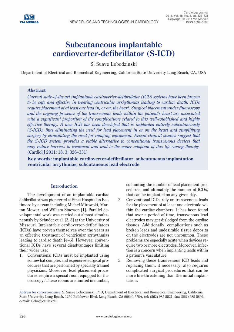
Non-transvenously implanted defibrillators offerpotential advantages over conventional ICDs (Fig. 1):1. Obviation of the need for intravascular sensing
and therapy electrodes, and elimination of therisks associated with lead placement within oron the heart.
2. Lowering incidence of inappropriate devicetherapy.
3. May reduce the surgical complication rate and//or make some complications e.g. infectionsless serious.
4. Easy to implant and explant the electrodes.The Cameron Health subcutaneous (S-ICD)
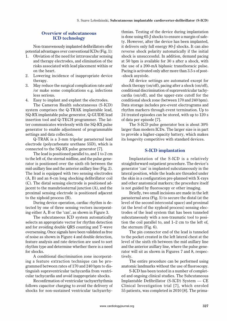
system comprises the Q-TRAK implantable lead,SQ-RX implantable pulse generator, Q-GUIDE leadinsertion tool and Q-TECH programmer. The lat-ter communicates wirelessly with the SQ-RX pulsegenerator to enable adjustment of programmablesettings and data collection.
Q-TRAK is a 3-mm tripolar parasternal leadelectrode (polycarbonate urethane 55D), which isconnected to the SQ-RX pulse generator [7].
The lead is positioned parallel to, and 1 to 2 cmto the left of, the sternal midline, and the pulse gene-rator is positioned over the sixth rib between themid-axillary line and the anterior axillary line (Fig. 2).The lead is equipped with two sensing electrodes(A, B) and an 8-cm long shocking defibrillator coil(C). The distal sensing electrode is positioned ad-jacent to the manubriosternal junction (A), and theproximal sensing electrode is positioned adjacentto the xiphoid process (B).
During device operation, cardiac rhythm is de-tected by one of three sensing vectors incorporat-ing either A, B or the ‘can’, as shown in Figure 3.
The subcutaneous ICD system automaticallyselects an appropriate vector for rhythm detectionand for avoiding double QRS counting and T-waveoversensing. Once signals have been validated as freeof noise as shown in Figure 4 and double detection,feature analysis and rate detection are used to sortrhythm type and determine whether there is a needfor shocks.
A conditional discrimination zone incorporat-ing a feature extraction technique can be pro-grammed between rates of 170 and 240 bpm to dis-tinguish supraventricular tachycardia from ventri-cular tachycardia and avoid inappropriate shocks.
Reconfirmation of ventricular tachyarrhythmiafollows capacitor charging to avoid the delivery ofshocks for non-sustained ventricular tachyarrhy-
thmias. Testing of the device during implantationis done using 65-J shocks to ensure a margin of safe-ty. However, after the device has been implanted,it delivers only full energy 80-J shocks. It can alsoreverse shock polarity automatically if the initialshock is unsuccessful. In addition, demand pacingat 50 bpm is available for 30 s after a shock, withthe use of a 200-mA biphasic transthoracic pulse.Pacing is activated only after more than 3.5 s of post--shock asystole.
All device settings are automated except forshock therapy (on/off), pacing after a shock (on/off),conditional discrimination of supraventricular tachy-cardia (on/off), and the upper-rate cutoff for theconditional shock zone (between 170 and 240 bpm).Data storage includes pre-event electrograms andrhythm markers through event termination. Up to24 treated episodes can be stored, with up to 120 sof data per episode [7].
The S-ICD pulse generator box is about 30%larger than modern ICDs. The larger size is in partto provide a higher-capacity battery, which makesits longevity competitive with standard devices.
S-ICD implantation
Implantation of the S-ICD is a relativelystraightforward outpatient procedure. The device’sgenerator ‘can’ is implanted subcutaneously in thelateral position, while the leads are threaded underthe skin in a configuration pre-planned with X-raysand other anatomical markers; the procedure itselfis not guided by fluoroscopy or other imaging.
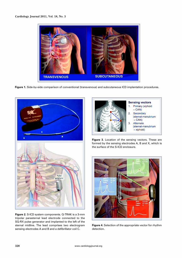
Briefly, two small incisions are made in the leftparasternal area (Fig. 5) to secure the distal (at thelevel of the second intercostal space) and proximal(at the level of the xyphoid process) sensing elec-trodes of the lead system that has been tunneledsubcutaneously with a non-traumatic tool to posi-tion the coil parallel to, and 1–2 cm to the left of,the sternum (Fig. 6).
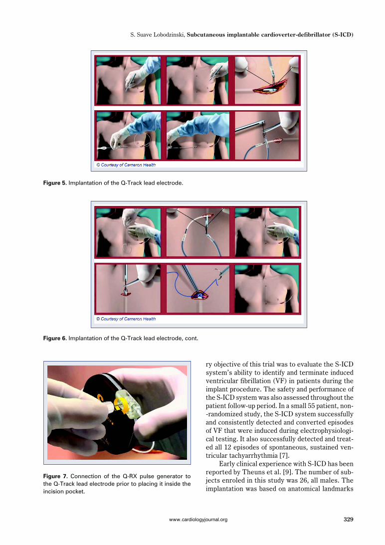
The pin connector end of the lead is tunneledto the pocket created in the left lateral chest at thelevel of the sixth rib between the mid axillary lineand the anterior axillary line, where the pulse gene-rator will sit as shown in Figures 7 and 8, respec-tively.
The entire procedure can be performed usinganatomic landmarks without the use of fluoroscopy.
S-ICD has been tested in a number of complet-ed and ongoing clinical studies. The SubcutaneousImplantable Defibrillator (S-ICD) System — CEClinical Investigation trial [7], which enroled55 patients, was completed in 2010 [8]. The prima-
328
Cardiology Journal 2011, Vol. 18, No. 3
www.cardiologyjournal.org
Figure 1. Side-by-side comparison of conventional (transvenous) and subcutaneous ICD implantation procedures.
Figure 2. S-ICD system components. Q-TRAK is a 3-mmtripolar parasternal lead electrode connected to theSQ-RX pulse generator and implanted to the left of thesternal midline. The lead comprises two electrogramsensing electrodes A and B and a defibrillator coil C.
Figure 3. Location of the sensing vectors. These areformed by the sensing electrodes A, B and X, which isthe surface of the S-ICD enclosure.
Figure 4. Selection of the appropriate vector for rhythmdetection.
329
S. Suave Lobodzinski, Subcutaneous implantable cardioverter-defibrillator (S-ICD)
www.cardiologyjournal.org
ry objective of this trial was to evaluate the S-ICDsystem’s ability to identify and terminate inducedventricular fibrillation (VF) in patients during theimplant procedure. The safety and performance ofthe S-ICD system was also assessed throughout thepatient follow-up period. In a small 55 patient, non--randomized study, the S-ICD system successfullyand consistently detected and converted episodesof VF that were induced during electrophysiologi-cal testing. It also successfully detected and treat-ed all 12 episodes of spontaneous, sustained ven-tricular tachyarrhythmia [7].
Early clinical experience with S-ICD has beenreported by Theuns et al. [9]. The number of sub-jects enroled in this study was 26, all males. Theimplantation was based on anatomical landmarks
Figure 5. Implantation of the Q-Track lead electrode.
Figure 6. Implantation of the Q-Track lead electrode, cont.
Figure 7. Connection of the Q-RX pulse generator tothe Q-Track lead electrode prior to placing it inside theincision pocket.

330
Cardiology Journal 2011, Vol. 18, No. 3
www.cardiologyjournal.org
only. There were no short-term procedure-relat-ed complications and no lead migration after useof suture sleeves. All VF episodes were accurate-ly detected using SQ-signals. There was no inap-propriate therapy caused by supraventricular ta-chycardia. The study authors concluded that theS-ICD system is a viable alternative to convention-al ICD systems for selected patients.
The primary objective of the ongoing S-ICD®
System IDE Clinical Study trial [10] is to evaluatethe safety and effectiveness of the S-ICD system.This is being conducted under an investigationaldevice exemption (IDE). It is a prospective, multi-center, single-arm design involving up to 330 sub-jects at up to 35 sites in the U.S., UK, Europe andNew Zealand.
In another study, early experience with S-ICDin three Dutch hospitals has been reported [11, 12].Of the 98 patients in the study, the first 17 werepart of the CE trial reported in the New EnglandJournal of Medicine in 2010 [7]. The remaining pa-tients were recruited after CE data collection wasclosed. In this 98-patient experience (78 males,20 females) from three centers in the Netherlands,reported at the Heart Rhythm Society 2011 Scien-tific Sessions, the device successfully terminatedall 42 episodes of sustained and non-sustained ven-tricular tachycardia in the cohort over an averageof nine months. Thirty-four spontaneous ventricu-lar arrhythmias (sustained and non-sustained) wereaccurately detected in six patients. A total of 23arrhythmic episodes were effectively treated inthree patients. Inappropriate therapy occurred ineight (early) patients due to oversensing. Of the98 patients, 62 (63%) received the device for pri-mary prevention; 40 (41%) patients had ischemic
cardiomyopathy. Other causes of cardiac disease in-cluded non-ischemic dilated cardiomyopathy (14%),Brugada syndrome (7%), and idiopathic VF (28%).Five patients in the series developed cutaneousinfections that were apparently device-related.Such infections with transvenous leads can be moreserious, as they can include sepsis or endocarditis;but their rate of occurrence is generally lower than5%. Eight patients experienced 22 inappropriateshocks; in all cases, the problem was resolved bya programming update or by lead repositioning [9].Lead migration was observed in three early pa-tients, with no recurrence since the use of an addi-tional suture sleeve at the xiphoid incision. Theresults of this study suggest that subcutaneousdefibrillation units are a viable alternative to con-ventional implantable cardiac defibrillators.
Limitations of the currentS-ICD system
Although S-ICD systems may mitigate someof the risks associated with conventional ICDs, theyprovide new shortcomings, such as inability to pro-vide long-term pacing therapy for bradyarrhy-thmias, or to be able to painlessly terminate mono-morphic ventricular arrhythmias using anti-tachy-cardia pacing. However, the S-ICD can institute‘backup’ transthoracic pacing for 30 s after a shock.Also, the S-ICD would not be suitable for patientswho require cardiac resynchronization therapy.
Regulatory status
The S-ICD system received CE approval in 2009and is commercially available in Europe. In the UK,
Figure 8. Chest X-ray of the patient’s torso with an implanted S-ICD and a corresponding frontal view [9].
331
S. Suave Lobodzinski, Subcutaneous implantable cardioverter-defibrillator (S-ICD)
www.cardiologyjournal.org
eligible patients may receive the S-ICD system asNational Health Service treatment. The first U.S. pa-tient was enroled on March 3, 2010, in CameronHealth’s FDA pivotal trial (IDE number G090013).Enrolment in this study was recently completed.
Conclusions
The S-ICD could be a new alternative therapyfor patients at risk of sudden cardiac death. Ongo-ing clinical evaluation and development are requiredbefore the role of S-ICDs as an adjunctive or pri-mary therapy can be fully defined. Further investi-gation is warranted to allow the more widespreaduse of ICDs in patients who have indications for pri-mary prevention devices.
Acknowledgements
I am grateful to Richard Sanders of CameronHealth for the care with which he reviewed theoriginal manuscript.
The author does not report any conflict of in-terest regarding this work.
References
1. Mirowski M, Mower MM, Staewen WS et al. Standby automaticdefibrillator: An approach to prevention of sudden coronarydeath. Arch Intern Med, 1970; 126: 158–161.
2. Schuder JC et al. Transthoracic ventricular defibrillation. In: Thedog with truncated and untruncated exponential stimuli. IEEETransactions on Bio-Medical Engineering, 1971; 18: 410–415.
3. Schuder JC. The role of an engineering oriented medical re-search group in developing improved methods and devices forachieving ventricular defibrillation: The University of Missouriexperience. Pacing Clin Electrophysiol, 1993;16: 95–124.
4. The Antiarrhythmics versus Implantable Defibrillators (AVID)Investigators. A comparison of antiarrhythmic drug therapy withimplantable defibrillators in patients resuscitated from near-fa-tal ventricular arrhythmias. N Engl J Med, 1997; 337: 1576––1583.
5. Moss AJ, Zareba W, Hall WJ et al. Prophylactic implantation ofa defibrillator in patients with myocardial infarction and reducedejection fraction. N Engl J Med, 2002; 346: 877–883.
6. Epstein AE, DiMarco JP, Ellenbogen KA et al. ACC/AHA/HRS2008 guidelines for device-based therapy of cardiac rhythm ab-normalities. J Am Coll Cardiol, 2008; 51: e1–e62.
7. Brady GH, Smith WM, Hood MA et al. An entirely subcutane-ous implantable cardioverter-defibrillator. N Engl J Med, 2010;363: 36–44.
8. www.clinicaltrials.gov, trial NCT01117792.9. Theuns DMAJ, Abkenari JD, Jordaens L. Initial clinical expe-
rience with a novel, totally subcutaneous implantable defibrilla-tor (S-ICD) system. ECS Stockholm, 01-09-2010.
10. www.clinicaltrials.gov, trial NCT01064076.11. Dabiri Abkenari L, Theuns DA, Valk SD et al. Clinical expe-
rience with a novel subcutaneous implantable defibrillator sys-tem in a single center. Clin Res Cardiol, 2011; 17: [Epub aheadof print].
12. Dabiri Abkenari L, Nordkamp LO, Boersma L et al. The subcu-taneous defibrillator: The Dutch experience. Heart RhythmSociety 2011 Scientific Sessions; May 4, 2011; San Francisco,CA.