Embed Size (px)
DESCRIPTION
SALIVARY GLAND DISORDERS. Done by:. Safieya Alhadab Aminah Alsulaiman. Supervised by. Prof.Ravi Kant. OBJECTIVES. Introduction. Anatomy. Disorders of glands. Clinical approach. Surgical aspect. INTRODUCTION. Salivary glands: are composed of 4 major glands, in addition to minor - PowerPoint PPT Presentation
Citation preview
• Introduction.• Anatomy.• Disorders of glands.• Clinical approach.• Surgical aspect.
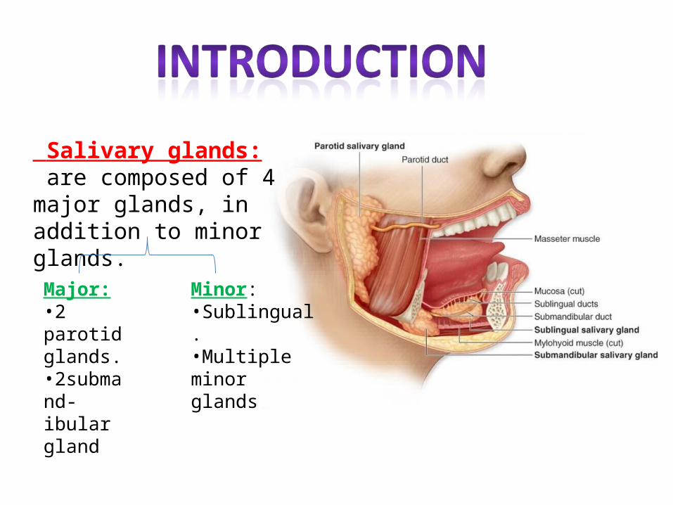
Salivary glands: are composed of 4 major glands, in addition to minor glands.
Major:•2 parotid glands.•2submand-ibular gland
Minor:•Sublingual.•Multiple minor glands
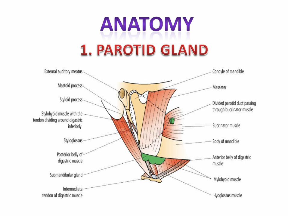
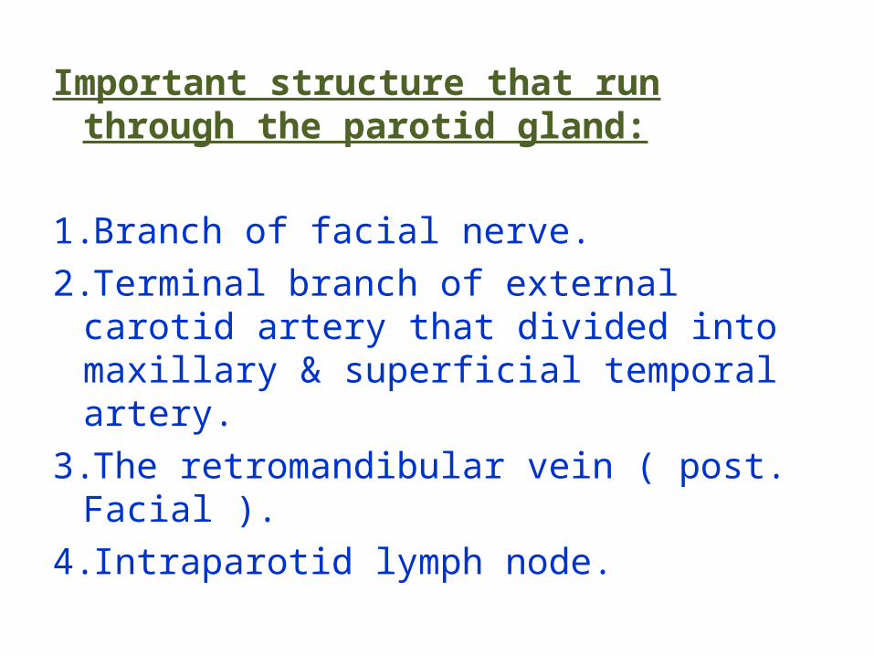
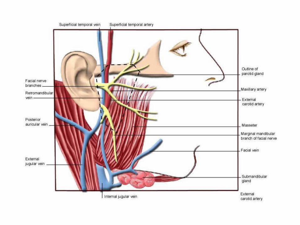
Important structure that run through the parotid gland:
1.Branch of facial nerve.2.Terminal branch of external carotid artery
that divided into maxillary & superficial temporal artery.
3.The retromandibular vein ( post. Facial ).4. Intraparotid lymph node.
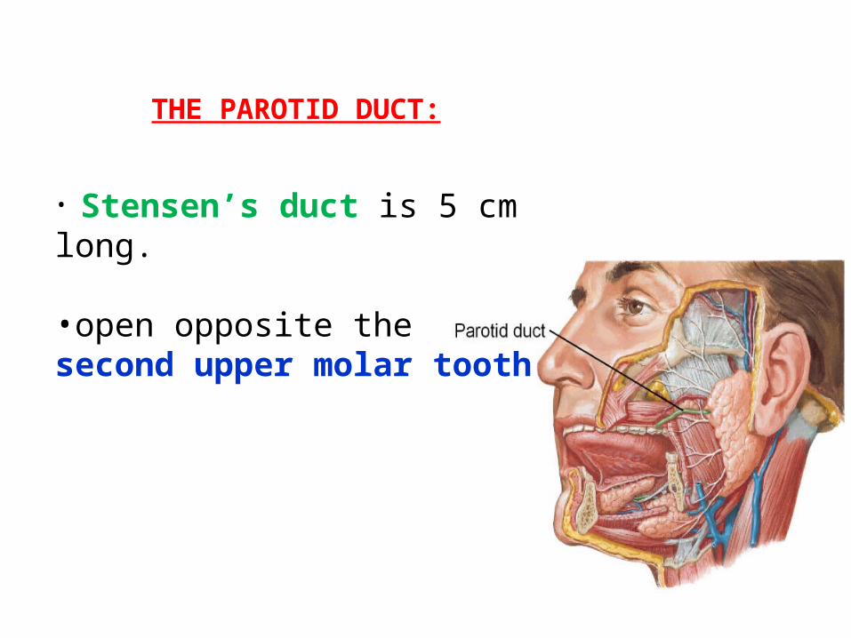
THE PAROTID DUCT:
• Stensen’s duct is 5 cm long.
•open opposite the second upper molar tooth
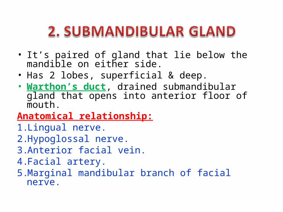
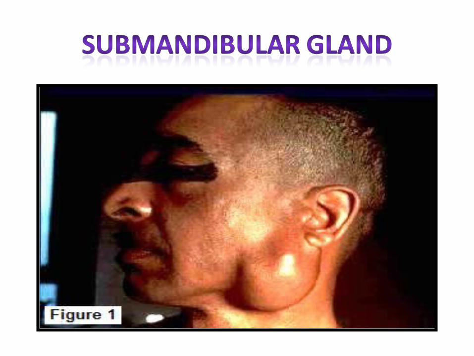
• It’s paired of gland that lie below the mandible on either side.
• Has 2 lobes, superficial & deep.• Warthon’s duct, drained submandibular gland that
opens into anterior floor of mouth.Anatomical relationship:1. Lingual nerve.2. Hypoglossal nerve.3. Anterior facial vein.4. Facial artery.5. Marginal mandibular branch of facial nerve.
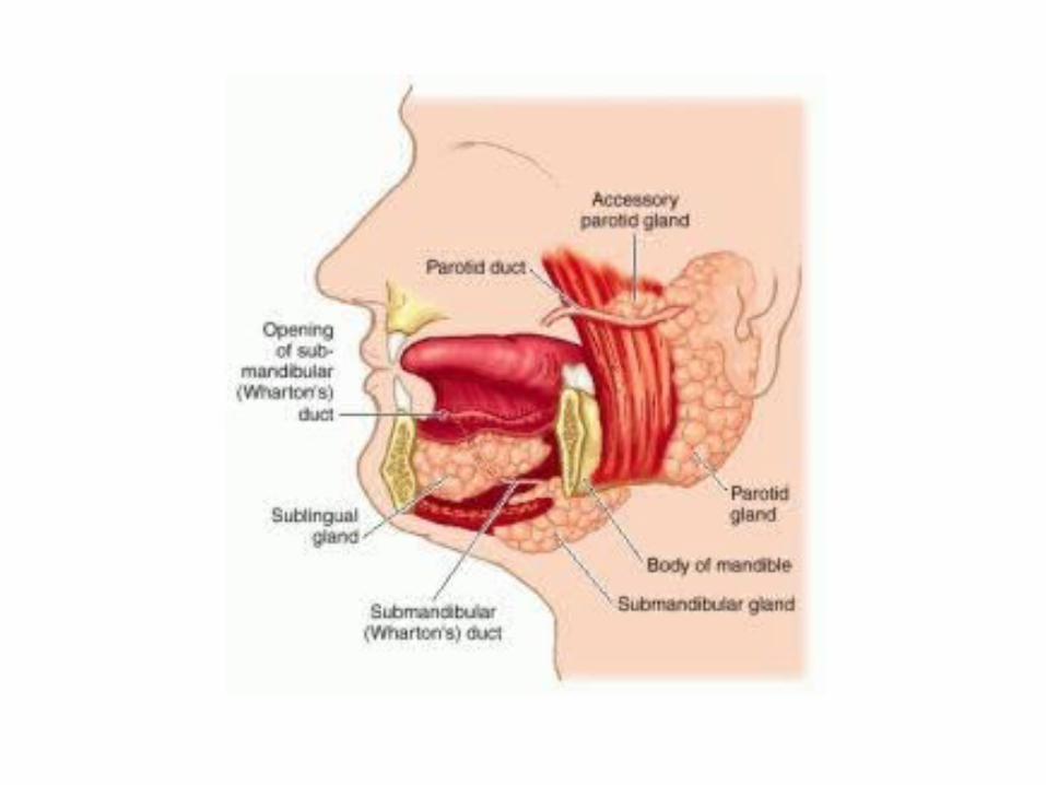
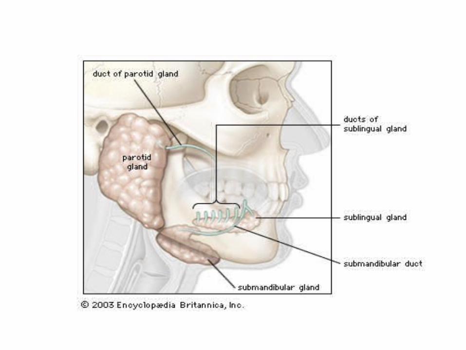
• Lie on the superior surface of the mylohyoid muscle and are separated from the oral cavity by a thin layer of mucosa. • The ducts of the sublingual glands are called Bartholin’s ducts.
•About 450 lie under the mucosa•They are distirbuted in the mucosa of the lips,
cheeks, palate, floor of mouth & retromolar area
•Also appear in oropharyanx, larynx & trachea
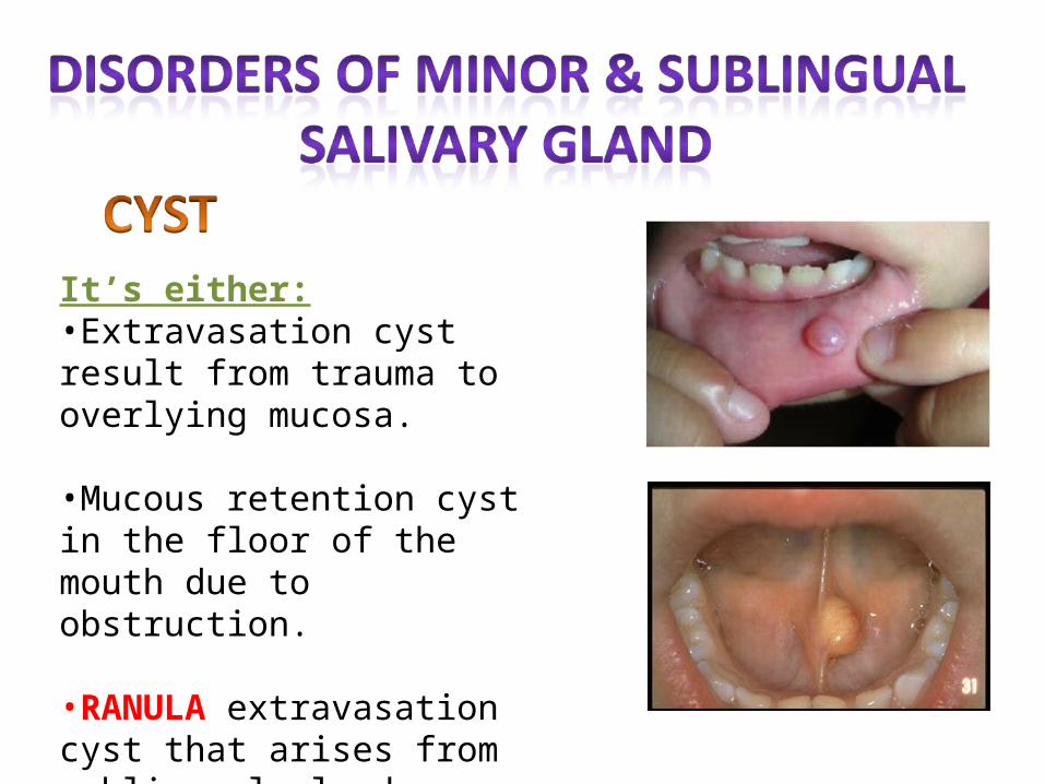
It’s either:•Extravasation cyst result from trauma to overlying mucosa.
•Mucous retention cyst in the floor of the mouth due to obstruction.
•RANULA extravasation cyst that arises from sublingual gland.
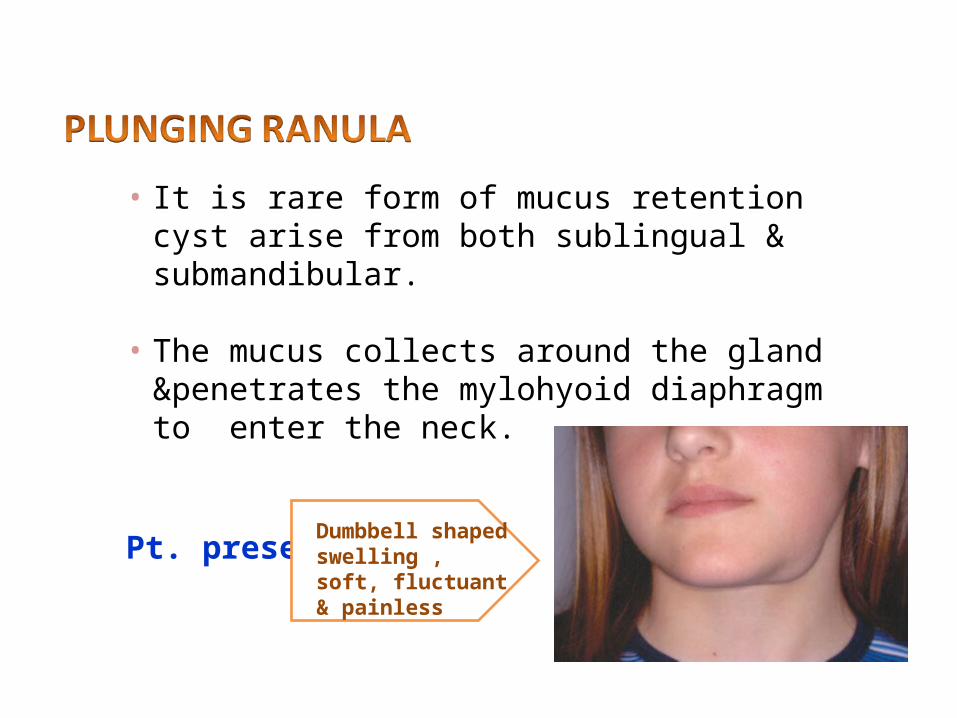
• It is rare form of mucus retention cyst arise from both sublingual & submandibular.
• The mucus collects around the gland &penetrates the mylohyoid diaphragm to enter the neck.
Pt. presents with
Dumbbell shaped swelling , soft, fluctuant & painless
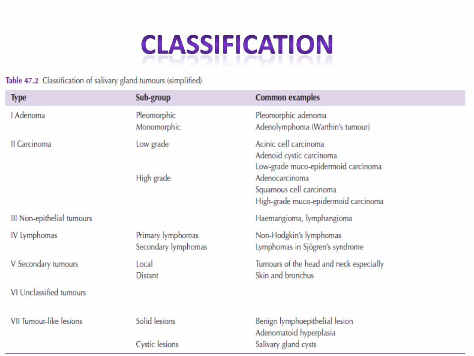
• Tumors of minor & sublingual salivary gland are extremely rare.
• 90% are malignant.
• Most common site: upper lip, palate & retromolar region.
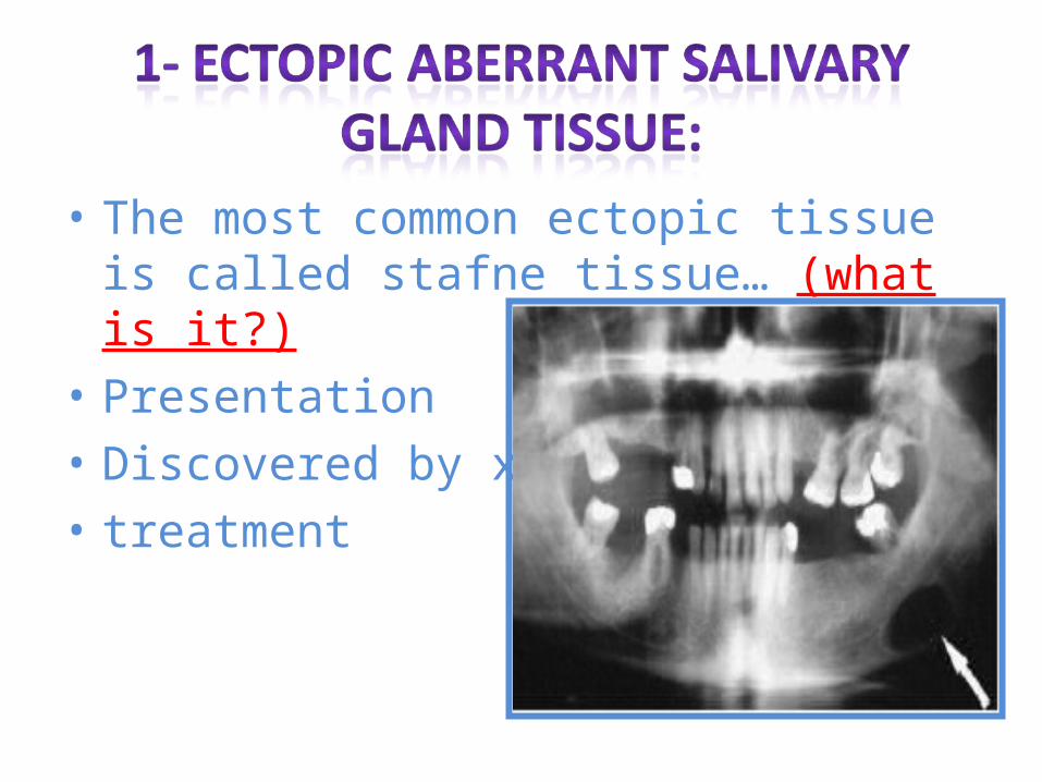
• The most common ectopic tissue is called stafne tissue… (what is it?)
• Presentation• Discovered by x-ray:• treatment
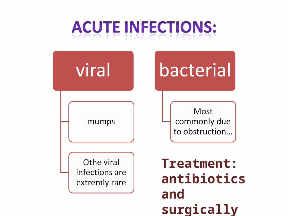
Treatment: antibiotics and surgically
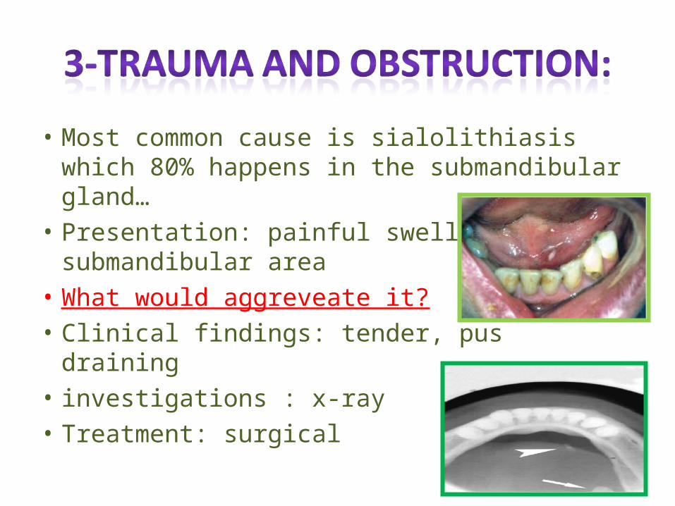
• Most common cause is sialolithiasis which 80% happens in the submandibular gland…
• Presentation: painful swelling in submandibular area
• What would aggreveate it?• Clinical findings: tender, pus draining• investigations : x-ray • Treatment: surgical
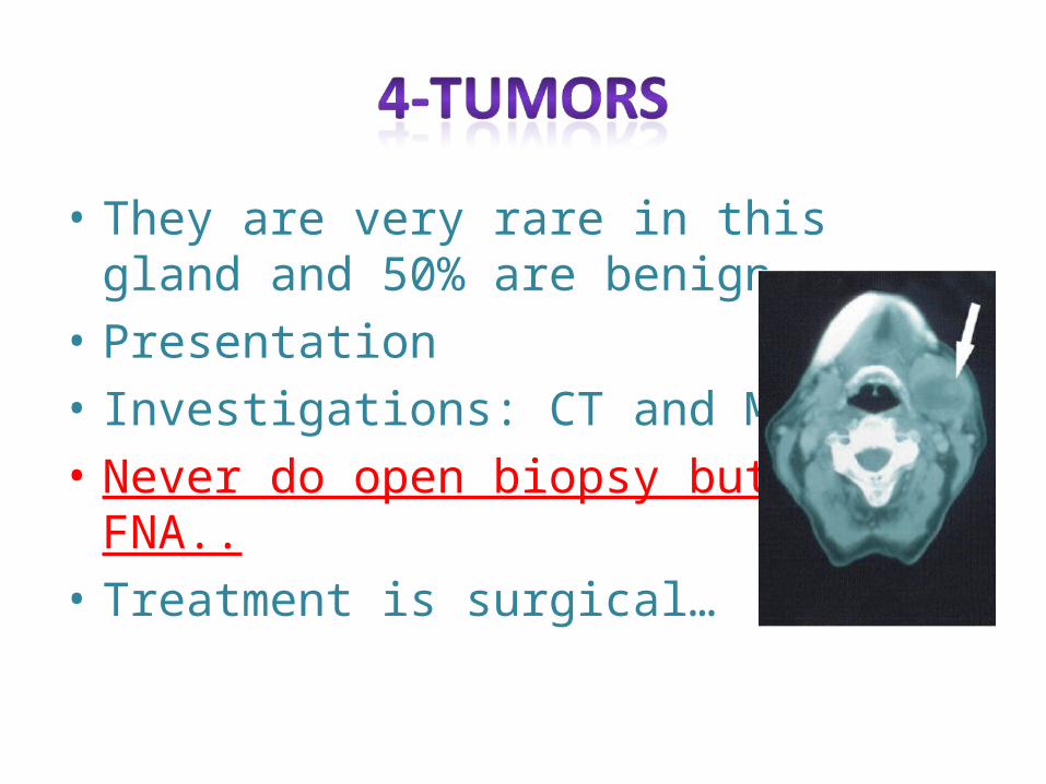
• They are very rare in this gland and 50% are benign…
• Presentation• Investigations: CT and MRI…• Never do open biopsy but do FNA..• Treatment is surgical…
•They extremly rare like agenesis, , duct atresia and congenital fistula formation…
A- viral infections:Mumps…Mode of infectionProdromal periodPresentationDiagnosisTreatment is conservativeComplications: Orchitis, oophoritis, pancreatitis, sensorineural deafness, nemimgoencephalitis but they are rare…
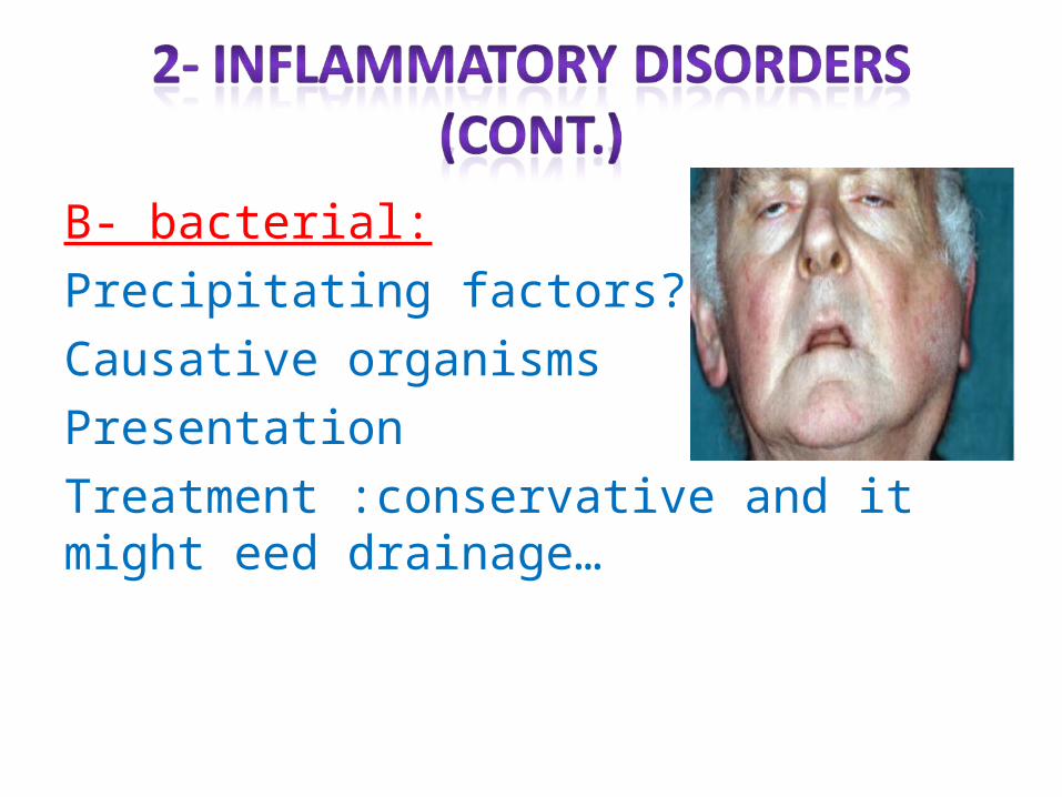
B- bacterial:Precipitating factors??!Causative organismsPresentationTreatment :conservative and it might eed drainage…
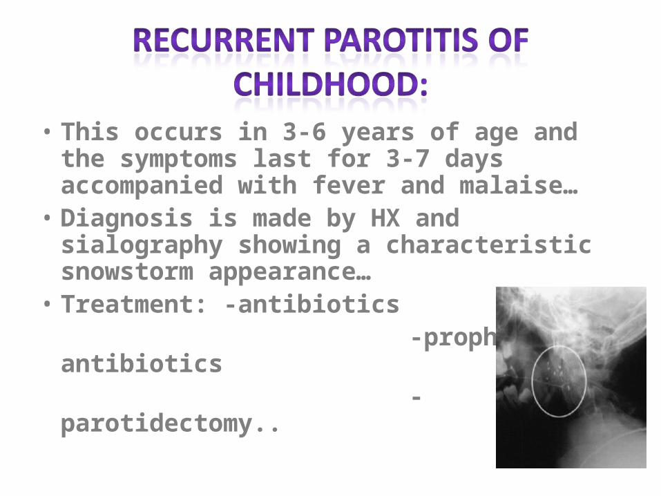
• This occurs in 3-6 years of age and the symptoms last for 3-7 days accompanied with fever and malaise…
• Diagnosis is made by HX and sialography showing a characteristic snowstorm appearance…
• Treatment: -antibiotics -prophylactic antibiotics -parotidectomy..
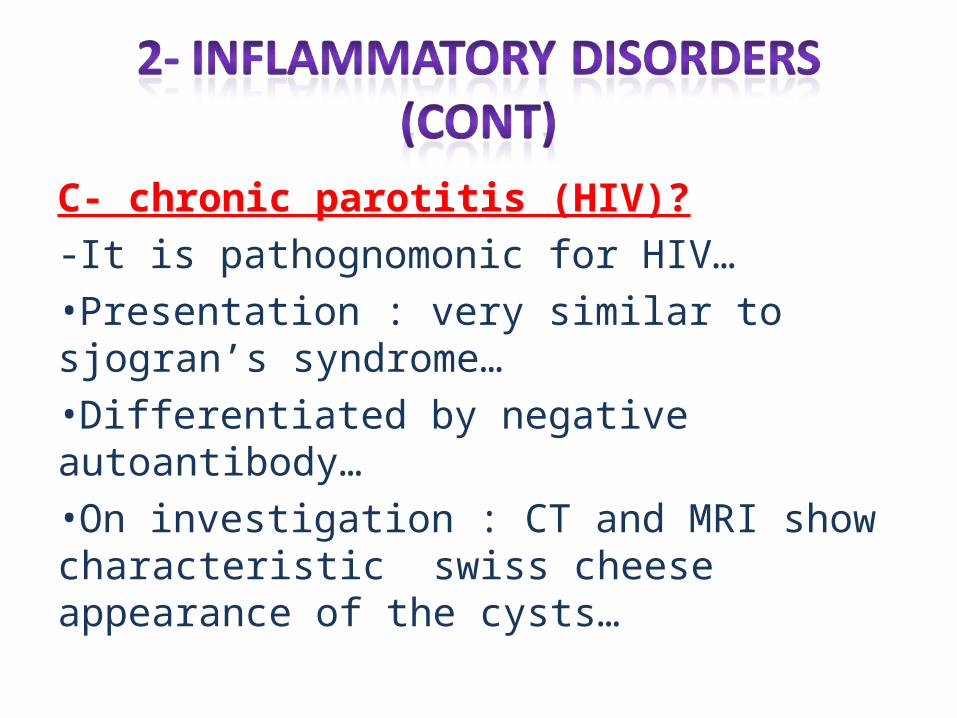
C- chronic parotitis (HIV)?-It is pathognomonic for HIV…•Presentation : very similar to sjogran’s syndrome…•Differentiated by negative autoantibody…•On investigation : CT and MRI show characteristic swiss cheese appearance of the cysts…
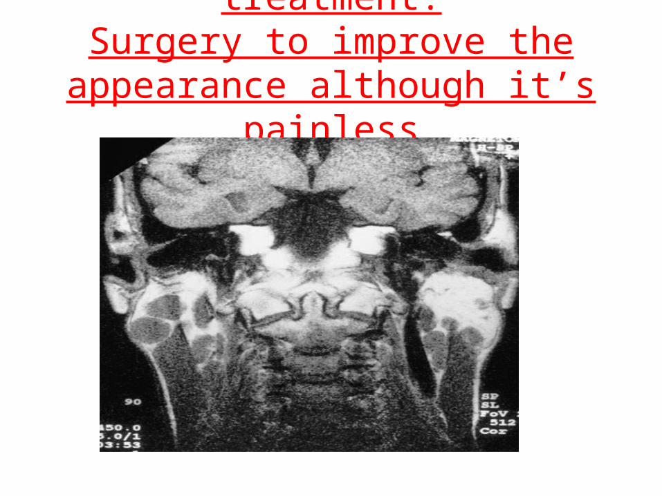
treatment:Surgery to improve the appearance
although it’s painless

A- papillary obstruction:It less common than in submandibular gland…Most commonly due to traumaPresentationTreatment is papillotomy…
B- stone formation:As mentioned before it is 80% in submandibular but only 20 % in parotidInvestigations:position…Treatment is surgical…
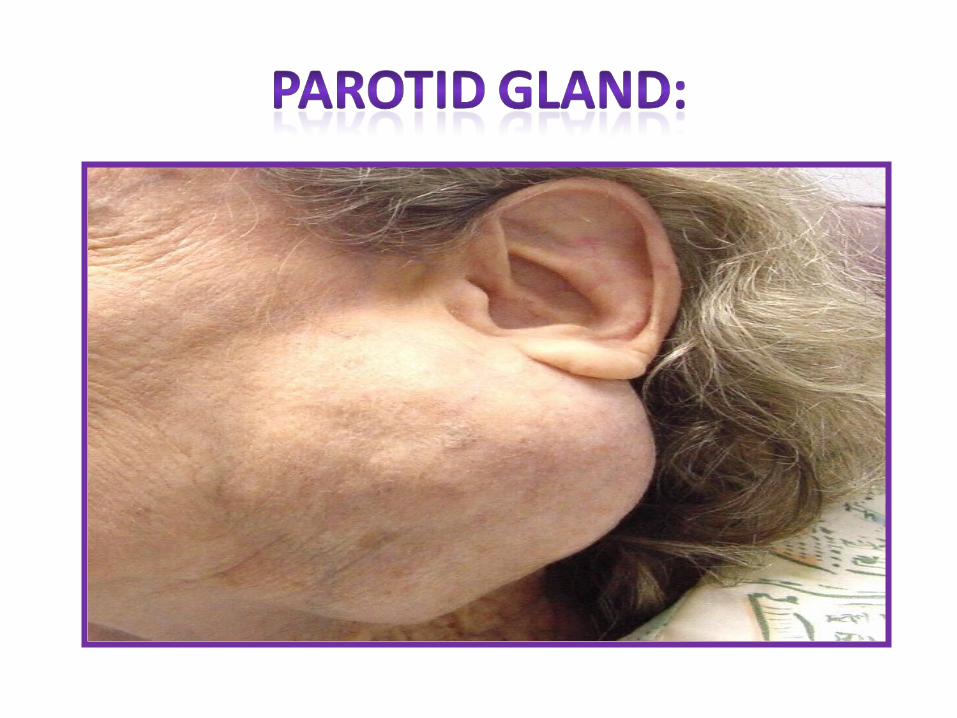
• The parotids are the commonest glands for tumors of salivary glands…

Slowly painless growing temor below the ear, or infront of it
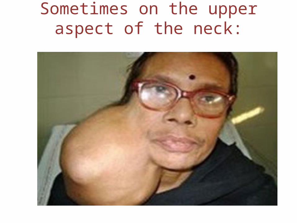
Sometimes on the upper aspect of the neck:
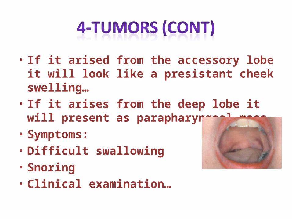
• If it arised from the accessory lobe it will look like a presistant cheek swelling…
• If it arises from the deep lobe it will present as parapharyngeal mass…
• Symptoms:• Difficult swallowing• Snoring• Clinical examination…
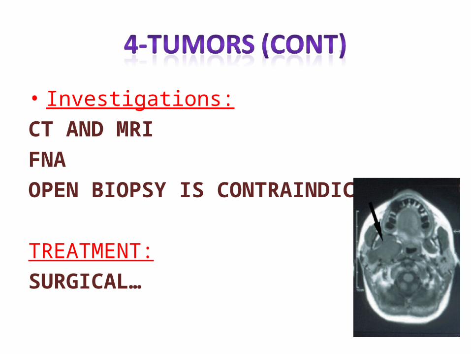
• Investigations:CT AND MRIFNAOPEN BIOPSY IS CONTRAINDICATED…
TREATMENT:SURGICAL…
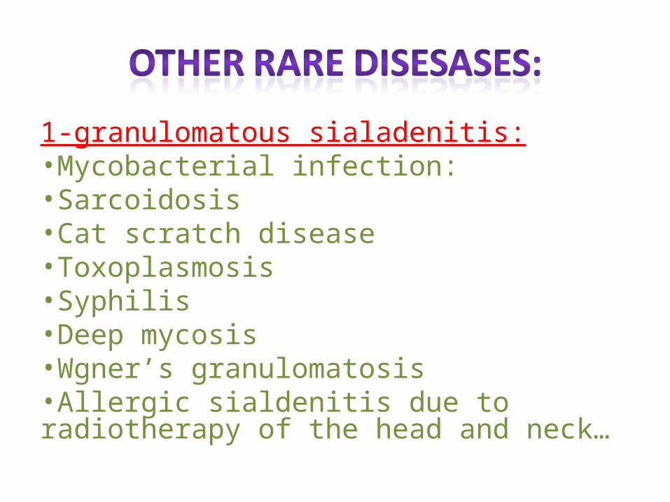
1-granulomatous sialadenitis:•Mycobacterial infection:•Sarcoidosis•Cat scratch disease•Toxoplasmosis•Syphilis •Deep mycosis•Wgner’s granulomatosis•Allergic sialdenitis due to radiotherapy of the head and neck…
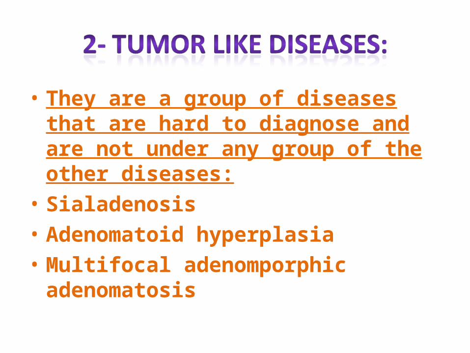
• They are a group of diseases that are hard to diagnose and are not under any group of the other diseases:
• Sialadenosis• Adenomatoid hyperplasia• Multifocal adenomporphic adenomatosis
• Sjogran’s syndrome…:• Benign lymphoepithilial lesions• Xerostomia• Sialorrhea
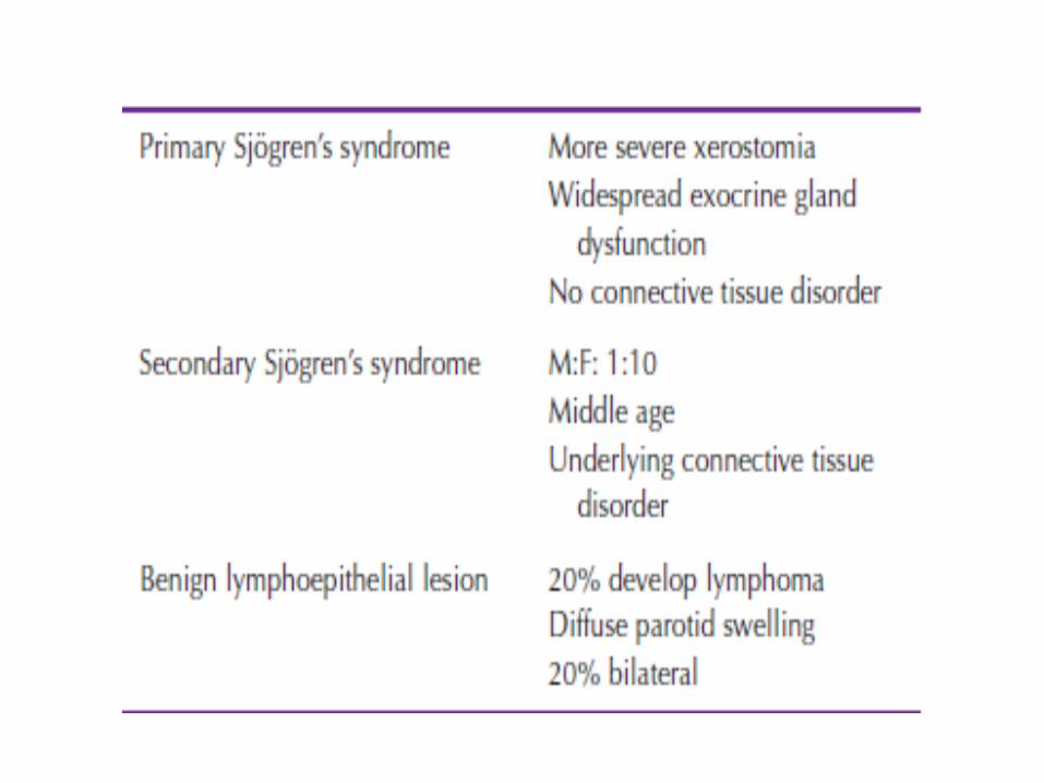
• It is an autoimmune condition causing progressive destruction of the salivary glands and the lacrimal glands…..
• Presentation is xerostomia and keratoconjunctivitis…
• They also present with pain and asendng infection
• .females more than males 10:1• Parotis is more common
• The charachtaristic feature is progressive lymphocytic infiltration acinar cell destruction and prolifration of duct epithilium…
• Diagnosis based on history…• Treatment remains symptomatic: Artificial tears…Salivary substitiuants or water…Floride to avoid dental carries…Complications are B cell lymphoma
• Normal salivary flow decreases with age…• Mostly in woman postmenopausal complaining of
burning tongue of mouth..• Causes: -chronic anxiety and depression.. -dehydration… -anticholinergic drugs… -sjogran’s syndrome… -radiotherapy of the neck and head
• Causes: some infections and drugs…• Drooling:In children that are mental handicapAlso in cerebral palsy
Management is surgical…Bilateral submandibular duct repositioning and sublingual
duct excision…Bilateral submandibualr gland excision…Bilateral submandibualr gland excision and repositioning
of the parotis duct…
• History.• Clinical examination.• Investigation.
• History of swellings / change over time?• Trismus?• Pain?• Variation with meals?• Bilateral?• Dry mouth? Dry eyes?• Recent exposure to sick contacts (mumps)?• Radiation history?• Current medications?
INSPECTION:• Asymmetry (glands, face, neck)• Diffuse or focal enlargement• Erythema extra-orally• Trismus• Medial displacement of structures intraorally?• Cranial nerve testing ( Facial , Hypoglossal
nerve)
PALPATION:• Palpate for cervical lymphadenopathy• Bimanual palpation of floor of mouth in a
posterior to anterior direction– Have patient close mouth slightly & relax oral
musculature to aid in detection– Examine for duct purulence
• Bimanual palpation of the gland (firm or spongy/elastic).
1. Plain occlusal film.2. CT Scan.3. Ultrasound.4. Sialography.5. Radionuclide Studies.6. Diagnostic Sialendoscopy2
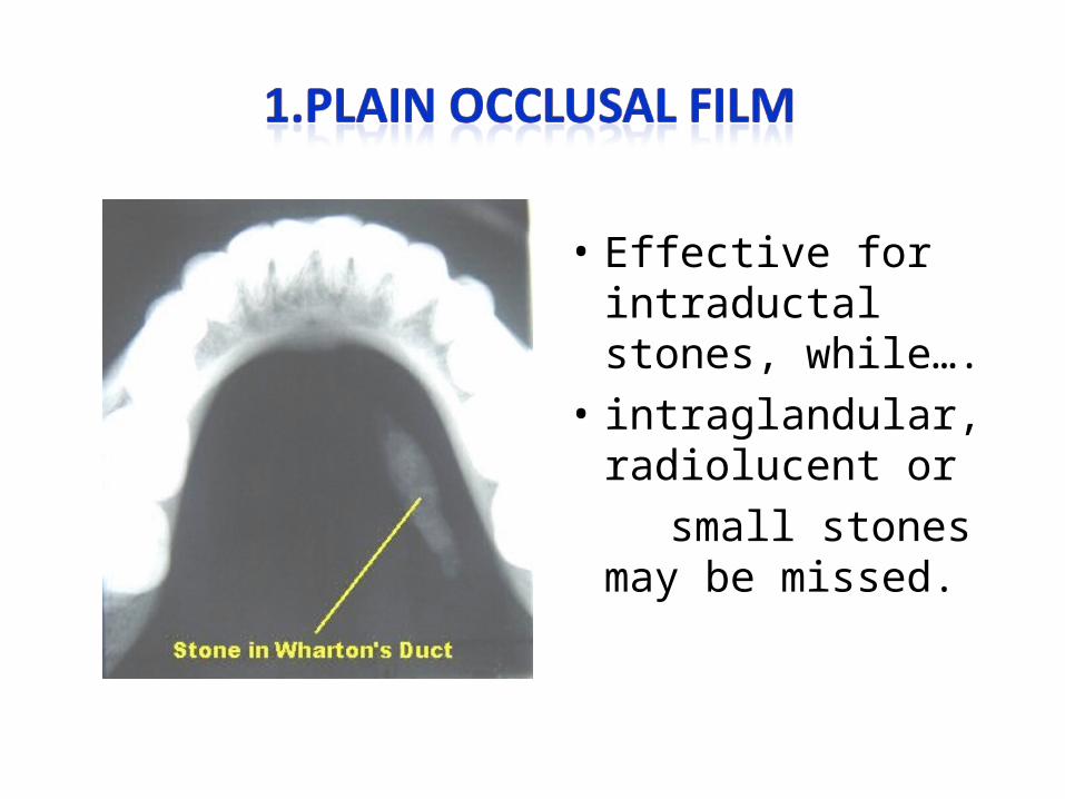
• Effective for intraductal stones, while….
• intraglandular, radiolucent or
small stones may be missed.
• Large stones or small CT slices done.
• Also used for inflammatory disorders
• Operator dependent, can detect small stones
(>2mm), inexpensive, non-invasive
• Consists of opacification of the ducts by a retrograde injection of a water-soluble dye.
• Provides image of stones and duct morphological structure
• May be therapeutic, but success of therapeutic sialography never documented
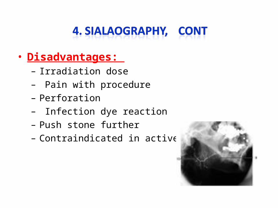
• Disadvantages: – Irradiation dose– Pain with procedure– Perforation– Infection dye reaction– Push stone further– Contraindicated in active infection.
• is useful preoperatively to determine if gland is functional.
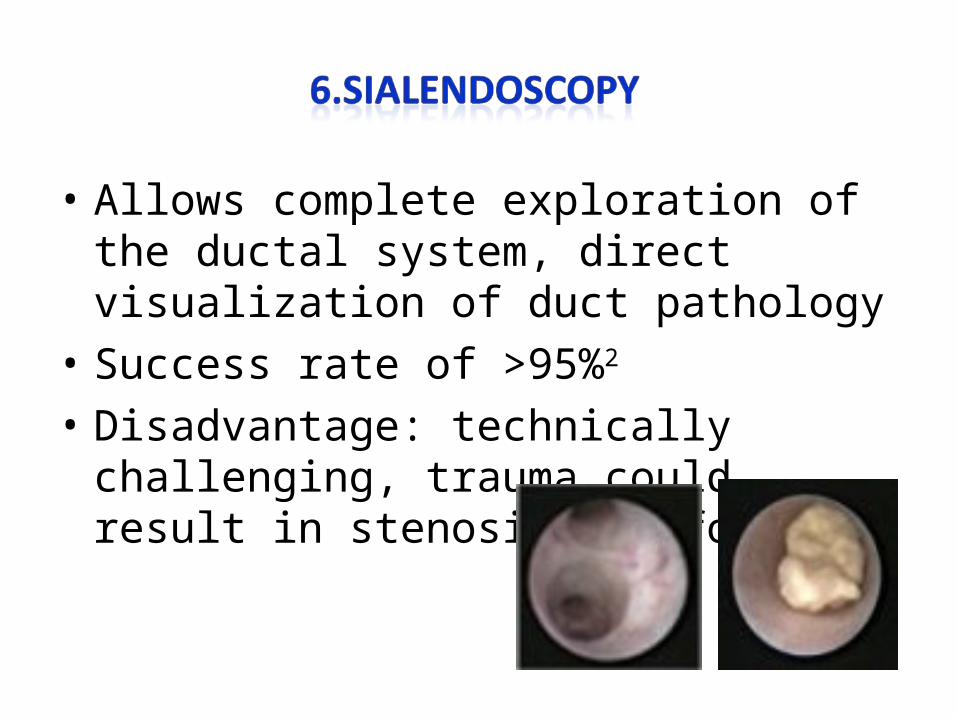
• Allows complete exploration of the ductal system, direct visualization of duct pathology
• Success rate of >95%2
• Disadvantage: technically challenging, trauma could result in stenosis, perforation
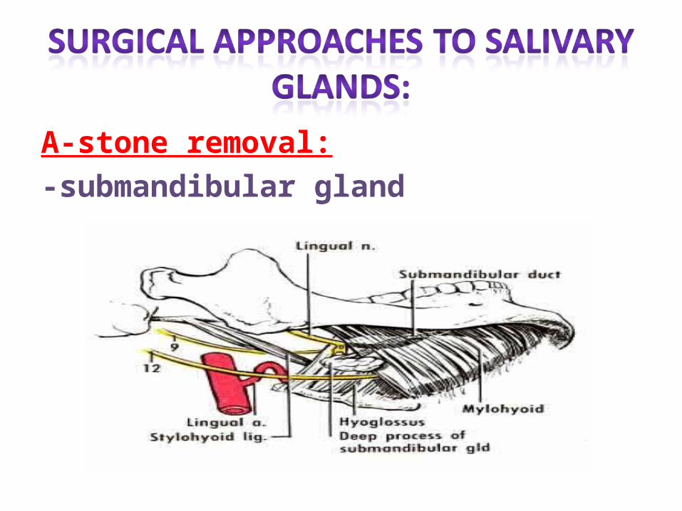
A-stone removal:-submandibular gland
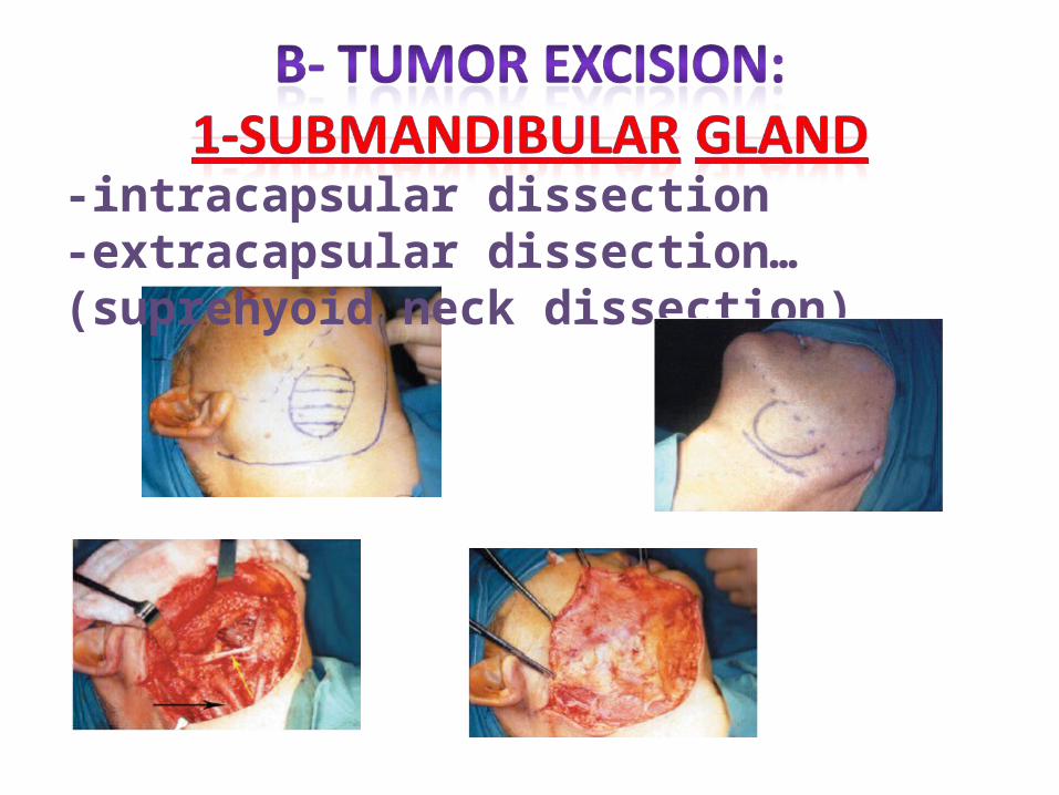
-intracapsular dissection -extracapsular dissection…(suprehyoid neck dissection)
• So what are the indications of removal of the submandibular
gland???
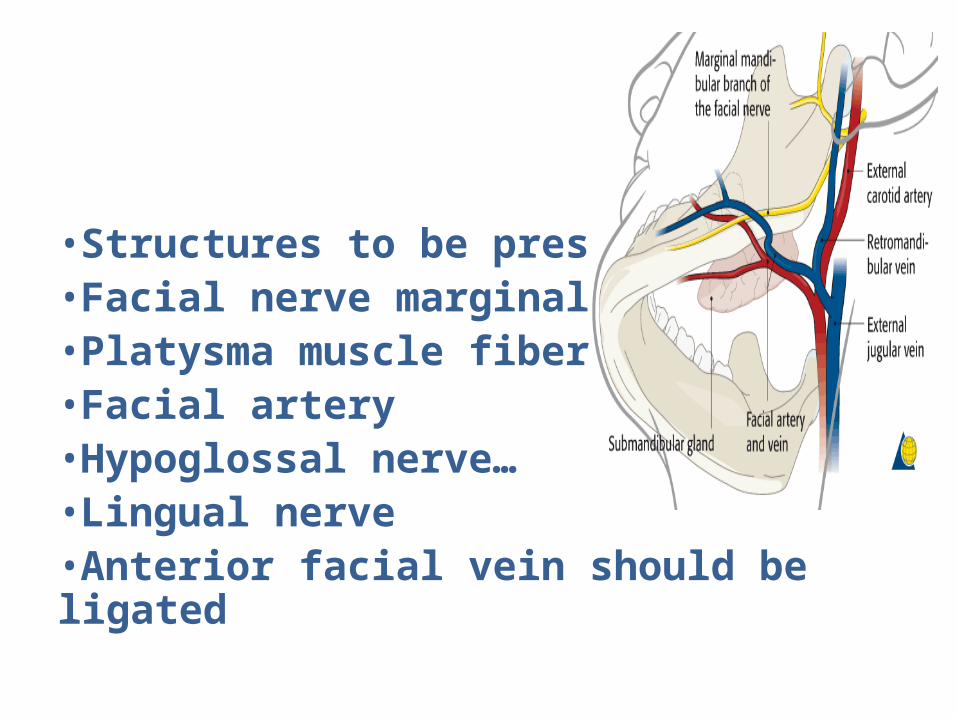
•Structures to be preserved:•Facial nerve marginal branch•Platysma muscle fibers…•Facial artery•Hypoglossal nerve…•Lingual nerve•Anterior facial vein should be ligated
• Hematoma• wound infection • marginal mandibular nerve injury• lingual nerve injusry • hypoglossal nerve injury• transection of the nerve to the myelohyoid
muscle causing submental skin anesthesia…
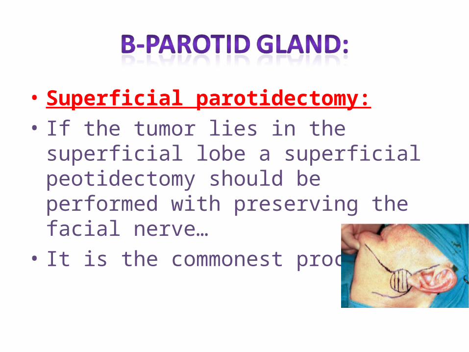
• Superficial parotidectomy:• If the tumor lies in the superficial lobe a
superficial peotidectomy should be performed with preserving the facial nerve…
• It is the commonest procedure…
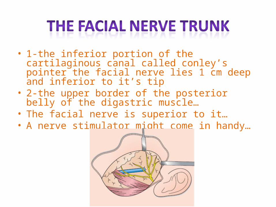
• 1-the inferior portion of the cartilaginous canal called conley’s pointer the facial nerve lies 1 cm deep and inferior to it’s tip
• 2-the upper border of the posterior belly of the digastric muscle…
• The facial nerve is superior to it…• A nerve stimulator might come in handy…
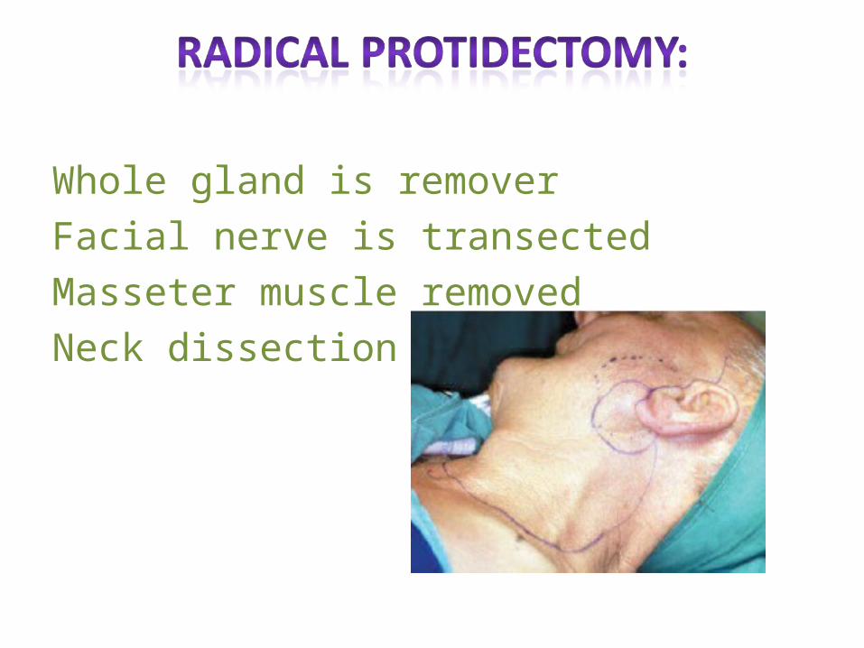
Whole gland is removerFacial nerve is transectedMasseter muscle removedNeck dissection
• Hematoma• Infection• Temporary facial nerve weakness.• Transection of the facial nerve and permenant
facial weakness..• Sialocele…• Facial numbness.• Permenant numbness of the ear lobe due to
transection of the great auricular nerve…• Frey’s syndrome
• Cause…• Prevention…• Treatment is incidence…• Antiperspirants like ALCL• Denervation by tympanic neurectomy• Injection of botulinum toxin to the skin area
refrences
• Baily and love’s• Schwart’s• Browse• Manual of clinical syrgery…