Embed Size (px)
Citation preview
DOI:10.5125/jkaoms.2010.36.3.228
228
Ⅰ. Introduction
Sarcomatoid carcinoma is a rare and occurs mainly in the
upper aerodigestive tract such as the oral cavity, esophagus
and vocal cords. It is a unique variant of squamous cell carci-
noma consisting of sarcomatoid proliferation of plemorphic
spindle-shaped cells sometimes with a biphasic appearance
presenting as a part of frank squamous cell carcinoma1,2. This
type of carcinoma has been described using various terms,
including spindle cell squamous carcinoma, carcinosarcoma,
pseudosarcoma, and pleomorphic carcinoma1.
We report the case of a patient with spindle cell squamous
cell carcinoma involving the mandible.
Ⅱ. Report of the case
An 80-year-old male visited to our hospital because of pain-
less submandibular swelling. The swelling appeared 2 weeks
ago and decreased after lower teeth extraction. His medical
history included surgical treatment for cerebral abscess after
admission one month ago. The patient didn’t take any medi-
cine. He was not a smoker.
On intraoral examination, tooth extracted area was complete-
ly healed state. Oral mucosa and gingiva appeared normal col-
or and texture but a hard mass was palpable on right lower
vestibular area. The tongue and floor of mouth was not elevat-
ed. On extraoral examination, a hard submandibular mass was
palpable but overlying skin was normal appearance. On
panoramic view, any pathologic findings were not showed. We
recommended further radiographic examinations to him and
his family but that was refused by him.
One week later, he was referred from the department of neu-
rosurgery to evaluate the right lower gingival mass after
admission. He was admitted to the department of neurosurgery
for seizure and abnormal behavior two days before. Clinical
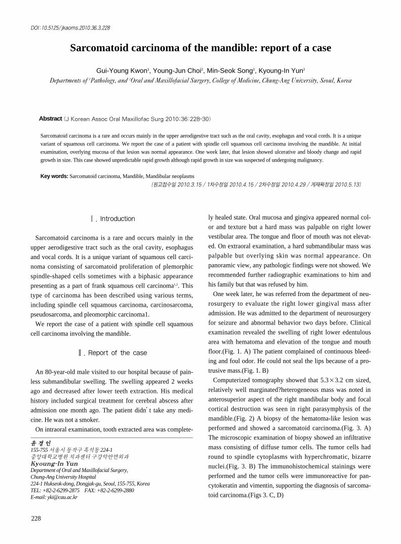
examination revealed the swelling of right lower edentulous
area with hematoma and elevation of the tongue and mouth
floor.(Fig. 1. A) The patient complained of continuous bleed-
ing and foul odor. He could not seal the lips because of a pro-
trusive mass.(Fig. 1. B)
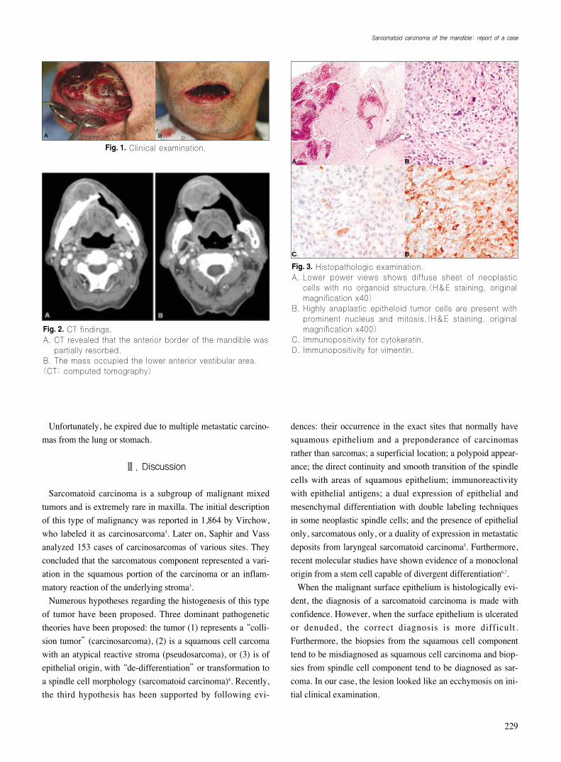
Computerized tomography showed that 5.3×3.2 cm sized,
relatively well marginated?heterogeneous mass was noted in
anterosuperior aspect of the right mandibular body and focal
cortical destruction was seen in right parasymphysis of the
mandible.(Fig. 2) A biopsy of the hematoma-like lesion was
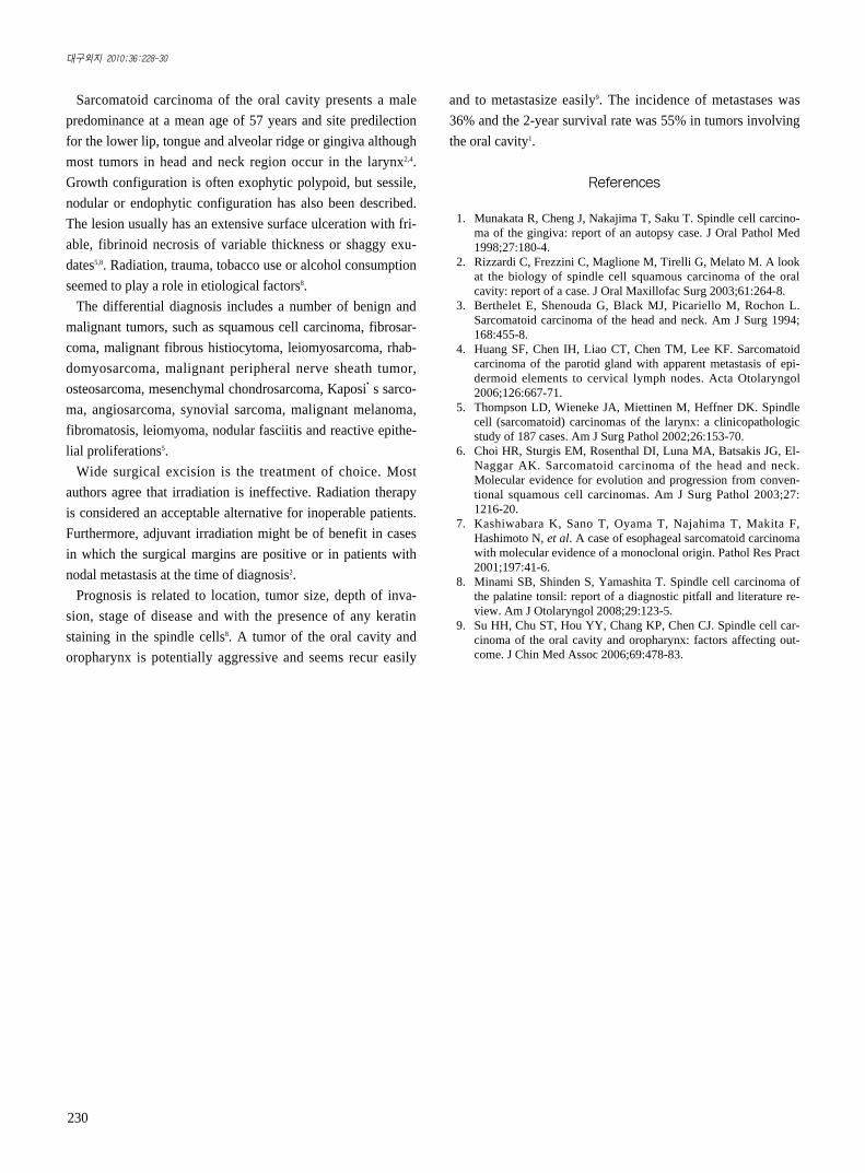
performed and showed a sarcomatoid carcinoma.(Fig. 3. A)
The microscopic examination of biopsy showed an infiltrative
mass consisting of diffuse tumor cells. The tumor cells had
round to spindle cytoplasms with hyperchromatic, bizarre
nuclei.(Fig. 3. B) The immunohistochemical stainings were
performed and the tumor cells were immunoreactive for pan-
cytokeratin and vimentin, supporting the diagnosis of sarcoma-
toid carcinoma.(Figs 3. C, D)
윤 경 인155-755 서울시동작구흑석동 224-1중앙 학교병원치과센터구강악안면외과Kyoung-In YunDepartment of Oral and Maxillofacial Surgery,Chung-Ang University Hospital224-1 Hukseok-dong, Dongjak-gu, Seoul, 155-755, KoreaTEL: +82-2-6299-2875 FAX: +82-2-6299-2880E-mail: [email protected]
Sarcomatoid carcinoma of the mandible: report of a case
Gui-Young Kwon1, Young-Jun Choi2, Min-Seok Song2, Kyoung-In Yun2
Departments of 1Pathology, and 2Oral and Maxillofacial Surgery, College of Medicine, Chung-Ang University, Seoul, Korea
Sarcomatoid carcinoma is a rare and occurs mainly in the upper aerodigestive tract such as the oral cavity, esophagus and vocal cords. It is a unique
variant of squamous cell carcinoma. We report the case of a patient with spindle cell squamous cell carcinoma involving the mandible. At initial
examination, overlying mucosa of that lesion was normal appearance. One week later, that lesion showed ulcerative and bloody change and rapid
growth in size. This case showed unpredictable rapid growth although rapid growth in size was suspected of undergoing malignancy.
Key words: Sarcomatoid carcinoma, Mandible, Mandibular neoplasms
[원고접수일 2010.3.15 / 1차수정일 2010.4.15 / 2차수정일 2010.4.29 / 게재확정일 2010.5.13]
Abstract (J Korean Assoc Oral Maxillofac Surg 2010;36:228-30)
229
Unfortunately, he expired due to multiple metastatic carcino-
mas from the lung or stomach.
Ⅲ. Discussion
Sarcomatoid carcinoma is a subgroup of malignant mixed
tumors and is extremely rare in maxilla. The initial description
of this type of malignancy was reported in 1,864 by Virchow,
who labeled it as carcinosarcoma3. Later on, Saphir and Vass
analyzed 153 cases of carcinosarcomas of various sites. They
concluded that the sarcomatous component represented a vari-
ation in the squamous portion of the carcinoma or an inflam-
matory reaction of the underlying stroma3.
Numerous hypotheses regarding the histogenesis of this type
of tumor have been proposed. Three dominant pathogenetic
theories have been proposed: the tumor (1) represents a “colli-
sion tumor”(carcinosarcoma), (2) is a squamous cell carcoma
with an atypical reactive stroma (pseudosarcoma), or (3) is of
epithelial origin, with “de-differentiation”or transformation to
a spindle cell morphology (sarcomatoid carcinoma)4. Recently,
the third hypothesis has been supported by following evi-
dences: their occurrence in the exact sites that normally have
squamous epithelium and a preponderance of carcinomas
rather than sarcomas; a superficial location; a polypoid appear-
ance; the direct continuity and smooth transition of the spindle
cells with areas of squamous epithelium; immunoreactivity
with epithelial antigens; a dual expression of epithelial and
mesenchymal differentiation with double labeling techniques
in some neoplastic spindle cells; and the presence of epithelial
only, sarcomatous only, or a duality of expression in metastatic
deposits from laryngeal sarcomatoid carcinoma5. Furthermore,
recent molecular studies have shown evidence of a monoclonal
origin from a stem cell capable of divergent differentiation6,7.
When the malignant surface epithelium is histologically evi-
dent, the diagnosis of a sarcomatoid carcinoma is made with
confidence. However, when the surface epithelium is ulcerated
or denuded, the correct diagnosis is more difficult.
Furthermore, the biopsies from the squamous cell component
tend to be misdiagnosed as squamous cell carcinoma and biop-
sies from spindle cell component tend to be diagnosed as sar-
coma. In our case, the lesion looked like an ecchymosis on ini-
tial clinical examination.
Fig. 2. CT findings.
A. CT revealed that the anterior border of the mandible was
partially resorbed.
B. The mass occupied the lower anterior vestibular area.
(CT: computed tomography)
Fig. 3. Histopathologic examination.
A. Lower power views shows diffuse sheet of neoplastic
cells with no organoid structure.(H&E staining, original
magnification x40)
B. Highly anaplastic epitheloid tumor cells are present with
prominent nucleus and mitosis.(H&E staining, original
magnification x400)
C. Immunopositivity for cytokeratin.
D. Immunopositivity for vimentin.
Fig. 1. Clinical examination.
Sarcomatoid carcinoma of the mandible: report of a case
구외지 2010;36:228-30
230
Sarcomatoid carcinoma of the oral cavity presents a male
predominance at a mean age of 57 years and site predilection
for the lower lip, tongue and alveolar ridge or gingiva although
most tumors in head and neck region occur in the larynx2,4.
Growth configuration is often exophytic polypoid, but sessile,
nodular or endophytic configuration has also been described.
The lesion usually has an extensive surface ulceration with fri-
able, fibrinoid necrosis of variable thickness or shaggy exu-
dates5,8. Radiation, trauma, tobacco use or alcohol consumption
seemed to play a role in etiological factors8.
The differential diagnosis includes a number of benign and
malignant tumors, such as squamous cell carcinoma, fibrosar-
coma, malignant fibrous histiocytoma, leiomyosarcoma, rhab-
domyosarcoma, malignant peripheral nerve sheath tumor,
osteosarcoma, mesenchymal chondrosarcoma, Kaposi’s sarco-
ma, angiosarcoma, synovial sarcoma, malignant melanoma,
fibromatosis, leiomyoma, nodular fasciitis and reactive epithe-
lial proliferations5.
Wide surgical excision is the treatment of choice. Most
authors agree that irradiation is ineffective. Radiation therapy
is considered an acceptable alternative for inoperable patients.
Furthermore, adjuvant irradiation might be of benefit in cases
in which the surgical margins are positive or in patients with
nodal metastasis at the time of diagnosis2.
Prognosis is related to location, tumor size, depth of inva-
sion, stage of disease and with the presence of any keratin
staining in the spindle cells8. A tumor of the oral cavity and
oropharynx is potentially aggressive and seems recur easily
and to metastasize easily9. The incidence of metastases was
36% and the 2-year survival rate was 55% in tumors involving
the oral cavity1.
References
1. Munakata R, Cheng J, Nakajima T, Saku T. Spindle cell carcino-ma of the gingiva: report of an autopsy case. J Oral Pathol Med1998;27:180-4.
2. Rizzardi C, Frezzini C, Maglione M, Tirelli G, Melato M. A lookat the biology of spindle cell squamous carcinoma of the oralcavity: report of a case. J Oral Maxillofac Surg 2003;61:264-8.
3. Berthelet E, Shenouda G, Black MJ, Picariello M, Rochon L.Sarcomatoid carcinoma of the head and neck. Am J Surg 1994;168:455-8.
4. Huang SF, Chen IH, Liao CT, Chen TM, Lee KF. Sarcomatoidcarcinoma of the parotid gland with apparent metastasis of epi-dermoid elements to cervical lymph nodes. Acta Otolaryngol2006;126:667-71.
5. Thompson LD, Wieneke JA, Miettinen M, Heffner DK. Spindlecell (sarcomatoid) carcinomas of the larynx: a clinicopathologicstudy of 187 cases. Am J Surg Pathol 2002;26:153-70.
6. Choi HR, Sturgis EM, Rosenthal DI, Luna MA, Batsakis JG, El-Naggar AK. Sarcomatoid carcinoma of the head and neck.Molecular evidence for evolution and progression from conven-tional squamous cell carcinomas. Am J Surg Pathol 2003;27:1216-20.
7. Kashiwabara K, Sano T, Oyama T, Najahima T, Makita F,Hashimoto N, et al. A case of esophageal sarcomatoid carcinomawith molecular evidence of a monoclonal origin. Pathol Res Pract2001;197:41-6.
8. Minami SB, Shinden S, Yamashita T. Spindle cell carcinoma ofthe palatine tonsil: report of a diagnostic pitfall and literature re-view. Am J Otolaryngol 2008;29:123-5.
9. Su HH, Chu ST, Hou YY, Chang KP, Chen CJ. Spindle cell car-cinoma of the oral cavity and oropharynx: factors affecting out-come. J Chin Med Assoc 2006;69:478-83.


![Case Report Prostatic Carcinosarcoma with Lung Metastases · 2019. 7. 31. · Case Reports in Oncological Medicine sarcomatoid carcinoma [ [] C. G. Rogers, A. Parwani, A. Tekes, M](https://img.pdfslide.net/doc/110x75/6106208aa9c710750f2e2726/case-report-prostatic-carcinosarcoma-with-lung-metastases-2019-7-31-case-reports.jpg)