Embed Size (px)
Citation preview
Schistocytes in megaloblastic anemia
Barbara J. Bain*
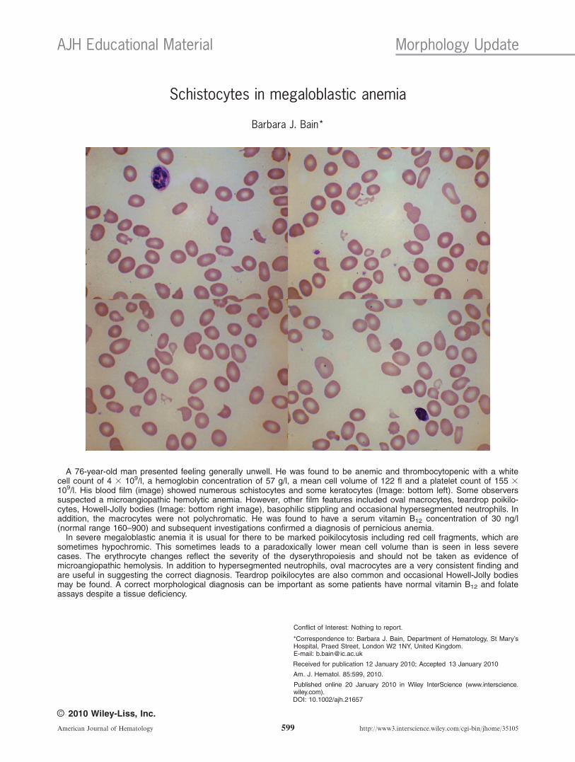
A 76-year-old man presented feeling generally unwell. He was found to be anemic and thrombocytopenic with a whitecell count of 4 3 109/l, a hemoglobin concentration of 57 g/l, a mean cell volume of 122 fl and a platelet count of 155 3109/l. His blood film (image) showed numerous schistocytes and some keratocytes (Image: bottom left). Some observerssuspected a microangiopathic hemolytic anemia. However, other film features included oval macrocytes, teardrop poikilo-cytes, Howell-Jolly bodies (Image: bottom right image), basophilic stippling and occasional hypersegmented neutrophils. Inaddition, the macrocytes were not polychromatic. He was found to have a serum vitamin B12 concentration of 30 ng/l(normal range 160–900) and subsequent investigations confirmed a diagnosis of pernicious anemia.In severe megaloblastic anemia it is usual for there to be marked poikilocytosis including red cell fragments, which are
sometimes hypochromic. This sometimes leads to a paradoxically lower mean cell volume than is seen in less severecases. The erythrocyte changes reflect the severity of the dyserythropoiesis and should not be taken as evidence ofmicroangiopathic hemolysis. In addition to hypersegmented neutrophils, oval macrocytes are a very consistent finding andare useful in suggesting the correct diagnosis. Teardrop poikilocytes are also common and occasional Howell-Jolly bodiesmay be found. A correct morphological diagnosis can be important as some patients have normal vitamin B12 and folateassays despite a tissue deficiency.
Conflict of Interest: Nothing to report.
*Correspondence to: Barbara J. Bain, Department of Hematology, St Mary’sHospital, Praed Street, London W2 1NY, United Kingdom.E-mail: [email protected]
Received for publication 12 January 2010; Accepted 13 January 2010
Am. J. Hematol. 85:599, 2010.
Published online 20 January 2010 in Wiley InterScience (www.interscience.wiley.com).DOI: 10.1002/ajh.21657
AJH Educational Material Morphology Update
VVC 2010 Wiley-Liss, Inc.
American Journal of Hematology 599 http://www3.interscience.wiley.com/cgi-bin/jhome/35105