Embed Size (px)
Citation preview
Antigenic Protein of Leucocytozoon caulleryi
schizont Inducing Cellular Immune Response:
TLR-2 and CD4 as Marker
Nunuk Dyah Retno Lastuti1a) Dony Chrismanto2)
Endang Suprihati1) 2)Study Program of Animal Health, Faculty of Vocation 1) Department of Parasitology, Faculty of Veterinary Medicine Universitas Airlangga, Surabaya
Universitas Airlangga, Surabaya Anwar Ma’ruf 3)
a) Corresponding Author: [email protected] 3) Department of Basic Medical Science
Universitas Airlangga, Surabaya
Abstract: Leucocytozoonosis is caused by Leucocytozoon
caulleryi and is responsible for death in chickens by bleeding.
Leucocytozoonosis is an endemic disease in Indonesia and
incidences have been reported in several regions in East and
Central Java. The financial losses caused by this disease
include growth disorders in chick, decreased egg production,
higher mortality rate and also a higher production cost. This
research aims to detect TLR-2 and CD4 expression as a
cellular immune response in rabbits immunised by the protein
of L.caulleryi schizont. It is needed as a preliminary research
study for molecular vaccine development which is
considerably effective when it comes to preventing
leucocytozoonosis occurance in Indonesia. This research study
was performed in several stages i.e. the isolation of
L.caulleryi schizont from a chicken liver infected with
leucocytozoonosis to be based on the clinical signs observed,
microscopic examination, and the pathological changes in the
other chicken organs. The purification of the soluble protein of
L.caulleryi schizont including the immunisation of the rabbits.
Each of the experimental rabbits was injected with 500 µg of
L.caulleryi schizont protein and added adjuvant complete with
a ratio of 1:1. Every two weeks the injection was performed
with the same protein with a dosage of 500 µg each and an
added adjuvant that was incomplete (the booster was
performed 5 times in 2 weeks). The examination of the
cellular immune response of CD4 and TLR-2 expression in the
rabbits’ T cells using an immunocytochemistry method
visualised by fluorescein isothiocyanate. The examination of
the results was done by immunocytochemistry showing TLR-2
and CD4 expression as yellow to green fluorescent colour,
mainly in the 5th booster where the activation of the CD4 co-
receptor and TLR-2 occurred. The conclusion shows that the
antigenic protein of L.caulleryi schizont has the ability to
induce a cellular immune response through the co-receptors
CD4 and TLR-2 in the rabbits’ T cells as the preliminary
research in the sub-unit vaccine development for
leucocytozoonosis in chickens.
KEY WORD: antigenic protein, Leucocytozoon caulleryi,
schizont, TLR-2, CD4.
I. INTRODUCTION
Leucocytozoonosis is one of the diseases caused by
Leucocytozoon caulleryi in poultry, which is transmitted by
flies Culicoides sp. or Simulium sp. Leucocytozoonosis is an
endemic disease in Indonesia and incidences have been
reported in several regions in East and Central Java.
Leucocytozoon parasites infect a large number of avian hosts,
including the domestic chicken, and causes a significant
economical loss to the poultry industry (1). The financial
losses impacted by this disease include growth disorders in
chicks, decreased egg production, higher mortality rate and
also a higher production cost (2, 3, 4, 6). The occurrence of
leucocytozoonosis in broiler is between 7-40%, while the
mortality rate in chicks is about 7-50% and in layer is about 2-
60% respectively (7). The clinical signs observed in chickens
are green faeces, depression, a loss of appetite, vomiting
blood, paralysis and death due to bleeding (8). In order to
overcome leucocytozoonosis in chickens, farmers have carried
out the dispensing of medications, eradicating flies using
insecticides, and improving the water irrigation in the area
around the henhouse, but the latter method is less efficient due
to the rapid growth of flies. Based on the vaccine developed
by Onaga et al (1999), chickens can be protected from
L.caulleryi infection by giving them a second generation
schizont extract and blood serum containing an antigen (5).
The weakness of this live vaccine administration is due to the
possibility of infection because the parasites may become
pathogens when the host’s condition is weakened. Molecular
vaccine development is considerably more effective when it
comes to preventing leucocytozoonosis occurance in
67Copyright © 2018, the Authors. Published by Atlantis Press. This is an open access article under the CC BY-NC license (http://creativecommons.org/licenses/by-nc/4.0/).
Advances in Social Science, Education and Humanities Research (ASSEHR), volume 981st International Conference Postgraduate School Universitas Airlangga:
Implementation of Climate Change Agreement to Meet Sustainable Development Goals (ICPSUAS 2017)
Indonesia. Based on the phylogenetic analysis of Cytochrom B
Leucocytozoon spp in broilers, it was shown that the
L.caulleryi from various endemic regions is highly
homologous (>95%) (6). Referring to the problems above, a
preliminary research about the rabbits’ immune response
induced by L. caulleryi schizont protein with TLR-2 and CD4
as marker to explore the cellular immune response is
necessary.
TLR is a membrane protein that helps receptor
recognition patterns in response to various molecule
derivatives from microbes and stimulated innate immunity due
to microbe molecule exposure. TLR is known to be a
recognition receptor which is involved in pathogen-associated
molecular patterns (PAMP) recognised by pattern recognition
molecules (PRMs). A phagocytes development system in
recognising pathogens can be stimulated any time to respond
as an inflammatory system. TLR stimulation through
microbial product initiates the signalling pathways which
activate not only the innate immunity but also adaptive
immunity (9, 10). CD4+T cells play a central role in immune
protection and the B cells to produce antibodies, to induce the
macrophages to develop enhanced microbicidal activity, to
recruit neutrophils, eosinophils, and basophils to the sites of
infection and inflammation, and through their production of
cytokines and chemokines (11). As a vaccine kit candidate, it
is required to know whether or not the L.caulleryi protein can
induce either humoral or cellular immune response, because
then a favourable immune response and immunogenic protein
can be explored. Immunogenic protein has main
characteristics such as a heavy molecule weight, homogeny
and a complex chemical structure and alienation (13, 15). It is
necessary to study whether the results of the immunogenic
proteins from a liver containing L. caulleryi can be developed
for use in a vaccine in the effort to overcome
Leucocytozoonosis, for example, with a vaccination program
for chickens with a vaccine sub-unit that is safe in its use.
II. METHODS
A. Isolation and identification of L.caulleryi schizont
Schizont L.caulleryi isolated from chicken liver infected by
leucocytozoonosis is based on the clinical signs observed,
microscopic examination and the pathological changes of
other chicken organs. Microscopic examination was
performed to detect any gametosit stadium developed in
eritrosit. Then, further assesment was done on several other
organs such as the liver, spleen and intestine to detect the
schizont stadium by crushing the organ and a pathological
examination. The chicken liver and spleen containing schizont
L.caulleryi was isolated in 50-100 mg or 0.05 ml cultured wet
pellets, with a 2-3 ml 2-Drehydration solution/sample buffer
added. Next, the sample was put on ice, sonicated for 30
seconds and cooled down to -80°C for 5 minutes. This
treatment was repeated four times. The sample was
centrifuged using a microcentrifuge (16.000 x g) for 20-30
minutes at 18-20 °C, and was then taken out from the
centrifuge, Supernatan was put into a clean tube and the
sample was stored at -80°C.
B. Immunization of rabbits
This research used five rabbits treated as per the animal
welfare concept. They were given a health examination based
on both clinical symptoms and laboratory tests. All of the
animals were handled in strict accordance with Ethical
Clearance and the experiment was approved by the Ethical
Committee of the Faculty of Veterinary Medicine, of the
Universitas Airlangga, No: 630-KE. Four-month old naïve
rabbits were prepared for the immunisation trial at the
laboratory of experimental animals, in the Faculty of
Veterinary Medicine at the Universitas Airlangga. Each of the
experimental rabbits were injected with 500 µg L.caulleryii
schizont protein (0.3 ml) with an added Freund adjuvant
complete (Sigma, USA) with a ratio of 1:1. The injection was
performed every two weeks with the same protein with a
dosage of 500 µg each and with the added adjuvant
incomplete (Sigma, USA). The immunisation (booster) was
performed 5 times in 2 weeks. Prior to the first injection, about
10 ml of rabbit blood was taken for TLR-2 and CD4
examination as preliminary data (control) and whole blood
examination was conducted at the end of the first booster until
the fifth booster (13, 15).
C. Examination of TLR 2 and CD4 expression using
Immunocytochemistry
The principle of immunocytochemistry examination is that it
is an immunology technique used to visualise specific proteins
or antigens in the cells using the first antibody
(www.abcam.com/index html). Several stages of the
examination will be explained: 1) the blood sample is washed
using 10% PBS-T20 five times, and the sample is fixated with
100% methanol (10 minutes) or with paraformaldehyde in
PBS pH 7,4 for 15 minutes at room temperature, 2) the sample
is washed twice using cold PBS, the sample is incubated for
10 minutes in PBS consisting of 0.1% Triton X-100 or 100
mM digitonin, and then the cells are washed in PBS three
times for 5 minutes, 3) the cells are incubated with 1% BSA in
PBS-T20 for 30 minutes, and incubated in conjugated
antibody TLR 2-FITC labelled (Abcam’s RabMab, USA) and
CD4-FITC labeled (Abcam’s RabMab, USA) and diluted in
1% BSA in PBS-T20 at room temperature for an hour or at
night at a temperature of 4°C, 4) the cells are washed three
times in PBS (5 minutes for each washing). The results were
examined by using a fluorescent microscope using a
magnification of 200 times, to find out whether the yellow to
green fluorescent colour from the T cells expresses TLR-2 and
CD4.
68
Advances in Social Science, Education and Humanities Research (ASSEHR), volume 98
III. RESULTS AND DISCUSSION
The cellular immune response was shown by the
expression of TLR-2 and CD4 in the rabbit T cells marked by
the yellow to green fluorescent colour after the rabbit
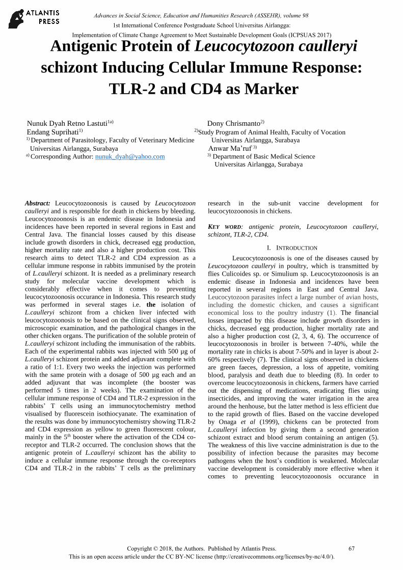
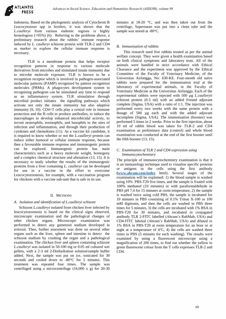
immunisation (Figure 1 and Figure 2).
Figure 1. TLR2 expression in rabbit T cells visualized by
Fluorescein Isothiocyanate (FITC) (200x magnification).
Figure 2. CD4 expression in rabbit T cells visualized by
Fluorescein Isothiocyanate (FITC) (200x magnification).
It has been known that the toll-like receptor (TLR) is
a receptor that can recognise the ligand from microbes or
parasites which are involved in innate immunity. Along with
new developments, TLR not only plays a role in innate
immunity but also in adaptive immunity (12, 13). The
examination results from the immunocytochemistry shows
that there is a yellow to green fluorescent colour visualised by
fluorescein isothiocyanate (FITC). It shows that the stadium
schizont L.caulleryi protein has a ligand that is recognised by
TLR 2 as a form of signal transduction which actívates and
induces a cellular immune response such as the lympocyte T
of the rabbits after immunisation. In accordance with the
principles of the immunisation method, which is an increasing
degree of immunity, it provides protective immunity by
inducing a memory response towards a specific pathogen with
a non-virulent or non-toxic antigen (14).
According the results of the research, it is shown that
the antibody TLR-2 can recognise the ligand from the protein
antigen L.caulleryi schizont by stimulating T cell activation,
marked by the presence of yellow to green fluorescent colour
which increased in accordance with the treatment from the
various boosters. When the antigen of L.caulleryi schizont
enters the body, it will be caught by macrophage or dendritic
cells and the phagocytes cells will be activated by TLR as a
signal transducer. L.caulleryi schizont possess a ligand or
pathogen-associated molecular pattern (PAMP) which is
recognised by TLR-2. Ligands that are recognised by TLR-2
consist of lipoprotein/lipopolypeptide, flagelin, ssRNA and
CpG DNA. The schizont of L.caulleryi is an intracellular
microorganism which contains antigens. When the antigen
enters the body, it will be caught by a macrophage and the
phagocytes cells will be activated by the TLR as a signalling
pathway. The signal produced by TLR will activate the
transcription factor NFB which stimulates cytokine
production (10, 16). NFB activation is initiated by a signal
which recruits MyD88 and interacts with the IL-1 receptor
associated kinase (IRAK). Autophosphorylation then occurs,
separating MyD88 and activating the TNF receptor associated
factor 6 (TRAF-6) to activate the IB kinase (IKK). Activated
IKK will activate NFB to transcript gene IL-12, IL-10, IL-4,
TNF-, IFN-. IL-2 roles will increase the cytolytic activity
from the cytolytic T lymphocytes. This will also promote Th1
cells development together with CD8 activation in order to
produce IL-2 which stimulates the proliferation and
differentiation of B cells that will produce antibodies. IL-4 is a
cytokine which is produced by subset Th2 from Th cells CD4
that function to induce Th2 cells differentiation and stimulate
IgE production. (17, 18).
The main function of CD4 is acting as a transduction
signal in antigen recognition and to strengthen the bond
between T cells and antigen-presenting cells (APC). APC
produces IFN- and IL-12 that stimulate the differentiation of
CD4+ cells into Th1 which plays a major role in delayed
hypersensitivity reactions. CD4+ T-cells produce a protein
named IL-4 cytokine which helps B lymphocytes in antibody
production and phagocytosis to destroy ingested microbes (11,
17). CD4 molecules as a co-receptor are a surface cell
molecule which are expressed by various types of cells in the
immune system which were formed by cluster differentiation.
The accessory molecule is used as a marker of Th cell
activation with B cells and cytotoxic T cells maturation,
which is responsible in regulating chronic inflammatory
reactions towards antigens through macrophage stimulation.
Lymphocytes B activation is marked by a significant increase
(p<0,05) in the antibody titer of rabbits injected with the
protein of L.caulleryi schizont (15, 19). As a vaccine kit
candidate, the schizont L.caulleryi antigen injected in to
rabbits needs to induce a cellular immune response, marked by
T cell lymphocte activation which expresses TLR 2 in
accordance with humoral immune response. There was an
antibody (IgG) titre enhancement produced by B lymphoctes
(14, 15).
69
Advances in Social Science, Education and Humanities Research (ASSEHR), volume 98
Conclusion
The antigenic protein of L.caulleryi schizont has the ability to
induce a cellular immune response through the expression of
TLR-2 and CD4 in the rabbits’ T cells. The TLR-2 signal
plays a role in innate immunity, but also in adaptive immunity.
The antigenic protein of L.caulleryi schizont may contain
ligand which acts as a receptor that is involved in pathogen-
associated molecular patterns (PAMP). This study is a
preliminary research to explore the immunogenic protein
which plays a role in immune system activation for future
studies in vaccine development to overcome
leucocytozoonosis in chickens.
Acknowledgment We would like to thank the Ministry of Research, Technology
and Higher Education in Indonesia. This study was supported
by research grant 2015. We would also like to thank the
Rector of Universitas Airlangga and the Director of Research
and Innovation Department, of Universitas Airlangga.
References
1. Abbas, A.K., Litchman, A.H. Cellular and Molecular Immunology.
5th. Ed. International Edition. Elsevier Saunders Inc. Philadelphia, Pennsylvania, 41-105, 411-432 (2005).
2. Gotanda T, Doi M, Eiguchi Y, Tanaka Y, Kobayashi S and Fujisaki Y, 2002. Characterization of Monoclonal Antibodies against the second generation schizonts of Leucocytozoon caulleryi. J Vet Med Sci. 64(3):281-283.
3. Imura T, Sato S, Sato Y, Sakamoto D, Isobe T, Murata K, Holder AA, Yukawa M. The apicoplast genome of Leucocytozoon caulleryi, a pathogenic apicomplexan parasite of the chicken. Parasitol Res 113:823-828, 2014.
4. Ito A, Gotanda T. The correlation of protective effects and antibody production in immunized chickens with recombinant R7 vaccine against Leucocytozoon caulleryi. J Vet Med Sci 64(5):405-411, 2002.
5. Lastuti, N.D.: Specific antigenic protein 57.3 kDa of Sarcoptes scabiei var. caprae as material candidate of scabies diagnostic kit for goat and Toll-like receptor mediated Immune Responses. Doctoral diss. Postgraduate Program. Universitas Airlangga (2009).
6. Lee HR, Koo BS, Jeon EO, Han MS, Min KC, Lee SB, Bae Y, Mo IP. Pathology and molecular characterization of recent
Leucocytozoon caulleryi cases in layer flocks. J Biomed Res, 30(6): 517–524, 2016.
7. Lastuti N.D, Suprihati E, Chrismanto D. Exploration of antigenic protein of Leucocytozoon caulleryi as diagnostic kit leucocytozoonosis on broiler chickens. Media Kedokteran Hewan 29 (3): 213-222 (2013).
8. Majewska M, Szczepanik M. The role of Toll-like receptors (TLR) in innate and adaptive immune responses and their function in immune response regulation. Postepy Hig Med Dosw (Online). 60:52-63 (2006).
9. Nakamura K, Ogiso M, Shibahara T, Kasuga H and Isobe T. Pathogenicity of Leucocytozoon caulleryi for specific Pathogen Free Laying Hens. J of Parasitol .87:1202-1204, 2001.
10. Nakata K, Watarai S, Kodama H, Gotanda T, Ito A, Kume K. Cellular immune responses in chickens induced by recombinant R7 Leucocytozoon caulleryi vaccine. J Parasitol 89(2):419-422 (2003).
11. Pasare C, Medzhitov,R. Control of B cell Responses by Toll-like receptor. Nature, 364-438, 2005.
12. Singh., Dimri, U., Sharma, B., Saxena, M.: Assessment of the cytokine profile in peripheral blood mononuclear cells of naturally Sarcoptes scabiei var.canis infested dogs. Veterinary Parasitology. 206:253-257 (2014).
13. Suprihati E. Phylogenetic analysis of gen cytochrome B Leucocytozoon sp. on broiler chicken in endemic area, Indonesia. Doctoral diss. Postgraduate Program. Universitas Airlangga, 2013.
14. Soekardono S, 1986. Chicken Leucocytozoonosis in Java and Bali. Doctoral diss. Postgraduate Program, Institut Pertanian Bogor, 12-15, 1986.
15. Vollmer, J.: CpG motifs to modulate innate and adaptive immune responses. Int Rev Immunol 25, 125-134 (2006).
16. Xiao, H, Li, X., Abbot, D.W. Analysis of TLR Expression, Regulation and Signalling. Signalling by Toll like receptor. Edited by Gregory WK. CRC Press. Taylor & Francis Group. Chapter 3, 39-55, 2008.
17. Yarovinsky, F.H., Kanzier., Hieny, S, Coffman, R.L, Sher, A.: Toll-like Receptor Recognation Regulated Immunodominance in an Antimicrobial CD4+Tcell Response. Immunity 25, 655-664 (2006).
18. Zhao W, Pang Q, Xu R, Liu J, Liu S, Li J, Monitoring the Prevalence of Leucocytozoon sabrazesi in Southern China and Testing Tricyclic Compounds against Gametocytes. PLoS ONE 11(8): e0161869. https://doi.org/10.1371/journal.pone.0161869, 2016.
19. Zhu J, Paul WE. CD4 T cells: fates, functions, and faults. Blood, Sep 1;112(5):1557-69. doi: 10.1182/blood-2008-05-078154. (2008).
70
Advances in Social Science, Education and Humanities Research (ASSEHR), volume 98