Embed Size (px)
Citation preview
Searching for microbesPart III.
Culture of bacteria & yeasts
Ondřej Zahradníčekto practical of VLLM0421c
contact to me:[email protected]
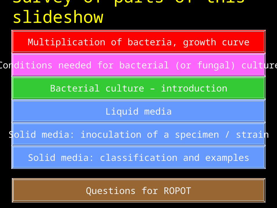
Survey of parts of this slideshow
Multiplication of bacteria, growth curve
Conditions needed for bacterial (or fungal) culture
Bacterial culture – introduction
Liquid media
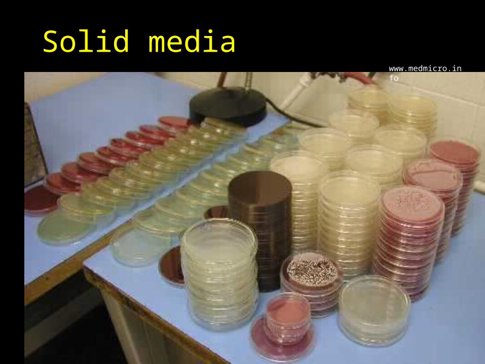
Solid media: inoculation of a specimen / strain
Solid media: classification and examples
Questions for ROPOT
Tale
• It was once a bacterium, and it was very small, and so without the microscope nobody has seen it, and in the microscope it was very difficult to see its shape. It was very unhappy, because it emphasized very much its beauty.
• Once, Mr. Koch came. He put the bacterium onto a solid medium and hide it into a thermostat. Bacterium was very happy and quickly started to multiply…
And Day After…• Mr. Koch came, opened the thermostat,
took out the Petri dish with medium, and he saw: his liked, tiny bacterium was visible by a naked eye! Of course, not as one bacterium, but as a strain of totally identical cells, that raised from that bacterium in form of a colony
• The bacterium was very happy, and it showed its beauty to mr. Koch. It showed him its shape, size, profile of its colony, pigments and many other things.

Multiplication of bacteria, growth curve
Multiplication of bacteria• Each bacteria has its generation period• During one gereration period one makes
two, in ten times of the period one makes 1024 bacteria (theoretically) and so on,.
• Ideal multiplication would exist only if we would add all the time nutrients and eventually oxygen and we would remove waste products.
Real growth curve
• Phase of latence – we let bacteria grow, but they still do not multiply
• Exponencial phase – growth accelerates
• Stational phase – they grow with the same speed all the time
• Slowing and stopping of growth – lack of nutrients, to many waste, or bacteria regulate themselves by „quorum sensing“

Conditions needed for bacterial (fun- gal) culture

Do the conditions for bacterial growth matter?
Of course they do! Majority of bacteria need their temperature, moisture, salts concetration and many other characteristics to be in a quite narrow range.
Various microbes need various conditions!
Values, that enable microbial survival, are not sufficient. They should be able to multiply.
lower survival limit (bactericidal)
upper survival limit (bactericidal)
lower growth limit (inhibitory)
lower growth limit (inhibitory)
Medically important bacteria• Temperature usually needed around 37 °C
– but bird pathogens more (42 °C), microbes coming from outer environment less (30 °C)
• Value of pH needed around pH 7– but gastric helicobacter by far less
• NaCl concentration needed around 0,9 % (physiological saline)– but staphylococci, that have to be able to
multiply on sweated skin, multiplies even at 10 % of salt!
In practice part of parameters (e. g. temperature) is derived from thermostat settings, and remainder (e. g. NaCl concentrations) by composition of the culture medium.
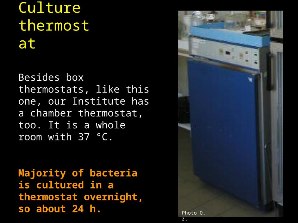
Culture thermostat
Besides box thermostats, like this one, our Institute has a chamber thermostat, too. It is a whole room with 37 °C.
Majority of bacteria is cultured in a thermostat overnight, so about 24 h.
Photo O. Z.
Example of use of relation to temparature in bacterial diagnostics
• Pseudomonas aeruginosa grows at 37 °C and 42 °C.
• On the other hand, Pseudomonas fluorescens grows at 4 °C and 37 °C.
Besides Pseudomonas, Listeria, too, and yeasts and molds grow at lower temperature.
Elevated temperature is suitable e. g. for Campylopacter

Influence of NaCl concentrations to the growth of some bacterial genera
6,5 %
10 %
Majority of Enterococci Staphylococci
bacteria
Besides various NaCl concentrations
• Adding of sodium azide enables growth of enterococci, but neither staphylococci, nor streptococci are able to grow
• Amikacin enables growth of streptococci and enterococci. Staphylococci, sensitive to amikacin, are eliminated.
A little about catabolism of bacteria
• We have three types of catabolism:– Fermentation – nutrients breakdown without need for
oxygen. Little energetically effective, but does not need oxygen. Forduct is e. g. lactate, ethanol etc.
– Aerobic respiration – little nutrients gives a lot of energy, but oxygen is needed. Product is CO2 and H2O
– Anaerobic respiration – another electron acceptor
• Type of catabolism is closely connected with relation to oxygen. Fermentating bacteria are usually facultatively or strictly anaerobic. On the other hand, aerobically respirating bacteria use to be strictly aerobic.
Relation to oxygen• Strict aerobes grow only in presence of
oxygen• Strict anaerobes they grow only in
environment without oxygen• Facultative anaerobes and
aerotolerant bacteria (it is not possible to differentiate them) grow at all conditions
• Microaerophile bacteria grow only in conditions with traces of oxygen
• Capnophile bacteria need more CO2
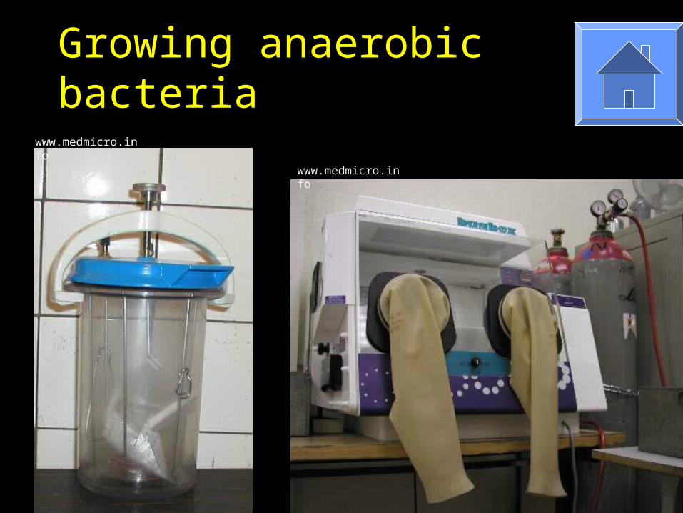
Growing anaerobic bacteria
www.medmicro.info
www.medmicro.info

Bacterial culture – introduction
Why we culture bacteria• Why bacteria are cultured in the
laboratory?– To keep them living and to multiply
them. This is gained by cultivation in both liquid and solid media (jelly-consistence media, based on agar algae)
– To obtain strain – solid media only; invented by Robert Koch
– To differentiate and divide them mutually –diagnostic and selective media are used, for identification
Specimen and strain• Specimen is taken from a patient. Specimen
contains cells macroorganism, various number of microbial species (zero to maybe twenty) and more items
• A strain – an isolate – is a population of one bacteria, isolated from a specimen on a solid medium
• To gain a strain, we have to grow a bacterium on a solid medium and inoculate carefully
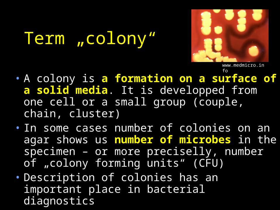
Term „colony“
• A colony is a formation on a surface of a solid media. It is developped from one cell or a small group (couple, chain, cluster)
• In some cases number of colonies on an agar shows us number of microbes in the specimen – or more preciselly, number of „colony forming units“ (CFU)
• Description of colonies has an important place in.bacterial diagnostics
www.medmicro.info
Solid mediawww.medmicro.info
Is it good, or bad, that various bacteria need different conditions?
• It is bad, because it complicates deffinition of conditions, that would be suitable for majority of bacteria
• It is good, because this enables to use it in diagnostics (e. g. growth ability on medium with 10 % NaCl differenciates well stafylococci)
Media globally versus media in medical microbiology
• In industrial microbiology or in some other applications we use mostly chemically defined media. We know their composition, and it is possible to observe, how much of something increased or decreased.
• In clinical microbiology we have no need to know a detailed composition. Often some parts of media are not definable (red blood cells, yeast extract).
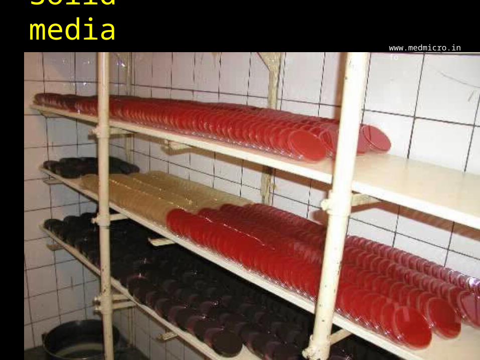
Liquid media and solid media
• Liquid media are based on je meat-peptonic broth (exctract of cooked beef meat + protein hydrolysate). They are used mostly to multiplication. It is difficult to evaluate the result, in fact, only „non turbid broth – turbid broth“ (growth – no growth)
• Majority of solid media are based on the same broth, but supplied by an agar alge extract. Bacteria grow slower on solid media, but the result is very variable, and it is possible to get a strain.
Various specimens – various cultivation• How the specimen type influences
culture?– Specimens, where microbes use to be
rare are inoculated into liquid media only. Microbes multiply quickly. Example: conjunctival swab
– Specimens, where the amout of microbes may vary, but even small amounts are important are cultured on both liquid and solid media. Example: wound swab
– Specimens, where usually we have many microbes, eventually even common microflora, are cultured on solid media only. Example: throat swab

Liquid media
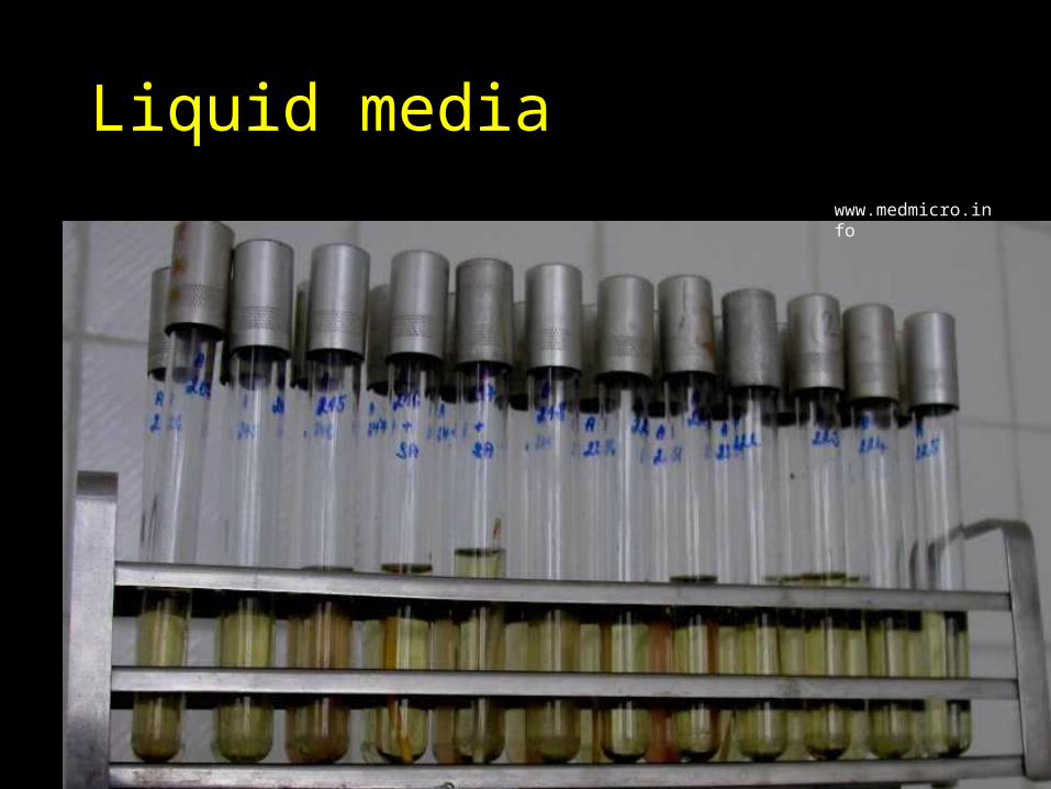
Liquid mediawww.medmicro.info
Classification of liquid media
• Liquid media have two categories only:
• multiplying media are common and universal. Example: broth for aerobic culture and VL-broth for anaerobic culture (VL = viande-levure, from french – contains meat-yeas extract)
• selectively multiplying media were developped to multilply some bacteria and to supress multiplication of other. Example: selenite broth for salmonella

Solid media: inoculation of specimen and strain
Solid mediawww.medmicro.info
Solid (agar) media• To have all advantages, given by solid
media, we have both the specimen musíme specimen (cultivation specimen strain), but also strain (cultivation strain strain) dilute properly at inoculation. Classical way of dilution inoculation is so named cross inoculation. In practice, usually e. g. one halfth of a plate is inoculated by a swab, and then diluted by a loop. Sometimes some discs and culture lines are added – not being a topic for today.
Why an isolated colony is so important
• Only ao we can identify larger number of mixed pathogens
• But also because only isolated colonies enable to observe typical colony characteristics.
The best clown is not able to show you his art, when kept with many other clowns in a small cupboard.
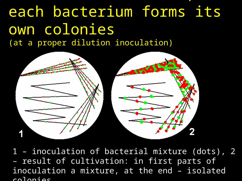
In case of a mixture, each bacterium forms its own colonies(at a proper dilution inoculation)
1 – inoculation of bacterial mixture (dots), 2 – result of cultivation: in first parts of inoculation a mixture, at the end – isolated colonies
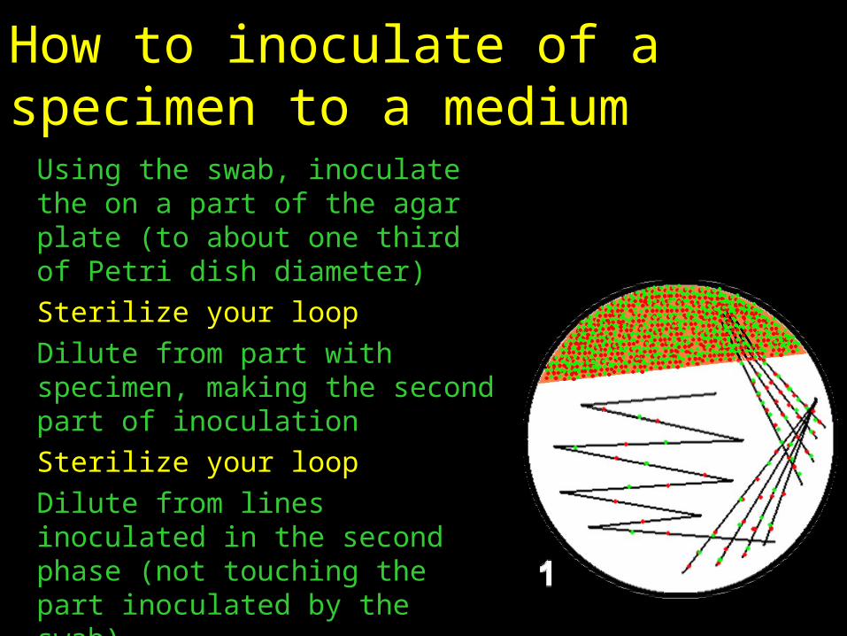
How to inoculate of a specimen to a medium
Using the swab, inoculate the on a part of the agar plate (to about one third of Petri dish diameter)Sterilize your loopDilute from part with specimen, making the second part of inoculationSterilize your loopDilute from lines inoculated in the second phase (not touching the part inoculated by the swab)Sterilize your loopInoculate the „serpent“ on the remaining part of the plate

How to reinoculate of an agar culture
Sterilize your loopTake the strainInoculate first phaseSterilize your loopDo not take the strain againInoculate second phaseSterilize your loopDo not take the strain againInoculate third phaseSterilize your loopDo not take the strain againInoculate the „serpent“
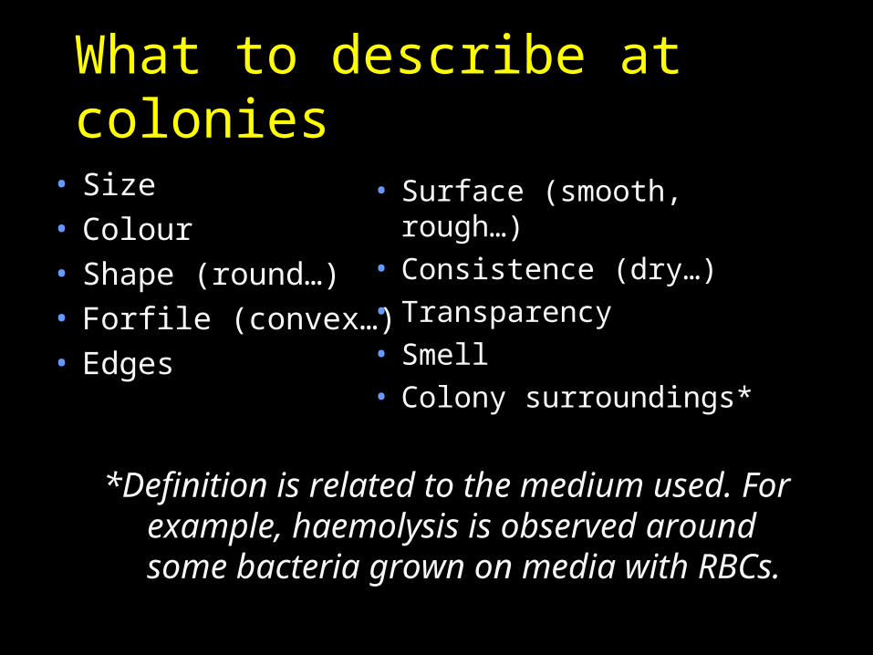
What to describe at colonies
• Size• Colour• Shape (round…)• Forfile (convex…)• Edges
• Surface (smooth, rough…)• Consistence (dry…)• Transparency• Smell• Colony surroundings*
*Definition is related to the medium used. For example, haemolysis is observed around some bacteria grown on media with RBCs.
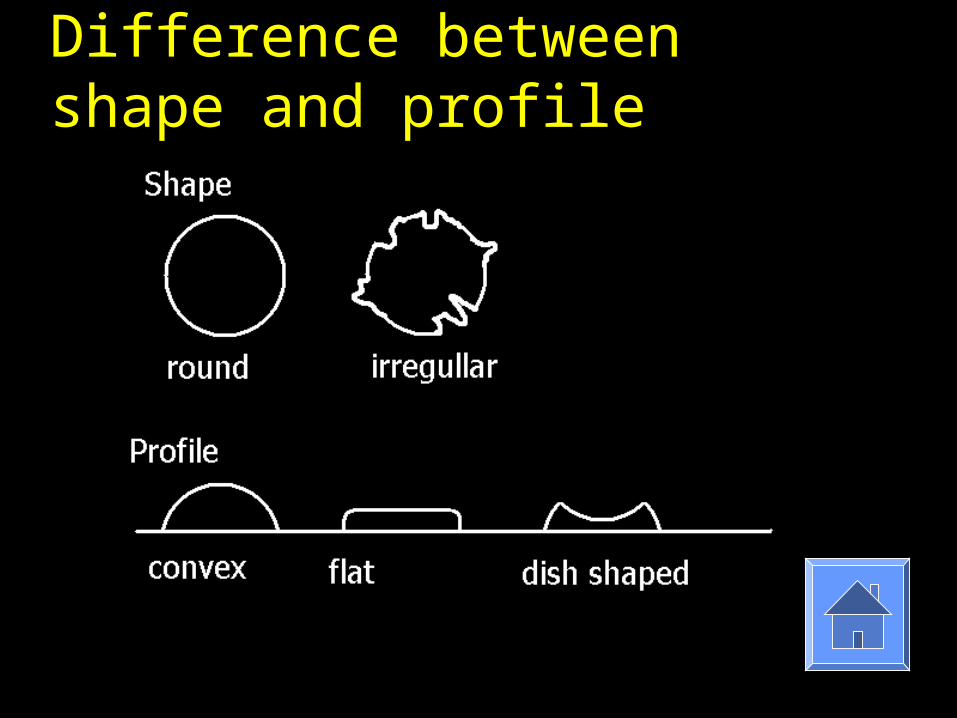
Difference between shape and profile

Solid media: classification and examples
Solid selective media• They have to select (separate) from
a bacterial mixture only one of several groups of genera
• An example is blood agar with 10 % NaCl used for stafylococci
• Sometimes, selectivity is reached by an antibiotic addition. Blood agar with amikacin is selective for streptococci and enterococci
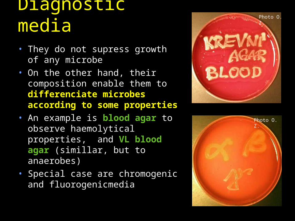
Diagnostic media• They do not supress growth of
any microbe• On the other hand, their
composition enable them to differenciate microbes according to some properties
• An example is blood agar to observe haemolytical properties, and VL blood agar (simillar, but to anaerobes)
• Special case are chromogenic and fluorogenicmedia
Photo O. Z.
Photo O. Z.
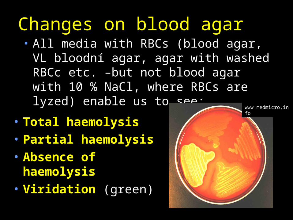
Changes on blood agar• All media with RBCs (blood agar, VL
bloodní agar, agar with washed RBCc etc. –but not blood agar with 10 % NaCl, where RBCs are lyzed) enable us to see:
• Total haemolysis• Partial haemolysis• Absence of
haemolysis• Viridation (green)
www.medmicro.info
Chromogenic and fluorogenic media
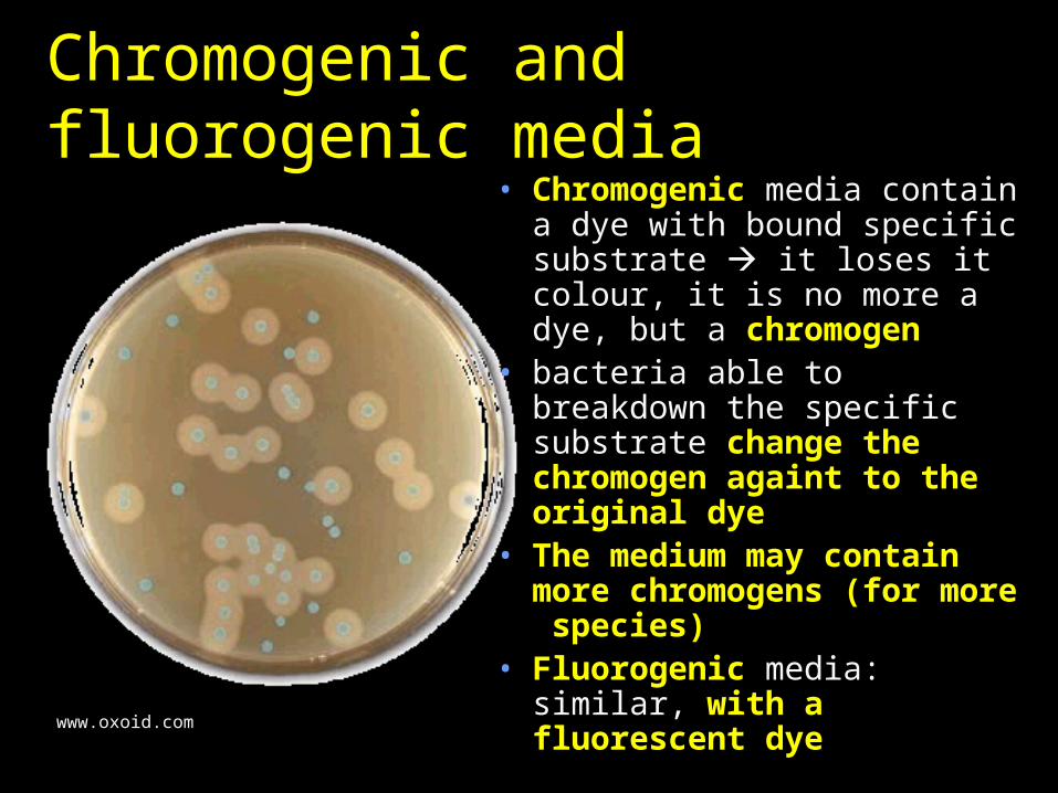
• Chromogenic media contain a dye with bound specific substrate it loses it colour, it is no more a dye, but a chromogen
• bacteria able to breakdown the specific substrate change the chromogen againt to the original dye
• The medium may contain more chromogens (for more species)
• Fluorogenic media: similar, with a fluorescent dye
www.oxoid.com
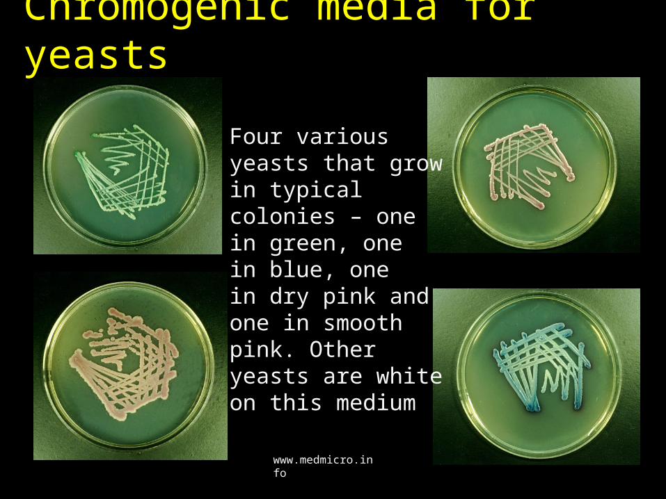
Chromogenic media for yeasts
Four various yeasts that grow in typical colonies – one in.green, one in.blue, one in.dry pink and one in smooth pink. Other yeasts are white on this medium
www.medmicro.info
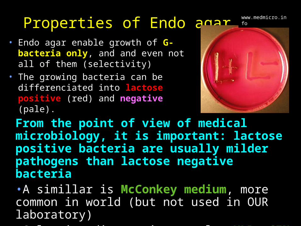
Properties of Endo agar• Endo agar enable growth of G-
bacteria only, and and even not all of them (selectivity)
• The growing bacteria can be differenciated into lactose positive (red) and negative (pale).
From the point of view of medical microbiology, it is important: lactose positive bacteria are usually milder pathogens than lactose negative bacteria•A simillar is McConkey medium, more common in world (but not used in OUR laboratory)•Selective diagnostic are also XLD, CIN media etc.
www.medmicro.info
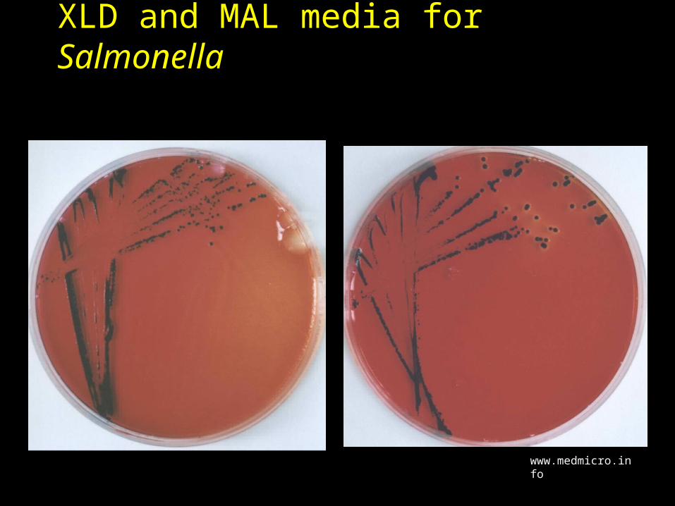
XLD and MAL media for Salmonella
www.medmicro.info
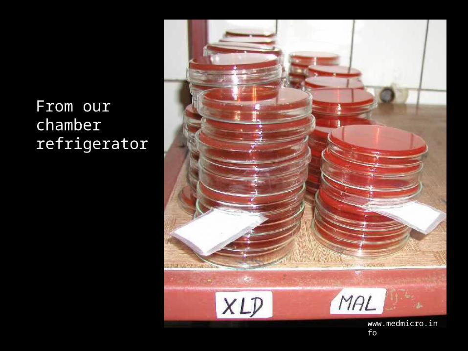
From our chamber refrigerator
www.medmicro.info
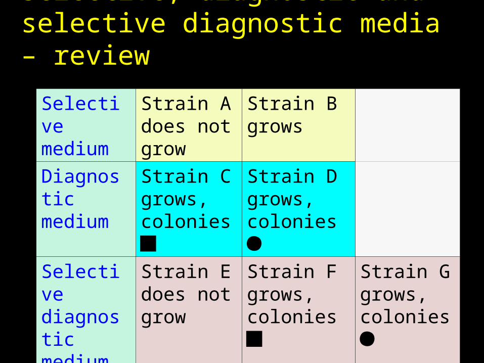
Selective, diagnostic and selective diagnostic media – review
Selective medium
Strain A does not grow
Strain B grows
Diagnostic medium
Strain C grows, colonies
Strain D grows, colonies
Selective diagnostic medium
Strain E does not grow
Strain F grows, colonies
Strain G grows, colonies
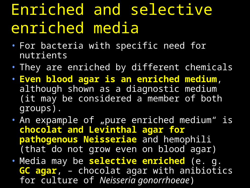
Enriched and selective enriched media • For bacteria with specific need for nutrients• They are enriched by different chemicals• Even blood agar is an enriched
medium, although shown as a diagnostic medium (it may be considered a member of both groups).
• An expample of „pure enriched medium“ is chocolat and Levinthal agar for pathogenous Neisseriae and hemophili (that do not grow even on blood agar)
• Media may be selective enriched (e. g. GC agar, – chocolat agar with anibiotics for culture of Neisseria gonorrhoeae)
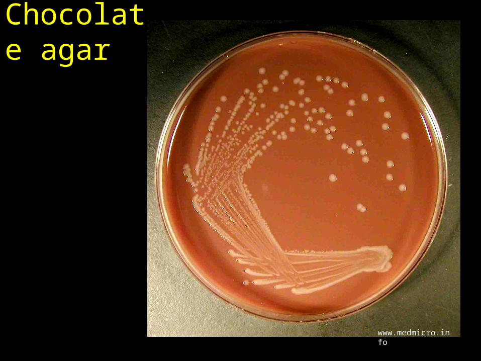
Chocolate agar
www.medmicro.info
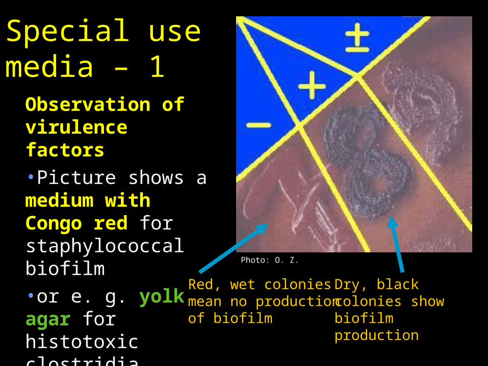
Dry, black colonies show biofilm production
Red, wet colonies mean no production of biofilm
Special use media – 1
Observation of virulence factors•Picture shows a medium with Congo red for staphylococcal biofilm•or e. g. yolk agar for histotoxic clostridia
Photo: O. Z.
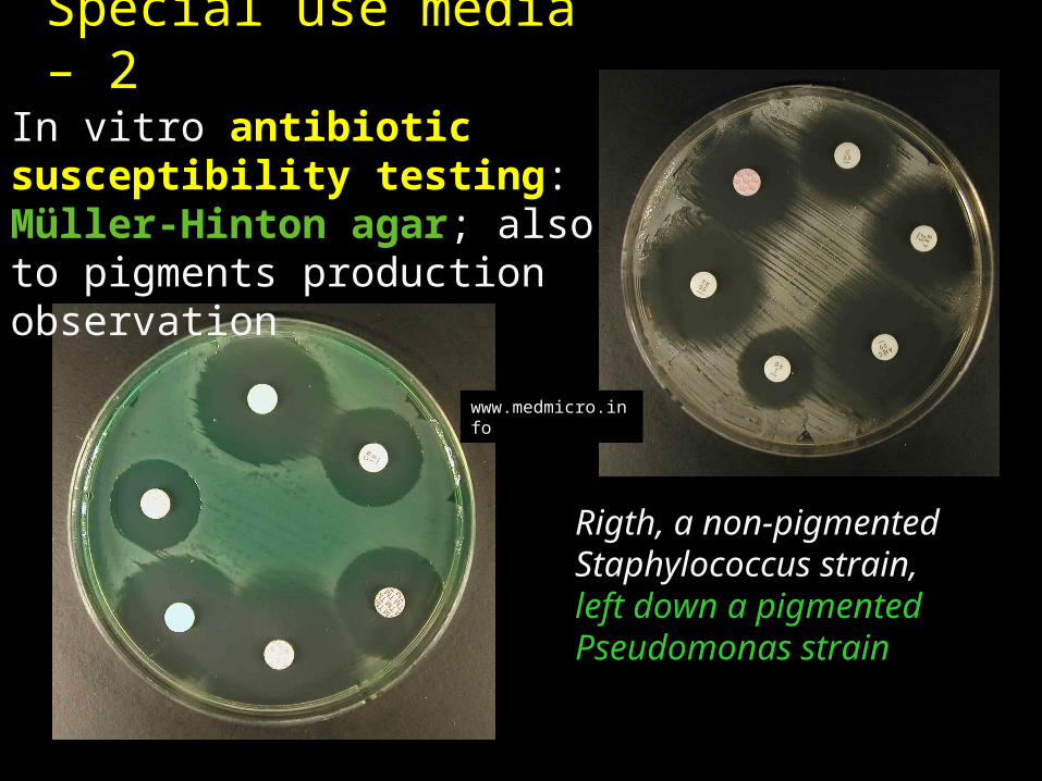
In vitro antibiotic susceptibility testing: Müller-Hinton agar; also to pigments production observation
Rigth, a non-pigmented Staphylococcus strain, left down a pigmented Pseudomonas strain
Special use media – 2
www.medmicro.info
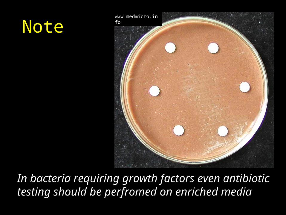
Note
In bacteria requiring growth factors even antibiotic testing should be perfromed on enriched media
www.medmicro.info
Modern trends in culture• Despite development of genetic
methods, cultivation still keeps its key role in mostly bacterial diagnostics
• Because of standardization, laboratories have to switch from „home made“ media to comercial products
• Chromogenic and fluorogenic media start to be used more and more, despite the price
Survey of the most important media1. Broth2. VL-broth3. Selenite broth4. Sabouraud5. Löwenstein-
Jenssen6. Blood agar (BA)7. Endo agar
8. MH9. BA + 10 % NaCl10. VLA (VL BA)11. XLD (and MAL)12. CHA13. Levinthal14. Slanetz-Bartley
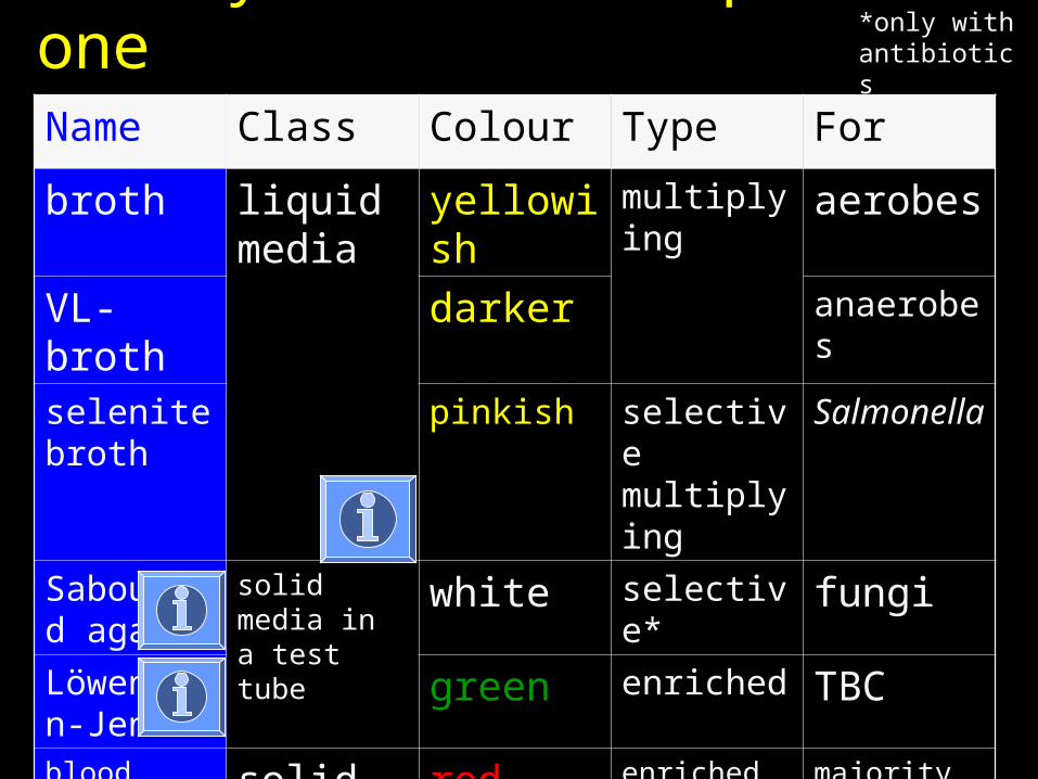
Survey of media – part oneName Class Colour Type For
broth liquid media
yellowish
multiplying
aerobes
VL-broth
darker anaerobes
selenite broth
pinkish selective multiplying
Salmonella
Sabouraud agar
solid media in a test tube
white selective* fungi
Löwentein-Jensen
green enriched TBC
bloodagar
solid media in.dish
red enriched + diagnostic
majority of bacteria
Endoagar
pink selective diagnostic
mostly enterobacteria
*only with antibiotics
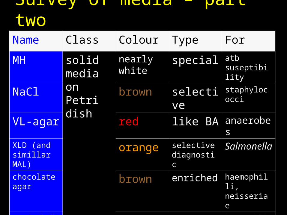
Survey of media – part twoName Class Colour Type For
MH solid media on Petri dish
nearly white
special atb suseptibility
NaCl brown selective
staphylococci
VL-agar red like BA anaerobes
XLD (and simillar MAL)
orange selective diagnostic
Salmonella
chocolate agar
brown enriched haemophilli, neisseriae
Levinthal agar
yellowish
enriched haemophilli
Slanetz-Bartley
pink selective diagnostic
enterococci
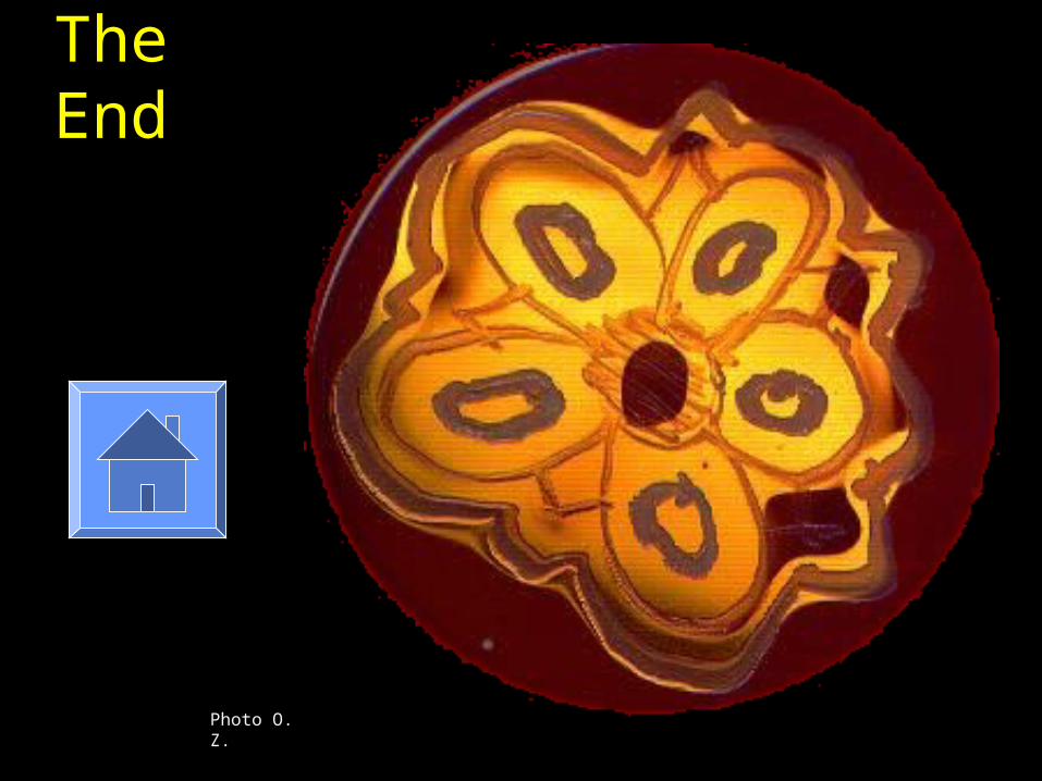
The End
Photo O. Z.

Robert Koch
Robert Koch (1843 – 1910)Bacteriologist Robert Koch discovered the anthrax disease cycle (1876); and the bacteria responsible for tuberculosis (1882) and cholera (1883). Koch formulated rules for the control of epidemics of cholera. "Koch's Postulates" (Kochsche Postulate, refined in 1884) are still the basic procedures used by modern epidemiologists and medical researchers: (1) Identify and specific organism, (2) obtain and pure culture of that organism, (3) reproduce the disease in experimental animals using the pure culture, and (4) recover the organism from the infected animals. http://www.general-anaesthesia.com/images/robert-koch.html
Once more Robert Koch
http://www.educationforum.co.uk/kochlesson.htm
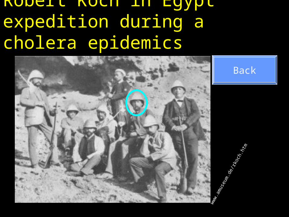
Robert Koch in Egypt expedition during a cholera epidemics
ww
w.a
mus
eum
.de/
rkoc
h.ht
m
Back
Solid media in a test tube? Why?
Among given media, two of theme are in test tubes, although they are solid. The reason is that they are used for slowly growing organisms. Both mycobacteria (Löwenstein-Jensen) and some molds (Sabouraud) grow slowly and the medium would be dry before the organism would grow on a Petri dishIn case of Hajna medium (see J04) the reason is different: the medium is used for biochemical testing and the difference between the lower part (no access to oxygen) and upper part (surface of medium) is important for its functionLöwenstein Jensen medium is also interesting as is it solid although agar is absent; it is solid because of coagulated eggs.
Back

Blood agars
• It is possible to use blood agar with red blood cells of various organisms (horses, chicken, cattle, and even humans). Nevertheless, the sheep RBCs are far most used ones
• It is possible to add blood cells to various bases. For example, if you add blood to VL broth (simplified), you get VL agar (VL blood agar)
• For haemolytical interaction testing (e. g. CAMP test, see P02) it is recommended to use agar with washed red blood cells.
Back

Endo agar and its principle• Endo agar contains lactose as a substrate• It also containts basic fuchsin• This fuchsin acts as factor of selectivity
• The same fuchsin (together with Na2SO3)
also acts as indicator (Schiff reagent). Bacteria forming lactaldehyde from lactose are visualised by purple colour
Endo agar should be kept away from light, otherwise it becomes purple without bacteria.
Back
Questions for ROPOT• Try to find answers to these questions• Then go to „ROPOT“ and try to answer the
questions (the ROPOT may be on IS MU something later than this slide-show)
• The formulation of questions on ROPOT may be different in details1. Why we cover VL broth with parafin oil?2. When we add red blood cells at preparing blood agar?3. Why gelatine is usually not used at making solid media?4. Microaerophilic and capnophilic conditions: is it the same?5. Staphylococci are adapted to life in skin of mammals. How do
microbiologists use this knowledge when cultivating them?6. What characteristic of bacterial colony cannot be seen by one’s eye?7. What characteristic of bacterial colony requires touching the colony?8. Why it is so important to obtain isolated colonies at cultivation?9. Blood agar is made of “basis for blood agar” (in fact it is nutrient
agar) and defibrinated sheep blood. Is it possible to add blood to other bases?
10.And one more secret question