Embed Size (px)
Citation preview
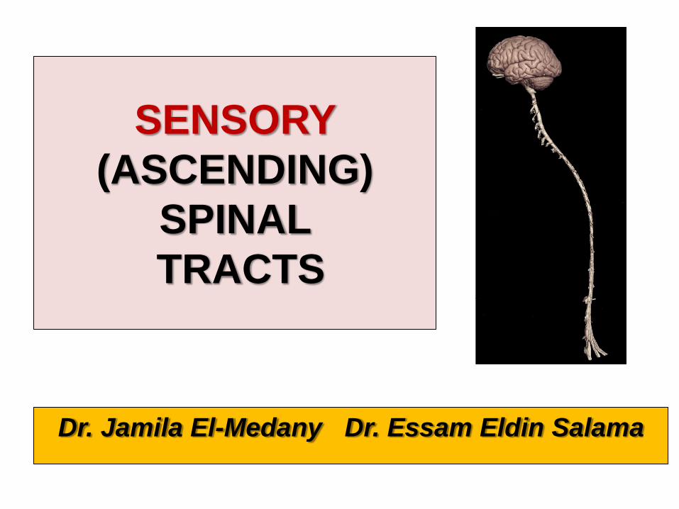
SENSORY
(ASCENDING)
SPINAL
TRACTS
Dr. Jamila El-Medany Dr. Essam Eldin Salama
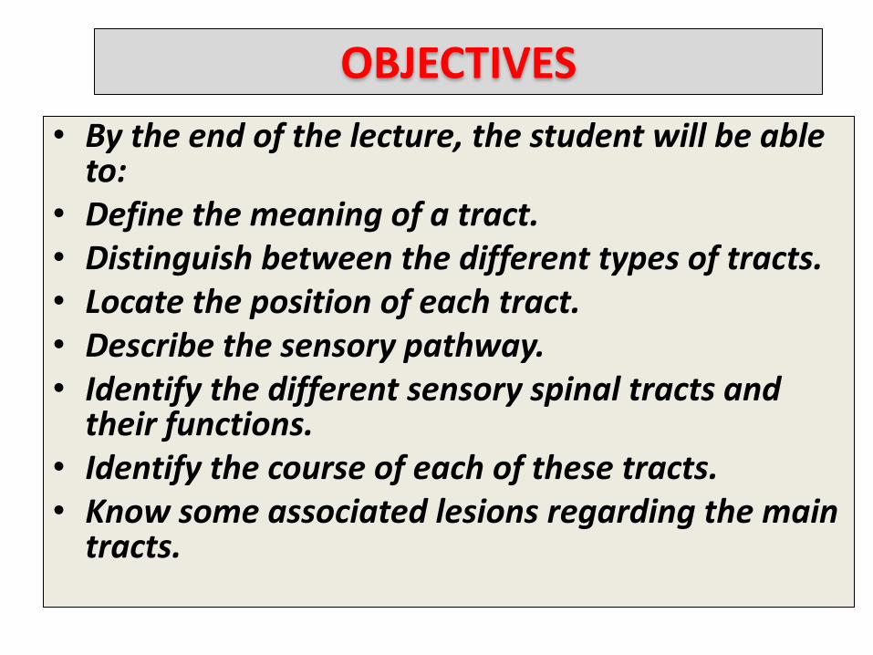
OBJECTIVES
• By the end of the lecture, the student will be able to:
• Define the meaning of a tract. • Distinguish between the different types of tracts. • Locate the position of each tract. • Describe the sensory pathway. • Identify the different sensory spinal tracts and
their functions. • Identify the course of each of these tracts. • Know some associated lesions regarding the main
tracts.
3
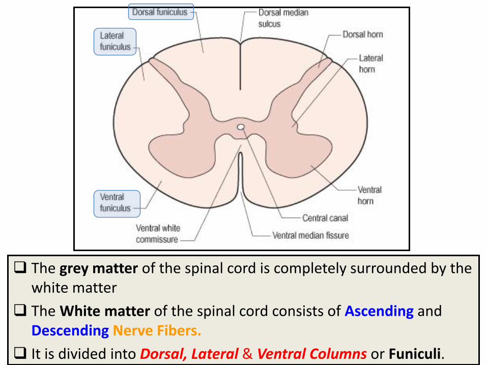
The grey matter of the spinal cord is completely surrounded by the white matter
The White matter of the spinal cord consists of Ascending and Descending Nerve Fibers.
It is divided into Dorsal, Lateral & Ventral Columns or Funiculi.
WHITE MATTER TRACTS
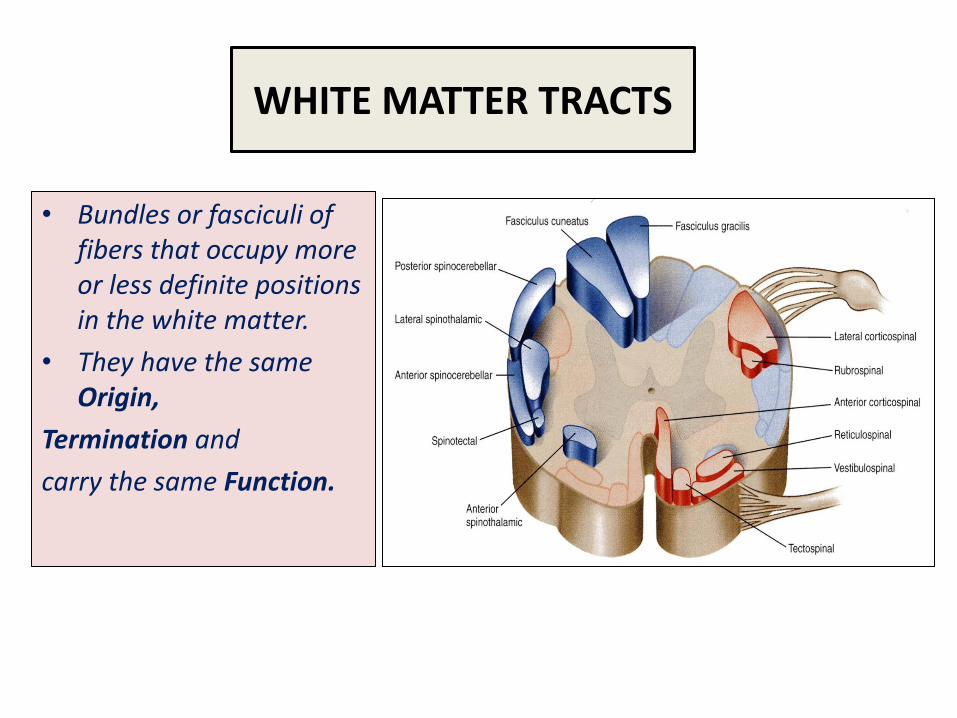
• Bundles or fasciculi of fibers that occupy more or less definite positions in the white matter.
• They have the same Origin,
Termination and
carry the same Function.
CLASSIFICATION OF WHITE MATTER TRACTS
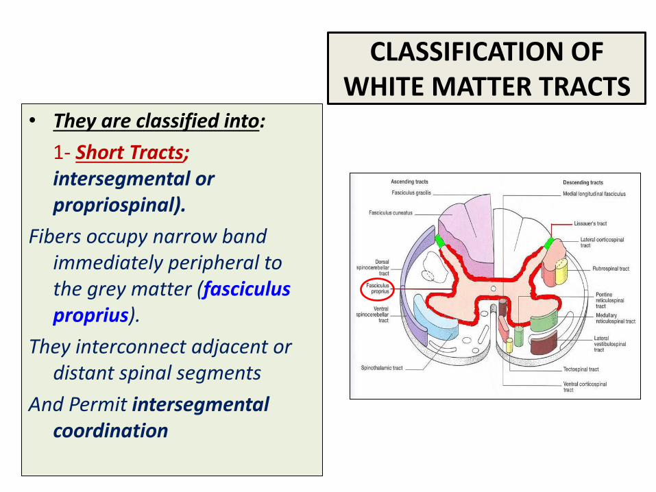
• They are classified into:
1- Short Tracts; intersegmental or propriospinal).
Fibers occupy narrow band immediately peripheral to the grey matter (fasciculus proprius).
They interconnect adjacent or distant spinal segments
And Permit intersegmental coordination
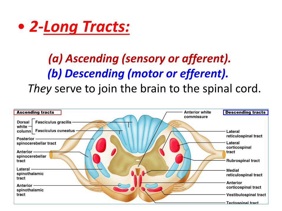
• 2-Long Tracts:
(a) Ascending (sensory or afferent). (b) Descending (motor or efferent). They serve to join the brain to the spinal cord.
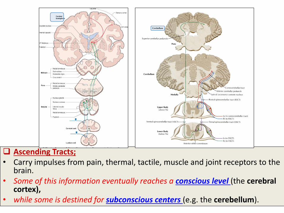
Ascending Tracts; • Carry impulses from pain, thermal, tactile, muscle and joint receptors to the
brain. • Some of this information eventually reaches a conscious level (the cerebral
cortex), • while some is destined for subconscious centers (e.g. the cerebellum).
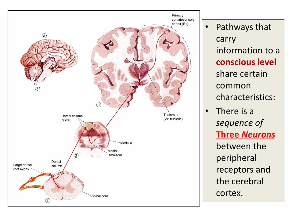
• Pathways that carry information to a conscious level share certain common characteristics:
• There is a sequence of Three Neurons between the peripheral receptors and the cerebral cortex.
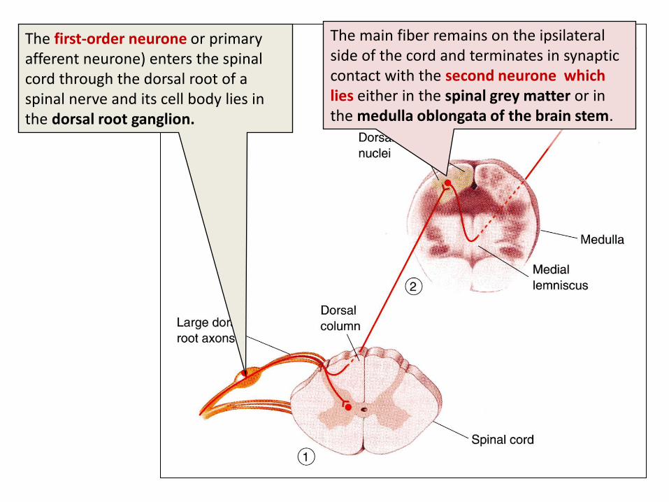
The first-order neurone or primary afferent neurone) enters the spinal cord through the dorsal root of a spinal nerve and its cell body lies in the dorsal root ganglion.
The main fiber remains on the ipsilateral side of the cord and terminates in synaptic contact with the second neurone which lies either in the spinal grey matter or in the medulla oblongata of the brain stem.
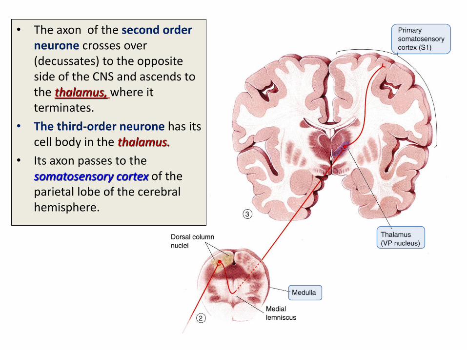
• The axon of the second order neurone crosses over (decussates) to the opposite side of the CNS and ascends to the thalamus, where it terminates.
• The third-order neurone has its cell body in the thalamus.
• Its axon passes to the somatosensory cortex of the parietal lobe of the cerebral hemisphere.
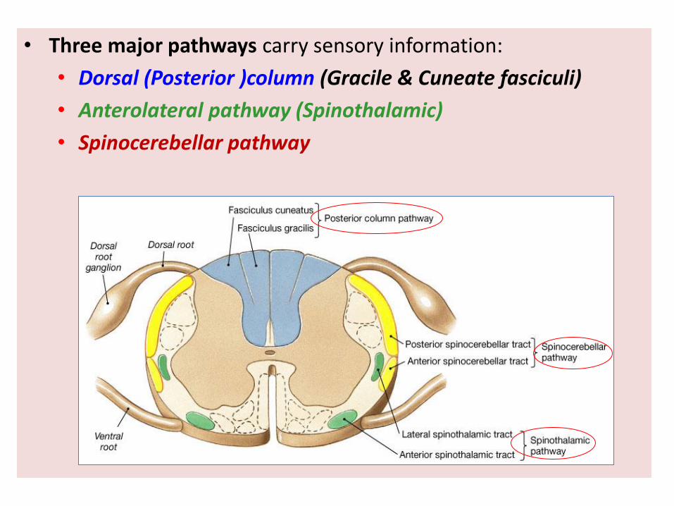
• Three major pathways carry sensory information:
• Dorsal (Posterior )column (Gracile & Cuneate fasciculi)
• Anterolateral pathway (Spinothalamic)
• Spinocerebellar pathway
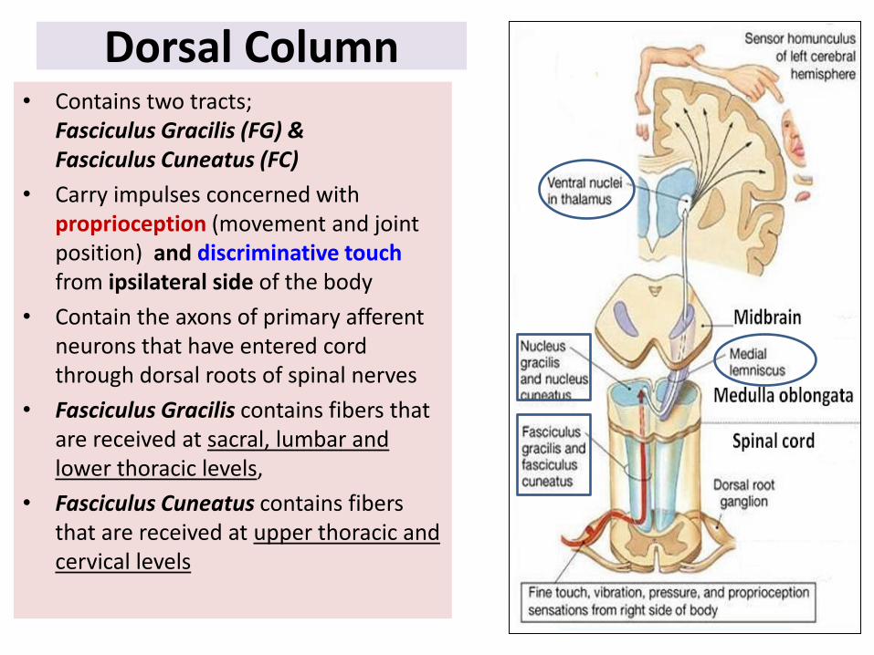
Dorsal Column • Contains two tracts;
Fasciculus Gracilis (FG) & Fasciculus Cuneatus (FC)
• Carry impulses concerned with proprioception (movement and joint position) and discriminative touch from ipsilateral side of the body
• Contain the axons of primary afferent neurons that have entered cord through dorsal roots of spinal nerves
• Fasciculus Gracilis contains fibers that are received at sacral, lumbar and lower thoracic levels,
• Fasciculus Cuneatus contains fibers that are received at upper thoracic and cervical levels
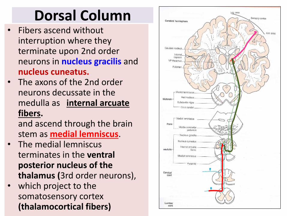
Dorsal Column • Fibers ascend without
interruption where they terminate upon 2nd order neurons in nucleus gracilis and nucleus cuneatus.
• The axons of the 2nd order neurons decussate in the medulla as internal arcuate fibers. and ascend through the brain stem as medial lemniscus.
• The medial lemniscus terminates in the ventral posterior nucleus of the thalamus (3rd order neurons),
• which project to the somatosensory cortex (thalamocortical fibers)
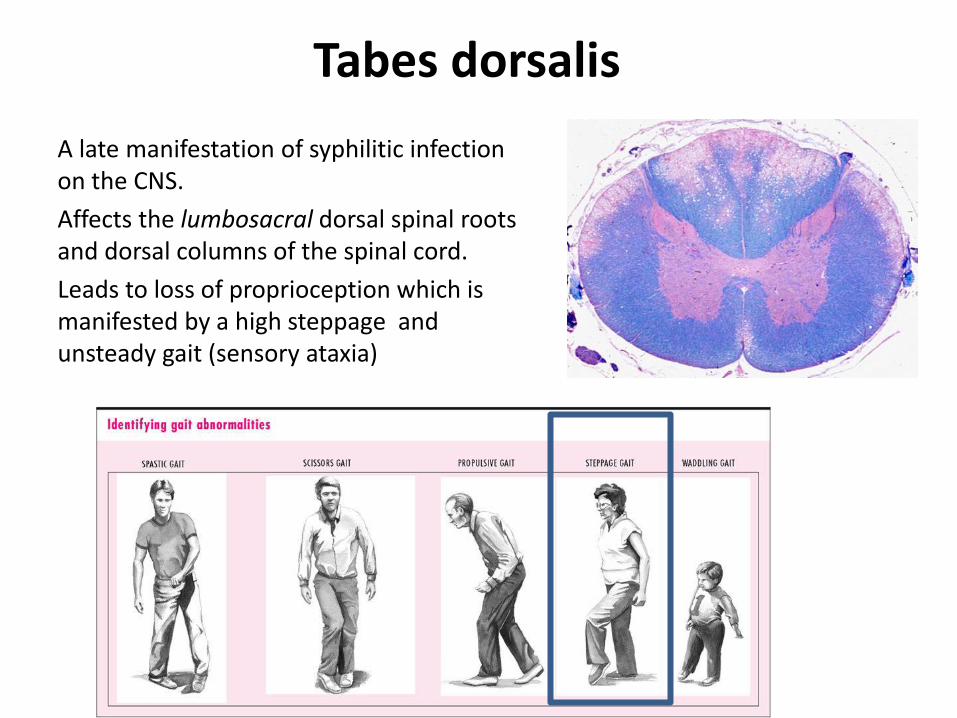
Tabes dorsalis
A late manifestation of syphilitic infection on the CNS.
Affects the lumbosacral dorsal spinal roots and dorsal columns of the spinal cord.
Leads to loss of proprioception which is manifested by a high steppage and unsteady gait (sensory ataxia)
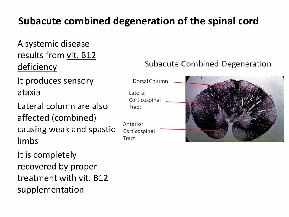
Subacute combined degeneration of the spinal cord
A systemic disease results from vit. B12 deficiency
It produces sensory ataxia
Lateral column are also affected (combined) causing weak and spastic limbs
It is completely recovered by proper treatment with vit. B12 supplementation
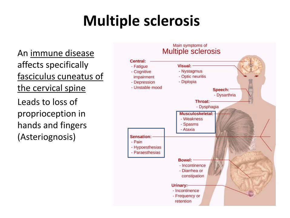
Multiple sclerosis
An immune disease affects specifically fasciculus cuneatus of the cervical spine
Leads to loss of proprioception in hands and fingers (Asteriognosis)
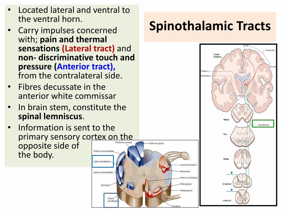
Spinothalamic Tracts • Located lateral and ventral to
the ventral horn. • Carry impulses concerned
with; pain and thermal sensations (Lateral tract) and non- discriminative touch and pressure (Anterior tract), from the contralateral side.
• Fibres decussate in the anterior white commissar
• In brain stem, constitute the spinal lemniscus.
• Information is sent to the primary sensory cortex on the opposite side of the body.
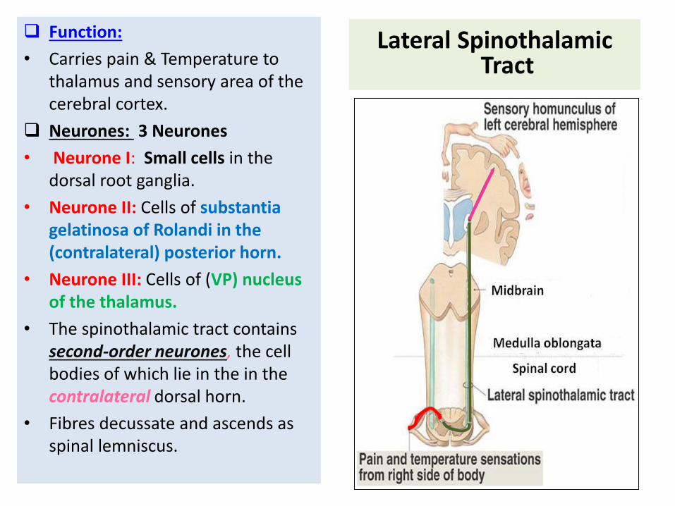
Lateral Spinothalamic Tract
Function:
• Carries pain & Temperature to thalamus and sensory area of the cerebral cortex.
Neurones: 3 Neurones
• Neurone I: Small cells in the dorsal root ganglia.
• Neurone II: Cells of substantia gelatinosa of Rolandi in the (contralateral) posterior horn.
• Neurone III: Cells of (VP) nucleus of the thalamus.
• The spinothalamic tract contains second-order neurones, the cell bodies of which lie in the in the contralateral dorsal horn.
• Fibres decussate and ascends as spinal lemniscus.
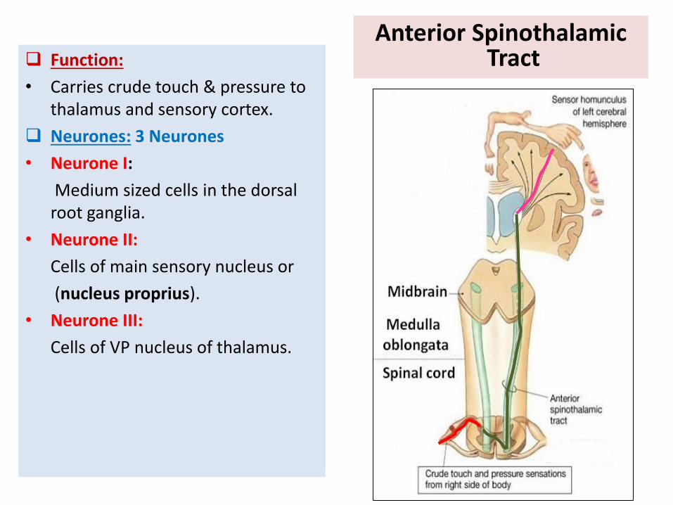
Anterior Spinothalamic Tract Function:
• Carries crude touch & pressure to thalamus and sensory cortex.
Neurones: 3 Neurones
• Neurone I:
Medium sized cells in the dorsal root ganglia.
• Neurone II:
Cells of main sensory nucleus or
(nucleus proprius).
• Neurone III:
Cells of VP nucleus of thalamus.
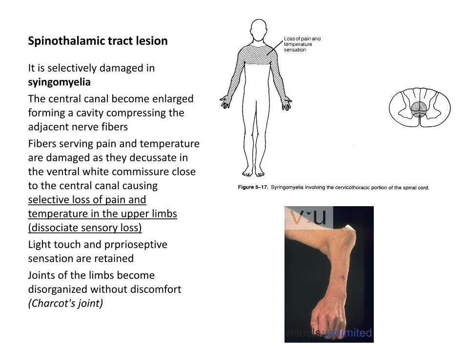
Spinothalamic tract lesion
It is selectively damaged in syingomyelia
The central canal become enlarged forming a cavity compressing the adjacent nerve fibers
Fibers serving pain and temperature are damaged as they decussate in the ventral white commissure close to the central canal causing selective loss of pain and temperature in the upper limbs (dissociate sensory loss)
Light touch and prprioseptive sensation are retained
Joints of the limbs become disorganized without discomfort (Charcot's joint)
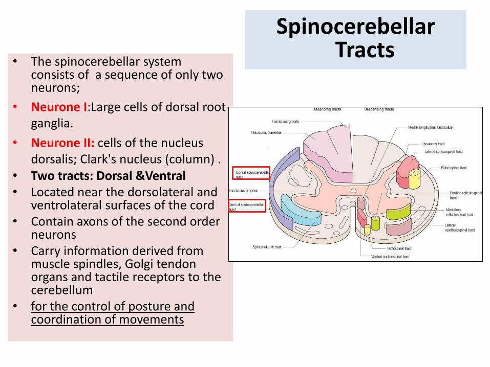
Spinocerebellar Tracts
• The spinocerebellar system consists of a sequence of only two neurons;
• Neurone I:Large cells of dorsal root ganglia.
• Neurone II: cells of the nucleus dorsalis; Clark's nucleus (column) .
• Two tracts: Dorsal &Ventral • Located near the dorsolateral and
ventrolateral surfaces of the cord • Contain axons of the second order
neurons • Carry information derived from
muscle spindles, Golgi tendon organs and tactile receptors to the cerebellum
• for the control of posture and coordination of movements
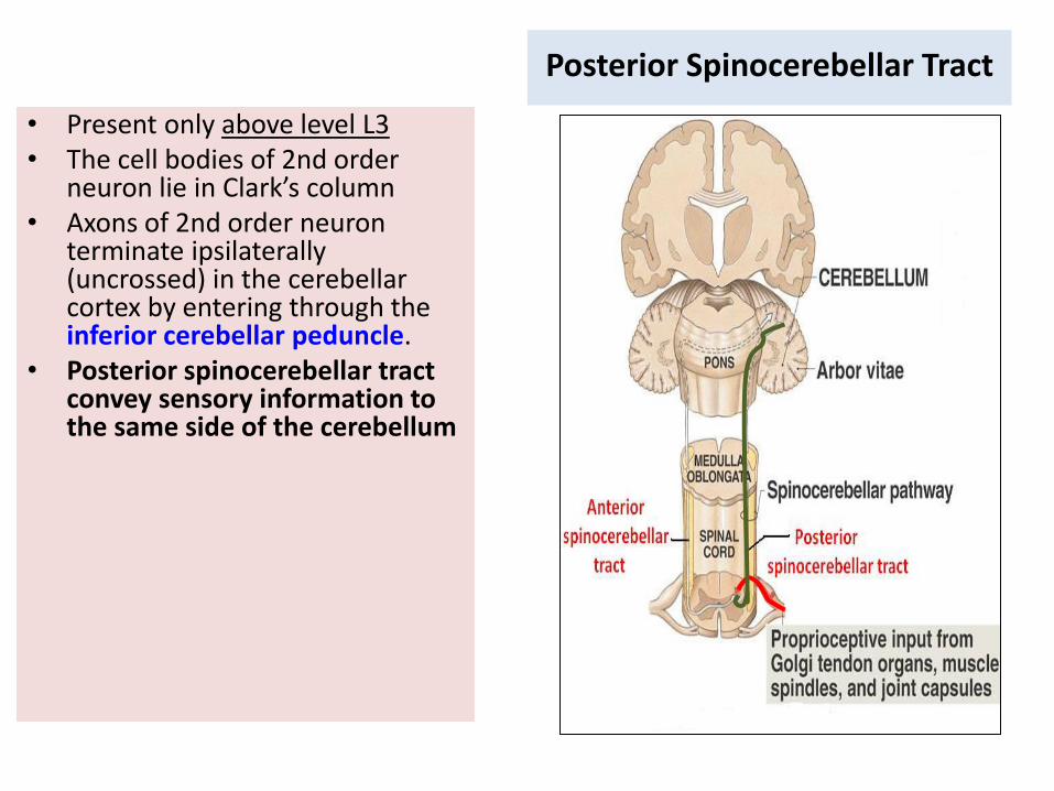
Posterior Spinocerebellar Tract
• Present only above level L3 • The cell bodies of 2nd order
neuron lie in Clark’s column • Axons of 2nd order neuron
terminate ipsilaterally (uncrossed) in the cerebellar cortex by entering through the inferior cerebellar peduncle.
• Posterior spinocerebellar tract convey sensory information to the same side of the cerebellum
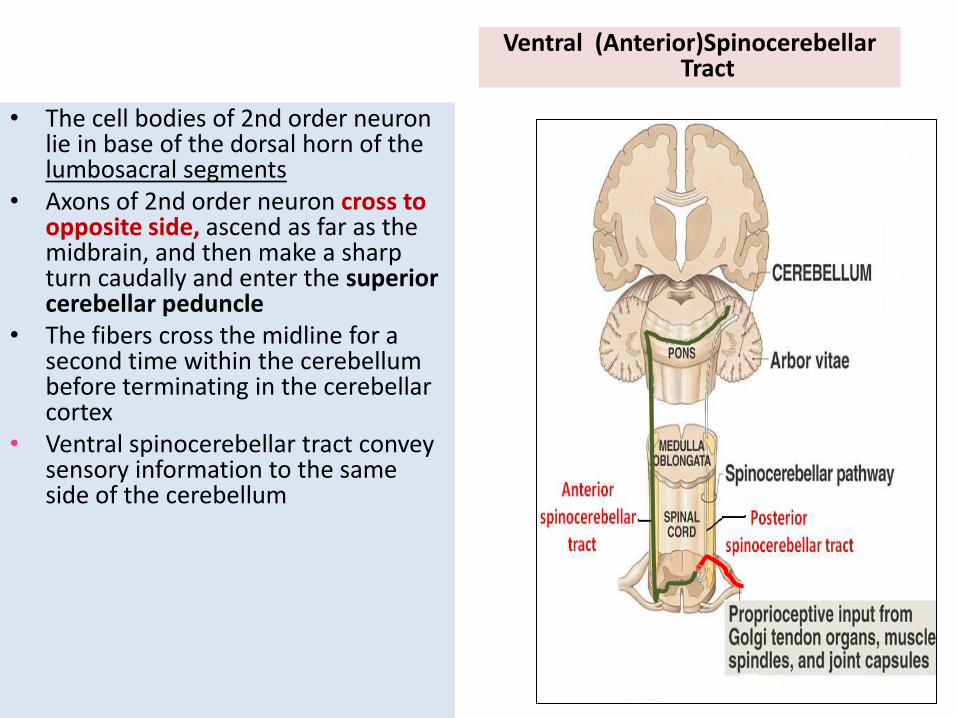
Ventral (Anterior)Spinocerebellar Tract
• The cell bodies of 2nd order neuron lie in base of the dorsal horn of the lumbosacral segments
• Axons of 2nd order neuron cross to opposite side, ascend as far as the midbrain, and then make a sharp turn caudally and enter the superior cerebellar peduncle
• The fibers cross the midline for a second time within the cerebellum before terminating in the cerebellar cortex
• Ventral spinocerebellar tract convey sensory information to the same side of the cerebellum
Lesion of the spinocerebellar tracts
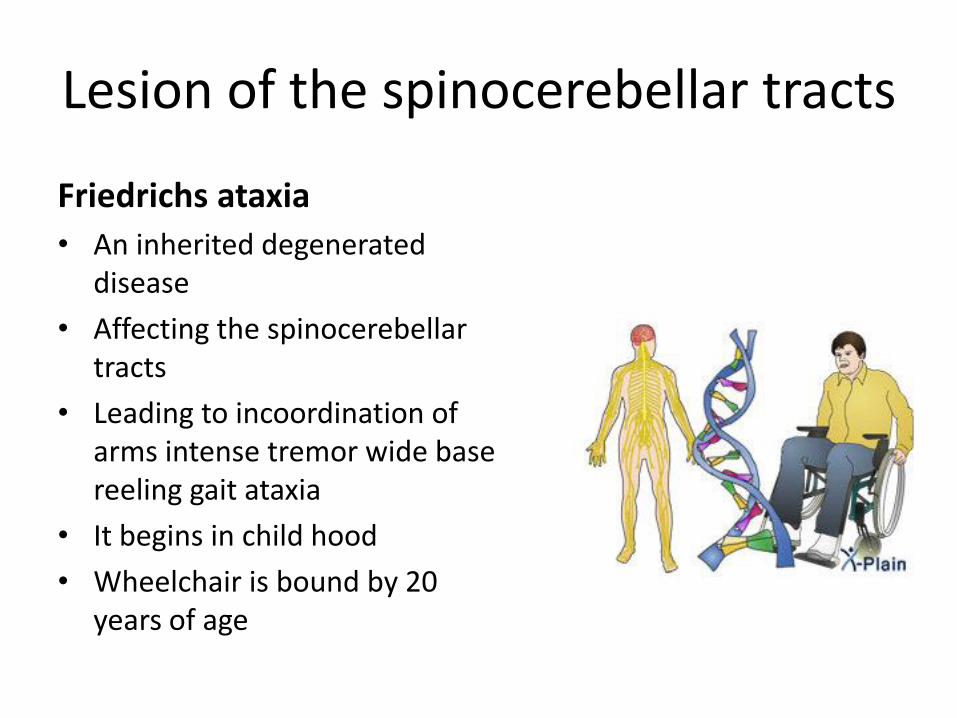
Friedrichs ataxia
• An inherited degenerated disease
• Affecting the spinocerebellar tracts
• Leading to incoordination of arms intense tremor wide base reeling gait ataxia
• It begins in child hood
• Wheelchair is bound by 20 years of age
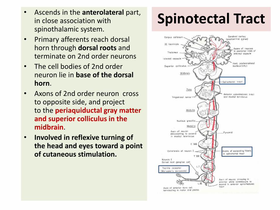
Spinotectal Tract • Ascends in the anterolateral part,
in close association with spinothalamic system.
• Primary afferents reach dorsal horn through dorsal roots and terminate on 2nd order neurons
• The cell bodies of 2nd order neuron lie in base of the dorsal horn.
• Axons of 2nd order neuron cross to opposite side, and project to the periaquiductal gray matter and superior colliculus in the midbrain.
• Involved in reflexive turning of the head and eyes toward a point of cutaneous stimulation.
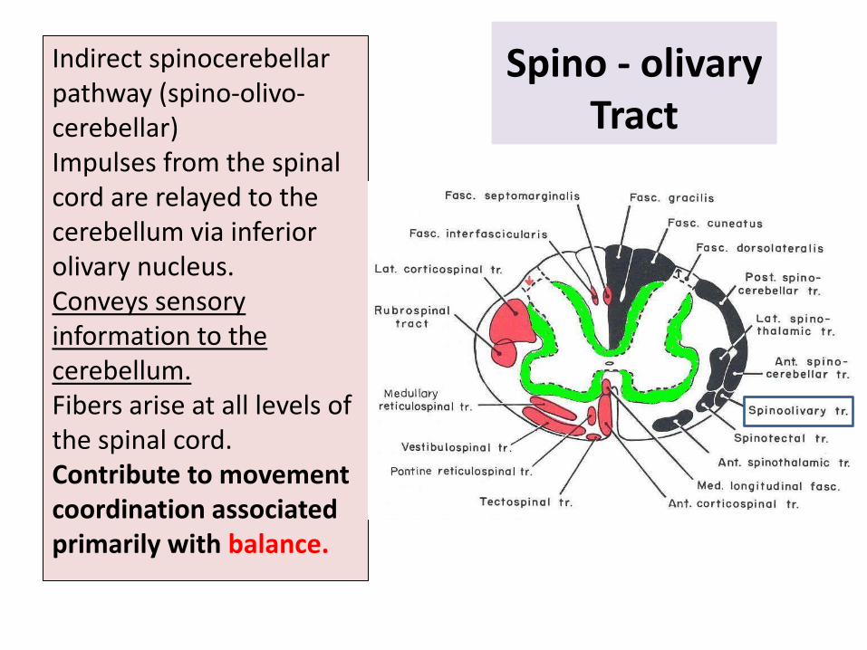
Spino - olivary Tract
Indirect spinocerebellar pathway (spino-olivo-cerebellar) Impulses from the spinal cord are relayed to the cerebellum via inferior olivary nucleus. Conveys sensory information to the cerebellum. Fibers arise at all levels of the spinal cord. Contribute to movement coordination associated primarily with balance.
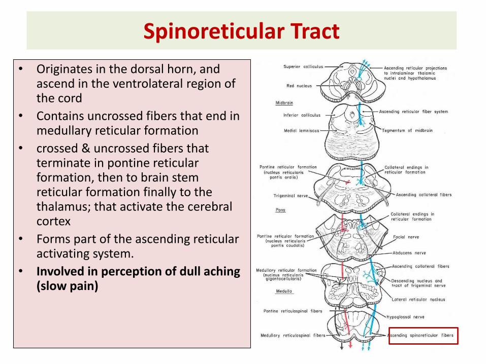
Spinoreticular Tract
• Originates in the dorsal horn, and ascend in the ventrolateral region of the cord
• Contains uncrossed fibers that end in medullary reticular formation
• crossed & uncrossed fibers that terminate in pontine reticular formation, then to brain stem reticular formation finally to the thalamus; that activate the cerebral cortex
• Forms part of the ascending reticular activating system.
• Involved in perception of dull aching (slow pain)

Thank you


![Post-doctoral position 2016 - Find a team - Inria white matter, gray matter, lesions or spinal cord tracts, using tools available from the literature [3,4] or adapted from similar](https://img.pdfslide.net/doc/110x75/5aebd9e77f8b9ab24d8f4e1f/post-doctoral-position-2016-find-a-team-inria-white-matter-gray-matter-lesions.jpg)