Embed Size (px)
Citation preview
ORIGINAL ARTICLE
Serial observations on an orthotopic gastric cancer model constructed using improved implantation technique
Yan Li, Bo Li, Yu Zhang, Chun-Ping Xiang, Yuan-Yuan Li, Xiao-Ling Wu
Yan Li, Xiao-Ling Wu, Department of Gastroenterology, the Sec-ond Affiliated Hospital, Chongqing Medical University, Chong-qing 400010, ChinaBo Li, Department of Surgery, the First People’s Hospital, Jiang-bei District, Chongqing 400020, China Yu Zhang, Department of Pathology, the First People’s Hospital, Jiangbei District, Chongqing 400020, ChinaChun-Ping Xiang, Department of Pathology, Children’s Hospi-tal, Chongqing Medical University, Chongqing 400014, ChinaYuan-Yuan Li, Department of Pathology, Chongqing Medical University, Chongqing 400016, ChinaAuthor contributions: Li Y conceived of the study, constructed animal models, and drafted the manuscript; Li B participated and established the animal model; Zhang Y and Xiang CP performed paraffin sections and hematoxylin and eosin staining; Li YY performed immunohistochemical staining; Wu XL guided and inspected the whole experiments. Supported by the Natural Science Foundation of China, No. 30830040Correspondence to: Xiao-Ling Wu, Professor, Department of Gastroenterology, the Second Affiliated Hospital, Chongqing Medical University, Chongqing 400010, China. [email protected]: +86-23-63693325 Fax: +86-23-63711527 Received: October 12, 2010 Revised: December 30, 2010Accepted: January 6, 2011Published online: March 21, 2011
Abstract AIM: To establish a gastric cancer nude-mouse model with improved orthotopic implantation and investigate its biological characteristics at different time points.
METHODS: Human gastric cancer SGC-7901 cell sus-pensions were injected subcutaneously into a nude mouse to develop solid tumors, and the tumor tissue pieces were implanted under the serous coat. The nude mice were then euthanized in group every two weeks to observe the primary tumor growth and metastases.
RESULTS: Within 2-4 wk, there were no obvious chang-
es about the primary tumor in stomach. At the sixth week, the primary tumor began to grow fast, resulting in incrassation of the gastric wall and stenosis of the gastric cavity, and metastases into the liver and lymph nodes were detected. The tumor, which compressed the adjacent organs, gradually became bigger and bigger followed by stenosis or vanishment of the gastric cavity from 8 to 12 wk. There were massive metastases, and the rate of metastasis was 58% in lymph nodes, 78% in liver, 39% in kidney, and 81% in peritoneum or septum.
CONCLUSION: A gastric cancer model is established, which can simulate the clinical tumor behavior and provide experimental carrier for clinical trials of gastric cancer treatment.
© 2011 Baishideng. All rights reserved.
Key words: Gastric cancer; Orthotopic implantation; Mouse model; Metastasis; Cell line
Peer reviewers: Fabio Grizzi, PhD, Laboratories of Quantita-tive Medicine, Istituto Clinico Humanitas IRCCS, Via Manzoni 56, 20089 Rozzano, Milan,Italy; Dr. Jianyuan Chai, PhD, MS, BS, Assistant Professor, Research (09-151), VA Long Beach Healthcare System, 5901 E. 7th St, Long Beach, CA 90822, United States
Li Y, Li B, Zhang Y, Xiang CP, Li YY, Wu XL. Serial observa-tions on an orthotopic gastric cancer model constructed using improved implantation technique. World J Gastroenterol 2011; 17(11): 1442-1447 Available from: URL: http://www.wjgnet.com/1007-9327/full/v17/i11/1442.htm DOI: http://dx.doi.org/10.3748/wjg.v17.i11.1442
INTRODUCTIONMetastasis is not only still the barrier to tumor therapy, but also linked with the prognosis of patients. Patients with gastric cancer hardly have symptoms in their early stage
1442
World J Gastroenterol 2011 March 21; 17(11): 1442-1447 ISSN 1007-9327 (print) ISSN 2219-2840 (online)
© 2011 Baishideng. All rights reserved.
Online Submissions: http://www.wjgnet.com/[email protected]:10.3748/wjg.v17.i11.1442
March 21, 2011|Volume 17|Issue 11|WJG|www.wjgnet.com
Li Y et al . Orthotopic gastric cancer model
so that they are always found with metastases after clinical examinations. Therefore, novel tumor models are needed to study the metastasis mechanism as well as therapeutic approaches. It is well known that orthotopic transplanta-tion technique has gain popularity in the field of the ani-mal model[1-12]. Orthotopic tumor models not only mimic clinical cancer course, but also promote metastasis[13]. In recent years, the procedure of orthotopic implantation has been improved from the“sewing”method to the“ad-hering”one[2-4,14]. The progress greatly shortens the course of surgical operation and decreases the mortality of ani-mals. To our knowledge, gastric cancer models of ortho-topic implantation with intact tumor tissue have been well established[1-3,5-8]. However, serial examination of tumor development and metastasis needs to be made in order to tailor the treatment strategies. Based on the previous researches, we intend to establish an orthotopic model of gastric cancer using tissue glue and observe tumor prog-ress and metastasis consecutively to detect an appropriate therapeutic target for clinical trials.
MATERIALS AND METHODSCell lineHuman gastric cancer cell lines SGC-7901 (poorly dif-ferentiated) was used for this study. The cell was obtained from the Centre of Cell Cultures of Chinese Academy of Medical Sciences, Shanghai, China, and cultured at 37℃ in a humidified air with 5% CO2, in RPMI-1640 medium (Gibco, Grand Island, NY) supplemented with 10% fetal calf serum (FCS; Gibco, Grand Island, NY), 100 U/mL penicillin, 100 μg/mL streptomycin, 2 mmol/L gluta-mine, and 1 mmol/L sodium pyruvate.
AnimalsFive to six-week-old male Balb/c nu-nu mice weighing 18-20 g were obtained from the Shanghai Experimental Animal Center of the Chinese Academy of Medical Sci-ences, China. All mice were maintained in a pathogen-free environment (temperature 25-27℃, humidity 45%-50%) and supplied with food and water ad libitum. All experi-mental procedures were conducted in conformity with institutional guidelines for the care and use of laboratory animals in Chongqing Medical University, China, and the National Institutes of Health Guide for Care and Use of Laboratory Animals.
Orthotopic implantation in stomachSGC-7901 cells were collected at the log phase and inject-ed subcutaneously into the mice at 107/0.2 mL. Two weeks later, tumors (about 2.0 cm × 2.0 cm × 1.0 cm) were har-vested from the mice under anesthesia and minced into small pieces (1mm3) in RPMI-1640 basal medium. All pro-cedures were then performed under anesthesia with Sumi-anxin Ⅱ (0.02 mL per animal; China Academy of Military Medical Sciences). For implantation, the mouse stomach was gently exteriorized via a left-side upper abdominal incision, and one small tissue pocket was formed in the
middle wall of the greater curvature using a microscissor. One tumor fragment was placed into the pocket and fixed with a drop of medical tissue glue (gifts from Shunkang Corporation of Biological Adhesive, Beijing, China). The quantity of the tissue adhesive should be strictly controlled to avoid adhering to adjacent normal tissues. The stomach was then relocated into the abdominal cavity followed by the abdominal closure with 4-0 absorbable sutures.
Evaluation of tumor growth and metastasisAll mice were divided into 6 groups of 6 animals each after orthotopic implantation. One group was sacrificed every two weeks. At autopsy, the primary tumor, lymph nodes, and other organs were examined in detail. The samples were fixed in 10% formalin for paraffin sections, and stained with hematoxylin and eosin for microscopic examination.
ImmunohistochemistryParaffin sections were examined histologically with the Cy-tokeratin 20 (CK20) mAb KS20.8 and Epithelial Membrane Antigen (EMA) mAb GP1.4 (MAB-0057, MAB-0061; Maixin Inc., Fuzhou, China), which are usually combined to diagnose the gastrointestinal adenocarcinoma. Ultrasen-sitive streptavidin-peroxidase (SP) kit (KIT-9710; Maixin Inc., Fuzhou, China) and DAB kit (DAB-0031; Maixin Inc., Fuzhou, China) were used in the immunohistochemical stain according to the manufacture’s instructions. CK20 and EMA expression was defined as positive if the stained region of tumor cells was in the cytoplasm.
Assessment of primary tumor growthThe primary tumor volume was calculated by the fol-lowing formula: V = 0.4 × ab2 (a: maximum diameter; b: minimum diameter)[15]. Then the tumor growth curve was depicted reflecting the tumor growth trend.
RESULTSMacroscopic examinationFrom 2 to 4 wk, the primary tumor developed without obvious volume change, and showed insufficient blood supply. The gastric cavity of tumor-bearing mice ap-peared almost normal in size. At the 2nd wk after ortho-topic implantation, metastatic infiltration was not found in all detected organs. However, tumor invasion into the liver was found at the 4th wk. Six weeks after trans-plantation, the tumor in situ grew increasingly in size, with sparse blood vessels. The stomach wall of mice was thickened and gastric cavity decreased, companied with enlargement of lymph nodes from gastric area and hilus pulmonis. The tumor invaded into the liver, resulting in metastatic nodules formation. At the 8th wk, the stomach tumor was characterized by large-size, irregular volume and rich blood supply. The resulting tumor squeezed the adjacent organs and deformed the stomach contributing to distal stenosis. The lymph nodes of the gastric area, hilus pulmonis and mesenterium presented enlargement caused by tumor metastasis. Metastatic lesions were
1443 March 21, 2011|Volume 17|Issue 11|WJG|www.wjgnet.com
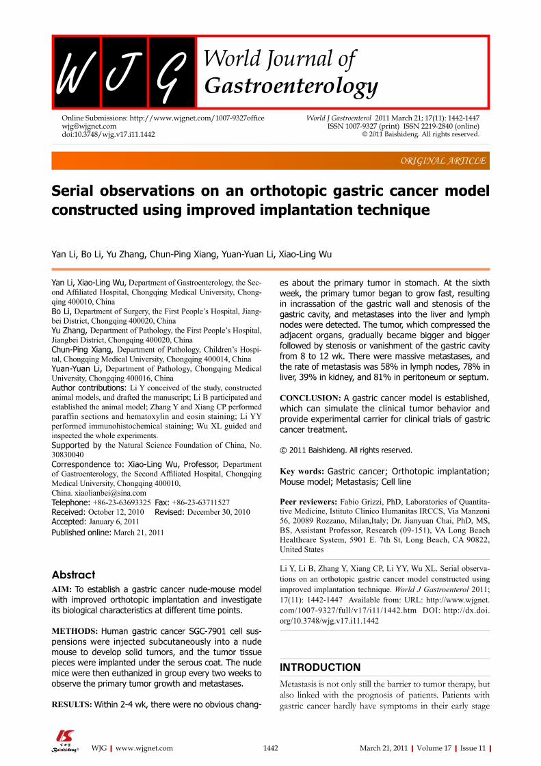
found in the organs such as liver, kidney, peritoneum and diaphragm. From 10 to 12 wk, the tumor grew fast and huge, occupying almost the upper abdomen and squeez-ing severely the surrounding organs. The necrosis from central tumor areas was macroscopically visible, indicat-ing that the tumor outgrew the sufficient blood supply. In most mice, the gastric cavity became narrow, even van-ished, and pylorus was obstructed partly or totally (Figure 1). The stomach of some mice was so severely disfigured that the greater and lesser curvature failed to be distin-guished. Enlargement of many lymph nodes was visible in the gastric area, hilus pulmonis and mesenterium. The metastatic infiltration into the liver and kidney was always detected, with characteristics of grey-white neoplastic nodules. At autopsy, a little clear and yellowish ascites was found (Table 1).
HistologyTumor cells spread with schistose, nest-like, or cable-like structure, with characteristics of nuclear polymorphism,
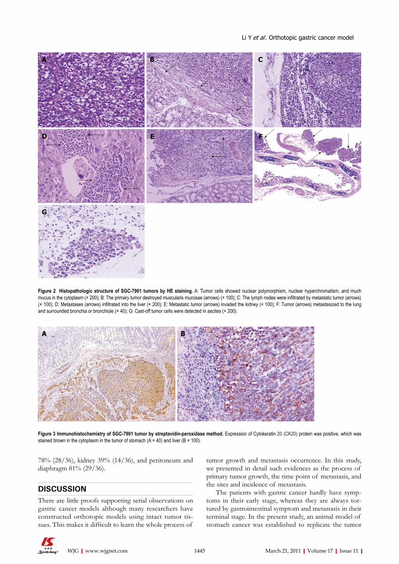
nuclear hyperchromatism, much red nucleolus, and rich pathological karyokinesis. Mucus secreted in the cytoplasm, which resulted in mucus lake, was visible in part of tumor cells (Figure 2A). Glandular differentiation and rich vas-cularity were present in tumor areas. The stomach tumor invaded the gastric wall following the disruption of the integrity of the mucous layer or muscularis mucosae (Fig-ure 2B). The lymph nodes were infiltrated and destroyed by metastatic tumor, characterized by the narrowness or diminishment of the lymph sinus (Figure 2C) Metastases were also detected in the liver (Figure 2D) and the kidney (Figure 2E), occasionally in the paranephros or pancreas (not shown). Metastatic lesions were always separated from adjacent normal tissues by fibrous integument, and lym-phoid infiltration was found in the peripheral tumor areas. Tumor metastasized to the lung and surrounded bronchia or bronchiole (Figure 2F). Smear of cast-off cells from ascites confirmed the malignant cells from the primary ad-enocarcinoma (Figure 2G).
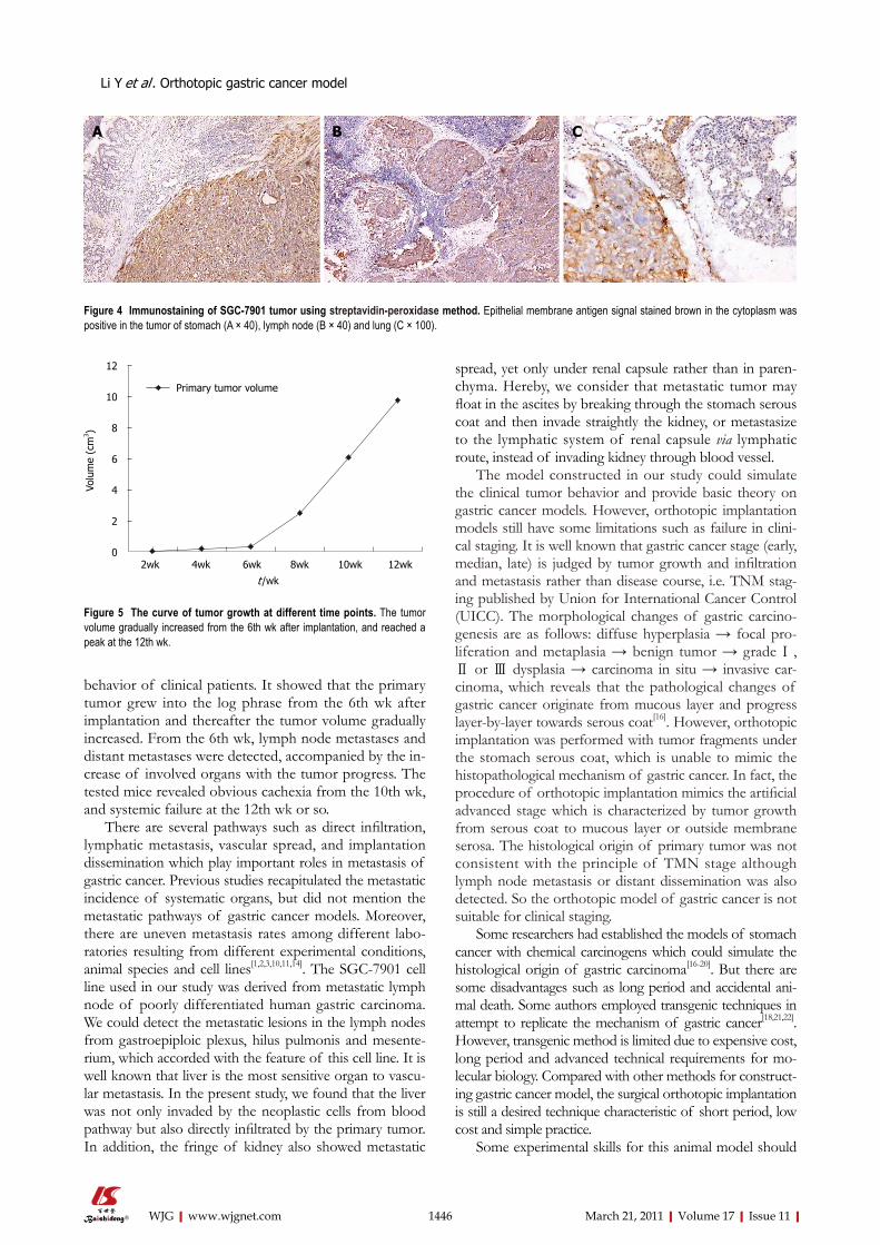
ImmunohistochemistryAll primary tumors and metastases from other organs by immunohistochemical staining showed the positive expression of CK-20 (Figure 3A and B) and EMA (Figure 4A-C) protein.
Varying volume of primary tumorThe tumor volume gradually increased from the 6th wk after implantation, and reached a peak when nude mice died at the 12th wk (Figure 5). From 10 to 12 wk, the model mice declined in their general conditions, accom-panied by cachexia and ascites.
Incidence of metastasisAfter all mice were sacrificed, the metastasis incidence of involved organs was analyzed and the following re-sults were obtained: lymph nodes 58% (21/36), liver
1444 March 21, 2011|Volume 17|Issue 11|WJG|www.wjgnet.com
Figure 1 Macroscopic examination of the primary tumor in SGC-7901 model. The gastric cavity occupied by primary tumor was found vanished, and pylorus was obstructed totally (arrows).
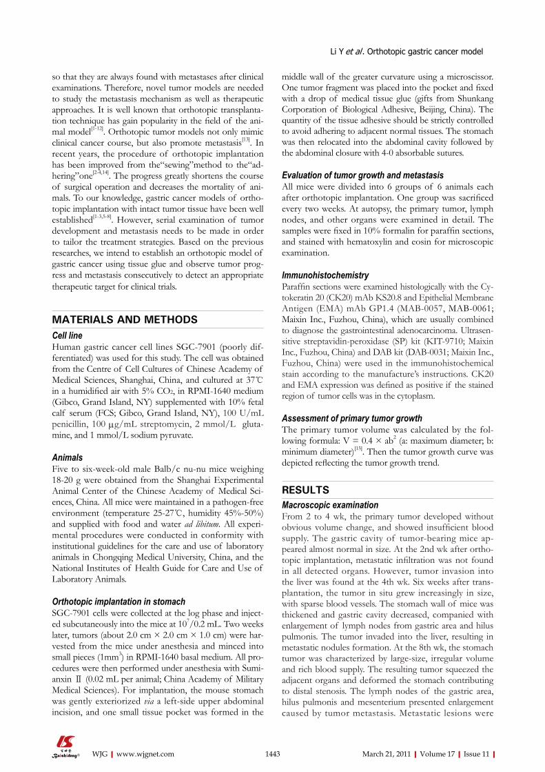
Table 1 Macroscopic observation on SGC-7901 gastric cancer model at different time points
Items 2 wk 4 wk 6 wk 8 wk 10 wk/12 wk
Primary tumor Shape Oval Oval Globular Irregularly lobular Irregularly lobular Color Gray-white Gray-white Gray-white Grayish yellow Grayish yellow Texture Stiff Stiff Stiff Stiff Stiff Vascularity Sparse Sparse Rich Richer RicherSection of stomach tumor Gastric cavity No change Mildly stenosis Obvious stenosis Obvious stenosis Narrow or vanished Greater and lesser curvature Distinguishable Distinguishable Distinguishable Distinguishable Undistinguishable Pylorochesis No No Partly Partly TotallyEffects on adjacent organs Abdominal cavity No effects No effects No effects Occupying upper abdomen Occupying whole abdomen Adhering extent Adhering to
liver lobesAdhering to liver lobes
Adhering to liver lobes
Adhering to liver lobes or posterior abdominal wall
Adhering to liver lobes or posterior abdominal wall
Compressed status No compression No compression No compression Partly oppressed Partly oppressedAscites Liquid quantity Zero Zero Zero Little Little Property - - - Clear and yellowish Clear and yellowishMetastasis Lymph nodes No No Yes Yes Yes Other organs No Yes Yes Yes Yes
Li Y et al . Orthotopic gastric cancer model
78% (28/36), kidney 39% (14/36), and peritoneum and diaphragm 81% (29/36).
DISCUSSIONThere are little proofs supporting serial observations on gastric cancer models although many researchers have constructed orthotopic models using intact tumor tis-sues. This makes it difficult to learn the whole process of
tumor growth and metastasis occurrence. In this study, we presented in detail such evidences as the process of primary tumor growth, the time point of metastasis, and the sites and incidence of metastasis.
The patients with gastric cancer hardly have symp-toms in their early stage, whereas they are always tor-tured by gastrointestinal symptom and metastasis in their terminal stage. In the present study, an animal model of stomach cancer was established to replicate the tumor
1445 March 21, 2011|Volume 17|Issue 11|WJG|www.wjgnet.com
Figure 2 Histopathologic structure of SGC-7901 tumors by HE staining. A: Tumor cells showed nuclear polymorphism, nuclear hyperchromatism, and much mucus in the cytoplasm (× 200); B: The primary tumor destroyed muscularis mucosae (arrows) (× 100); C: The lymph nodes were infiltrated by metastatic tumor (arrows) (× 100); D: Metastases (arrows) infiltrated into the liver (× 200); E: Metastatic tumor (arrows) invaded the kidney (× 100); F: Tumor (arrows) metastasized to the lung and surrounded bronchia or bronchiole (× 40); G: Cast-off tumor cells were detected in ascites (× 200).
A B C
FED
G
A B
Figure 3 Immunohistochemistry of SGC-7901 tumor by streptavidin-peroxidase method. Expression of Cytokeratin 20 (CK20) protein was positive, which was stained brown in the cytoplasm in the tumor of stomach (A × 40) and liver (B × 100).
Li Y et al . Orthotopic gastric cancer model
behavior of clinical patients. It showed that the primary tumor grew into the log phrase from the 6th wk after implantation and thereafter the tumor volume gradually increased. From the 6th wk, lymph node metastases and distant metastases were detected, accompanied by the in-crease of involved organs with the tumor progress. The tested mice revealed obvious cachexia from the 10th wk, and systemic failure at the 12th wk or so.
There are several pathways such as direct infiltration, lymphatic metastasis, vascular spread, and implantation dissemination which play important roles in metastasis of gastric cancer. Previous studies recapitulated the metastatic incidence of systematic organs, but did not mention the metastatic pathways of gastric cancer models. Moreover, there are uneven metastasis rates among different labo-ratories resulting from different experimental conditions, animal species and cell lines[1,2,3,10,11,14]. The SGC-7901 cell line used in our study was derived from metastatic lymph node of poorly differentiated human gastric carcinoma. We could detect the metastatic lesions in the lymph nodes from gastroepiploic plexus, hilus pulmonis and mesente-rium, which accorded with the feature of this cell line. It is well known that liver is the most sensitive organ to vascu-lar metastasis. In the present study, we found that the liver was not only invaded by the neoplastic cells from blood pathway but also directly infiltrated by the primary tumor. In addition, the fringe of kidney also showed metastatic
spread, yet only under renal capsule rather than in paren-chyma. Hereby, we consider that metastatic tumor may float in the ascites by breaking through the stomach serous coat and then invade straightly the kidney, or metastasize to the lymphatic system of renal capsule via lymphatic route, instead of invading kidney through blood vessel.
The model constructed in our study could simulate the clinical tumor behavior and provide basic theory on gastric cancer models. However, orthotopic implantation models still have some limitations such as failure in clini-cal staging. It is well known that gastric cancer stage (early, median, late) is judged by tumor growth and infiltration and metastasis rather than disease course, i.e. TNM stag-ing published by Union for International Cancer Control (UICC). The morphological changes of gastric carcino-genesis are as follows: diffuse hyperplasia → focal pro-liferation and metaplasia → benign tumor → grade Ⅰ , Ⅱ or Ⅲ dysplasia → carcinoma in situ → invasive car-cinoma, which reveals that the pathological changes of gastric cancer originate from mucous layer and progress layer-by-layer towards serous coat[16]. However, orthotopic implantation was performed with tumor fragments under the stomach serous coat, which is unable to mimic the histopathological mechanism of gastric cancer. In fact, the procedure of orthotopic implantation mimics the artificial advanced stage which is characterized by tumor growth from serous coat to mucous layer or outside membrane serosa. The histological origin of primary tumor was not consistent with the principle of TMN stage although lymph node metastasis or distant dissemination was also detected. So the orthotopic model of gastric cancer is not suitable for clinical staging.
Some researchers had established the models of stomach cancer with chemical carcinogens which could simulate the histological origin of gastric carcinoma[16-20]. But there are some disadvantages such as long period and accidental ani-mal death. Some authors employed transgenic techniques in attempt to replicate the mechanism of gastric cancer[18,21,22]. However, transgenic method is limited due to expensive cost, long period and advanced technical requirements for mo-lecular biology. Compared with other methods for construct-ing gastric cancer model, the surgical orthotopic implantation is still a desired technique characteristic of short period, low cost and simple practice.
Some experimental skills for this animal model should
1446 March 21, 2011|Volume 17|Issue 11|WJG|www.wjgnet.com
A B C
Figure 4 Immunostaining of SGC-7901 tumor using streptavidin-peroxidase method. Epithelial membrane antigen signal stained brown in the cytoplasm was positive in the tumor of stomach (A × 40), lymph node (B × 40) and lung (C × 100).
Primary tumor volume
12
10
8
6
4
2
0
Volu
me
(cm
3 )
2wk 4wk 6wk 8wk 10wk 12wk
Figure 5 The curve of tumor growth at different time points. The tumor volume gradually increased from the 6th wk after implantation, and reached a peak at the 12th wk.
t /wk
Li Y et al . Orthotopic gastric cancer model
be noticed. For example, necrotic area should be removed from tumor tissues used for orthotopic implantation; the quantity of medical glue should be strictly controlled to avoid adhering to adjacent organs; and the middle part of the greater gastric curvature is the first choice for ortho-topic implantation because of the operational convenience and rich blood supply.
In conclusion, we can succeed in establishing a gas-tric cancer model if only we master the experimental skills above and avoid the adverse factors.
COMMENTSBackgroundMetastasis is still the barrier to the treatment of patients with gastric cancer. Gastric cancer models of orthotopic implantation with intact tumor tissues have been well established. Moreover, the procedure of orthotopic implantation has been improved from the “sewing” method to the “adhering” one. However, there are little evidences regarding serial observations on gastric cancer models to tailor treatment strategies. Research frontiersIt is well known that orthotopic transplantation technique plays an important role in establishing animal models of various tumors. Gastric cancer model of orthotopic implantation could mimic clinical cancer process and contribute to metastasis occurrence. The procedure greatly facilitates surgical operation course and decreases the mortality of animals. Innovations and breakthroughsOrthotopic implantation with “glue paste technique” is a popular and new method in recent years which is characterized by easy performance, low cost and short operation course. This study makes it possible to help researchers learn the whole process of tumor development and metastasis, thus choosing the appropriate therapy target for clinical trials. ApplicationsSerial observations on the models can provide evidences for researchers to treat stomach cancer and test the therapeutic effects. Peer reviewThe study is interesting and well done. The model used by the Authors well mimics the dynamics of local growth and spread of gastric cancer cells at both lymph-nodes and distant organs.
REFERENCES1 Shi J, Wei PK, Zhang S, Qin ZF, Li J, Sun DZ, Xiao Y, Yu
ZH, Lin HM, Zheng GJ, Su XM, Chen YL, Liu YF, Xu L. OB glue paste technique for establishing nude mouse human gastric cancer orthotopic transplantation models. World J Gastroenterol 2008; 14: 4800-4804
2 Chen YL, Wei PK, Xu L, Su XM. [Nude mouse model of hu-man gastric carcinoma metastasis constructed by orthotopic transplantation using organism glue paste technique]. Ai Zheng 2005; 24: 246-248
3 Bhargava S, Hotz B, Buhr HJ, Hotz HG. An orthotopic nude mouse model for preclinical research of gastric cardia can-cer. Int J Colorectal Dis 2009; 24: 31-39
4 Yang B, Tuo S, Tuo CW, Zhang N, Liu QZ. A liver-metastat-ic model of human primary gastric lymphoma in nude mice orthotopically constructed by using histologically intact pa-tient specimens. Chin J Cancer 2010; 29: 579-584
5 Illert B, Otto C, Thiede A, Timmermann W. Detection of disseminated tumor cells in nude mice with human gastric cancer. Clin Exp Metastasis 2003; 20: 549-554
6 Takemura S, Yashiro M, Sunami T, Tendo M, Hirakawa K. Novel models for human scirrhous gastric carcinoma in
vivo. Cancer Sci 2004; 95: 893-9007 Furukawa T, Fu X, Kubota T, Watanabe M, Kitajima M,
Hoffman RM. Nude mouse metastatic models of human stomach cancer constructed using orthotopic implantation of histologically intact tissue. Cancer Res 1993; 53: 1204-1208
8 Koyama T, Tsubota A, Nariai K, Yoshikawa T, Mitsunaga M, Sumi M, Nimura H, Yanaga K, Yumoto Y, Mabashi Y, Takahashi H. Detection of sentinel nodes by a novel red-fluorescent dye, ATX-S10Na (Ⅱ), in an orthotopic xenograft rat model of human gastric carcinoma. Lasers Surg Med 2007; 39: 76-82
9 Hoffman RM. Orthotopic metastatic mouse models for anti-cancer drug discovery and evaluation: a bridge to the clinic. Invest New Drugs 1999; 17: 343-359
10 An Z, Jiang P, Wang X, Moossa AR, Hoffman RM. Develop-ment of a high metastatic orthotopic model of human renal cell carcinoma in nude mice: benefits of fragment implanta-tion compared to cell-suspension injection. Clin Exp Metasta-sis 1999; 17: 265-270
11 Rashidi B, Gamagami R, Sasson A, Sun FX, Geller J, Moossa AR, Hoffman RM. An orthotopic mouse model of remetas-tasis of human colon cancer liver metastasis. Clin Cancer Res 2000; 6: 2556-2561
12 Fu X, Guadagni F, Hoffman RM. A metastatic nude-mouse model of human pancreatic cancer constructed orthotopi-cally with histologically intact patient specimens. Proc Natl Acad Sci USA 1992; 89: 5645-5649
13 Kozlowski JM, Fidler IJ, Campbell D, Xu ZL, Kaighn ME, Hart IR. Metastatic behavior of human tumor cell lines grown in the nude mouse. Cancer Res 1984; 44: 3522-3529
14 Fujihara T, Sawada T, Hirakawa K, Chung YS, Yashiro M, Inoue T, Sowa M. Establishment of lymph node metastatic model for human gastric cancer in nude mice and analysis of factors associated with metastasis. Clin Exp Metastasis 1998; 16: 389-398
15 Kyriazis AP, Kyriazis AA, McCombs WB 3rd, Kereiakes JA. Biological behavior of human malignant tumors grown in the nude mouse. Cancer Res 1981; 41: 3995-4000
16 Kossoy G, Ben-Hur H, Elhayany A, Schneider DF, Zusman I. The morphological pathway for mouse forestomach cancer. Oncol Rep 2006; 15: 479-483
17 Zaidi NH, O'Connor PJ, Butler WH. N-methyl-N'-nitro-N-nitrosoguanidine-induced carcinogenesis: differential pat-tern of upper gastrointestinal tract tumours in Wistar rats after single or chronic oral doses. Carcinogenesis 1993; 14: 1561-1567
18 Pritchard DM, Przemeck SM. Review article: How useful are the rodent animal models of gastric adenocarcinoma? Aliment Pharmacol Ther 2004; 19: 841-859
19 Tsuzuki T, Egashira A, Igarashi H, Iwakuma T, Nakatsuru Y, Tominaga Y, Kawate H, Nakao K, Nakamura K, Ide F, Kura S, Nakabeppu Y, Katsuki M, Ishikawa T, Sekiguchi M. Spontaneous tumorigenesis in mice defective in the MTH1 gene encoding 8-oxo-dGTPase. Proc Natl Acad Sci USA 2001; 98: 11456-11461
20 Xu X, Brodie SG, Yang X, Im YH, Parks WT, Chen L, Zhou YX, Weinstein M, Kim SJ, Deng CX. Haploid loss of the tu-mor suppressor Smad4/Dpc4 initiates gastric polyposis and cancer in mice. Oncogene 2000; 19: 1868-1874
21 Koike K, Hinrichs SH, Isselbacher KJ, Jay G. Transgenic mouse model for human gastric carcinoma. Proc Natl Acad Sci USA 1989; 86: 5615-5619
22 Maruta F, Sugiyama A, Ishida K, Ikeno T, Murakami M, Kawasaki S, Ota H, Tatematsu M, Katsuyama T. Timing of N-methyl-N-nitrosourea administration affects gastric car-cinogenesis in Mongolian gerbils infected with Helicobacter pylori. Cancer Lett 2000; 160: 99-105
S- Editor Tian L L- Editor Ma JY E- Editor Ma WH
1447 March 21, 2011|Volume 17|Issue 11|WJG|www.wjgnet.com
COMMENTS
Li Y et al . Orthotopic gastric cancer model