Embed Size (px)
Citation preview
www.elsevier.com/locate/jcfJournal of Cystic Fibrosis xx (2014) xxx–xxx
JCF-01010; No of Pages 9
Review
Serum antibodies to Pseudomonas aeruginosa in cystic fibrosis as adiagnostic tool: A systematic review
Renan Marrichi Mauch a, Carlos Emilio Levy b,⁎
a Faculty of Medical Sciences, State University of Campinas, Brazilb Departament of Clinic Pathology, Hospital de Clínicas, Faculty of Medical Sciences, State University of Campinas, Brazil
Received 26 November 2013; received in revised form 20 January 2014; accepted 21 January 2014
Abstract
Background: A systematic literature review of the last 40 years on the research of serum antibodies to Pseudomonas aeruginosa in cystic fibrosisand its utility as a diagnostic tool.Methods: Research papers in English, Portuguese, and Spanish were accessed through electronic databases (PubMed, Medline, LILACS, andSciELO).Results: 26 studies were assessed. ELISA technique was the most commonly used technique to detect serum P. aeruginosa antibodies. The mostconsistent results were those in which the response against the antigen St-Ag:1–17 was evaluated. The accuracy levels of the ELISA techniqueremain controversial, but most studies showed a good correlation between antibody titers and microbiological culture.Conclusions: The detection of serum antibodies to P. aeruginosa shows capacity for early detection of this pathogen and potential utility andviability of incorporation in the diagnostic routine of patients with cystic fibrosis.© 2014 European Cystic Fibrosis Society. Published by Elsevier B.V. All rights reserved.
Keywords: Pseudomonas aeruginosa; Cystic fibrosis; Serum antibodies; Review
Contents
1. Introduction . . . . . . . . . . . . . . . . . . . . . . . . . . . . . . . . . . . . . . . . . . . . . . . . . . . . . . . . . . . . . . . 02. Methods . . . . . . . . . . . . . . . . . . . . . . . . . . . . . . . . . . . . . . . . . . . . . . . . . . . . . . . . . . . . . . . . 03. Data synthesis . . . . . . . . . . . . . . . . . . . . . . . . . . . . . . . . . . . . . . . . . . . . . . . . . . . . . . . . . . . . . 0
3.1. Studies addressed, casuistries and methods used . . . . . . . . . . . . . . . . . . . . . . . . . . . . . . . . . . . . . . . . . 03.2. Diagnostic and prognostic value . . . . . . . . . . . . . . . . . . . . . . . . . . . . . . . . . . . . . . . . . . . . . . . . . 03.3. Early detection of the P. aeruginosa infection . . . . . . . . . . . . . . . . . . . . . . . . . . . . . . . . . . . . . . . . . . 03.4. Antibody titers and clinical status . . . . . . . . . . . . . . . . . . . . . . . . . . . . . . . . . . . . . . . . . . . . . . . . 03.5. Antibody response against the infection treatment . . . . . . . . . . . . . . . . . . . . . . . . . . . . . . . . . . . . . . . . 03.6. Accuracy . . . . . . . . . . . . . . . . . . . . . . . . . . . . . . . . . . . . . . . . . . . . . . . . . . . . . . . . . . . . . 0
4. Discussion . . . . . . . . . . . . . . . . . . . . . . . . . . . . . . . . . . . . . . . . . . . . . . . . . . . . . . . . . . . . . . . 05. Conclusions . . . . . . . . . . . . . . . . . . . . . . . . . . . . . . . . . . . . . . . . . . . . . . . . . . . . . . . . . . . . . . . 0References . . . . . . . . . . . . . . . . . . . . . . . . . . . . . . . . . . . . . . . . . . . . . . . . . . . . . . . . . . . . . . . . . . 0
Abbreviations: CF, Cystic fibrosis; BAL, Bronchoalveolar lavage; ELISA, Enzyme-linked immunosorbent assay; CIE, Crossed immune electrophoresis; RIA,Radioimmunoassay; ExoA, Exotoxin A; ELA, Elastase; AP, Alkaline protease; TTSS, Type three secretion system; PCR, Polymerase chain reaction; PPV, Positivepredictive value; NPV, Negative predictive value⁎ Corresponding author at: Department of Clinical Pathology, Faculty of Medical Sciences, State University of Campinas, Campinas, SP, Brazil.
E-mail address: [email protected] (C.E. Levy).
1569-1993/$ -see front matter © 2014 European Cystic Fibrosis Society. Published by Elsevier B.V. All rights reserved.http://dx.doi.org/10.1016/j.jcf.2014.01.005
Please cite this article as: Mauch RM, Levy CE, Serum antibodies to Pseudomonas aeruginosa in cystic fibrosis as a diagnostic tool: A systematic review, J Cyst Fibros(2014), http://dx.doi.org/10.1016/j.jcf.2014.01.005
2 R.M. Mauch, C.E. Levy / Journal of Cystic Fibrosis xx (2014) xxx–xxx
1. Introduction
Pseudomonas aeruginosa pulmonary infection is responsi-ble for elevated morbidity and mortality among cystic fibrosis(CF) patients [1,2]. In childhood, 10 to 30% of these patientsare colonized and in adulthood, 80 to 90% are infected withthis bacterium. Patients infected with P. aeruginosa generallypresent a reduction of about 10 years in life expectancy whencompared with the non-infected ones [3].
When chronic infection [4] is established, P. aeruginosa ispractically impossible to be eradicated [1,2], although, its clearancefrom the respiratory tract is possible through early interventionwith antibiotic therapy, as soon as the pathogen settles in theorganism. Thus, early aggressive treatment is recommended, beingpossible to delay the chronic colonization and progression of thepulmonary disease [5].
Detection of P. aeruginosa in the diagnostic routine is mademostly through sputum culture — spontaneously expectorated orinduced by inhalation of hypertonic saline (3–7%); however, manypatients – especially children under 7 years – are incapable ofproducing an expectorated sputum specimen. BAL, the goldstandard, is an option, but it is invasive and usually employed onlywhen there is a compelling reason to obtain a respiratory sampleand other approaches have failed. For this reason, oropharyngeal(OP) swabs are used as a surrogate for sputum or BAL fluidspecimens in these individuals, sampling the microbial flora of theupper respiratory tract, which is assumed to reflect that of the lowerrespiratory tract [6]. Despite the good specificity reported for thismethod, in comparison to BAL culture, it is known that a negativeculture from the upper airways does not exclude the presence ofP. aeruginosa in the lower airways [7,8]. Sampling errors duringthe BAL and the frequent insufficient sample obtained throughoropharyngeal swab can lead to false negative results [7]. Thedifficulty in obtaining representative respiratory specimens fromthe airways of infants and children indicates the need for the use ofmethods that can complement or be an alternative to microbiolog-ical culture [9].
The detection of serum antibodies against P. aeruginosahas emerged as a possible auxiliary method to assess the earlyeradication therapy [10,11]. Highly sensitive methods fordetection of antibodies against several P. aeruginosa antigensmay complement the monitoring methods currently used [11].Positive antibody titer results and culture-negative samples ofrespiratory secretion should alert health professionals to perform amore thorough search for a probable infection, by repeating thetest or by using more sensitive and specific methods. In contrast,increasing antibody levels are associated with a greater likelihoodof persistent P. aeruginosa chronic infection [9]. Elevatedantibody titers at the time of initial OP culture may be a usefultool for CF clinicians and researchers monitoring patients atrisk for subsequent infection. However, their use in routinepractice remains controversial [9].
Thus, the aim of this systematic review was to collect studiespublished in the last 40 years addressing the detection of serumantibodies to P. aeruginosa and to evaluate its utility for earlydetection of this bacterium and the diagnostic and prognosticvalue in CF patients.
Please cite this article as: Mauch RM, Levy CE, Serum antibodies to Pseudomonas ae(2014), http://dx.doi.org/10.1016/j.jcf.2014.01.005
2. Methods
Research papers in English, Portuguese, and Spanish in theperiod from 1973 to 2013 were reviewed. The search forreferences was made through electronic database exploration(PubMed, Medline, LILACS and SciELO), using the keywords“Cystic Fibrosis”, “Pseudomonas aeruginosa”, “serology”, and“serum antibodies”, and their correspondent translations in variedcombinations, from November to August of 2013. In addition, thereferences of all papers were consulted in the search of new papersto include.
Then, we began the paper selection process, by reviewing titlesand abstracts. The first inclusion criterion was the identification ofpotentially relevant papers, considering those in which the studyassessed the utility of serum antibody detection in the diagnosticroutine of pulmonary disease. The recuperation criteria for com-plete papers were the following types of study: cohort, longitudi-nal, case–control, descriptive, experimental and cross-sectional,whose results addressed such subject. The selection was based onthe limits of the compliance of issues in relation to objectives,excluding those in which, despite appearing in the search results,did not address the issue from the diagnostic point of view of serumantibody detection. We also excluded review articles, studiesaddressing the immune response to P. aeruginosa but not related toCF, or related to CF but without clinical application or applied tothe diagnosis and studies not related to the CF pulmonary disease.
3. Data synthesis
3.1. Studies addressed, casuistries and methods used
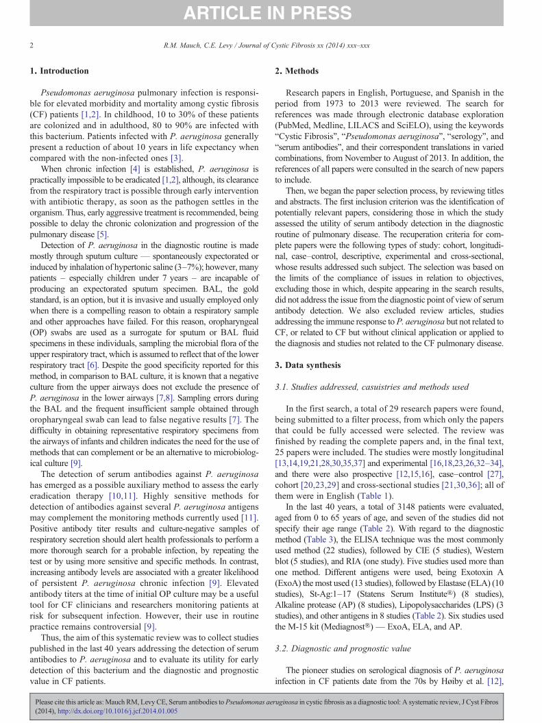
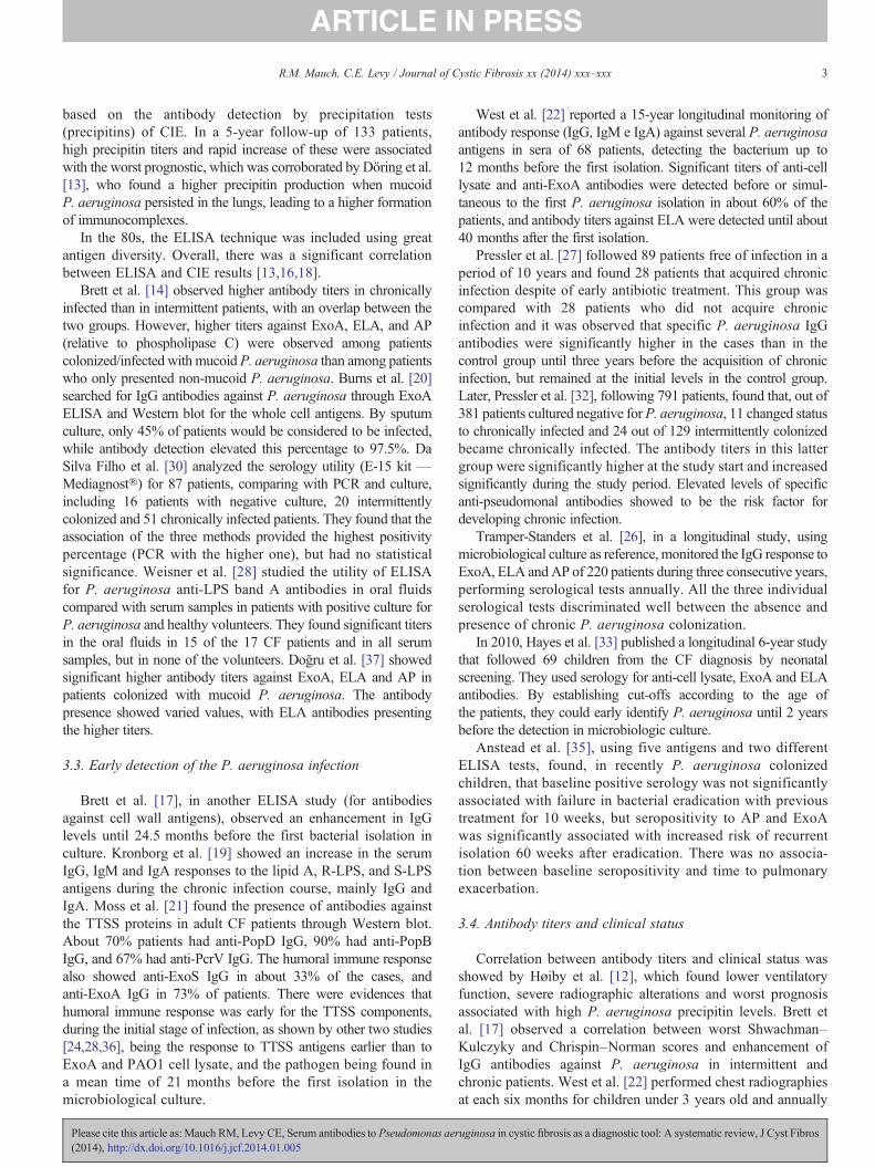
In the first search, a total of 29 research papers were found,being submitted to a filter process, from which only the papersthat could be fully accessed were selected. The review wasfinished by reading the complete papers and, in the final text,25 papers were included. The studies were mostly longitudinal[13,14,19,21,28,30,35,37] and experimental [16,18,23,26,32–34],and there were also prospective [12,15,16], case–control [27],cohort [20,23,29] and cross-sectional studies [21,30,36]; all ofthem were in English (Table 1).
In the last 40 years, a total of 3148 patients were evaluated,aged from 0 to 65 years of age, and seven of the studies did notspecify their age range (Table 2). With regard to the diagnosticmethod (Table 3), the ELISA technique was the most commonlyused method (22 studies), followed by CIE (5 studies), Westernblot (5 studies), and RIA (one study). Five studies used more thanone method. Different antigens were used, being Exotoxin A(ExoA) the most used (13 studies), followed by Elastase (ELA) (10studies), St-Ag:1–17 (Statens Serum Institute®) (8 studies),Alkaline protease (AP) (8 studies), Lipopolysaccharides (LPS) (3studies), and other antigens in 8 studies (Table 2). Six studies usedthe M-15 kit (Mediagnost®)— ExoA, ELA, and AP.
3.2. Diagnostic and prognostic value
The pioneer studies on serological diagnosis of P. aeruginosainfection in CF patients date from the 70s by Høiby et al. [12],
ruginosa in cystic fibrosis as a diagnostic tool: A systematic review, J Cyst Fibros
3R.M. Mauch, C.E. Levy / Journal of Cystic Fibrosis xx (2014) xxx–xxx
based on the antibody detection by precipitation tests(precipitins) of CIE. In a 5-year follow-up of 133 patients,high precipitin titers and rapid increase of these were associatedwith the worst prognostic, which was corroborated by Döring et al.[13], who found a higher precipitin production when mucoidP. aeruginosa persisted in the lungs, leading to a higher formationof immunocomplexes.
In the 80s, the ELISA technique was included using greatantigen diversity. Overall, there was a significant correlationbetween ELISA and CIE results [13,16,18].
Brett et al. [14] observed higher antibody titers in chronicallyinfected than in intermittent patients, with an overlap between thetwo groups. However, higher titers against ExoA, ELA, and AP(relative to phospholipase C) were observed among patientscolonized/infected with mucoid P. aeruginosa than among patientswho only presented non-mucoid P. aeruginosa. Burns et al. [20]searched for IgG antibodies against P. aeruginosa through ExoAELISA and Western blot for the whole cell antigens. By sputumculture, only 45% of patients would be considered to be infected,while antibody detection elevated this percentage to 97.5%. DaSilva Filho et al. [30] analyzed the serology utility (E-15 kit —Mediagnost®) for 87 patients, comparing with PCR and culture,including 16 patients with negative culture, 20 intermittentlycolonized and 51 chronically infected patients. They found that theassociation of the three methods provided the highest positivitypercentage (PCR with the higher one), but had no statisticalsignificance. Weisner et al. [28] studied the utility of ELISAfor P. aeruginosa anti-LPS band A antibodies in oral fluidscompared with serum samples in patients with positive culture forP. aeruginosa and healthy volunteers. They found significant titersin the oral fluids in 15 of the 17 CF patients and in all serumsamples, but in none of the volunteers. Doğru et al. [37] showedsignificant higher antibody titers against ExoA, ELA and AP inpatients colonized with mucoid P. aeruginosa. The antibodypresence showed varied values, with ELA antibodies presentingthe higher titers.
3.3. Early detection of the P. aeruginosa infection
Brett et al. [17], in another ELISA study (for antibodiesagainst cell wall antigens), observed an enhancement in IgGlevels until 24.5 months before the first bacterial isolation inculture. Kronborg et al. [19] showed an increase in the serumIgG, IgM and IgA responses to the lipid A, R-LPS, and S-LPSantigens during the chronic infection course, mainly IgG andIgA. Moss et al. [21] found the presence of antibodies againstthe TTSS proteins in adult CF patients through Western blot.About 70% patients had anti-PopD IgG, 90% had anti-PopBIgG, and 67% had anti-PcrV IgG. The humoral immune responsealso showed anti-ExoS IgG in about 33% of the cases, andanti-ExoA IgG in 73% of patients. There were evidences thathumoral immune response was early for the TTSS components,during the initial stage of infection, as shown by other two studies[24,28,36], being the response to TTSS antigens earlier than toExoA and PAO1 cell lysate, and the pathogen being found ina mean time of 21 months before the first isolation in themicrobiological culture.
Please cite this article as: Mauch RM, Levy CE, Serum antibodies to Pseudomonas ae(2014), http://dx.doi.org/10.1016/j.jcf.2014.01.005
West et al. [22] reported a 15-year longitudinal monitoring ofantibody response (IgG, IgM e IgA) against several P. aeruginosaantigens in sera of 68 patients, detecting the bacterium up to12 months before the first isolation. Significant titers of anti-celllysate and anti-ExoA antibodies were detected before or simul-taneous to the first P. aeruginosa isolation in about 60% of thepatients, and antibody titers against ELA were detected until about40 months after the first isolation.
Pressler et al. [27] followed 89 patients free of infection in aperiod of 10 years and found 28 patients that acquired chronicinfection despite of early antibiotic treatment. This group wascompared with 28 patients who did not acquire chronicinfection and it was observed that specific P. aeruginosa IgGantibodies were significantly higher in the cases than in thecontrol group until three years before the acquisition of chronicinfection, but remained at the initial levels in the control group.Later, Pressler et al. [32], following 791 patients, found that, out of381 patients cultured negative for P. aeruginosa, 11 changed statusto chronically infected and 24 out of 129 intermittently colonizedbecame chronically infected. The antibody titers in this lattergroup were significantly higher at the study start and increasedsignificantly during the study period. Elevated levels of specificanti-pseudomonal antibodies showed to be the risk factor fordeveloping chronic infection.
Tramper-Standers et al. [26], in a longitudinal study, usingmicrobiological culture as reference, monitored the IgG response toExoA, ELA andAP of 220 patients during three consecutive years,performing serological tests annually. All the three individualserological tests discriminated well between the absence andpresence of chronic P. aeruginosa colonization.
In 2010, Hayes et al. [33] published a longitudinal 6-year studythat followed 69 children from the CF diagnosis by neonatalscreening. They used serology for anti-cell lysate, ExoA and ELAantibodies. By establishing cut-offs according to the age ofthe patients, they could early identify P. aeruginosa until 2 yearsbefore the detection in microbiologic culture.
Anstead et al. [35], using five antigens and two differentELISA tests, found, in recently P. aeruginosa colonizedchildren, that baseline positive serology was not significantlyassociated with failure in bacterial eradication with previoustreatment for 10 weeks, but seropositivity to AP and ExoAwas significantly associated with increased risk of recurrentisolation 60 weeks after eradication. There was no associa-tion between baseline seropositivity and time to pulmonaryexacerbation.
3.4. Antibody titers and clinical status
Correlation between antibody titers and clinical status wasshowed by Høiby et al. [12], which found lower ventilatoryfunction, severe radiographic alterations and worst prognosisassociated with high P. aeruginosa precipitin levels. Brett etal. [17] observed a correlation between worst Shwachman–Kulczyky and Chrispin–Norman scores and enhancement ofIgG antibodies against P. aeruginosa in intermittent andchronic patients. West et al. [22] performed chest radiographiesat each six months for children under 3 years old and annually
ruginosa in cystic fibrosis as a diagnostic tool: A systematic review, J Cyst Fibros
Table 1Study index, according to author, design, local, objective and results obtained.
Author Study design Local Objective Main results
Høiby et al. [12] Prospective Copenhagen,Denmark
Detection of anti-pseudomonal antibodies by CIE. Poor prognosis in cystic fibrosis was associatedwith high numbers of precipitins that are rapidlyincreasing.
Döring et al. [13] Longitudinal Copenhagen,Denmark
To measure humoral antibody titers to AP andELA and to determine the number of differentprecipitins to the St-Ag:1–17
After onset of chronic P. aeruginosa lung infection,the clinical state of most patients revealed decliningand specific antibody production increase in allpatients.
Brett et al. [14] Longitudinal Leeds, UK To measure immunological changes associatedwith colonization and infection of the respiratorytract by P. aeruginosa and to assess the value ofthese measurements in monitoring the progress ofinfection
High antibody titers associated with worst clinicalstatus and low titers associated with better clinicalstatus both in chronic infected and intermittentpatients
Hollsing et al. [15] Prospective Danderyd,Sweden
To elucidate the clinical value of the determinationof antibodies against various antigens in relation tosigns of infection and antimicrobial therapy
Serum antibodies against phospholipase C seem tobe a valid indicator of chronic P. aeruginosacolonization.
Pedersen et al. [16] Experimental Copenhagen,Denmark
Development of an ELISA test to detect anti-pseudomonal antibodies, comparing with CIE
Sensitivity and specificity to detection of infectionsimilar to CIE, but simpler
Brett et al. [17] Prospective Leeds, UK To use an ELISA test for monitoring antibodytiters in CF patients from whom P. aeruginosawas isolated for the first time during a study periodof 3 years
Antibody titers altered even 24 months before thefirst isolation in culture. Levels decreased afterantibiotic therapy
Formsgaard et al. [18] Experimental Copenhagen,Denmark
Development of an ELISA test to detect anti-LPSIgG and IgM P. aeruginosa antibodies
Enhancement in IgG anti-LPS P. aeruginosa anti-bodies in the establishment and course of chronicinfection, with marked affinity and specificity
Kronborg et al. [19] Longitudinal Copenhagen,Denmark
To investigate the appearance of specificantibodies to endotoxic lipid A as well as tothe core and O saccharides of P. aeruginosa inserum and sputum samples during lung infection
Enhanced antibody response to all LPS antigensstudied during the course of chronic infection
Burns et al. [20] Cohort Multicenter (USA) To examine the agreement among P. aeruginosaisolates from upper and lower airways; tocharacterize indirect infection markers
Greater infection evidence when culture andserology are combined than when only culture isperformed; 97.5% of the children until 3 years oldanalyzed were infected.
Moss et al. [21] Cross-sectional Milwaukee, USA Investigation of TTSS proteins through themeasure of immune response against TTSScomponents
Evidences that antibody response is early to theTTSS components. Type III cytotoxins contributeto the pathogenicity of P. aeruginosa in acute lunginfections.
West et al. [22] Longitudinal Multicenter (USA) To evaluate the relationship between the productionof antibody response against P. aeruginosa andclinical factors associated with pulmonary infectionsin patients with CF diagnosed in early life
Enhanced antibody titers before the first isolationin culture (ExoA until 6 months, cell lysate until12 months) in patients recently diagnosed withCF
Johansen et al. [23] Cohort Copenhagen,Denmark
To investigate the effects of increasingly intensivetreatment regimens on the anti-Pseudomonasantibody response and survival of patients afteracquisition of P. aeruginosa chronic lunginfection
Lower antibody response and longer survival afteracquisition of chronic lung infection in patientswho were treated intensively
Corech et al. [24] Experimental Milwaukee, USA To test if identification of the response to TTSSproteins can lead to early detection of P. aeruginosainfection than has been measured for the analysis ofother pseudomonal antigens
Response to TTSS components earlier than toother antigens in the initial stages of infection,with reduced prevalence during chronic infection.
Kappler et al. [25] Experimental Munich, Germany Validation of an ELISA commercialized test to detectanti-Pseudomonas antibodies and P. aeruginosainfection
Higher sensitivity, specificity and PPV when the 3serological assays were combined. Higher PPVsfor intermittent patients, but low VPP for patientsfree of infection.
Tramper-Standers etal. [26]
Experimental Utrecht,Netherlands
Evaluation of a serological commercialized test toearly P. aeruginosa detection and chroniccolonization
All three individual serological tests discriminatedwell between the absence and presence of chronicP. aeruginosa colonization
Pressler et al. [27] Case–control Copenhagen,Denmark
To analyze risk factors for the development ofchronic P. aeruginosa infection
Specific anti-pseudomonal IgG serum antibodieswere significantly higher in the study group thanin controls already 3 years prior to onset ofchronic infection
Weisner et al. [28] Longitudinal London, UK To investigate the viability of the use of oral fluidsamples to detect anti-LPS P. aeruginosaantibodies; to compare these results with serumantibodies
Immunoblotting and ELISA are sensitive proce-dures for the detection of antibodies to A-bandLPS of P. aeruginosa in oral fluid and serum frompatients with CF.
4 R.M. Mauch, C.E. Levy / Journal of Cystic Fibrosis xx (2014) xxx–xxx
Please cite this article as: Mauch RM, Levy CE, Serum antibodies to Pseudomonas aeruginosa in cystic fibrosis as a diagnostic tool: A systematic review, J Cyst Fibros(2014), http://dx.doi.org/10.1016/j.jcf.2014.01.005
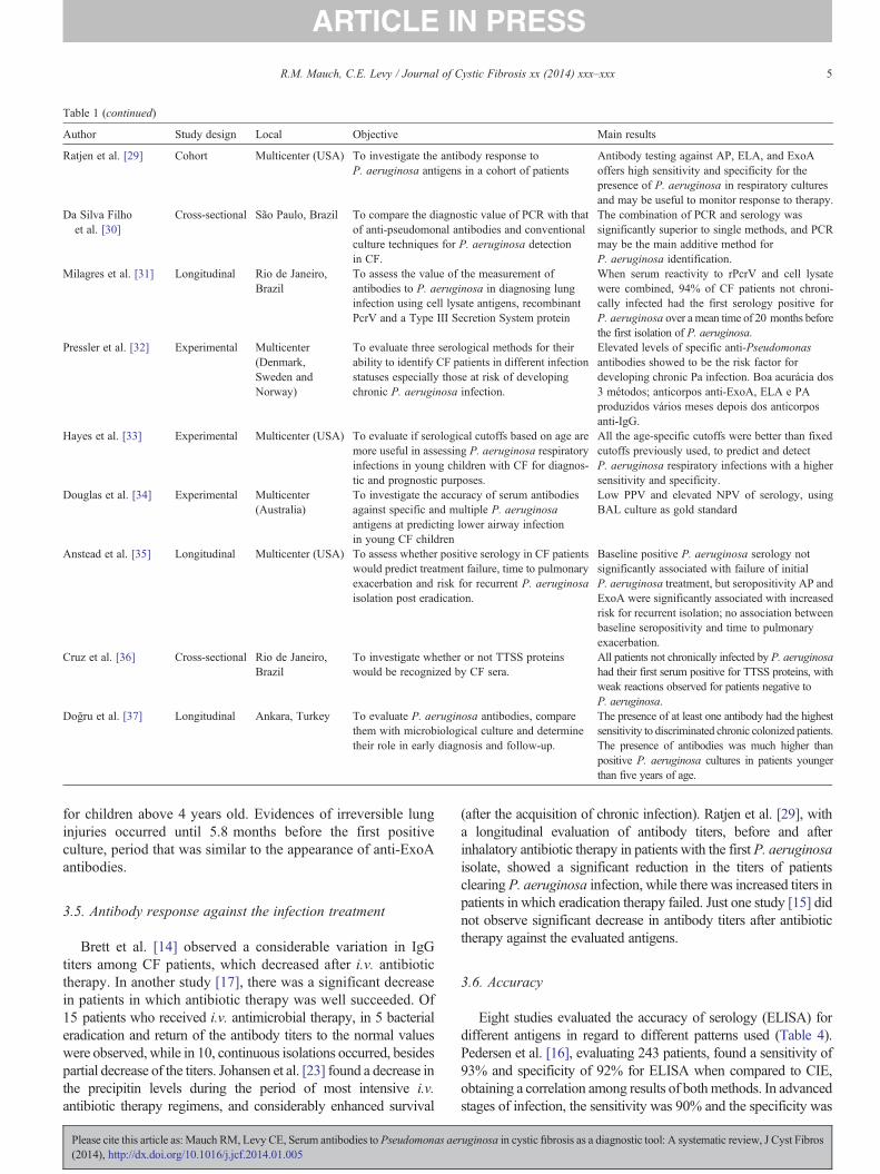
Table 1 (continued)
Author Study design Local Objective Main results
Ratjen et al. [29] Cohort Multicenter (USA) To investigate the antibody response toP. aeruginosa antigens in a cohort of patients
Antibody testing against AP, ELA, and ExoAoffers high sensitivity and specificity for thepresence of P. aeruginosa in respiratory culturesand may be useful to monitor response to therapy.
Da Silva Filhoet al. [30]
Cross-sectional São Paulo, Brazil To compare the diagnostic value of PCR with thatof anti-pseudomonal antibodies and conventionalculture techniques for P. aeruginosa detectionin CF.
The combination of PCR and serology wassignificantly superior to single methods, and PCRmay be the main additive method forP. aeruginosa identification.
Milagres et al. [31] Longitudinal Rio de Janeiro,Brazil
To assess the value of the measurement ofantibodies to P. aeruginosa in diagnosing lunginfection using cell lysate antigens, recombinantPcrV and a Type III Secretion System protein
When serum reactivity to rPcrV and cell lysatewere combined, 94% of CF patients not chroni-cally infected had the first serology positive forP. aeruginosa over a mean time of 20 months beforethe first isolation of P. aeruginosa.
Pressler et al. [32] Experimental Multicenter(Denmark,Sweden andNorway)
To evaluate three serological methods for theirability to identify CF patients in different infectionstatuses especially those at risk of developingchronic P. aeruginosa infection.
Elevated levels of specific anti-Pseudomonasantibodies showed to be the risk factor fordeveloping chronic Pa infection. Boa acurácia dos3 métodos; anticorpos anti-ExoA, ELA e PAproduzidos vários meses depois dos anticorposanti-IgG.
Hayes et al. [33] Experimental Multicenter (USA) To evaluate if serological cutoffs based on age aremore useful in assessing P. aeruginosa respiratoryinfections in young children with CF for diagnos-tic and prognostic purposes.
All the age-specific cutoffs were better than fixedcutoffs previously used, to predict and detectP. aeruginosa respiratory infections with a highersensitivity and specificity.
Douglas et al. [34] Experimental Multicenter(Australia)
To investigate the accuracy of serum antibodiesagainst specific and multiple P. aeruginosaantigens at predicting lower airway infectionin young CF children
Low PPV and elevated NPV of serology, usingBAL culture as gold standard
Anstead et al. [35] Longitudinal Multicenter (USA) To assess whether positive serology in CF patientswould predict treatment failure, time to pulmonaryexacerbation and risk for recurrent P. aeruginosaisolation post eradication.
Baseline positive P. aeruginosa serology notsignificantly associated with failure of initialP. aeruginosa treatment, but seropositivity AP andExoA were significantly associated with increasedrisk for recurrent isolation; no association betweenbaseline seropositivity and time to pulmonaryexacerbation.
Cruz et al. [36] Cross-sectional Rio de Janeiro,Brazil
To investigate whether or not TTSS proteinswould be recognized by CF sera.
All patients not chronically infected by P. aeruginosahad their first serum positive for TTSS proteins, withweak reactions observed for patients negative toP. aeruginosa.
Doğru et al. [37] Longitudinal Ankara, Turkey To evaluate P. aeruginosa antibodies, comparethem with microbiological culture and determinetheir role in early diagnosis and follow-up.
The presence of at least one antibody had the highestsensitivity to discriminated chronic colonized patients.The presence of antibodies was much higher thanpositive P. aeruginosa cultures in patients youngerthan five years of age.
5R.M. Mauch, C.E. Levy / Journal of Cystic Fibrosis xx (2014) xxx–xxx
for children above 4 years old. Evidences of irreversible lunginjuries occurred until 5.8 months before the first positiveculture, period that was similar to the appearance of anti-ExoAantibodies.
3.5. Antibody response against the infection treatment
Brett et al. [14] observed a considerable variation in IgGtiters among CF patients, which decreased after i.v. antibiotictherapy. In another study [17], there was a significant decreasein patients in which antibiotic therapy was well succeeded. Of15 patients who received i.v. antimicrobial therapy, in 5 bacterialeradication and return of the antibody titers to the normal valueswere observed, while in 10, continuous isolations occurred, besidespartial decrease of the titers. Johansen et al. [23] found a decrease inthe precipitin levels during the period of most intensive i.v.antibiotic therapy regimens, and considerably enhanced survival
Please cite this article as: Mauch RM, Levy CE, Serum antibodies to Pseudomonas ae(2014), http://dx.doi.org/10.1016/j.jcf.2014.01.005
(after the acquisition of chronic infection). Ratjen et al. [29], witha longitudinal evaluation of antibody titers, before and afterinhalatory antibiotic therapy in patients with the first P. aeruginosaisolate, showed a significant reduction in the titers of patientsclearing P. aeruginosa infection, while there was increased titers inpatients in which eradication therapy failed. Just one study [15] didnot observe significant decrease in antibody titers after antibiotictherapy against the evaluated antigens.
3.6. Accuracy
Eight studies evaluated the accuracy of serology (ELISA) fordifferent antigens in regard to different patterns used (Table 4).Pedersen et al. [16], evaluating 243 patients, found a sensitivity of93% and specificity of 92% for ELISA when compared to CIE,obtaining a correlation among results of bothmethods. In advancedstages of infection, the sensitivity was 90% and the specificity was
ruginosa in cystic fibrosis as a diagnostic tool: A systematic review, J Cyst Fibros
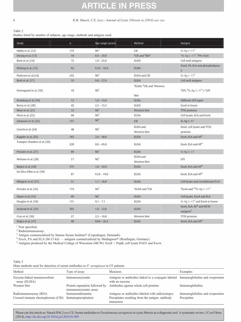
Table 2Studies listed by number of subjects, age range, methods and antigens used.
Study n Age range (years) Method Antigen
Høiby et al. [12] 133 NE1 CIE St-Ag:1–173
Doring et al. [13] 10 8,0 – 29,0 aCIE and bRIA2 aSt-Ag:1–173, bPA e ELA
Brett et al. [14] 75 1,0 – 25,0 ELISA Cell wall antigens
Holsing et al. [15] 62 0,32 – 32,0 ELISAExoA, PA, ELA and phospholipase
C
Pedersen et al.[16] 243 NE1 ELISA and CIE St-Ag:1–173
Brett et al. [17] 33 0,8 – 27,0 ELISA Cell wall antigens
Formsgaard et al. [18] 10 NE1
aELISA, bCIE and cWestern
blot
aLPS, bSt-Ag:1–173 e cLPS
Kronborg et al. [19] 12 1,0 – 15,0 ELISA Different LPS types
Burns et al. [20] 42 2,5 – 15,5 ELISA ExoA in house
Moss et al. [21] 33 NE1 Western blot TTSS proteins
West et al. [22] 68 NE1 ELISA Cell lysate, ELA and ExoA
Johansen et al. [23] 157 NE1CIE St-Ag:1-17
Corech et al. [24] 48 NE1ELISA and
Western blot
ExoA, cell lysate and TTSS
proteins
Kappler et al. [25] 183 2,0 – 38,0 ELISA ExoA, ELA and AP4
Tramper-Standers et al. [26] 220 0,0 – 65,0 ELISA ExoA, ELA and AP4
Pressler et al. [27] 89 NE1 ELISA St-Ag:1–17
Weisner et al. [28] 17 NE1ELISA and
Western blotLPS
Ratjen et al. [29] 375 1,0 – 52,0 ELISA ExoA, ELA and AP4
Da Silva Filho et al. [30] 87 0,24 – 19,0 ELISA ExoA, ELA and AP4
Milagres et al. [31] 51 1,1 – 16,8 ELISA Cell lysate and recombinant PcrV
Pressler et al. [32] 719 NE1 aELISA and bCIE aExoA and a,bSt-Ag:1–173
Hayes et al. [33] 69 NE1 ELISA Cell lysate, ExoA and ELA
Douglas et al. [34] 131 0,1 – 7,1 ELISA St-Ag:1–173 and ExoA in house
Anstead et al. [35] 303 1,0 – 12,0 ELISAExoA, ELA, AP4 and MCW
antigens5
Cruz et al. [36] 27 2,5 – 16,8 Western blot TTSS proteins
Doğru et al. [37] 90 0,64 – 26,3 ELISA ExoA, ELA and AP4
1 Non specified.2 Radioimmunoassay.3 Antigen commercialized by Statens Serum Institute® (Copenhagen, Denmark).4 ExoA, PA and ELA (M-15 kit) — antigens commercialized by Mediagnost® (Reutlingen, Germany).5 Antigens produced by the Medical College of Wisconsin (MCW): ExoS + PopB, cell lysate PAO1 and ExoA.
Table 3Main methods used for detection of serum antibodies to P. aeruginosa in CF patients.
Method Type of assay Measures Examples
Enzyme-linked immunosorbentassay (ELISA)
Immunoenzymatic Antigens or antibodies linked to a conjugate labeledwith an enzyme
Immunoglobulins and exoproteins
Western blot Protein separation followed byimmunoenzymatic assay
Antibodies against whole cell proteins Immunoglobulins
Radioimmunoassay (RIA) Immunoradiometric Antigens or antibodies labeled with radioisotopes Immunoglobulins and exoproteinsCrossed immune electrophoresis (CIE) Immunoprecipitation Precipitates resulting from the antigen–antibody
interactionPrecipitins
6 R.M. Mauch, C.E. Levy / Journal of Cystic Fibrosis xx (2014) xxx–xxx
Please cite this article as: Mauch RM, Levy CE, Serum antibodies to Pseudomonas aeruginosa in cystic fibrosis as a diagnostic tool: A systematic review, J Cyst Fibros(2014), http://dx.doi.org/10.1016/j.jcf.2014.01.005
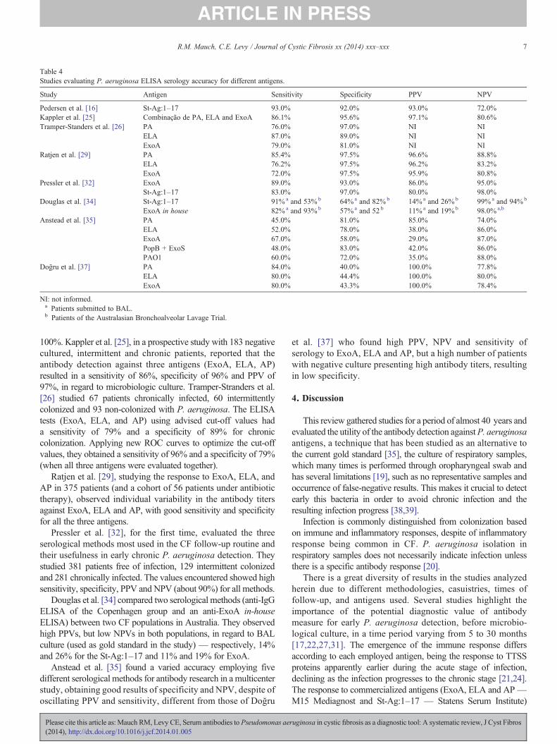
Table 4Studies evaluating P. aeruginosa ELISA serology accuracy for different antigens.
Study Antigen Sensitivity Specificity PPV NPV
Pedersen et al. [16] St-Ag:1–17 93.0% 92.0% 93.0% 72.0%Kappler et al. [25] Combinação de PA, ELA and ExoA 86.1% 95.6% 97.1% 80.6%Tramper-Standers et al. [26] PA 76.0% 97.0% NI NI
ELA 87.0% 89.0% NI NIExoA 79.0% 81.0% NI NI
Ratjen et al. [29] PA 85.4% 97.5% 96.6% 88.8%ELA 76.2% 97.5% 96.2% 83.2%ExoA 72.0% 97.5% 95.9% 80.8%
Pressler et al. [32] ExoA 89.0% 93.0% 86.0% 95.0%St-Ag:1–17 83.0% 97.0% 80.0% 98.0%
Douglas et al. [34] St-Ag:1–17 91%a and 53%b 64%a and 82%b 14%a and 26%b 99%a and 94%b
ExoA in house 82%a and 93%b 57%a and 52 b 11%a and 19%b 98.0%a,b
Anstead et al. [35] PA 45.0% 81.0% 85.0% 74.0%ELA 52.0% 78.0% 38.0% 86.0%ExoA 67.0% 58.0% 29.0% 87.0%PopB + ExoS 48.0% 83.0% 42.0% 86.0%PAO1 60.0% 72.0% 35.0% 88.0%
Doğru et al. [37] PA 84.0% 40.0% 100.0% 77.8%ELA 80.0% 44.4% 100.0% 80.0%ExoA 80.0% 43.3% 100.0% 78.4%
NI: not informed.a Patients submitted to BAL.b Patients of the Australasian Bronchoalveolar Lavage Trial.
7R.M. Mauch, C.E. Levy / Journal of Cystic Fibrosis xx (2014) xxx–xxx
100%. Kappler et al. [25], in a prospective study with 183 negativecultured, intermittent and chronic patients, reported that theantibody detection against three antigens (ExoA, ELA, AP)resulted in a sensitivity of 86%, specificity of 96% and PPV of97%, in regard to microbiologic culture. Tramper-Stranders et al.[26] studied 67 patients chronically infected, 60 intermittentlycolonized and 93 non-colonized with P. aeruginosa. The ELISAtests (ExoA, ELA, and AP) using advised cut-off values hada sensitivity of 79% and a specificity of 89% for chroniccolonization. Applying new ROC curves to optimize the cut-offvalues, they obtained a sensitivity of 96% and a specificity of 79%(when all three antigens were evaluated together).
Ratjen et al. [29], studying the response to ExoA, ELA, andAP in 375 patients (and a cohort of 56 patients under antibiotictherapy), observed individual variability in the antibody titersagainst ExoA, ELA and AP, with good sensitivity and specificityfor all the three antigens.
Pressler et al. [32], for the first time, evaluated the threeserological methods most used in the CF follow-up routine andtheir usefulness in early chronic P. aeruginosa detection. Theystudied 381 patients free of infection, 129 intermittent colonizedand 281 chronically infected. The values encountered showed highsensitivity, specificity, PPV and NPV (about 90%) for all methods.
Douglas et al. [34] compared two serological methods (anti-IgGELISA of the Copenhagen group and an anti-ExoA in-houseELISA) between two CF populations in Australia. They observedhigh PPVs, but low NPVs in both populations, in regard to BALculture (used as gold standard in the study) — respectively, 14%and 26% for the St-Ag:1–17 and 11% and 19% for ExoA.
Anstead et al. [35] found a varied accuracy employing fivedifferent serological methods for antibody research in a multicenterstudy, obtaining good results of specificity and NPV, despite ofoscillating PPV and sensitivity, different from those of Doğru
Please cite this article as: Mauch RM, Levy CE, Serum antibodies to Pseudomonas ae(2014), http://dx.doi.org/10.1016/j.jcf.2014.01.005
et al. [37] who found high PPV, NPV and sensitivity ofserology to ExoA, ELA and AP, but a high number of patientswith negative culture presenting high antibody titers, resultingin low specificity.
4. Discussion
This review gathered studies for a period of almost 40 years andevaluated the utility of the antibody detection againstP. aeruginosaantigens, a technique that has been studied as an alternative tothe current gold standard [35], the culture of respiratory samples,which many times is performed through oropharyngeal swab andhas several limitations [19], such as no representative samples andoccurrence of false-negative results. This makes it crucial to detectearly this bacteria in order to avoid chronic infection and theresulting infection progress [38,39].
Infection is commonly distinguished from colonization basedon immune and inflammatory responses, despite of inflammatoryresponse being common in CF. P. aeruginosa isolation inrespiratory samples does not necessarily indicate infection unlessthere is a specific antibody response [20].
There is a great diversity of results in the studies analyzedherein due to different methodologies, casuistries, times offollow-up, and antigens used. Several studies highlight theimportance of the potential diagnostic value of antibodymeasure for early P. aeruginosa detection, before microbio-logical culture, in a time period varying from 5 to 30 months[17,22,27,31]. The emergence of the immune response differsaccording to each employed antigen, being the response to TTSSproteins apparently earlier during the acute stage of infection,declining as the infection progresses to the chronic stage [21,24].The response to commercialized antigens (ExoA, ELA and AP—M15 Mediagnost and St-Ag:1–17 — Statens Serum Institute)
ruginosa in cystic fibrosis as a diagnostic tool: A systematic review, J Cyst Fibros
8 R.M. Mauch, C.E. Levy / Journal of Cystic Fibrosis xx (2014) xxx–xxx
showed good sensitivity and specificity [16,25,26,29,32]. Theresponse to St-Ag:1–17 occurred several months before the firstmicrobiological isolation, and the response to ExoA, ELA and APoccurred several months after the response to St-Ag:1–17,since they are antigens regulated by quorum-sensing, i.e., needa higher microorganism density to lead to the response[13,32,40]. Antibodies to ELA appeared later (a mean of41.1 months after the first microbiological isolation), beingmore frequent in chronic infected patients, and may be usefulin the diagnostic of chronic infection [22,27]. The enhance-ment of antibodies against ExoA and AP was associated withthe risk of recurrent P. aeruginosa isolation, so patients withnew acquisition of the pathogen may benefit from a closemonitoring of these antibodies [35]. In some cases, antibodyresponse can be transitory and, especially in children between4 and 6 years, only one positive result may not represent chronicinfection or intermittent colonization [26]. Da Silva Filho et al. [30]tested serology for ExoA, ELA, and AP, and compared it to PCRandmicrobiological culture, finding that the association of the threemethods guided to the higher positivity percentage, being PCR theone with higher positivity, but with no statistical significance. Asthe study was not longitudinal, they could not take any otherconclusions, despite considering that serology should not beuseful to detect intermittent colonization cases, since the immuneresponse appears just in persistent or recurrent infection cases.Douglas et al. [34] questioned the use of serology forP. aeruginosadetection, but recognized that they used BAL culture as the goldstandard, and this method may lead to false positive results due tocontamination of upper airways and false negatives, resulting frominappropriate samples [7]. In general, good accuracy measures areshown by most studies, being serology for antibody detectionrecommended as a diagnostic tool in the follow-up routine. It isrecommended, for non-colonized patients (in the microbiologicalpoint of view), that the antibody monitoring be performed at leastannually for patients with negative culture and more frequently forintermittent patients [25,27].
It is observed that there is a relationship between enhancedantibody titers against P. aeruginosa antigens (St-Ag:1–17[12], cell wall antigens [17] and ExoA [22]) and clinicalstatus, where patients presented lower ventilatory function, worstShwachman–Kulczyki score and severe radiographic alterations,even in a period of almost 6 months before the first positiveP. aeruginosa culture [22], which is associated with the worstprognosis and deterioration of pulmonary function [41]. There is aproposal to associate the X-ray score and antibody titers for theearly diagnosis of P. aeruginosa infection [22]. Pressler et al. [32]compared two ELISA tests to distinguish between intermittentcolonization and chronic infection. Patients with intermittentculture and higher antibody titers against the St-Ag:1–17 may beindicative of status change to chronically infected, being thisresponse earlier than to ExoA, ELA, and AP. Culture limitationscan make it difficult to identify chronically infected patients,which, without immunological results, can be wrongly classifiedas intermittent or even free of infection or never colonized[9,30,32].
With regard to the response of antibody titers against infectiontreatment, there was decreasing IgG titers to cell wall antigens and
Please cite this article as: Mauch RM, Levy CE, Serum antibodies to Pseudomonas ae(2014), http://dx.doi.org/10.1016/j.jcf.2014.01.005
to St-Ag:1–17 after i.v. [14,17,23], and inhalatory [29] antibiotictreatments, with survival of patients who presented lower titersbeing longer, in chronic infection cases [23], and enhanced titers inpatients in which eradication therapy failed [29]. When thereis a rise in the antibody titers, early antibiotic treatment mustbe considered, since in 80% of the cases it is possible toprevent chronic infection [21,29,32,42]. However, there is agreater consensus that serology results must be confirmed by alongitudinal follow-up, employing other auxiliary diagnosticresources, and thus, should not be adopted as the only criterionfor diagnosis and treatment [28,30,31,33,43].
5. Conclusions
Although we found a low number of papers addressing thediagnostic utility of detection of serum antibodies to P. aeruginosa,we recognized consistent results, especially in the studies using theserology for antibody detection against St-Ag:1–17, since theresearches employing this antigen had continuity over the courseof 30 years, with significant casuistries and early P. aeruginosadetection, with good clinical correlation when compared withlongitudinal microbiological culture (prospective microbiologicalanalysis). We also highlight the utility of the ELISA technique foranti-pseudomonal antibodies, which is a simple and commerciallyavailable method and does not demand great investments. Theaccuracy values (sensitivity, specificity, PPV and NPV) of theELISA technique remain controversial, because of the diversityof antigens, cut-off values and casuistries employed; however,considering that most studies showed a good correlation betweenanti-pseudomonal antibody titers and clinical status, both forsuccess in eradication and for progress to chronic infection orexacerbations, this resource may be useful in early detection or toalert the clinician to seek for other evidences of P. aeruginosacolonization or infection, and also to provide parameters toevaluate the colonization/infection status [44,45]. An impor-tant gap is the lack of studies comparing serology, microbi-ological culture and molecular resources for search of evidencesof P. aeruginosa presence, like PCR and RT-PCR of respiratorysamples, mainly in longitudinal studies with significant casuist-ries, to better elucidate some apparently inconsistent results ofserology. The evidences of potential utility of P. aeruginosaserology, technical and financial viability, give support to suggestthe incorporation of this diagnostic tool in the follow-up routineof CF patients in the effort to reduce the prevalence of chronicP. aeruginosa infection and for best understanding of thecolonization/infection process.
References
[1] Kiska DL, Riddell SW. Practical laboratory aspects of cystic fibrosismicrobiology: an update, part I. Clin Microbiol Newsl 2012;34(4):27–31.
[2] Gilligan PH, Kiska DL, Appleman MD. Cystic fibrosis microbiology. In:Appleman MD, editor. Cumulative techniques and procedures in clinicalmicrobiology. Washington, DC: ASM Press; 2006. p. 1–36.
[3] Brennan AL, Geddes DM. Cystic fibrosis. Curr Opin Infect Dis2002;15:175–82.
ruginosa in cystic fibrosis as a diagnostic tool: A systematic review, J Cyst Fibros
9R.M. Mauch, C.E. Levy / Journal of Cystic Fibrosis xx (2014) xxx–xxx
[4] Lee TWR, Brownlee KG, Conway SP, Denton M, Littlewood JM.Evaluation of a definition for chronic Pseudomonas aeruginosa infectionin cystic fibrosis patients. J Cyst Fibros 2003;2(1):29–34.
[5] Döring G. Prevention of Pseudomonas aeruginosa infection in cysticfibrosis patients. IJMM 2010;300:573–7.
[6] Hauser A, Jain M, Bar-Meir M. Clinical significance of microbialinfection and adaptation in cystic fibrosis. Clin Microbiol Rev2011;24(1):29–70.
[7] Rosenfeld M, Emerson J, Accurso F, Armstrong D, Castile R, GrinwoodK, et al. Diagnostic accuracy of oropharyngeal cultures in infants andyoung children with cystic fibrosis. Pediatr Pulmonol 1999;28:321–8.
[8] Tramper-Standers GA, van der Ent CK, Wolfs TFW. Detection ofPseudomonas aeruginosa in patients with cystic fibrosis. J Cyst Fibros2005;4:37–43.
[9] Da Silva Filho LVF, Ferreira FA, Reis FJC, Britto MCAM, Levy CE,Clark O, et al. Pseudomonas aeruginosa infection in patients with cysticfibrosis: scientific evidence regarding clinical impact, diagnosis andtreatment. J Bras Pneumol 2013;39(4):495–512.
[10] Döring G, Taccetti G, Campana S, Festini F, Mascherini M. Eradication ofPseudomonas aeruginosa in cystic fibrosis patients. Eur Respir J2005;27(3):653.
[11] Ratjen F, Doring G, Nikolaizik WH. Effect of inhaled tobramycin on earlyPseudomonas aeruginosa colonisation in patients with cystic fibrosis.Lancet 2001;358:983–4.
[12] Høiby N, Flensborg EW, Beck B, Friis B, Jacobsen SV, Jacobsen L.Pseudomonas aeruginosa in cystic fibrosis. Diagnostic and prognosticsignificance of Pseudomonas aeruginosa precipitins determinedby means of crossed immunoelectrophoresis. Scand J Respir Dis1977;58:65–79.
[13] Doring G, Høiby N. Longitudinal study of immune response to P.aeruginosa antigens in cystic fibrosis. Infect Immun 1983;42:197–201.
[14] Brett MM, GhoneimATM, Littlewood JM. Serum antibodies to Pseudomonasaeruginosa in cystic fibrosis. Arch Dis Child 1986;61:1114–20.
[15] Hollsing AE, Granström M, Vasil ML, Wretlind B. Prospective study ofserum antibodies to Pseudomonas aeruginosa exoproteins in cysticfbrosis. J Clin Microbiol 1987;25:1868–74.
[16] Pedersen S, Espersen F, Høiby N. Diagnosis for chronic Pseudomonasaeruginosa infection in cystic fibrosis by enzyme-linked immunosorbentassay. J Clin Microbiol 1987;25(10):1830–6.
[17] Brett MM, Ghoneim ATM, Littlewood JM. Prediction and diagnosis ofearly infection in cystic fibrosis: a follow-up study. J Clin Microbiol1988;26:1565–70.
[18] Formsgaard A, Høiby N, Shand GH, Conrad RS, Galanos C. Longitudinalstudy of antibody response to lipopolysaccharides during chronicPseudomonas aeruginosa lung infection in cystic fibrosis. Infect Immun1988;56(9):2270–8.
[19] Kronborg G, Formsgaard A, Galanos C, Freudenberg MA, Høiby N.Antibody response to lipid A, core, and O sugars of the Pseudomonasaeruginosa lipopolysaccharide in chronically infected cystic fibrosispatients. J Clin Microbiol 1992;30(7):1848–55.
[20] Burns JL, Gibson RL, McNamara S, Yin D, Emerson J, Rosenfeld M,et al. Longitudinal assessment of Pseudomonas aeruginosa in youngchildren with cystic fibrosis. J Infect Dis 2001;183:444–52.
[21] Moss J, Ehrmantraut ME, Banwart BD, Frank DW, Barbieri JT. Sera fromadult patients with cystic fibrosis contains antibodies to Pseudomonasaeruginosa type III apparatus. Infect Immun 2001;69:1185–8.
[22] West SE, Zeng L, Lee BL, Kosorok MR, Laxova A, Rock MJ, et al.Respiratory infections with Pseudomonas aeruginosa in children withcystic fibrosis: early detection by serology and assessment of risk factors.JAMA 2002;287:2958–67.
[23] Johansen HK, Norregaard L, Gotzsche PC, Pressler T, Koch C, Høiby N.Antibody response to Pseudomonas aeruginosa in cystic fibrosis patients:a marker of therapeutic success? A 30-year cohort study of survival inDanish of patients after onset of chronic Pseudomonas aeruginosa lunginfection. Pediatr Pulmonol 2004;37:427–32.
[24] Corech R, Rao A, Laxova A, Moss J, Rock MJ, Li Z, et al. Early immuneresponse to the components of the type III system of Pseudomonas aeruginosain children with cystic fibrosis. J Clin Microbiol 2005;43:3956–62.
Please cite this article as: Mauch RM, Levy CE, Serum antibodies to Pseudomonas ae(2014), http://dx.doi.org/10.1016/j.jcf.2014.01.005
[25] Kappler M, Kraxner A, Reinhardt D, Ganster B, Griese M, Lang T.Diagnostic and prognostic value of serum antibodies against Pseudomo-nas aeruginosa in cystic fibrosis. Thorax 2006;61:684–8.
[26] Tramper-Stranders GA, van der Ent CK, Slieker MG, Terheggen-LagroSW, van Berkhout FT, Kimpen JL, et al. Diagnostic value of serologicaltest against Pseudomonas aeruginosa in a large cystic fibrosis population.Thorax 2006;61:689–93.
[27] Pressler T, Frederiksen B, Skov M, Garred P, Koch C, Høiby N. Early riseof anti-Pseudomonasantibodies and a mucoid phenotype of Pseudomonasaeruginosa are risk factors for development of chronic lung infection - acase control study. J Cyst Fibros 2006;5:9–15.
[28] Weisner AM, Chart H, Bush A, Davies JC, Pitt TL. Detection ofantibodies to Pseudomonas aeruginosa in serum and oral fluid frompatients with cystic fibrosis. J Med Microbiol 2007;56:670–4.
[29] Ratjen F, Walter H, Haug M, Meinser MC, Grasemann H, Döring G.Diagnostic value of serum antibodies in early Pseudomonas aeruginosainfection in cystic fibrosis patients. Pediatr Pulmonol 2007;42:349–55.
[30] da Silva Filho LV, Tateno AF, Martins KM, Azzuz Chernishev AC,Garcia DO, Haug M, et al. The combination of PCR and serologyincreases the diagnosis of Pseudomonas aeruginosa colonization/infectionin cystic fibrosis. Pediatr Pulmonol 2007;42(10):938–44.
[31] Milagres LG, Castro TL, Garcia D, Cruz AC, Higa L, Folescu T, et al.Antibody response to Pseudomonas aeruginosa in children with cysticfibrosis. Pediatr Pulmonol 2009;44(4):392–401.
[32] Pressler T, Karpati F, Granstrom M, Knudsen PK, Lindbald A, Hjelte L, et al.Scandinavian CF Study Consortium. Diagnostic significance of measurementsof specific IgG antibodies to Pseudomonas aeruginosa by three differentserological methods. J Cyst Fibros 2009(8):37–42.
[33] Hayes Jr D, Farrell PM, Li Z, West SE. Pseudomonas aeruginosaserologicalanalysis in young children with cystic fibrosis diagnosed through newbornscreening. Pediatr Pulmonol 2010;45(1):55–61.
[34] Douglas TA, Brennan S, Berry L, Winfield K, Wainwright CE, Grinwood K,et al. Value of serology in predicting Pseudomonas aeruginosa infection inyoung children with cystic fibrosis. Thorax 2010;65(11):985–90.
[35] Anstead M, Heltshe S, Khan U, Barbieri JT, Langkramp M, Döring G,et al. Pseudomonas aeruginosa serology and risk for re-isolation in theEPIC trial. J Cyst Fibros 2012;12(2):147–53.
[36] Cruz AC, Neves BC, Higa LYS. Type III apparatus of Pseudomonasaeruginosa as a tool to diagnose pulmonary infection in cystic fibrosispatients. APMIS 2012;120:622–7.
[37] Doğru D, Peckan S, Yalçin E, Ozçelik U, Kiper N, Gürcan N, et al. Therole of serum Pseudomonas aeruginosa antibodies in the diagnosis andfollow-up of cystic fibrosis. TJP 2013;55:50–7.
[38] Li Z, Kosorok MR, Farell PM, Laxova A, West SE, Green CG, et al.Longitudinal development of mucoid Pseudomonas aeruginosa infection andlung disease progression in childrenwith cystic fibrosis. JAMA2005;293:581–8.
[39] Milagres L, Garcia D, Castro T, Tavares K, Leão R, Folescu T, et al. Infecçãopulmonar por Pseudomonas aeruginosa na fibrose cistica: diagnósticosorológico e conduta. Pediatria (Sao Paulo) 2009;30(1):56–65.
[40] Jimenez PN, Koch G, Thompson JA, Xavier KB, Cool RH, Quax WJ. Themultiple signaling systems regulating virulence in Pseudomonas aeruginosa.MMBR 2012;76(1):46–65.
[41] Winnie GB, Cowan RG. Respiratory tract colonization with Pseudomonasaeruginosa in cystic fibrosis: correlations between anti-Pseudomonasaeruginosa antibody levels and pulmonary function. Pediatr Pulmonol1991;10:92–100.
[42] Tramper-Standers GA, van der Ent CK, Molin S, Yang L, Hansen SK,Rau MH, et al. Initial Pseudomonas aeruginosa infection in patients withcystic fibrosis: characteristics of eradicated and persistent isolates. ClinMicrobiol Infect 2012;18:567–74.
[43] Farrel PM, Govan JRW. Pseudomonas serology: confusion, controversy,and challenges. Thorax 2006;61:645–7.
[44] Proesmans M, Balinska-Mizkiewicz W, Dupont L, Bossuyt X, Verhaegen J,Høiby N, et al. Evaluating the “Leeds criteria” for Pseudomonas aeruginosainfection in a cystic fibrosis centre. Eur Respir J 2006;27:937–43.
[45] Pressler T, Bohmova C, Conway S, Dumcius S, Hjelte L, Høiby N, et al.Chronic Pseudomonas aeruginosainfection definition: EuroCareCF Work-ing Group report. J Cyst Fibros 2011;10(2):S75–8.
ruginosa in cystic fibrosis as a diagnostic tool: A systematic review, J Cyst Fibros