Embed Size (px)
Citation preview
Serum high-sensitivity C-reactive protein concentration of
Chinese chronic-renal-failure patients with
atherosclerotic vascular disease or
cardiac valve calcification
CHAN Fat-Yiu
A Thesis Submitted in Partial Fulfilment of the Requirements for
the Degree of Master of Science in Clinical Biochemistry
Department of Chemical Pathology
Faculty of Medicine
• T h e Chinese University of Hong Kong
February 2002
The Chinese University of Hong Kong holds the copyright of this thesis. Any person(s)
intending to use a part or whole of the materials in the thesis in a proposed publication
must seek copyright release from the Dean of the Graduate School.

( g ( o 8 B ^ S : U N i v ^ “ i M
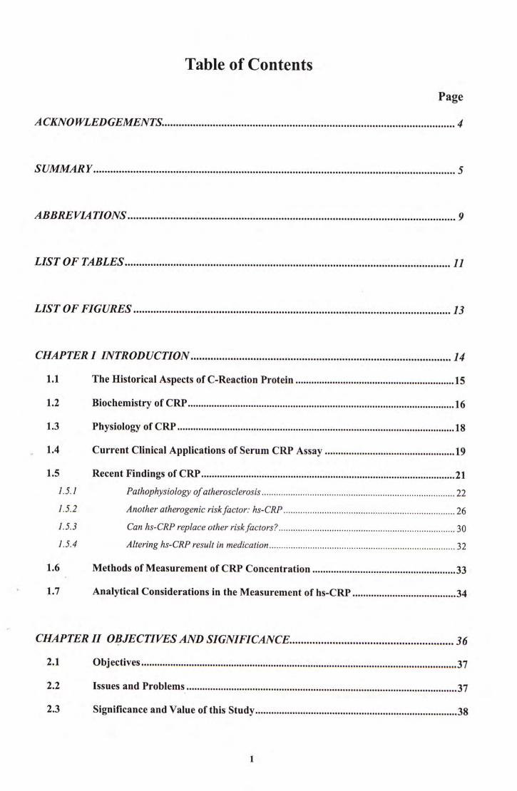
Table of Contents
Page
ACKNOWLEDGEMENTS. 4
SUMMARY 5
ABBREVIATIONS 9
LIST OF TABLES 11
LIST OF FIGURES 13
CHAPTER I INTRODUCTION 14
1.1 The Historical Aspects of C-Reaction Protein 15
1.2 Biochemistry ofCRP 16
1.3 Physiology of CRT 18
1.4 Current Clinical Applications of Serum CRP Assay 19
1.5 Recent Findings of CRP 21
1.5.1 Pathophysiology of atherosclerosis 22
1.5.2 Another atherogenic risk factor: hs-CRP 26
1.5.S Can hs-CRP replace other risk factors? 30
1.5.4 Altering hs-CRP result in medication 32 1.6 Methods of Measurement of CRP Concentration 33
, 1.7 Analytical Considerations in the Measurement of hs-CRP 34
CHAPTER II OBJECTIVES AND SIGNIFICANCE 36
2.1 Objectives
2.2 Issues and Problems 37
2.3 Significance and Value of this Study 38
1
CHAPTER III MA TERIALS AND METHODS I
Setting up the serum hs-CRP assay on the Hitachi 911 Analyzer 39
3.1 Materials
S.1.1 Reagents from Roche Diagnostics 40
Reagents for the Beckman Coulter Array ©Analyzer 40
3.1.3 In-house reagents 41
3.2. Apparatus and Equipment
3.2.1 Hitachi 911 Analyzer
^•2.2 Beckman Coulter Array ® 360 Analyzer 42
3.3 The Tina-quant a C-Reactive Protein (Latex) Ultrasensitive Assay 42
3.3.1 Priniciple of the Dual-Radius Enhanced Latex (DuRELTM) technology 42
3-3.2 Assessment of Analytical Performance 45
CHAPTER IV MA TERIALS AND METHODS II
Serum hs-CRP in Chinese chronic-renal-failure patients with
atherosclerotic vascular disease or cardiac valve calcification 48
4.1 Patient Recruitment
4.2. Blood Specimens
4.3 Assay Methods
4.3.1 hs-CRP
4.3.2 TC
4.3.3 TG 51
4.3.4 HDL-C
4.3.5 LDL-C 52
4.3.6 Apo A-1 52
4.3.7 Apo B 53
4.3.8 Lp(a) 53
4.4 Ultrasound measurement of carotid artery inter-media thickness 53
, 4.5 Statistical analysis
CHAPTER V RESUTLSI
Setting up the serum hs-CRP assay on the Hitachi 911 Analyzer 55
5.1 Imprecision 56
2
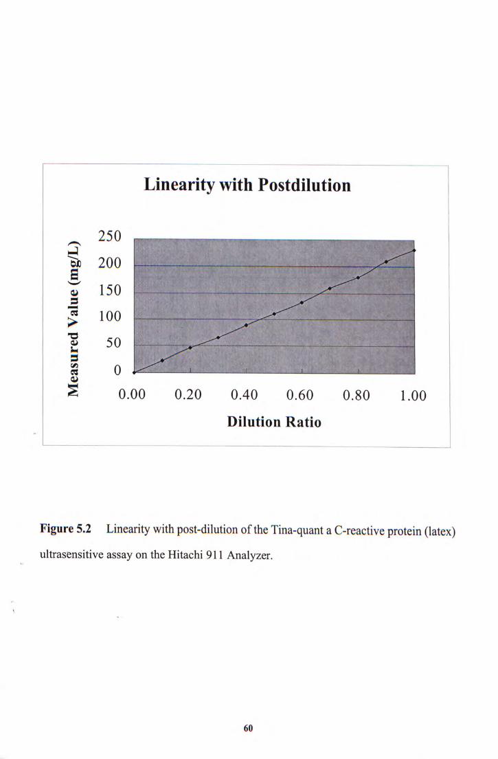
5.2 Linearity
5.3 Recovery
5.4 Detection Limit
5.5 Carry-over
CHAPTER VI RESULTS II
Serum hs-CRP in Chinese chronic-renal-failure patients with
atherosclerotic vascular disease or cardiac valve calcification 63
6.1 Patient Recruitment 64
6.2 Chinese chronic-renal-failure patients with AVD 64
6.3 Chinese chronic-renal-failure patients with CVC 65
CHAPTER VII DISCUSSION I
Performance of the serum hs-CRP assay on the Hitachi 911 Analyzer 75
7.1 Imprecision, Detection Limit, Linearity, and Recovery of hs-CRP Assay 76
7.1.1 Imprecision 76
7.1.2 Detection Limit 76
7.1.3 Linearity nc
7.1.4 Recovery nn
7.2 Overall Performance 77
CHAPTER VIII DISCUSSION II
Serum hs-CRP in Chinese chronic-renal-failure patients with
atherosclerotic vascular disease or cardiac valve calcification 79
8.1 CAPD Patients
8.2 Serum hs-CRP Concentration of AVD and CVC Patients 81
8.3 Other risk factors in AVD and CVC Patients 82
8.4 Conclusion 83
REFERENCES
3
ACKNOWLEDGEMENTS
I would like to express my sincere gratitude to Professor Christopher W K Lam,
Chairman of the Department of Chemical Pathology, Prince of Wales Hospital, the
Chinese University of Hong Kong, who supervised this work, for his continuous
guidance, discussion and encouragement during the course of this research study. I am
grateful to Miss Lydia C W Lit, Scientific Officer (Medical) of the Department of
Chemical Pathology, Prince of Wales Hospital, for her tuition and valuable advice in my
setting up and evaluating the hs-CRP assay.
My sincere thanks also go to Dr. Angela Y M Wang, Senior Medical Officer,
Department of Medicine and Therapeutics, Prince of Wales Hospital, for providing me
with the serum samples and clinical information of her patients, as well as her
enlightening discussion of the significance of the results.
I am indebted to all staff members of the Department of Chemical Pathology at the
Prince of Wales Hospital. In particular, Miss Lydia L F Tang and Miss Iris H S Chan
gave me very importance help and taught me the operation of Hitachi 911 Analyzer and
Beckman Coulter Array® 360 Analyzer, respectively.
Especially I wish to thank my family members for their strong support and
encouragement, and for their understanding and endurance during the period of my
MSc Course.
Once,again, thank you all who in some way have forced me, helped me, or
encouraged me to have done what I really was not too capable of doing.
4
SUMMARY
Inflammation is a key feature of the development of atherosclerosis. Serum
high-sensitivity C-reactive protein (hs-CRP) concentration can provide a simple
method for assessing low-grade systemic inflammation and represents an independent
risk factor for cardiovascular events. The baseline concentration of serum hs-CRP can
also predict future risk of the development of symptomatic peripheral vascular disease
and coronary heart disease.
We examined the potential relationship between serum hs-CRP concentration and
atherosclerotic vascular disease and cardiac valve calcification in 268 Chinese
chronic-renal-failure patients on continuous ambulatory peritoneal dialysis (CAPD).
Analytical performance of Tina-quant a C-reactive protein (latex) ultrasensitive
assay on the Hitachi 911 analyzer was first evaluated. Within- and between- run
imprecision (CV, n = 20) ranged from 1.07% to 4.69% at low, medium and high serum
hs-CRP concentrations (1.9/2.0,4.3, 14.6/14.9 mg/L). The assay was linear from 0.03
to 35 mg/L and to 230 mg/L (re-run with post-dilution) with a detection limit of 0.03
mg/L. The recovery of hs-CRP ranged from 98.1% to 110.5% and 94.9% to 100.3%
(re-run with post-dilution). The assay was satisfactorily commissioned for routine
service and research use.
The study population consisted of 140 male and 128 female (mean 士 SD age = 54 土
12 years, range 20-81) Chinese chronic-renal-failure patients on CAPD. Serum hs-CRP
concentrations of the atherosclerotic vascular disease (AVD) patients were significantly
5
higher than those of non-AVD group [median (interquartile range) of 3.59 (0.92 _ 8.21)
vs 1.43 (0.73 - 3.72) mg/L, p < 0.05, Mann-Whitney U-test]. Likewise, cardiac valve
calcification (CVC) patients had significantly higher serum hs-CRP concentration than
the non-CVC group [median (interquartile range) of 1.81 (0.77 - 5.45) vs 1.43 (0.72 -
3.40) mg/L, p < 0.05,Mann-Whitney U-test). There were no significant differences
between AVD and Non-AVD patients and between CVC and Non-CVC patients with
respect to carotid artery inter-media thickness (IMT) and other serum lipids,
lipoproteins and apolipoproteins.
The results of this research study increase our understanding of the pathogenesis of
atherosclerosis. Since hs-CRP is a sensitive marker of inflammation, there is a practical
clinical application of serum hs-CRP in the risk stratification of Chinese
chronic-renal-failure patients on CAPD.
6
撮要
發炎是心血管病的重要特徵。量度血淸內的hs-CRP濃度,提供了一個簡單方法去
評定全身性的低程度發炎’同時它在心血管病中也是一個獨立的風險因子;另一方面,
量度血淸內的hs-CRP底線濃度可預測患血管病及冠心病的風險。
我們測試了 268名中國籍患有慢性賢衰歇及持續性活動性腹膜透析(CAPD)病人中
的血淸hs-CRP濃度與血管病和心瓣躬化的關係。
我們先評估 Tina-quant a C-reactive protein (latex) ultrasensitive 測試劑在 Hitachi 911
分析機中的分析表現。在低〈1.9/2.0 mg/L)、中〈4.3 mg/L)及高〈14.6/14.9 m g / L �血
淸hs-CRP濃度中,同批操作及隔次操作的精確度�CV,n=20�由1.07%-4.69% ; hs-CRP
測試劑的直線性由0.03-35 mg/L及至230 mg/L〈經稀釋後再測試〉,最低血淸hs-CRP
濃度可量度値爲0.03 mg/L ; hs-CRP測試劑的復原性由98.1%-110.5%及94.9%-100.3%
〈經稀釋後再測試〉。我們得出這hs-CRP測試劑在分析表現中的結果爲滿意。
在硏究樣本中,有140名男性及128名女性〈平均値±標準差年齡=54±12年,間距
20-81歲〉的中國籍慢性賢衰歇及持續性活動性腹膜透析病人。有血管病的病人,他們
的血淸hs-CRP濃度明顯地高於那些沒有血管病的病人〈中位數(四分位數間距)爲
3.59(0.92-8.21)對 1.43(0.73-3.72) mg/L,P<0.05,Mann-Whitney U-Test��同樣’心瓣躬
化病人的血淸hs-CRP濃度明顯地高於沒有心瓣躬化病人〈中位數(四分位數間距)爲
1.81(0.77-5.45)對 1.43(0.72-3.40) mg/L,P<0.05 ’ Mann-Whitney U-Test��我們也發現這
兩組類病人中他f!的頸動脈的內中膜厚度(IMT)及其他血脂因素沒有統計學上的分別。
這項硏究的結果,增加了我們對血管病的病理性認識�hs-CRP是一個高度敏感的
7

發炎性標記’在臨床上可應用在中國籍慢性腎衰歇及持纊性活動性KM透析病人中的風
險分層法。
8
ABBREVIATIONS
Apolipoprotein A-1 (Apo A-1)
Apolipoprotein B (Apo B)
Atherosclerotic vascular disease (AVD)
Cardiac valve calcification (CVC)
Carotid artery inter-media thickness (IMT)
Cerebrovascular accident (CVA)
•• Cerebrovascular disease (CVD)
Continuous ambulatory peritoneal dialysis (CAPD)
Coronary artery disease (CAD)
High density lipoprotein cholesterol (HDL-C)
High-sensitivity C-reactive protein (hs-CRP)
Interleukin-1 (IL-1)
9
Interleukin-6 (IL-6)
Lipoprotein(a) (Lp(a))
Low density lipoproteins (LDL)
Low density lipoprotein-cholesterol (LDL-C)
Myocardial infarction (MI)
Peripheral vascular disease (PVD)
Total cholesterol (TC)
Triglyceride (TG)
10
LIST OF TABLES
Page
Table 1.1 Risk factors for endothelial dysfunction 22
Table 5.1 Within-run and between-run imprecision of Tina-quant a
C-reactive protein (latex) ultrasensitive assay on the Hitachi
911 analyzer. 55
Table 5.2 Recovery of hs-CRP Assay 59
Table 5.3 Recovery with post-dilution of hs-CRP Assay 60
Table 6.1 Serum hs-CRP concentration in AVD and Non-AVD, CVC
and Non-CVC of Chinese chronic-renal-failure patients on
CAPD. 64
Table 6.2 Results of serum hs-CRP concentration, average IMT and TC,
TG, HDL-C,LDL-C, Apo A-1, Apo B and Lp(a) concentrations
in AVD and Non-AVD of Chinese chronic-renal-failure patients
� on CAPD expressed as median (interquartile range) and their
statistical analysis.
Table 6.3 Results of serum hs-CRP concentration, average IMT and TC,
TG, HDL-C, LDL-C, Apo A-1, Apo B and Lp(a) concentrations
in CVC and Non-CVC of Chinese chronic-renal-failure patients
11

Page
on CAPD expressed as median (interquartile range) and their
statistical analysis. 72
12
LIST OF FIGURES
Page
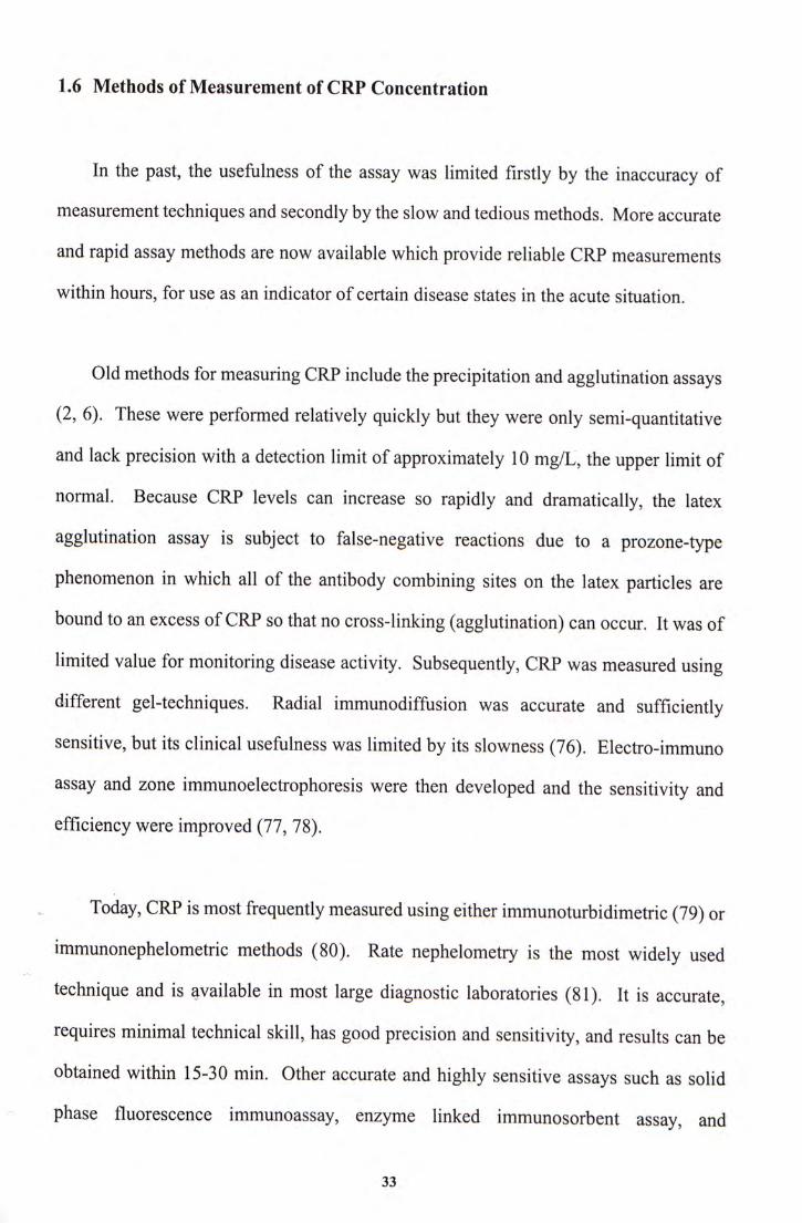
Figure 1.1 Schematic organization of the five identical CRP subunits 15
Figure 1.2 Relative risks for future myocardial infarction 29
Figure 3.1 Principle of the Dual-Radius Enhanced Latex (DuREL™) technology for the measurement of serum hs-CRP concentration. 42
Figure 5.1 Linearity of Tina-quant a C-reactive protein (latex) ultrasensitive
assay on the Hitachi 911 Analyzer. 57
Figure 5.2 Linearity with post-dilution of Tina-quant a C-reactive protein
(latex) ultrasensitive assay on the Hitachi 911 Analyzer. 58
%
13

CHAPTER I
INTRODUCTION
•
14
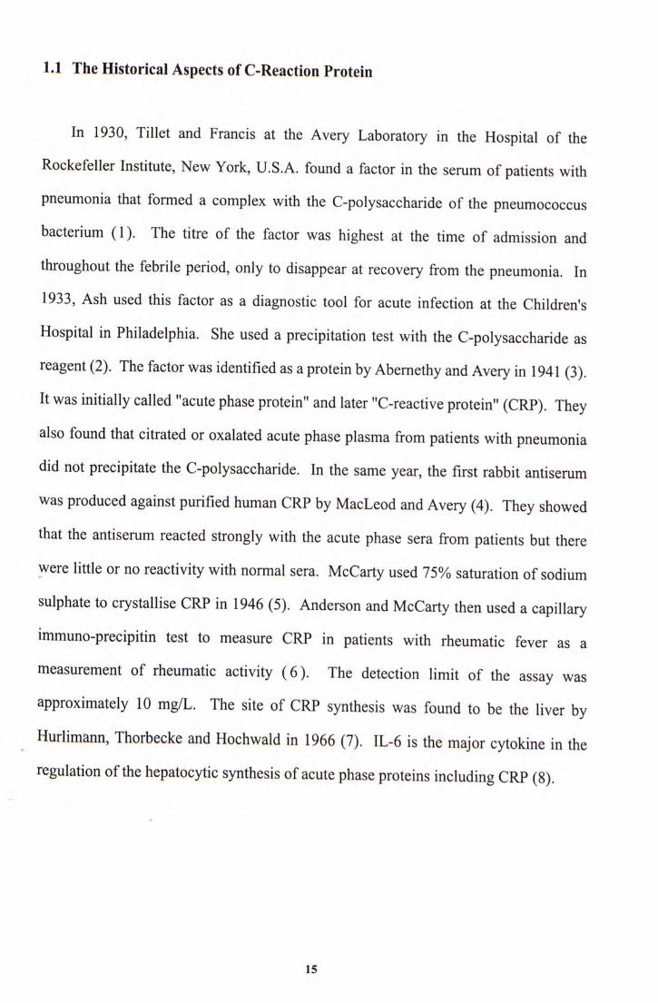
1.1 The Historical Aspects of C-Reaction Protein
In 1930, Tillet and Francis at the Avery Laboratory in the Hospital of the
Rockefeller Institute, New York, U.S.A. found a factor in the serum of patients with
pneumonia that formed a complex with the C-polysaccharide of the pneumococcus
bacterium (1). The titre of the factor was highest at the time of admission and
throughout the febrile period, only to disappear at recovery from the pneumonia. In
1933, Ash used this factor as a diagnostic tool for acute infection at the Children's
Hospital in Philadelphia. She used a precipitation test with the C-polysaccharide as
reagent (2). The factor was identified as a protein by Abemethy and Avery in 1941 (3).
It was initially called "acute phase protein" and later "C-reactive protein" (CRP). They
also found that citrated or oxalated acute phase plasma from patients with pneumonia
did not precipitate the C-polysaccharide. In the same year, the first rabbit antiserum
was produced against purified human CRP by MacLeod and Avery (4). They showed
that the antiserum reacted strongly with the acute phase sera from patients but there
}vere little or no reactivity with normal sera. McCarty used 75% saturation of sodium
sulphate to crystallise CRP in 1946 (5). Anderson and McCarty then used a capillary
immuno-precipitin test to measure CRP in patients with rheumatic fever as a
measurement of rheumatic activity (6) . The detection limit of the assay was
approximately 10 mg/L. The site of CRP synthesis was found to be the liver by
Hurlimann, Thorbecke and Hochwald in 1966 (7). IL-6 is the major cytokine in the
regulation of the hepatocytic synthesis of acute phase proteins including CRP (8).
15
1.2 Biochemistry of CRP
A single CRP gene which spans about 2.5 kbp is located proximally on the long
arm of chromosome 1. There are 2 exons separated by a single intron. The exon
encodes a signal peptide consisting of 18 amino acids followed by the first 2 amino
acids of the protein. The exon encodes 204 amino acids (9,10).
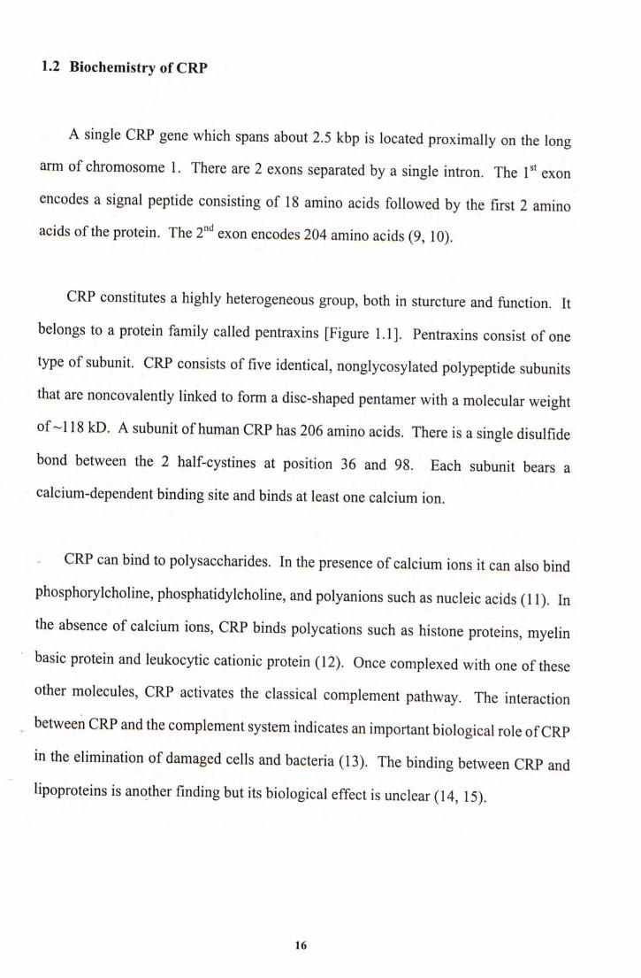
CRP constitutes a highly heterogeneous group, both in sturcture and function. It
belongs to a protein family called pentraxins [Figure 1.1]. Pentraxins consist of one
type ofsubunit. CRP consists of five identical, nonglycosylated polypeptide subunits
that are noncovalently linked to form a disc-shaped pentamer with a molecular weight
of � 1 1 8 kD. A subunit of human CRP has 206 amino acids. There is a single disulfide
bond between the 2 half-cystines at position 36 and 98. Each subunit bears a
calcium-dependent binding site and binds at least one calcium ion.
CRP can bind to polysaccharides. In the presence of calcium ions it can also bind
phosphorylcholine, phosphatidylcholine, and polyanions such as nucleic acids (11). In
the absence of calcium ions, CRP binds polycations such as histone proteins, myelin
basic protein and leukocytic cationic protein (12). Once complexed with one of these
other molecules, CRP activates the classical complement pathway. The interaction
.be tween CRP and the complement system indicates an important biological role of CRP
in the elimination of damaged cells and bacteria (13). The binding between CRP and
lipoproteins is another finding but its biological effect is unclear (14,15).
16
Figure 1.1 Schematic organization of the five identical CRP-subunits
MllkMf^
V
17
1.3 Physiology ofCRP
CRP is an acute-phase protein produced mainly by the liver in response to cytokine
production (IL-6, IL-1, tumor necrosis factor) during tissue injury, inflammation,
infection or malignancy (16,17). A small number of normal human peripheral blood
lymphocytes have been shown to produced CRP (18).
Normally, CRP is found in serum at very low concentrations (<5 mg/L). It is also
found in all age groups with no difference in the mean serum concentration between
men and women, but the level of it is higher in late pregnancy (16). Serum CRP
concentration varies widely in normal subjects and the mean level tends to increase
slightly with age. In inflammatory disorders, there is a rise of CRP from 0.5-3 mg/L to
more than 200 mg/L (sometimes 1000 mg/L). The level of CRP is related to the extent
and severity of the inflammatory activity. CRP concentration begins to increase 6-8 h
after the onset of an inflammatory process. The plasma level can double at least every 8
h, reaching a maximum after 48-72 h (19). Then it returns to its baseline level in about
2 weeks after the resolution of inflammatory activity. The half-life of CRP in the
circulation is about 19 h during the healing phase of an uncomplicated inflammatory
process (20). The only condition that interferes with the "normal" CRP response is
severe hepatocellular impairment. Recently, a study showed that the baseline CRP
�concentrations in healthy people have remarkable stability throughout the day and year
(21, 22). It means the baseline CRP concentration is not subject to diurnal and seasonal
variations.
CRP has two major activities in relation to different cell types. One is related to its
scavenger function with an opsonic effect for macrophages with respect to molecules,
18
cell fragments, and for altered cells (23, 24). The other activity ofCRP is to modulate
the activity of different cell types either through released tuftsin-like peptides from CRP
or through the expression of neo-antigens on the CRP molecule (25, 26, 27).
Collectively, these properties confer an important role for CRP in the recognition of
microbial organisms and as an immunomodulator in host defense. CRP may also be
important in the recognition of necrotic tissues. Serum CRP concentration is increased
in postmenopausal women under hormone replacement therapy (28).
1.4 Current Clinical Applications of Serum CRP Assay
The main clinical applications are as follows (16):
入 4,1 To assess treatment of bacterial infections with antibiotics
Bacterial infections are usually a potent stimulus of CRP production. In
monitoring the resolution of pyelonephritis, pulmonary infection, pelvic infection and
meningitis, a return to normal CRP concentration can be used as a guide to
discontinuation of antibiotic treatment (29, 30, 31).
V
"•2 To detect intrauterine infections with concomitant premature amniorrhexis
An elevated serum CRP concentration seems to be a useful indicator of intrauterine
infection and preterm rupture of the membranes and can be used to select patients for
antibiotic therapy against chorioamnionitis when the foetus is still in utero and to
19
evaluate the effect of treatment (32). Early chorioamnionitis is not reliably predicted by
ESR,WBC, differential count, platelet count, or bacterial culture from the vagina or the
amniotic fluid. Serum CRP concentration often increases before other laboratory and
clinical parameters in these patients.
“•3 To differentiate between active and inactive forms of disease with
concurrent infection
Serum CRP concentration has been used to monitor patients with chronic
inflammatory conditions. In SLE with systemic sclerosis or ulcerative colitis, serum
CRP concentration has been found to be relatively low in relation to the disease activity
(33, 34). However, if these patients suffer an intercurrent infection, the serum CRP
concentration shows a "normal" inflammatory increase.
To therapeutically monitor rheumatic disease and assess anti-inflammatory
therapy
In rheumatoid arthritis clinical assessment of disease activity correlates well with
serum CRP concentration (35, 36). Serum CRP concentration is elevated in 64% with
radiological progression and a fall in CRP correlates with cessation of radiological
deterioration after 6 months of treatment (37).
20
1-4.5 To determine the presence of postoperative complications at an early stage,
such as infected wounds,thrombosis and pneumonia
Serum CRP concentration has been studied in a variety of surgical conditions. All
patients undergoing surgery show a rapid rise in serum CRP concentration from 6 h
postoperatively with maximum levels at 48 h, and in uncomplicated cases showing a
rapid return to normal levels on the third postoperative day (38). Postoperative
complications are often accompanied by a persistently elevated CRP even after the third
postoperative day, or a secondary rise (39). Serial measurement of serum CRP
concentration is useful and better than WBC and other acute phase proteins in detecting
and monitoring infectious and/or ischemic/necrotic postoperative complications.
i'4,6 To distinguish between infection and bone-marrow transplant rejection
• In a study of 41 bone marrow transplant patients there was a significant elevation in
the serum CRP level in 38 of the 40 episodes of infection but in none of the 15 episodes
of acute graft-versus-host disease (40).
1.5 Recent Findings of CRP
Atherosclerosis is a process in which the artery is hardened and narrowed due to
the formation of atheroma. Consequently, sufficient oxygen-rich blood is prevented to
reach the end organ. Oxygen deprivation (called ischemia) causes injury to tissues of
the organ. If the artery becomes completely occluded, damage becomes so extensive
21
that cell death, e.g. myocardial infarction (MI), cerebrovascular accident (CVA), and
peripheral vascular disease (PVD), occurs. Recent evidence gathered in apparently
healthy adults using high-sensitivity C-reactive protein (hs-CRP) method (CRP
measured at levels within the normal range especially at 1 mg/L) indicates that CRP is
a strong independent risk factor for subsequent development of MI, CVA and PVD (41).
i . 5.1 Pathophysiology^ of atherosclerosis
In 1976, Ross and Glomset proposed a theory of atherogenesis described as the
"response-to-injury hypothesis", which hypothesised that endothelial injury led to
repetitive denudations and ulcerations, being responsible for local activation of platelets
and release of growth factors that resulted in intimal hyperplasia, lipid accumulation
and thrombus formation (42). This hypothesis initially proposed that endothelial
denudation was the first step in atherosclerosis. The most recent version of this
hypothesis emphasised on endothelial dysfunction rather than denudation (43,44). The
functional status of the endothelium might be as important as its anatomic integrity in
the pathogenesis of many diseases. Therefore, endothelial dysfunction was introduced
to describe the potential for vascular endothelium to undergo phenotypic modulation to
a non-adaptive state, characterized by the loss or dysregulation of critical homeostatic
�m e c h a n i s m s , which are normally operative in healthy endothelial cells.
Before the development of atherosclerosis, endothelial dysfunction has occurred.
A list of pathophysiological stimuli for endothelial dysfunction have been identified (45,
46, 47). These stimuli [Table 1.1] appear to act on vascular endothelial cells to elicit
basic alterations in the cellular structure and function that are common to a variety of
22
response-to-injury processes, including acute and chronic inflammatory reactions,
wound healing and angiogenesis.
At an early stage, impaired endothelium function may promote the development of
atherosclerosis through its effects on the vascular wall, platelet and leukocyte adhesion,
vascular smooth muscle cell growth, and coagulation, triggered by the large repertoire
of its biologically active products. The injury induces the endothelium to have
procoagulant instead of anticoagulant properties. In fact, the lesions of atherosclerosis
represent a series of highly specific cellular and molecular responses that can best be
described, in aggregate, as an inflammatory disease. Arterial inflammation has only
recently been identified as a possible trigger or it plays a major role in atherogenesis
(48). Researchers have speculated for years that inflammation in some way has a role in
the development of heart disease.
If the offending agents are not removed, the inflammatory response stimulates
migration and proliferation of smooth-muscle cells that become intermixed with the
area of inflammation to form an intermediate lesion. If these responses continue, they
can thicken the artery wall, which compensates by gradual dilation, so that up to a point,
the lumen remains unaltered, a phenomenon termed "remodeling" (49).
23

Table 1.1 Risk factors for endothelial dysfunction (44)
(1) Atherosclerotic risk factors Hypertension Cigarette smoking Hypercholesterolemia Diabetes mellitus Male gender Increased age Family history of atherosclerosis
(2) Chronic infections Herpes viruses Cytomegalovirus Respiratory syncytial virus Helicobacter pylori C. pneumoniae
(3) Environmental factors Hypoxia Turbulent flow Oxidants
(4) Genetic factors Hyperhomocysteinaemia
V
Elevated plasma lipoprotein (a) Familial hypercholesterolemia Familial hypertriglyceridemia
24
The injuries to the arteries signal the immune system to release monocytes and T
lymphocytes at the sites. These cells migrate between the endothelium and become
localised subendothelially. They are trapped by adhesive glycoproteins expressed on
the surface of the endothelial cells as a result of injury. Monocytes transform into
macrophages. Macrophages literally engulf foreign debris and become foam cells that
attach to smooth muscle cells causing them to build up. The immune system, sensing
further harm, releases other factors called cytokines, which attract more white blood
cells and perpetuate the whole cycle. They also stimulate the liver to produce CRP and
blood-clotting factors called fibrinogen. Fibrinogen and other clotting factors are acute
phase proteins and CRP stimulates production by mononuclear cells of tissue factor, an
important initiator of coagulation. It also interacts with low density lipoproteins (LDL)
and damaged cell membranes to activate the complement system.
Cycles of accumulation of mononuclear cells, migration and proliferation of
smooth-muscle cells, and formation of fibrous tissue lead to further enlargement and
restructuring of the lesion, so that it becomes covered by a fibrous cap that overlies a
core of lipid and necrotic tissue - a so-called advanced, complicated lesion. Injured
inner walls fail to produce enough nitric oxide, a substance critical for maintaining
blood vessel elasticity. The arteries become calcified and lose elasticity. At some point,
the artery can no longer compensate by dilation; the lesion may then intrude into the
�l u m e n and alter the flow of blood.
The stability of the lesion depends on the strength and thickness of the fibrous cap
relative to the size of the lipid core. If the fibroatheroma is weakened by increased
tensile forces or by enzymatic degradation of the fibrous cap, then the plaque may
rupture and expose highly thrombogenic material from its lipid core.
25
Hemorrhage-hematoma and thrombus formation may eventually occlude the vascular
lumen, giving rise to myocardial infarction (MI) consequent to coronary artery disease
(CAD), ischemic stroke (CVA), or peripheral vascular disease (PVD).
L 5.2 Another atherogenic risk factor: hs-CRP
From the above discussion, inflammation appears to have a key role in the
progression of atherosclerosis and destabilization of atheromas. Serologic markers of
the inflammation have been associated with CAD risk. IL-1 (50) and IL-6 (51) are
increased in subjects with CHD and are markers for increased risk of cardiovascular
events. Other inflammatory markers such as fibrinogen (52) have also been associated
with an increased risk of future coronary events in healthy subjects and in patients with
unstable angina. Of these serum markers of inflammation, CRP has the largest body of
literature supporting its relationship with atherosclerosis and clinical utility in assessing
cardiovascular risk.
Assessment of the acute phase response by the measurement of the acute phase
proteins (e.g. CRP) in serum is a reliable way of detecting and assessing the severity of
inflammation and tissue damage. CAD, CVA and PVD are frequently at the end of a
�l o n g process of inflammation-mediated atherosclerosis. Studies utilizing hs-CRP
assays (CRP measured by high-sensitivity assays which detect levels of CRP within the
normal range with high precision, especially 1 mg/L or below) have demonstrated that
level previously considered normal (i.e., <3 mg/L) may be associated with future
cardiovascular disease (53, 54). It is associated with increased levels of hs-CRP. It may
indicate the degree of inflammation occurring in the lining of the arteries. Therefore,
26
modest elevation in serum CRP concentration significantly predicts future coronary
events (55).
1.5.2.1. hs-CRP in patients with CAD
The association between low-grade inflammation and the progression and
complications of atherosclerosis has been well established. In patients with unstable
angina,hs-CRP is an important predictor of MI and CAD death (56, 57, 58, 59). The
European Concerted Action on Thrombosis (ECAT) and Disabilities Angina Pectoris
Study suggested that cardiovascular events were higher in those stable and unstable
angina patients with higher CRP levels (60). There was about a two-fold increase in the
risk of a coronary event in patients whose CRP concentration was in the fifth quintile
( � 3 . 6 mg/L), compared with the first four quintiles. A third of the events occurred
among patients who had a CRP concentration of more than 3.6 mg/L.
Inflammation may have a crucial role in adverse CAD outcome after coronary
intervention. Preprocedural serum CRP concentration, an easily measurable marker of
acute phase response, is a powerful independent predictor of both early and late
outcome in patients undergoing repeated coronary revascularization (coronary artery
bypass graft surgery and percutaneous transluminal coronary angioplasty), suggesting
that early complications and clinical restenosis are markedly influenced by the
preprocedural degree of inflammatory cell activation (61, 62,63). Increased hs-CRP in
patients with and without unstable angina before coronary revascularization has been
associated with higher in-hospital morbidity and restenosis. The local inflammatory
response from plaque disruption, angioplasty, and coronary stent deployment results in
27
endothelial dysfunction and intimal proliferation leading to restenosis. Therefore,
patients with increased hs-CRP may have an exaggerated inflammatory response,
resulting in accelerated atheroma progression and bypass graft disease after
revascularization.
1.5.2.2. hs-CRP in patients without clinical CAD
Increase in serum hs-CRP concentration has been associated with increased
cardiovascular events in asymptomatic subjects. In the Physician's Health Study (PHS),
Rider et al. (64, 65) showed that CRP was elevated at baseline among apparently
healthy individuals who subsequently developed first-ever heart attack compared to
those who did not during the 14 years of the trial. Men in the highest CRP quartile
(2 .11 mg/L) had two times the risk of CVA, three times the risk of MI and four times
the risk of developing severe PVD compared to the men in the lowest quartile ( 0.55
mg/L). These risks were stable over long periods and were not modified by smoking or
other cardiovascular risk factors.
The MONICA (Monitoring Trends and Determinants in Cardiovascular Disease)
Augsburg Cohort Study also confirmed the value of the prognostic relevance of hs-CRP
�i n predicting CHD events in a large, randomly selected cohort of initially healthy
middle-aged men (54). Baseline hs-CRP concentration was measured in 936 healthy,
middle-aged men .who were followed for 8 years. The investigators demonstrated that a
1 SD increase in the log-transformed value of hs-CRP was associated with a 50%
increase in CHD risk.
28
A prospective, nested case-control study actually compared the reliability of 12
different "markers" for heart disease (66). Among those were cholesterol, cigarette
smoking, obesity, and diabetes - all known risk factors for CAD. Just slightly elevated
levels of serum CRP was found to be the most significant, strongest, powerful and
univariate predictor of the risk of cardiovascular events. The risk of heart attack was
more than four times higher for 28,263 apparently healthy postmenopausal women with
the highest levels of hs-CRP, compared to those with the lowest levels. The study
concludes that the addition of hs-CRP to screening based on serum lipids may provide
an improved method of identifying persons at risk for future cerebral cardiovascular
events.
In a prospective, nested case-control study in the Cardiovascular Health Study and
Rural Health Promotion Project, serum hs-CRP concentrations were associated with
increased coronary events, especially in those with evidence of subclinical disease at
baseline in women but not in men (67).
In brief, higher level of serum CRP, though usually within the reference range for
healthy individuals, has been found to be associated with increased risk in apparently
healthy people and also in CAD patients. Therefore, higher level of serum CRP is at
increased risk of developing CAD, CVA and PVD. Measurement of the serum level of
�C R P has been used to predict the risk of infarction and death among high-risk patients
and ischemic complications in those with stable and unstable angina. The small
elevations in CRP can help to identify people at risk long before symptoms arise (68).
29
1.5.3 Can hs-CRP replace other risk factors?
As many as 30% to 50% of all first-time heart attack victims do not fit into any of
the known risk categories, but researchers are finding more evidence that hs-CRP can
better predict who will have a heart attack, even in those with no other risk factors (68).
The baseline serum total cholesterol level (TC) doesn't predict which patients would
have CAD. However, CRP level does. If CRP is higher than normal, the risk for heart
attack is higher. This is true even if a person has normal cholesterol levels. These
observations have important implications for the assessment of patients and for
treatment.
hs-CRP is one of the few risk factors in the blood that provides information over
and above that given by TC- to high-density lipoprotein cholesterol (HDL-C) ratio. It
should not replace cholesterol, but it should be added to it for the determining CAD
risk. Simultaneous measurements of hs-CRP and lipids (TC:HDL-C ratio) predict
future vascular risk better than lipid measurements alone (69). The relative risk of
developing a future MI among those with high levels of both CRP and TC (RR = 5.0)
were greater than the product of the individual risk factor, (RR of CRP = 1.5; R R o f T C
=2.3) (70).
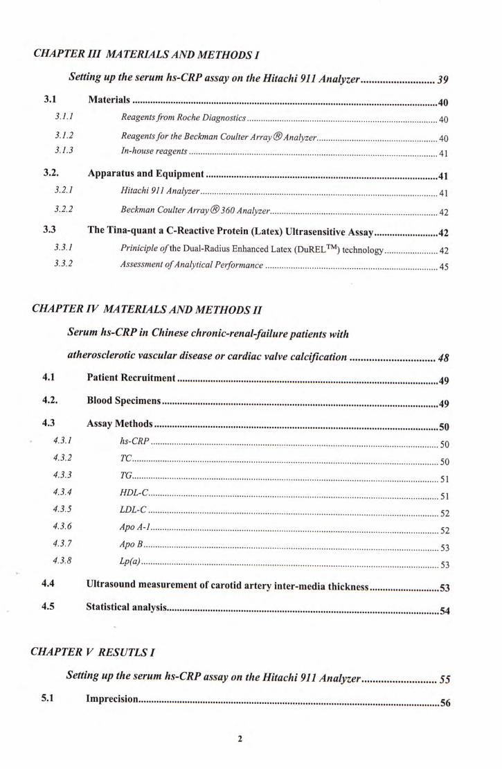
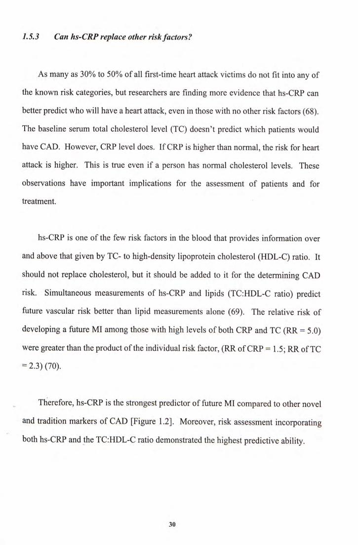
, Therefore, hs-CRP is the strongest predictor of future MI compared to other novel
and tradition markers of CAD [Figure 1.2]. Moreover, risk assessment incorporating
both hs-CRP and the TC:HDL-C ratio demonstrated the highest predictive ability.
30
Lipoprotein(a) 1~||
Total homocysteine •
TC •
Fibrinogen
tPA antigen ^
TCiHDLC n
hs-CRP —
hs-CRP + TC:HDLC H I ,
0 1.0 2.0 4 .0 6.0
Relative Risk for Future Myocardial Infarction
Figure 1.2 Relative risks for future myocardial infarction among apparently healthy middle-aged men in the Physicians' Health Study according to baseline levels of lipoprotein(a), total plasma homocysteine, total cholesterol (TC), fibrinogen, tissue-type plasminogen activator(tPA) antigen, the ratio of total cholesterol to high-density lipoprotein cholesterol (HDLC), and high-sensitivity C-reactive protein (hs-CRP) (71). For consistency, risks are computed for men in the top compared with the bottom quartile for each marker.
31
1.5,4 AItering hs-CRP result in medication
Some researchers believe that patients with high serum hs-CRP concentration can
benefit from taking cholesterol-lowering drugs - statins, even if their cholesterol levels
are normal. This drug may reduce chronic, low-grade inflammation associated with the
atherosclerotic process in the arteries. They are just as effective at lowering CRP - and
reducing the CRP-associated risk for heart attack - as they are for lowering cholesterol.
Prescribing statins boost survival rates for all patients regardless of their cholesterol
levels and have an anti-inflammatory effect that is protective, over and above their role
in lowering cholesterol ( 72 , 73 ). Other agents like aspirin, nonsteroidal
anti-inflammatory drugs and glucocorticoids may also reduce CRP concentration.
However, CRP goes up in the presence of inflammation, a one-time finding of elevated
CRP does not necessarily confirm an increased risk for heart attacks. So Jialal et al. (74)
recommends at least two CRP measurements to confirm the true level. Recent medical
events resulting in tissue injury, infections, or inflammation (which may cause elevated
CRP levels) should also be considered when interpreting results.
In addition, the use of aspirin was associated with significant reduction in the risk
of MI only among men in the highest quartile (64). Patients with CRP concentrations >
2.1 mg/L achieved the greatest benefit from aspirin prophylaxis, whereas those with
, l e v e l s < 0.55 mg/L attained no significant benefit. The reduction in thrombotic events
associated with the use of aspirin raises the possibility that anti-inflammatory agents
such as aspirin may provide clinical benefit for the prevention of cardiovascular disease.
Similar data concerning healthy middle-aged women were reported in 1998 (75).
32
1.6 Methods of Measurement of CRP Concentration
In the past, the usefulness of the assay was limited firstly by the inaccuracy of
measurement techniques and secondly by the slow and tedious methods. More accurate
and rapid assay methods are now available which provide reliable CRP measurements
within hours, for use as an indicator of certain disease states in the acute situation.
Old methods for measuring CRP include the precipitation and agglutination assays
(2, 6). These were performed relatively quickly but they were only semi-quantitative
and lack precision with a detection limit of approximately 10 mg/L, the upper limit of
normal. Because CRP levels can increase so rapidly and dramatically, the latex
agglutination assay is subject to false-negative reactions due to a prozone-type
phenomenon in which all of the antibody combining sites on the latex particles are
bound to an excess of CRP so that no cross-linking (agglutination) can occur. It was of
limited value for monitoring disease activity. Subsequently, CRP was measured using
different gel-techniques. Radial immunodiffusion was accurate and sufficiently
sensitive, but its clinical usefulness was limited by its slowness (76). Electro-immuno
assay and zone immunoelectrophoresis were then developed and the sensitivity and
efficiency were improved (77,78).
� Today, CRP is most frequently measured using either immunoturbidimetric (79) or
immunonephelometric methods (80). Rate nephelometry is the most widely used
technique and is available in most large diagnostic laboratories (81). It is accurate,
requires minimal technical skill, has good precision and sensitivity, and results can be
obtained within 15-30 min. Other accurate and highly sensitive assays such as solid
phase fluorescence immunoassay, enzyme linked immunosorbent assay, and
33

radioimmunoassay are also available. However, they are slower to perform and
demand greater technical skill than rate nephelometry (82, 83).
1.7 Analytical Considerations in the Measurement of hs-CRP
Most of the current CRP assays can measure levels which are increased up to
1,000-fold in response to infection or tissue destruction, but are not sensitive enough for
concentrations within the normal range and therefore cannot be used for cardiovascular
risk assessment. Mildly elevated of CRP may already be present as a risk of
atherosclerosis. An assay must be able to measure with high precision at much lower
CRP levels. In the last several years, new tests for measuring "high-sensititivity CRP"
have been developed. High-sensitivity CRP assays detect levels of CRP within the
normal range at concentrations proven to predict future cardiovascular events. Called
"high-sensititivity CRP" (hs-CRP), this is the parameter that now has the cardiologists'
strong interest. The first assay received the U.S. Food and Drug Administration
clearance on October 25, 1999 for cardiac risk stratification. In the past few years,
several assays for hs-CRP have been developed by the different manufacturers. hs-CRP
is relatively easy to measure and is an inexpensive assay. The World Health
Organization (WHO) International Reference Standared for CRP Immunoassay 85 /
� 5 0 6 is available now for standardization of hs-CRP assay (84). Discrepancies between
methods that claim the use of calibrators that are traceable to the same reference source
are not that uncommon (85).
The performance characteristics of a good CRP assay for cardiovascular risk
assessment are high sensitivity (<1 mg/L) and good precision within the decision range
34

(2 to 4 mg/L) (86,87). It should have a good linearity throughout the dynamic range
down to the detection limit, with satisfactory recovery rates for all possible dilution
steps (88, 89).
35

CHAPTER II
OBJECTIVES AND SIGNIFICANCE
36
2.1 Objectives
(1) To adapt the serum hs-CRP assay onto the Hitachi 911 Analyzer in order to develop
an automated and sensitive method for measurement of CRP concentrations below
those routinely measured.
(2) To assess the usefulness of serum hs-CRP concentration as a risk factor for
atherosclerotic vascular disease (AVD) and cardiac valve calcification (CVC) in a
high risk group namely, Chinese chronic-renal-failure patients.
2.2 Issues and Problems
Serum CRP concentration increases even in trivial inflammation such as the
common cold (90), and in tissue damage such as that induced by surgery or marathon
running (91 ). However, follow-up studies have shown that serum hs-CRP
concentration is stable over long periods, as long as measurements are not made within
two to three weeks of a clinical infection or inflammation that will elevate usual levels
(92,93). If the risk of atherosclerosis in patients is assessed, the clinical state must be
investigated. Therefore, increases in serum CRP concentration are non-specific and
�shou ld not be interpreted without a complete clinical history. When serum hs-CRP
concentration is used to assess the risk of cardiovascular and peripheral vascular disease,
previous values should be compared. Any recent medical events that may have
temporarily elevated serum CRP concentrations should be taken into account. It is
important for a patient to be metabolically stable before being tested for cardiac risk
prevention.
37
2.3 Significance and Value of this Study
The current literature supports the hypothesis that atherosclerosis is, in part, an
inflammatory disease.
The measurement of serum hs-CRP which is an inflammatory marker can assess
the risk of atherosclerosis. Patients who may benefit from primary prevention can be
identified. The cost-to-benefit ratio in the use of statins can be improved.
38
CHAPTER III
MATERIALS AND METHODS I
Setting up the serum hs-CRP assay on the Hitachi 911 Analyzer
39
3.1 Materials
3, L1 Reagents from Roche Diagnostics
Tina-quant a C-reactive protein (latex) ultrasensitive assay reagent (Cat. No.
1972855) for serum hs-CRP assay, C.f.a.s. Proteins (Cat. No. 1355279) for serum
hs-CRP calibration, and CRP T Control N (Cat. No. 2064677/0766321) for serum
hs-CRP control were purchased from Roche Diagnostics GmbH, D-68298 Mannheim,
Germany.
Cholesterol CHOD-PAP assay reagent (Cat. No. 1489437) for serum cholesterol
assay, Triglycerides GPO-PAP assay reagent (Cat. No. 1488899) for serum
triglycerides assay, and HDL-Cholesterol Plus assay reagent (Cat. No. 1930729) for
serum HDL-cholesterol assay were also purchased from Roche Diagnostics GmbH,
D-68298 Mannheim, Germany.
3丄2 Reagents for the Beckman Coulter Array® Analyzer
The following reagents were purchased from Beckman Coulter, Inc., Fullerton, CA
�92834-3100, U.S.A.
-Lipoprotein(a) (LPA) assay reagent (P/N 465360) for serum lipoprotein(a) assay
-Lipoprotein(a) Calibrator (P/N 465365) for serum lipoprotein(a) calibration
-Vigil™ Protein Control - Level 1 (P/N 441391) for serum lipoprotein(a) control.
-Apolipoprotein A-1 assay reagent (P/N 449300) for serum apolipoprotein A-1 assay
40
-Apolipoprotein B (APB) assay reagent (P/N 449310) for serum apolipoprotein B
assay
-Apolipoprotein Calibrator (APO CAL) (P/N 469283) for serum apolipoprotein A-1
and apolipoprotein B calibrations
-Vigil™ Lipid Control - Lipids and Lipoprotein Control Serum - Level 1 (P/N 469905)
and Level 2 (P/N 465980) for serum apolipoprotein A-1 and apolipoprotein B
controls
-APO Diluent (P/N 449380)
-Buffer (P/N 663600)
3.L3 In-house reagents
Normal saline (9.0 g/L of NaCl) was prepared using glass-distilled, de-ionized
water and analytical grade chemicals from Sigma Chemical Co, St Louis, MO 63178,
USA.
3.2. Apparatus and Equipment
3.2.1 Hitachi 911 Analyzer
The Hitachi 911 Analyzer (Roche Diagnostics Corporation, Indianapolis, IN
46250-0457, U.S.A.) was used for the measurement of serum hs-CRP, TC, TG and
HDL-C.
41
3.2.2 Beckman Coulter Array ® 360 Analyzer
The Beckman Coulter Array® 360 analyzer (Beckman Coulter, 200 South
Kraemer Boulevard, Brea, California 92621-6209, 1-800-526-3821, U.S.A.) was used
for the measurement of Lp(a), apo A-1, apo B. •
3.3 The Tina-quant a C-Reactive Protein (Latex) Ultrasensitive Assay
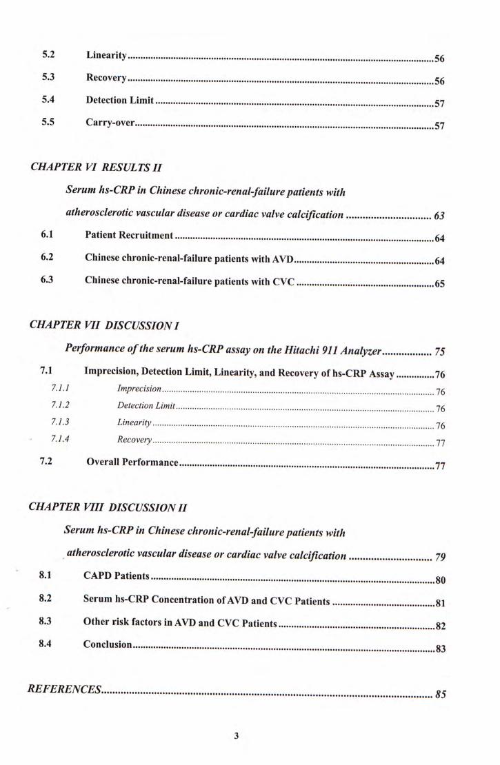
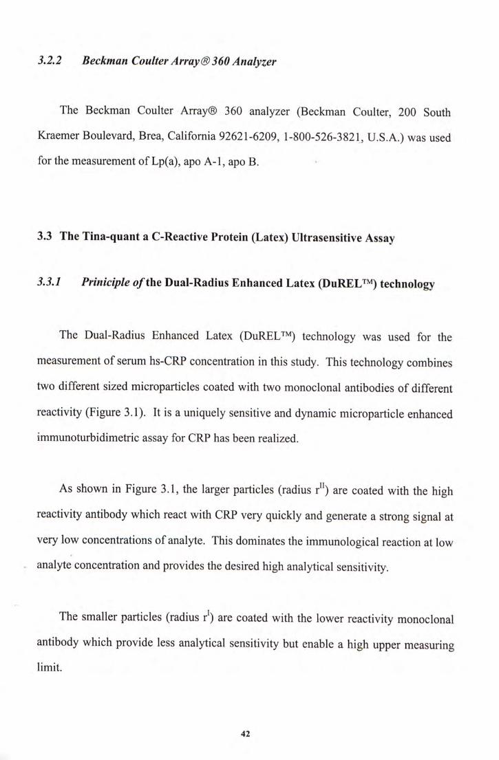
3.3.1 Priniciple of the Dual-Radius Enhanced Latex (DuREL™) technology
The Dual-Radius Enhanced Latex (DuREL™) technology was used for the
measurement of serum hs-CRP concentration in this study. This technology combines
two different sized microparticles coated with two monoclonal antibodies of different
reactivity (Figure 3.1). It is a uniquely sensitive and dynamic microparticle enhanced
immunoturbidimetric assay for CRP has been realized.
As shown in Figure 3.1, the larger particles (radius r") are coated with the high
reactivity antibody which react with CRP very quickly and generate a strong signal at
very low concentrations of analyte. This dominates the immunological reaction at low
> analyte concentration and provides the desired high analytical sensitivity.
The smaller particles (radius r!) are coated with the lower reactivity monoclonal
antibody which provide less analytical sensitivity but enable a high upper measuring
limit.
42
The concentration of CRP is determined by comparing it with a known standard.
All dilutions and steps necessary for preparing the test mixture and for performing
the measurement are carried out automatically by the instrument.
43
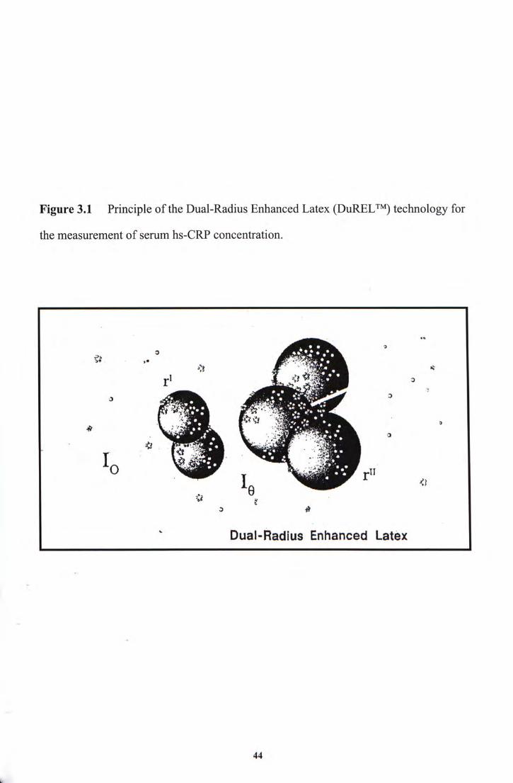
Figure 3.1 Principle of the Dual-Radius Enhanced Latex (DuREL™) technology for
the measurement of serum hs-CRP concentration.
\%My: � Dual-Radius Enhanced Latex
44
3.3,2 Assessment of Analytical Performance
3.3.2.1 Imprecision
The within-run imprecision was assessed by analyzing CRP T Control N (4.34
mg/L), and low (1.88 mg/L) and high (14.58 mg/L) levels of pooled patient serum 20
times within a run, while the between-run imprecision was obtained by analyzing the
same samples in 20 different runs over 20 days. Imprecision of within-run and
between-batch were calculated as coefficient of variation (CV) from results of the 20
replicate analyses.
3.3.2.2 Linearity
Two patient sera with concentrations of 74.86 mg/L and 231.53 mg/L were used
for linearity testing at low end and with post-dilution areas respectively. Dilution by
volume with a low CRP concentration was performed to obtain values down to 0.2%
and 10% of original concentration of 74.86 mg/L and 231.53 mg/L respectively. Each
sample was prepared by direct dilution from the original high sample. The serum
hs-CRP concentration of the diluted samples was measured. Duplicate measurements
�o f serum hs-CRP concentration were performed for each specimen prepared. All the
data points should be plotted for visual inspection of linear performance. The
coefficient of correlation, r,was calculated.
45
3.3.2.3 Recovery
Two pools of patient sera A and B were used as baseline specimens for study of
recovery at low end and with post-dilution areas respectively. To each pool, patient sera
with hs-CRP concentrations of 74.86 mg/L and 231.53 mg/L were added in different
volume at low end and with post-dilution areas. For each specimen prepared, duplicate
assays were performed and the two individual values averaged.
The amount recovered was determined by subtracting the amount originally
present in the respective pools from the amount measured or "observed". Per cent
recovery was obtained by dividing this figure by the amount added and multiplying by
100. These individual recoveries were averaged to give an estimate of recovery.
3.3.2.4 Detection Limit
This is the smallest single result which, with stated probability (commonly 95%),
can be distinguished from a suitable zero blank. It was estimated using the following
equation:
‘ xi = Xbi + ksbi
where Xbi is the mean of the blank measurements, Sbi is the SD of the blank
measurements, and k i s a numerical factor chosen based on the confidence level desired.
In practice, k is assigned a value of either 95% or 99%, respectively. We measured
hs-CRP in 30 samples of normal saline.
46
3.3.2.5 Carryover
This parameter should be tested when the instrumentation used is subject to
contamination of one sample by another. A batch analysis was made with the following
sequence of low (L) and high (H) pools: L1L2L3 H1H2L4 H3H4L5 LgLyLg H5H6L9
HyHgLio HgHioLn. Results of the five L pools which follow other L pools (L2, L3,Le,
L7, Lg) were calculated for their mean and SD. The average was compared with the
mean obtained from the five L pools which follow the H pools (L4, L5, L9, Lio, Li 1). The
difference was the carryover error.
47
CHAPTER IV
MATERIALS AND METHODS II
Serum hs-CRP in Chinese chronic-renal-failure patients with atherosclerotic vascular disease or cardiac valve calcification
V
48
4.1 Patient Recruitment
The study population consisted of 140 male and 128 female (mean 士 SD age = 54 士
12 years, range 20-81) Chinese chronic-renal-failure patients on CAPD. They were
recruited at the Nephrology Clinic of the Prince of Wales Hospital.
Atherosclerotic vascular disease (AVD) was diagnosed by the presence of ischemic
heart disease with a history of angina, previous myocardial infarction, coronary artery
bypass surgery or stenting, or peripheral vascular disease with or without amputation. It
was confirmed by direct patient enquiry and detailed review of hospital case records
and computer records of all hospital admissions in each patient.
For the diagnosis of cardiac valve calcification (CVC), echocardiography was
performed using a Vingmed GE System V sonographic machine using a 3.3MHz probe.
All echocardiographic images were analyzed by cardiologists who were blinded to
clinical details of patients. CVC was defined as bright echoes on one or more cusps of
more than 1mm in either mitral or aortic valves or both. Sensitivity and specificity for
echocardiographic detection of calcium in both the mitral and aortic valve have been
reported to be 76% and 89 to 94%, respectively (94).
V
4.2. Blood Specimens
A 10 mL venous blood sample was collected from each patient and allowed to clot
for about one hour. Serum was separated by centrifugation at 4000 r.p.m. for 15
minutes at 4°C. All sera were stored at -70°C before they were assayed for serum
49
hs-CRP, TC, TG, HDL-C, Apo A-1, Apo B and Lp(a).
4.3 Assay Methods
The Hitachi 911 Analyzer was used for measurements of serum hs-CRP, TC, TG
and HDL-C.
The Beckman Coulter Array® 360 System (Array rate nephelometer) was used for
measurements of Apo A-1, Apo B and Lp(a).
4.3.1 hs-CRP
The hs-CRP assay provided a quantitative measurement of serum hs-CRP by
means of particle enhanced immunoturbidimetry, using the Hitachi 911 anazyer. The
Dual-Radius Enhanced Latex (DuRELtm) technology was used in the measurement of
serum hs-CRP of this study. The principle has been described in the Section III -
Materials and Methods I.
4.3.2 TC
Serum total cholesterol was assayed enzymatically using cholesterol esterase and
cholesterol oxidase. Firstly, cholesterol esters were cleaved by the action of cholesterol
esterase to yield free cholesterol and fatty acids. Then, cholesterol was converted by
50
oxygen with the aid of cholesterol oxidase to cholest-4-en-3-one and hydrogen
peroxide. Hydrogen peroxide formed a red dye compund by reacting with
4-aminophenazone and phenol under the catalytic action of peroxidase. The color
intensity was directly proportional to the concentration of cholesterol and was
determined photometrically.
4.3.3 TG
A lipoprotein lipase was used for the rapid and complete hydrolysis of triglycerides
to glycerol followed by oxidation to dihydroxyacetone phosphate and hydrogen
peroxide. The hydrogen peroxide reacted with 4-aminophenazone and 4-chlorophenol
under the catalytic action of peroxidase to form a red dyestuff (Trinder endpoint
reaction).
4.3.4 HDL-C
In the presence of magnesium ions, the reactivity of cholesterol, especially in
chylomicrons and very low density lipoprotein (VLDL), was reduced by sulfated
a-cyclodextrin without the need for precipitation of lipoprotein aggregates. Sulfated V
a-cyclodextrin and dextran sulfate selectively was formed with LDL, VLDL, and
"" chylomicrons which were resistant to PEG-medified enzymes to water-soluble
complexes. The cholesterol concentration of HDL-cholesterol was determined
enzymatically by cholesterol esterase and cholesterol oxidase coupled with PEG to the
amino groups (approx. 40%). When cholesterol esterase and cholesterol oxidase
51
enzymes were modified by PEG, they showed selective catalytic activities toward
lipoprotein fractions, with the reactivity increasing in the order: LDL < VLDL «
chylomicrons < HDL. Cholesterol ester were broken down quantitatively into free
cholesterol and fatty acids by cholesterol esterase. In the presence of oxygen,
cholesterol was oxidized by cholesterol oxidase to A'^-cholestenone and hydrogen
peroxide. In the presence of peroxidase, the hydrogen peroxide was reacted with
4-aminophenazone and HSDA to form a purple blue dye. The color intensity of this dye
was directly proportional to the cholesterol concentration and was measured
photometrically.
4.3.5 LDL-C
LDL-C was calculated using the Friedewald formula (95):
“ LDL-C = TC - (HDL-C + TG/2.2)
where all quantities were expressed in mmol/L. This formula was not used if the
triglyceride concentration exceeded 4.5 mmol/L.
V
4.3.6 Apo A-1
The rate of increase in light scattered from particles suspended in solution was
measured as a result of complexes formed during an antigen-antibody reaction.
52
4.3.7 Apo B
The rate of increase in light scattered from particles suspended in solution was
measured as a result of complexes formed during an antigen-antibody reaction.
4J.8 Lp(a)
The rate of increase in light scattered from particles suspended in solution was
measured as a result of complexes formed during an antigen-antibody reaction.
Lipoprotein(a) (sample) + Anti-Lp(a) (antibody)—
[Lipoprotein(a)-Anti-Lp(a)]-Complex
4.4 Ultrasound measurement of carotid artery inter-media thickness
The inter-media thickness (IMT) was measured bilaterally on three segments: the
1-cm section of the common carotid artery immediately proximal to the beginning of
the dilation of the bifurcation, a 1-cm section of the bifurcation immediately proximal V
to the tip of the flow divider, and the 1-cm section of the internal carotid artery
immediately distal to the tip of the flow divider. The mean of 6 six measurements gave
the value of IMT of a patient (96). The equipment used was the 7.5 MHz transducer of
the Philips CD800 ultrasound scanner (Best, Netherland).
53
4.5 Statistical analysis
Statistical analysis of data obtained was performed with a personal computer using
SPSS for Windows, Release 10.0.1,Standard Version.
The non-parametric Mann-Whitney U-test was applied to two groups of
independent ranked data to test the null hypothesis that they were samples from the
same population of ranked data, e.g. AVD vs Non-AVD and CVC vs Non-CVC serum
hs-CRP concentrations of Chinese chronic-renal-failure patients on CAPD. Unless
otherwise specified, results were presented as median (interquartile range). All
probability (P) values were two-tailed and P <0.05 was deemed statistically significant.
Patients have been recruited at random. Serum hs-CRP concentrations were
skewed rightward. The distribution of concentration values would be expected in
non-parametric form. The cut-off value of serum hs-CRP concentration was setted at
•• 15 mg/L (41, 68, 69, 97). Median plasma concentrations were computed and the
significance of any difference in the distributions and in median values between AVD
and Non-AVD and between CVC and non-CVC subjects were assessed by the use of the
Mann-Whitney U-test.
54
CHAPTER V
RESUTLSI
Setting up the serum hs-CRP assay on the Hitachi 911 Analyzer
55
The Tina-quant A C-Reactive Protein (Latex) Ultrasensitive Assay using Hitachi
911 Analyzer was evaluated and the results are presented below:
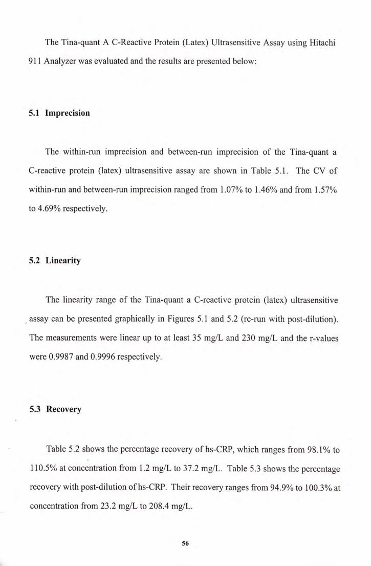
5.1 Imprecision
The within-run imprecision and between-run imprecision of the Tina-quant a
C-reactive protein (latex) ultrasensitive assay are shown in Table 5.1. The CV of
within-run and between-run imprecision ranged from 1.07% to 1.46% and from 1.57%
to 4.69% respectively.
5.2 Linearity
The linearity range of the Tina-quant a C-reactive protein (latex) ultrasensitive
assay can be presented graphically in Figures 5.1 and 5.2 (re-run with post-dilution).
The measurements were linear up to at least 35 mg/L and 230 mg/L and the r-values
were 0.9987 and 0.9996 respectively.
5.3 Recovery V
Table 5.2 shows the percentage recovery of hs-CRP, which ranges from 98.1% to
110.5% at concentration from 1.2 mg/L to 37.2 mg/L. Table 5.3 shows the percentage
recovery with post-dilution of hs-CRP. Their recovery ranges from 94.9% to 100.3% at
concentration from 23.2 mg/L to 208.4 mg/L.
56
5.4 Detection Limit
The detection limit was estimated by assaying blank solutions repeatedly to obtain
a value (x", = 0.01 mg/L) and SD (彻=0.01 mg/L) for the blank with k assigned the
value of 2 to give a confidence limit of 95%. Then, the detection limit � was
calculated by xi = x � + ks / as 0.03 mg/L.
5.5 Carry-over
The percentage of carryover error was found to be 1%.
57
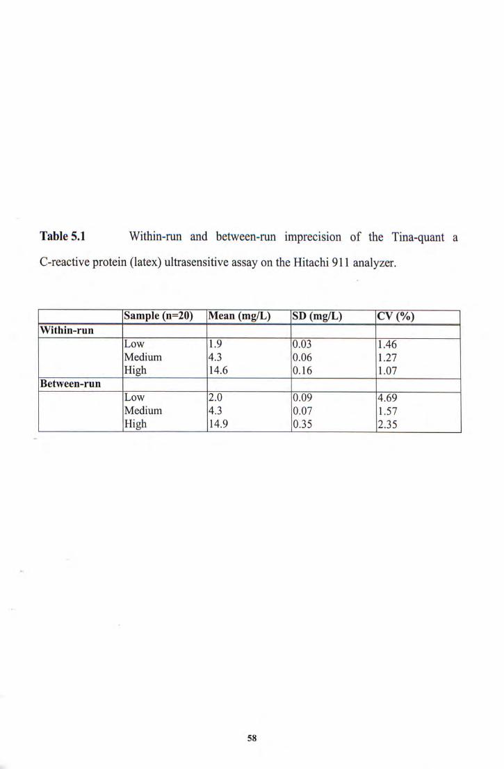
Table 5.1 Within-run and between-run imprecision of the Tina-quant a
C-reactive protein (latex) ultrasensitive assay on the Hitachi 911 analyzer.
iSampIe (n=20) |Mean (mg/L) |SD (mg/L) |CV (%) Within-run
L ^ 19 M 3 146 Medium 4.3 0.06 1.27
Between-run L ^ ZO ^ ^ Medium 4.3 0.07 1.57
[ l ^ [ 0 ^
�
58
Linearity 4 5
0.00 0.20 0.40 0.60
Dilution Ratio
Figure 5.1 Linearity of the Tina-quant a C-reactive protein (latex) ultrasensitive
� assay on the Hitachi 911 Analyzer.
59
Linearity with Postdilution
250
^ 0.00 0.20 0.40 0.60 0.80 1.00
Dilution Ratio
Figure 5.2 Linearity with post-dilution of the Tina-quant a C-reactive protein (latex)
ultrasensitive assay on the Hitachi 911 Analyzer,
60
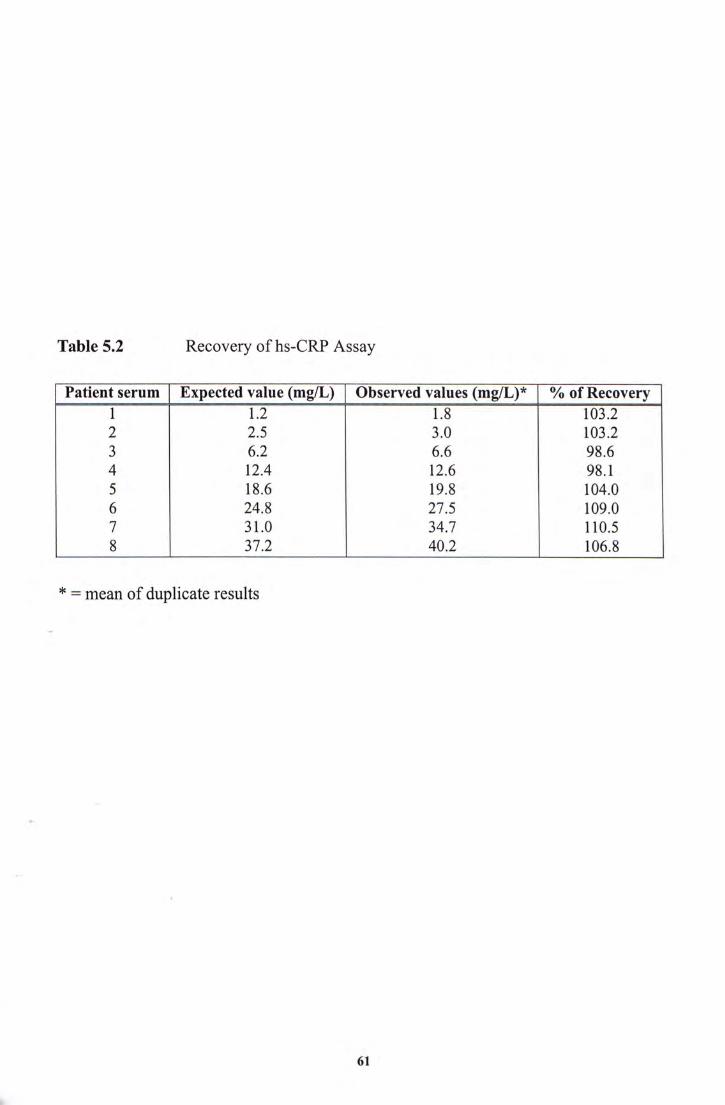
Table 5.2 Recovery of hs-CRP Assay
Pat ient serum Expected value (mg/L) Observed values (mg/L)* % of Recovery 1 n n 2 2.5 3.0 103.2 3 6.2 6.6 98.6 4 12.4 12.6 98.1 5 18.6 19.8 104.0 6 24.8 27.5 109.0 7 31.0 34.7 110.5 8 ^ 106.8
* = mean of duplicate results
61
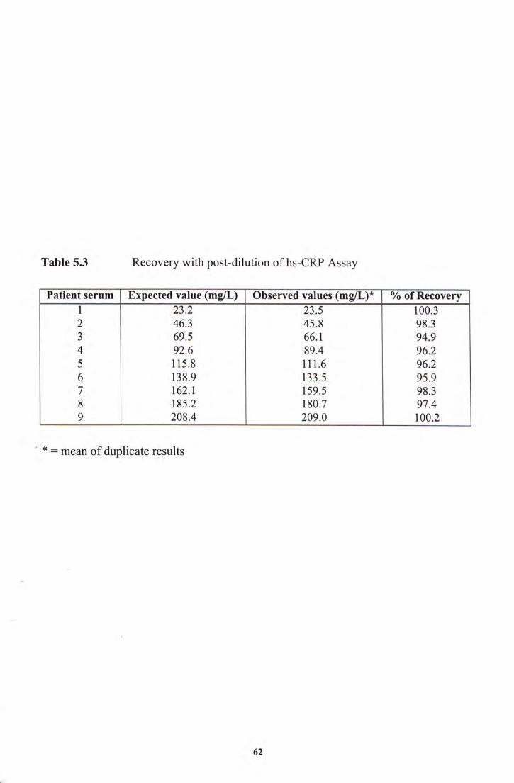
Table 5.3 Recovery with post-dilution of hs-CRP Assay
Pat ient serum Expected value (mg/L) Observed values (mg/L)* % of Recovery 1 2I2 ^ r ^ 2 46.3 45.8 98.3 3 69.5 66.1 94.9 4 92.6 89.4 96.2 5 115.8 111.6 96.2 6 138.9 133.5 95.9 7 162.1 159.5 98.3 8 185.2 180.7 97.4 9 208.4 209.0 100.2
”* = mean of duplicate results
62
CHAPTER VI
RESULTS II
Serum hs-CRP in Chinese chronic-renal-failure patients with atherosclerotic vascular disease or cardiac valve calcification
63
6.1 Patient Recruitment
Two hundred and sixty-eight Chinese chronic-renal-failure patients on CAPD (140
male and 128 female, age = 54 土 12 years, range 20-81) were recruited as described in
Section 4.1.
6.2 Chinese chronic-renal-failure patients with AVD
Results of the above measurements (totaling 9 assays on each of the 268 patients)
are listed on Table 6.1.
As shown on Table 6.2,serum hs-CRP concentration of the AVD group had
significantly higher than that of non-AVD group [median (interquartile range) of 3.59
(0.92 - 8.21) vs 1.43 (0.73 - 3.72) mg/L, p < 0.05, Mann-Whitney U-test] of the Chinese
•“ chronic-renal-failure patients on CAPD.
The IMT and serum TC, TG, HDL-C, LDL-C, Apo AI, Apo B and Lp(a) between
AVD and Non-AVD groups were compared by performing Mann-Whitney U-test
(Table 6.2). No statistically significant difference in the IMT and in serum TC, TG,
� HDL-C, LDL-C, Apo AI and Apo B concentrations were noted. AVD patients had
significantly difference in serum Lp(a) concentration compared to non-AVD patients (p
< 0.05). �
64
6.3 Chinese chronic-renal-failure patients with CVC
Results of the above measurements (totaling 9 assays on each of the 268 patients)
are listed on Table 6.1.
As shown on Table 6.3, serum hs-CRP concentration of the CVC group had
significantly higher than that of non-CVC group [median (interquartile range) of 1.81
(0.77 - 5.45) vs 1.43 (0.72 - 3.40) mg/L, p < 0.05, Mann-Whitney U-test] of the Chinese
chronic-renal-failure patients on CAPD.
The IMT and serum TC, TG, HDL-C, LDL-C, Apo AI, Apo B and Lp(a) between
CVC and Non-CVC groups were compared by performing Mann-Whitney U-test
(Table 6.3). Between the two groups, there was no significant difference in the IMT and
in serum TC, TG, HDL-C, LDL-C, Apo A-1, Apo B and Lp(a) concentrations (p >
0.05).
V
65
Table 6.1 Serum hs-CRP concentration in AVD and Non-AVD, CVC and
Non-CVC of Chinese chronic-renal-failure patients on CAPD. IMT and the following
serum lipids, lipoproteins and apolipoproteins were also measured:
TC
TG
HDL-C
LDL-C (calculated)
Apo A-1
Apo B
Lp(a)
66
Table 6.1 — Subject I Sex I Age AVD hs-CRP Average ~ ~ T C “ T G ~ ~ HDL-C LDL-C ApoA-1 I Apo B I Lp(a)
(l=yes’ calcification (mg/L) IMT (mmol/L) (mmol/L) (mmol/L) (mmol/L) (mg/dL) (mg/dL) (mg/dL)
0=no) (l=yes, 0=no) (mm)
^ ! 68 1 0 _ ^ ^ ^ ^ ^ ^ ^ 2j2_ 143.01 96.0 TLTO ! ^ L 1 56.01 NA 5.41 Ua 1.21 3.55 ~ ~ ^ ^ ^ ^ ^ _ _ ^ 0 ^ _ 0.70 6.24 1.04 1.59 " 4 . 1 8 1 0 5 . 0 26.40 I ! 0 0_0.32 NA ^ 1.99 m 122.0 ^ ! 0 0__OM NA 3.63— 2.07 0.69 2.01 100.0 ^ ^ ^ 0 ^ 7-25 I 5.37 2.08 __0j 36.10 ! ^ ^ 0 0__0.55 NA— 3.15 0.7万 0.87 L% 102.0 ? ^ ^ L 0 0.65 ^ 4.44 “ 0.92 138 2.65 M ~ 9 P ^ L 1 15.78 ^ ^ 6.95 I M 1.23 4.62 132.0 129.0 152.60
^ _ _ ^ 0 NA 1.77 ^ 4.01 m 0.74 ^ ^ 8 7: 6 ~ 1 ^ I I I ^ 0 0 28.16 NA 6.42 2.90 0.98 4.13 IIO.O^T^ ^
^ ^ 0 N 八 1.65 L^ 5.56 r^ 3.90 一106.0 120.0 ~~im ! ^ 0 NA 66.78 ^ 3.98 Bo OCT 2.18 ~~100.0 89.8 ~ ^
!1 ^ ^ 0 0 _ _ 0 . 4 4 NA 3.72 0.46 1.48 2.03 118.0 ~
H ! ^ 0 L_5.54 NA 5.09 2.05 1.04 HJ 109.0 90.7 19.10 ! ! ^ 0 0__5.09 NA— 4.00 1.33 1.23 j j j 125.0 ~~ !Z I ! 0 ^_4.93 NA~ 4.79 1.03 1.48 lE 127.0 ~ 7 L 2 ~ ^ !? I ^ 0 L__5.75 NA~ 3.81 3.07 0.61 im 140.0 109.0 14.80 !! ^ ^ L NA 1.07 NA 5.96 r^ 098 4.15 ^ ^ ! ^ L L_Z:^ ^A_^ZHM___^H^I^^I;;;^^^ ^ ! 0 Q 12.35 ^ 4.15 1.89— 0.90 2 M 106.0 101.0 HW
^ ! ^ Q Q 1-33 NA 5.84 1.90 1.31 一 3.67 118^ 89^ 37.60
^ ! ^ 0 0__0:^ NA 5.81— 1.49 1.30 3.84 ~ ~ ^ 103.0 33.60 ^ ! ^ 0 L _ _ ^ NA 5.23 3.13 0.83 ~ ~ ~ ~ 1 1 ^ 0 130.0^32\
^ _ _ ^ L NA 0.95 NA 5.19 2.18 0.81 3.40 106.0 ~TTLO~3^ ! ^ 0 L _ ^ ^ 7.12 2.97 U S 4.60. 133.0 158.0 49.10
^ ! ^ L ^ _ _ ^ NA 2.77 ~~113 0.58 r ^ 129.0 ~ ~ ^ . ^ ^ ^ 0 0__ ^ 5.68 0.72 h^ 4.04 124.0^
^ I ^ 0 0 0.36 ^ 6.19 a^ 2.41 3.59 ~~~14^ ^ ! ^ L Q 0.80 NA 5.39 U5 1.96 2.91 153.0 ^ ! 0 ^__0:^ ^__6.97 1.31 "T^ 4.39— 164.0 105.0 44.40 ^ ! ^ 0 0_!i9 NA 7.54 1.20 T ^ 5.16— 135.0 117.0 52.70 ^ ! ^ L 1 94.24 ^ 3.32 1.27 1.01 1.74— 95.9 49.5 62.50 ^ ! 0 L __0.68 1.65 — 4.11 1.70 0.86 2.48 817 ^ ^ _ _ ^ L ^ _ ^ NA 4.31 2.23 0.75 ^ I 47 NiA 3.96 NA 3.46 1.38 0.54 ^ ^ T ^ !! ^__44 1 1 34.27 I 5.39 1.03' 3.64 ;; ! 38 |M 57 0 [_Om ^ 5.33 2.09 3.45" 119.0 103.0 23.00
.|F 55 0 0 _ _ ^ N A 3.8I 1 3 0 0.87 1.90 1 14 .0 fOO
� _ ! _ _ 0 0 5.68 NA 6.68 ~~119 1.34 ~ ~ ^ 丽 117.0 17.00 ^ _ _ ^ 0 ^!^_yn___0.98 4.36 \J4 0.76— 2.82 8 5 . 8 8 4 T ^
_ ! _ _ 0 1 33.14 NA 4 . 0 3 O j ^ ^ _ _ ^ 0 L__ ^ 6.32 3.18 m ^^ m 124.0 1220
^^_!__IZ _ _ 1 3.54 NA 3.99 __Oj3 ^ ^ ^ ^ ^ ^ J j j Q ^ ^: ^ ! U 0 ^__Sm NA ^ 1.25 1.55 ^ 145.0
_ ! _ _ ^ 0 Q 2.84 NA 6.34 ^ ^ ^ ^ 108.0 16.60 1 ! _ ! _ _ _ _ 0 NAITO NA ~ ~ ~ ^ ~ ~ ~ ^ ~ ~ ~ ~ ~ ^ 73.2 5200
^ _ _ ^ L 0__em ^ 3.29 2.02 1.45 n s r o o ! ^ 0 0_2.01 NA 一 7.96 3.32"___O:^!!!^ 177.0 19.10 ! ^ 0 L _ _ ^ NA 4.62 0.56 ^ ^ ^
5丨 |M 55| Q| 0| 0.811 na| 4.9I| 1.5I| 1.2I| 3.Q2| 129.O| 86.O| ^
67
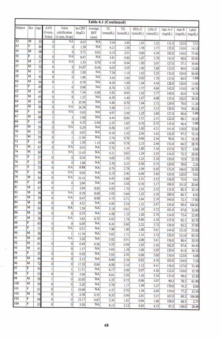
Table 6.1 (Continued) "“ Subject I Sex I Age I AVD I hs-CRP I Average I TC I TG | HDL-C LDL-C Apo A-1 I Apo B I Lp(a)
(l=yes’ calcification (mg/L) IMT (mmol/L) (mmol/L) (mmol/L) (mmol/L) (mg/dL) (mg/dL) (mg/dL)
0=no) (l=yes, 0=no) (mm)
52 |M 551 01 NA| 0.971 NAI 5.叫 了圳 i .oi | 3.22I i31.o| m i T ^
“ ! _ _ ^ 0 0 _ ^ ^ _ _ 6 . 2 1 1.96 156 3.77 一 135.0 116.0 14.10
^ ^ _ _ _ ^ L 0 _ _ ^ _ _ 6.10 2.01 4.39 函 118.0 77.40
“ _ ! _ _ ^ 0 NA 0.47 NA 5.81 ~ ~ 1 . 6 7 1 : ^ 98.6 53 90
_ ^ _ _ E 0 ^ _ _ _ _ _ 0 . 7 0 4.16 ~ ~ a ^ 1.80 ~ ~ m 127.0 57.1 34.60
!Z ^ _ _ ^ 0 Q 14-07 0.48 6.05 ^ l.OQ ^ H l O _ ^ _ _ Z Z 0 0 3.20 ~~NA ~ ~ ^ ~~Em ~~~L^ ^ 129.0 56.90
!! ^ _ _ 0 0__1:09 NA 3.42 L ^ 0 . 9 0 — 1.78 1 1 3 . 0 m ~ T M
^ ^___^ 0 L__0.79 NA ^ 1.69 1.04 4.48 128.0 1200 13 90 ^ ! _ _ L 0 0-96 NA 6.76 m 1.57 119.0 44.70 ^ ^ _ _ 0 0 _ _ I M _ _ 0 . 8 8 5.82 ~ ~ 1 . 6 2 — 3.77 140.0 ^ ~ ^
“ ^ _ _ ^ 0 0 _ NA 6.38 L^ 1.67 "T08 102.0 39 50 ^ _ _ ^ _ _ ^ 0 1 25.94 NA ~ ~ ^ ~ ~ ~ ~ m 78.0 1120
^ _ _ ^ _ _ ^ _ 0 NA 34.34 ~ ~ ~ N A 5 . 2 0 _ _ L L I Z I ^ I Z I J ^ _ _ 1 2 8 ^ 79.R 9.9 9.0 ^ Z ^ 0 ^ _ _ ^ ^ 4.97 2.49 L^ 2.60 ~ ~ W ^ ^ ~ T M - _ _ ^ _ _ ^ L 1 3.98 NA 4.44 ~ ~ 1 . 5 1 T S T 68.1 18.20
! 0 0 _ ^ _ _ ^ 2.19 1.65 ~ I K O 4 M ~ T M
- _ ^ _ _ ^ _ _ 0 NA 0.29__NA ~ ~ ^ f^ 1.60 4.2T MTO 118.0 32.80 - _ ^ _ _ « 0 0 Q-87 NA 6.10 ~ ~ R ^ 2.04 ^ 97.3 72 10
_ _ ^ ^ _ _ ^ _ _ 0 L^l:^::^;:^:^;^^;^^;;^^;;^;^^ 76.1 16.30 _ _ ! _ _ 0 0 _ L ^ ___L]^ ___4.96 078 Z15 ^ 64.3 28.70 _ _ _ ^ ___0 ^ 7 6 . 5 6.86
- _ _ _ ^ _ _ ^ L NA 1 1 . 4 5 _ _ L ^ ^ 114.0 72.3 28.00 11 ! ?Z 0 0 _ ^ ^ _ _ 1.50 1.23 118 ^ - _ _ ! _ _ ^ 0 0 1 . 8 6 _ _ ^ _ 2 . 3 6 ^ 丽 5 8 . 6 2.26
! ! _! _ _ ^ _ _ 0 BZO 166.0 25.80 ! ^ 0 ^ _ _ o m ^ _ _ 6 . 1 9 2.90 m 120.0 120 0 ~ m
! ? _ _ ^ _ _ ^ 0 NA 3 1 . 4 3 _ _ N A ~ ~ J : ^ ~ ~ 1 . 5 1 2:^ 73.9 9 61
_ ! _ _ 0 0 _ _ ^ ^ _ _ _ 5 . 4 4 3.48 "OTO 3.17 ~ 108.0 101.0 2040
!I ^ _ _ ^ 0 L _ _ ^ _ _ ^ _ _ _ 4 . 8 3 1.70 ~ ~ L ^ 2.52 803^110 ‘ ^ _ _ ^ _ _ ^ 0 NA 0 . 7 8 _ _ ^ _ _ 3 . 9 2 ~ ~ L ^ ~ ~ ^ 函 607 8 92
^ _ _ ^ 0 NA 0 . 4 7 _ ^ _ _ 4 . 7 5 0.71 L ^ ^ 丽 72 3 17 80
^ _ _ ^ _ _ ^ _ _ 0 Q 4.51__NA ^ B4 133 90.4 104.00 ^ _ _ ^ _ _ ^ _ _ 0 ^^ 7.59 __NA ^ oIT Lio m ^ _ _ ^ _ _ ^ _ _ 0 0 0.73__NA i: TB L^ m - _ _ - _ _ ^ L NA 3 . 6 3 _ _ _ _ 4 . 0 3 Tj4 ^
! ^ 0 0 _ _ ^ 6.24 0.66 m ^ m ^ T T O ^ _ ! _ _ ^ _ _ 0 NA 0 . 5 1 _ _ N A ^ ~ ^
^ _ _ ^ _ _ ^ L _ _ N A ^ TtT T U ^ 121.0 69.10 ^ _ _ ^ _ _ ^ _ _ L __NA ^ ^ ^ ^ _ _ r _ _ ^ 0 0 0 . 4 9 _ _ 0 . 5 6 4:^ ^ ^
S _ _ ^__11 L 0 1.17 _ _ ^ ___4.65 L^ TOS ~ ^ 81.8 36.10 ^ _ _ I _ _ ^ _ _ _ 0 __NA ^ ^ ^ ^ ! ^ _ ^ _ _ _ _ _ 0 0 2 . 1 3 _ _ N A ^ ~ B 9 ^ ^ M 9 ^ _ _ ^ _ _ _ _ 0 ^ 丽 117.0 31:40
‘ _ _ I _ _ ^ L 旧 1 _ _ _ ^ _ _ ^ ~ ~ ^ ^ 1110 110.0 15.70
^ _ _ ‘ _ _ _ _ _N A ^ B 6 Lie 0 4 丽 99.6 15.20
- - _ _ ^ ^ ^ 0 % ^ - ^ ― ^ _ _ N A ^ ni7 r ^ ^
_ ‘ _ _ ^ _ ^ ___NA ~ ~ ^ OTO r ^ ~ 丽 77.2 13.10
^ ^ ― ^ _ _ 0 _ _ 0 . 5 5 ^ ― ^ m ^ 167.0 6 9 . 2 - 1 ^
_ ‘ _ _ ^ _ _ ^ _ _ _ ^ 3.34 R ^ L ^ 函 68.5 2.73
I 川4 |F 6 3 | Q| o| 3.00I NA| 6 . 1 3 | 2 . 1 2 I 0 . 8 5 1 ^
68
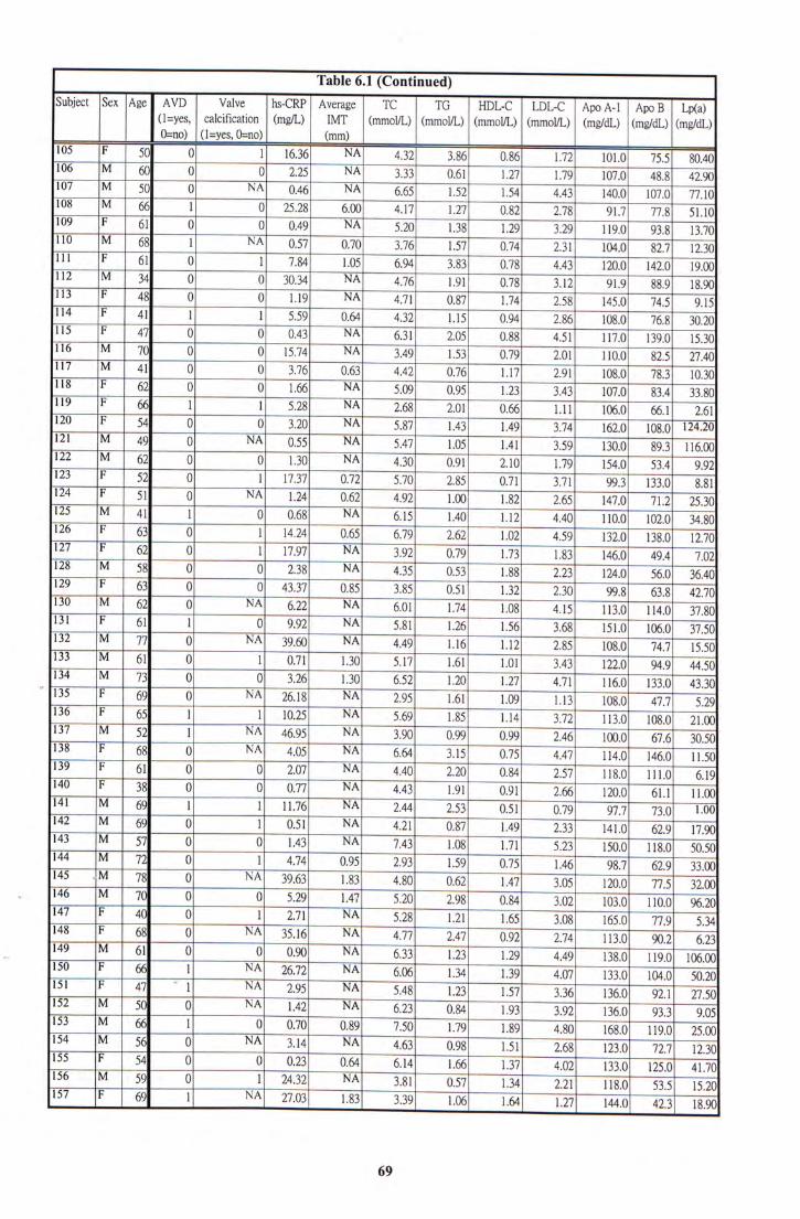
Table 6.1 (Continued) "“ Subject I Sex I Age AVD " " “ hs-CRP Average TC""“TCHDL-C LDL-C Apo A-1 I Apo B I Lp(a)
(l=yes’ calcification (mg/L) IMT (mmol/L) (mmol/L) (mmol/L) (mmol/L) (mg/dL) (mg/dL) (mg/dL)
Q=no) (l=yes, 0=no) (mm)
105 |F 50| 0| 11 16.361 NAI 4.32| 3.86| 0.86| ~ 7 5 . 5 ! 80.40
! ^ _ ^ _ _ ^ 0 0 _ _ ^ NA 3.33 ~ ~ 1 . 2 7 ~ [ J 9 _ _ ^ _ _ ^ 0 NA 0.46 NA 6.65 _ _ ^ _ ^ ^
! ^ _ ^ _ _ ^ L 0 25.28 6.00 4.17 1.27 0.82 2.78 ^ 77.8 51.10
_ Z ^ 0 0_0.49 NA ^ 1.38 r ^ 119.0 _ ^ _ _ _ ^ L ^ _ _ ^ 0.70 3.76 [57 0.74 I M
^ ! ^ 0 L _ _ 1-Q5 6.94 0.78 4.43 ["20.0 142.0 19.00 _ ^ _ _ ^ 0 Q 30.34 NA ^ ^ __TI2 91.9 ~
m _ ! 0 0 __hll NA 4.71 1.74 BS 145.0 74.5 ^ ^ ! 11 L L _ _ ^ 0.64 4.32 1.15 0.97 2.86 76.8 30.20
_ ! IZ 0 0 _ _ 0 . 4 3 N A ~ 6.31 2.05 O M ' ^ ^ ^ ^ ^ ^ I V I O 139.0 15.30 _ ^ _ _ 0 0 15.74 NA 3.49 F ^ 0.79 M \ M
M ^ ^ _ _ 1 1 0 0 _ ^ 0.63 4.42 0.76 1.17 2.91 " “ ~ 7 8 . 3 10.30
! ! ! _ ! ^ 0 0 _ _ \ M NA 5.09 0.95 1.23 3.43 107.0 ~ ~
1 1 ! _ I ^ L L _ _ ^ NA 2.68 m 0.66 uT 106.0 ~ ~ ^ m _ ! ^ 0 0 _ _ ^ NA 5.87 MS 1.49 3.74 ^ ^ 1 6 2 . 0 108.0 124.20
_ ^ _ _ 0 NA 0.55 ~ ^ ~ ~ ~ ^ ~ ~ L O S ~ M T ~ ~ i M 89.3 116.00 ! ^ ? _ ^ _ _ ^ 0 0__ NA 4.30 ~ ~ ^ 2.10 T^ ~~15^ ~ ^ ^
_ ! ^ 0 1 17.37 0.72 5.70 0.71 _ 3.71 ^
_ ! 0 ^ _ _ ^ 0.62 4.92 1.00 1.82 2.65 147.0
_ ^ _ _ 1 1 L 0 _ ^ NA 6.15 L ^ 1.12 4.40 ~ ~ I M 102.0 34.80
_ ! ^ 0 1 14.24 0.65 6.79 2.62 1.02" 4.59 138.0 12.70
_ ! ^ 0 1 17.97 NA 3.92 1.73 ~ ~ \ M 1 4 ^ T F F I
_ ^ _ _ ^ 0 0 2.38 NA 4.35 ~ ~ 1 . 8 8 ~ ~ 2.23 124.0
_ ! ^ 0 0 43.37 0.85 3.85 OIT 1.32 ^ ^ ^ ^ _ _ ^ 0 ! _ _ ^ 114.0 37.80
^ ! ^ L 0 _ _ ^ ^ 5.81 1.26 L ^ 3.68 丽 106.0 37.50 _ ^ _ _ Z Z 0 NA 39.60 NA 4.49 TT^ 1 . 1 2 一 2.85 108.0
_ ^ _ _ ^ 0 L _ ^ 1.30 5.17 r ^ 1.01 ~ ~ ^ . . _ _ ^ _ _ 0 Q 3.26 ~ ~ ~ 1 3 0 ~ ~ ^ 1.20 1.27 ~ ~ W \ ^ ^ U ^ 133.0 43.30
_ ! ® 0 NA 26.18 ^ 2.95 1.61 1:09 113 ~ ~ ~ 历 ^
^ _ _ ! ^ L 1 10.25 NA 5.69 L ^ 1.14 M 113.0 108.0 21.00 ! ^ _ ^ _ _ ^ L NA 46.95 ^ 3.90 0.99 ~ ~ 2 . 4 6
_ ! ^ 0 ^ _ _ 4 : 0 5 NA 6.64 3.15 0.75 ~ ~ ~ ^
! ! ! _ ! ^ 0 0 _ ^ ^ 4.40 2.20 2.57 118.0 111.0 ^
_ ! _ _ ^ 0 0 0.77 ^ _ _ 4 . 4 3 1.91 ^ 函 611 1100
_ ^ _ _ ® L 1 11.76 NA 2.44 0.51 ^ ^ 7 3 0 H O O _ ^ _ _ ® 0 I 0.51 ^ 4.21 ~ 0 8 7 1.49 ^ 62.9 17 90
!^__ ^ _ _ ^ _ _ 0 0 1.43 _ _ N A ~ ~ ~ ^ ~ ~ R 5 8 LTT ~ ~ ^ 118.0 50.50
_ ^ _ _ V L 0 1 4.74 _ _ 0 . 9 5 2.93 ~ ~ L ^ 0.75 T ^ ^ 62.9 33 00
145 .|M 78 0 NA 39.63 _ _ 1 . 8 3 ~ ~ 4 : ^ ~ ~ 1 . 4 7 105 函 77.5 3200
� ! ^^ - _ _ 0 0 5 . 2 9 _ ^ _ _ 5 . 2 0 ^ A ^ ^ 1 0 ^ 110.0 96.20
! ! ! ^ ! ^ 0 L _ _ ^ ^ 5.28 1.21 R ^ ^ F L 9 ^
_ _! _ _ ^ 0 NA 35.16 __NA ~ ~ ~ ^ ^ ~ ~ ~ ~ ^ ^ ^ l i l o 90.2 6 23 _ _ - _ _ ^ 0 0 0.90 _ _ N A ~ ~ ^ ~ ~ ~ 1 3 ~ ~ ^ 丽 119.0 106.00
I f ? _ ! _ _ ^ L NA 2 6 . 7 2 _ _ N A ^ L ^ ^ ^
_! _ _ I Z _ 1 I i ^ - ^ i - ^ o _ ^ _ _ ^ 0 ^__^___,;;;^;;^^;^^^^;^^^;;^^^^ 93.3 9 05
M _ ^ _ _ ^ L 0 _ _ ^ _ _ _ ^ 7.50 1.79 R ^ ^ 函 119.0 25 00 _ ^ _ _ ^ 0 na 3.14__NA ~~i:^ ~ ~ ~ ~ ~ ~ ~ ~ ^
— _ ! _ _ ^ 0 0 0 . 2 3 _ _ ^ _ _ 6 . 1 4 1.66 L ^ ^ ^ 丽 125.0 41 70
I F ^ _ ^ ^ ^ _ _ ^ _ _ 0 L ^ ^:;; ^ ];; ^ ^;; ^ ^;;; ^ 2 2 1 丽 53.5 15 20 157 |F 69| 1| NA| 27.03| 1.83| 3.39| 1.06| 1.641 \.2l\ 144.O| 42.3| 18.90
69
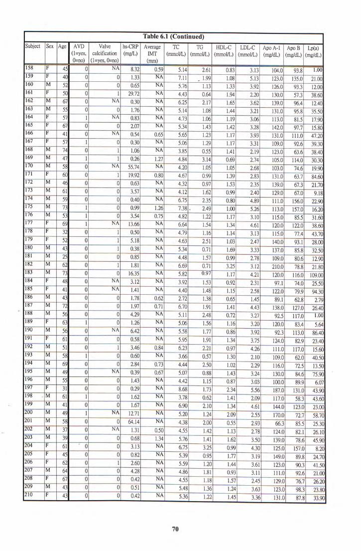
Table 6.1 (Continued) "“ Subject Sex Age AVD Valve hs-CRP Average TC“TG""“ HDL-C LDL-C I Apo A-1 I Apo B I Lp(a)
(l=yes’ calcification (mg/L) IMT (mmol/L) (mmol/L) (mmol/L) (mmol/L) (mg/dL) (mg/dL) (mg/dL) 0=no) (l=yes, 0=no) (mm)
I ^ 0 NA 8.32 _ 0.59 5.14 2.61 0.83 3.13 104.0 93.8 1-00 159 F 40 ~~ 0 "O 1.33 21.00
_ M 52 0 0 0.65 ^ 5.76 U3 1.33— 3.92 ~126.0 93.3 ~ilOO I ^ 0 1 29.72 ^ 4.43 1.94 2.20 ~ ~ 1 3 0 0 ^ ^ ^
_ ^ ^ 0 NA 0.30 NA 6.25 2.17 3.62 ^ _ ^ ^ 0 0 1.76 NA 5.14 LOS 1.44 _ 3.21 LEO 95.8 35.50
_ I ^ L NA 0.83 丽 4.73 1.06 L19 3.06 """TLLO K 5 ~ P M
_ _ ! ^ 0 0 2.07 NA 5.34 L ^ 1.42. 3.28 97.7 15.80
_ ^ 0 ^ _ _ 0 . 5 4 0.65 _ 5.65 1.23 1.17 131.0 111.0 47.20
! ^ _ ! 1 0 030 N A ' 5.06 1.17 3.31 109.0" 92.6 39.30
_ _ ^ 0 [ _ 1 . 0 6 NA 3.85 0.55 1.41 Z19 123.0 63.6 38.40
^ IZ L 1 0.26 ] J F 4.84" 3.14 0.69 2.74" 1 0 5 . 0 1 1 4 . 0 30.30
I Z ? _ _ ^ ^ 0 NA 55.74 NA 4.20 LOS 1.05 —— 168 103.0 _ 74.6 19.90
! I ^ 0 1 19.92 OTSO 4.67 0.99 1.39 2.83 "l3L0 63.7 84.60
I Z ? _ ^ 0 0 0.63 ^ 4.32— 0.97 1.53 2.35 ~ ^ ^ T ^ _ ^ ^ 0 0 3.57 ^ 4.12— 1.62 0.99 2.40 ~ ~ m ~ ^
I Z ^ _ ^ ^ 0 0 0.40 NA 6.75 2.35 0.80 M FLL.O 156.0 22.90
!]I_ ^__Z^ 1 0 ~ 1 - 2 6 " 7 .38:~~1 .00 5.26 113.0" 157.0 16.20 I Z ^ _ ^ « }_ 0__3.54 0.75 — 4.82 1.22 1.17 Ho 115.0 ~~SiS^L^ IZZ I ^ NA 13.66— NA 6.64 1.54 — 1.34 4.61 ~122.0 38.60
_ F 飞 0 1— 0.50 NA 4.79 1.T6 1.14 3.13 115.0 77.4 43.70
_ I ^ 0 1 5.18 NA 4.63 ^ 1.03" 2.47 ~ ~ M O . Q 93.1
_ ^ _ _ 1 3 0 ^ _ 0 . 3 8 NA ^ 0.71 _ 1.69 ^ 137.0
^ ^ 0 0 _0.85 NA— 4.48 i.sT 0.99 一 2.78 109.0 806
_ ^ ^ 0 1 1.81 NA 6.69 0.71 3.25" 3.12 ~ ~ ^ 78.8 21.80
_ ^ _ _ Z 3 0 0 16.35 NA 5.82 0.97 1.17 4.21 120.0 116.0 109.00 _ ! 48 0 ^ _ 3 . 1 2 NA 3.92 1.53 0.92 2.31 97.1 ~ ~
I 11 0 NA 1 . 4 1 — NA 4.40 1.48 — 1.15 2.58 " m 94.30
_ M 43 0 一 0 1.78 0.62 1.38 0.65 L ^ M
. _ ^ 72 0 0 1.97 0.71 6.70 1.91 1.41 ~ 4.43 138.0 127.0 26.40
“ M S6 0 0 4.29 ^ 5.11' 2.48 3.27 ^ ~ n i o T M
189 F ^ I " ~ 0 1.26 ^ 5.06 1.56 1.16 3 . 2 0 [ " 2 0 . 0 83.4 5 M
_ M 56 0 ^ _ 6 M NA 5.58 \ j l 0.86 3.92 92.3 113.0 86.40
_ Q Q 0.58 NA 5.95 1.34. 3.75 — ~ 8 2 . 9 23.40 _ M__M 0 [_3.46 0.84 6.23 2.21 0.97 4.26 111.0~~117:0~[5: _ M _ _ ^ L 0_0.60 NA ^ 0.57- 1.30 110 109.0 ~~ _ ^ ® 0 0 _ _ m 0.73 4.44 2.50 1.02 2.29 ~ ~ 1 1 ^ 72.5 13.50
_ M _ _ 4 9 0 ^ _ 0 . 3 9 0 .67— 5.07 0.88 1.43 130.0
_ ^ ^ 0 0 _ _ 1 . 4 3 N A ~ - 4.42 1.15 0.87 3.03 100.0 ~ ~ ^
1!Z_! ^ 0 0 _ _ ^ NA 8.68 1.73 2.34 5.56 187.0 ^ _ _ _ _ ^ L 0__ !:! 3.78 0.62 1.41 2.09 _ 117.0 58.3 43.60
� !! ! _ ^ _ _ 1 1 0 0 _ _ NA 6.90 l i o 1.34 m 144.0 123.0 23.00 ^ _ ^ _ _ 1 9 L NA 12.71 NA 5.20 L ^ 2.09— 2.55 170.0 ^ _ ^ _ _ ^ 0 0 64.14 NA 4.38 m 0.55 2.93 ^ K s ^ ^
, _ ^ _ _ E . 0 ^ _ 0.50 4.55 1:42 1 . 1 3 ~ 2.78 124.0
^ _ ^ _ _ ^ 0 0 _ ^ 1.34 5.76 MT 1.62— 3.50 139.0 _ ! 61 �0 0 _ _ ^ NA 6.75 ^ 0.99 ^ 125.0 ^
_ ! 0 0 _ _ ^ NA 5.39 0.95 1.77 " 3.19 ~ ~ 8 9 . 8 24.70
_ ! ^ 0 L _ _ ^ NA 5.59 L^ 1.44 3.61 123.0 _ ^ _ _ ^ 0 0__ NA 4.86 ilT 0.93— 3.11 111.0
^ _ ! ^ 0 0 _ _ 2 : 1 2 ^ 4.55 1.18 LS? 2.45
_ ^ _ _ 0 Q 0.51 NA 5.48 L36 1.24 "T^ 函 98.3 23.80 210 |F 43| 0| Q| 0.42| Na| 5.36| 1.22! 1.45| 3.36| 131.o| 87.8| 33.90
70
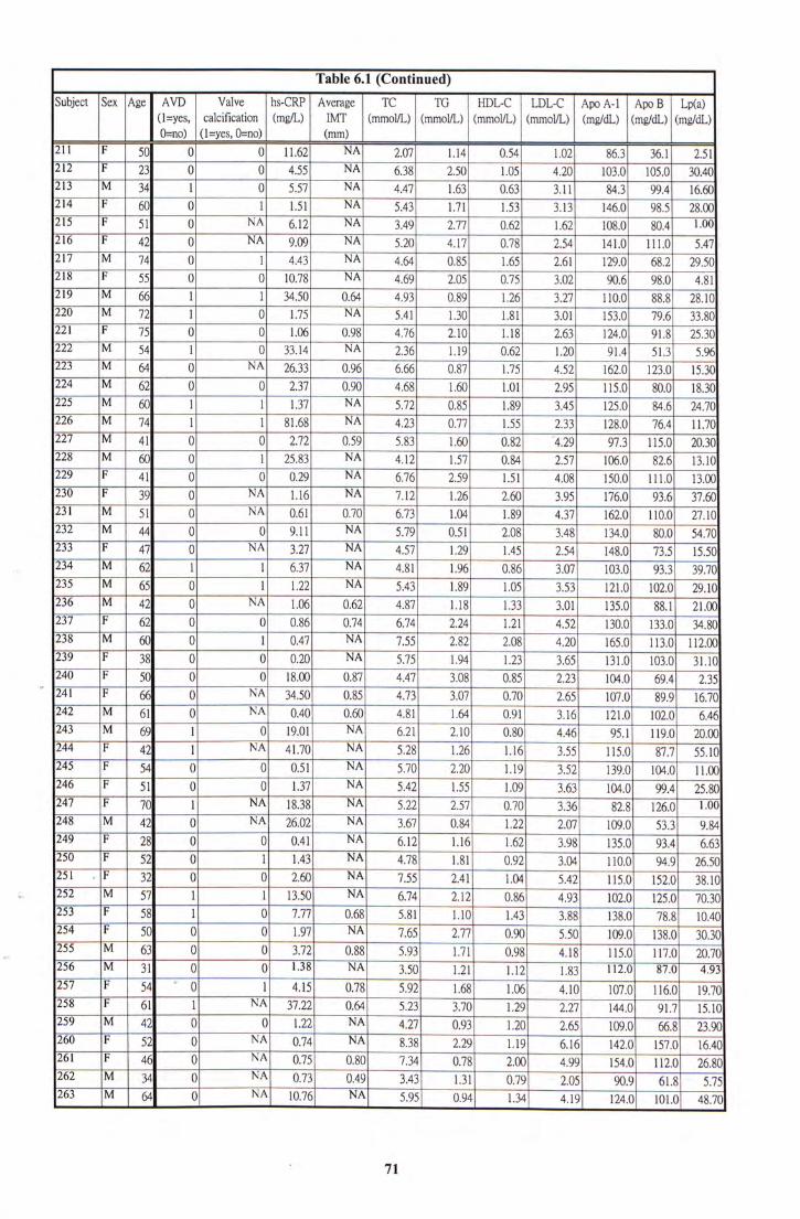
Table 6.1 (Continued) "“
Subject Sex ~ AVD “ hs-CRP Average TCTC""“ HDL-C LDL-C Apo A-1 I Apo B I Lp(a) (L=YES, CALCIFICATION (MG/L) IMT (MTNOL/L) (MMOL/L) (MMOL/L) (MMOL/L) (MG/DL) (MG/DL) (MG/DL) 0=NO) (L=YES, 0=NO) (MM)
^ F ^ 0 0 11.62 NA _ 2.07 1.14 0.54 1.02 86.3 36.1 2.51
212 F ^ 0 0 4.55 NA 6.38 ^ L05 ~ ~ M
213 M M 1 _ 0 ~ ~ N A 4.47 一 1.63 0.63 3.11 — 84.3 ^ ~
214 F ^ 0 1 ~ ~ L M NA 5.43 1.71 1.53 3.13 1 4 ^ 9 8 . 5 28.00
215 F 51 0 NA 6.12 NA 3.49 2J1 804 ^
216 F ^ 0— NA ~ ~ ^ NA 5,20 4.17 0.78 ~ U L O ^
217 M 0 ~ 1 4.43 NA 4.64 — 0.85 165 m 68.2 29.50
218 F 召 0 0 10.78 NA — 4.69 2.05 M M ^ ILL
219 M —品 1 ~ 1 0.64 4.93 一 0.89 1.26 3.27 U ^ ^ ^ 8 8 . 8 28.10
220 1 0 1.污 NA 5.41 Oo ]Jl m ~ ^ ~ ^ 221 F ~ 5 Q 0 1.06 0.98 — 4.76 2.L"0 U 8 91.8 25.30
222 M M 1— 0 3114 NA 2.36 1.19 0.62 L ^ K A ^ 5 1 . 3 5.96
223 M 0 ~ NA 2 K B 0.96 6.66 — 0.87 1.75 ^52 ~ 1 5 ^
224 M ^ 0 0 ^ 2 J J 0.90 4.68 1.历 1.01 M 115.0 ^ ^ ^ ~
225 M FFL 1 _ 1 ~ ~ N A 5.72 0.85 1.89 3A5 84^6 ~ ^
226 M ^ 1 ~ 1 ~ ~ N A 4.23 一 Qjy ^ ^ ~ I L T O
227 M 41 0 0 2.12 0.59 5.83 1.60 0.82 W3 ~ 1 1 5 . 0 20.30
228 M FFL 0 1—25.83 NA 4.12 1.57 0.84 B V 816 “ M O
229 F ^ 0 0 0.29 NA 6.76 2.59 LSL I H ~ U L O “
230 F ^ 0 NA ^ U 6 NA 7.12 — 1.26 2.60 3.95 93.6 37.60
231 M S\ 0 ~ N A — 0.61 0.70' 6.73 1.04 1.89 110.0 27.10
232 M ^ O _ 0 — 9 . 1 1 NA 5.79 一 0.51 2.08 3.48 80.0 54.70
233 F ^ 0 — NA 3.27 NA — 4.57 1.45 154 115 “ 1 1 5 0
234 M ^ 1 — 1 ^ NA 4.81 1.96 0.86 M 1 0 ^ ^ 9 3 . 3 39.70
235 M ^ 0 ~ 1 1.22 N A ~ 5.43 1.89 1.05 ~ 1 0 2 . 0 29.10
236 M ^ 0 — NA 1;06 0.62 4.87 L.TS 1.33 — 3.01 135.0 21.00
237 F ^ 0 ~ 0 " W 0.74 6 . 7 4 — 2.24 l i T ^ ~ ^
238 M 0 1 N A 1.55 2 M 2.08 4.20 ""113 .0 112.00
239 F ^ 0 — 0 N A ~ 5.75 1.23 — 3.65 13L0 ~ 1 0 3 . 0 31.10
2 4 0 F ~ 5 0 0 0 18.00 ~ 0 .87 4.47 3 .08 0 .85 ^ ^ B S
‘ 2 4 1 F ^ 0 — NA ~ L 4 . 5 0 0.85 — 4.73 3.07 0.70 M ~
242 M ^ 0 NA 0.40 0.60 M 1.64 0.91 3.16 ~ ^
243 M 1 0 NA 6.21 2.10 0.80 ^ ^ 119.0 20.00
^ ~ ~ P ^ ― 1 NX 4 I . 7 O " ~ ~ M 5.28 1.26 1;16 3.55 U ^ 87.7 55.10
^ ^ F 0 "0 0.51 ^ 5.70 2.20 1 9 3.52 104.0 11.00 246 F M 0 0 1.37— NA 5.42 1.55 109 3.63 104.0 ~ ~ ^ . 4 25.80
247 F TO 1 — NA ~ N A 5.22 2SF 0.70 3.36 M ~ L O O
248 M 42 0 — NA 2 6 M NA 3.67 0."^ 1.22 2.07 ^ M
249 F ^ 0 0 0.41" NA 6.12 1.16 _ 1.62 198 1 3 5 . 0 ^ ^
250 F ^ 0 _ 1 ~ ~ 1 . 4 3 N A ~ 4.78 1.81 0.92 3.04 I W 9 4 . 9 26.50
^ ~ ~ " F ^ 0 "0 2.60 一 NA 7.55" 2.41 LW 5.42— 115.0 152.0 38.10
� 252 M FT 1 1 13.50 NA ~ ~ 6.74 2.12" 0.86 4:^ 102.0
253 F ^ 1 _ 0 7.77 0.68 ^ 1.10 1.43 188 138.0
254 ~ F ^ 0 0 1.97 NA 7.65 " IT? 0.90 _ 5.50 ~~~IFFL.O 138.0 M 0 "0 3.72 一 0.88 5.93 " 1.71 4.18 ~ 115.0 117.0 20.70
256 M 0 0 1.38 NA ^ m i m ULO ~ W T O ~ ^
^ F ^ ‘ 0 "T 4.15 — 0.78 5.92" 1.68 4.10— 107.0 116.0 19.70
258 F M 1 NA Y F ^ 0.64 _ 5.23 1.29 2.27 ^ ~
259 M ^ 0 — 0 1.22 NA ^ 0 .93 ' 1.20 2.65
260 F 52 0 NA 0.74 ^ 8.38' 2.29 1.19 6.16 ~ ~ ^ P ^ 0 _ _ 0 . 7 5 0.80 ~ 7.34 0:78 2.00 4.99 154.0 112.0 26.80 262 M ^ 0 NA 0.73 0.49 ^ 1.31 — 0.79 2":05 ^ ^ 5^5 263 |m I 64| o| 叫 10.76! H 5.951 o.94 1.34 U 9 ~ 函 48.70
71
Table 6.1 (Continued)
Subject Sex Age AVD Valve hs-CRP Average TC“TCHDL-C LDL-C I Apo A-1 I Apo B I Lp(a) (l=yes, calcification (mg/L) IMT (mmol/L) (mmol/L) (mmol/L) (mmol/L) (mg/dL) (mg/dL) (mg/dL) 0=no) (l=yes, 0=no) (mm)
^ M " ~ 4 4 ~ 0 NA 3.15 — 0.50 3.87 L 3 4 = 1.05 2.22 113.0 60.5 12.60
265 M TI 0— 1 ~ ~ ^ NA 3.41 1,04 1.17 H I O 51.9 38.20
266 M 52 1— 0 0 5 0 NA 5.25 1.53 1.50 106 91.0 29.00
F 0 0 13.23— NA 4.33 2.04 0.90 2.51 109.0 ^ ^ 22.20
268 |F I 47| Q| O| 32.981 0.671 4.461 0.811 2.00 H O I M
NA = not yet assessed or measured
t
72
Table 6.2 Results of serum hs-CRP concentration, average IMT and TC, TG,
HDL-C, LDL-C, Apo A-1, Apo B and Lp(a) concentrations in AVD and
Non-AVD of Chinese chronic-renal-failure patients on CAPD expressed
as median (interquartile range) and their statistical analysis.
hs-CRP Average T C T G ~ HDL-C LDL-C Apo A-11 Apo B I Lp (a)
(mg/L) IMT (mm) (mmoI/L) (mmol/L) (mmol/L) (mmol/L) (mg/dL) (mg/dL) (mg/dL)
159 0.73 5.06 1.40 1.14 3.07 ^ 86 28.4
(0.92-8.21) (0.64-1.25) (4.19-5.77) (1.15-2.02) (0.84-1.40) (2.43-3.70) (104-133) (75-105) (15.6-42.1)
Non-AV r43 O s H s H i 0 9 3 l 6 123 % l l J “
D (0.73-3.72) (0.64-0.96) (4.40-6.14) (1.00-2.12) (0.90-1.53) (2.48-3.95) (109-136) (74-111) (11.0-34.8)
Mann-
Whitney P < 0.05 P = 0.97 P = 0.12 P = 0.83 P = 0.14 P = 0.36 P = 0.07 P = 0.49 P < 0.05
U-test NS NS NS NS NS NS NS
NS: Not significant
73
Table 6.3 Results of serum hs-CRP concentration, average IMT and TC, TG,
HDL-C,LDL-C, Apo A-1, Apo B and Lp(a) concentrations in CVC and
Non-CVC of Chinese chronic-renal-failure patients on CAPD expressed
as median (interquartile range) and their statistical analysis.
hs-CRP Average T C “ T G " " “ HDL-C LDL-C I Apo A-11 Apo B I Lp (a)
(mg/L) IMT (mm) (mmoI/L) (mmol/L) (mmol/L) (mmol/L) (mg/dL) (mg/dL) (mg/dL)
CVC 1.81 0.79 4.90 1.46 1.17 3.06 122 88 ^ ^ I F "
(0.77-5.45) (0.65-1.01) (4.22-5.94) (0.99-2.11) (0.91-1.54) (2.46-3.90) (108-136) (74-109) (12.2-37.8)
Non-CV 143 ^ 5 M LSS 0 9 JTl 120 Tl ^ “
C (0.72-3.40) (0.64-0.90) (4.42-6.16) (1.15-2.08) (0.90-1.48) (2.50-4.08) (109-136) (76-113) (11.0-37.1)
Mann-
Whitney P<0.05 P = 0.17 P = 0.06 P = 0.90 P = 0.40 P = 0.06 P = 0.37 P = 0.30 P = 0.58
U-test NS NS NS NS NS NS NS NS
NS: Not significant
74
CHAPTER VII
DISCUSSION I
Performance of the serum hs-CRP assay on the Hitachi 911 Analyzer
75
7.1 Imprecision, Detection Limit, Linearity, and Recovery of hs-CRP Assay
7.1.1 Imprecision
The CV of within-run and between-run imprecision ranged from 1.07% to
1.46% and from 1.57% to 4.69% respectively. They were satisfactory and judged
acceptable when compared with the manufacturer's claim.
7.1.2 Detection Limit
The absorbance at zero hs-CRP concentration was the reagent blank value,
which was reproducible and satisfactory low, so that omission of this blank absorbance
should not be a significant source of error. The value was 0.03 mg/L. It was
satisfactory and was judged acceptable when compared with the manufacturer's claim
7.1.3 Linearity
The assay has excellent linearity to 35 mg/L, which meet the specification for
� linearity to 20 mg/L, and was also up to 230 mg/L after dilution.
This linearity limit was adequate for patient study in which the measurement
of serum hs-CRP as the inflammatory marker is enough in assessing the risk of
atherosclerosis. Patient to be benefited from primary prevention can be identified.
76
7,1,4 Recovery
I
Recovery is a ratio index that reflects the amount that was measured out in the
assay to its actual amount being existed. In the experiments, the individual recoveries
were averaged to give an estimate of 104%, which corresponds to a 4% proportional
error. Under the post-dilution recovery study, the averaged value is 98%, which
corresponds to a 2% proportional error. The allowable total error cannot be available
and thus the analytical goal is not defined now. However, the performance is acceptable
compared with the manufacturer's claim.
7.2 Overall Performance
The analytical performance of the Tina-quant a C-reactive protein (latex)
ultrasensitive assay on the Hitachi 911 analyzer, as discussed above, was satisfactory.
“By using two different sized microparticles covalently coated with two monoclonal
antibodies of different reactivity, high analytical sensitivity and a high upper measuring
limit could be simultaneously attained, resulting in a remarkably wide dynamic range.
The hs-CRP assay's wide dynamic range will additionally lead to a reduction in the
number of repeated analyses,thus improving the efficiency of CRP determinations in
� clinical laboratories.
Comparable with the traditional method by ELISA, our assay is not tedious,
technically demanding, or time-consuming. There are no extra investments necessary
and the assay is cost-effective. It is highly precise, reproducible, fully automated,
capable of measuring large numbers of samples relatively quickly, and capable of
77
measuring CRP with a detection limit of 0.03 mg/L. It is useful in assessing and
predicting risk of coronary and cerebrovascular disease in apparently healthy
populations.
In brief, convenience and robustness of the assay make it well suited for
routine use in the clinical laboratory.
78
CHAPTER VIII
DISCUSSION II
Serum hs-CRP in Chinese chronic-renal-failure patients with atherosclerotic vascular disease or cardiac valve calcification
79
8.1 CAPD Patients
The patients in this study have been recruited from the Nephrology Clinic of the
Prince of Wales Hospital. All of them have been treated by CAPD.
Cardiovascular disease is the most common cause of death in patients on CAPD
(98). Information regarding risk factors is crucial for planning rational interventional
studies or the therapeutic strategy. Because major endpoints include death and the
development of morbid cardiovascular events, it is very important to identify potential
risk factors. To determine risk factors for AVD and CVC, patients with AVD or CVC
were compared to those without in this study.
Diagnosis of AVD was made by Dr Angela Y M Wang using established criteria. It
was diagnosed by the presence of ischemic heart disease with a history of angina,
previous myocardial infarction, coronary artery bypass surgery or stenting, or
peripheral vascular disease with or without amputation. It was confirmed by direct
patient enquiry and detailed review of hospital case records and computer records of all
hospital admissions in each patient. For the diagnosis of CVC, all echocardiographic
images were analyzed by cardiologists who were blinded to clinical details of patients.
CVC was defined as bright echoes on one or more cusps of more than 1mm in either
�m i t r a l or aortic valves or both. Sensitivity and specificity for echocardiographic
detection of calcium in both the mitral and aortic valve have been reported to be 76%
and 89 to 94%, respectively (94). All AVD and CVC diagnoses were provided from Dr
Angela Y M Wang.
80
8.2 Serum hs-CRP Concentration of AVD and CVC Patients
As explained in Section 4.1, a detailed medical history and thorough physical
examination are essential for diagnosis of AVD. However, the degree and severity of
AVD cannot be accurately defined and monitored. For the diagnosis of CVC, all
echocardiographic images should be analyzed. However, it requires considerable
personnel training.
Serum hs-CRP represents a clinical marker of inflammation including that in the
arteries. Inflammation of the arteries may explain heart disease in people without other
known risk factors ~ people with normal serum cholesterol concentration, low blood
pressure and in good physical condition. These patients make up a third of all heart
attack cases. In this study, it can be used for the risk assessment of AVD and CVC.
The relationships between hs-CRP and AVD and CVC were evaluated. We
•“ detected a highly significant association between serum hs-CRP concentration and
AVD [median (interquartile range) of 3.59 (0.92 - 8.21) vs 1.43 (0.73 - 3.72) mg/L, p <
0.05, Section 6.2] and between serum hs-CRP concentration and CVC [median
(interquartile range) of 1.81 (0.77 - 5.45) vs 1.43 (0.72 - 3.40) mg/L, p < 0.05, Section
6.3] of the Chinese chronic-renal-failure patients on CAPD.
Serum hs-CRP concentration cannot serve as a specific indicator of AVD and CVC
and is of limited usefulness in confirming the diagnosis of AVD and CVC. Serum CRP
concentration increases even in trivial inflammation such as the common cold (90), and
in tissue damage such as that induced by surgery or marathon running (91). However,
follow-up studies have shown that serum hs-CRP concentration is stable over long
81
periods, as long as measurements are not made within two to three weeks of a clinical
infection or inflammation that will elevate usual levels (92, 93). If the risk of
atherosclerosis in CAPD patients is assessed, the clinical state must be investigated.
Therefore, increases in serum CRP concentrations are non-specific and should not be
interpreted without a complete clinical history. When serum hs-CRP concentration is
used to assess the risk of cardiovascular and peripheral vascular disease, previous
values should be compared. Any recent medical events that may have temporarily
elevated serum CRP concentrations should be taken into account. It is important for a
patient to be metabolically stable before being tested for cardiac risk prevention.
The cut-off concentration of serum hs-CRP was setted at 15 mg/L. Values of
hs-CRP over 15 mg/L is commonly used to represent clinically relevant inflammation
for example an active infection or intense, nonvascular inflammation (41,68,69, 97).
8.3 Other risk factors in AVD and CVC Patients
The relationships between other risk factors and AVD and CVC were also
evaluated. They were not severe enough to result in significant difference in AVD and
CVC of the Chinese chronic-renal-failure patients on CAPD. The reasons for this
� inconsistency between studies may reflect differences in population characteristics and
sample sizes.
82
A positive association between serum hs-CRP concentration and common carotid
artery intima-media thickness has been documented in coronary atherosclerosis (99,
100). However, there were no strong associations between IMT and AVD and CVC. It
was because the sample size of IMT was too small.
Researchers have identified a new simple blood test that may be even better at
predicting heart attack risk than a cholesterol test. Serum CRP is a stronger predictor of
risk than was the regular cholesterol levels, and that is very important because almost
half of all heart attacks occur among people who have normal cholesterol levels. This
new parameter, combined with a screening for high serum cholesterol concentration,
could help clinicians to make more reliable risk assessment. Therefore, high levels of
both CRP and cholesterol could be a more sensitive risk indicator of coronary artery
disease. However, we found that the traditional risk factors including TC, TG, HDL-C,
LDL-C, Apo A-1, Apo B and Lp(a) cannot be given out a significant relationship in
AVD and CVC of the Chinese chronic-renal-failure patients on CAPD. Therefore,
serum hs-CRP concentration has been shown to discriminate groups with far higher
relative risk compared to conventional indicators including TC, TG, HDL-C, LDL-C,
Apo A-1, Apo B and Lp(a).
8.4 Conclusion
Apart from the acknowledged use for the detection and monitoring of
inflammatory processes, sensitive measurements have been used and discussed for
prognostic risk assessment of cardiovascular disease. The data in this study indicate
that among CAPD patients, elevated baseline serum hs-CRP concentrations predict
83
future risk of developing symptomatic AVD and CVC. In previous studies from other
research groups, elevated baseline serum hs-CRP concentrations were found to predict
risk of future myocardial infarction and thromboembolic stroke (64). Thus, these data
extend the role of serum hs-CRP as a marker of the risks of AVD and CVC among
otherwise Chinese chronic-renal-failure patients on CAPD and provide evidence that
this effect is not limited to the coronary and cerebral circulations. Taken together, these
observations and those from prior studies support the hypothesis that serum hs-CRP
may serve as a molecular marker for underlying systemic atherosclerosis.
Scientists are still trying to understand how inflammation plays a role in
atherosclerosis. In the meantime, researchers believe this new test can help identify
thousands of healthy people who may be at risk of heart attack or the different groups of
disease of patient who may be at risk of atherosclerosis.
V
84
REFERENCES
1. Tillett WS, Francis T. Serological reactions in pneumonia with a non-protein somatic fraction of pneumococcus. J Exp Med 1930;52:561-571.
2. Ash R. Nonspecific precipitins for pneumococcic fraction C in acute infections J Infect Dis 1933;53:89-97.
3. Abemethy TJ, Avery OT. The occurrence during acute infections of a protein not normally present in the blood. I. Distribution of the reactive protein in patients' sera and the effect of calcium on the flocculation reaction with C-polysaccharide of pneumococcus. J Exp Med 1941;73:173-182.
4. Macleod CM, Avery OT. The occurrence during acute infections of a protein not normally present in blood. III. Immunological properties of the C-reactive protein and its differentiation from normal blood proteins. J Exp Med 1941;73:191-200.
5. McCarty M. The occurrence during acute infections of a protein not normally present in blood. IV. Crystallization of the C-reactive protein. J Exp Med 1947;85:491-498.
6. Anderson HC,McCarty M. Determination of C-reactive protein in the blood as a measure of the activity of the disease process in acute rheumatic fever Am J Med 1950;8:445-455.
7. Hurlimann J, Thorbecke GJ, Hochwald GM. The liver as the site of C-reactive protein formation. J Exp Med 1966;123:365-378.
8. Kishimoto T. The biology of interleukin-6. Blood 1989;74:1-10.
9. Whitehead AS, Bruns GAP, Markham AF, Colten HR, Woods DE. Isolation of human C-reactive protein complementary DNA and localisation of the gene to chromosome 1. Science 1983;221:69-71.
� 10. Floyd Smith G, Whitehead AS, Colten HR, France U. The human C-reactive protein gene (CRP) and serum amyloid P component gene (APCS) are located on the proximal long arm of chromosome 1. Immunogenetics 1986;24:171-176.
11. Du Clos TW, Zlock LT,Rubin RL. Analysis of the binding of C-reactive protein to histones and chromatin. J Immunol 1988;141:4266-4270.
12. DiCamelli R, Potempa LA, Siegel J, Suyehira L, Petras K, Gewurz H. Binding reactivity of C-reactive protein for polycations. J Immunol 1980;125:1933-1938.
85
13. Jiang H,Robey FA, Gewurz H. Localization of sites through which C-reactive protein binds and activates complement to residues 14-26 and 76-92 of the human Clq A chain. J Exp Med 1992;175:1373-1379.
14. Rowe IF, Soutar AK, Trayner IM, Baltz ML, de Beer FC,Walker L, Bowyer D, Herbert J, Feinstein A, Pepys MB. Rabbit and rat C-reactive proteins bind apolipoprotein B-containing lipoproteins. J Exp Med 1984;159:604-616.
15. Mookerjea S, Francis J, Hunt D, Yang CY, Nagpurkar A. Rat C-reactive protein causes a charge modification of LDL and stimulates its degradation by macrophages. Arteriosclerosis and thrombosis 1994;14:282-287.
16. Young B,Gleeson M, Cripps AW. C-reaction protein: A critical Review. Pathology 1991;23:118-124.
17 Bienvenu J, Monneret G, Fabien N, Revillard JP. The clinical usefulness of the measurement of cytokines. Clin Chem Lab Med 2000;38:267-285.
18. Kuta AE, Baum LL. C-reactive protein is produced by a small number of normal human peripheral blood lymphocytes. J Exp Med 1986;164:321-326.
19. Gabay C, Kushner I. Acute-phase proteins and other systemic responses to inflammation. N Engl J Med 1999;340:448-454.
20. Pepys MB. C-reactive protein fifty years on. Lancet 1981;1:653-657.
21. Meier-Ewert HK,Ridker PM, Rifai N, Price N, Dinges DF, Mullington JM. Absence of diurnal variation of C-reactive protein concentrations in healthy human subjects. Clin Chem 2001;47:426-430.
22 Frohlich M, Sund M,Thorand B, Hutchinson WL, Pepys MB, Koenig W. Lack of seasonal variation in C-reaction protein. Clin Chem 2002;48:575-577.
23. Hokama Y,Coleman MK, Riley RF. In vitro effect of C-reactive protein on phagocytosis. JBact 1962;83:1017-1024.
24: Muller H, Fehr J. Binding of C-reactive protein to human polymorpho-nuclear leukocytes: Evidence for association of binding sites with Fc receptors. J Immunol 1986;136:2202-2207.
25. Fiedel BA. Influence of tuftsin-like synthetic peptides derived from C-reactive protein (CRP) on platelet behaviour. Immunology 1988;136:2551-2555.
26. Bray RA, Samberg NL, Gewurz H, Potempa LA, Landay AL. C-reactive protein antigenicity on the surface of human peripheral blood lymphocytes.
86
Characterization of lymphocytes reactive with anti-neo-CRP. J Immunol 1988;140:4271-4278.
27. Potempa LA, Zeller JM, Fiedel BA, Kinoshita CM, Gewurz H. Stimulation of human neutrophils, monocytes, and platelets by modified C-reactive protein (CRP) expressing a neoantigenic specificity. Inflammation 1988;12:391-405.
28. Ridker PM, Hennekens CH, Rifai N, Buring JE, Manson JE. Hormone replacement therapy and increased plasma concentration of C-reactive protein. Circulation 1999;100:713-716.
29 Hellerstein S,Duggan E, Welchert E, Mansour F. Serum C-reactive protein and the site of urinary tract infections. J Pediatrics 1982;100:21-25.
30 Melbye H, Straume B, Aasebo U, Brox J. The diagnosis of adult pneumonia in general practice. The diagnostic value of history, physical examination and some blood tests. Scand J Prim Health Care 1988;6:111-117.
31 Lehtinen M, Laine S, Heinonen PK, Teisala K, Miettinen A, Aine R, Punnonen R, Gronroos P, Paavonen J. Serum C-reactive protein determination in acute pelvic inflammatory disease. Am J Obstet Gynecol 1986;154:158-159.
32 Romem Y,Artal R. C-reactive protein as a predictor for chorioamnionitis in cases of premature rupture of membranes. Am J Obstet Gynecol 1984;150:546-550.
33 Whicher JT, Bell AM, Martin MRF, Marshall LA, Dieppe PA. Prostaglandins cause an increase in serum acute-phase proteins in man, which is diminished in systemic sclerosis. Clin Sci 1984;66:165-171.
34 Fagan EA,Dyck RF, Maton PN, Hodgson HJF, Chadwick VS, Petrie A, Pepys MB. Serum levels of C-reactive protein in Crohn's disease and ulcerative colitis. Eur J Clin Invest 1982;12:351-359.
35 Amos RS, Constable TJ,Crockson RA, Crockson AP, McConkey B. Rheumatoid arthritis: relation of serum C-reactive protein and erythrocyte sedimentation rates to radiographic changes. Brit Med J 1977;1:195-197.
V
36 Dawes PT,Fowler PT, Clarke S, Fisher J, Lawton A, Shadforth MF. Rheumatoid arthritis: treatment which controls the C-reactive protein and erythrocyte sedimentation rate reduces radiological progression. Br J Rheumatol 1986;25:44-49.
37 Mallya RK, de Beer FC, Berry H, Hamilton ED, Mace BE, Pepys MB. Correlation of clinical parameters of disease activity in rheumatoid arthritis with serum concentration of C-reactive protein and erythrocyte sedimentation rate. J
87
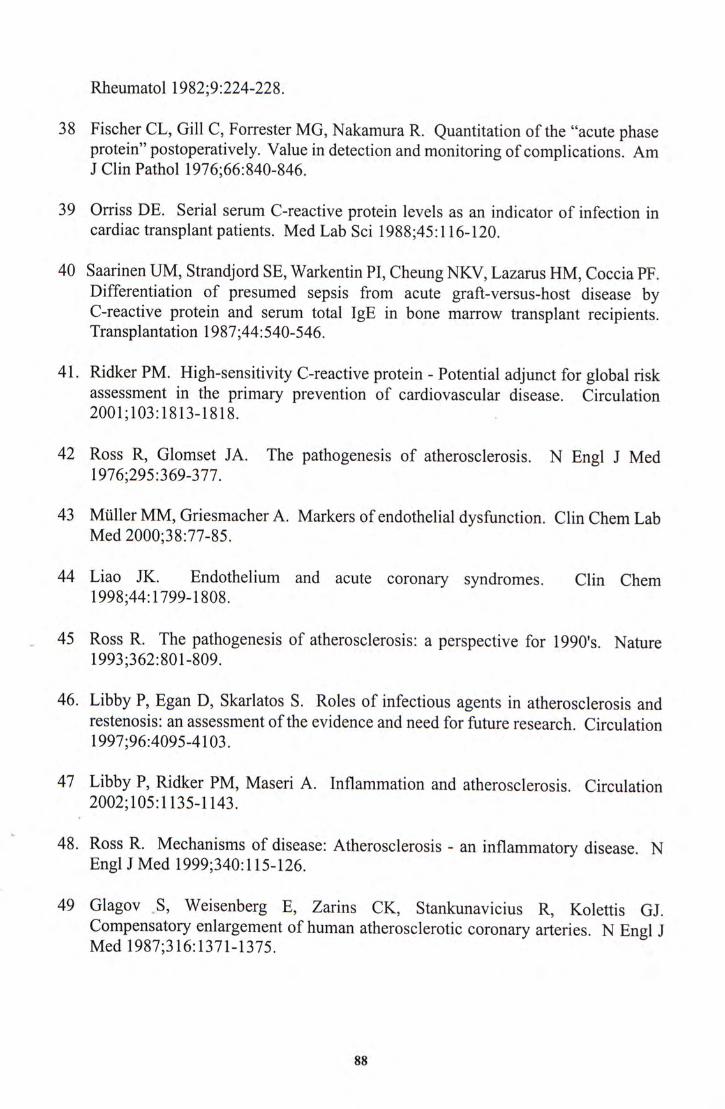
Rheumatol 1982;9:224-228.
38 Fischer CL, Gill C,Forrester MG, Nakamura R. Quantitation of the "acute phase protein" postoperatively. Value in detection and monitoring of complications. Am J Clin Pathol 1976;66:840-846.
39 Orriss DE. Serial serum C-reactive protein levels as an indicator of infection in cardiac transplant patients. Med Lab Sci 1988;45:116-120.
40 Saarinen UM, Strandjord SE,Warkentin PI, Cheung NKV, Lazarus HM, Coccia PF. Differentiation of presumed sepsis from acute graft-versus-host disease by C-reactive protein and serum total IgE in bone marrow transplant recipients. Transplantation 1987;44:540-546.
41. Ridker PM. High-sensitivity C-reactive protein - Potential adjunct for global risk assessment in the primary prevention of cardiovascular disease. Circulation 2001;103:1813-1818.
42 Ross R, Glomset JA. The pathogenesis of atherosclerosis. N Engl J Med 1976;295:369-377.
43 Mtiller MM, Griesmacher A. Markers of endothelial dysfunction. Clin Chem Lab Med 2000;38:77-85.
44 Liao JK. Endothelium and acute coronary syndromes. Clin Chem 1998;44:1799-1808.
45 Ross R. The pathogenesis of atherosclerosis: a perspective for 1990's. Nature 1993;362:801-809.
46. Libby P,Egan D, Skarlatos S. Roles of infectious agents in atherosclerosis and restenosis: an assessment of the evidence and need for future research. Circulation 1997;96:4095-4103.
47 Libby P, Ridker PM, Maseri A. Inflammation and atherosclerosis. Circulation 2002;105:1135-1143.
48. Ross R. Mechanisms of disease: Atherosclerosis - an inflammatory disease. N Engl J Med 1999;340:115-126.
49 G l a g o v � S , Weisenberg E, Zarins CK, Stankunavicius R, Kolettis GJ. Compensatory enlargement of human atherosclerotic coronary arteries. N Engl J Med 1987;316:1371-1375.
88
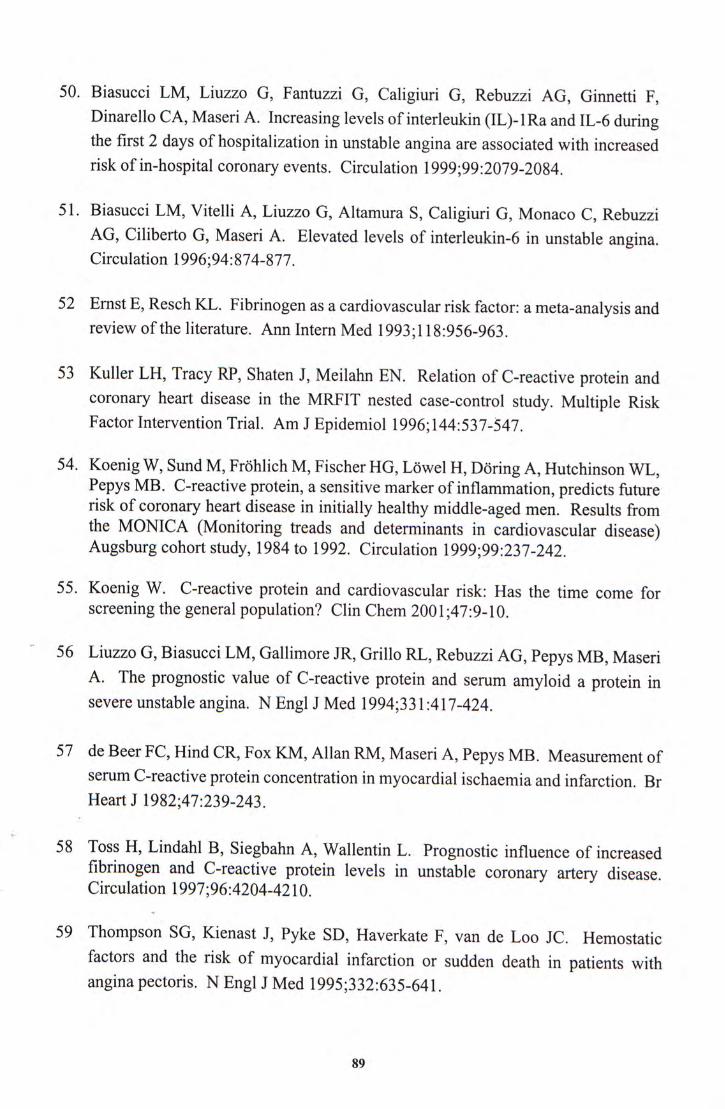
50. Biasucci LM, Liuzzo G, Fantuzzi G, Caligiuri G, Rebuzzi AG, Ginnetti F, Dinarello CA,Maseri A. Increasing levels of interleukin (IL)-lRa and IL-6 during the first 2 days of hospitalization in unstable angina are associated with increased risk of in-hospital coronary events. Circulation 1999;99:2079-2084.
51. Biasucci LM, Vitelli A, Liuzzo G, Altamura S, Caligiuri G, Monaco C, Rebuzzi AG, Ciliberto G,Maseri A. Elevated levels of interleukin-6 in unstable angina. Circulation 1996;94:874-877.
52 Ernst E, Resch KL. Fibrinogen as a cardiovascular risk factor: a meta-analysis and review of the literature. Ann Intern Med 1993;118:956-963.
53 Kuller LH, Tracy RP, Shaten J, Meilahn EN. Relation of C-reactive protein and coronary heart disease in the MRFIT nested case-control study. Multiple Risk Factor Intervention Trial. Am J Epidemiol 1996;144:537-547.
54. Koenig W, Sund M,Frohlich M,Fischer HG, Lowel H, Doring A, Hutchinson WL, Pepys MB. C-reactive protein, a sensitive marker of inflammation, predicts future risk of coronary heart disease in initially healthy middle-aged men. Results from the MONICA (Monitoring treads and determinants in cardiovascular disease) Augsburg cohort study, 1984 to 1992. Circulation 1999;99:237-242.
55. Koenig W. C-reactive protein and cardiovascular risk: Has the time come for screening the general population? Clin Chem 2001;47:9-10.
56 Liuzzo G, Biasucci LM, Gallimore JR, Grillo RL, Rebuzzi AG, Pepys MB, Maseri A. The prognostic value of C-reactive protein and serum amyloid a protein in severe unstable angina. N Engl J Med 1994;331:417-424.
57 de Beer FC,Hind CR, Fox KM, Allan RM, Maseri A, Pepys MB. Measurement of serum C-reactive protein concentration in myocardial ischaemia and infarction. Br Heart J 1982;47:239-243.
58 Toss H, Lindahl B, Siegbahn A, Wallentin L. Prognostic influence of increased fibrinogen and C-reactive protein levels in unstable coronary artery disease Circulation 1997;96:4204-4210.
59 Thompson SG,Kienast J, Pyke SD, Haverkate F, van de Loo JC. Hemostatic factors and the risk of myocardial infarction or sudden death in patients with angina pectoris. N Engl J Med 1995;332:635-641.
89
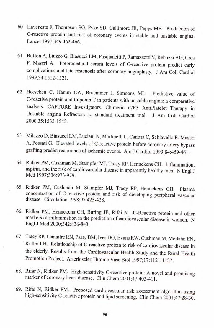
60 Haverkate F, Thompson SG, Pyke SD, Gallimore JR, Pepys MB. Production of C-reactive protein and risk of coronary events in stable and unstable angina. Lancet 1997;349:462-466.
61 Buffon A, Liuzzo G, Biasucci LM,Pasqualetti P, Ramazzotti V,Rebuzzi AG, Crea F,Maseri A. Preprocedural serum levels of C-reactive protein predict early complications and late restenosis after coronary angioplasty. J Am Coll Cardiol 1999;34:1512-1521.
62 Heeschen C,Hamm CW, Bruemmer J,Simoons ML. Predictive value of C-reactive protein and troponin T in patients with unstable angina: a comparative analysis. CAPTURE Investigators. Chimeric c7E3 Antiplatelet Therapy in Unstable angina Refractory to standard treatment trial. J Am Coll Cardiol 2000;35:1535-1542.
63 Milazzo D,Biasucci LM, Luciani N, Martinelli L, Canosa C, Schiavello R, Maseri A,Possati G. Elevated levels of C-reactive protein before coronary artery bypass grafting predict recurrence of ischemic events. Am J Cardiol 1999;84:459-461.
64. Ridker PM,Cushman M, Stampfer MJ, Tracy RP, Hennekens CH. Inflammation, aspirin,and the risk of cardiovascular disease in apparently healthy men N Engl J Med 1997;336:973-979.
65. Ridker PM, Cushman M,Stampfer MJ, Tracy RP, Hennekens CH. Plasma concentration of C-reactive protein and risk of developing peripheral vascular disease. Circulation 1998;97:425-428.
66. Ridker PM, Hennekens CH, Buring JE,Rifai N. C-Reactive protein and other markers of inflammation in the prediction of cardiovascular disease in women N Engl J Med 2000;342:836-843. ‘
67 Tracy RP, Lemaitre RN, Psaty BM,Ives DG, Evans RW, Cushman M, Meilahn EN, ‘Kuller LH. Relationship of C-reactive protein to risk of cardiovascular disease in the elderly. Results from the Cardiovascular Health Study and the Rural Health Promotion Project. Arterioscler Thromb Vase Biol 1997;17:1121-1127.
68. Rifar N,Ridker PM. High-sensitivity C-reactive protein: A novel and promising marker of coronary heart disease. Clin Chem 2001;47:403-411.
69. Rifai N,Ridker PM. Proposed cardiovascular risk assessment algorithm using high-sensitivity C-reactive protein and lipid screening. Clin Chem 2001;47:28-30.
90
70. Ridker PM, Glynn RJ, Hennekens CH. C-reactive protein adds to the predictive value of total and HDL cholesterol in determining risk of first myocardial infarction. Circulation 1998;97:2007-2011.
71 Ridker PM. Evaluating novel cardiovascular risk factors. Can we better predict heart attack? Ann Intern Med 1999;130:933-937.
72. Ridker PM, Rifai N, Pfeffer MA, Sacks FM, Moye LA, Goldman S,Flaker GC, Braunwald E. Inflammation, pravastatin, and the risk of coronary events after myocardial infarction in patients with average cholesterol levels Circulation 1998;98:839-844.
73. Ridker PM, Rifai N, Pfeffer MA, Sacks F, Braunwald E. Long-term effects of pravastatin on plasma concentration of C-reaction protein. Circulation 1999;100:230-235.
74. Jialal I, Stein D,Balis D, Grundy SM, Adams-Huet B,Devaraj S. Effect of hydroxymethyl glutaryl coenzyme A reductase inhibitor therapy on high sensitive C-reactive protein levels. Circulation 2001;103:1933-1935.
75. Ridker PM, Buring JE,Shih J, Matias M, Hennekens CH. Prospective study of C-reaction protein and the risk of future cardiovascular events among apparently healthy women. Circulation 1998;98:731-733.
76 Nilsson LA. Comparative testing of precipitation methods for quantitation of C-reactive protein in blood serum. Acta Pathol Microbiol Scand 1968;73:129-144.
77 Kindmark CO, Thorell JI. Quantitative determination of individual serum proteins by radio-electroimmunoassay and use of '^^I-labeled antibodies (application to C-reactive protein). Scand J Clin Lab Invest 1972;124:49-53.
78 Hansson LO, Lindquist L, Linne T,Sego E. Quantitation of C-reactive protein in cerebrospinal fluid and serum by zone immunoelectrophoresis assay (ZIA) J Immunol Methods 1987;100:191-195.
79 Otsuji S,Shibata H,Umeda M. Turbidimetric immunoassay of serum C-reactive protein. Clin Chem 1982;28:2121-2124.
80 Kapmeyer W,Grenner G, Becker W. The nephelometric determination of C-reactive protein, antistreptolysin 0,and rheumatoid factors by latex agglutination test. Clin Chem 1983;29:1189 (Abstract).
81. Gill CW,Bush WS, Burleigh WM, Fischer CL. An evaluation of a C-reactive protein assay using a rate immunonephelometric procedure. Am J Clin Pathol 1981;75:50-55. •
91
82 Abe A, Kawade M,Seishima M, Makino K,Maeda S, Nakamura S. A highly sensitive enzyme linked immunosorbent assay (ELISA) of human C-reactive protein (CRP) and its level in cord blood serum. Clin Chem 1987;33:919-925.
83 Claus DR, Osmand AP, Gewurz H. Radioimmunoassay of human C-reactive protein and levels in normal sera. J Lab Clin Med 1976;87:120-128.
84 WHO Expert Committee on Biological Standardization, 37th report. WHO Technical Report Series 760. Geneva: WHO, 1987:21-22.
85 Ledue TB, Weiner DL, Sipe J, Poulin SE, Collins MF, Rifai N. Analytical evaluation of particle-enhanced immunonephelometric assays for C-reactive protein, serum amyloid A, and mannose binding protein in human serum. Ann Clin Biochem 1998;35:745-753.
86. Rifai N, Tracy RP, Ridker PM. Clinical efficacy of an automated high-sensitivity C-reactive protein assay. Clin Chem 1999;45:2136-2141.
87. Roberts WL, Sedrick R, Moulton L, Spencer A,Rifai N. Evaluation of four automated high-sensitivity C-reactive protein methods: Implications for clinical and epidemiological applications. Clin Chem 2000;46:461-468.
88. Eda S, Kaufmann J, Roos W, Pool S. Development of a new microparticle enhanced turbidimetric assay for C-reactive protein with superior features in analytical sensitivity and dynamic range. J Clin Lab Anal 1998;12:137-144.
89. Roberts WL, Moulton L, Law TC, Farrow G, Cooper-Anderson M,Savory J, Rifar N. Evaluation of nine automated high-sensitivity C-reactive protein methods: Implications for clinical and epidemiological applications. Part 2. Clin Chem 2001;47:418-425.
90. Whicher JT, Chambers RE, Higginson J, Nashef L, Higgins PG. Acute phase response of serum amyloid A protein and C-reactive protein to the common cold and influenza. J Clin Pathol 1985;38:312-316.
91. Strachan AF, Noakes TD, Kotzenberg G, Nel AE, de Beer FC. C-reactive protein ‘concentrations during long distance running. Br Med J 1984;289:1249-1254.
92. Macy EM, Haye TE,Tracy RP. Variability in the measurement of C-reactive protein in healthy subjects: Implications for reference intervals and epidemiologic applications. Clin Chem 1997;43:52-58.
93. Ockene IS, Matthews CE, Rifai N,Ridker PM, Reed G, Stanek E. Variability and classification accuracy of serial high-sensitivity C-reactive protein measurements in healthy adults. Clin Chem 2001;47:444-450.
92
94 Wong M, Tei C, Shah PM. Sensitivity and specificity of two-dimensional echocardiography in the detection of valvular calcification. Chest 1983;84:423-427.
95 Friedewald WT, Levy RI, Fredrickson DS. Estimation of the concentration of low-density lipoprotein cholesterol in plasma without use of the preparative ultracentrifuge. Clin Chem 1972;18:499-502.
96 Hunt KJ, Evans GW, Folsom AR, Sharrett AR, Chambless LE, Tegeler CH, Heiss G. Acoustic shadowing on B-mode ultrasound of the carotid artery predicts ischemic stroke: the Atherosclerosis Risk in Communities (ARJC) study. Stroke 2001;32:1120-1126.
97 Yu H, Rifai N. High-sensitivity C-reactive protein and atherosclerosis: from theory to therapy. Clin Biochem 2000;33:601-610.
98 Harnett JD, Foley RN, Kent GM, Barre PE, Murray D, Parfrey PS. Congestive heart failure in dialysis patients: Prevalence, incidence, prognosis and risk factors. Kidney Int 1995;47:884-890.
99 Blackburn R, Giral P,Bruckert E, Andre JM, Gonbert S, Bernard M, Chapman MJ, Turpin G. Elevated C-reactive protein constitutes an independent predictor of advanced carotid plaques in dyslipidemic subjects. Arterioscler Thromb Vase Biol 2001;21:1962-1968.
100 Hashimoto H, Kitagawa K, Hougaku H, ShimizuY, Sakaguchi M, Nagai Y, lyama S, Yamanishi H, Matsumoto M, Hori M. C-reactive protein is an independent predictor of the rate of increase in early carotid atherosclerosis. Circulation 2001;104:63-67.
93

• . � ‘>• »
; V ‘ _
‘ . . . V
. . . V • .1 ‘
’ -
• . . • . - • .
-F • . . _ V.
i ••
.‘:,,‘:.:.->::'::,:. •.
. ' � • . . . • • •• V . • »
二 • , . . . : . . : , “ . •
. . . . -.
. . . . � . - :
... . . . .
. - . - ; . 、 . - ; • •-- . • ‘ -
.•,. .... . ’•• : ::
.’..... 乂 ’
• . . ... r:”iK .:..:
• . . . . . . • .
. -V-. - • ’ • -,.:.:. •••
� • ‘ . . • S ' i '� • ^ 、么、广.

CUHK L i b r a r i e s
D D 3 T 3 3 3 S S