Embed Size (px)
Citation preview
Serveur Académique Lausannois SERVAL serval.unil.ch
Author Manuscript Faculty of Biology and Medicine Publication
This paper has been peer-reviewed but dos not include the final publisher
proof-corrections or journal pagination.
Published in final edited form as:
In the absence of a copyright statement, users should assume that standard copyright protection applies, unless the article contains
an explicit statement to the contrary. In case of doubt, contact the journal publisher to verify the copyright status of an article.
Title: Hematopoietic stem cell function and survival depend on c-Myc
and N-Myc activity.
Authors: Laurenti E, Varnum-Finney B, Wilson A, Ferrero I, Blanco-
Bose WE, Ehninger A, Knoepfler PS, Cheng PF, MacDonald HR,
Eisenman RN, Bernstein ID, Trumpp A
Journal: Cell stem cell
Year: 2008 Dec 4
Volume: 3
Issue: 6
Pages: 611-24
DOI: 10.1016/j.stem.2008.09.005
Hematopoietic Stem Cell Function and Survival Depend on c-Mycand N-Myc Activity
Elisa Laurenti1, Barbara Varnum-Finney3, Anne Wilson4, Isabel Ferrero4, William E. Blanco-Bose1, Armin Ehninger1, Paul S. Knoepfler5,6,7, Pei-Feng Cheng5, H. Robson MacDonald4,Robert N. Eisenman5, Irwin D. Bernstein3, and Andreas Trumpp1,2,*
1 Ecole Polytechnique Fédérale de Lausanne (EPFL), ISREC, Swiss Institute for Experimental CancerResearch, School of Life Science, CH-1066 Epalinges, Switzerland 2 Division of Cell Biology, DeutschesKrebsforschungszentrum (DKFZ), DKFZ-ZMBH Alliance, Im Neuenheimer Feld 280, D-69120 Heidelberg,Germany 3 Clinical Research Division, Fred Hutchinson Cancer Research Center, 1100 Fairview AvenueNorth, Seattle, WA 98109, USA 4 Ludwig Institute for Cancer Research Ltd., Lausanne Branch, Universityof Lausanne, CH-1066 Epalinges, Switzerland 5 Division of Basic Sciences, Fred Hutchinson CancerResearch Center, MS A2-025, POB 19024, Seattle, WA 98109-1024, USA
SUMMARYMyc activity is emerging as a key element in acquisition and maintenance of stem cell properties.We have previously shown that c-Myc deficiency results in accumulation of defective hematopoieticstem cells (HSCs) due to niche-dependent differentiation defects. Here we report that immature HSCscoexpress c-myc and N-myc mRNA at similar levels. Although conditional deletion of N-myc in thebone marrow does not affect hematopoiesis, combined deficiency of c-Myc and N-Myc (dKO) resultsin pancytopenia and rapid lethality. Interestingly, proliferation of HSCs depends on both myc genesduring homeostasis, but is c-Myc/N-Myc independent during bone marrow repair after injury.Strikingly, while most dKO hematopoietic cells undergo apoptosis, only self-renewing HSCsaccumulate the cytotoxic molecule GranzymeB, normally employed by the innate immune system,thereby revealing an unexpected mechanism of stem cell apoptosis. Collectively, Myc activity (c-Myc and N-Myc) controls crucial aspects of HSC function including proliferation, differentiation,and survival.
INTRODUCTIONLong-term hematopoietic stem cells (HSCs) are defined by their unique ability to self-renewwhile concomitantly differentiating to generate all mature blood cell types. HSCs possess theclonal capacity to provide life-long reconstitution of all hematopoietic lineages upontransplantation into lethally irradiated mice (Purton and Scadden, 2007; Shizuru et al., 2005;Till and Mc Culloch, 1961). To therapeutically take advantage of the exclusive regenerativeproperties of LT-HSCs, it is fundamental to elucidate the mechanisms by which these cellsmaintain the balance between self-renewal and differentiation. The advent of geneticallyengineered mice has facilitated identification of several molecules that play a role in stem cellmaintenance and function. For example, mice lacking signaling components such as TPO-
*Correspondence: [email protected] address: Department of Cell Biology and Human Anatomy, University of California School of Medicine, Davis, CA 95616-8643,USA7Present address: Institute for Pediatric Regenerative Medicine, Shriners Hospital for Children Northern California, 2425 StocktonBoulevard, Sacramento, CA 95817, USA
NIH Public AccessAuthor ManuscriptCell Stem Cell. Author manuscript; available in PMC 2009 December 4.
Published in final edited form as:Cell Stem Cell. 2008 December 4; 3(6): 611–624. doi:10.1016/j.stem.2008.09.005.
NIH
-PA Author Manuscript
NIH
-PA Author Manuscript
NIH
-PA Author Manuscript
cMPL (Qian et al., 2007; Yoshihara et al., 2007), Ang1-Tie2 (Arai et al., 2004), or SCF-cKit(Thoren et al., 2008), or nuclear regulators such as FOXO proteins (Tothova et al., 2007) orBmi-1 (Park et al., 2003), have impaired HSC function. Most of these molecules participate incell-cycle control, regulation of apoptosis, and response to oxidative stress, or interact with thesurrounding niche environment (Orford and Scadden, 2008; Wilson and Trumpp, 2006). InHSCs, these processes are tightly regulated, most likely through distinct mechanisms duringhomeostasis or under stress conditions. For instance, myeloablative chemotherapy transientlyinduces cell-cycle and cell-surface marker changes in HSCs, allowing them to enter anactivated state in order to re-establish normal hematopoiesis (Randall and Weissman, 1997;Venezia et al., 2004).
The Myc family members, c-Myc, N-Myc, and L-Myc (DePinho et al., 1987), encode basichelix-loop-helix leucine zipper transcription factors that are potent oncogenes. Myc proteinshave been implicated in many biological processes, such as proliferation, cellular growth,angiogenesis, apoptosis, differentiation, and regulation of chromatin structure (Eisenman,2001; Knoepfler, 2007; Murphy et al., 2005; Nilsson and Cleveland, 2003). Moreover, c-Myc(or N-Myc) has been identified as an important element in the induced reprogramming of adultfibroblasts into embryonic stem cells like iPS cells (Knoepfler, 2008; Lewitzky and Yamanaka,2007). While many studies have shed light on the mechanisms by which overexpression ofMyc promotes tumorigenesis (Pelengaris et al., 2002), its physiological role still remainselusive in many tissues in vivo. While L-Myc appears dispensable during development (Hattonet al., 1996), deletion of c-myc or N-myc leads to embryonic lethality (Charron et al., 1992;Dubois et al., 2008; Trumpp et al., 2001). We have previously reported that deleting c-myc inthe adult bone marrow (BM) via the inducible MxCre-loxP system unexpectedly results in anaccumulation of functionally defective HSCs (Wilson et al., 2004). In the absence of c-Myc,differentiation of these cells into more committed progenitors is inhibited as they upregulatea number of adhesion molecules that anchor them in the niche, thus preventing theirdifferentiation. Surprisingly, and in contrast to differentiated progenitors, c-Myc-deficientHSCs can still divide, and their proliferation capacity is not affected. Since N-myc is expressedin normal and c-Myc-deficient HSCs (Ivanova et al., 2002; Wilson et al., 2004), we havegenetically addressed the individual role of N-Myc, as well as that of c-Myc and N-Myctogether, for HSC self-renewal, survival, and differentiation.
RESULTSExpression of c-myc, N-myc, and L-myc in Hematopoietic Lineages
To address whether, in addition to c-Myc, other Myc family members are involved incontrolling HSC and progenitor function, the expression levels of c-myc, N-myc, and L-mycwere determined by qRT-PCR in various stem and progenitor populations isolated by FACS(Figure 1A). Identical amplification efficiencies for all three genes were established to not onlyquantitatively determine the expression of each gene, but also to compare the relative amountsof each transcript expressed in each individual cell type. Total myc mRNA levels (c- + N- +L-myc) were found to be highest in the most immature HSCs and progressively decreasedduring differentiation (Figure 1A). While c-myc and N-myc are detected in most progenitorsubsets, L-myc only modestly contributes to the general Myc activity in CLPs, megakaryocytes,and macrophages (3%–5% of total Myc) and is not expressed in any stem/progenitor cells. Thehighest expression levels of total myc transcripts are found in the most immature HSCs(CD34− and CD34+LT-HSCs), where c-myc and N-myc contribute approximately equalamounts. During the initial differentiation step into ST-HSCs, the total myc level drops byapproximately 30% but then remains constant in more committed progenitor stages (MPPs andLMPPs). This is mostly due to the progressive decrease of N-Myc transcripts while c-MycmRNA expression remains stable. Surprisingly, despite c-Myc being essential for proliferation
Laurenti et al. Page 2
Cell Stem Cell. Author manuscript; available in PMC 2009 December 4.
NIH
-PA Author Manuscript
NIH
-PA Author Manuscript
NIH
-PA Author Manuscript
of most differentiated hematopoietic cell types, it is only expressed at modest levels in maturecell types (Figures 1A and 1B). These data show that expression of N-myc is essentiallyrestricted to HSCs, thus raising the possibility that in addition to c-Myc, N-Myc may also playan important role in HSC function.
N-Myc Is Not Essential for Steady-State HematopoiesisTo investigate the role of N-Myc during hematopoiesis, adult MxCre;N-mycflox/flox mice weregenerated and treated with pIpC to conditionally eliminate this gene in all hematopoietic celltypes, including HSCs (Figure 1C) (Knoepfler et al., 2002; Kühn et al., 1995). Five weekspostdeletion of N-myc, total numbers of cells in the BM, thymus, or spleen of mutant micewere indistinguishable from those of control littermates (Figure 1D). In addition, similarproportions of all major BM populations (Figure 1F), including stem/progenitor cell types suchas LSK, LSK-SP (Side Population), or LSK-SLAM (LSK CD150+CD48−) populations (Figure1E) were observed in control and mutant mice. While deletion of N-myc was complete (FigureS1A available online), no significant compensatory upregulation of either c-myc or L-myc wasobserved (data not shown). To address whether N-Myc-deficient HSCs are functional, limitingdilution assays in a competitive chimera setting were performed. N-Myc-deficient BM cellswere indistinguishable from control BM cells in their capacity to engraft (>2% donorcontribution after 21 weeks) (Table S2). In summary, these data suggest that deletion of N-myc alone has no major impact on HSC numbers and does not affect the capacity to maintainadult hematopoiesis.
Simultaneous Elimination of Both c-Myc and N-Myc Leads to Rapid Hematopoietic Failure,Including HSC Loss
We have previously shown that c-Myc-deficient HSCs have a severe niche-dependentdifferentiation defect, indicating that c-Myc activity is required for proper HSC function.Interestingly, c-Myc-deficient HSCs proliferate normally, suggesting that the remaining N-Myc expression may be sufficient to maintain self-renewing divisions in c-Myc null HSCs.Moreover, the fact that both c-myc and N-myc transcripts are equally expressed in HSCs raisesthe possibility that both genes control HSC self-renewal and possibly other HSC functions ina cooperative or additive manner. To genetically address this hypothesis, MxCre;c-mycflox/flox;N-mycflox/flox double knockout (dKO) mice were generated (Trumpp et al., 2001;Knoepfler et al., 2002). Treatment of these mice with pIpC resulted in the loss of both c-mycand N-myc transcripts from day 4 onward. Although deletion of both c-myc and N-myc wasvery efficient (90% and 98%, respectively) as measured by qRT-PCR, no significant (p = 0.28)compensatory upregulation of L-Myc was observed in dKO BM subsets (Figure S1B).Simultaneous deletion of c-myc and N-myc rapidly induced severe pancytopenia (much fasterthan after deletion of c-Myc alone; Wilson et al. [2004]), as dKO mice died as early as 12 daysafter Cre induction (Figure 2A). Just prior to death, dKO mice were severely anemic, and theirbone cavities were almost completely devoid of hematopoietic cells, further confirmingcomplete BM failure upon loss of both genes (Figure 2C). Total BM cellularity rapidlydecreased (>90%) 2 weeks postdeletion (Figure 2B). All major BM cell types, includingmyeloid, lymphoid, and erythroid cells, were progressively lost, albeit with distinct kinetics(Figure 2B and Figure S2A). Cell counts in peripheral hematopoietic organs, such as spleenand peripheral blood, were also strongly reduced in dKO animals (Figures S2B and S2C).Collectively, these results show that simultaneous elimination of c-Myc and N-Myc in the adultBM results in rapid hematopoietic failure and subsequent lethality 2 weeks postdeletion.
At the stem/progenitor level, the LSK compartment was no longer recognizable in dKO miceas early as day 4 postdeletion (Figure 2D, left panels). Instead, Lin−Sca1+cKit− cellsaccumulated, suggesting that c-Kit expression is lost in mutant LSKs. Downregulation of c-Kit cell surface expression on functional HSCs has also been reported in response to 5-FU-
Laurenti et al. Page 3
Cell Stem Cell. Author manuscript; available in PMC 2009 December 4.
NIH
-PA Author Manuscript
NIH
-PA Author Manuscript
NIH
-PA Author Manuscript
induced myelosuppression. Such severe loss of myeloid cells is thought to activate dormantHSCs, inducing them to cycle and generate progenitors and new mature cells to quicklyreachieve homeostasis (Randall and Weissman, 1997; Wilson et al., 2008). Since simultaneousloss of c-Myc and N-Myc results in a similar reduction of BM cells to that observed aftertreatment with 5-FU, HSCs could still be present in dKO mice, but they would have alsodownregulated surface expression of the cKit receptor by an analogous mechanism. dKO BMwas thus reanalyzed using the SLAM receptors CD150 and CD48 (Kiel et al., 2005). As allLin−Sca1+CD150+ cells in control mice are also cKit+, HSCs can be phenotypically identifiedwithout cKit by analyzing the Lin−Sca1+CD150+CD48− population (hereafter called SLAM-HSC). Around 40% to 60% of SLAM-HSCs are still present in dKO mice shortly after deletion.However, their number steadily declines over time, and almost all SLAM-HSCs are lost byday 8 postdeletion (Figure 2E). Relative and absolute numbers of SLAM-HSCs in the spleenalso decreased significantly (Figure S2D and data not shown), indicating that there was nosignificant mobilization of dKO HSCs from the BM to this organ and no subsequentextramedullary hematopoiesis. In summary, these results demonstrate that simultaneousdeletion of both c-myc and N-myc results not only in rapid hematopoietic failure, but also inthe complete loss of phenotypic HSCs.
c-Myc;N-Myc Double-Deficient HSCs and Differentiated Cells Display Aberrant Proliferationand Enter an Apoptosis Program
To determine the effects of total loss of Myc activity on cellular proliferation, the cell-cyclestatus of all major hematopoietic populations, including HSCs, was determined in dKO animals6 days after pIpC injection. As expected, a highly significant reduction of cells in the S/G2/Mphases of the cell cycle was observed in the dKO myelomonocytic population (~3-fold; p <0.01; Figure 3A). In contrast, dKO SLAM-HSCs cycled significantly more than control HSCs(~4-fold; p < 0.001; Figure 3B). To ensure that these unexpectedly proliferating HSCs had notsimply escaped deletion of either c-myc or N-myc, the proliferative BrdU+ populations of HSCs(Lin−Sca1+CD150+BrdU+) from control and dKO mice were isolated by FACS and analyzedby immunofluorescence for the expression of c-Myc and N-Myc proteins. While abundantexpression of both c-Myc and N-Myc was observed in more than 80% of control cells, HSCsisolated from dKO mice showed only very low or undetectable levels of c-Myc or N-Mycprotein, confirming that MxCre-mediated deletion of both genes was highly efficient. Thesedata demonstrate that, surprisingly, dKO SLAM-HSCs are able to proliferate even in theabsence of both c-Myc and N-Myc (Figures 3C and 3D).
We also analyzed whether an increase in the relative amount of apoptosis occurred upondeletion of c-myc and N-myc by performing TUNEL and AnnexinV analyses ex vivo and afterin vitro culture. dKO granulocytes, erythroblasts, and all B cell stages except the most matureBM B cells (IgM+) showed a substantial increase in apoptosis from day 4 postdeletion onward,with a further increase by day 6 (Figures 3E and 3F and data not shown). Moreover, increasedcell death was also observed in the dKO LT-HSC (Lin−Sca1+CD150+CD48−CD34−)population as early as 4 days after the first pIpC injection (Figure 3G). Apoptosis was alsoinduced in cultured mutant progenitors upon MxCre induction by IFNα, indicating that celldeath induced by the loss of c-Myc and N-Myc is likely to be cell-autonomous and is not aresult of putative alterations of the stem cell environment (Figure S3A). In addition to TUNELassays, AnnexinV/7AAD analysis independently confirmed these results (data not shown). Insummary, most of the major hematopoietic lineages, including HSCs, undergo apoptosis withina few days of the simultaneous loss of c-myc and N-myc. This not only provides a plausibleexplanation for the rapid general BM failure, but also reveals that while the presence of eitherN-Myc or c-Myc alone ensures survival, hematopoietic cells lacking both proteins are quicklylost due to cell death.
Laurenti et al. Page 4
Cell Stem Cell. Author manuscript; available in PMC 2009 December 4.
NIH
-PA Author Manuscript
NIH
-PA Author Manuscript
NIH
-PA Author Manuscript
Bone Marrow Failure upon Loss of c-Myc and N-Myc Is Hematopoietic Cell AutonomousThe MxCre transgene induces significant deletion also outside the hematopoietic system,including the BM stromal compartment and the liver (Kühn et al., 1995). Nevertheless, c-myc and N-myc deletion in nonhematopoietic cell types does not contribute to the severephenotype observed, as all the defects described above for dKO mice could be qualitativelyand quantitatively recapitulated in noncompetitive BM transplantation experiments in whichthe c-myc and N-myc genes were deleted after reconstitution (Figure S4A).
To determine the functional capacity of dKO HSCs in a competitive situation, competitive BMtransplantation experiments were performed (Figure 4A). CD45.2+ recipient mice were lethallyirradiated and reconstituted with either a 1:1 or a 3:1 mixture of experimental, but not yetdeleted, BM cells (MxCre; c-mycflox/flox;N-mycflox/flox;CD45.1+/2+) together with nondeletablecontrol (c-mycflox/flox;N-mycflox/flox;CD45.2+) cells (mixed chimeras). After stablereconstitution was established, deletion of both myc genes was induced by pIpC and the BManalyzed 14 or 21 days later (Figure 4A). In this set-up, the nondeletable control (“wild-type”)cells maintain hematopoiesis even if the dKO cells are lost after pIpC treatment and, therefore,prevent the development of a severe BM phenotype. This strategy conveniently permitsdifferentiation between cell autonomous effects due to loss of c-Myc and N-Myc, and anyeffects secondary to the severe BM cytopenia that occurs in dKO mice and non-mixedchimeras. As expected, these competitive chimeric mice did not appear anemic upon deletionand had normal total BM cell numbers (data not shown). However, dKO donor cells werespecifically lost as their relative chimerism decreased by 70% on day 14 and 90% 21 days afterthe first pIpC injection (Figure 4B). Furthermore, dKO donor cells proliferated significantlyless compared to control cells and displayed increased apoptosis (Figure S5), in agreementwith the results observed in dKO mice and nonmixed chimeras. In summary, these data showthat dKO BM cells are lost even in a wild-type microenvironment (competitive andnoncompetitive chimeras) and are unable to compete with wild-type hematopoietic cells in asteady-state (mixed BM chimera) or cytopenic (dKO and non-competitive transplantation)situation.
Role of Myc Proteins in HSC Proliferation and Survival during Homeostasis and StressResponse
In contrast to dKO mice and nonmixed chimeras, no downregulation of cKit cell surfaceexpression was observed in dKO SLAM-HSCs in mixed chimeras (Figure 4C). Since theseanimals are not anemic, these data confirm that loss of cKit expression in dKO mutants is asecondary consequence of the BM hypoplasia similar to what is observed after 5-FU-inducedBM ablation (Randall and Weissman, 1997) and is not a direct effect of Myc activity on c-Kitexpression or cell-surface localization. Importantly, significant numbers of dKO SLAM-HSCswere still present in competitive chimeras. In comparison to their initial numbers beforedeletion, the total number of dKO SLAM-HSCs (LSKCD150+CD48−) had only halved by day14 after deletion and only further decreased to 25% by day 21 (Figure 4E), a rather modestreduction compared to dKO mutants where all HSCs are lost within 8 days (Figure 2E).Analysis of the proportion of apoptotic cells in mixed chimeras revealed that in contrast toLin− progenitors in which apoptotic rates are normal, dKO HSCs displayed a 2.5-fold increasein cell death compared to control cells (Figure 4F), highly similar to the situation in dKO mutantmice (Figure 3G). However, in striking contrast to HSCs in dKO mice, which show increasedproliferation, the percentage of actively cycling dKO SLAM-HSCs (%S/G2/M) in mixed BMchimeras was strongly reduced compared to controls (~3-fold; p < 0.001; Figure 4G). Theseresults suggest that c-Myc and N-Myc are indeed critical for the proliferation of HSCs, butonly during homeostasis (mixed BM chimeras) and not in situations of severe BM stress (dKOanimals). This result supports the notion that Myc-dependent and Myc-independent HSCproliferation programs can be engaged during homeostasis and repair.
Laurenti et al. Page 5
Cell Stem Cell. Author manuscript; available in PMC 2009 December 4.
NIH
-PA Author Manuscript
NIH
-PA Author Manuscript
NIH
-PA Author Manuscript
Proliferating Cells with HSC Phenotype Are More Dependent on Myc Activity Than QuiescentHSCs
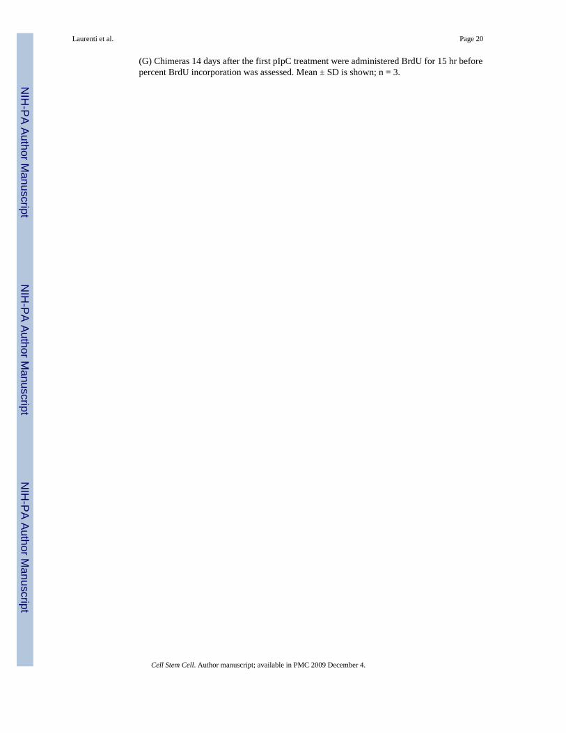
To investigate whether c-Myc- and N-Myc-deficient HSCs retain any long-term repopulationability, serial transplantations were performed. BM from the primary competitive chimerasdescribed above was harvested 14 days after pIpC treatment and transferred into lethallyirradiated (CD45.2+) recipients and analyzed 12 weeks later (Figures 5A and 5B). As expected,no contribution from dKO-deficient cells to either the myeloid or erythroid compartments wasobserved (Figure 5B and data not shown), showing that no stem cell activity is retained in dKOSLAM-HSCs. Nevertheless, dKO SLAM-HSCs were still present in secondary recipientsalthough their number had decreased about 6-fold compared to that at the time of transfer(Figure 5C). These results demonstrate that although HSCs lacking c-myc and N-myc can beserially transplanted, they do not give rise to differentiated progeny and, thus, have lost one ofthe critical features of stem cells.
The persistence of dKO dormant/quiescent HSCs indicates that these cells are less susceptibleto the loss of both myc genes compared to actively proliferating HSCs. To test this hypothesis,a “label-retaining cell” experiment (LRC; Arai et al., 2004; Wilson et al., 2008) that permitsdistinction between quiescent and self-renewing stem cells, was performed on mixed chimerasto monitor the outcome of c-Myc/N-Myc deletion on long term dormant HSCs while avoidingputative cytopenia-induced effects (Figure 5D). After elimination of both myc genes, about60% of the self-renewing SLAM-HSCs (BrdU−), but none of the dKO LRC SLAM-HSCs,were lost 14 days after the first pIpC injection (Figure 5E). These results indicate that duringhomeostasis, deletion of both c-myc and N-myc rather modestly, if at all, impairs maintenanceof the quiescent, and therefore most immature, HSCs but strongly affects self-renewal andsurvival capacities of activated HSCs. Thus, Myc activity becomes critical during the activationprocess promoting quiescent HSCs into a self-renewal mode.
Myc Depletion Results in Upregulation of Granzyme B Expression in HSCsHSC survival can be increased by expression of the antiapoptotic factor BCL-2 (Domen andWeissman, 2000) and can be strongly inhibited by an increase in Reactive Oxygen Species(ROS) (Ito et al., 2006; Tothova et al., 2007). While overexpression of BCL-2 (via the H2K-BCL-2 transgene) rescued apoptosis in dKO B lymphocytes, it did not diminish the apoptosisrate of SLAM-HSCs, nor did it rescue their rapid decline in numbers (Figure 6A and data notshown). To test whether the apoptosis induced upon loss of c-Myc and N-Myc in HSCs ismediated by upregulation of ROS, the intracellular concentration of ROS in dKO SLAM-HSCswas determined by DCF-DA staining. However, no significant differences could be detectedbetween control and dKO SLAM-HSCs (Figure 6B), indicating that apoptotic cell death ofdKO HSCs is not mediated by an increase in ROS.
To elucidate the mechanisms underlying apoptosis of dKO HSCs, the global gene expressionprofiles of FACS-purified SLAM-HSCs and committed progenitors isolated from control anddKO mice were established using Affymetrix arrays (Figure 6C). As expected, c-myc and N-myc transcripts were strongly downregulated while L-myc expression was not significantlychanged, in agreement with the data obtained by qRT-PCR (Figure S1B). In addition, dKOHSCs showed higher mRNA levels of the gene encoding the CDK inhibitor p21CIP, which isknown to be repressed by c-Myc (Oskarsson et al., 2006; Wu et al., 2003). An HSC signaturewas generated by comparing the global gene expression profiles of wild-type LT-HSCs andprogenitors. The expression of genes present in this wild-type HSC signature was onlymarginally changed in dKO-SLAM-HSCs, indicating that no significant loss of stem cellidentity occurs upon loss of Myc activity (data not shown). This is consistent with our findingthat the most quiescent dKO HSCs are maintained. Interestingly, the expression of 31 genesencoding ribosomal proteins was significantly downregulated (Figure S3), suggesting that
Laurenti et al. Page 6
Cell Stem Cell. Author manuscript; available in PMC 2009 December 4.
NIH
-PA Author Manuscript
NIH
-PA Author Manuscript
NIH
-PA Author Manuscript
ribosome biogenesis is strongly inhibited as a result of c-myc and N-myc gene loss. Intriguingly,the most upregulated transcript upon loss of both Myc family members was the gene encodingfor Granzyme B (GrB, 150-fold and 130-fold as determined by microarray and qRT-PCRanalysis, respectively; Figure 6C and Figure S7A), a highly potent cytotoxic molecule. Theserine-protease Granzyme B plays a key role in the granule-exocytosis pathway, which is usedby activated cytotoxic T lymphocytes (CTLs) and natural killer (NK) cells to eliminate cellsinfected with intracellular pathogens or tumor cells (Russell and Ley, 2002). Normal SLAM-HSCs do not express GrB as determined by intracellular FACS staining (Figures 6D and 6E;Revell et al., 2005). In contrast, most dKO SLAM-HSCs show highly elevated levels ofintracellular GrB protein (Figures 6D and 6E). Strikingly, GrB upregulation upon c-myc andN-myc deletion is specific to HSCs since no increased GrB could be detected in any other dKOhematopoietic cell type (Figure 6F). Importantly, GrB is also upregulated in dKO HSCs presentin mixed chimeras (Figure 6G, left panel), and deletion of both Myc genes in culturedprogenitors in vitro also resulted in the upregulation of intracellular GrB protein (Figure 6G,right panel), indicating a cell-autonomous effect of Myc deficiency on GrB production.
Processing of an N-terminal propeptide by the protease cathepsin C (or DPPI) is required forthe proteolytical activity of GrB (Caputo et al., 1993; Pham and Ley, 1999). Cathepsin Ctranscripts are significantly upregulated (2.5-fold) in dKO SLAM-HSCs, while the expressionlevel of the principal GrB inhibitor, Serine Protease Inhibitor 6 (Spi-6), is unchanged (Figure6C and Figure S7A). Several groups have recently reported GrB-leakage-induced cell deathin NK cells and CTLs, in which functionally active GrB escapes into the cytoplasm, thusprovoking apoptosis of the GrB producing cell itself. The major determinant of suchendogenous GrB-mediated killing seems to be an excess of free active GrB over its cytoplasmicinhibitor Spi-6 (Ida et al., 2003; Laforge et al., 2006; Phillips et al., 2004; Zhang et al., 2006),which correlates with diffuse cytoplasmic localization of the GrB protein. In agreement withthis notion, in the vast majority of dKO progenitor cells (70.3% ± 0.4), GrB protein is locatedthroughout the cytoplasm (diffuse pattern, Figure 6H, bottom panels, Figure S7B), in sharpcontrast to the specifically localized and punctuate pattern observed in most (85.1% ± 2.0)activated NK cells (Figure 6H, top right panel; Figure S7B). All conditions for cell-autonomouskilling by GrB are thus met in dKO HSCs. To assess whether GrB+ dKO HSCs are indeeddying as a consequence of GrB upregulation, HSCs were simultaneously assayed for TUNELand GrB expression (Figures 6I–6K). Around 80% of dying dKO SLAM-HSCs were also GrB+ (Figure 6J). Moreover, the percentage of TUNEL+ cells among GrB+ dKO SLAM-HSCsincreased over time, from about 40% at day 4 to around 70% at day 6 post-pIpC injection(Figure 6K), indicating that GrB upregulation precedes HSC apoptosis. Taken together, ourresults suggest that Myc depletion results in the massive increase of catalytically active GrBin SLAM-HSCs. As GrB is one of the most toxic molecules known, we conclude that Mycactivity is required to ensure stem cell survival via repression of Granzyme B, thus identifyingan unexpected mechanism of HSC apoptosis.
DISCUSSIONIn this paper, we show that c-myc and N-myc are coexpressed in the most immaturehematopoietic stem-progenitor populations and provide genetic evidence that total Mycactivity (c-Myc, N-Myc, and L-Myc) controls HSC function at various levels (Figure 7). Wedemonstrate that c-Myc and N-Myc are required for HSC proliferation, metabolic growth,differentiation, long-term self-renewal activity, and survival of stem cells by inhibiting GrB-mediated apoptosis.
Laurenti et al. Page 7
Cell Stem Cell. Author manuscript; available in PMC 2009 December 4.
NIH
-PA Author Manuscript
NIH
-PA Author Manuscript
NIH
-PA Author Manuscript
c-myc and N-myc Are Coexpressed in HSCs, and Both Contribute to Stem Cell FunctionOur analysis of the precise expression pattern of the Myc family members throughouthematopoiesis has led to a number of important observations. First, no hematopoietic cell typeexpresses only N-myc or L-myc without c-myc. Consistent with the genetic data discussedbelow, this suggests that c-Myc provides the baseline Myc function in hematopoietic cells. Theadditional expression of either or both of the other two myc genes through their distinctregulatory regions is likely to fine-tune total Myc activity in these cell types. Second, thestriking coexpression of c-myc and N-myc in immature HSCs combined with the rapid decreaseof N-myc (but not c-myc) transcripts at the onset of HSC differentiation suggests that N-Mycplays a critical role in HSC function. However, when ablated in the presence of c-Myc, N-myc is dispensable for stem cell maintenance during steady-state conditions or following stressaccompanied by transplantation (Figure 7C and Table S2). In contrast, when N-myc is deletedin the absence of c-Myc (dKO situation), HSC proliferation, survival, and differentiation areseverely impaired (Figures 7D and 7E). As deletion of c-myc alone (Figure 7B) does not alterHSC proliferation and survival rates (Wilson et al., 2004), our dKO data reveal a role for N-myc expression in sustaining the survival and proliferation of c-myc null HSCs. Moreover, nocompensatory upregulation of one family member is observed upon deletion of the other,implying that when N-myc is deleted, the unchanged c-myc transcript levels are sufficient formaintaining all aspects of HSC maintenance. In addition, when c-myc is deleted, the normallyequivalent levels of N-myc transcripts can provide enough Myc activity to ensure HSC survivaland proliferation but are not sufficient to permit exit from the niche and differentiation pastthe ST-HSC stage (Wilson et al., 2004). How can we explain such a discrepancy? Gene-replacement studies in which the coding sequences and intervening intron of c-myc werereplaced by those of N-myc (NCR allele) exclude the possibility that the N-Myc proteinfunctionally differs from c-Myc, since mice homozygous for this NCR allele are viable andproduce normal proportions of all differentiated cell types (Malynn et al., 2000). Our ownanalysis of NCR BM confirms these data as no obvious differentiation defect in their HSC andprogenitor compartments was detectable (data not shown). Thus, N-Myc, if expressed in theexact pattern as endogenous c-Myc, can provide all c-Myc functions during steady-statehematopoiesis, including exit of HSCs from the niche. Nevertheless, it remains possible thatc-Myc and N-Myc activities are quantitatively different in stem cells as a number of studieshave reported that N-Myc transactivation properties are weaker than those of c-Myc (Cole andMcMahon, 1999; Malynn et al., 2000). Alternatively, differential posttranscriptional regulationof c-Myc and N-Myc degradation may result in a predominance of c-Myc over N-Myc at theprotein level in HSCs (Zhao et al., 2008). Further experiments will be required to address theseappealing aspects.
A third interesting observation is that total myc expression peaks in the most immature HSCsand decreases in progenitors and more mature cell types. Thus, absolute myc transcript levelspositively correlate with multipotency and self-renewal activity. These results are consistentwith reports suggesting Myc genes as stem cell markers (Bettess et al., 2005; Cartwright et al.,2005; Ivanova et al., 2002; Knoepfler et al., 2006; Ramalho-Santos et al., 2002). Moreover,although not absolutely required, c-Myc or N-Myc have been shown to participate inreprogramming tail-tip fibroblasts into iPS cells, multipotent cells highly similar to ESCs(Lewitzky and Yamanaka, 2007). The mechanism by which Myc activity promotes thisreprogramming remains hypothetical, but its positive effects on cell-cycle and metabolicgrowth, as well as its recently uncovered effect on chromatin structure, have been suggestedto be causally involved (Knoepfler, 2008). Our study confirms that Myc activity is critical forstem cell maintenance as total loss of Myc strongly affects both self-renewal division andsurvival of activated HSCs, suggesting that Myc expression is tightly regulated in order tobalance scheduled differentiation with maintenance of a rarely self-renewing stem cell pool.
Laurenti et al. Page 8
Cell Stem Cell. Author manuscript; available in PMC 2009 December 4.
NIH
-PA Author Manuscript
NIH
-PA Author Manuscript
NIH
-PA Author Manuscript
Interestingly, deletion of c-myc and N-myc in more committed progenitors (Lin−) severelydiminishes their proliferation capacity but does not compromise their survival, indicating thatin highly cycling progenitors (in contrast to HSCs), Myc principally controls proliferation rates.Similarly, most lineage committed cells (Lin+) are exquisitely sensitive to c-Myc levels as theyundergo G0 arrest after elimination of c-Myc (Trumpp et al., 2001; Wilson et al., 2004).Collectively, our data show that proliferation of most hematopoietic cell types, includinghomeostatic HSCs, is Myc dependent. Nonetheless, we also provide evidence that dKO HSCsretain their ability to rapidly cycle in response to injury signals. Indeed, simultaneous deletionof c-myc and N-myc in a noncompetitive setting causes severe pancytopenia due to cell-cycleexit and apoptosis of lineage committed cell types. As a consequence, dKO stem cells become“activated” in order to repair the injured BM (like 5-FU treated HSCs) (Randall and Weissman,1997; Venezia et al., 2004). This is accompanied by downregulation of c-Kit cell-surfaceexpression and enhanced proliferation (Figure 7D). Since this proliferative burst occurs withoutupregulation of the barely detectable L-myc transcripts, our data strongly suggest that situationsexist where hematopoietic cell cycling can occur in a Myc-independent manner. In agreementwith recent gene-profiling studies (Venezia et al., 2004), this observation implies that differentproliferation/self-renewal programs control HSC cycling during homeostasis or in an injuryresponse situation. Importantly, the injury-activated and proliferating dKO HSCs all undergoapoptosis, leading to the complete loss of all HSCs in the “straight” mutants. In contrast, incompetitive chimeras where no cytopenia (injury) develops, quiescent dKO HSCs aremaintained, suggesting that the loss of the Myc-mediated apoptotic response is linked to anactivated self-renewal program (Figure 7). In conclusion, myc genes appear to be both criticaland pleiotropic control elements regulating not only homeostatic HSC cycling, but also otherkey stem cell functions such as differentiation and survival.
Myc Activity Is Essential for HSC Survival by Repressing a GrB-Mediated Apoptosis PathwayGrB, one of the most toxic proteases known, is, so far, only thought to be produced by NKcells and CTLs, major components of the innate immune system. NK cells possess finely tunedproduction, shuttling, and targeting pathways that permit delivery of GrB granules to targetcells without any self-inflicted damage. Here we demonstrate that the vast majority (80%) ofapoptotic dKO HSCs express significant levels of intracellular GrB protein. Although GrBexpression has not yet been reported in murine stem cells, GrB expression has been observedin human CD34+ cells after challenge with a combination of chemotherapeutic drugs and G-CSF (Berthou et al., 1995). As no increase in Spi-6, which not only inactivates GrB proteolyticactivity but also contributes to its compartmentalization into exocytic granules (Sun et al.,1997; Zhang et al., 2006), is detected upon loss of both myc genes, HSCs are unlikely to beequipped with the molecular machinery necessary to compartmentalize GrB and/or counteractits devastating proteolytic effects. As a consequence, aberrant production of GrB is most likelythe main cause of cell death of Myc-deficient HSCs, thus revealing a previously unrecognizedmechanism of stem cell apoptosis.
Importantly, GrB accumulation is restricted to the stem cell compartment. However a varietyof other cell types undergo apoptosis in the absence of Myc, implying that distinct apoptoticpathways are elicited in different Myc-deficient cellular contexts. Indeed, our results suggestthat BCL2 overexpression rescues apoptosis in lymphoid cells at different stages of maturation,but not in HSCs or in myeloid cells. Moreover, granulocytic cells, but not HSCs or lymphoidcells, show upregulation of ROS (data not shown). Although it is possible that Myc directlycontrols cell-type-specific antiapoptotic target genes, it seems more likely that the total absenceof Myc activity generates a general cellular emergency situation that then secondarily elicitsa cell death program inherent to each hematopoietic cell type. Our microarray profiling datahave revealed the downregulation of 31 different ribosomal proteins in dKO HSCs, consistentwith previous reports showing that Myc controls transcription mediated by all three RNA
Laurenti et al. Page 9
Cell Stem Cell. Author manuscript; available in PMC 2009 December 4.
NIH
-PA Author Manuscript
NIH
-PA Author Manuscript
NIH
-PA Author Manuscript
polymerases (Oskarsson and Trumpp, 2005). An interesting possibility is that the absence ofall Myc activity results in the inability of dormant HSCs to increase protein biosynthesis uponactivation/stress signals. This metabolic/growth impairment might in turn trigger GrB-mediated apoptosis. This is consistent with our finding that the most quiescent/dormant HSCs,in which protein metabolism is naturally low compared to actively self-renewing HSCs(Passegue et al., 2005; Wilson et al., 2008), are significantly less affected by simultaneous c-myc and N-myc deletion than activated HSCs. Future experiments will address whetherinduction of GrB accumulation in dKO HSCs is due to their inability to grow in the absenceof Myc, combined with activating/stress signals caused by the resultant severe cytopenia or bypI-pC/IFNa treatment that also activates quiescent HSCs in vivo (M.A.G. Essers, S. Offner,W.E.B.-B., Z. Waibler, U. Kalinke, M.A. Duchosal, and A.T., unpublished data).
Collectively, our genetic data not only show that the absolute Myc levels resulting from thecombined expression of the c-myc and N-myc genes are critical for several biological functionsof HSCs, but also identify GrB as a novel pathway used to eliminate defective stem cells invivo and may raise new possibilities to target malignant cells including leukemic stem cells.
EXPERIMENTAL PROCEDURESConditional Deletion of c-myc and N-myc
Six- to twelve-week-old N-mycflox/flox (±MxCre) or c-mycflox/flox N-mycflox/flox(±MxCre) micewere used throughout our study (Knoepfler et al., 2002; Trumpp et al., 2001). Deletion of theflox alleles was induced with pIpC treatment (Invitrogen). Control and experimental littermatemice were injected i.p. (up to five times) every 2 days with 10 μg/g mouse (or 1 μg/g mousein competitive transplantation settings). For day 4 analyses, two injections on 2 consecutivedays were performed at 5 μg/g mouse.
Flow Cytometry and Purification of HSCsBM was taken from hindlegs, forelegs, and/or backbone. Single-cell suspensions were madeby crushing bones after removal of muscle and connective tissue. For a complete list ofantibodies used, refer to the Supplemental Data.
Transplantation AssaysIn the noncompetitive setting, 5 × 10⁶ Thy1 depleted CD45.2+ c-mycflox/flox N-mycflox/flox ±MxCre were injected i.v. into CD45.1+ lethally irradiated (900 rads in two doses of 450 rads4 hr apart) recipient mice that had been treated with 100 μg anti-NK1.1 mAb 24 hr beforehand.Similarly, in the competitive setting, a mixture of 3 × 10⁶ Thy1-depleted CD45.1/2+ c-mycflox/flox N-mycflox/flox ± MxCre and 3 × 10⁶ Thy1 depleted competitive wild-type BM (c-myc flox/flox N-mycflox/flox; CD45.2+) was i.v. transferred into CD45.2+ lethally irradiatedrecipient animals. Mice were maintained on antibiotic (Bactrim, Roche, Basel, Switzerland)-containing water for 3 weeks and reconstitution of peripheral blood, BM, and spleen wasanalyzed at the indicated time points. Limiting dilution experiments are described in theSupplemental Data.
BrdU Uptake and Label-Retaining Cell AssaysIn vivo 5-Bromodeoxy-uridine (BrdU, Sigma) labeling was performed by injecting control andexperimental mice with 200 μl (1.8 mg BrdU/ml) i.p. then giving water containing 1 mg/mlBrdU and 5% glucose for 15 hr. For the LRC assays, control and experimental mice not yettreated with pIpC were maintained on 1 mg/ml BrdU and 5% glucose drinking water for 10–14 days, then on normal water for the entire period of chase (70 days). In both assays, BrdUstaining was quantitated by FACS, combining staining of adequate cell surface markers with
Laurenti et al. Page 10
Cell Stem Cell. Author manuscript; available in PMC 2009 December 4.
NIH
-PA Author Manuscript
NIH
-PA Author Manuscript
NIH
-PA Author Manuscript
BrdU intracellular staining using the BrdU-APC staining kit according to the manufacturer’sinstructions (BD Biosciences).
c-Myc/N-Myc ImmunofluorescenceExperimental and control mice were labeled with BrdU for 15 hr as described above. FollowingBM isolation and depletion of Lin+ cells, cells were stained with CD150-APC and Sca1-biotinantibodies. Secondary staining was performed with a SAV-APCCy7 conjugate. BrdUintracellular staining was carried out using a BrdU-FITC staining kit (BD Biosciences).Lin−Sca1+CD150+BrdU+ cells were then sorted on a FACS Aria Flow Cytometer (BD) andfinally spun on slides with a cytocentrifuge. c-Myc/N-Myc immunofluorescent staining wasperformed as described in Figure S6.
Cell-Cycle AnalysisCell-cycle analysis was performed by surface staining to define BM subsets, followed byfixation and permeabilization with Cytofix/Cytoperm (BD Biosciences). Intracellular stainingwith Hoechst 33342 (Molecular Probes, Invitrogen) was done in Permwash (BD Biosciences)for 30 min.
Apoptosis AssaysFollowing cell surface staining, cells were incubated with 7-AAD and AnnexinV-Cy5 (BDBiosciences) following the manufacturer’s instructions. TUNEL labeling by FACS wasperformed with the in situ cell death detection kit (Roche) according to the manufacturer’sprotocol.
Determination of Intracellular Reactive Oxygen SpeciesBM samples prestained for the cell-surface markers of interest were loaded with 5 μM DCF-DA (Sigma), incubated on a shaker for 30 min at 37° C, and then immediately analyzed on aFACS Canto instrument (BD).
GrB Intracellular StainingFor FACS analyses, total BM or Lin− cells were stained for surface markers and intracellularGrB as described (Revell et al., 2005) with a directly conjugated GrB-PE antibody (CalTag,Invitrogen). Mouse IgG1-PE was used as the appropriate IgG control. For immunofluorescenceassays, Lin− cells were fixed in 3.7% PFA, then cytospun onto slides. Followingpermeabilization in PBS + 0.2% Triton X-100 and blocking with PBS + 15% Goat Serum, theslides were stained overnight with anti-murine GrB biotinylated Ab (R&D Systems), then for1 hr with SAV-Alexa 568. Nuclei were counterstained with DAPI.
Microarray Analysis and RT-PCRTotal RNA isolation was performed from the indicated populations with TRizol reagent(Invitrogen) according to the manufacturer’s instructions. Microarray analysis raw data areavailable for download from Gene Expression Omnibus (http://ncbi.nlm.nih.gov/geo; geneaccession number: GSE12538). Microarray and real-time PCR protocols are described in theSupplemental Data.
Statistical AnalysisAll analyses were performed using two-tailed t tests assuming equal variance. Statisticalsignificance is indicated by *p < 0.1, **p < 0.05, and ***p < 0.01.
Laurenti et al. Page 11
Cell Stem Cell. Author manuscript; available in PMC 2009 December 4.
NIH
-PA Author Manuscript
NIH
-PA Author Manuscript
NIH
-PA Author Manuscript
Supplementary MaterialRefer to Web version on PubMed Central for supplementary material.
AcknowledgementsThe authors thank Sandra Offner and Christelle Dubey for animal husbandry, genetic screening, and technical help;Drs. Jos Domen for the H2K-BCL-2 mice; Fred Alt for the NCR mice; Mark Murphy for the initial breeding of thedKO mice; Keith Harshman, Otto Hagenbüchle, and other members of the Lausanne DNA Array Facility (DAFL) fortheir microarray service; and all members of the EPFL-MIM facility for outstanding help with microscopy andproviding reagents. We would like to thank Steven Merlin and Joanna Roberts for FACS sorting, Tobias Vogt forTBP primers, and Rama for the mouse cartoon sketches. This work was supported in part by grants to A.T. from theSwiss National Science Foundation, the Swiss Cancer League, the EU- FP6 Program “INTACT,” and the EU-FP7Program “EuroSyStem,” and by NIH grants PO1 HL084205 (to R.N.E. and I.D.B.) and RO1CA20525 (to R.N.E.).
ReferencesArai F, Hirao A, Ohmura M, Sato H, Matsuoka S, Takubo K, Ito K, Koh GY, Suda T. Tie2/angiopoietin-1
signaling regulates hematopoietic stem cell quiescence in the bone marrow niche. Cell 2004;118:149–161. [PubMed: 15260986]
Berthou C, Marolleau JP, Lafaurie C, Soulie A, Dal Cortivo L, Bourge JF, Benbunan M, Sasportes M.Granzyme B and perforin lytic proteins are expressed in CD34+ peripheral blood progenitor cellsmobilized by chemotherapy and granulocyte colony-stimulating factor. Blood 1995;86:3500–3506.[PubMed: 7579456]
Bettess MD, Dubois N, Murphy MJ, Dubey C, Roger C, Robine S, Trumpp A. c-Myc is required for theformation of intestinal crypts but dispensable for homeostasis of the adult intestinal epithelium. MolCell Biol 2005;25:7868–7878. [PubMed: 16107730]
Caputo A, Garner RS, Winkler U, Hudig D, Bleackley RC. Activation of recombinant murine cytotoxiccell proteinase-1 requires deletion of an amino-terminal dipeptide. J Biol Chem 1993;268:17672–17675. [PubMed: 8349649]
Cartwright P, McLean C, Sheppard A, Rivett D, Jones K, Dalton S. LIF/STAT3 controls ES cell self-renewal and pluripotency by a Myc-dependent mechanism. Development 2005;132:885–896.[PubMed: 15673569]
Charron J, Malynn BA, Fisher P, Stewart V, Jeannotte L, Goff SP, Robertson EJ, Alt FW. Embryoniclethality in mice homozygous for a targeted disruption of the N-myc gene. Genes Dev 1992;6:2248–2257. [PubMed: 1459450]
Cole MD, McMahon SB. The Myc oncoprotein: a critical evaluation of transactivation and target generegulation. Oncogene 1999;18:2916–2924. [PubMed: 10378688]
DePinho R, Mitsock L, Hatton K, Ferrier P, Zimmerman K, Legouy E, Tesfaye A, Collum R,Yancopoulos G, Nisen P, et al. Myc family of cellular oncogenes. J Cell Biochem 1987;33:257–266.[PubMed: 3034933]
Domen J, Weissman IL. Hematopoietic stem cells need two signals to prevent apoptosis; BCL-2 canprovide one of these, Kitl/c-Kit signaling the other. J Exp Med 2000;192:1707–1718. [PubMed:11120768]
Dubois NC, Adolphe C, Ehninger A, Wang RA, Robertson EJ, Trumpp A. Placental rescue reveals a solerequirement for c-Myc in embryonic erythroblast survival and hematopoietic stem cell function.Development 2008;135:2455–2465. [PubMed: 18550708]
Eisenman RN. Deconstructing myc. Genes Dev 2001;15:2023–2030. [PubMed: 11511533]Hatton KS, Mahon K, Chin L, Chiu FC, Lee HW, Peng D, Morgenbesser SD, Horner J, DePinho RA.
Expression and activity of L-Myc in normal mouse development. Mol Cell Biol 1996;16:1794–1804.[PubMed: 8657155]
Ida H, Nakashima T, Kedersha NL, Yamasaki S, Huang M, Izumi Y, Miyashita T, Origuchi T, KawakamiA, Migita K, et al. Granzyme B leakage-induced cell death: a new type of activation-induced naturalkiller cell death. Eur J Immunol 2003;33:3284–3292. [PubMed: 14635036]
Laurenti et al. Page 12
Cell Stem Cell. Author manuscript; available in PMC 2009 December 4.
NIH
-PA Author Manuscript
NIH
-PA Author Manuscript
NIH
-PA Author Manuscript
Ito K, Hirao A, Arai F, Takubo K, Matsuoka S, Miyamoto K, Ohmura M, Naka K, Hosokawa K, IkedaY, et al. Reactive oxygen species act through p38 MAPK to limit the lifespan of hematopoietic stemcells. Nat Med 2006;12:446–451. [PubMed: 16565722]
Ivanova NB, Dimos JT, Schaniel C, Hackney JA, Moore KA, Lemischka IR. A stem cell molecularsignature. Science 2002;298:601–604. [PubMed: 12228721]
Kiel MJ, Yilmaz OH, Iwashita T, Yilmaz OH, Terhorst C, Morrison SJ. SLAM family receptorsdistinguish hematopoietic stem and progenitor cells and reveal endothelial niches for stem cells. Cell2005;121:1109–1121. [PubMed: 15989959]
Knoepfler PS. Myc goes global: new tricks for an old oncogene. Cancer Res 2007;67:5061–5063.[PubMed: 17545579]
Knoepfler PS. Why Myc? An Unexpected Ingredient in the Stem Cell Cocktail. Cell Stem Cell 2008;2:18–21. [PubMed: 18371417]
Knoepfler PS, Cheng PF, Eisenman RN. N-myc is essential during neurogenesis for the rapid expansionof progenitor cell populations and the inhibition of neuronal differentiation. Genes Dev2002;16:2699–2712. [PubMed: 12381668]
Knoepfler PS, Zhang XY, Cheng PF, Gafken PR, McMahon SB, Eisenman RN. Myc influences globalchromatin structure. EMBO J 2006;25:2723–2734. [PubMed: 16724113]
Kühn R, Schwenk F, Aguet M, Rajewsky K. Inducible gene targeting in mice. Science 1995;269:1427–1429. [PubMed: 7660125]
Laforge M, Bidere N, Carmona S, Devocelle A, Charpentier B, Senik A. Apoptotic death concurrent withCD3 stimulation in primary human CD8+ T lymphocytes: a role for endogenous granzyme B. JImmunol 2006;176:3966–3977. [PubMed: 16547231]
Lewitzky M, Yamanaka S. Reprogramming somatic cells towards pluripotency by defined factors. CurrOpin Biotechnol 2007;18:467–473. [PubMed: 18024106]
Malynn BA, de Alboran IM, O’Hagan RC, Bronson R, Davidson L, DePinho RA, Alt FW. N-myc canfunctionally replace c-myc in murine development, cellular growth, and differentiation. Genes Dev2000;14:1390–1399. [PubMed: 10837031]
Murphy MJ, Wilson A, Trumpp A. More than just proliferation: Myc function in stem cells. Trends CellBiol 2005;15:128–137. [PubMed: 15752976]
Nilsson JA, Cleveland JL. Myc pathways provoking cell suicide and cancer. Oncogene 2003;22:9007–9021. [PubMed: 14663479]
Orford KW, Scadden DT. Deconstructing stem cell self-renewal: genetic insights into cell-cycleregulation. Nat Rev Genet 2008;9:115–128. [PubMed: 18202695]
Oskarsson T, Essers MA, Dubois N, Offner S, Dubey C, Roger C, Metzger D, Chambon P, Hummler E,Beard P, et al. Skin epidermis lacking the c-Myc gene is resistant to Ras-driven tumorigenesis butcan reacquire sensitivity upon additional loss of the p21Cip1 gene. Genes Dev 2006;20:2024–2029.[PubMed: 16882980]
Oskarsson T, Trumpp A. The Myc trilogy: lord of RNA polymerases. Nat Cell Biol 2005;7:215–217.[PubMed: 15738972]
Park IK, Qian D, Kiel M, Becker MW, Pihalja M, Weissman IL, Morrison SJ, Clarke MF. Bmi-1 isrequired for maintenance of adult self-renewing haematopoietic stem cells. Nature 2003;423:302–305. [PubMed: 12714971]
Passegue E, Wagers AJ, Giuriato S, Anderson WC, Weissman IL. Global analysis of proliferation andcell cycle gene expression in the regulation of hematopoietic stem and progenitor cell fates. J ExpMed 2005;202:1599–1611. [PubMed: 16330818]
Pelengaris S, Khan M, Evan G. c-MYC: more than just a matter of life and death. Nat Rev Cancer2002;2:764–776. [PubMed: 12360279]
Pham CT, Ley TJ. Dipeptidyl peptidase I is required for the processing and activation of granzymes Aand B in vivo. Proc Natl Acad Sci USA 1999;96:8627–8632. [PubMed: 10411926]
Phillips T, Opferman JT, Shah R, Liu N, Froelich CJ, Ashton-Rickardt PG. A role for the granzyme Binhibitor serine protease inhibitor 6 in CD8+ memory cell homeostasis. J Immunol 2004;173:3801–3809. [PubMed: 15356127]
Purton LE, Scadden DT. Limiting factors in murine hematopoietic stem cell assays. Cell Stem Cell2007;1:263–270. [PubMed: 18371361]
Laurenti et al. Page 13
Cell Stem Cell. Author manuscript; available in PMC 2009 December 4.
NIH
-PA Author Manuscript
NIH
-PA Author Manuscript
NIH
-PA Author Manuscript
Qian H, Buza-Vidas N, Hyland CD, Jensen CT, Antonchuk J, Mansson R, Thoren LA, Ekblom M,Alexander WS, Jacobsen SEW. Critical Role of Thrombopoietin in Maintaining Adult QuiescentHematopoietic Stem Cells. Cell Stem Cell 2007;1:671–684. [PubMed: 18371408]
Ramalho-Santos M, Yoon S, Matsuzaki Y, Mulligan RC, Melton DA. “Stemness”: transcriptionalprofiling of embryonic and adult stem cells. Science 2002;298:597–600. [PubMed: 12228720]
Randall TD, Weissman IL. Phenotypic and functional changes induced at the clonal level in hematopoieticstem cells after 5-fluorouracil treatment. Blood 1997;89:3596–3606. [PubMed: 9160664]
Revell PA, Grossman WJ, Thomas DA, Cao X, Behl R, Ratner JA, Lu ZH, Ley TJ. Granzyme B and thedownstream granzymes C and/or F are important for cytotoxic lymphocyte functions. J Immunol2005;174:2124–2131. [PubMed: 15699143]
Russell JH, Ley TJ. Lymphocyte-mediated cytotoxicity. Annu Rev Immunol 2002;20:323–370.[PubMed: 11861606]
Shizuru JA, Negrin RS, Weissman IL. Hematopoietic stem and progenitor cells: clinical and preclinicalregeneration of the hematolymphoid system. Annu Rev Med 2005;56:509–538. [PubMed: 15660525]
Sun J, Ooms L, Bird CH, Sutton VR, Trapani JA, Bird PI. A new family of 10 murine ovalbumin serpinsincludes two homologs of proteinase inhibitor 8 and two homologs of the granzyme B inhibitor(proteinase inhibitor 9). J Biol Chem 1997;272:15434–15441. [PubMed: 9182575]
Thoren LA, Liuba K, Bryder D, Nygren JM, Jensen CT, Qian H, Antonchuk J, Jacobsen SE. Kit regulatesmaintenance of quiescent hematopoietic stem cells. J Immunol 2008;180:2045–2053. [PubMed:18250409]
Till JE, Mc Culloch E. A direct measurement of the radiation sensitivity of normal mouse bone marrowcells. Radiat Res 1961;14:213–222. [PubMed: 13776896]
Tothova Z, Kollipara R, Huntly BJ, Lee BH, Castrillon DH, Cullen DE, McDowell EP, Lazo-KallanianS, Williams IR, Sears C, et al. FoxOs are critical mediators of hematopoietic stem cell resistance tophysiologic oxidative stress. Cell 2007;128:325–339. [PubMed: 17254970]
Trumpp A, Refaeli Y, Oskarsson T, Gasser S, Murphy M, Martin GR, Bishop JM. c-Myc regulatesmammalian body size by controlling cell number but not cell size. Nature 2001;414:768–773.[PubMed: 11742404]
Venezia TA, Merchant AA, Ramos CA, Whitehouse NL, Young AS, Shaw CA, Goodell MA. Molecularsignatures of proliferation and quiescence in hematopoietic stem cells. PLoS Biol2004;2:e301.10.1371/journal.pbio.0020301 [PubMed: 15459755]
Wilson A, Trumpp A. Bone-marrow haematopoietic-stem-cell niches. Nat Rev Immunol 2006;6:93–106.[PubMed: 16491134]
Wilson A, Murphy MJ, Oskarsson T, Kaloulis K, Bettess MD, Oser GM, Pasche AC, Knabenhans C,Macdonald HR, Trumpp A. c-Myc controls the balance between hematopoietic stem cell self-renewaland differentiation. Genes Dev 2004;18:2747–2763. [PubMed: 15545632]
Wilson A, Laurenti E, Oser GM, van der Wath RC, Blanco-Bose WE, Dunant C, Bockamp E, Liò P,MacDonald HR, Trumpp A. Hematopoietic stem cells reversibly switch from dormancy to self-renewal during homeostasis and repair. Cell. 2008in press
Wu S, Cetinkaya C, Munoz-Alonso MJ, von der Lehr N, Bahram F, Beuger V, Eilers M, Leon J, LarssonLG. Myc represses differentiation-induced p21CIP1 expression via Miz-1-dependent interaction withthe p21 core promoter. Oncogene 2003;22:351–360. [PubMed: 12545156]
Yoshihara H, Arai F, Hosokawa K, Hagiwara T, Takubo K, Nakamura Y, Gomei Y, Iwasaki H, MatsuokaS, Miyamoto K, et al. Thrombopoietin/MPL Signaling Regulates Hematopoietic Stem CellQuiescence and Interaction with the Osteoblastic Niche. Cell Stem Cell 2007;1:685–697. [PubMed:18371409]
Zhang M, Park SM, Wang Y, Shah R, Liu N, Murmann AE, Wang CR, Peter ME, Ashton-Rickardt PG.Serine protease inhibitor 6 protects cytotoxic T cells from self-inflicted injury by ensuring theintegrity of cytotoxic granules. Immunity 2006;24:451–461. [PubMed: 16618603]
Zhao X, Heng JI, Guardavaccaro D, Jiang R, Pagano M, Guillemot F, Iavarone A, Lasorella A. TheHECT-domain ubiquitin ligase Huwe1 controls neural differentiation and proliferation bydestabilizing the N-Myc oncoprotein. Nat Cell Biol 2008;10:643–653. [PubMed: 18488021]
Laurenti et al. Page 14
Cell Stem Cell. Author manuscript; available in PMC 2009 December 4.
NIH
-PA Author Manuscript
NIH
-PA Author Manuscript
NIH
-PA Author Manuscript
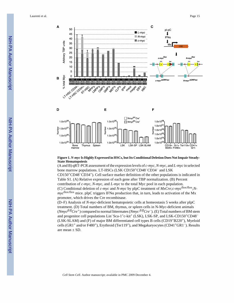
Figure 1. N-myc Is Highly Expressed in HSCs, but Its Conditional Deletion Does Not Impair Steady-State Hematopoiesis(A and B) qRT-PCR assessment of the expression levels of c-myc, N-myc, and L-myc in selectedbone marrow populations. LT-HSCs (LSK CD150+CD48−CD34− and LSKCD150+CD48−CD34+). Cell surface marker definition of the other populations is indicated inTable S1. (A) Relative expression of each gene after TBP normalization. (B) Percentcontribution of c-myc, N-myc, and L-myc to the total Myc pool in each population.(C) Conditional deletion of c-myc and N-myc by pIpC treatment of MxCre;c-mycflox/flox;N-mycflox/flox mice. pIpC triggers IFNα production that, in turn, leads to activation of the Mxpromoter, which drives the Cre recombinase.(D–F) Analysis of N-myc-deficient hematopoietic cells at homeostasis 5 weeks after pIpCtreatment. (D) Total numbers of BM, thymus, or spleen cells in N-Myc-deficient animals(Nmycfl/flCre+) compared to normal littermates (Nmyc fl/flCre−). (E) Total numbers of BM stemand progenitor cell populations Lin−Sca-1+c-kit+ (LSK), LSK-SP, and LSK-CD150+CD48−(LSK-SLAM) and (F) of major BM differentiated cell types B cells (CD19+B220+), Myeloidcells (GR1+ and/or F480+), Erythroid (Ter119+), and Megakaryocytes (CD41+GR1−). Resultsare mean ± SD.
Laurenti et al. Page 15
Cell Stem Cell. Author manuscript; available in PMC 2009 December 4.
NIH
-PA Author Manuscript
NIH
-PA Author Manuscript
NIH
-PA Author Manuscript
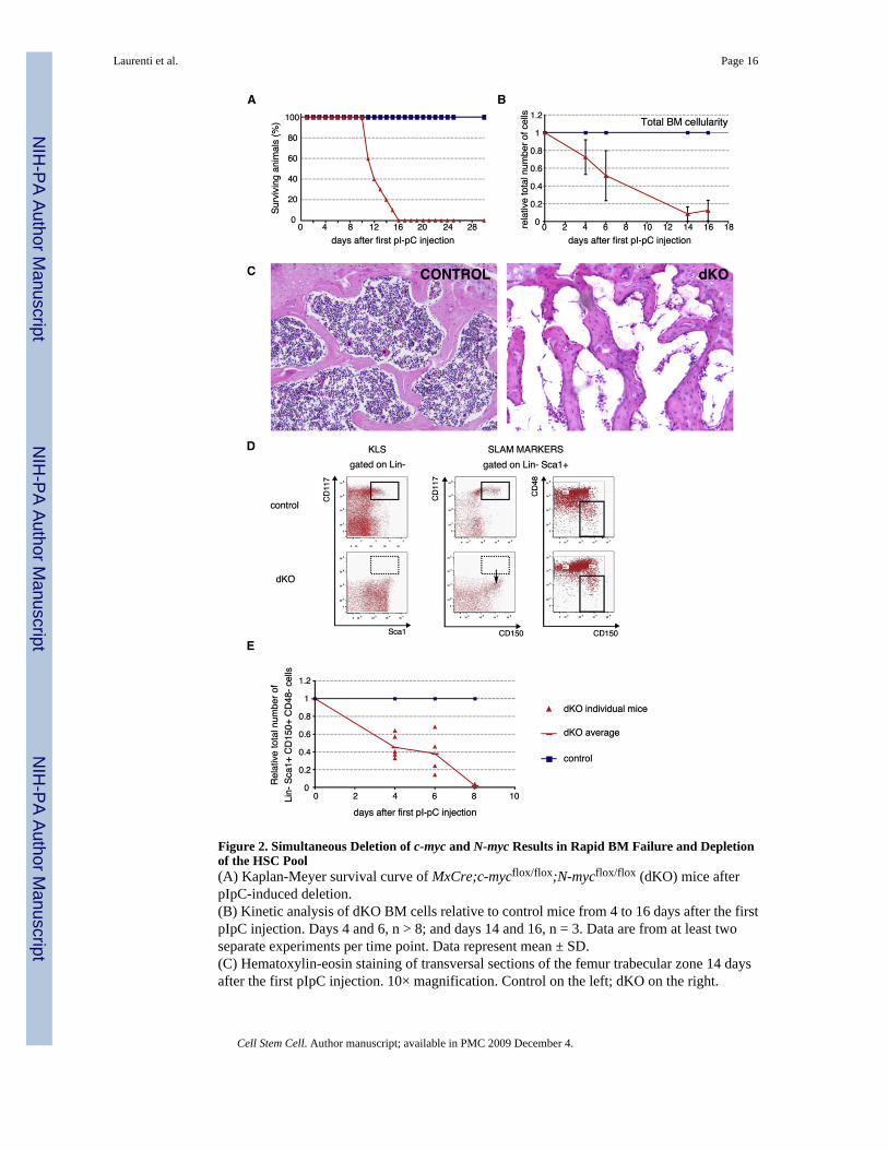
Figure 2. Simultaneous Deletion of c-myc and N-myc Results in Rapid BM Failure and Depletionof the HSC Pool(A) Kaplan-Meyer survival curve of MxCre;c-mycflox/flox;N-mycflox/flox (dKO) mice afterpIpC-induced deletion.(B) Kinetic analysis of dKO BM cells relative to control mice from 4 to 16 days after the firstpIpC injection. Days 4 and 6, n > 8; and days 14 and 16, n = 3. Data are from at least twoseparate experiments per time point. Data represent mean ± SD.(C) Hematoxylin-eosin staining of transversal sections of the femur trabecular zone 14 daysafter the first pIpC injection. 10× magnification. Control on the left; dKO on the right.
Laurenti et al. Page 16
Cell Stem Cell. Author manuscript; available in PMC 2009 December 4.
NIH
-PA Author Manuscript
NIH
-PA Author Manuscript
NIH
-PA Author Manuscript
(D) Representative FACS profiles of early hematopoietic stem/progenitor cells isolated fromcontrol and dKO mice 6 days after pIpC treatment (bottom). BM cells were stained withLineage, cKit (CD117), Sca1, CD150, and CD48 antibodies and gated as indicated. cKit+Sca1+ labeling alone would erroneously predict a complete loss of stem cells and earlyprecursors (left panel). The use of the SLAM markers CD150 and CD48 shows that the dKOLin−Sca1+ compartment downregulates cKit cell-surface expression (middle panel) but stillcontains a subset of HSCs (Lin−Sca1+CD150+CD48−, right panel).(E) Quantification of the number of SLAM-HSCs post pIpC induction. Data are from at leasttwo experiments per time point, 2 < n < 4 per genotype and time point.
Laurenti et al. Page 17
Cell Stem Cell. Author manuscript; available in PMC 2009 December 4.
NIH
-PA Author Manuscript
NIH
-PA Author Manuscript
NIH
-PA Author Manuscript
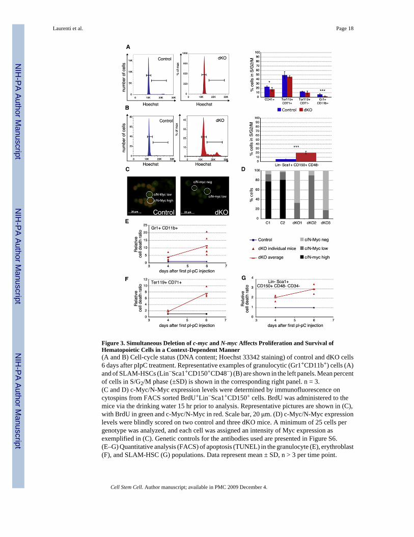
Figure 3. Simultaneous Deletion of c-myc and N-myc Affects Proliferation and Survival ofHematopoietic Cells in a Context-Dependent Manner(A and B) Cell-cycle status (DNA content; Hoechst 33342 staining) of control and dKO cells6 days after pIpC treatment. Representative examples of granulocytic (Gr1+CD11b+) cells (A)and of SLAM-HSCs (Lin−Sca1+CD150+CD48−) (B) are shown in the left panels. Mean percentof cells in S/G2/M phase (±SD) is shown in the corresponding right panel. n = 3.(C and D) c-Myc/N-Myc expression levels were determined by immunofluorescence oncytospins from FACS sorted BrdU+Lin−Sca1+CD150+ cells. BrdU was administered to themice via the drinking water 15 hr prior to analysis. Representative pictures are shown in (C),with BrdU in green and c-Myc/N-Myc in red. Scale bar, 20 μm. (D) c-Myc/N-Myc expressionlevels were blindly scored on two control and three dKO mice. A minimum of 25 cells pergenotype was analyzed, and each cell was assigned an intensity of Myc expression asexemplified in (C). Genetic controls for the antibodies used are presented in Figure S6.(E–G) Quantitative analysis (FACS) of apoptosis (TUNEL) in the granulocyte (E), erythroblast(F), and SLAM-HSC (G) populations. Data represent mean ± SD, n > 3 per time point.
Laurenti et al. Page 18
Cell Stem Cell. Author manuscript; available in PMC 2009 December 4.
NIH
-PA Author Manuscript
NIH
-PA Author Manuscript
NIH
-PA Author Manuscript
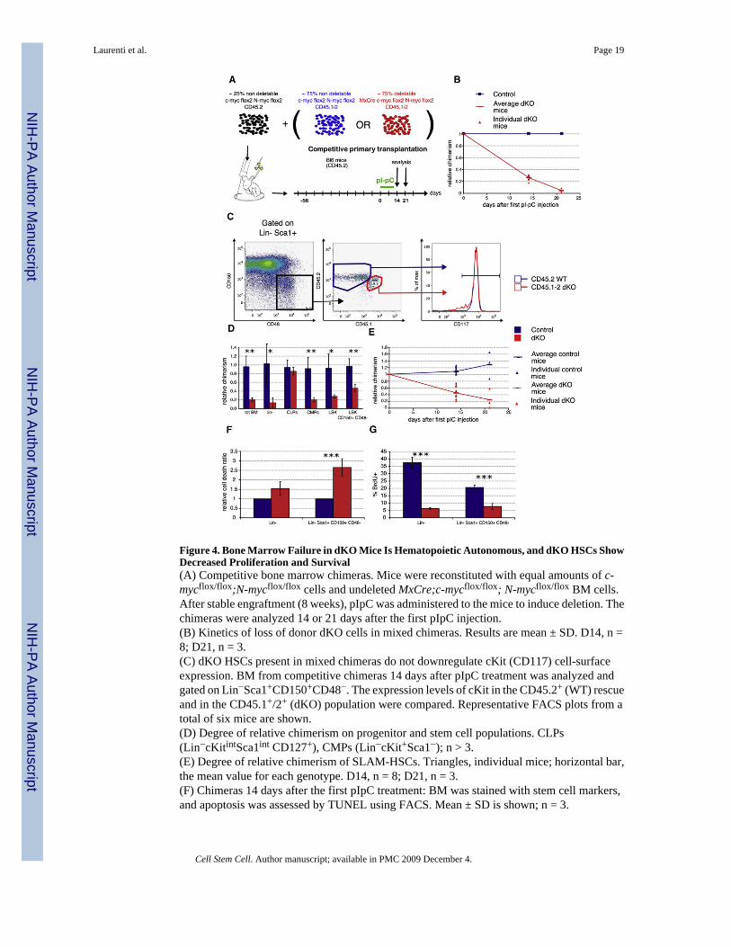
Figure 4. Bone Marrow Failure in dKO Mice Is Hematopoietic Autonomous, and dKO HSCs ShowDecreased Proliferation and Survival(A) Competitive bone marrow chimeras. Mice were reconstituted with equal amounts of c-mycflox/flox;N-mycflox/flox cells and undeleted MxCre;c-mycflox/flox; N-mycflox/flox BM cells.After stable engraftment (8 weeks), pIpC was administered to the mice to induce deletion. Thechimeras were analyzed 14 or 21 days after the first pIpC injection.(B) Kinetics of loss of donor dKO cells in mixed chimeras. Results are mean ± SD. D14, n =8; D21, n = 3.(C) dKO HSCs present in mixed chimeras do not downregulate cKit (CD117) cell-surfaceexpression. BM from competitive chimeras 14 days after pIpC treatment was analyzed andgated on Lin−Sca1+CD150+CD48−. The expression levels of cKit in the CD45.2+ (WT) rescueand in the CD45.1+/2+ (dKO) population were compared. Representative FACS plots from atotal of six mice are shown.(D) Degree of relative chimerism on progenitor and stem cell populations. CLPs(Lin−cKitintSca1int CD127+), CMPs (Lin−cKit+Sca1−); n > 3.(E) Degree of relative chimerism of SLAM-HSCs. Triangles, individual mice; horizontal bar,the mean value for each genotype. D14, n = 8; D21, n = 3.(F) Chimeras 14 days after the first pIpC treatment: BM was stained with stem cell markers,and apoptosis was assessed by TUNEL using FACS. Mean ± SD is shown; n = 3.
Laurenti et al. Page 19
Cell Stem Cell. Author manuscript; available in PMC 2009 December 4.
NIH
-PA Author Manuscript
NIH
-PA Author Manuscript
NIH
-PA Author Manuscript
(G) Chimeras 14 days after the first pIpC treatment were administered BrdU for 15 hr beforepercent BrdU incorporation was assessed. Mean ± SD is shown; n = 3.
Laurenti et al. Page 20
Cell Stem Cell. Author manuscript; available in PMC 2009 December 4.
NIH
-PA Author Manuscript
NIH
-PA Author Manuscript
NIH
-PA Author Manuscript
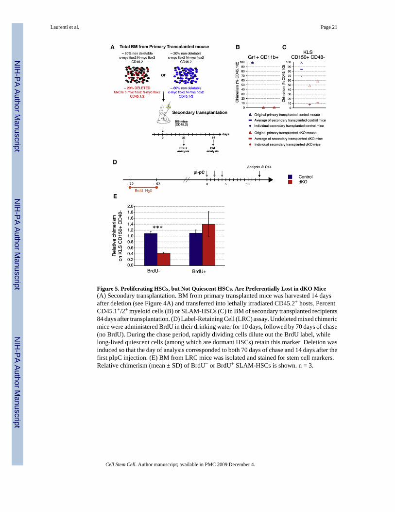
Figure 5. Proliferating HSCs, but Not Quiescent HSCs, Are Preferentially Lost in dKO Mice(A) Secondary transplantation. BM from primary transplanted mice was harvested 14 daysafter deletion (see Figure 4A) and transferred into lethally irradiated CD45.2+ hosts. PercentCD45.1+/2+ myeloid cells (B) or SLAM-HSCs (C) in BM of secondary transplanted recipients84 days after transplantation. (D) Label-Retaining Cell (LRC) assay. Undeleted mixed chimericmice were administered BrdU in their drinking water for 10 days, followed by 70 days of chase(no BrdU). During the chase period, rapidly dividing cells dilute out the BrdU label, whilelong-lived quiescent cells (among which are dormant HSCs) retain this marker. Deletion wasinduced so that the day of analysis corresponded to both 70 days of chase and 14 days after thefirst pIpC injection. (E) BM from LRC mice was isolated and stained for stem cell markers.Relative chimerism (mean ± SD) of BrdU− or BrdU+ SLAM-HSCs is shown. n = 3.
Laurenti et al. Page 21
Cell Stem Cell. Author manuscript; available in PMC 2009 December 4.
NIH
-PA Author Manuscript
NIH
-PA Author Manuscript
NIH
-PA Author Manuscript
Figure 6. c-Myc and N-Myc Promote Survival of HSCs and Prevent GrB Accumulation(A) Absolute numbers, relative to control mice, of SLAM-HSCs in dKO and H2K::BCL2 dKOmice, 6 days after pIpC treatment. Mean ± SD is shown; n ≥ 3.(B) SLAM-HSCs from dKO and control animals were stained with DCF-DA to measure theintracellular concentration of ROS. A representative FACS plot is shown; n = 3.(C) Differential expression levels of selected genes as analyzed by microarray analysis 4 daysafter the first pIpC injection.(D) Intracellular FACS staining to determine expression of Granzyme B (GrB) protein (blueline, control; red line, dKO; tinted, isotype control; n ≥3 for each condition).
Laurenti et al. Page 22
Cell Stem Cell. Author manuscript; available in PMC 2009 December 4.
NIH
-PA Author Manuscript
NIH
-PA Author Manuscript
NIH
-PA Author Manuscript
(E) Quantification of GrB expression at day 4 and day 6. Percent GrB+ in gatedLin−Sca1+CD150+CD48− cells. The Lin cocktail includes DX5, NK1.1, CD3, CD4, and CD8antibodies in order to eliminate any possible contamination from canonical GrB-producingcells, such as CTLs and NK cells. Blue triangles, individual control mice; red circles, dKOmice. Horizontal bars indicate averages of at least 6 mice per genotype.(F) Intracellular GrB staining on indicated cell types 4 days after pIpC treatment.(G) Representative GrB expression profiles gated on LSKCD150+CD48− in mixed chimeras(left panel) and in cultured SLAM HSCs 48 hr after IFNα addition.(H) Representative ictures of GrB by immunofluorescence. Lin− cells or activated NK cells(DX5+, positive control) were cytospun onto slides and stained for GrB protein and DAPI(blue). Scale bar, 5 μm.(I–K) GrB/TUNEL double staining on gated Lin−Sca1+CD150+CD48− cells at 4 and 6 daysafter c-myc and N-myc deletion. (I) Representative FACS plots at day 6. (J and K)Quantification of percentage of GrB+ cells among TUNEL+ SLAM HSCs ([J], mean ± SD)and of TUNEL+ cells among GrB+ SLAM HSCs (K). Day 4, n = 3; day 6, n = 2 for controlsand n = 5 for dKO.
Laurenti et al. Page 23
Cell Stem Cell. Author manuscript; available in PMC 2009 December 4.
NIH
-PA Author Manuscript
NIH
-PA Author Manuscript
NIH
-PA Author Manuscript
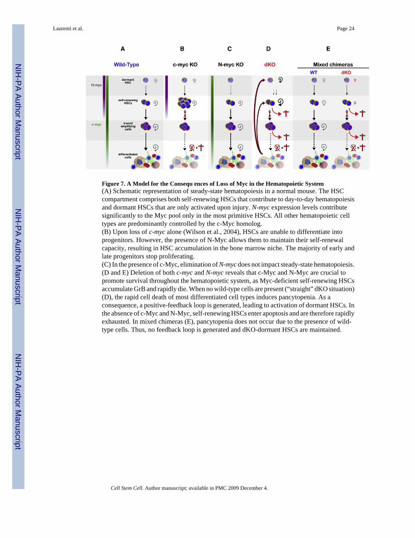
Figure 7. A Model for the Consequ ences of Loss of Myc in the Hematopoietic System(A) Schematic representation of steady-state hematopoiesis in a normal mouse. The HSCcompartment comprises both self-renewing HSCs that contribute to day-to-day hematopoiesisand dormant HSCs that are only activated upon injury. N-myc expression levels contributesignificantly to the Myc pool only in the most primitive HSCs. All other hematopoietic celltypes are predominantly controlled by the c-Myc homolog.(B) Upon loss of c-myc alone (Wilson et al., 2004), HSCs are unable to differentiate intoprogenitors. However, the presence of N-Myc allows them to maintain their self-renewalcapacity, resulting in HSC accumulation in the bone marrow niche. The majority of early andlate progenitors stop proliferating.(C) In the presence of c-Myc, elimination of N-myc does not impact steady-state hematopoiesis.(D and E) Deletion of both c-myc and N-myc reveals that c-Myc and N-Myc are crucial topromote survival throughout the hematopoietic system, as Myc-deficient self-renewing HSCsaccumulate GrB and rapidly die. When no wild-type cells are present (“straight” dKO situation)(D), the rapid cell death of most differentiated cell types induces pancytopenia. As aconsequence, a positive-feedback loop is generated, leading to activation of dormant HSCs. Inthe absence of c-Myc and N-Myc, self-renewing HSCs enter apoptosis and are therefore rapidlyexhausted. In mixed chimeras (E), pancytopenia does not occur due to the presence of wild-type cells. Thus, no feedback loop is generated and dKO-dormant HSCs are maintained.
Laurenti et al. Page 24
Cell Stem Cell. Author manuscript; available in PMC 2009 December 4.
NIH
-PA Author Manuscript
NIH
-PA Author Manuscript
NIH
-PA Author Manuscript