Embed Size (px)
Citation preview
Setor 04. Farmacologia Cardiovascular e Renal 04.001LOW-DOSE INTRAVENOUS NITRITE IMPROVES HEMODYNAMICS IN A CANINE MODEL OF ACUTE PULMONARY THROMBOEMBOLISM (APT) Dias-Junior, C. A.1; Florencio, B. C.1; Gladwin, M. T.2; Tanus-Santos, J. E.3 - 1USP - Farmacologia; 2NIH - Clinical Lung; 3FMRP - USP - Farmacologia Introduction:APT-induced pulmonary hypertension may result from active pulmonary vasoconstriction, which can be counteracted by activating the nitric oxide NO-cGMP pathway. Recent studies have demonstrated that nitrite is reduced to NO under conditions of hypoxia and acidosis. We hypothesized that nitrite infused intravenously could attenuate the hemodynamic changes associated with APT. Methods:APT was induced in mongrel dogs with autologous blood clots injected into the right atrium. After APT was induced (or saline injected), the dogs received an intravenous nitrite (or saline) infusion (6.75 micromol/kg over 15 min and then 0.28 micromol/kg/min) and hemodynamic evaluations were carried out for two hours. Plasma nitrite concentrations were measured by chemiluminescence. Results:APT decreased cardiac index (CI) and increased pulmonary vascular resistance index (PVRI). However, nitrite infusion increased CI by 28%, reduced the PVRI by 48% and the systemic vascular resistance index (SVRI) by 21% in embolized dogs. In non-embolized control dogs the same nitrite infusion decreased MAP and CI (all P<0.05). The nitrite infusion increased plasma nitrite concentrations by approximately 2 microM, and produced dose-dependent effects on PVRI, MAP, and SVRI. Discussion:These results suggest that a low dose nitrite infusion produces beneficial hemodynamic effects in APT; and as well a new therapeutic application for nitrite and support emerging evidence for a potent physiological vasoactivity of nitrite. Supported by: CNPq
04.002 DYNAMICS OF SOLUBLE GUANYLATE CYCLASE IN SEPSIS: A WINDOW OF THERAPEUTIC OPPORTUNITY Fernandes, D.1; Sordi, R.1; Duma, D.1; Heckert, B. T.1; Villela, C. G.2; Barja Fidalgo, T. C.2; Assreuy, J.1 - 1UFSC - Farmacologia; 2UERJ - Farmacologia Overproduction of nitric oxide (NO) is the one of the most important elements involved in hypotension and hyporesponsiveness to vasoconstrictors, which are hallmarks of sepsis and septic shock. Activation of guanylate cyclase (GC) accounts for several of NO effects. In spite of being of potential importance as an effective therapeutic strategy for septic shock treatment, GC inhibition is still controversial. Vasoconstrictive responses to phenylephrine (Phe) were reduced by 50% 8 and 24 h after LPS injection, thus reproducing an important finding in human septic shock. Methylene blue (MB, a GC inhibitor, 15 µmol/kg, i.v.) restored the reactivity to Phe in rats injected with LPS 24 h earlier (control 44.5 ± 2.3; LPS 24.3 ± 3.0; LPS+MB 40.5 ± 1.9 mmHg, n=6), but failed to do so in animals injected with LPS 8 h earlier. This prompted us to study guanylate cyclase activity and expression during endotoxaemia. Sodium nitroprusside (SNP; 100 µM) increased cGMP levels in lungs harvested from normal rats (vehicle 0.4 ± 0.1; SNP 4.6 ± 1.3 pmol/mg protein, n=4) or those injected with LPS 24 h before (data not shown). However, SNP failed in increasing lung cGMP levels of rats treated with LPS 8 h before (control 1.2 ± 0.2; SNP 1.3 ± 0.1 pmol/mg protein, n=4). Immunoblotting revealed that GC protein levels were lower (~40%) than controls in lungs harvested from rats injected 8 h earlier and were back to normal values in rats injected 24 h earlier with LPS. Thus, the refractoriness in MB effect in hyporeactivity to Phe at 8 h was mirrored by decreased GC activity and protein levels. The mRNA levels to GC increased 24 h after LPS (~50%). Thus, the recovery in sGC activity 24 h after LPS appears to be due to expression of new GC protein. To evaluate MB effect in mortality, animals were submitted to cecal ligation and puncture (CLP, a model of sepsis). When MB was given 8 h after CLP, survival rate was reduced (CLP 25%; CLP+MB 8 h 10%, n=20). In rats which received MB 20 h after the surgery, survival was significantly improved (CLP 25%; CLP+MB 20 h 55%, n=20). Therefore, differential responsiveness to soluble guanylate cyclase during the course of sepsis may determine the success or failure of therapy with guanylate cyclase inhibitors. Interestingly, MB is effective at later stages of sepsis, exactly when other therapeutic alternatives meet with failure. Thus, MB may be a useful therapeutic strategy if administered at the proper window of opportunity. Supported by: CAPES, FAPESC, CNPQ and PRONEX
04.003 EXPRESSION OF Na+/K+-ATPase α SUBUNIT ISOFORMS AND p38-MAP KINASE IN HEARTS FROM ENDOTHELIAL NITRIC OXIDE SYNTHASE GENE KNOCKOUT MICE Rezende, D. C.1; Caricati-Neto, A. C.2; Jurkiewicz, A.2; Noel, F.1; Quintas, L. E. M.1 - 1UFRJ - Farmacologia Básica e Clínica; 2UNIFESP - EPM - Farmacologia INTRODUCTION: Systemic hypertension has been related to changes in expression/activity of myocardial Na+/K+-ATPase and mitogen-activated protein kinases (MAPKs). Nevertheless, the pattern of expression depends on the experimental hypertensive model. Here we evaluated the protein expression of Na+/K+-ATPase α isoforms and activated and total p38-MAPK in hearts of hypertensive mice knocked out for the endothelial nitric oxide synthase gene (eNOS/KO). METHODS: Hearts from male 12-week-old eNOS/KO or control (C57BL/6J) mice were homogenized and passed through differential centrifugation to obtain cytosolic and particulated fractions. Samples ran on 7.5 or 10% SDS-PAGE followed by immunoblotting with anti-Na+/K+-ATPase α1 and α2 isoforms and anti-p38-MAPK or –phosphop38-MAPK antibodies. RESULTS: Preliminary data show that protein expression of Na+/K+-ATPase α1 and α2 isoforms were largely reduced in eNOS/KO hearts (for both, about 15% of control level, p<0.05, n=3). In contrast, no significant alteration of the density of p38-MAPK active (phosphorylated) form was detected. DISCUSSION: In eNOS/KO model, cardiac downregulation of Na+/K+-ATPase a isoforms may represent an adaptation to pressure overload in order to generate a positive inotropic effect. The mechanical stress induced by overload, however, does not activate p38-MAPK signaling. Studies with Ca2+-ATPases and other MAPKs are in progress. Supported by: FAPESP, FAPERJ, CNPq
04.004 ENDOTHELIUM MODULATES THE VASORELAXATION INDUCED BY NITRIC OXIDE DONOR. Bonaventura, D.1; De Lima, R. G.1; Da Silva, R. S.1; Bendhack, L. M.1 - 1USP - FCFRP Introduction: many substances are released by endothelium including nitric oxide (NO) and cyclooxygenase (COX) products that control the vascular tone. The aim of this study was to investigate if the endothelium modulates the relaxation induced by the NO donor [Ru(terpy)(bdq)NO]3+ (TERPY)1 in isolated rat aortic rings. Methods: contractile responses were induced by phenylephrine (10-7M) in intact (E+) and denuded (E-) aortic rings. On the top of this contraction, cumulative concentration-effect curves for TERPY (10-9-3x10⁻⁴M) were constructed. E+ were incubated with indomethacin (Indo 10-5M), non selective COX inhibitor, SQ29548 (3x10⁻⁶M), or AH6809 (10-5M), selective antagonists of thromboxane A2 (TXA2) and prostaglandin F2a (PGF2a) receptors, respectively. We analyzed the maximum effect (ME) induced by TERPY its and potency (pD2). Results: the relaxation induced by TERPY was concentration-dependent and was less potent in E+ (6.10±0.06, n=17) than in E- (6.64±0.07, n=10). No differences in ME were observed in both E+ (103.0±0.9%) and in E- (104.4±1.5%). In the presence of INDO, the relaxation induced by TERPY (ME: 103.3 ±1.8% and pD2: 6.79±0.17, n=5) in E+ did not differ of E-. Similar results were observed for SQ29548 (ME: 105.1±2.0% and pD2: 6.85±0.15, n=5). However, AH6809 had no effect in the relaxation induced by TERPY (ME: 100.9±1.1% and pD2: 6.19±0.17, n=8). Conclusions: relaxation induced by TERPY is less potent in E+ than in E-, and its effect is related to TXA2, which is produced by cyclooxygenase. References: de Lima, R.G., Sauaia, M., Bonaventura, D., Tedesco, A.C., Bendhack, L.M., da Silva, R.S., 2006. Influence of ancillary ligand L in the nitric oxide photo-release by the [Ru(L)(terpy)NO]3+ complex and its vasodilator activity based on visible light irradiation. Inorganica Chimica Acta 359, 2543-2549. Supported by: Fapesp and CNPq.
04.005 CONVERSION OF ANGIOTENSIN I TO II IS ALTERED IN MESENTERIC ARTERIAL BED PERFUSATE FROM SPONTANEOUSLY HYPERTENSIVE RATS (SHR). Sivieri-Jr., D. O.1; Bispo-da-Silva, L. B.1; Becari, C.1; Oliveira, E. B.2; Salgado, M. C. O.1 - 1FMRP - USP - Farmacology; 2FMRP - USP - Biochemistry and Immunology Introduction: The mesenteric arterial bed perfusate (MABP) posses many soluble proteases which are involved in vasoactive peptides metabolism. We evaluated the conversion of angiotensin (Ang) I to II in MABP from SHR. Methods: The mesentery from normotensive Wistar (NWR, n=7) and SHR (n=8) was isolated and reperfused with Krebs solution for 2 h. Then, the perfusate was concentrated and incubated with Ang I (30 nmol). The reactions products were analyzed by HPLC (nmol), in the absence or presence of the angiotensin-converting enzyme (ACE) inhibitor captopril (CPT: 10 mM) and/or the serine protease inhibitor chymostatin (CHY: 100 mM). Results: It was observed two populations of SHR perfusates concerning Ang I consumption: a high consumer (HC: Ang I consumption >90%) and a low consumer (LC: Ang I consumption <30%) group. While Ang II generation was similar in LC and NWR (2.02±0.59 and 2.22±0.36), it was increased in HC (5.60±1.74). CPT did not alter Ang II generation in all groups. However, CHY decreased Ang II generation in NWR, LC and HC (0.77±0.29; 0.75±0.29 and 2.35±0.52*, respectively; *p<0.05) and the association with CPT did not induce further inhibition. Discussion: ACE is not involved in Ang II generation in MABP and this activity is related to a chymostatin-sensitive one. In addition, there are two populations of MABP in SHR and in one of them the conversion of Ang I to II is increased. Supported by: CAPES and FAPESP.
04.006 EFEITOS DA TESTOSTERONA NA EXPRESSÃO DE VCAM-1, ICAM-1E INOS E NA ATIVAÇÃO DE MAPKS EM CÉLULAS DE MÚSCULO LISO VASCULAR. Schuldt, E. Z.1; Yogi, A.1; Montezano, A. C. I.1; Chignalia, A. Z.1; Kharma, P. M.1; Carvalho, M. H. C.1; Nigro, D.1; Fortes, Z. B.1; Tostes, R. C. A.1 - 1USP - Farmacologia Introdução: As ações vasculares da testosterona são complexas e associadas à dose, tempo de exposição, gênero animal e presença de doença vascular. O objetivo deste estudo foi analisar os efeitos da testosterona em células de músculo liso vascular (CMLV) na expressão protéica de moléculas envolvidas no processo inflamatório e ativação de MAPKs. Métodos: Culturas de CMLV do leito mesentérico de ratos Wistar e da aorta de coelhos (RASM) foram estimuladas com testosterona (1 nM a 10 mcM), nos tempos de 2 e 4 h (CMLV) ou 1 a 60 min (RASM). Proteínas de CMLV e RASM foram submetidas à técnica de Imunoblot, incubadas com anticorpos anti-VCAM-1, ICAM-1, iNOS e p38, ERK 1/2 e Src (formas fosforilada e não fosforilada) à 4oC por 24 h. Após incubação com os anticorpos secundários, os sinais foram revelados por quimioluminescência e os resultados expressos como percentagem do veículo. Resultados: A estimulação de RASM com testosterona 10-7, mas não 10-8 M, promoveu aumento na fosforilação da enzima ERK1/2 (p<0,05), sem alterar os níveis de expressão de ERK1/2 não-fosforilada. Testosterona, 10-7 e 10-8 M, também induziu ativação, representada pela fosforilação, das enzimas p38MAPK e c-SRC, em células RASM (p<0,05). A testosterona não alterou a expressão protéica de VCAM-1, ICAM-1 ou iNOS após 2 h de estimulação. No entanto, após 4 h, testosterona diminuiu a expressão de ICAM-1 e iNOS e aumentou a expressão de VCAM-1, em todas as concentrações utilizadas. Discussão: A ativação da via das MAPKs na primeira hora de estímulo sugere uma subsequente modulação na expressão de moléculas pró-inflamatórias em CMLV. Apoio Financeiro: CNPq, FAPESP
04.007 MUSCARINIC RECEPTOR (MR) RESPONSIVENESS IN DETRUSOR SMOOTH MUSCLE (DSM) OBTAINED FROM NITRIC OXIDE (NO)-DEFICIENT AND SPONTANEOUSLY HYPERTENSIVE RATS (SHR). Monica, F. Z. T.1; Nucci, G. de1; Zanesco, A.2; Bricola, A. A. de O.3; Antunes, E.1 - 1UNICAMP - Pharmacology; 2UNESP - Physical Education; 3PUCCamp - Pharmacology Goals: We have previously shown that long-term NO inhibition significantly increases the sensitivity of DSM for the muscarinic agonist carbachol (CCH), evidencing an experimental model of hyperactive bladder. Since this supersensitivity may reflect the hypertensive state, the aim of this work was to investigate contractile responses of DSM induced by in rats made hypertensive by long-term NO inhibition in comparison with SHR rats. Methods: Wistar male rats were treated orally with L-NAME (20mg/rat/day) for 30 days. Age-matched control animals received tap water alone. Bladders from all groups L-NAME, SHR and normotensive Wistar Kyoto (WKY) were removed. Concentration response curve to CCH (1 nM-30 µM) were obtained, and pEC50 and maximal responses (Emax) were calculated. Results: Both L-NAME-treated rats and SHR presented a marked arterial hypertension. Long-term NO inhibition increased the CCH potency (6.09±0.02 vs 6.82±0.06), without modifying the Emax (ctl: 3.50±0.10 vs treated: 3.40±0.07). Contractile response to CCH were similar in both WKYs and SHR (pEC50 5.63±0.04 vs 5.67±0.12 and the (Emax: 1.69±0.08 vs 1.53±0.13 mN/mg wet weight), respectively. Conclusion: NO exerts a modulatory effect on the contractility mediated by MR, but these receptors does not appear to contribute to bladder dysfunction in SHR. The relationship between hypertension and overactive bladder remains to be confirmed. Supported by: FAPESP
04.008 VITAMIN E EFFECTS ON LEUKOCYTE-ENDOTHELIAL CELL INTERACTIONS IN DOCA-SALT HYPERTENSION Carneiro, F. S.1; Callera, G. E.2; Souza, H. P. de3; Rodrigues, S.1; Montezano, A. C. I.2; Nigro, D.1; Fortes, Z. B.1; Carvalho, M. H. C.1; Tostes, R. C. A.1 - 1USP - Farmacologia; 2University of Ottawa - Kidney Research Center; 3FM - USP - LIM - 51 Introduction: Vascular oxidative stress, decreased acetylcholine (ACh) vasodilation and altered leukocyte-endothelial cell interactions in DOCA rats are associated with activation of the endothelin system. Since reactive oxigen species (ROS) play a key role in these alterations, we hypothesized that vitamin E (VE) ameliorates impaired endothelium-dependent dilation and leukocyte-endothelial cell interactions in DOCA hypertension. Methods and results: DOCA and control (C) rats were treated with VE (200 mg/Kg/day) or vehicle during 5 weeks. VE treatment normalized the increased ROS generation, evaluated by lucigenin, as well as the impaired ACh relaxation in DOCA aorta. VE normalized the decreased rolling (DOCA:99±17, C:210±6, DOCA VE:184±16) and attenuated the increased adhesion (DOCA:15.8±2.6, C:4.0±1.0, DOCA VE:8.8±1.4) in DOCA, as shown by intravital microscopy. Flow citometry analysis identified decreased L-selectin (C:22.6±1.9 vs DOCA:15.7±2.6) and increased CD18 (C:22.8±2.06 vs DOCA:45.4±11.2) protein expression in DOCA leukocytes. VE normalized CD18 (DOCA VE:23.6±2.3), but not L-selectin (DOCA VE:11.1±0.9), expression and also the increased vascular eNOS and ICAM-1 gene expression in DOCA rats. Discussion: ROS play a direct role on the impaired vascular reactivity and leukocyte behavior in DOCA hypertension. ROS effects may be mediated by decreased NO bioavailability and by changes in celular adhesion molecules expression. Supported by: FAPESP, CNPq.
04.009 A ROLE FOR MATRIX METALLOPROTEINASE-9 IN THE HEMODYNAMIC CHANGES FOLLOWING ACUTE PULMONARY EMBOLISM Fortuna, G.1; Dias-Junior, C. A.1; Lopes, L. F.2; Gerlach, R. F.3; Tanus-Santos, J. E.2 - 1USP - Farmacologia; 2FMRP - USP - Farmacologia; 3FORP - USP - Morfologia Matrix metalloproteinases (MMPs) modulate vascular contractility and may affect acute pulmonary embolism (APE)-induced pulmonary hypertension. We examined the effects of the administration of doxycycline (a MMP inhibitor) following APE in anesthetized dogs. Methods: Sham operated dogs (N=5) received only saline. APE was induced by intravenous injections of microspheres in amounts to increase mean pulmonary artery pressure (MPAP) by 20 mmHg, and embolized dogs received saline (Emb group, N=8), or doxycycline (10 mg/kg, i.v.) 5 or 30 min of APE (Emb + Doxy 5 and Emb + Doxy 30 groups, N=9 and 8, respectively). Hemodynamic evaluation was performed at baseline and 5-120 after APE. Gelatin zymography of MMP-2 and MMP-9 from plasma samples was performed. Results: No significant hemodynamic changes were found in Sham animals. Embolization increased MPAP by 218±16% and the pulmonary vascular resistance index (PVRI) by 289±42% in Emb group (both P<0.05). Doxycyline increased the cardiac index by 24±5% and reduced PVRI by 23±4% 120 min of APE in Doxy 30 + Emb group. In addition, doxycyline reduced MPAP and PVRI 30 min after APE with maximum effects seen 120 min after APE (25 ± 4% decrease in MPAP and 33 ± 6% decrease in PVRI; both P<0.05) in Doxy + 5 group. Plasma pro-MMP-9 and MMP-9 levels increased only in Emb group and MMP-2 remained unaltered. Conclusions: Our study shows that doxycycline attenuates APE-induced pulmonary hypertension, and indicates that MMP-9 has a role in APE-induced pulmonary hypertension. MMP-9 may be a pharmacological target in APE. Keywords: Doxycyclin, Matrix metalloproteinases, Pulmonary Embolism, Pulmonary Hypertension.
04.010 ANGIOTENSIN II INDUCES KININ B1 RECEPTORS Ceravolo, G. S.1; Nigro, D.1; Tostes, R. C. A.1; Fortes, Z. B.1; Carvalho, M. H. C.1 - 1USP - Farmacologia The kinin B1 receptor (B1R) is normally absent under physiological conditions, but is highly inducible during inflammatory conditions or tissue damage. The present study was designed to explore the effect of angiotensin II (AII) infusion on B1R protein expression in the cardiovascular system of rats. Methods: Male Wistar rats received 400ng/kg/min of AII (AII rats) or saline (S rats) infusion during 14 days, via mini-osmotic pump. The blood pressure levels (BP) were determined by the tail cuff method at day 0, 7 and 13 after implants. At 14th day the animals were anesthetized, and aorta was excised for determination of B1R expression, superoxide anion (O-2) and nitric oxide (NO) generation by DABK. Aortic rings were also mounted in organ bath, pre-contracted with phenylephrine and cumulative concentration-curves to DABK were performed. Results: AII rats had higher BP than S at days 7 (121±1.5vs167±1.2mmHg) and 13 (118±2.2vs182±5.9mmHg). Aorta of AII rats presented expression of B1R in endothelium and an increased generation of O-2 when compared with S rats. DABK promoted dilatation in aortic rings with endothelium of AII rats and NO generation. In aorta of S rats DABK had any effect. Conclusion: These results provide evidences that AII increased O-2 generation concomitantly with an increasing modulation of cardiovascular B1R protein expression. We have also shown that activation of B1R causes endothelium-dependent vasodilatation via NO generation in aortic rings. These data suggest the existence of a new site of interaction between kinins and angiotensins. Supported by: FAPESP/CNPQ-PRONEX/CAPES
04.011 EFFECT OF PHOSPHODIESTERASE (PDE) 5 INHIBITORS IN THE ISOLATED RABBIT PULMONARY ARTERY Flores Toque, H. A.1; Priviero, F. B. M.1; Teixeira, C. E.2; Antunes, E.1; Nucci, G. de1 - 1UNICAMP - Farmacologia; 2Medical College of Georgia - Physiology Goal: Sildenafil (SILD), Tadalafil (TADA) and Vardenafil (VARD) are selective PDE5 inhibitors. Because PDE5 is abundant in lung, this work aimed to investigate the effects of SILD, TADA and VARD on NO/cGMP-dependent relaxations of rabbit pulmonar artery (RbPA) rings. Method: Endothelium-intact (E+) and denuded (E-) rings of RbPA were mounted in organ baths. Concentration response curves (CRC) to SILD, TADA and VARD (1 nM to 10 µM) were obtained in the presence or in the absences of L-NAME and BAY 41-2272. Results: In E+ rings, SILD, TADA and VARD induced relaxations with potency (pEC50) values of 7.97 ± 0.07, 7.94 ± 0.06 and 8.23 ± 0.07, respectively. L-NAME (100 µM) caused a rightward shift in the CRC for SILD, TADA and VARD (13, 3 and 13-fold, respectively). Endothelium denudation caused a rightward shift in the CRC for SILD, TADA and VARD (pEC50: 6.87 ± 0.09; 7.53 ± 0.05; 7.13 ± 0.10, respectively). Addition of BAY-41-2272 (30 nM) in E- enhanced the pEC50 for SILD, TADA and VARD. GTN-induced relaxations were enhanced by SILD, TADA and VARD (0,1 µM) in E- rings. Although SILD, TADA and VARD showed similar potencies and maximal responses, endothelium denuded caused 6 rightward shift in the curve to TADA, whereas the relaxant response evoked by VARD and SILD were approximately 21 and 9-fold to the right. Conclusion: The findings show that inhibition of the NO/cGMP signaling pathway markedly affect VARD and SILD-induced relaxations, but not those in response to TADA. In E- RbPA rings, relaxing effects of PDE5 inhibitors are restored by adding the sGC activator BAY 41-2272. Supported by: Fapesp
04.012 AÇÃO DE NOVOS PROTÓTIPOS N-ACILIDRAZÔNICOS 1,3-BENZODIOXÓLICOS NA REATIVIDADE VASCULAR DE RATOS Beiral, H. J. V.1; Kummerle, A. E.2; Fraga, C. A. M.2; Barreiro, E. J.2; Sudo, R. T.1; Zapata-Sudo, G.1 - 1UFRJ - Farmacologia Básica e Clínica; 2UFRJ - Faculdade de Farmácia - LASSBio Introdução. O composto-protótipo N-acilidrazônico, 3,4-metilenodioxibenzoil-2-tienilidrazona (LASSBio-294) apresentou propriedades inotrópica positiva e vasodilatadora. Com a intenção de otimizar o efeito vasodilatador deste protótipo foram planejados e sintetizados novos análogos estruturais (LASSBio-897, 1026, 1027, 1029) para serem avaliados quanto a seus efeitos na contratilidade dos músculos liso vascular e cardíaco. Métodos: Aorta e músculo papilar de ratos Wistar (200-250g) foram dissecados e preparados para registro de tensão isométrica. Os abalos do músculo papilar obtidos na ausência e presença dos derivados foram digitalizados e armazenados em computador. Após período de estabilização dos anéis de aorta, a preparação foi contraída com 10 µM de fenilefrina seguida da exposição aos derivados testes. Resultados: Todos os derivados reduziram a contratura induzida pela fenilefrina em anéis de aorta com endotélio íntegro. Na concentração de 50 µM, LASSBio-1027 e 1029 promoveram 100% de relaxamento enquanto LASSBio-1026 apenas 30%. A concentração necessária para induzir 50% de relaxamento muscular por LASSBio-1029 e 897 foi de 7,3±0,4 e 0,46±0,02 µM. LASSBio-1029 também apresentou efeito cardionotrópico negativo. onclusões. LASSBio-897 foi o derivado mais potente em promover vasodilatação em anéis de aorta pré-contraídas com fenilefrina. Sua potência para a ação vasodilatadora foi cerca de 16 vezes maior quando comparada ao protótipo LASSBio-294. Apoio Financeiro: IM-INOFAR, CAPES, Pronex-Rio, CNPq, FAPERJ
04.013 CLINICAL EVIDENCE FOR LEAD-INDUCED INHIBITION OF NITRIC OXIDE FORMATION Cau Sertorio, J. T.1; Barbosa Jr, F.2; Gerlach, R. F.3; Tanus-Santos, J. E.1 - 1FMRP - USP - Farmacologia; 2FCFRP - USP - Toxicologia; 3FORP - USP - Morfologia Introduction Lead exposure has been associated with increased cardiovascular risk, which may result, at least in part, from lead-induced increases in oxidative stress and depressed nitric oxide (NO) availability. However, no previous clinical study has examined whether lead exposure is associated with significant effects on biomarkers of NO activity. Methods We investigated whether there is an association between the circulating concentrations of nitrites, nitrates, and cGMP and the concentrations of lead in whole blood (B-Pb) or plasma (P-Pb) from 62 lead-exposed subjects (30 men and 32 women). P-Pb was determined by inductively coupled plasma mass spectrometry (ICP-MS) and B-Pb by graphite furnace atomic absorption spectrometry (GF AAS). Plasma nitrite and nitrate concentrations were measured using an ozone-based chemiluminescence assay. Plasma cGMP concentrations were measured using a commercial enzyme immunoassay. Results We found a negative correlation between plasma nitrite and B-Pb concentrations (r=-0.358; P=0.004), and between plasma nitrite and P-Pb concentrations (r=-0.264; P=0.038). However, no significant correlations were found between plasma nitrate or cGMP and B-Pb or P-Pb concentrations (all P>0.05). Discussion These findings suggest a significant inhibitory effect of lead exposure on NO formation and provide clinical evidence for a biological mechanism possibly involved the association between lead exposure and increased cardiovascular risk. Acknowledgments FAPESP.
04.014 ROLE OF MUSCARINIC RECEPTORS ON ELECTRICALLY- INDUCED ARRHYTHMIAS IN RAT RIGHT ATRIUM DURING POSTNATAL DEVELOPMENT. Faria, D. M.1; Galvao, K. M.2; Godoy, C. M. G.1; Caricati-Neto, A. C.2 - 1UMC - Núcleo de Pesquisas Tecnológicas (NPT); 2UNIFESP - EPM - Farmacologia Introduction and Goals: On the basis that acetylcholine is involved in electrically-induced arrhythmias in rat right atrium (Godoy et al., Cardiovasc Pharmacol 34: 475, 1999) and responses mediated by cardiac muscarinic receptors are changed by aging (Brodde & Leineweber, Eur J Pharmacol 500: 167, 2004), we investigated if responses of muscarinic receptors in this arrhythmias are changed during postnatal development. Methods: Right atrium of rats of 5, 15, 30 and 100 day old (N=8, for each age) were isolated and mounted in perfusion chamber containing Krebs-Henseleit solution (36.5oC, pH 7.4, 95% O2 + 5% CO2) between two platinum electrodes for electrical field stimulation (250 rectangular voltage pulses, 66.7 Hz, 5 ms duration) for induction of atrial arrhythmia. The number of trains and the train stimulus strength necessary to induce arrhythmia were determined for every age. Effects of agonist (Carbachol 1 microM) and antagonist (Atropine 1 microM) of muscarinic receptors on atrial arrhythmia were studied. Results: The stimulus amplitude of the stimulation train necessary for atrial arrhythmia induction were, 5, 5, 3 and 2-fold the atrial threshold, respectively for 5, 15, 30 and 100 day old animals. Accordingly, the numbers of train applications necessary to induce arrhythmia were (mean±SEM) 4.8±1.3, 4.2±0.7, 2.6±1.1 and 6.5±0.6, respectively for 5, 15, 30 and 100 day old animals. Pretreatment of atria with carbachol (1 microM for 30 min) decreased the number of stimulation trains necessary to induce arrhythmia to 1.6±0.5, 1.7±0.5, 1.4±0.3 and 1.6±0.6 respectively for 5, 15, 30 and 100 day old animals. Pretreatment of atria with atropine (1 microM for 30 min) inhibited atrial arrhythmia induction in rats of 5, 15, 30 and 100 day old, even after the application of up to 20 stimulation trains. Conclusion: These results suggest that muscarinic receptors facilitate electrically-induced arrhythmias in rat right atrium in similar way for both adult and young rats. Supported by: FAEP/UMC and FAPESP
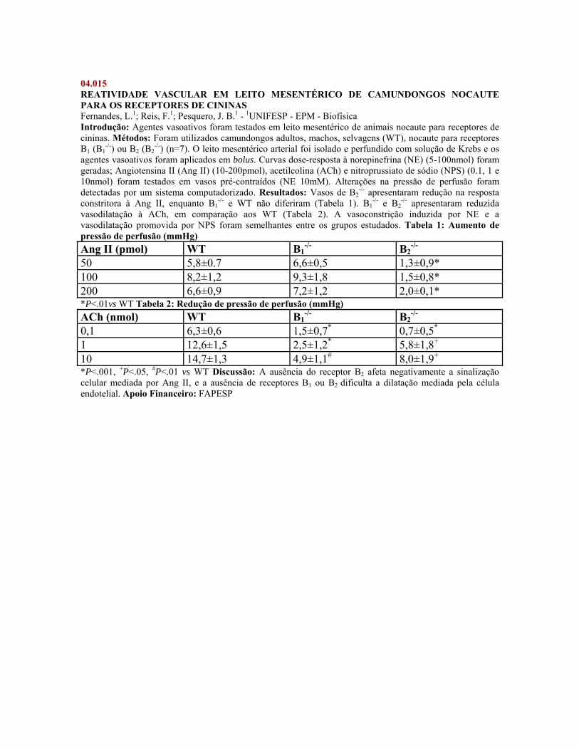
04.015 REATIVIDADE VASCULAR EM LEITO MESENTÉRICO DE CAMUNDONGOS NOCAUTE PARA OS RECEPTORES DE CININAS Fernandes, L.1; Reis, F.1; Pesquero, J. B.1 - 1UNIFESP - EPM - Biofísica Introdução: Agentes vasoativos foram testados em leito mesentérico de animais nocaute para receptores de cininas. Métodos: Foram utilizados camundongos adultos, machos, selvagens (WT), nocaute para receptores B1 (B1
-/-) ou B2 (B2-/-) (n=7). O leito mesentérico arterial foi isolado e perfundido com solução de Krebs e os
agentes vasoativos foram aplicados em bolus. Curvas dose-resposta à norepinefrina (NE) (5-100nmol) foram geradas; Angiotensina II (Ang II) (10-200pmol), acetilcolina (ACh) e nitroprussiato de sódio (NPS) (0.1, 1 e 10nmol) foram testados em vasos pré-contraídos (NE 10mM). Alterações na pressão de perfusão foram detectadas por um sistema computadorizado. Resultados: Vasos de B2
-/- apresentaram redução na resposta constritora à Ang II, enquanto B1
-/- e WT não diferiram (Tabela 1). B1-/- e B2
-/- apresentaram reduzida vasodilatação à ACh, em comparação aos WT (Tabela 2). A vasoconstrição induzida por NE e a vasodilatação promovida por NPS foram semelhantes entre os grupos estudados. Tabela 1: Aumento de pressão de perfusão (mmHg) Ang II (pmol) WT B1
-/- B2-/-
50 5,8±0.7 6,6±0,5 1,3±0,9* 100 8,2±1,2 9,3±1,8 1,5±0,8* 200 6,6±0,9 7,2±1,2 2,0±0,1* *P<.01vs WT Tabela 2: Redução de pressão de perfusão (mmHg) ACh (nmol) WT B1
-/- B2-/-
0,1 6,3±0,6 1,5±0,7* 0,7±0,5* 1 12,6±1,5 2,5±1,2* 5,8±1,8+ 10 14,7±1,3 4,9±1,1# 8,0±1,9+ *P<.001, +P<.05, #P<.01 vs WT Discussão: A ausência do receptor B2 afeta negativamente a sinalização celular mediada por Ang II, e a ausência de receptores B1 ou B2 dificulta a dilatação mediada pela célula endotelial. Apoio Financeiro: FAPESP
04.016 eNOS GENOTYPE DEPENDENT DECREASES IN PLASMA MMP-9 LEVELS BY ATORVASTATIN Lopes, L. F.1; Souza da Costa, D. C.1; Sandrim, V. C.1; Rego, E. M.2; Gerlach, R. F.3; Tanus-Santos, J. E.1 - 1FMRP - USP - Farmacologia; 2FMRP - USP - Clínica Médica; 3FORP - USP - Morfologia Introduction: Anti-inflammatory effects produced by statins cholesterol-independent, result at least in part from increased endothelial nitric oxide production. These effects may be modulated by polymorphisms in the endothelial nitric oxide synthase (eNOS) gene. Here we examined whether the T-786C polymorphism of eNOS gene affects the concentrations of MMP-2, MMP-9, and TIMP-1. Methods: Healthy male volunteers (N=200), Caucasians, non-smokers, were genotyped for the T-786C polymorphism by restriction fragment length polymorphism. Subjects with TT (N=15) or CC (N=15) genotype were randomized to receive placebo for 14 days followed by 14 days of atorvastatin, 10 mg/day p.o. The concentrations of TIMP-1 were measured with ELISA kit and MMP-2 and MMP-9 by gelatin zymography. Results: Atorvastatin significantly reduced the concentrations of MMP-9 in subjects with CC (but not TT) genotype (P<0.05). No significant effects were found on the concentrations of pro-MMP-9, pro-MMP-2, and TIMP-1. Discussion: The significant decrease in MMP-9 activity in subjects with CC genotype without significant changes in TIMP-1 suggests that treatment with atorvastatin reduced net MMP-9 activity. These findings may be of major clinical importance because MMPs have been involved in cardiovascular diseases. Supported by: FAPESP-CAPES-CNPq
04.017 INFLUÊNCIA DA IDADE NO EFEITO CARDIOTÓXICO DA BUPIVACAÍNA Kiuchi, M. G.1; Zapata-Sudo, G.1; Trachez, M. M.1; Sudo, R. T.1 - 1UFRJ - Farmacologia Básica e Clínica INTRODUÇÃO: A suscetibilidade de crianças e neonatos a cardiotoxicidade pela bupivacaína é controversa. Comparamos o efeito da RS(±) e S(-) bupivacaína (bupi) na regulação intracelular de Ca2+ de miócitos cardíacos de ratos de 2 e 16 semanas (sem) de idade. MÉTODOS: Feixes de ventrículo permeabilizados com saponina foram preparados para registro de tensão isométrica. Protocolos foram elaborados para investigar o efeito dos anestésicos na liberação de Ca2+ pelo retículo sarcoplasmático (RS) e na sensibilidade das miofibrilas a este íon. RESULTADOS: A RS(±) e a S(-) bupi estimularam a liberação de Ca2+ pelo RS não havendo diferença estereosseletiva em ratos de mesma idade. Porém, este efeito foi maior em ratos de 2 sem do que 16 sem (P<0,01). Quanto a sensibilidade das mifibrilas ao Ca2+ a RS(±) e S(-) bupi deslocaram a curva de pCa para a esquerda, em relação ao controle (P<0,001). O valor do pCa50 foi aumentado (P<0,001) de 5,77±0,02 para 6,15±0,04 mM pela S(-) bupi e de 5,80±0,04 para 6,14±0,02 mM pela RS(±) bupi em animais de 2 sem. Nos de 16 sem, o pCa50 aumentou de 5,83±0,05 para 6,18±0,04 mM pela S(-) bupi, e de 5,79±0,02 para 6,15±0,02 mM pela RS(±) bupi. Portanto, a variação da sensibilidade das miofibrilas ao Ca2+ induzidas pelos anestésicos não foi modificada pela idade. CONCLUSÃO: A S(-) e RS(±) bupi induzem de forma equipotente a liberação de cálcio pelo RS, sendo esta mais acentuada em animais jovens do que em adultos. O aumento da sensibilidade das proteínas contráteis ao Ca2+ induzida pelos anestésicos independe da idade. Apoio Financeiro: CAPES, CRISTÁLIA, FUJB, CNPq
04.018 SISTEMA RENINA-ANGIOTENSINA (SRA) E REATIVIDADE VASCULAR EM SERPENTES BRASILEIRAS Perussi, A. P.1; Burckhardt, P. L.1; Breno, M. C.1 - 1Instituto Butantan - Farmacologia INTRODUÇÃO: O SRA atua na regulação da pressão arterial após a conversão do peptídeo angiotensina I (AI) em II (AII), e posterior interação deste último com os receptores AT1 e AT2. Este sistema está presente em mamíferos e não-mamíferos e o seu estudo comparado em vertebrados pode elucidar modificações/adaptações ocorridas ao longo da escala filogenética. Na serpente Bothrops jararaca (Bj) detectou-se a presença do SRA e de um receptor AT atípico, enquanto em Crotalus durissus terrificus (Cdt) existe um receptor diferente do AT1. O presente estudo buscou aprofundar o conhecimento e a caracterização de elementos do SRA em diferentes serpentes brasileiras. MÉTODOS: Foram realizadas curvas concentração-efeito à AI (10-10-3x10⁻⁶ M) ou AII (10-10-10-6M) em anéis de aorta de Bj e Cdt, na ausência e na presença de PD123319 (antagonista seletivo AT2) ou captopril (bloqueador da enzima conversora de angiotensina). Em aorta de Oxyrhopus guibei (Og) foram obtidas curvas concentração-efeito apenas à AII. RESULTADOS E DISCUSSÃO: AII produziu contração dependente da concentração (pD2 7,90 ± 0,06 n=6) em Og, enquanto PD123319 em altas concentrações (pKb 10-5= 4,24; 3x10-5= 4,38; 10-4=4,06; n=5) deslocou à direita a curva para AII em Cdt. Captopril (10-6M, n=6) deslocou à direita a curva para AI (pD2 6,96 ± 0,09 para pD2 5,94 ± 0,15) em aorta de Bj. Os dados indicam: presença de receptor de AII funcionalmente ativo no sistema cardiovascular de Og; caracterização de um receptor de AII atípico em Cdt, similar ao descrito para Bj; presença de enzima conversora tecidual de angiotensina em Bj. Apoio Financeiro: CNPq e FUNDAP
04.019 EVALUATION OF AORTIC METALLOPROTEINASE-2 (MMP-2)ACTIVITY IN TWO KIDNEY ONE CLIP (2K1C) HYPERTENSION RATS Mazzaron de Castro, M.1; Rizzi, E.2; Lopes, L. F.1; Gerlach, R. F.3; Bendhack, L. M.4; Tanus-Santos, J. E.1 - 1FMRP - USP - Farmacologia; 2USP - Farmacologia; 3FORP - USP - Morfologia; 4USP - FCFRP Introduction: Altered MMPs activity may contribute to some cardiovascular dysfunctions. We evaluated whether increased MMPs activity is related to renovascular hypertension (2K1C experimental model). Methods: Hypertension was induced in male Wistar rats by clipping the right renal artery. Sham rats underwent the same surgical procedure, except for the placement of the artery clip. Rats were randomly assigned to one of four groups as: 2K1C and sham rats (received water); D2K1C and Dsham rats (received 30 mg/kg/day of doxycycline (D), 2 months). Systolic blood pressure (SBP) was monitored weekly. Endothelium-dependent (EDR) and –independent (IDR) relaxations were evaluated with concentration-response curves to acetylcholine (Ach) and sodium nitruprussiate, respectively. Aorta MMP-2 activity was carried out by gelatin zymography. Results: After 3-weeks of D intake a significant attenuation of SBP was observed in hypertensive rats (209 vs 168 mmHg; p<0.05), but not in Dsham (106 vs 103 mmHg). EDR induced by Ach was lower in 2K1C compared with sham (69 ± 3% vs 107 ± 2%; p<0.01). D increased EDR on D2K-1C (110 ± 3%), but not in Dsham (107 ± 2%). Conversely, IDR was similar for all groups (p>0.05). While no significant changes were observed in sham and Dsham, 2K1C presented significant increase in aortic pro-MMP-2 and MMP-2 levels (p<0.008). However, D did not avoid the increase pro-MMP-2 and MMP-2 levels in D2K1C (p>0.7). Conclusion: Our results suggest that MMPs may play a significant role in the development of hypertension. Supported by: Fapesp
04.020 HYPOREACTIVITY IN SEPSIS: INVOLVEMENT OF POTASSIUM CHANNELS Sordi, R.1; Fernandes, D.1; Assreuy, J.1 - 1UFSC - Farmacologia Introduction: Septic shock represents a major cause of death in intensive care units and it is usually associated with hypotension and loss of vascular reactivity to vasoconstrictors. Membrane ion channels appear to be important effectors of this condition. The aim of the present study was to investigate the involvement of potassium channels (KC) in vascular changes during sepsis. Methods: Sepsis was induced by cecal ligation and puncture (CLP) in female Wistar rats. A dose-response curve to phenylephrine (Phe, 3, 10 and 30 nmol/kg, i.v.) was obtained. Then, glibenclamide (GLB, ATP-sensitive KC blocker, 40 mg/kg, i.p.), tetraethylammonium (TEA, a non-selective KC blocker, 100 µmol/kg, i.v.) or 4-aminopyridine (4-AP, voltage-sensitive KC blocker, 1 µmol/kg, i.v.) were administrated and a second dose-response curve to Phe was obtained. Plasma urea, creatinine and nitrite/nitrate (NOx) levels were determined. The protocol was performed at 12 and 24 h after CLP. Results: Plasma NOx levels were 20.1 ± 2.4, 118.5 ± 18.8 and 131.7 ± 36.1 µM for Sham, CLP 12 h and CLP 24 h, respectively. CLP groups also exhibited significant increase in plasma urea (24.24 ± 1.9; 83.96 ± 11.62 and 56.31 ± 6.0 mg/dL for Sham, CLP 12 h and CLP 24 h, respectively) and creatinine (0.19 ± 0.03; 0.35 ± 0.06 and 0.35 ± 0.05 mg/dL for Sham, CLP 12 h and CLP 24 h respectively). Vasoconstrictive effects to Phe were reduced by 40-50% in all time periods. Twelve hours after surgery, none of potassium channels blockers reversed the hyporeactivity to Phe, however TEA and GLB reversed this hyporeactivity 24 h after CLP procedure. None of potassium channels inhibitors has improved the refractory hypotension. Discussion: After CLP procedure plasma NOx, urea and creatinine increased, indicating organ damage, and animals developed hypotension and hyporeactivity to vasoconstritors, thus reproducing some important characteristics of clinical septic shock. KC are not important for hypotension nor for hyporeactivity to Phe 12 h after CLP procedure. However, at later times, ATP-sensitive KC appear to have a prominent role in this hyporeactivity. A better understanding about the relationship among KC and vascular responsiveness may lead to development of improved strategies for the management of septic shock. Supported by: CAPES, CNPq, FAPESC and PRONEX.
04.021 HIPOTENSÃO ARTERIAL INDUZIDA PELO VENENO DE BOTHROPS LANCEOLATUS Oliveira, R. F. M.1; Moreno Junior, H.1; Hyslop, S.1; Lôbo de Araújo, A.1 - 1UNICAMP - Farmacologia Introdução: envenenamentos causados por serpentes do gênero Bothrops provocam graves efeitos locais (dor, edema, necrose) e efeitos sistêmicos que incluem coagulopatia, hemorragia e insuficiência renal aguda. As complicações sistêmicas dependem da severidade do acidente; nos casos mais graves observa-se hipotensão, choque hipovolêmico, podendo ocorrer morte da vítima. Neste trabalho, o objetivo foi caracterizar o efeito do veneno de Bothrops lanceolatus na pressão arterial de ratos anestesiados. Material e Método: ratos Wistars (200-250 g) foram anestesiados com uretana (1,2 g/kg, i.p.). Uma veia femoral e uma artéria carótida foram canuladas para administração do veneno total e registro da pressão arterial, respectivamente. A injeção do veneno foi feita in bolus, seguida da lavagem da cânula com salina.O mesmo procedimento foi realizado para o grupo controle (salina).O veneno foi injetado nas doses de 0,1 a 12,8 mg/kg. Resultados: para todas as doses observou-se queda na pressão arterial; na dose de 6,4mg/kg houve a maior queda (49,8%, n=10) tanto em relação a pressão arterial média, quanto em relação a pressão sistólica e pressão diastólica. Na dose de 12,8mg/kg houve morte dos animais. Discussão: o veneno total de Bothrops lanceolatus quando administrado via intravenosa induz queda da pressão arterial de ratos anestesiados. Deve-se ainda avaliar quais mediadores endógenos estão envolvidos neste mecanismo hipotensor e qual o papel das frações protéicas, isoladas do veneno, neste processo. Apoio Financeiro: Capes
04.022 TACHYPHYLAXIS TO ANGIOTENSIN II IN AORTIC RINGS INVOLVES MEMBRANE CAVEOLAE IN NORMOTENSIVE BUT NOT IN HYPERTENSIVE RATS Linder, A. E.1; Leite, R.1; Webb, R. C.1 - 1Medical College of Georgia - Physiology Introduction: We have previously observed that Angiotensin II (Ang II) fails to induce reproducible contractions in rat aortic rings (ROR). This phenomenon, called tachyphylyxis, was prevented by caveolae disruption and inhibition of Ang II type 1 receptor (AT1R) internalization induced by methyl-beta-cyclodextrin (CD). Whereas it has been shown increased Ang II internalization in hypertension, decreased number of caveolae have been shown to be associated with vascular diseases. We hypothesized that, in hypertension, tachyphylaxis to Ang II is increased due to increased AT1R internalization independently of caveolae. Methods: Endothelium-denuded ROR from normotensive (N) and hypertensive [Ang II (60 ng/Kg/day)-treated for 14 days] (H) rats were exposed to increasing concentrations of Ang II (1 nmol/L to 1 mmol/L) to generate two cumulative concentration-effect curves (CEC I and CEC II). A 90-min interval separated CEC I and CEC II. CEC II was performed after a 60 min pre-incubation with vehicle or CD (10 mmol/L). Results: Ang II induces tachyphylactic contractile responses in ROR from N and H rats. When CEC II after vehicle is expressed as % of CEC I, we clearly demonstrate that tachyphylaxis is increased in ROR from H rats. Whereas CD prevented the tachyphylactic contractions to Ang II in ROR from N animals, it had no effect on vessels from H rats. Conclusion: Our data indicate that the increased tachyphylactic responses to Ang II in hypertensive ROR may be associated with increased receptor internalization via a caveolae-independent pathway. Supported by: HL-74167
04.023 THE ROLE OF ENDOTHELIUM IN THE INCREASED IN VASCULAR HYPER-REACTIVITY FOLLOWING ARTERIAL BALLOON INJURY Olivon, V. C.1; de Andrade, C. R.2; Fukada, S. Y.3; Cunha, F. de Q.4; Ramalho, L.4; Oliveira, A. M. de1 - 1USP - FCFRP; 2USP - INCOR; 3FMRP - USP - Farmacologia; 4USP - FMRP INTRODUCTION: The adverse functional effects of balloon catheter injury (BCI) include simple procedure failure, compromise of vessel lumen, restenosis and vascular hyper-reactivity distant of the injury and delayed effects(a). Balloon catheter injury induced an increased Phe-induced contraction in contralateral carotid arteries while compared to control artery (b). METHODS: Arteries from control adult rats and animals that underwent unilateral balloon catheter injury during 4 days. To study role of endothelium in vascular reactivity carotids were removed and placed in a organ chamber with or without endothelium and in presence or absence of L-NAME (inespecific nitric oxide inhibitor). Imunohistochimistry was realized for eNOS, iNOS, nNOS and nitrotyrosine. RESULTS: Phe Emax was increased after balloon injury in contralateral (0,61 ± 0,06 g) when compared to control (0,39 ± 0,02 g). In the absence of endothelium Emax was 0,58 ± 0,02 g in control, while in contralateral 0,40 ± 0,03g. In the presence of endothelium and L-NAME was increased in control (0,63± 0,05) and there was no difference in contralateral (0,58±0,05). The eNOS, iNOS, nNOS and nitrotyrosine expression were decreased. DISCUSSION: Reduction of NO biodisponibility is related on hyper-reactivity to Phe in contralateral after balloon catheter injury. (a)WILSON,A.J. Br. J. of Phamacology, 142,3-4,2004 (b) ACCORSI-MENDONCA,D. British Journal of Pharmacology, 142, 79-88, 2004. Supported by: CNPq
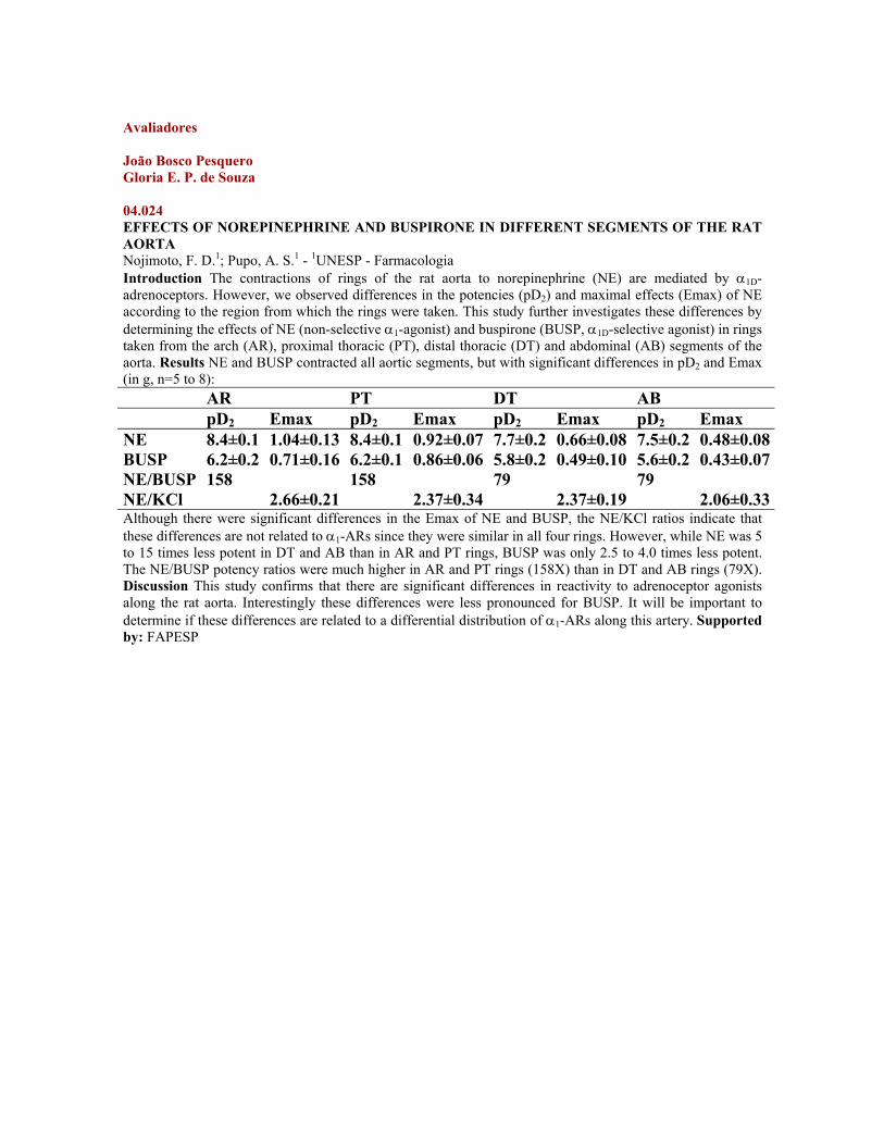
Avaliadores João Bosco Pesquero Gloria E. P. de Souza 04.024 EFFECTS OF NOREPINEPHRINE AND BUSPIRONE IN DIFFERENT SEGMENTS OF THE RAT AORTA Nojimoto, F. D.1; Pupo, A. S.1 - 1UNESP - Farmacologia Introduction The contractions of rings of the rat aorta to norepinephrine (NE) are mediated by α1D-adrenoceptors. However, we observed differences in the potencies (pD2) and maximal effects (Emax) of NE according to the region from which the rings were taken. This study further investigates these differences by determining the effects of NE (non-selective α1-agonist) and buspirone (BUSP, α1D-selective agonist) in rings taken from the arch (AR), proximal thoracic (PT), distal thoracic (DT) and abdominal (AB) segments of the aorta. Results NE and BUSP contracted all aortic segments, but with significant differences in pD2 and Emax (in g, n=5 to 8): AR PT DT AB pD2 Emax pD2 Emax pD2 Emax pD2 Emax NE 8.4±0.1 1.04±0.13 8.4±0.1 0.92±0.07 7.7±0.2 0.66±0.08 7.5±0.2 0.48±0.08BUSP 6.2±0.2 0.71±0.16 6.2±0.1 0.86±0.06 5.8±0.2 0.49±0.10 5.6±0.2 0.43±0.07NE/BUSP 158 158 79 79 NE/KCl 2.66±0.21 2.37±0.34 2.37±0.19 2.06±0.33Although there were significant differences in the Emax of NE and BUSP, the NE/KCl ratios indicate that these differences are not related to α1-ARs since they were similar in all four rings. However, while NE was 5 to 15 times less potent in DT and AB than in AR and PT rings, BUSP was only 2.5 to 4.0 times less potent. The NE/BUSP potency ratios were much higher in AR and PT rings (158X) than in DT and AB rings (79X). Discussion This study confirms that there are significant differences in reactivity to adrenoceptor agonists along the rat aorta. Interestingly these differences were less pronounced for BUSP. It will be important to determine if these differences are related to a differential distribution of α1-ARs along this artery. Supported by: FAPESP
04.025 EFEITOS DA TESTOSTERONA SOBRE A GERAÇÃO DE ESPÉCIES REATIVAS DE OXIGÊNIO EM CÉLULAS DA MUSCULATURA LISA VASCULAR DE RATOS WISTAR. Chignalia, A. Z.1; Schuldt, E. Z.1; Montezano, A. C. I.1; Yogi, A.1; Nigro, D.1; Carvalho, M. H. C.1; Fortes, Z. B.1; Muscara, M. N.1; Lopes, L. R.1; Tostes, R. C. A.1 - 1USP - Farmacologia Introdução: Altos níveis circulantes de testosterona são associados a doenças cardiovasculares como arteriosclerose, hipertensão e pré-eclampsia. Os andrógenos são capazes de induzir a formação de espécies reativas de oxigênio (EROs) em vários tecidos, mas existem poucas evidências a este respeito no sistema cardiovascular. Objetivo: Avaliar se a testosterona é capaz de induzir a formação de EROs em células de músculo liso vascular (CMLV). Métodos: As CMLV foram isoladas do leito mesentérico de ratos Wistar (12-16 semanas), após pré-digestão com solução enzimática para remoção da camada endotelial. As mesmas foram mantidas em cultura em meio Eagle modificado por Dulbecco e utilizadas nas passagens 4-7. A produção das EROs foi avaliada pelo método da hidroetidina e pela presença de substâncias reativas ao ácido tiobarbitúrico (TBARS). Resultados: A testosterona aumentou a produção de EROs nas CMLV e induziu a lipoperoxidação de modo concentração- (10-9 a 10-6 M) e tempo-dependente (1,5 a 12 horas) (p<0,05). A geração de EROs também ocorreu após estimulação com testosterona conjugada a albumina (T-BSA). O bloqueio dos receptores para andrógenos (AR) com flutamida 10uM diminuiu (p<0,05) a geração de EROs em ambos ensaios experimentais. Discussão: A testosterona aumenta a geração das EROs em CMLV. Este efeito parece ser mediado por AR presentes na membrana plasmática. Apoio Financeiro: FAPESP, CNPq.
04.026 EFEITO DO TEMPOL NO DESENVOLVIMENTO DA HIPERTENSÃO EXPERIMENTAL E DISFUNÇÃO ENDOTELIAL DE ANIMAIS 2R-1C. Costa, C. A.1; Silva, J. G. M.1; Ognibene, D. T.1; Tano, T.1; Soares de Moura, R.2; Resende, A. C.1 - 1UERJ - Farmacologia e Psicobiologia; 2UERJ - Farmacologia Introdução: Na hipertensão renal experimental dois rins, um clip (2R-1C), uma disfunção endotelial é observada pelo impedimento do relaxamento induzido por substâncias vasodilatadoras. Como o estresse oxidativo parece estar elevado nesse modelo de hipertensão, tivemos como objetivo avaliar o efeito do tratamento crônico do tempol, um superóxido desmutase mimético, sobre o desenvolvimento da hipertensão e disfunção endotelial. Método e Resultados: Ratos Wistar machos (150-180 g) controles (sham operados, n=5) ou submetidos à cirurgia 2R, 1C receberam tratamento diário (40 dias) com veículo (2R-1C, n=5) ou Tempol (2R-1C + Tempol, n=5) e tiveram a pressão arterial sistólica (PAS), diastólica e média aferidas por pletismografia de cauda. Os efeitos vasodilatadores da acetilcolina (Ach, 1-100 nmol) e nitroglicerina (NG, 1-100 nmol) foram estudados em leito arterial mesentérico (LAM) perfundido (McGregor, J. Physiol., 177:21,1965). A PAS (mm Hg) de animais 2R,1C foi maior (p<0.05) que a dos animais controles (189,6±5 vs 115,9±4) e o tempol preveniu o desenvolvimento da hipertensão (116,6±3). O efeito vasodilatador (% de relaxamento) reduzido da ACh (p<0.05) em animais 2R, 1C (10 pmol: 63,2±3 vs 10,5±3) foi parcialmente (p<0.05) recuperado pelo tempol (46,4±6), assim como o da NG (10 nmol: controle 46,05 vs 2R-1C 20±2 vs 2R-1C + tempol 38,7±4). Conclusão: Nosso estudo demonstra que o tempol previne o desenvolvimento da hipertensão e melhora a disfunção endotelial em animais 2R-1C Apoio Financeiro: FAPERJ
04.027 EFFECTS OF MATRIX METALLOPROTEINASES (MMPs) INHIBITION IN A CANINE MODEL OF ACUTE PULMONARY EMBOLISM (APE) Florencio, B. C.1; Dias-Junior, C. A.1; Lopes, L. F.2; Gerlach, R. F.3; Tanus-Santos, J. E.2 - 1USP - Farmacologia; 2FMRP - USP - Farmacologia; 3FORP - USP - Morfologia Introduction: APE is an important cause of morbidity and death in the world. Recent studies have addressed the relevance of pulmonary artery vasoconstriction in APE. A group of zinc-dependent endopeptidases called MMPs are involved in the degradation of components of extracellular matrix and it may play a role in the pulmonary vascular contractility. We examined the effects of the administration of doxycycline (Doxy - a MMPs inhibitor) following APE. Methods: Sham operated dogs (N=4) received only saline. APE was induced by autologous blood clots injected into the right atrium in the Emb group (N=9); Doxy group (N=3) received only doxy infusion; and Doxy + Emb group (N=10) received doxy before APE. Gelatin zymography of MMP-2 and MMP-9 from plasma samples was performed. Results: No significant hemodynamic changes were found in sham and Doxy groups. Embolization increased MPAP (from 7±1 to 28±2 mmHg) and the pulmonary vascular resistance index (PVRI, from 185±23 to 917±103 dynscm) in Emb group (both P<0,05). Doxy improved the cardiac index, reduced PVRI to 707±114 dynscm and MPAP to 22±2 mmHg, 120 min after APE in Doxy+Emb group (both P<0.05). Plasma pro-MMP-9 and MMP-9 levels increased in Emb group 120 min after APE and MMP-2 remained unaltered. However, doxy inhibited the MMP increase in the Doxy+Emb group. Discussion: Doxy-induced inhibition of MMPs attenuated the hemodynamic changes associated with APE, and indicates that MMP-9 may be a pharmacological target in APE. Supported by: FAPESP
04.028 NOVO DOADOR DE ÓXIDO NÍTRICO (NO) PROMOVE VASODILATAÇÃO VIA REDUÇÃO DO INFLUXO E AUMENTO DO ARMAZENAMENTO DE Ca2+. Girolineto, B. M. P.1; Vercesi, J. A.1; De Lima, R. G.2; Da Silva, R. S.3; Bendhack, L. M.2 - 1FCFRP - USP - Física e Química; 2USP - FCFRP; 3USP - Física e Química Introdução:O NO promove vasodilatação pela redução da concentração citoplasmática de Ca2+ e ativação de canais de K+. Analisamos os efeitos do NO liberado do composto [Ru (NH.NHq) (terpy)NO+](PF6)3 (terpy) sobre o influxo e liberação de Ca2+ intracelular em aorta de rato. Métodos:Estudamos os efeitos do terpy sobre a contração ativada com fenilefrina (PHE) em meio zero-Ca2+ e influxo de Ca2+ extracelular ativado com PHE 100 nM ou KCl 60 mM. Resultados:A contração ativada com PHE em zero-Ca2+ foi de 0,77 ± 0,08 g (n=5) na ausência de terpy e de 1,29 ± 0,22g (n=5) após incubação com terpy. Porém, o influxo de Ca2+ estimulado com PHE foi reduzido pelo terpy tanto no efeito máximo (Emax) de 2,16 ± 0,23g (n=5) para 1,50 ± 0,21g (n=5) como na potência (pD2) de 3,16 ± 0,03 (n=5) para 3,06 ± 0,04 (n=5). Por outro lado, o terpy aumentou o influxo de Ca2+ extracelular estimulado com KCl (Emax: de 2,13 ± 0,47g; n=5 para 3,14 ± 0,58g; n=5) e pD2 de 3,11 ± 0,05 (n=5) para 3,27 ± 0,06 (n=5). Discussão:A PHE estimula a liberação de Ca2+ intracelular e influxo de Ca2+ extracelular via canais de Ca2+ operados por receptores. Alta concentração extracelular de KCl ativa canais de Ca2+ operados por voltagem e inibe canais de K+. Conclusão:Os resultados indicam que o doador de NO promove relaxamento da aorta de rato pelo aumento do armazenamento de Ca2+ nos estoques intracelulares sensíveis à PHE e redução do influxo Ca2+ extracelular estimulado com PHE e envolve a ativação de canais para K+. Apoio Financeiro: CNPq e FAPESP.
04.029 EFEITOS DE TRATAMENTO CRÔNICO COM ATENOLOL E ENALAPRIL SOBRE A DENSIDADE CAPILAR MUSCULAR E CUTÂNEA DE RATOS ESPONTANEMENTE HIPERTENSOS (SHR) Sabino, B. D.1; Tibiriça, E.1 - 1FIOCRUZ - Fisiologia e Farmacodinâmica Introdução: A rarefação dos vasos da microcirculação é uma alteração característica da hipertensão arterial primária. Investigamos os efeitos de um tratamento crônico com os anti-hipertensivos atenolol (ATE) e enalapril (ENA) sobre a densidade capilar média (DCM) cutânea e muscular esquelética (grácil) de SHR. Métodos: Ratos SHR machos com 12-14 semanas, receberam tratamento oral com ATE (50 mg/kg/dia), ENA (10 mg/kg/dia) ou veículo (grupo controle) durante 4 semanas. Após o término do tratamento avaliou-se a DCM funcional através de microscopia intravital por epi-iluminação com fluorescência. Resultados: O tratamento reduziu a pressão arterial sistólica dos ratos SHR [196 ± 6 mmHg para 162 ± 4 mmHg (n=10) e de 204 ± 6 mmHg para 156 ± 4 mmHg (n=10), com ATE ou ENA, respectivamente, P<0.05]. Foi observado aumento da DCM muscular esquelética de ratos SHR tratados com ATE ou ENA (302 ± 16 e 285 ± 9 capilares/mm2, respectivamente) comparados com o grupo controle (248 ± 11 capilares/mm2, P<0.05). Observou-se aumento da DCM cutânea (269 ± 16 e 283 ± 17 capilares/mm2, tratados com ATE e ENA, respectivamente) comparados com o grupo controle (201 ± 14 capilares/mm2, P<0.05). Discussão: O presente estudo demonstrou que o tratamento crônico com os anti-hipertensivos ATE ou ENA induz aumento da DCM funcional cutânea e muscular esquelética em ratos SHR. Estamos investigando os efeitos sobre a densidade capilar estrutural destes tratamentos através de análises histológicas. Apoio Financeiro: IOC/FIOCRUZ
04.030 RELAXATION ACTIVATED BY A NEW NITRIC OXIDE DONOR IS NOT ALTERED IN HYPERTENSIVE L-NAME RATS AORTA. Ferezin, C. Z.1; Bonaventura, D.2; De Lima, R. G.3; Da Silva, R. S.4; Bendhack, L. M.3 - 1FCFRP - USP - Física e Química; 2USP - Farmacologia; 3USP - FCFRP; 4USP - Física e Química Introduction: The ruthenium complex [Ru(NH.NHq)(terpy)NO+]PF6)3 (Terpy) releases nitric oxide (NO) inducing vascular relaxation. In this study, we evaluated the relaxation induced by Terpy in isolated rat aorta after chronic inhibition of NO synthesis with the inhibitor L-NAME. Methods: Rats were made hypertensive by administration of L-NAME (50 mg kg−1 per day) for 3 weeks in drinking water and normotensive (control) rat received only water. We analyzed the relaxation induced by Terpy in denuded rat aortic rings. In phenylephrine-contracted arteries, cumulative concentration-effect curves for Terpy were obtained before and after incubation for 30 min with the selective inhibitor of soluble guanylyl-cyclase, ODQ (10-6M) or the NO scavenger oxyhemoglobin (HbO2 10-5M). We analyzed the maximum effect (ME) and potency (pD2) induced by Terpy. Results: L-NAME treated rats presented an increase in blood pressure and decrease in body weight. The relaxation induced by Terpy was similar between L-NAME and control rat aorta rings and it was not altered by HbO2. However, ODQ inhibited the relaxation induced by Terpy in control (ME: 96,8 ± 1,7 and pD2: 5,2 ± 0,2) and in L-NAME aortic rings (ME: 97,0 ± 4,7 and pD2: 5,4 ± 0,4). Conclusion: These results indicate that the relaxation to the NO donor Terpy is not altered in the aortas of rats submitted to chronic inhibition of NO synthase. Terpy releases NO extracellularly and its relaxation involves guanylyl-cyclase activation. Supported by: FAPESP, CNPq and Universidade de São Paulo.
04.031 ALTERNATIVE PATHWAY TO ACE FOR ANGIOTENSIN II GENERATION IN SHR CAROTID ARTERY. Becari, C.1; Sivieri-Jr., D. O.1; Bispo-da-Silva, L. B.1; Salgado, M. C. O.1 - 1FMRP - USP - Farmacologia Introduction: Alternative pathways to angiotensin-converting enzyme (ACE) involved in angiotensin (Ang) II generation have been extensively demonstrated and include serine proteases like human chymase and rat elastase-2. We investigated the Ang II forming enzymes in carotid artery isolated from spontaneously hypertensive rats (SHR). Methods: Cumulative concentration curves (10-10-10-6M) to Ang I, Ang II or [Pro11-D-Ala12]-Ang I (PDA, an ACE-resistant substrate that is cleaved by chymase and elastase-2) were obtained in carotid rings from SHR in the absence or presence of proteases inhibitors. mRNA expression for the different components of renin-angiotensin system was obtained by RT-PCR. Results: Ang II and its precursors produced a concentration-dependent vasoconstrictor effect in carotid of SHR that was abolished by losartan (1 µM). Captopril (10 µM) altered the responses induced by Ang I (PD2=8.7±0.19 vs 7.03±0.14 in controls, p<0.001) but did not affect those induced by PDA and Ang II. In the presence of the serine protease inhibitor chymostatin (100 µM), the effects induced by Ang II was not altered while the concentration-response curve to Ang I was shifted to the right (PD2=8.4±0.12 vs 7.9±0.05, p<0.05). The mRNA for rat elastase-2, ACE, AT1 and AT2 Ang II receptors were detected in carotid arteries. Discussion: Ang II formation from Ang I is essentially dependent on ACE although an alternative chymostatin-sensitive pathway, most probably elastase-2, is also present in SHR carotid artery. Supported by: FAPESP.
04.032 TYPE 1 DIABETIC PATIENTS HAVE IMPAIRED MICROVASCULAR FUNCTION IN THE LOWER EXTREMITIES Gomes, M. B.1; Tibiriça, E.2 - 1UERJ - Endocrinologia; 2FIOCRUZ - Fisiologia e Farmacodinâmica Introduction: Impaired microvascular function and structure is known to be correlated with late complications in patients with type 1 diabetes mellitus (DM1). Methods: We studied skin capillary density and recruitment in response to arterial and venous occlusion in the upper and lower extremities of patients with DM1. This cross-sectional observational study included 34 (28.2 ± 10.7 years) consecutive outpatients with DM1 (duration 12.8 ± 8.6 years) under treatment and 33 age- and sex-matched healthy controls. We used intravital video microscopy to measure basal (functional) and maximal (during venous occlusion) skin capillary densities and capillary recruitment (after post-occlusive reactive hyperemia) in the dorsum of the fingers and toes. Results: Baseline capillary densities (number/mm2) were not different between controls and patients, either in the fingers (121.0 ± 3.6 and 124.1 ± 4.1, respectively; P=0.572) or toes (82.6 ± 3.6 and 92.6 ± 3.9; P=0.066). In contrast, capillary recruitment (% increase of number/mm2) was significantly higher in controls compared to patients both in fingers (7.7 ± 1.4 and 1.4 ± 1.0, respectively; P <0.001) or toes (9.7 ± 2.8 and 0.1 ± 1.8; P=0.005). During venous occlusion, capillary density increased significantly in toes of controls but not DM1 patients (10.7 ± 2.7 and -3.1 ± 2.3; P<0.001). Discussion: It is concluded that patients with DM1 present structural capillary rarefaction in the lower but not upper extremities. Moreover, functional capillary reserve is absent in both extremities of DM1 patients suggesting that diabetic capillaries at rest are already recruited maximally. Supported by: FIOCRUZ
04.033 INFLUENCE OF NITRIC OXIDE (NO) ON THE RELAXATION OF RAT ISOLATED DETRUSOR SMOOTH MUSCLE (DSM) MEDIATED BY β-ADRENERGIC RECEPTOR (β-AR) AGONISTS. Bau, F. R.1; Monica, F. Z. T.1; Nucci, G. de1; Bricola, A. A. de O.2; Zanesco, A.3; Antunes, E.1 - 1UNICAMP - Pharmacology; 2PUCCamp - Pharmacology; 3UNESP - Physical Education Goals: Long-term NO inhibition has been associated with hypersensitivity of DSM to muscarinic agonists. β-AR activation leads to DSM relaxation, thus contributing to urine storage during bladder filling. The aim of this study was to examine the effect of long-term NO inhibition on the relaxation responses of the DSM induced by β-AR. Method: Male Wistar rats (200-350g) were treated orally with L-NAME (20 mg/rat/day) for 30 days. Age-matched control animals received tap water alone. DSM strips were pre-contracted with KCl (80 mM), and concentration-response cumulative curves in DSM to isoptroterenol (ISO) (non-selctive β-agonist; 100 pM-10 µM) and BRL 37344 (β3-agonist; 100 pM-10 µM) were done. The of pEC50 and maximal responses (Emax) were calculated. Results: The long-term treatment of L-NAME caused a significant increase in the tail blood pressure. The potency of ISO in control (6.40±0.05; n=5-6) did not differ from L-NAME-treated rats (6.39±0.17; n=6). The Emax for ISO was also unaffected by L-NAME treatment (105.72±4.86 and 103.03±4.98, to control and L-NAME-treatment, respectively). There were no significant differences in the pEC50 values and Emax for BRL 37344 from controls and L-NAME-treated rats (pEC50: 7.17±0.33 to 6.61±0.19; Emax: 76.54±5.23 and 61.89±4.62 respectively). Conclusion: NO does not modulate β-AR responses in the DSM.
04.034 RELAXING EFFECTS OF SILDENAFIL ANALOGUES IN THE RABBIT ISOLATED AORTA Flores Toque, H. A.1; Priviero, F. B. M.1; Teixeira, C. E.2; Antunes, E.1; Nucci, G. de1 - 1UNICAMP - Farmacologia; 2Medical College of Georgia - Physiology Goal: Sildenafil (SILD), a cGMP-specific phosphodiesterase type 5 (PDE5) inhibitor, is used as oral therapy for penile erectile dysfunction due to its ability to relax the erectile time. However, several side effects have been related. In this work, we aimed to investigate the effects of two SILD anologues (nomely SILD-11 and -12) in rabbit aorta (RbA). Method: Thoracic RbA was isolated and cut in rings. Aortic rings (AR) were mounted in organ baths filled with Krebs Solution. Data were recorded in a PowerLabâ system. Concentration-response curves (CRC) to SILD, SILD-11 and SILD-12 (1 nM to 10 mM) were constructed in the absence or in the presence of L-NAME (NO synthesis inhibitor) or ODQ (soluble guanylyl cyclase inhibitor) in endothelium-intact and -denuded (E-) aortic rings. Results: In the E+, SILD-11 and SILD-12 induced relaxations in a concentration-dependent manner, with potency (pEC50) values of 7.10 ± 0.10 and 7.21 ± 0.09, respectively, which were similar to SILD (7.25 ± 0.07). The maximal responses (Emax) to SILD-11 (65 ± 5%) and SILD-12 (69 ± 6%) were also similar to sildenafil (76 ± 8%). Endothelium denudation reduced the Emax values and caused a rightward shift in the CRC for SILD, SILD-11 and SILD-12 (4, 5 and 4-fold, respectively). Addition of either L-NAME or ODQ reduced pEC50 in E+ rings at the same magnitude as the endothelium denudation. However, no additional shift was seen in E- rings. Conclusions: The vasorelaxant responses induced by SILD-11 and SILD-12 are similar to sildenafil and partly involve endothelium intregrity. Supported by: Fapesp
04.035 ESTUDO DA PARTICIPAÇÃO DO ÓXIDO NÍTRICO NA ATIVIDADE CARDIOVASCULAR INDUZIDA POR MILONINA EM RATOS Cavalcante, H. M. M.1; Lima, R. P. C.1; Furtado, F. F.1; Ribeiro, T. P.1; Nunes, X. P.1; Barbosa Filho, J. M.1; Medeiros, I. A.1 - 1UFPB - Tecnologia Farmacêutica Introdução: Estudo preliminar demonstrou que milonina, alcalóide obtido das folhas de Cissampelos sympodialis Eichl., produziu efeito vasorelaxante dependente de endotélio em artéria mesentérica superior de rato. Métodos: Ratos Wistar (250-350g) foram anestesiados com tiopental sódico (45mg/Kg, i.v.) e catéteres de polietileno foram inseridos na aorta abdominal e veia cava inferior, para medida da pressão arterial e administração de drogas. Anéis (2-3 mm) de artéria mesentérica superior de rato foram obtidos, suspensos por hastes de platina, mantidos em cubas com solução Tyrode, gaseificada com carbogênio, a 37°C, sob tensão de 0,75g. Todos os dados foram registrados em sistema computacional de aquisição e tratamento. Resultados: Milonina (0,1; 0,5; 1; 5 e 10 mg.Kg-1 i.v., randomicamente) produziu hipotensão (-7±1,3; -9±0,5; -14± 0,5; -17±1,5 e -40±0,9 mmHg) associada com bradicardia (-9±0,9; -12±0,5; -21±7,4; -35±4,1 e -140±9 bpm) A hipotensão foi significantemente atenuada após L-NAME (20 mg.Kg-1,i.v.). Em anéis mesentéricos, milonina (10-14-3.10-4 M) antagonizou (CI50=2,3±0,4x10⁻⁶M) as contrações induzidas por fenilefrina (10 µM). Esta efeito vasorelaxante foi atenuado após o pré-tratamento dos anéis intactos com L-NAME (100 µM), hidroxicobalamina (30µM) ou ODQ (10µM) (3,2±0,5x10⁻⁵, 3,1±0,8x10⁻⁵, 2,3±0,6x10⁻⁵ M, respectivamente). Discussão: Esses resultados sugerem que o efeito hipotensor induzido por milonina é provavelmente devido a vasodilatação periférica, a qual é, em parte, a liberação de NO pelo endotélio vascular. Apoio Financeiro: CNPq
04.036 CARDIAC TROPONIN I RELEASE IS RELATED TO THE SEVERITY OF ACUTE PULMONARY THROMBOEMBOLISM (APT) Uzuelli, J. A.1; Dias-Junior, C. A.2; Tanus-Santos, J. E.1 - 1FMRP - USP - Farmacologia; 2USP - Farmacologia Introduction: patients with pulmonary thromboembolism have elevated cardiac troponin I concentrations. However, a precise relationship between the plasma levels of troponin I and the severity of APT has not been addressed yet. Methods: APT was induced in mongrel dogs with increasing autologous blood clots volumes (0, 1, 3, and 5 ml/kg) injected into the right atrium (Control, Emb1, Emb3, and Emb5 groups, respectively). Hemodynamic evaluations were carried out for two hours. Serum troponin I concentrations were measured by fluorometric enzyme immunoassay. Results: Control group no showed significant changes throughout study period. APT produced dose-dependent pulmonary hypertension. Mean pulmonary arterial pressure increased from 10±3 mmHg to 18±4, 25±3 and 29±2 mmHg in Emb1, Emb3 and Emb5 groups, respectively. Correspondingly, troponin I increased from non detectable concentrations to 1±0,2, 1,6±0,4 and 3±0,8 ng/mL in Emb1, Emb3 and Emb5 groups, respectively. Discussion: Our findings suggest that troponin I increases after APT depend on the severity of lung embolization, thus suggesting that troponin I is a marker of severity in APT. Supported by: CAPES and CNPq
04.037 EFFECTS INDUCED BY MESOIONIC 2-(4-CHLOROPHENYL)-3-METHYL-4-(4-METHOXYPHENYL)-1;3-THIAZOLIUM-5-THYOLATE (CMMTT) IN DIFFERENT MODELS OF HYPERTENSION Cavalcante, K. V. M.1; Correia, N. A.2; Silva Filho, J. C.3; Dias, K. L. G.1; Lira, B. F.1; Silva, D. F.4; Araujo, I. G. A.1; Medeiros, I. A.1 - 1UFPB - Tecnologia Farmacêutica; 2UFPB - Fisiologia e Patologia; 3UFPB - Tecnologia Farmaceutica; 4UFPB - Tecnologia Farmacêutica / Ciências Farmacêuticas Introduction: Hypertension is a major risk factor for cardiovascular mortality and morbidity and several classes of antihypertensive have been investigated. This study was designed to investigate the effects induced by CMMTT on cardiovascular parameters (blood pressure and heart rate) in two-kidney, one-clip renovascular hypertensive (2K1C-Goldblatt) and L-NAME-7th days hypertensive rats. Methods: 2K1C was induced by clipping the left renal artery during 4 weeks, while control rats were sham-operated. L-NAME 7th days hypertension was obtained after treatment of the rats with L-NAME (100 mg/kg/day, by gavage), while the control group received water. At the end of 4th week or 7th day, the animals were submitted to a surgery for insertion of polyethylene catheters into the abdominal aorta and inferior vena cava for blood pressure recordings and administration of drugs. Results: Acute administration of CMMTT (0.001; 0.005; 0.01; 0.05; 0.1; 0.5; 1; 5; 10 mg/Kg – i.v.) was able to induce hypotension followed by an increase in the heart rate in 2k1C hypertensive rats, and this response was of similar magnitude when compared to controls. However, in the L-NAME 7th day hypertension model, the hypotensive effect was significantly higher when compared to controls. Conclusion: Our findings indicate that L-NAME hypertensive rats are more sensitive to CMMTT compared to 2K1C hypertensive rats. Nevertheless, additional experiments are necessary to clarify the mechanisms involved in this response. Supported by: CAPES, CNPq, UFPB
04.038 ALTERATIONS IN THE RAT CORONARY VASODILATATORY CAPACITY DURING THE EARLY PHASE OF CARDIAC HYPERTROPHY. Bispo-da-Silva, L. B.1; Sivieri-Jr., D. O.1; Becari, C.1; Prado, C. M.2; Rossi, M. A.2; Salgado, M. C. O.1 - 1FMRP - USP - Farmacologia; 2FMRP - USP - Patologia Introduction: Bradykinin (BK) type-2 receptor (B2) is present in cardiomyocyte and nonmyocyte cells of the heart and BK posses cardiac antitrophic and vasodilatatory actions. We evaluated cardiac B2 receptor mRNA expression and the reactivity of coronary arterial bed to BK, ATP and sodium nitroprusside (SNP), 1 day after suprarenal aorta coarctation (CO) Methods: Rats were submitted to CO or to sham surgery (S). Cardiomyocyte diameter was determined, 1 and 7 days after CO. Cardiac B2 mRNA expression was determined by RT-PCR. Blood pressure was measured in anesthetized rats. Vasodilatatory responses to BK, ATP and SNP were studied in isolated hearts. Results: CO increased cardiac B2 receptor mRNA expression (1.8-folds, n=3-4) and blood pressure (131±4 vs 118±4 mmHg, n=10). Cardiomyocyte diameter increased only 7 days after surgery (12.9±0.3 vs 11.8±0.2 μm, n=7-10). Two populations of CO animals concerning coronary response to BK (60 pmol) were observed: one hyporesponsive (fall in perfusion pressure in %: 19.8±1.6, 5 of 7 animals) and other normoresponsive (35.7±3.6%; 2 of 7 animals) when compared with S (41.9±2.8%, n=6). The same profile was observed with ATP and SNP administration. Discussion: The increase in B2 receptor mRNA expression observed in the early phase of cardiac hypertrophy is not related to increases on coronary responsiveness to BK. Moreover, most of the CO animals exhibited vascular alteration characterized by a decrease in coronary vasodilatatory capacity. Supported by: Capes and FAPESP.
04.039 FEN: UMA ARILETANOLAMINA ORTO-SUBSTITUÍDA EQUIPOTENTE AO PROPRANOLOL Kiuchi, M. G.1; Gonçalves, B. T.2; Silva, J. F. M.2; Sudo, R. T.1; Zapata-Sudo, G.1 - 1UFRJ - Farmacologia Básica e Clínica; 2UFRJ - Química Orgânica/Instituto de Química INTRODUÇÃO: As ariloxipropanolaminas apresentam maior potência como beta-bloqueadores que as ariletanolaminas. Este trabalho apresenta seis ariletanolaminas orto-substituídas que foram avaliadas na contratilidade do músculo papilar de ratos e comparadas ao propranolol. MÉTODOS: Músculos papilares foram dissecados e montados em cubas preenchidas com solução Tyrode (pH= 7,4, a 37°C), oxigenada (95%O2/5%CO2) para registro de tensão isométrica. Os abalos musculares foram obtidos com estimulação elétrica (40-50 V, 2 ms de duração e 1,0 Hz) e armazenados em computador para análise utilizando o programa Axoscope. Após estabilização dos abalos musculares, concentrações crescentes das ariletanolaminas foram adicionadas a preparação (10 a 500 µM). A amplitude dos abalos foi comparada antes e depois da exposição às substâncias. RESULTADOS: Dentre as ariletanolaminas testadas, a FEN foi mais potente em reduzir a contratilidade do músculo papilar. A concentração inibitória de 50% da amplitude dos abalos musculares (IC50) foi de 38.6 ± 11.1; 184.9 ± 64.8; 447,2 ± 64,9; 79,2 ± 9,6; 102,9 ± 25,0 e 289,6 ± 89,7 µM para FEN; 4-MeO-FEN; 4-iPr-FEN; 4-Cl-FEN; 4-NO2-FEN e 4-Me-FEN, respectivamente. FEN foi equipotente ao propranolol que apresentou IC50=39.0 ± 4.3 µM. Arritmias foram observados na presença de 4-iPr-FEN, 4-NO2-FEN e 4-Me-FEN. CONCLUSÃO: FEN que possui núcleo ariletanolamina mostrou-se com potência similar ao propranolol em deprimir a atividade cardíaca sugerindo atividade beta-bloqueadora. Apoio Financeiro: IM-INOFAR, CAPES, Pronex-Rio, CNPq, FAPERJ
04.040 IMPROVEMENT OF ERECTILE RESPONSES IN RATS SUBMITTED TO REGULAR PHYSICAL TRAINING. Claudino, M. A.1; Pena, C. B.1; Camargo, E.1; Priviero, F. B. M.1; Teixeira, C. E.2; Nucci, G. de1; Zanesco, A.1; Antunes, E.1 - 1UNICAMP - Pharmacology; 2Medical College of Georgia - Physiology Goals: We have previously shown that regular physical exercise significantly increases the relaxations of isolated rat corpus cavernosum mediated by endogenous nitric oxide release (Claudino et al., 2004), but no studies investigable the effects of exercise training on the in vivo erectile responses. Thus, the aim of this work was evaluate the erectile response in rats submitted to treadmill run training. Methods: Wistar rats were divided into 2 groups: sedentary (SD) and trained (TR) groups. The training program consisted in 8 weeks of treadmill run training, 5 days/week, and each session lasted 60 min. The erectile function was assessed by measuring the rise in intracavernous pressure (ICP) following cavernous nerve electrical stimulation. Plasma nitrite and nitrate (NOx) concentration was quantified by Griess methods. Blood pressure was monitored by both a tail-cuff method and by systemic mean arterial pressure (MAP). Results: A significant increase in ICP was observed in trained animals (2960 ± 247 mmHg.s; 0.38 ± 0.04 ICP/MAP), compared with sedentary group (2107 ± 219 mmHg.s; 0.32 ± 0.03 ICP/MAP). The treadmill run training also significantly increased the plasma NOx levels by approximately 30 % compared with sedentary group. Systolic bloody pressure and systemic mean arterial pressure did not change by physical training. Conclusions: Our findings suggest that dynamic exercise improve the erectile responses in vivo, by mechanisms possibly involving overproduction of nitric oxide. Supported by: FAPESP
04.041 SEROTONIN-INDUCED CONTRACTION IS ENHANCED IN SINOAORTIC DENERVATED RAT AORTA Rocha, M. L.1; Bendhack, L. M.2 - 1USP - FMRP; 2USP - FCFRP Introduction: The sinoaortic denervation (SAD) induces arterial pressure lability (APL) without sustained hypertension. The aim of the present study was to verify the vascular responses to serotonin (SER), angiotensin II (AII) and KCl in aortas from SAD and sham-operated (SO) rats and to verify the effect of the endothelium removal on this response. Methods: The arterial pressure was recorded 3 days after the SAD or sham-operation. Aortas were quickly removed and concentration-effect curves to SER (10-8 to 10-4 M), AII (10-10 to 10-7 M) and KCl (4.7 to 120 mM) were constructed in intact endothelium (E+) or denuded arteries (E-) from SAD and SO rats. We analyzed the maximum effect (Emax) and potency (pD2) of the contractile agents. Results: Only the SAD rats presented arterial pressure lability and both rat groups remained normotensive. The pD2 and Emax induced by AII and KCl were similar for SAD and SO either in E+ and E-. However, in the response induced by SER the SAD rat aortas E- presented higher value of Emax (2.08±0.16g, n=6) than SO (1.26±0.1g, n=7). No differences were observed between SAD E+ and SO E+. The pD2 values to SER were similar in E- and E+ SAD and SO. Oscillatory contractions were induced by all the contractile agents in SAD and SO rat aortas with and without endothelium, when the agonists and KCl were used in the intermediary range of concentration. Conclusions: Contractile responses to AII and KCl were similar in SAD and SO rat aortas, independently of endothelium. The efficacy of SER is higher in SAD than in SO, only in denuded arteries. Supported by: FAPESP and CNPq.
04.042 EVALUATION OF CARDIAC REMODELLING AFTER NO SYNTHASE INHIBITION BY RAMAN SPECTROSCOPY Ramos, L.1; Labat, R.1; Bittar, R.2; Martin, A.2; Lopes-Martins, R. A. B.1 - 1USP - Farmacologia; 2UNIVAP - IP&D Introduction: It has been clearly established that chronic inhibition of nitric oxide synthesis results in increases in blood pressure, changes in myocardial contractility, cardiac remodeling and fibrosis. The experimental treatment of rats with L-Arginine analogs is one of the most common models employed to induce hypertension, however, the presence of cardiac hypertrophy still controversial. The aim of the present study was to verify the effects of nitric oxide inhibition through oral L-NAME administration on the cardiac tissue of rats, and the possible prevention by L-Arginine. Methods: Thirty male Wistar rats were used. Saline, L-NAME or L-NAME + L-Arginine were orally administered by gavage daily for 4 weeks. At the end of the treatments the animals were anesthetized, intubated and artificially ventilated. The invasive arterial pressure was monitored. After the end of the hemodynamic recordings the animals were sacrificed by a lethal dose of anesthetics. The hearts were removed. Soon after, the hearts were dissected, obtaining the total heart weights. The quantitative evaluation of the myocardial collagen was made using the classical Hematoxilin-Eosin and Sirius red dye. We also used the Raman spectroscopy (FT-Raman Spectrometer RFS 100; Bruker, Germany) as a second technique to evaluate collagen deposition. Results: The administration of L-NAME induced increases in arterial pressure that could be partially reverted by L-arginine. We didn’t observe cardiac hypertrophy, but histological analyses showed a wide but diffuse increases in interstitial collagen in L-NAME treated group, partially prevented by Arginine administration. These results were better demonstrated by FT-Raman Spectroscopy that revealed a sharp increase in collagen contents in L-NAME treated hearts. Conclusion: Our results demonstrates that NO-synthesis inhibition was able to produce cardiac remodeling well demonstrated by Raman Spectroscopy. Supported by: FAPESP 05/02117-6
04.043 CHARACTERIZATION OF THE PRESSOR RESPONSE TO URIDINE ADENOSINE TETRAPHOSPHATE (UP4A) IN VARIOUS BLOOD VESSELS Leite, R.1; Linder, A. E.1; Todd Jr, J. K.1; Webb, R. C.1 - 1Medical College of Georgia - Physiology The uridine adenosine tetraphosphate (Up4A), a dinucleotide that contains both purine and pirimidine moieties, described as a novel potent nonpeptidic vasoconstrictor has been shown to be released from the endothelium upon chemical and mechanical stimulation, suggesting a potential role as an endothelium-derived contracting factor (EDCF). It has been shown that Up4A has a potent vasoconstrictor effect in isolated perfused rat kidney but its effect in rat and mouse aorta has not been characterized yet. In this study we characterize the Up4A contractile response in isolated aortic rings from normotensive animals. Aortic rings were isolated from Sprague Dawley rats (250-275 g) and C57BL/6 mice (30-35g), cleaned and prepared for isometric tension recordings. Concentration-effect curves to Up4A (10-8 to 10-4M) were performed in aortic rings both in the presence and in the absence of the endothelium before and after treatment with an ATP sensitive P2X1 receptor blocker NF279 (10-4M), NOS inhibitor L-NNA (10-4M), Rho Kinase inhibitor Y-27632 (10-6M) or NADPH oxidase inhibitor apocynin (10-4M). Concentration-effect curves to tempol (2.5X10⁻³-1.510-2M) were performed in endothelium denuded aortic rings contracted with Up4A (10-5M). Up4A contractile response is not tachyphylactic and is significantly potentiated by endothelium removal or treatment with L-NNA. Treatment with NF279, Y-27632 or apocynin significantly impaired the Up4A-induced contraction in these vessels. Tempol caused relaxation in a concentration dependent manner in endothelium denuded aortic rings contracted with Up4A (maximum relaxation 39%). These data suggest that the functional endothelium and nitric oxide are key modulators of the contractile effect of the putative EDCF Up4A, and that this effect is mediated mainly by P2X1 receptor and involves superoxide formation and Rho Kinase pathway activation. Supported by: NIH (P01 HL074167)

04.044 VASCULAR HYPER-REACTIVITY FOLLOWING ARTERIAL BALLOON INJURY: ROS AND PROSTANOIDS PARTICIPATION ON DISTANT AND DALAYED EFFECTS Olivon, V. C.1; de Andrade, C. R.2; Fukada, S. Y.3; Cunha, F. de Q.4; Ramalho, L.4; Laurindo, F. R. M.2; Oliveira, A. M. de1 - 1USP - FCFRP; 2USP - INCOR; 3FMRP - USP - Farmacologia; 4USP - FMRP INTRODUCTION:Balloon catheter injury induced hyper-reactivity to Phenylefrine (Phe)(a) in contralateral (CL) artery and an increase in the innervation of these arteries as well as an increased of density of PGP, CGRP and SP(b) contained in these nerves. It is well known that SP, one pro-inflammatory agents, induces ROS production. METHODS:Arteries from control (CO) adult rats and arteries contralateral (CL) to balloon catheter injury after 4 days. The study was performed “in vitro” using arteries with endothelium (E+) and without endothelium (E-) and in presence or absence of SC-560 (COX-1 selective inhibitor) and SC-236 (COX-2 selective inhibitor). The ROS production was measuring reactive oxygen species by oxidative fluorescent dye dihydroethidium (DHE) and HPLC analysis. RESULTS: Phe Emax was increased after balloon injury in CL when compared to CO (see table) Control Emax Phe Emax Phe + SC-560 Emax Phe + SC-236 E+ 0,39 ± 0,02 g 0,38±0,02g 0,36±0,02g E- 0,58 ± 0,02 g 0,61±0,03g 0,63±0,06g Contralateral Emax Phe Emax Phe + SC-560 Emax Phe + SC-236 E+ 0,61 ± 0,06 g* 0,54±0,03g 0,40±0,03g E- 0,40 ± 0,03g* 0,43±0,02g* 0,36±0,05g*
*significant difference when compared to respective control (One-way ANOVA –Newman-Keuls Post Test). In ROS production was observed a significant increase in DHE fluorescence (2151,64±110,17 in CL and 1471,6050 ± 62,18 in CO – arbitrarily measured) and O2
- formation (367,2±202,27 in CL and 507,6 ±129,1 in CO) in HPLC analysis. DISCUSSION: Data obtained indicate that prostanoids are involved on hyper-reactivity to Phe in CL after balloon injury. Additionally, ROS production can contribute on this enhancement. We have also shown this effect is dependent on endothelium. (a) ACCORSI-MENDONCA,D. et. al. British Journal of Pharmacology, 142, 79-88, 2004. (b) MILNER, P. et. al.. J. Vasc. Res. , 34, 31-34, 1997 Supported by: CNPq

04.045 NICOTINE DOES NOT STIMULATE MATRIX METALLOPROTEINASE (MMP)-9 RELEASE BY PLATELETS Palei, A. C. T.1; Gerlach, R. F.2; Tanus-Santos, J. E.1 - 1FMRP - USP - Farmacologia; 2FORP - USP - Morfologia e Histologia Introduction: Nicotine is known to activate platelets. We have previously shown that nicotine increases MMP-9 levels in plasma when it is incubated with whole blood. Here we examined whether nicotine induces the release of MMP-2 and MMP-9 by platelets. Methods: Blood samples were drawn from eight healthy volunteers into citrate tubes and centrifuged (800 g for 15 min) to separate platelet rich plasma (PRP) samples, which incubated at room temperature for 30 min with vehicle, nicotine 50 nM, or 150 nM. After incubation, PRP samples were centrifuged (2000 g for 5 min) and gelatin zymography of MMP-2 and MMP-9 from supernatant were performed under non-reducing conditions on 7% polyacrylamide co-polymerized with gelatin 1%, as the substrate. Gels were washed Triton X-100, incubated at 37°C for 16 h in Tris–CaCl2 buffer and stained with Coomassie Blue. Enzyme activity was assayed by densitometry. The results for each subject were normalized by the results obtained after incubation with vehicle. Results: Incubation with nicotine 50 nM and 150 nM produced no significant % changes in MMP-2 (99±4% and 98±5% of vehicle, respectively) and in MMP-9 (93±14% and 122±21% of vehicle, respectively) activities. Discussion: Our results show that nicotine does not affect plasma MMP levels by inducing platelet release of MMP-2 and MMP-9. Supported by: CNPq, FAPESP, CAPES.
04.046 EFEITOS DO EXTRATO HIDRO-ALCOÓLICO DO CAROÇO DO AÇAÍ (Euterpe oleracea) NO DESENVOLVIMENTO DA HIPERTENSÃO ARTERIAL DE RATOS SHR E NA LIBERAÇÃO DE NO POR CÉLULAS ENDOTELIAIS Rocha, A. P. M.1; Carvalho, L. C. dos R. M. de1; Sousa, M. A. V.1; Sousa, P. J. C.2; Madeira, S. V. F.1; Schini-Kerth, V.3; Tano, T.1; Resende, A. C.1; Soares de Moura, R.4 - 1UERJ - Farmacologia e Psicobiologia; 2UFPA - Farmácia; 3Université Luis Pasteur - Pharmacologie; 4UERJ - Farmacologia Introdução: Resultados anteriores demonstram que o extrato hidro-alcoólico de caroço de açaí (ASE) induz efeito vasodilatador e anti-hipertensivo em ratos tratados com L-NAME, 2R-1C e SHR. Desta forma avaliamos o efeito do tratamento crônico com ASE, logo após o final da amamentação, e uma possível ação liberadora de óxido nítrico (NO) em células endoteliais umbilicais humanas. Metodologia e Resultados: Ratos SHR (n = 9), imediatamente após desmame, foram tratados com ASE (200 mg/kg/dia pela via oral, na água de beber e ração ad libitum) durante 3 meses, quando tiveram sua pressão arterial medida (mm Hg) pelo método não invasivo. O mesmo procedimento foi feito com um grupo controle (n = 8) que recebeu somente água e alimentação ad libitum. Os ratos tratados com ASE apresentaram pressões sistólica (157,27± 9,35), diastólica (121,79 ± 10,48) e média (129,9 ± 8,36) significativamente inferior aos ratos controles que apresentaram pressões sistólica (168,45 ± 5,13), diastólica (125,12 ± 6,52) e média (139,28 ± 5,87) (p< 0,05). A formação de NO por células endoteliais do cordão umbilical humano, induzida pelo ASE foi avaliada pela técnica de ressonância de spin eletrônico, em células tratadas com o extrato e com o L-NAME mais o extrato. Células endoteliais tratadas com 100 μg/ml de ASE apresentaram um aumento de 100% na formação de NO quando comparadas com o controle. No entanto, a liberação de NO induzida pelo ASE foi reduzida em células tratadas com L-NAME (100 μg/ml). Conclusão: A análise dos resultados nos permite sugerir que os efeitos anti-hipertensivo e vasodilatador do ASE, possa ser dependente de uma liberação significativa de NO. Apoio Financeiro: CNPq, FAPERJ
04.047 ALTERAÇÕES DA REATIVIDADE VASCULAR COM NICOTINA: EFEITOS DA INIBIÇÃO DE METALOPROTEINASES. Jacob Ferreira, A. L. B.1; Martinez, M. L. L.1; Palei, A. C. T.1; Gerlach, R. F.2; Tanus-Santos, J. E.1 - 1FMRP - USP - Farmacologia; 2FORP - USP - Morfologia e Histologia Nicotina pode ativar e aumentar a expressão das metaloproteinases da matriz extracelular (MMPs). A ativação de MMPs produz tendências à vasoconstrição. No presente estudo, examinamos os efeitos da inibição de MMPs, pela doxiciclina, nas alterações de reatividade vascular associadas ao tratamento com nicotina. Ratos Wistar foram divididos em 4 grupos, recebendo água ou nicotina (em água, 10mg/kg/dia), combinado com tratamento com salina ou doxiciclina 30mg/kg (i.p.), durante 30 dias. Os animais foram decapitados, tendo sua aorta extirpada. Um anel desta foi imediatamente utilizado para a verificação da reatividade vascular à acetilcolina (Ach) (10-10M a 10-5M), utilizando-se fenilefrina 10-4M para induzir contração da aorta previamente. O grupo nicotina-salina apresentou um efeito máximo de relaxamento bem menor que o grupo controle água-salina; e a Ach apresentou menor potência de relaxamento sobre este grupo. O grupo nicotina-doxiciclina apresentou uma ligeira, porém significativa, melhora na reatividade vascular à Ach, que se apresentou mais potente, quando comparada ao grupo nicotina-salina. Estes achados sugerem que as MMPs participam, ao menos em parte, da redução de reatividade vascular associada ao tratamento com nicotina. Apoio Financeiro: CNPq, FAPESP, CAPES.
04.048 TIENILHIDRAZONAS REDUZEM DEPRESSÃO CARDÍACA INDUZIDA PELO INFARTO DO MIOCÁRDIO. Gabriel, D.1; Silva, J. S.1; Kummerle, A. E.2; Fraga, C. A. M.2; Barreiro, E. J.2; Sudo, R. T.2; Zapata-Sudo, G.1 - 1UFRJ - Faculdade de Farmácia - Farmacologia Básica e Clínica; 2UFRJ - Faculdade de Farmácia - LASSBio Introdução: O acúmulo de Ca2+ no retículo sarcoplasmático (RS) é reduzido em músculo cardíaco de ratos submetidos ao infarto do miocárdio (IM) levando a depressão cardíaca. As alterações funcionais e histológicas dos músculos cardíacos foram avaliadas em ratos infartados tratados ou não com as substâncias N-acilidrazonas, LASSBio294 e LASSBio785. Métodos: Ratos Wistar machos foram submetidos a ligadura da artéria coronária descendente (grupo I). As substâncias (1 mg/kg) foram administradas via i.p. logo após a cirurgia durante 2 ou 4 semanas. Para a análise histopatológica, os corações foram corados com hematoxilina e eosina ou tricrômio de Gomori. Fibras cardíacas desnudas foram preparadas para registro de tensão isométrica. O acúmulo de Ca2+ no RS foi avaliado em fibras de ratos falso-operados (FO) e I tratados ou não com as substâncias. Resultados: O IM provocou o aparecimento de intenso infiltrado celular e depósito de colágeno que foi reduzido após tratamento com LASSBio294 e 785. A relação peso do coração e corporal dos ratos foi reduzida de 7,6±0,5 do grupo I para 4,0±0,3 g/kg do grupo I tratado com LASSBio294 semelhante ao grupo FO (4,0±0,1g/kg). A redução do acúmulo de Ca2+ causado pelo IM foi revertido com tratamento dos ratos I com LASSBio294 e 785. A resposta contrátil da cafeína aumentou de 10,0±1,0% do grupo I para 63,0±1,0% da resposta máxima no grupo I tratado com LASSBio294. Conclusão: LASSbio294 e 785 parecem retardar o processo de fibrose e impedir a depressão cardíaca após IM. Apoio Financeiro: IM-INOFAR, CAPES, Pronex-Rio, CNPq, FAPERJ.
04.049 ATIVIDADE CARDIOVASCULAR DO EREMANTOLÍDEO C EM ANIMAIS HIPERTENSOS De Paula, D. C. C.1; Vidal, A. T.1; Guimarães, H. N.2; Saude-Guimaraes, D. A.1; Grabe-Guimaraes, A.1 - 1UFOP - Farmácia; 2UFMG - Engenharia de Elétrica A insuficiência cardíaca (IC) pode ser tratada por digitálicos, que apresentam baixo índice terapêutico. O Eremantolídeo C (EREC) isolado de Lychonophora trichocarpha apresenta em sua estrutura química um anel lactônico de cinco membros semelhante aos digitálicos. O objetivo do presente trabalho foi avaliar a atividade cardiovascular do EREC e sua segurança terapêutica. Para a determinação da toxicidade aguda do EREC, camundongos albinos receberam por via IP as doses 8 x 10⁻², 16 x 10-2, 4 x 10⁻¹, 8 x 10⁻¹, 16 x 10-1, 5, 10 e 20 mg/kg. Nenhum dos animais veio a óbito e não foram observados sinais de toxicidade. Para a avaliação da atividade cardiovascular do EREC, ratos Wistar machos foram tratados pelo L-NAME (60 mg/kg/dia por 7 dias) e 3 meses após, foram anestesiados, os sinais de pressão arterial (PA) e ECG (DII) foram obtidos, e receberam o EREC (1 mg/kg, IV). Os animais tratados com L-NAME (n=4) apresentaram supra ou infra-desnivelamento do segmento ST do ECG, hipertensão arterial (PAS=144 ± 6,8 e PAD=92 ± 7,8 mmHg) que não foi alterada até 30 min após a administração do EREC (PAS=152 ± 3,8 e PAD=102 ± 17,4 mmHg). Não foram observadas alterações significativas para a FC (374 + 10,7 X 359 + 7,8) e QT do ECG (controle 4,79x10-2 X 4,69x10-2 após EREC). Os resultados demonstram a segurança do EREC em animais apresentando o fator de risco hipertensão arterial para a IC. Indicam ainda a maior segurança terapêutica do EREC em relação aos digitálicos, já que as maiores doses avaliadas não induziram óbito nos camundongos. Sugerimos o EREC como um promissor objeto de estudo como inotrópico para a terapêutica da IC. Apoio Financeiro: PIP/UFOP, FAPEMIG.
04.050 DIFFERENCES BETWEEN ANGIOTENSIN I AND II METABOLISM IN CARDIAC PERFUSATE OF RATS. Sivieri-Jr., D. O.1; Bispo-da-Silva, L. B.1; Becari, C.1; Oliveira, E. B.2; Salgado, M. C. O.1 - 1FMRP - USP - Farmacology; 2FMRP - USP - Biochemistry and Immunology Introduction: We evaluated angiotensin (Ang) I and II metabolism in cardiac perfusate (CP) from normotensive Wistar (NWR, n=8) and spontaneously hypertensive rats (SHR, n=6). Methods: The heart was isolated and reperfused with Krebs solution for 2 h. Then, the perfusate was concentrated and incubated with Ang I or II (30 nmol). The reaction products were analyzed by HPLC (nmol), in the absence or presence of proteases inhibitors [captopril (CPT), phosphoramidon (PHO) and MGTA: 10 mM; Chymostatin (CHY): 100 mM]. Results: The products formed from Ang I were (NWR vs. SHR): Ang II (2.4±0.2 vs. 2.97±0.51), Ang 1-9 (0.87±0.4 vs. 0.73±0.12), Ang 1-7 (1.01±0.21 vs. 0.38±0.03; p<0.05) and Ang 5-10 (0.77±0.15; only in NWR). CPT decreased Ang II formation in both groups, while CHY decreased it only in SHR. None of the inhibitors used altered Ang 1-9, Ang 1-7 and Ang 5-10 production. Ang II metabolism in PC from SHR was less (p<0.05) than NWR and only two fragments were generated: Ang 1-7 (0.34±0.07 vs. 1.28±0.26) and Ang 5-8 (0.45±0.04 vs. 1.38±0.28) and none of the inhibitors used altered this profile. Discussion: Although different carboxy- and endo-peptidases are involved in Ang degradation in CP, angiotensin-converting enzyme is the major proteolytic activity involved in Ang I metabolism in both groups. In addition, a serine protease is also involved in Ang I to II conversion, only in CP from SHR. Supported by: CAPES and FAPESP.
04.051 EFFECTIVE TREATMENT INCREASES SKIN CAPILLARY DENSITY IN ESSENTIAL HYPERTENSIVE PATIENTS Debbabi, H.1; Levy, B. I.1; Tibiriça, E.2 - 1Université Paris VII - INSERM U689; 2FIOCRUZ - Fisiologia e Farmacodinâmica Microvascular rarefaction is a hallmark of essential hypertension (EH). We measured skin capillary density in non-diabetic hypertensive subjects with effective antihypertensive treatment and evaluated possible correlations with arterial blood pressure (BP). This cross-sectional observational study included 76 (55 ± 1 years) consecutive outpatients with EH under chronic antihypertensive treatment (BP<140/90 mmHg), 24 age- and sex-matched patients with recently discovered and never-treated EH and 70 normotensive (BP<140/90 mmHg) age- and sex-matched healthy controls. We used intravital video microscopy to measure basal and maximal (after venous occlusion) skin capillary densities in the dorsum of the fingers. Baseline and maximal capillary densities (number/mm2) were significantly lower (59.6 ± 2.0 and 62.0 ± 1.9) in untreated than in treated EH patients (74.0 ±1.4 and 79.4 ± 1.5; P<0.001) and than in normotensives (68.2 ± 1.5 and 72.4 ± 1.5; P<0.001). Based on multiple regression analysis, after adjustment to tobacco consumption, aortic (but not brachial) systolic BP was inversely correlated with basal and post-occlusive capillary densities in normotensive subjects. In hypertensives, this correlation disappears and capillary density was influenced by two independent variables, antihypertensive drug treatment and overweight. In hypertensives, capillary density is reduced in association with a cluster of cardiovascular risk factors involving tobacco consumption and obesity. The increased capillary density in treated hypertensives suggests that a cause to effect relationship between BP and capillary density should be evaluated in a long term prospective follow-up. Supported by: INSERM - FIOCRUZ
04.052 EFFECT INDUCED BY N-P-NITROPHENYLMALEIMIDE (4-NO2-NPM) ON BLOOD PRESSURE AND HEART RATE IN RATS Ribeiro, E. A. N.1; Furtado, F. F.2; Oliveira, A. P.3; Cechinel-Filho, V.4; Noldin, V. F.5; Medeiros, I. A.6 - 1UFPB - Ciências Farmacêuticas / Laboratório Tecnologia Farmacêutica; 2UFPB - LTF; 3UFPB - Ciências Farmacêuticas; 4UNIVALI - NIQFar; 5UNIVALI - Centro Ciências da Saúde; 6UFPB - Tecnologia Farmacêutica Introduction: The maleimide 4-NO2-NPM was synthesized through the reaction between the anhidride maleic and 4-nitre anilyne. Several biological activities such as analgesic, anti-spasmodic, ansiolitic have been attributed to the maleimides. Cardiovascular effects of intravenous treatment with 4-NO2-NPM were investigated in normotensive rats. Methods: Male Wistar rats (250-350 g) were anesthetized and polyethylene catheters were inserted into the abdominal aorta and into the inferior vena cava for blood pressure measurements and administration of drugs. Blood pressure and heart rate were obtained by using a computer set and CVMS software. Results: In normotensive rats (n=6), 4-NO2-NPM (0,01; 0,05; 0,1; 0,5, 1 mg/kg, i.v.) induced a significant and dose-independent hypotension (-21±7; -12±2; -10±2, -17±4, -40±6 %) associated with bradycardia (-35±12; -6±2; -10±3, -9±1 and -60±11 %, n=6, respectively). Hypotensive (-11±2; -4±1; -6±2; -7±2, -9±4 %) and bradicardic (-1,4±0,4; -1,2±0,3; -0,9±0,3; -1,4±0,3, -2±0,6 %) response were significantly attenuated with atropine (2 mg/Kg; i.v.) or hexamethonium (20 mg/Kg; i.v.) (-5± 1; -4±2; -3±0,7; -5±1; -20±3 % or -3±1; -2±0,2; -2±0,8, -1±0,3; -10±3 %, n=6, respectively). The hypotensive response was atenuated after nitric oxide (NO) synthase blockade, L-NAME (20mg/Kg, i.v.). Conclusion: These results indicate that hypotension induced by 4-NO2-NPM, is probably due to a decrease of peripheral vascular resistance and bradycardia, as a consequence of cardiac muscarinic receptor activation. Supported by: CAPES and CNPq.
04.053 INCREASED SENSITIVITY TO THE NO-INDEPENDENT SGC STIMULATOR BAY 41-2272 IN AORTA FROM eNOS MICE. Priviero, F. B. M.1; Teixeira, C. E.1; Hilgers, R. H. P.1; Semze, S. M.1; Webb, R. C.1 - 1Medical College of Georgia - Physiology AIM: We investigated the mechanisms by which endogenous nitric oxide (NO) affects the vasorelaxation elicited by the sGC stimulator BAY 41-2272 in wild-type (WT) mice and endothelium or neuronal NO synthase knockout mice (eNOS and nNOS, respectively). METHODS: Endothelium-intact (E+) and denuded (E-) rings were mounted in myographs. Data were recorded in a PowerLab system. cGMP was measured using EIA kits. Real-time PCR and western blot were used to assess gene and protein expression of sGC subunits. RESULTS: BAY 41-2272 (0.001-1 µM) relaxed E+ rings with pEC50 values of 7.53 ± 0.03 (WT), 7.76 ± 0.06 (eNOS) and 7.56 ± 0.04 (nNOS). In E- rings, the curve for BAY 41-2272 was shifted to the right in WT (7.08 ± 0.05) and nNOS (7.15 ± 0.03) but not in eNOS (7.71 ± 0.06). The sGC inhibitor ODQ (10 µM) displaced the curve for BAY 41-2272 to the right in both E+ and E- rings of WT (30- and 10-fold), eNOS (80- and 20-fold) and nNOS (30- and 6-fold). The NO synthesis inhibitor L-NAME (100 µM) inhibited the relaxations of BAY 41-2272 in rings from WT and nNOS, but not eNOS. Incubation of E- rings with BAY 41-2272 (0.001-1 µM) or sodium nitroprusside (SNP, 0.001-1 µM) caused a rightward shifts in the contractile responses to phenylephrine (PE, 0.001-10 µM). Co-incubation of BAY 41-2272 and SNP caused a synergistic rightward shift in the curves to PE (5.6-fold) along with an increase in cGMP levels in an ODQ-sensitive manner. Increase in cGMP in response to BAY 41-2272 was significantly higher in eNOS aorta, compared to WT and nNOS. Real-time PCR and Western blot analysis revealed a decreased expression of both subunits of sGC at the mRNA and protein level, respectively. CONCLUSION: BAY 41-2272 relaxes the mouse aorta synergistically with NO. Despite the decreased expression of sGC, this study demonstrates that higher enzyme activity accounts for the increased sensitivity to NO-independent sGC stimulators in eNOS aorta. Supported by: HL-74167
04.054 EFFECTS OF THE TOTAL VENOM OF BOTHROPS MARAJOENSIS CARDIOVASCULAR SYSTEM IN NORMOTENSIVE RATS. Aguiar, F. L. N.1; Evangelista, I. L.2; Santos, C. F.1; Kerntof, M. R.1; Monteiro, H. S. A.2; Nascimento, N. R. F.1 - 1UECE - Instituto Superior de Ciências Biomédicas ; 2UFC - Farmacologia e Fisiologia Envenoming by Bothrops leads to systemic manifestations being responsible for the primary cause of death after snakebite. Bothrops marajoensis (Bm) is a snake found in “Marajó” island located in North Brazil. In the present study the effects of the whole venom (BmWV) was evaluated in arterial blood pressure (MAP), eletrocardiographic parameters (ECG) and isolated perfused heart. MAP was registered by means of a pressure transducer and the ECG was registered using subcutaneous platinum electrodes in a DII lead. The isolated heart was perfused in an open Langendorff system. The injection of BmWV (1, 3, 30 and 100 mg/Kg) induced a dose-related decrease in MAP (from 104.3±11.5 mmHg to 85.1 ± 17, 78±17, 55.7±8.1 and 45.9±7.5 mmHg, respectively). The main alterations found in the ECG was atrioventricular blockade and atrial tachycardia, In higher doses (30 and 100 mg/Kg ) we observed QRS widening and ST elevation and extra-systoles (less than 25%). The injection of BmWV in the perfused heart at concentrations ranging from 0.01 to 10 mg induced cardiac depression with 75.2 ± 11% (p<0.05) reduction in amplitude contraction with an increase of 88.1 ± 4.5% in coronary perfusion pressure (p<0.05). This data suggests that the cardiovascular effects of BmWV are related to a direct cardiac depression probably related to coronary spasms and also to cardiac action potential conduction blockade. Supported by: Capes, CNPq, Funcap
04.055 PRODUÇÃO DE O2
- REDUZ EFEITO RELAXANTE DO DOADOR DE ÓXIDO NÍTRICO NA AORTA DE RATOS 2R-1C. Rodrigues, G. J.1; De Lima, R. G.1; Lunardi, C. N.1; Da Silva, R. S.1; Bendhack, L. M.1 - 1FCFRP - USP - Física e Química Introdução: Ânions O2
- reagem com óxido nítrico (NO) produzindo um poderoso oxidante, o peroxinitrito. O NO pode ser armazenado em cavéolas nas células do músculo liso vascular. Investigamos a interferência do O2
- no relaxamento induzido pelo doador de NO [Ru(NH.NHq)(terpy)NO+]PF6)3 (Terpy) quando as cavéolas estão desorganizadas pela methyl-β-ciclodextrina (CD) e o efeito do antioxidante vitamina-C (VitC) em aortas de ratos hipertensos 2R-1C e normotensos 2R. Métodos: Em aortas sem endotélio contraídas com fenilefrina, realizamos curvas concentração-efeito para o Terpy na ausência e após incubação com CD (10mM) por 60 min e/ou com vitC (100mM) por 20 min. Analisamos a potência (pD2) e o efeito máximo (EM) para o Terpy. Resultados: O pD2 do Terpy foi menor na aorta de ratos 2R-1C (6,55 ± 0,06; n=7) do que em 2R (7,05 ± 0,07; n=6), mas o EM foi semelhante nos dois grupos. Em 2R a CD reduziu o pD2 para 6,50 ± 0,09 (n=6) e o EM de 108,6 ± 2,0% para 97,9 ± 1,6% (n=6). A CD não teve efeito sobre o relaxamento ao Terpy em 2R-1C. A combinação de CD e vitC aumentou o pD2 em 2R para 6,98 ± 0,04 (n=5) e em 2R-1C de 6,34 ± 0,11 (n=6) para 6,83 ± 0,15 (n=5), sem alterar o EM em relação à CD. A vitC isoladamente, aumentou o pD2 do Terpy somente na aorta de 2R-1C (6,91 ± 0,06; n=6) sem efeito sobre o EM. Conclusões: Na aorta de ratos 2R-1C a cavéola pode estar desorganizada e não sofrer o efeito da CD. Ocorre maior concentração de O2
- sensível à vitC na aorta de ratos hipertensos 2R-1C. Apoio Financeiro: CAPES, FAPESP, CNPq.
04.056 AMLODIPINE IMPAIRS THE ANTIMIGRATORY EFFECT OF POTASSIUM DICLOFENAC IN WISTAR RATS. Rodrigues, S. F.1; Ceravolo, G. S.1; Rastelli, V. M. F.1; Carvalho, M. H. C.1; Nigro, D.1; Tostes, R. C. A.1; Nucci, G. de2; Fortes, Z. B.1 - 1USP - Farmacologia; 2UNICAMP - Farmacologia Verapamil does not interfere with antimigratory effect of diclofenac in Wistar rats [Martinez et al.,Hypertension, 34:997,1999]. To investigate if this occurs with other calcium channel blocker we tested amlodipine on diclofenac effect. Male Wistar rats were divided into four groups: vehicle, potassium diclofenac 1mg/kg, amlodipine 10mg/kg and diclofenac plus amlodipine, treated for 15 days (v.o.). The blood pressure (BP) was evaluated by indirect tail-cuff method. Leukocyte rolling, adherence and migration were studied by intravital microscopy. Plasma concentration of diclofenac and amlodipine were analyzed by mass spectometry. Neither treatment altered the BP or leukocyte rolling. Amlodipine reduced leukocyte migration (6.6±1.0,n=13*) vs vehicle (10.8±0.8,n=21). Diclofenac decreased adherence (6.6±0.8,n=12*) vs vehicle (10.5±0.6,n=15) and also migration (6.6±0.7,n=12*). However, when amlodipine and diclofenac were combined, the reduction of adherence (9.3±0.8,n=10) and migration (9.3±0.6,n=13) induced by diclofenac were lost. The plasma concentration of diclofenac associated (219±62ng/mL,n=7) or not (217±38ng/mL,n=7) to amlodipine was similar. In conclusion, in contrast to verapamil, amlodipine impairs the reduction of leukocyte adherence and migration caused by diclofenac in Wistar rats, in a dose that does not interfere with the BP, possibly by to interferer with the expression of any adhesion molecule. Key words: Leukocyte, Wistar, amlodipine, diclofenac. *P<0.05. Acknowledgement: FAPESP/PRONEX
04.057 AUMENTO DA REATIVIDADE VASCULAR E DA HIPOTENSÃO ARTERIAL INDUZIDO PELO TRAMADOL EM RATOS HIPERTENSOS Raimundo, J. M.1; Trachez, M. M.2; Pontes, L. B.1; Sudo, R. T.1; Zapata-Sudo, G.1 - 1UFRJ - Farmacologia Básica e Clínica; 2UFF - Anestesiologia Introdução. Tramadol, em concentrações clinicamente relevantes, não produz efeitos tóxicos no sistema cardiovascular em ratos Wistar normotensos. Este trabalho investiga os efeitos cardiovasculares do tramadol em ratos espontaneamente hipertensos (SHR). Métodos. Aortas isoladas de ratos SHR (220-280g) foram preparadas para registro de tensão isométrica. A contratura da aorta induzida por fenilefrina (Fe; 10 µM) ou KCl (40 mM) foi medida antes e após a exposição a concentrações cumulativas de tramadol (0,1–1 mM). Os parâmetros hemodinâmicos, após injeção venosa de tramadol (1–10 mg/kg), foram avaliados através do registro do eletrocardiograma e da medida das pressões sistólica (PS) e diastólica (PD) em ratos SHR. Resultados. Tramadol promoveu relaxamento vascular não dependente do endotélio. A concentração inibitória média (CI50) foi 0,47±0,08 e 0,44±0,03 mM em aortas com e sem endotélio contraídas com Fe. Quando a contratura da aorta foi induzida por KCl, a CI50 foi 0,13±0,01 mM (P<0,05). Em ratos Wistar normotensos, tramadol produziu relaxamento em aortas com endotélio somente com 1 mM. Na dose de 10 mg/kg, PS e PD foram reduzidas de 155,0±5,2 para 117,0±8,9 mmHg (P<0,01) e de 107,0±7,1 para 45,0±12,9 mmHg (P<0,05), respectivamente, em ratos SHR. Em contraste, nos ratos normotensos, não foram observadas alterações significativas na pressão arterial. Discussão. O efeito vasodilatador do tramadol foi mais intenso nos ratos SHR resultando em maior redução da PD. Apoio Financeiro: Cristália, CAPES, FAPERJ, CNPq e FUJB.
04.058 MOLECULAR EVIDENCE FOR THE PARTICIPATION OF THE NO-PATHWAY IN THE VASORELAXANT RESPONSE INDUCED BY CMMTT Cavalcante, K. V. M.1; Correia, N. A.2; Dias, K. L. G.1; Lira, B. F.1; Silva, D. F.3; Neto, M. A.4; Bendhack, L. M.5; Medeiros, I. A.1 - 1UFPB - Tecnologia Farmacêutica; 2UFPB - Fisiologia e Patologia; 3UFPB - Tecnologia Farmaceutica; 4FCFRP - USP - Física e Química; 5USP - FCFRP Introduction: Previous studies realized by our group demonstrated an important participation to the endothelium, through the NO-cGMP pathway, in the vasorelaxant effect induced by Mesoionic 2-(4-chlorophenyl)-3-methyl-4-(4-methoxyphenyl)-1;3-thiazólium-5-thiolate (CMMTT) in isolated rat superior mesenteric arteries. The aim of this study was to confirm, the participation of the calcium and NO-pathway activation in the vasorelaxant response induced by CMMTT. Methods: Rat superior mesenteric artery rings were removed, placed in a 12-well plates containing Tyrode’s solution, and placed in humidified atmosphere of 5% CO2 and 95% air, at 37ºC and the total amount of NOx in the medium was determined using a purge system (model NOA 280i). Endothelial cells were isolated from superior mesenteric arteries of rats, maintained in Petri dishes and loaded with FURA-2/AM to measure intracellular calcium concentrations. Results: CMMTT (10-6-10-5M) was able to significantly increase NOx levels in isolated rat mesenteric artery rings, and these effects were completely abolished after removal of vascular endothelium. However, CMMTT was not able to induce intracellular calcium concentration increases in endothelial cells of mesenteric arteries loaded with FURA-2 AM. Conclusion: These results show an important participation of the endothelium with increase NOx levels CMMTT-induced in rat superior mesenteric artery rings ; nevertheless are necessary new studies by investigate the mechanism envolved by activation of the NOS. Supported by: CAPES, CNPq, UFPB, USP-RP
04.059 ROLE OF BETA-ADRENERGIC AND MUSCARINIC RECEPTORS ON ARRHYTHMIA INDUCED ELECTRICALLY IN THE RIGHT ATRIUM OF SPONTANEOUSLY HYPERTENSIVE RAT (SHR). Viviani, A. G.1; Galvao, K. M.2; Godoy, C. M. G.1; Caricati-Neto, A. C.2 - 1UMC - Núcleo de Pesquisas Tecnológicas (NPT); 2UNIFESP - EPM - Farmacologia Introduction and Goals: Arterial hypertension is characterized by several autonomic dysfunctions and represents main risk factor in the development of atrial arrhythmia (Andrews et al., Mount Sinai J Med 73: 482, 2006). In order to investigate the role of autonomic mechanisms on the atrial arrhythmia in hypertension, we studied the effects of blockers of beta-adrenergic (propranolol) and muscarinic (atropine) receptors on arrhythmia electrically induced in the right atrium of hypertensive (SHR) and normotensive (NWR) rats. Methods: Right atrium of SHR (N=36) and NWR (N=10), age: 9 months, were isolated and mounted in perfusion chamber containing Krebs-Henseleit solution (at 36.5oC, pH 7.4, 95% O2 + 5% CO2) between two platinum electrodes for electrical field stimulation (250 rectangular voltage pulses, 66.7 Hz, 5 ms duration) for induction of atrial arrhythmia. On this arrhythmia, the effects of propranolol (0.6microM) and atropine (1microM) were studied. Results: The rate of arrhythmia induction (RAI: number of successful arrhythmia induction experiments divided by the total number of experiments, multiplied by 100%) was higher in SHR (95%) than in NWR (86%). Stimulation strength (SS: amplitude of the voltage pulses) necessary to induce arrhythmia was equal to 5-fold atrium stimulation threshold in NWR and SHR. RAI, but not SS, was changed by pretreatment of atria with propranolol (0.6microM) or atropine (1microM) in NWR and SHR. Propranolol (0.6microM) reduced RAI (80%) in SHR (from 95% to 80%) and increased (from 86% to 100%) in NWR. Atropine (1microM) augmented RAI in NWR (from 86% to 100%), but not in SHR. Conclusion: These results suggest that right atrium of hypertensive animals are more susceptible to arrhythmias in relation to normotensive animals. In addition, the results suggest that, in relation to NWR, the autonomic modulation of atrial activity, mediated by muscarinic and beta-adrenergic receptors, is attenuated in hypertensive animals. Supported by: FAEP/UMC, CAPES and FAPESP
04.060 AÇÃO DE DERIVADOS DA ISATINA NA REATIVIDADE VASCULAR Pontes, L. B.1; Sudo, R. T.1; Ribeiro, N.2; Pinto, A. C.2; Zapata-Sudo, G.1 - 1UFRJ - Farmacologia Básica e Clínica; 2UFRJ - Instituto de Química Introdução. A isatina é uma substância endógena que interfere com a concentração intracelular de GMPc através da inibição de receptores de peptídeos natriuréticos. Como o nível de GMPc apresenta um papel importante no relaxamento vascular, os cetais sintetizados foram testados na reatividade vascular em ratos. Métodos. Aortas isoladas de ratos Wistar (220-280g) foram preparadas para registro de tensão isométrica. A contratura da aorta induzida por fenilefrina (10 µM) foi medida antes e após a exposição a concentrações cumulativas da isatina e dos derivados testados (0,5–1000 µM). Em algumas preparações, as aortas foram pré-incubadas com NG-nitro-l-arginine methyl ester (L-NAME) e 1H-[1,2,4]-oxadiazole-[4,3-α]-quinoxalin-1-one (ODQ), antagonistas da óxido nítrico sintase e da guanilato ciclase, respectivamente. Resultados. Os cetais ao contrário da isatina promoveram relaxamento do músculo liso vascular de forma dependente da concentração. Os derivados 5Cl-CEG, OMe-CEG e 4,6Br-CPD na concentração de 100 µM reduziram a resposta contrátil vascular para 20,9±8,6; 54,9±7,3 e 43,3±3,9% do controle (P<0,05). A ação vasodilatadora do 5Cl-CEG foi parcialmente dependente do endotélio. A concentração inibitória média foi de 122,0±25,6 µM em aortas sem endotélio e de 138,9±26,0; 136,9±17,5 e 190,5±7,4 µM na presença de L-NAME, ODQ e indometacina, respectivamente. Discussão. A liberação de óxido nítrico, e conseqüente ativação da guanilato ciclase e a produção de prostaciclina parecem estar envolvidos no mecanismo de vasodilatação do 5Cl-CEG. Apoio Financeiro: CAPES, FAPERJ, CNPq, FUJB.
04.061 CARDIOPROTECTIVE EFFECTS OF FENTANYL: PHARMACOLOGICAL EVIDENCE FOR THE INVOLVEMENT OF CENTRAL AND PERIPHERAL OPIOID RECEPTORS Lessa, M. A.1; Tibiriça, E.1 - 1FIOCRUZ - Fisiologia e Farmacodinâmica We investigated the involvement of central versus peripheral opioid receptors (OR) in the effects of fentanyl (FENT) in a model of myocardial ischemia/reperfusion (I/R) injury associated to pharmacologically-induced sympathetic overactivity through intracerebroventricular (icv) injection of L-glutamate in anesthetized rabbits submitted to 35 min of coronary occlusion followed by 120 min of reperfusion. Rabbits received naloxone HCl icv (n=9) or naloxone methiodide iv (n=9), a quaternary compound that does not cross the blood-brain barrier, 5 min before FENT treatment (5 or 50 µ/kg, iv; n=9 each). Infarct area was reduced only by FENT 50 (from 51 ± 2 to 24 ± 2%). This protective effect was abolished by peripheral (42 ± 4%) but not central OR blockade (32 ± 3%). The number of premature ventricular complexes (PVCs) during the ischemic period (54 ± 3) was reduced by FENT 50 (19 ± 7), an effect blunted by central (40 ± 3) but not peripheral (18 ± 7) blockade of OR. Mortality rate (50%) and incidence of ventricular tachycardia (55%) were completely abolished by FENT 50, but were not modified neither by central nor by peripheral OR blockage. Statistics analyses were performed using two-way ANOVA and Fisher´s Exact Test. It is concluded that fentanyl presents cardioprotective effects mainly characterized by central antiarrhythmic and peripheral antiischemic actions. Supported by: CAPES
04.062 RESPONSIVENESS OF RAT PULMONARY AND MESENTERIC ARTERIES AFTER PULMONARY ISCHEMIA REPERFUSION: EFFECT OF PHYSICAL TRAINING Delbin, M. A.1; Moraes, C.1; Camargo, E.2; Antunes, E.2; Nucci, G. de2; Zanesco, A.1 - 1UNESP - Educação Física; 2UNICAMP - Farmacologia The aim of this work was to evaluate the effect of physical preconditioning in the responsiveness of rat pulmonary and mesenteric rings submitted to lung ischemia reperfusion (IR). METHODS: Rats were divided in Sham (SHAM/SD); sedentary IR (SD/IR) and trained IR (TR/IR) animals. Run training (RT) consisted in 60 minutes/day, 5 days/week for 8 weeks. Left pulmonary IR was performed by occluding for 90 minutes and reperfusion for 2 hours. Concentration-response curves to acetylcholine (ACh), sodium nitroprusside (SNP) and phenylephrine (PHE) were obtained. RESULTS: RT reduced body weight gain (SD: 399±4.1; TR: 343±5.1). In mesenteric rings: an increase of potency for ACh was found in RT group (TR/IR: 7.25±0.06) compared to sedentary groups (SD/IR: 6.95±0.03; SHAM/SD: 7.05±0.10) without changes in the EMAX. Neither the potency nor EMAX were modified for SNP in all groups. The potency for PHE was increased in RT group (7.18±0.06) compared to SD/IR group (6.83±0.05). In pulmonary rings, no changes for ACh were seen in all groups. On the other hand, the potency for SNP was significantly increased in TR/IR group (8.23±0.06) compared to SD/IR group (7.85±0.04). Contractile responses mediated by PHE were markedly decreased in IR groups (SD/IR: 6.75±0.06, TR/IR: 6.62±0.04) compared to SHAM/SD (7.33±0.05). CONCLUSION: Our findings show that RT associated with lung IR promotes differentiate relaxing and contractile responses in mesenteric and pulmonary rings. Supported by: FAPESP
04.063 HIPORREATIVIDADE À ANGIOTENSINA II EM AORTA DE RATAS AO FINAL DA GESTAÇÃO DEPENDE DE ÓXIDO NÍTRICO ENDOTELIAL Correa, J. W. N.1; Salgado, M. C. O.1 - 1USP - FMRP Introdução: Avaliar a participação de prostanóides, NO e dos receptores AT2 da angiotensina (Ang) II na resposta à Ang II em aorta isolada de ratas Wistar grávidas (19o-20o dia de gestação, G) e não grávidas na fase estro do ciclo estral (NG). MÉTODOS: Após determinação da pressão arterial (PA), foram obtidas curvas concentração-resposta à Ang II em anéis de aorta com ou sem endotélio de G e NG na presença ou ausência dos antagonistas dos receptores AT1 (losartan, 1 mM) e AT2 (PD123,319, 1 mM), dos inibidores da ciclooxigenase (diclofenaco, 100 mM) e da NOS (L-NNA, 100 mM). RESULTADOS: Ratas G apresentaram PA mais baixa que as NG (84±5 vs 116±9 mmHg, N=12, P<0,0001). As respostas vasoconstritoras à Ang II estavam deprimidas em aorta de G comparadas a NG (Emáx= 245±34 vs 579±77 mg, N=9, P<0,05) e não foram afetadas por PD123,319 (313±54 vs 464±84 mg, N=9) ou diclofenaco (251±49 vs 495±44 mg, N=7). Entretanto, a remoção da camada endotelial (1140±135 vs 1284±86 mg, N=6) e L-NNA (1089±41 vs 1113±109 mg, N=4) isoladamente ou associado a diclofenaco (996±162 vs 1169±112 mg, N=4) potencializaram as respostas à Ang II, abolindo as diferenças entre G e NG. Os efeitos induzidos por Ang II foram abolidos na presença de losartan. CONCLUSÕES: A produção de NO endotelial, e não a de prostanóides ou ativação dos receptores AT2, é fator decisivo para o desenvolvimento da hiporreatividade aórtica à Ang II ao final da gestação de ratas. Apoio Financeiro: CNPq, Fapesp
04.064 OSCILLATORY CONTRACTIONS INDUCED BY PHENYLEPHRINE IN SINOAORTIC DENERVATED (SAD) RAT AORTAS ARE DEPEND ON Ca2+ AND K+ FLUXES Rocha, M. L.1; Melo, M. C. C.2; Bendhack, L. M.3 - 1USP - FMRP; 2FCFRP - USP - Física e Química; 3USP - FCFRP Introduction: SAD rat aortas present spontaneous oscillatory contractions (OC), which are intensified by phenylephrine (PHE). The aim of this study was to investigate the contribution of Ca2+ and K+ channels to OC caused by PHE in isolated denuded SAD rat aorta. Methods: We have determined in which concentration of PHE the OC occurs in higher frequency. OC are represented as the number of ocurrences during 10 min and its amplitude in mN. We also verified the effects of the Ca2+ channels blocker verapamil (1 microM), K+ channels activator pinacidil (1 microM) and the non-selective K+ channels blocker tetraethylammonium (TEA 5mM) on the OC. Results: The OC were increased and more intense with phenylephrine on the range of 10-9 to 3x10⁻⁸ M and the frequency was 41±4 in 10 min with amplitude of 4.2±0.5 mN (n=6). This frequency was increased to 63±7 and the amplitude to 6.4±0.5 mN (n=6) after incubation with TEA. Verapamil (n=5) and pinacidil (n=4) totally blocked the OC. Conclusion: Our results indicate that aorta from SAD rats present OC that are dependent on the extracellular Ca2+ sensitive to verapamil. It also involves K+ channels sensitive to pinacidil and TEA. Supported by: FAPESP and CNPq.
04.065 AVALIAÇÃO DA CARDIOTOXICIDADE DE ANTIMÔNIO TRIVALENTE LIVRE E ENCAPSULADO EM LIPOSSOMAS EM RATOS WISTAR Maciel, N. R.1; Vidal, A. T.1; Frezard, F.2; Guimarães, H. N.3; Grabe-Guimaraes, A.1; Silva-Barcellos, N. M.1 - 1UFOP - Farmácia; 2UFMG - Fisiologia e Biofísica; 3UFMG - Engenharia de Elétrica O antimônio trivalente (SbIII) parece ser a forma responsável pelos efeitos terapêuticos e tóxicos do stibogluconato sódico, fármaco usado na terapêutica da leishmaniose. Um dos benefícios clínicos da terapia baseada em lipossomas é a redução da toxicidade das substâncias encapsuladas, através da liberação gradual do princípio ativo. Assim, o objetivo deste trabalho foi comparar a cardiotoxicidade do SbIII na forma livre (SbIIILivre) com o SbIII encapsulado em lipossomas (SbIIILipo). Para isto, foi determinada a priori a Dose Máxima Tolerada (DMT), em ratos Wistar, que foi de 17 mg/kg. Para avaliação da cardiotoxicidade, os animais foram anestesiados pelo tiopental e tiveram catéteres implantados na veia e artéria femorais, para injeção do fármaco e registro da PA, respectivamente. Eletrodos foram inseridos no tecido subcutâneo para aquisição do ECG (DII). Os animais receberam 17 mg/kg IV de SbIIILivre ou SbIIILipo. A avaliação dos ECGs mostrou uma variação máxima induzida pelo SbIIILivre de 25 % do intervalo QT e 10 % do QTc (índice de Fridericia) em 5 min, 45 % de PR e 14 % de QRS em 60 min e redução de 31 % da FC em 60 min, 48 % da PAS e 45 % da PAD em 10 min. No grupo que recebeu 17 mg/Kg de SbIIILipo não foram observadas variações significativas, sendo as variações máximas observadas de 1 % para o QT, -1 % para o QTc, 5 % para o PR, 1 % para o QRS e queda de 6 % de FC, 10 % de PAS e 13 % de PAD. Os resultados sugerem que a encapsulação em lipossomas de SbIII reduz sua cardiotoxicidade. Apoio Financeiro: PIBIC/CNPq, UFOP.
04.066 VASORELAXATION INDUCED BY OXIME DERIVATIVE OF LAPACHOL IN ISOLATED RAT AORTA Magalhaes, N. M.1; Assis, V. L.1; Dantas, B. P. V.1; Moreira, A. C. P.1; Araujo, I. G. A.1; Medeiros, I. A.2; Camara, C. A.1; Alencar, J. L. de3 - 1UFPB - LTF; 2UFPB - Tecnologia Farmacêutica; 3UFPB - Medicina Interna Introduction:The endothelium releases a variety of relaxing and contracting factors.The most widely known endothelium-derived relaxing factor is nitric oxide(NO).Some compounds can act as donating agents,like oximes,organic nitrates, among others,whose biotransformation into NO or NO-related vasorelaxant species in blood vessels is independent of nitric oxide synthase(NOS)activating.In the present work,the effect of a oxime synthesized in LTF was evaluate in isolated rat aorta.Materials and Methods:Rings of thoracic aorta with(control)and without intact endothelium were mounted in organ baths filled with Krebs solution and tension-2g.The rings were pre-contracted with phenylephrine(PHE 0.1μM)and oxime was added in tonic stage of contraction in a cumulative manner(1nM-0.1μM).Results:In isolated aorta rings,with endothelium intact, the oxime induced concentration-dependent relaxation of the contractions induced by PHE [Emáx=50.52±10.38%,EC50=3.6±2x10⁻⁶μM].The potency and efficacy of the oxime on PHE induced contractions was increased by removal of the vascular endothelium[Emáx=87±9.9%,EC50=6.8±1.3x10⁶μM].A similar effect was observeted when the rings were pre-treated with L-NAME (300μM) [Emáx=92.30±5.6%,EC50=7.0±2.5x10⁻⁶μM].This effect was significantly different as compared to control.Conclusion:These results showed that oxime, a derivative of lapachol,induced a concentration-dependent vasorelaxant effect which was attenuated in presence of endothelium.The vasorelaxation in presence of inhibitor suggest that oxime acted independent of nitric oxide synthase pathway.
04.067 IMPAIRED RELAXATION IN 2K-1C RATS AORTA INVOLVES A POSSIBLE β2-ADRENOCEPTORS DOWNREGULATION Rascado, R. R.1; Bendhack, L. M.2 - 1USP - FMRP; 2USP - FCFRP Introduction: β-adrenoceptors (β-AR) regulation of vascular relaxation has been extensively demonstrated and the different subtypes of β-AR, β1-,β2- and β3-β4-(atypical) are characterized in the blood vessels. In hypertension the impaired activaction of β-AR could be responsible for damages in vascular reactivity. Methods: Therefore, we have studied the effects of the selective β-AR agonist terbutaline (TER, β2), BRL 37344 (BRL, β3), cyanopindolol (CYA, β4) in inducing relaxation of aortic rings isolated from renal hypertensive rats (2K-1C) and sham operated rats (2K). We compared the relaxant effects of the selective agonist with the non-selective isoprenaline (ISO, β1,β2). The relaxation induced by β-AR agonists ISO, TER, BRL or CYA (0.1nM to 50mM) was analyzed in prostaglandin (PGF2α) pre-contracted aortic rings. Results: In 2K aortic rings the maximum effect (ME) induced by ISO (67.2±2.8% n=4) was higher than in 2K-1C (46.8±2.6% n=4). Similarly, the ME of TER was higher in 2K (41.0±3.3% n=5) than in 2K-1C (21.7±5.3% n=8). On the other hand, the relaxation induced by BRL was similar in 2K (23.3±1.4% n=5) and 2K-1C (25.6±1.6% n=6). The CYA was ineffective to induced relaxation in both groups 2K (5.2±1.3% n=4) and 2K-1C (2.0±0.25% n=3). Discussion: These results demonstrate that the activation of β2-AR is more important in 2K than in 2K-1C aortic rings, suggesting a possible β2-AR down-regulation. The participation of β3-AR is similar in 2K and 2K-1C aortic rings and the β4-AR are not important in aortic rings. Supported by: FAPESP and CNPq.
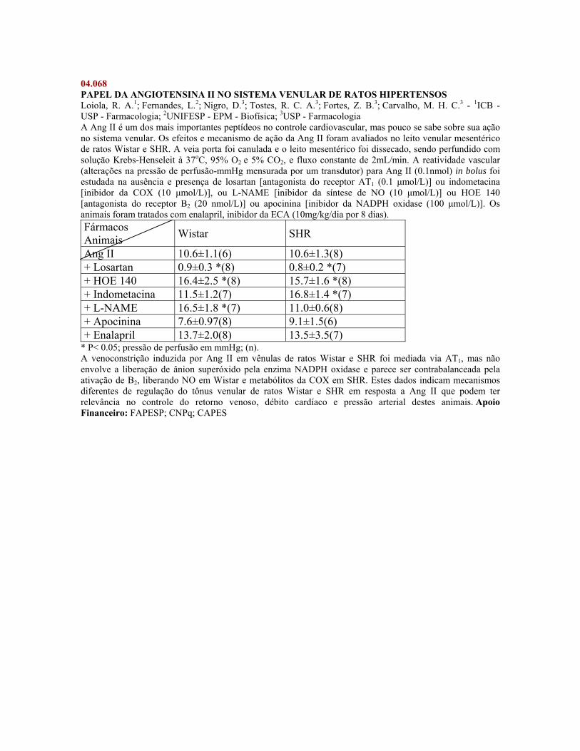
04.068 PAPEL DA ANGIOTENSINA II NO SISTEMA VENULAR DE RATOS HIPERTENSOS Loiola, R. A.1; Fernandes, L.2; Nigro, D.3; Tostes, R. C. A.3; Fortes, Z. B.3; Carvalho, M. H. C.3 - 1ICB - USP - Farmacologia; 2UNIFESP - EPM - Biofísica; 3USP - Farmacologia A Ang II é um dos mais importantes peptídeos no controle cardiovascular, mas pouco se sabe sobre sua ação no sistema venular. Os efeitos e mecanismo de ação da Ang II foram avaliados no leito venular mesentérico de ratos Wistar e SHR. A veia porta foi canulada e o leito mesentérico foi dissecado, sendo perfundido com solução Krebs-Henseleit à 37oC, 95% O2 e 5% CO2, e fluxo constante de 2mL/min. A reatividade vascular (alterações na pressão de perfusão-mmHg mensurada por um transdutor) para Ang II (0.1nmol) in bolus foi estudada na ausência e presença de losartan [antagonista do receptor AT1 (0.1 μmol/L)] ou indometacina [inibidor da COX (10 μmol/L)], ou L-NAME [inibidor da síntese de NO (10 μmol/L)] ou HOE 140 [antagonista do receptor B2 (20 nmol/L)] ou apocinina [inibidor da NADPH oxidase (100 μmol/L)]. Os animais foram tratados com enalapril, inibidor da ECA (10mg/kg/dia por 8 dias). Fármacos Animais Wistar SHR
Ang II 10.6±1.1(6) 10.6±1.3(8) + Losartan 0.9±0.3 *(8) 0.8±0.2 *(7) + HOE 140 16.4±2.5 *(8) 15.7±1.6 *(8) + Indometacina 11.5±1.2(7) 16.8±1.4 *(7) + L-NAME 16.5±1.8 *(7) 11.0±0.6(8) + Apocinina 7.6±0.97(8) 9.1±1.5(6) + Enalapril 13.7±2.0(8) 13.5±3.5(7)
* P< 0.05; pressão de perfusão em mmHg; (n). A venoconstrição induzida por Ang II em vênulas de ratos Wistar e SHR foi mediada via AT1, mas não envolve a liberação de ânion superóxido pela enzima NADPH oxidase e parece ser contrabalanceada pela ativação de B2, liberando NO em Wistar e metabólitos da COX em SHR. Estes dados indicam mecanismos diferentes de regulação do tônus venular de ratos Wistar e SHR em resposta a Ang II que podem ter relevância no controle do retorno venoso, débito cardíaco e pressão arterial destes animais. Apoio Financeiro: FAPESP; CNPq; CAPES
04.069 ESTUDO COMPARATIVO DO EFEITO CARDIOVASCULAR DO ÓLEO ESSENCIAL DE VÁRIAS PARTES DE Ocotea duckei Vattimo (LAURACEAE) EM RATOS Cunha, R. M.1; Ribeiro, T. P.2; Barbosa-Filho, J. M.3; Medeiros, I. A.3 - 1UFPB / UFAC - LTF; 2UFPB - LTF; 3UFPB - Tecnologia Farmacêutica Introdução: Ocotea duckei, conhecida como louro de cheiro, é usada popularmente para a redução de neuralgias, dispepsias e dor. Objetivo: Comparar os efeitos cardiovasculares do óleo essencial de várias partes de Ocotea duckei (OEOD) em ratos. Metodologia: Catéteres foram inseridos na aorta abdominal e veia cava inferior de ratos Wistar (250-300g), para registro da pressão arterial e frequência cardíaca, e infusão de drogas. Anéis de artéria mesentérica foram isolados, suspensos por fios de algodão e mantidos em cubas contendo solução de Tyrode, 37° C, aeradas com carbogênio, sob tensão de 0,75 g. Resultados: Em ratos não anestesiados OEOD (1, 5, 10, 15 mg/kg, i.v.) das partes da planta produziu hipotensão significante (n=6, p<0,05): folhas (7±1, 15±2, 21±2 e 37±3 %), frutos (6±1, 8±3, 18±3 e 26±3 %), caule (8±1, 25±3, 38±7 e 27±5 %) e raiz (4±2, 20±2, 33±2 e 25±5 %), e bradicardia (p<0,05): folhas (3±1,9±2, 18±4 e 53±4 %); frutos (3±2, 3±1, 12±3 e 35±2 %); caule (5±2, 22±2, 53±3 e 49±4 %) e raiz (3±1, 30±4, 57±3 e 35±2 %). Em anéis de artéria mesentérica com endotélio intacto (n=5), OEOD (0,1; 0,3; 1; 10; 30 100 mg/mL) de folhas, fruto, caule ou raiz, inibiu as contrações induzidas por Fen (10 mM) (CI50=31±2, 49±5, 30±6 ou 17±5 µg/ml) ou por KCl (80 mM) (CI50=5±1, 14±3, 7±2 ou 5±2 µg/ml). A remoção do endotélio não atenuou nenhum dos efeitos. Conclusão: OEOD de todas as partes testadas induziu hipotensão e bradicardia. Todos os óleos testados foram, no mínimo, três vezes mais potente em relaxar as contrações induzidas por KCl do que as induzidas por Fen. Apoio Financeiro: CAPES/CNPq.
04.070 THE REDUCTION ON VASCULAR TONUS PRODUCED BY STEVIOSIDE IS INFLUENCED BY NO SYNTHESIZED IN ENDOTHELIUM Amaral, V.1; Bornia, E. C. S.1; Bazotte, R. B.1; Santos, I. L.1; Alves-do-Prado, W.1 - 1UEM - Farmácia e Farmacologia Introduction: Stevioside reduces the arterial blood pressure when is administered intravenously in rats. The hypotensive effect of sweeter is determined by a reduction on Ca++ influx in peripheral vessels. The objective this study was to verify the effects of the stevioside in the isolated aortic rings preparations of rats pretreated with blockers of the L-arginine-NOS-NO-GC-cGMP pathway. Methods: Thoracic aorta rings preparations of rats with (E) or without (E*) intact endothelium were used in current study. The intact endothelium was considered when the addition of acetylcholine (1 μM) in the bath was able to produce 100 % relaxing of preparations pre- contracted with norepinephrine (10-8M). In the preparations without endothelium, this was removed by gentle rubbing and success of remotion was confirmed by the failure of acetylcholine (1 μM) to relax the rings pre- contracted by norepinephrine. The isometric tension (Grass, FT 03) was recorded on the computer equipped with Chart Software of Powerlab and data were submitted to ANOVA followed by Bonferroni (P<0.05). Results: Stevioside (10-3M) reduced the tension produced by norepinephrine in both types of preparations (52.8± 2.80%, n=10; E; 66.5± 3.97%, n=10; E*). L-NOARG (10-4M) or ODQ (10μM) antagonized the reduction on tension induced by stevioside in E preparations, but did not modify the effect of sweeter in E* preparations. On the other hand, it was verified that L-NOARG or ODQ increased (176.3±13.0%, n=10, L-NOARG, E; 150.3± 16.6%, n=10, L-NOARG, E*; 159.6±7.80%, n=10, ODQ, E; 136.5±21.8%, n=10, ODQ, E*) the pre- contraction induced by norepinephrine in E and E* preparations. Discussion: Since the increase produced by L-NOARG or ODQ on pre- contraction induced by norepinephrine was not able to impair the effect of stevioside in the denuded aortic rings preparations, data indicate that is unlikely that only the increase on pre- contraction produced by combined administrations of those agents had determined the antagonism by L-NOARG or ODQ recorded in the intact aortic rings preparations. It is concluded that the reduction on vascular tension produced by stevioside on the aortic rings preparations of rats is not dependent on intact endothelium, but it is strongly influenced by NO synthesized in such tissue. Supported by: CNPq (400875/2002)
04.071 ESTUDO DA PREVALÊNCIA DA HIPERTENSÃO NO POSTO DE SAÚDE CENTRAL DE POCONÉ-MT DE JUNHO Á OUTUBRO DE 2005 Barros, W. M.1; Nunes, A. A. A.2; Trevisan, T. C.3; Vanzeler, M. L. A.4 - 1UNIVAG - GPA - CIÊNCIAS BIOLÓGICAS ; 2UNIVAG - GPA - Ciências da Agrárias e Biológicas; 3SECRETÁRIA DE SAÚDE - MT - SES - MT; 4UFMT - Ciências Médicas A hipertensão arterial (HA) no Brasil pode ser considerada um problema para saúde pública no Brasil. Esta fato pode ser confirmado pela grande prevalência da doença, alto custo do tratamento, letalidade e pelo fato de produzir lesões permanentes. O conhecimento da prevalência da HA pode ser um importante valor para orientar o planejamento das políticas de saúde.Objetivo: descrever a prevalência da hipertensão, na população de Poconé-MT. Método: Foi realizado um estudo com base de indivíduos cadastrados no Posto de Saúde Central de Poconé-MT, analisando prontuários médicos de 80 pessoas, e preenchando um formulário de auto-preenchimento.Resultados: Os resultados observados foram: 57,5% do sexo feminino, 42,5 do sexo masculino; os fatores de riscos associados mais citados foram: idade avançada, hereditariedade, diabetese e os anti-hipertensivos mais receitados foram hidroclotiazida (25%), captopril (25 %) e enalapril (17,85 %).Conclusão: A prevalência da HA em Poconé é decorrente de hereditariedade, de maior incidência no sexo feminino, idosos, e de maus hábitos, o que podem comprometer e prejudicar a qualidade de vida. Apoio Financeiro: UNIVAG - CENTRO UNIVERSITÁRIO
04.072 VASCULAR REACTIVITY FROM TRAINED HIGH CALORIC-FED RATS Moraes, C.1; Claudino, M. A.2; Camargo, E.2; Vanzela, E. C.3; Carneiro, E. M.3; Boschero, A. C.3; Nucci, G. de3; Antunes, E.2; Zanesco, A.1 - 1UNESP - Educação Física; 2UNICAMP - Farmacologia; 3UNICAMP - Fisiologia e Biofísica Introduction:Our aim was to investigate the therapeutic effect of exercise on the reactivity of mesenteric and aortic rings from high caloric-fed rats. Methods:Male Wistar were divided in: Sedentary (SD);Trained (TR);Sedentary diet (SDD) and Trained diet (TRD) groups.Run training (RT) consisted of 66%VO2max (60min/day,5days/week) for 4 weeks concomitantly with normal chow or high caloric diet.Triglycerides, glucose, testosterone, and nitrate/nitrite (NOx) concentration were measured.Concentration-response curves to acetylcholine (ACh),sodium nitroprusside (SNP) and phenylephrine (PHE) were obtained in mesenteric and aortic rings. Results:High caloric diet increased triglycerides (SD:82±13;SDD:111±9mg/dl) whereas RT reduced significantly (TR:62±8;TRD:51±3mg/dl).Glucose and testosterone were not modified,but insulin was augmented by high caloric diet (SD:0.6±0.1;SDD:1.2±0.1ng/ml) and RT did not modified it. NOx was increase in TRD group (SD:27±4;TR:30±4;SDD:30±4;TRD:42±4μM).Hyper caloric diet impaired ACh Emax in mesenteric rings (SD:88±2;SDD:76±2%) and RT was effective to improve these responses (TR:93±1;TRD:91±3%),but the pEC50 was not altered.In aortic rings only RT modified the Emax (SD:75±2;TR:88±2;SDD:77±2;TRD:90±3%) and the pEC50 was increased about 2.5-fold only in TR group. SNP and PHE Emax and pEC50 were not modified in booth arteries. Discussion:RT completely abolished the hypertriglyceridemia, slightly reduced insulinemia and increased the endothelium-dependent response for ACh in mesenteric and aortic rings from high caloric-fed rats. Supported by: FAPESP/CAPES


![FARMACOLOGIA Farmacologia Basica_apostila[1]](https://img.pdfslide.net/doc/110x75/5571f8c649795991698e0e1f/farmacologia-farmacologia-basicaapostila1.jpg)

![Farmacologia farmacologia para enfermagem[1]](https://img.pdfslide.net/doc/110x75/55656d9ad8b42a7b518b4b48/farmacologia-farmacologia-para-enfermagem1.jpg)


![FARMACOLOGIA - Farmacologia Para Enfermagem[1]](https://img.pdfslide.net/doc/110x75/5572116d497959fc0b8ef613/farmacologia-farmacologia-para-enfermagem1-55c1e72cb7d96.jpg)