Embed Size (px)
Citation preview
228
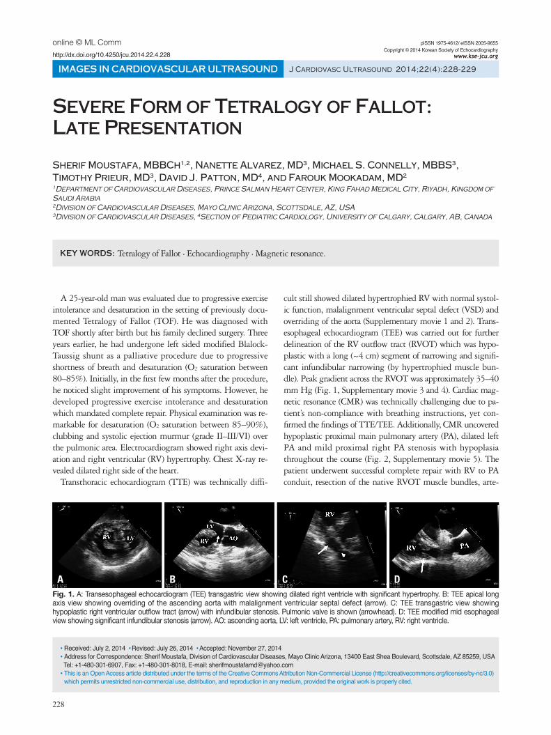
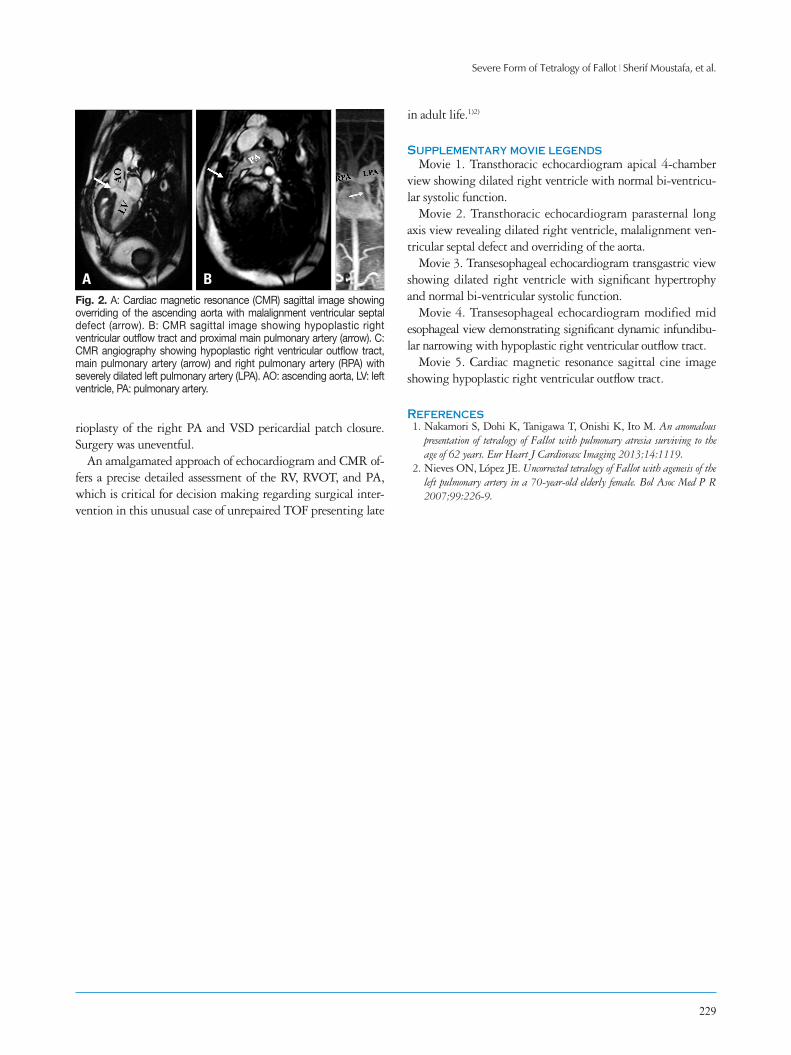
cult still showed dilated hypertrophied RV with normal systol-ic function, malalignment ventricular septal defect (VSD) and overriding of the aorta (Supplementary movie 1 and 2). Trans-esophageal echocardiogram (TEE) was carried out for further delineation of the RV outflow tract (RVOT) which was hypo-plastic with a long (~4 cm) segment of narrowing and signifi-cant infundibular narrowing (by hypertrophied muscle bun-dle). Peak gradient across the RVOT was approximately 35–40 mm Hg (Fig. 1, Supplementary movie 3 and 4). Cardiac mag-netic resonance (CMR) was technically challenging due to pa-tient’s non-compliance with breathing instructions, yet con-firmed the findings of TTE/TEE. Additionally, CMR uncovered hypoplastic proximal main pulmonary artery (PA), dilated left PA and mild proximal right PA stenosis with hypoplasia throughout the course (Fig. 2, Supplementary movie 5). The patient underwent successful complete repair with RV to PA conduit, resection of the native RVOT muscle bundles, arte-
A 25-year-old man was evaluated due to progressive exercise intolerance and desaturation in the setting of previously docu-mented Tetralogy of Fallot (TOF). He was diagnosed with TOF shortly after birth but his family declined surgery. Three years earlier, he had undergone left sided modified Blalock-Taussig shunt as a palliative procedure due to progressive shortness of breath and desaturation (O2 saturation between 80–85%). Initially, in the first few months after the procedure, he noticed slight improvement of his symptoms. However, he developed progressive exercise intolerance and desaturation which mandated complete repair. Physical examination was re-markable for desaturation (O2 saturation between 85–90%), clubbing and systolic ejection murmur (grade II–III/VI) over the pulmonic area. Electrocardiogram showed right axis devi-ation and right ventricular (RV) hypertrophy. Chest X-ray re-vealed dilated right side of the heart.
Transthoracic echocardiogram (TTE) was technically diffi-
pISSN 1975-4612/ eISSN 2005-9655 Copyright © 2014 Korean Society of Echocardiography
www.kse-jcu.orghttp://dx.doi.org/10.4250/jcu.2014.22.4.228
IMAGES IN CARDIOVASCULAR ULTRASOUND J Cardiovasc Ultrasound 2014;22(4):228-229
Severe Form of Tetralogy of Fallot: Late Presentation
Sherif Moustafa, MBBCh1,2, Nanette Alvarez, MD3, Michael S. Connelly, MBBS3, Timothy Prieur, MD3, David J. Patton, MD4, and Farouk Mookadam, MD2
1Department of Cardiovascular Diseases, Prince Salman Heart Center, King Fahad Medical City, Riyadh, Kingdom of Saudi Arabia2Division of Cardiovascular Diseases, Mayo Clinic Arizona, Scottsdale, AZ, USA3Division of Cardiovascular Diseases, 4Section of Pediatric Cardiology, University of Calgary, Calgary, AB, Canada
KEY WORDS: Tetralogy of Fallot · Echocardiography · Magnetic resonance.
•Received: July 2, 2014 •Revised: July 26, 2014 •Accepted: November 27, 2014 •Address for Correspondence: Sherif Moustafa, Division of Cardiovascular Diseases, Mayo Clinic Arizona, 13400 East Shea Boulevard, Scottsdale, AZ 85259, USA Tel: +1-480-301-6907, Fax: +1-480-301-8018, E-mail: [email protected]•This is an Open Access article distributed under the terms of the Creative Commons Attribution Non-Commercial License (http://creativecommons.org/licenses/by-nc/3.0) which permits unrestricted non-commercial use, distribution, and reproduction in any medium, provided the original work is properly cited.
Fig. 1. A: Transesophageal echocardiogram (TEE) transgastric view showing dilated right ventricle with significant hypertrophy. B: TEE apical long axis view showing overriding of the ascending aorta with malalignment ventricular septal defect (arrow). C: TEE transgastric view showing hypoplastic right ventricular outflow tract (arrow) with infundibular stenosis. Pulmonic valve is shown (arrowhead). D: TEE modified mid esophageal view showing significant infundibular stenosis (arrow). AO: ascending aorta, LV: left ventricle, PA: pulmonary artery, RV: right ventricle.
A B C D
online © ML Comm
Severe Form of Tetralogy of Fallot | Sherif Moustafa, et al.
229
in adult life.1)2)
Supplementary movie legendsMovie 1. Transthoracic echocardiogram apical 4-chamber
view showing dilated right ventricle with normal bi-ventricu-lar systolic function.
Movie 2. Transthoracic echocardiogram parasternal long axis view revealing dilated right ventricle, malalignment ven-tricular septal defect and overriding of the aorta.
Movie 3. Transesophageal echocardiogram transgastric view showing dilated right ventricle with significant hypertrophy and normal bi-ventricular systolic function.
Movie 4. Transesophageal echocardiogram modified mid esophageal view demonstrating significant dynamic infundibu-lar narrowing with hypoplastic right ventricular outflow tract.
Movie 5. Cardiac magnetic resonance sagittal cine image showing hypoplastic right ventricular outflow tract.
References1. Nakamori S, Dohi K, Tanigawa T, Onishi K, Ito M. An anomalous
presentation of tetralogy of Fallot with pulmonary atresia surviving to the age of 62 years. Eur Heart J Cardiovasc Imaging 2013;14:1119.
2. Nieves ON, López JE. Uncorrected tetralogy of Fallot with agenesis of the left pulmonary artery in a 70-year-old elderly female. Bol Asoc Med P R 2007;99:226-9.
rioplasty of the right PA and VSD pericardial patch closure. Surgery was uneventful.
An amalgamated approach of echocardiogram and CMR of-fers a precise detailed assessment of the RV, RVOT, and PA, which is critical for decision making regarding surgical inter-vention in this unusual case of unrepaired TOF presenting late
Fig. 2. A: Cardiac magnetic resonance (CMR) sagittal image showing overriding of the ascending aorta with malalignment ventricular septal defect (arrow). B: CMR sagittal image showing hypoplastic right ventricular outflow tract and proximal main pulmonary artery (arrow). C: CMR angiography showing hypoplastic right ventricular outflow tract, main pulmonary artery (arrow) and right pulmonary artery (RPA) with severely dilated left pulmonary artery (LPA). AO: ascending aorta, LV: left ventricle, PA: pulmonary artery.
A B C