Embed Size (px)
Citation preview
CASE REPORT Open Access
Severe postpartum disruption of the pelvic ring:report of two cases and review of the literatureZhiyong Hou1*, John T Riehl2, Wade R Smith2, Kent A Strohecker2, Patrick J Maloney2
Abstract
Pelvic dislocations are rare during labor, and the treatment is controversial. We report two cases of young womenwho sustained postpartum disruption of the pelvic ring: one case is an 8.8 cm wide separation of the pubicsymphysis with sacroiliac joint disruption underwent surgical stabilization and the second case with 4.0 cmdisruption being treated non-operatively. These cases illustrated of importance of accurate diagnosis, carefulphysical exam, fully informed consent and specific treatment for this condition.
BackgroundDisruption of the symphysis pubis is a rare injury duringchildbirth with an incidence of 0.005% to 0.8% [1-3]. Itis usually seen in elderly (older than 35) primagravidaand there are few reports of this injury in youngerpatients [4,5]. Conservative treatment was reported tobe successful in most cases, and operative managementwas considered in severe displaced case. We report twocases of young women who sustained postpartum dis-ruption of the pelvic ring: one case is an 8.8 cm wideseparation of the pubic symphysis with sacroiliac jointdisruption underwent surgical stabilization and the sec-ond case with 4.0 cm disruption being treated non-operatively. These cases illustrated of importance ofaccurate diagnosis, careful physical exam, fully informedconsent and specific treatment for this condition.
Case presentationCase 1A 26-year-old female patient had a history of two pre-vious uncomplicated pregnancies 4 and 7 years prior.She underwent a vaginal delivery, delivering a healthymacrosomic fetus with a vacuum assistance at an out-side hospital. The patient labored for 50 minutes andreceived epidural anesthesia for pain control peripartum.Immediately after delivering a healthy male (9 Lb 42 oz)she developed sudden severe pain over the pubic sym-physis and buttocks. She also complained of left lower
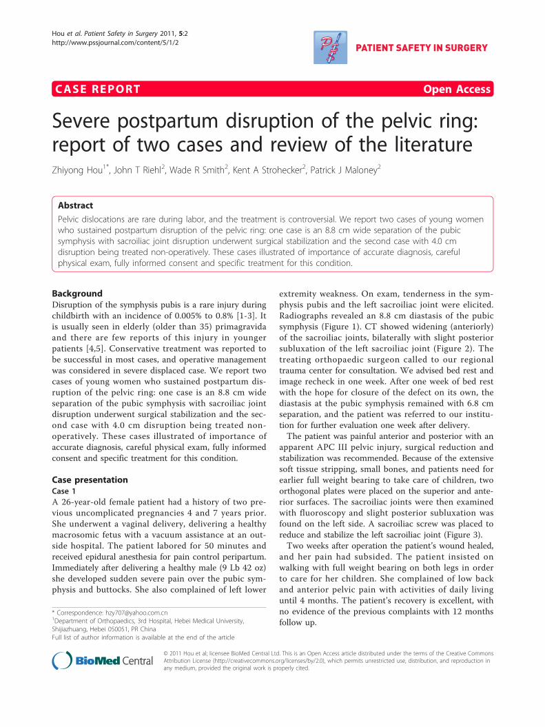
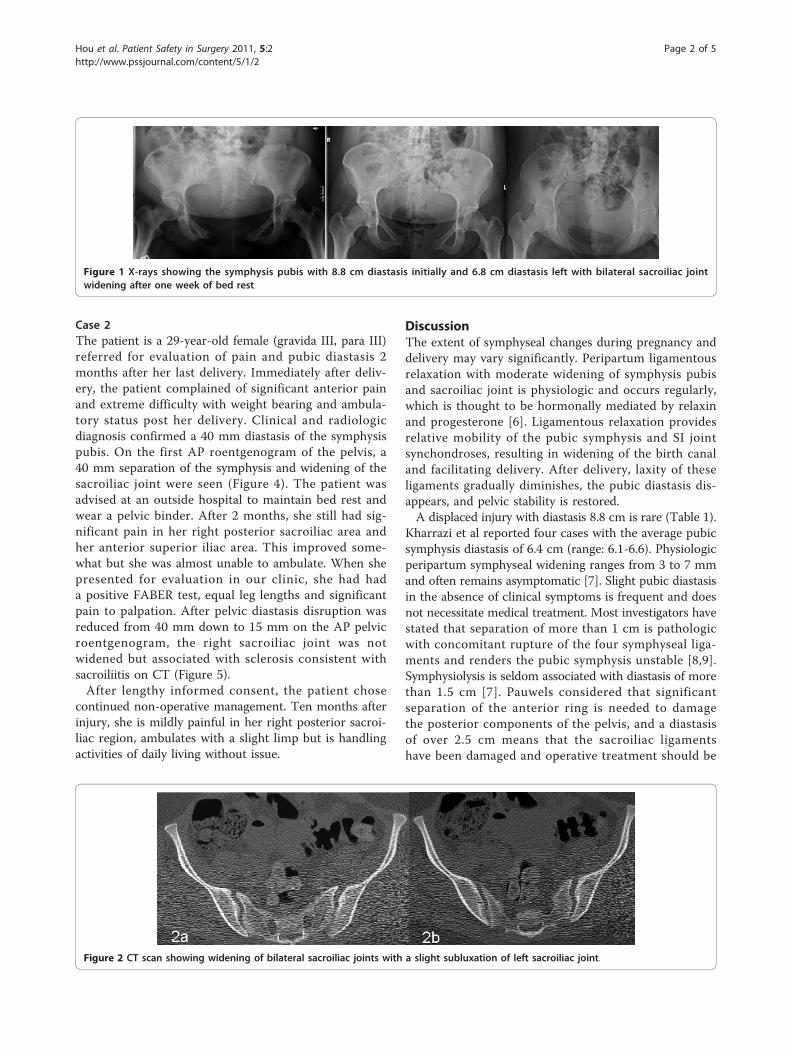
extremity weakness. On exam, tenderness in the sym-physis pubis and the left sacroiliac joint were elicited.Radiographs revealed an 8.8 cm diastasis of the pubicsymphysis (Figure 1). CT showed widening (anteriorly)of the sacroiliac joints, bilaterally with slight posteriorsubluxation of the left sacroiliac joint (Figure 2). Thetreating orthopaedic surgeon called to our regionaltrauma center for consultation. We advised bed rest andimage recheck in one week. After one week of bed restwith the hope for closure of the defect on its own, thediastasis at the pubic symphysis remained with 6.8 cmseparation, and the patient was referred to our institu-tion for further evaluation one week after delivery.The patient was painful anterior and posterior with an
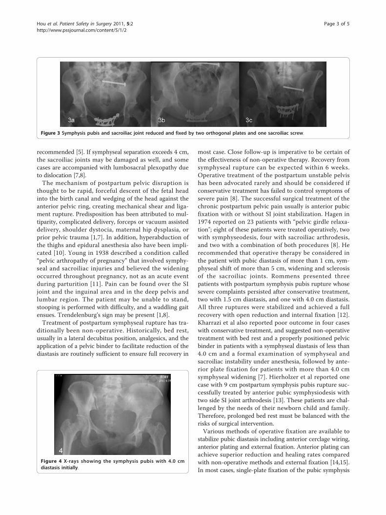
apparent APC III pelvic injury, surgical reduction andstabilization was recommended. Because of the extensivesoft tissue stripping, small bones, and patients need forearlier full weight bearing to take care of children, twoorthogonal plates were placed on the superior and ante-rior surfaces. The sacroiliac joints were then examinedwith fluoroscopy and slight posterior subluxation wasfound on the left side. A sacroiliac screw was placed toreduce and stabilize the left sacroiliac joint (Figure 3).Two weeks after operation the patient’s wound healed,
and her pain had subsided. The patient insisted onwalking with full weight bearing on both legs in orderto care for her children. She complained of low backand anterior pelvic pain with activities of daily livinguntil 4 months. The patient’s recovery is excellent, withno evidence of the previous complaints with 12 monthsfollow up.
* Correspondence: [email protected] of Orthopaedics, 3rd Hospital, Hebei Medical University,Shijiazhuang, Hebei 050051, PR ChinaFull list of author information is available at the end of the article
Hou et al. Patient Safety in Surgery 2011, 5:2http://www.pssjournal.com/content/5/1/2
© 2011 Hou et al; licensee BioMed Central Ltd. This is an Open Access article distributed under the terms of the Creative CommonsAttribution License (http://creativecommons.org/licenses/by/2.0), which permits unrestricted use, distribution, and reproduction inany medium, provided the original work is properly cited.
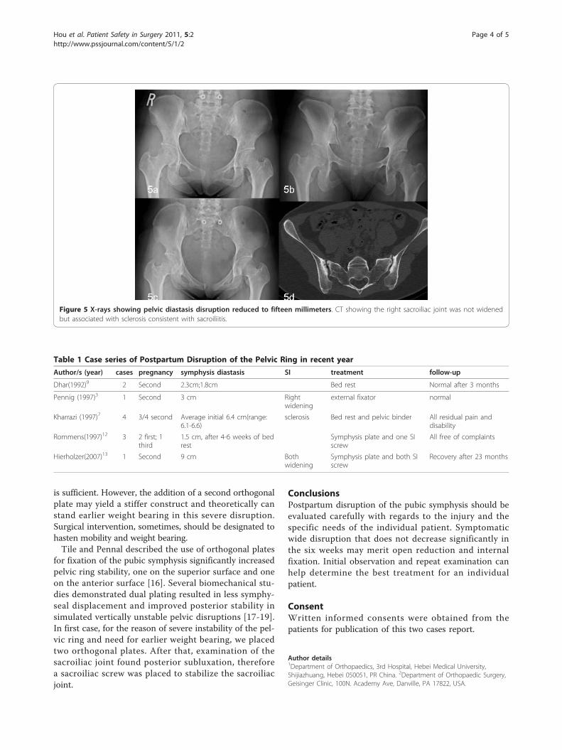
Case 2The patient is a 29-year-old female (gravida III, para III)referred for evaluation of pain and pubic diastasis 2months after her last delivery. Immediately after deliv-ery, the patient complained of significant anterior painand extreme difficulty with weight bearing and ambula-tory status post her delivery. Clinical and radiologicdiagnosis confirmed a 40 mm diastasis of the symphysispubis. On the first AP roentgenogram of the pelvis, a40 mm separation of the symphysis and widening of thesacroiliac joint were seen (Figure 4). The patient wasadvised at an outside hospital to maintain bed rest andwear a pelvic binder. After 2 months, she still had sig-nificant pain in her right posterior sacroiliac area andher anterior superior iliac area. This improved some-what but she was almost unable to ambulate. When shepresented for evaluation in our clinic, she had hada positive FABER test, equal leg lengths and significantpain to palpation. After pelvic diastasis disruption wasreduced from 40 mm down to 15 mm on the AP pelvicroentgenogram, the right sacroiliac joint was notwidened but associated with sclerosis consistent withsacroiliitis on CT (Figure 5).After lengthy informed consent, the patient chose
continued non-operative management. Ten months afterinjury, she is mildly painful in her right posterior sacroi-liac region, ambulates with a slight limp but is handlingactivities of daily living without issue.
DiscussionThe extent of symphyseal changes during pregnancy anddelivery may vary significantly. Peripartum ligamentousrelaxation with moderate widening of symphysis pubisand sacroiliac joint is physiologic and occurs regularly,which is thought to be hormonally mediated by relaxinand progesterone [6]. Ligamentous relaxation providesrelative mobility of the pubic symphysis and SI jointsynchondroses, resulting in widening of the birth canaland facilitating delivery. After delivery, laxity of theseligaments gradually diminishes, the pubic diastasis dis-appears, and pelvic stability is restored.A displaced injury with diastasis 8.8 cm is rare (Table 1).
Kharrazi et al reported four cases with the average pubicsymphysis diastasis of 6.4 cm (range: 6.1-6.6). Physiologicperipartum symphyseal widening ranges from 3 to 7 mmand often remains asymptomatic [7]. Slight pubic diastasisin the absence of clinical symptoms is frequent and doesnot necessitate medical treatment. Most investigators havestated that separation of more than 1 cm is pathologicwith concomitant rupture of the four symphyseal liga-ments and renders the pubic symphysis unstable [8,9].Symphysiolysis is seldom associated with diastasis of morethan 1.5 cm [7]. Pauwels considered that significantseparation of the anterior ring is needed to damagethe posterior components of the pelvis, and a diastasisof over 2.5 cm means that the sacroiliac ligamentshave been damaged and operative treatment should be
Figure 1 X-rays showing the symphysis pubis with 8.8 cm diastasis initially and 6.8 cm diastasis left with bilateral sacroiliac jointwidening after one week of bed rest.
Figure 2 CT scan showing widening of bilateral sacroiliac joints with a slight subluxation of left sacroiliac joint.
Hou et al. Patient Safety in Surgery 2011, 5:2http://www.pssjournal.com/content/5/1/2
Page 2 of 5
recommended [5]. If symphyseal separation exceeds 4 cm,the sacroiliac joints may be damaged as well, and somecases are accompanied with lumbosacral plexopathy dueto dislocation [7,8].The mechanism of postpartum pelvic disruption is
thought to be rapid, forceful descent of the fetal headinto the birth canal and wedging of the head against theanterior pelvic ring, creating mechanical shear and liga-ment rupture. Predisposition has been attributed to mul-tiparity, complicated delivery, forceps or vacuum assisteddelivery, shoulder dystocia, maternal hip dysplasia, orprior pelvic trauma [1,7]. In addition, hyperabduction ofthe thighs and epidural anesthesia also have been impli-cated [10]. Young in 1938 described a condition called“pelvic arthropathy of pregnancy” that involved symphy-seal and sacroiliac injuries and believed the wideningoccurred throughout pregnancy, not as an acute eventduring parturition [11]. Pain can be found over the SIjoint and the inguinal area and in the deep pelvis andlumbar region. The patient may be unable to stand,stooping is performed with difficulty, and a waddling gaitensues. Trendelenburg’s sign may be present [1,8].Treatment of postpartum symphyseal rupture has tra-
ditionally been non-operative. Historically, bed rest,usually in a lateral decubitus position, analgesics, and theapplication of a pelvic binder to facilitate reduction of thediastasis are routinely sufficient to ensure full recovery in
most case. Close follow-up is imperative to be certain ofthe effectiveness of non-operative therapy. Recovery fromsymphyseal rupture can be expected within 6 weeks.Operative treatment of the postpartum unstable pelvishas been advocated rarely and should be considered ifconservative treatment has failed to control symptoms ofsevere pain [8]. The successful surgical treatment of thechronic postpartum pelvic pain usually is anterior pubicfixation with or without SI joint stabilization. Hagen in1974 reported on 23 patients with “pelvic girdle relaxa-tion"; eight of these patients were treated operatively, twowith symphyseodesis, four with sacroiliac arthrodesis,and two with a combination of both procedures [8]. Herecommended that operative therapy be considered inthe patient with pubic diastasis of more than 1 cm, sym-physeal shift of more than 5 cm, widening and sclerosisof the sacroiliac joints. Rommens presented threepatients with postpartum symphysis pubis rupture whosesevere complaints persisted after conservative treatment,two with 1.5 cm diastasis, and one with 4.0 cm diastasis.All three ruptures were stabilized and achieved a fullrecovery with open reduction and internal fixation [12].Kharrazi et al also reported poor outcome in four caseswith conservative treatment, and suggested non-operativetreatment with bed rest and a properly positioned pelvicbinder in patients with a symphyseal diastasis of less than4.0 cm and a formal examination of symphyseal andsacroiliac instability under anesthesia, followed by ante-rior plate fixation for patients with more than 4.0 cmsymphyseal widening [7]. Hierholzer et al reported onecase with 9 cm postpartum symphysis pubis rupture suc-cessfully treated by anterior pubic symphysiodesis withtwo side SI joint arthrodesis [13]. These patients are chal-lenged by the needs of their newborn child and family.Therefore, prolonged bed rest must be balanced with therisks of surgical intervention.Various methods of operative fixation are available to
stabilize pubic diastasis including anterior cerclage wiring,anterior plating and external fixation. Anterior plating canachieve superior reduction and healing rates comparedwith non-operative methods and external fixation [14,15].In most cases, single-plate fixation of the pubic symphysis
Figure 4 X-rays showing the symphysis pubis with 4.0 cmdiastasis initially.
Figure 3 Symphysis pubis and sacroiliac joint reduced and fixed by two orthogonal plates and one sacroiliac screw.
Hou et al. Patient Safety in Surgery 2011, 5:2http://www.pssjournal.com/content/5/1/2
Page 3 of 5
is sufficient. However, the addition of a second orthogonalplate may yield a stiffer construct and theoretically canstand earlier weight bearing in this severe disruption.Surgical intervention, sometimes, should be designated tohasten mobility and weight bearing.Tile and Pennal described the use of orthogonal plates
for fixation of the pubic symphysis significantly increasedpelvic ring stability, one on the superior surface and oneon the anterior surface [16]. Several biomechanical stu-dies demonstrated dual plating resulted in less symphy-seal displacement and improved posterior stability insimulated vertically unstable pelvic disruptions [17-19].In first case, for the reason of severe instability of the pel-vic ring and need for earlier weight bearing, we placedtwo orthogonal plates. After that, examination of thesacroiliac joint found posterior subluxation, thereforea sacroiliac screw was placed to stabilize the sacroiliacjoint.
ConclusionsPostpartum disruption of the pubic symphysis should beevaluated carefully with regards to the injury and thespecific needs of the individual patient. Symptomaticwide disruption that does not decrease significantly inthe six weeks may merit open reduction and internalfixation. Initial observation and repeat examination canhelp determine the best treatment for an individualpatient.
ConsentWritten informed consents were obtained from thepatients for publication of this two cases report.
Author details1Department of Orthopaedics, 3rd Hospital, Hebei Medical University,Shijiazhuang, Hebei 050051, PR China. 2Department of Orthopaedic Surgery,Geisinger Clinic, 100N. Academy Ave, Danville, PA 17822, USA.
Table 1 Case series of Postpartum Disruption of the Pelvic Ring in recent year
Author/s (year) cases pregnancy symphysis diastasis SI treatment follow-up
Dhar(1992)9 2 Second 2.3cm;1.8cm Bed rest Normal after 3 months
Pennig (1997)3 1 Second 3 cm Rightwidening
external fixator normal
Kharrazi (1997)7 4 3/4 second Average initial 6.4 cm(range:6.1-6.6)
sclerosis Bed rest and pelvic binder All residual pain anddisability
Rommens(1997)12 3 2 first; 1third
1.5 cm, after 4-6 weeks of bedrest
Symphysis plate and one SIscrew
All free of complaints
Hierholzer(2007)13 1 Second 9 cm Bothwidening
Symphysis plate and both SIscrew
Recovery after 23 months
Figure 5 X-rays showing pelvic diastasis disruption reduced to fifteen millimeters. CT showing the right sacroiliac joint was not widenedbut associated with sclerosis consistent with sacroiliitis.
Hou et al. Patient Safety in Surgery 2011, 5:2http://www.pssjournal.com/content/5/1/2
Page 4 of 5
Authors’ contributionsAll authors contributed equally to this case report. All authors read andapproved the final version of the manuscript.
Competing interestsThe authors declare that they have no competing interests.
Received: 29 October 2010 Accepted: 13 January 2011Published: 13 January 2011
References1. Boland BF: Rupture of the symphysis pubis articulation during delivery.
Surg Gynecol Obstet 1933, 57:517-522.2. Scriven MW, Jones DA, McKnight L: The importance of pubic pain
following childbirth: a clinical and ultrasonographic study of diastasis ofthe pubic symphysis. J R Soc Med 1995, 88:28-30.
3. Pennig D, Gladbach B, Majchrowski W: Disruption of the pelvic ringduring spontaneous childbirth. A case report. J Bone Joint Surg Br 1997,79:438-440.
4. Blum M, Orovano N: Open rupture of the symphysis pubis duringspontaneous delivery. Acta Obstet Gynecol Scand 1976, 55:77-79.
5. Pauwels F: Beitrag zur Klarung der Beanspruchung des Beckens,insbesondere der Beckenfugen. In Gesammelte Abhandlungen zurfunktionellen Anatomie des Bewegungsapparates. Edited by: Pauwels F. Berlin,etc: Springer-Verlag; 1965:183-196.
6. Putschar WG: The structure of the human symphysis pubis with specialconsideration of parturition and its sequelae. Am J Phys Anthrop 1976,45:589-594.
7. Kharrazi FD, Rodgers WB, Kennedy JG, Lhowe DW: Parturition-inducedpelvic dislocation: a report of four cases. J Orthop Trauma 1997,11:277-281.
8. Hagen R: Pelvic girdle relaxation from an orthopaedic point of view. ActaOrthop Scand 1974, 45:550-563.
9. Dhar S, Anderton JM: Rupture of the symphysis pubis during labor. ClinOrthop Relat Res 1992, 283:252-257.
10. Cappiello GA, Oliver BC: Rupture of the symphysis pubis caused byforceful and excessive abduction of the thighs with labor epiduralanesthesia. J Florida Med Assoc 1995, 82:261-263.
11. Young J: Relaxation of the pelvic joints in pregnancy: pelvic arthropathyof pregnancy. J Obstet Gynecol Br Emp 1940, 47:493-524.
12. Rommens PM: Internal fixation in postpartum symphysis pubis rupture:report of three cases. J Orthop Trauma 1997, 11:273-276.
13. Hierholzer C, Ali A, Toro-Arbelaez JB, Suk M, Helfet DL: Traumaticdisruption of pubis symphysis with accompanying posterior pelvic injuryafter natural childbirth. Am J Orthop 2007, 36:E167-70.
14. Matta JM, Saucedo T: Internal fixation of pelvic ring fractures. Clin OrthopRelat Res 1989, 242:83-97.
15. Moed BR, Kellam JF, McLaren A, Tile M: Internal fixation for the injuredpelvic ring. In Fractures of the Pelvis and Acetabulum. Edited by: Tile M,Helfet DL, Kellam JF. Baltimore, MD: Williams 2003:217-293.
16. Tile M, Pennal GF: Pelvic disruption: principles of management. ClinOrthop Relat Res 1980, 151:56-64.
17. Varga E, Hearn T, Powell J, Tile M: Effects of method of internal fixation ofsymphyseal disruptions on stability of the pelvic ring. Injury 1995,26:75-80.
18. Schopfer A, Hearn TC, DiAngelo D, Tile M: Biomechanical comparison offixation methods of vertically unstable pelvic ring disruptions. Paperpresented at: 59th Meeting of the American Academy of Orthopaedic SurgeonsWashington, DC; 1992.
19. Hearn TC, Willet K, Schopfer A, DiAngelo D, Powell JN, Tile M: Mechanicalresponse of the intact, disrupted and internally fixed pelvic ring tostance related loading. Surgery of the Pelvis and Acetabulum: AnInternational Consensus, Final Program and Syllabus Pittsburgh, PA; 1992, 86.
doi:10.1186/1754-9493-5-2Cite this article as: Hou et al.: Severe postpartum disruption of thepelvic ring: report of two cases and review of the literature. PatientSafety in Surgery 2011 5:2.
Submit your next manuscript to BioMed Centraland take full advantage of:
• Convenient online submission
• Thorough peer review
• No space constraints or color figure charges
• Immediate publication on acceptance
• Inclusion in PubMed, CAS, Scopus and Google Scholar
• Research which is freely available for redistribution
Submit your manuscript at www.biomedcentral.com/submit
Hou et al. Patient Safety in Surgery 2011, 5:2http://www.pssjournal.com/content/5/1/2
Page 5 of 5