Embed Size (px)
Citation preview
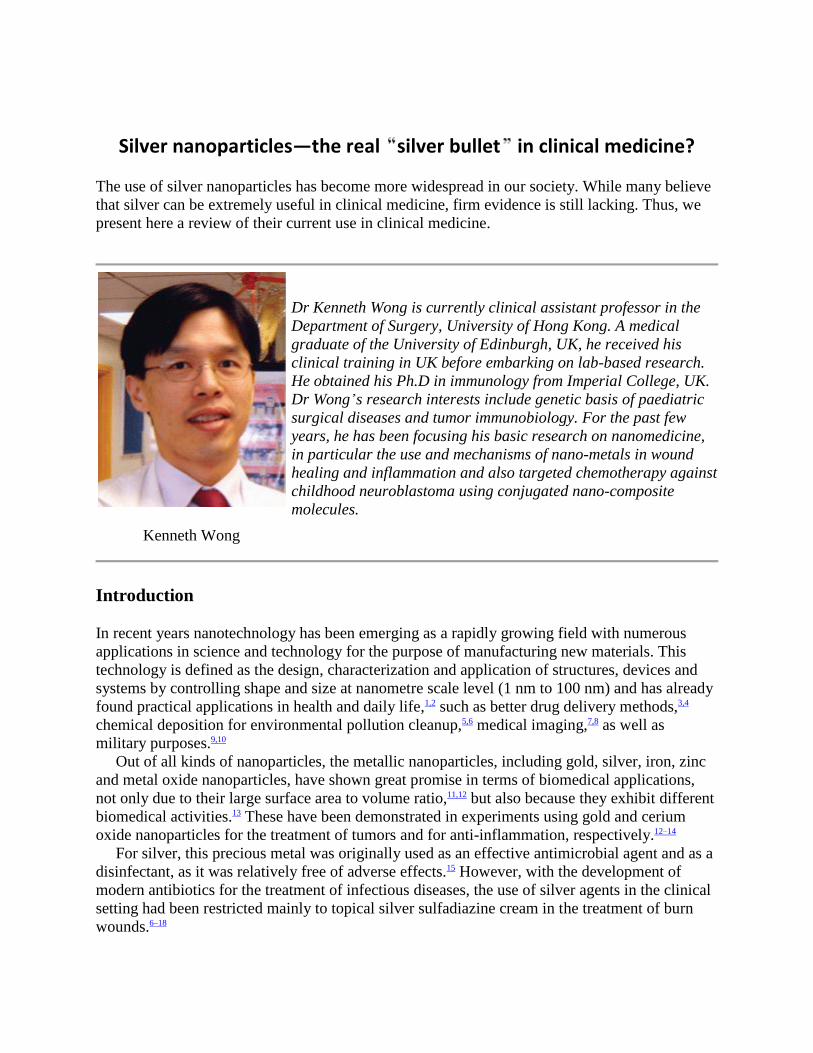
Silver nanoparticles—the real silver bullet in clinical medicine?
The use of silver nanoparticles has become more widespread in our society. While many believe
that silver can be extremely useful in clinical medicine, firm evidence is still lacking. Thus, we
present here a review of their current use in clinical medicine.
Kenneth Wong
Dr Kenneth Wong is currently clinical assistant professor in the
Department of Surgery, University of Hong Kong. A medical
graduate of the University of Edinburgh, UK, he received his
clinical training in UK before embarking on lab-based research.
He obtained his Ph.D in immunology from Imperial College, UK.
Dr Wong s research interests include genetic basis of paediatric
surgical diseases and tumor immunobiology. For the past few
years, he has been focusing his basic research on nanomedicine,
in particular the use and mechanisms of nano-metals in wound
healing and inflammation and also targeted chemotherapy against
childhood neuroblastoma using conjugated nano-composite
molecules.
Introduction
In recent years nanotechnology has been emerging as a rapidly growing field with numerous
applications in science and technology for the purpose of manufacturing new materials. This
technology is defined as the design, characterization and application of structures, devices and
systems by controlling shape and size at nanometre scale level (1 nm to 100 nm) and has already
found practical applications in health and daily life,1,2 such as better drug delivery methods,3,4
chemical deposition for environmental pollution cleanup,5,6 medical imaging,7,8 as well as
military purposes.9,10
Out of all kinds of nanoparticles, the metallic nanoparticles, including gold, silver, iron, zinc
and metal oxide nanoparticles, have shown great promise in terms of biomedical applications,
not only due to their large surface area to volume ratio,11,12 but also because they exhibit different
biomedical activities.13 These have been demonstrated in experiments using gold and cerium
oxide nanoparticles for the treatment of tumors and for anti-inflammation, respectively.12–14
For silver, this precious metal was originally used as an effective antimicrobial agent and as a
disinfectant, as it was relatively free of adverse effects.15 However, with the development of
modern antibiotics for the treatment of infectious diseases, the use of silver agents in the clinical
setting had been restricted mainly to topical silver sulfadiazine cream in the treatment of burn
wounds.6–18
From the 1990's, there has been a resurgence of the promotion of silver (as colloidal silver) as
an alternative medicine treatment. It has been marketed with claims of it being an essential
mineral supplement, or that it can treat various diseases.19,20 Although colloidal silver products
are legally available as health supplements, it is illegal in the U.S. to make such claims of
medical effectiveness for colloidal silver.
The commercial product referred to as colloidal silver includes solutions that contain
various concentrations of ionic silver compounds, silver colloids or silver compounds bound to
proteins. Unlike clinical drug production, the manufacturing of such products is not standardized
and thus results in various concentrations and also particle sizes.
At present, there are no evidence-based medical uses for ingested colloidal silver. Indeed, the
U.S. National Center for Complimentary and Alternative Medicine has issued an advisory
indicating that the marketing claims made about colloidal silver are scientifically unsupported.19
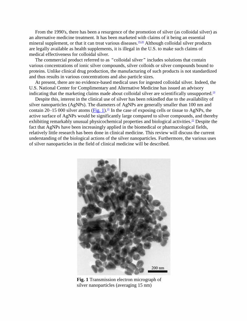
Despite this, interest in the clinical use of silver has been rekindled due to the availability of
silver nanoparticles (AgNPs). The diameters of AgNPs are generally smaller than 100 nm and
contain 20–15 000 silver atoms (Fig. 1).21 In the case of exposing cells or tissue to AgNPs, the
active surface of AgNPs would be significantly large compared to silver compounds, and thereby
exhibiting remarkably unusual physicochemical properties and biological activities.22 Despite the
fact that AgNPs have been increasingly applied in the biomedical or pharmacological fields,
relatively little research has been done in clinical medicine. This review will discuss the current
understanding of the biological actions of the silver nanoparticles. Furthermore, the various uses
of silver nanoparticles in the field of clinical medicine will be described.
Fig. 1 Transmission electron micrograph of
silver nanoparticles (averaging 15 nm)
produced by the reduction method.
Synthesis of silver nanoparticles
a. Chemical and physical synthesis methods of AgNPs. For biological use, the main aim of
making AgNPs will be for them to be stable in solution, so that each silver nanoparticle can
thoroughly be exposed to the cells in tissue and exert their maximal bio-effects. Since Turkevich
et al. first reported their preparation of AgNPs based on the reduction of silver nitrate with
citrate, similar updated methods have also been reported.23–25 Nowadays, AgNPs of different sizes
and shapes can be made.
In addition to chemical synthesis of AgNPs, Yen et al. reported the production of AgNPs by
physical manufacturing. First, silver bulk material was ground into the silver target materials.
Then they were vaporized to the atomic level by an electrically gasified method under vacuum
then further condensed in the presence of inert gas, and piled up to form AgNPs. The sizes of
AgNPs could be effectively managed depending on the evaporation time and electric current
used. The AgNPs were collected in a cold trap and centrifuged to obtain the final product.22
b. Biosynthesis of AgNPs from staphylococcus aureus and fungi. Apart from chemical and
physical methods, AgNPs can also be synthesized using a reduction of aqueous Ag ions with the
culture supernatants of Staphylococcus aureus.23,24 The supernatant was added separately to the
reaction vessel containing silver nitrate. The bioreduction of the silver ions in solution was
monitored and the spectra measured in a UV-vis spectrophotometer at a resolution of 1 nm.
Furthermore, Gajbhiye even reported the use of fungus Alternaria alternata to produce AgNPs.25
Biological properties of silver nanoparticles
a. Anti-bacterial properties of silver nanoparticles. The utilization of silver as a disinfecting
agent is not new, and silver compounds were shown to be effective against both aerobic and
anaerobic bacteria by precipitating bacterial cellular proteins and by blocking the microbial
respiratory chain system.26–32 Before the advent of silver nanoparticles, silver nitrate was an
effective antibacterial agent used clinically.33–39 Afterwards, the use of silver agents decreased as
antibiotics came into prominence during the last century. Nonetheless, the combination of silver
and sulfonamide to form silver sulfadiazine, has remained useful in the treatment of burns, even
to this day.40–42 Silver returned to prominence recently due to the emergence of antibiotic-
resistant bacteria as a result of the overuse of antibiotics.43,44 With the advancement of
nanotechnology, the interest in the use of the anti-bacterial efficiency of silver nanoparticles has
been rekindled. Compared with silver compounds, the mechanism for the antimicrobial action of
AgNPs may be similar, although neither is properly understood. However, because of the larger
surface area to volume ratio, AgNPs may have much better efficiency.21,45 The possible
mechanisms of action are:
1. Better contact with the microorganism—nanometer scale silver provides an extremely large
surface area for contact with bacteria. The nanoparticles get attached to the cell membrane and
also penetrate inside the bacteria;44,46
2. Bacterial membranes contain sulfur-containing proteins and AgNPs, like Ag+, can interact
with them as well as with phosphorus-containing compounds like DNA, perhaps to inhibit the
function;47,48
3. Silver (nanoparticles or Ag+) can attack the respiratory chain in bacterial mitochondria and
lead to cell death;49
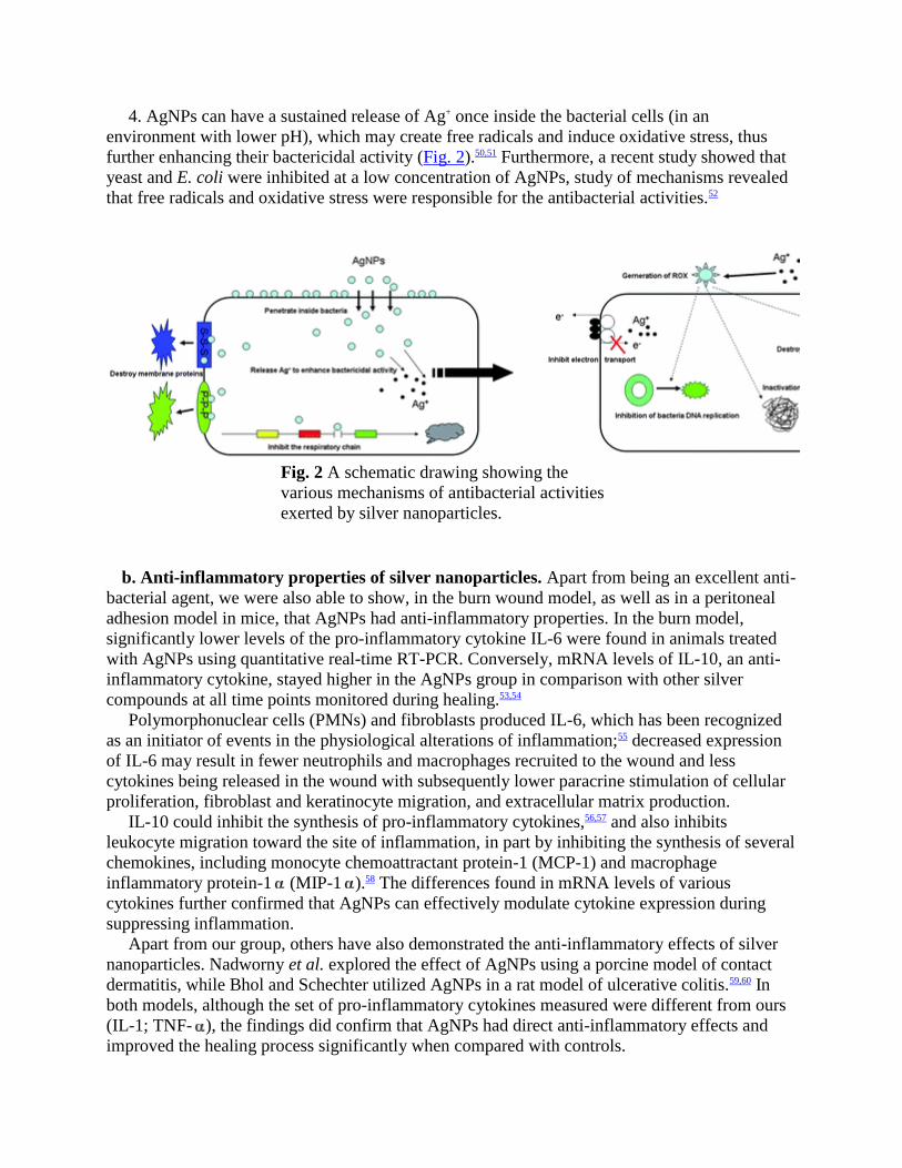
4. AgNPs can have a sustained release of Ag+ once inside the bacterial cells (in an
environment with lower pH), which may create free radicals and induce oxidative stress, thus
further enhancing their bactericidal activity (Fig. 2).50,51 Furthermore, a recent study showed that
yeast and E. coli were inhibited at a low concentration of AgNPs, study of mechanisms revealed
that free radicals and oxidative stress were responsible for the antibacterial activities.52
Fig. 2 A schematic drawing showing the
various mechanisms of antibacterial activities
exerted by silver nanoparticles.
b. Anti-inflammatory properties of silver nanoparticles. Apart from being an excellent anti-
bacterial agent, we were also able to show, in the burn wound model, as well as in a peritoneal
adhesion model in mice, that AgNPs had anti-inflammatory properties. In the burn model,
significantly lower levels of the pro-inflammatory cytokine IL-6 were found in animals treated
with AgNPs using quantitative real-time RT-PCR. Conversely, mRNA levels of IL-10, an anti-
inflammatory cytokine, stayed higher in the AgNPs group in comparison with other silver
compounds at all time points monitored during healing.53,54
Polymorphonuclear cells (PMNs) and fibroblasts produced IL-6, which has been recognized
as an initiator of events in the physiological alterations of inflammation;55 decreased expression
of IL-6 may result in fewer neutrophils and macrophages recruited to the wound and less
cytokines being released in the wound with subsequently lower paracrine stimulation of cellular
proliferation, fibroblast and keratinocyte migration, and extracellular matrix production.
IL-10 could inhibit the synthesis of pro-inflammatory cytokines,56,57 and also inhibits
leukocyte migration toward the site of inflammation, in part by inhibiting the synthesis of several
chemokines, including monocyte chemoattractant protein-1 (MCP-1) and macrophage
inflammatory protein-1 (MIP-1 ).58 The differences found in mRNA levels of various
cytokines further confirmed that AgNPs can effectively modulate cytokine expression during
suppressing inflammation.
Apart from our group, others have also demonstrated the anti-inflammatory effects of silver
nanoparticles. Nadworny et al. explored the effect of AgNPs using a porcine model of contact
dermatitis, while Bhol and Schechter utilized AgNPs in a rat model of ulcerative colitis.59,60 In
both models, although the set of pro-inflammatory cytokines measured were different from ours
(IL-1; TNF- ), the findings did confirm that AgNPs had direct anti-inflammatory effects and
improved the healing process significantly when compared with controls.
Nonetheless, in a peritoneal adhesion model, we provided further evidence for, and
contributed to the understanding of, anti-inflammation properties of AgNPs.54 Here, the
mechanisms of the anti-inflammation effects were suggested to be through a reduction of IFN-
and TNF- via macrophages.
Applications of silver nanoparticles in medicine
In the past, silver was used for a variety of clinical conditions including epilepsy, venereal
infections, acnes and leg ulcers. Silver foil was applied to surgical wounds for improved healing
and reduced post-operative infections, while silver and lunar caustic (pencil containing silver
nitrate mitigated with potassium nitrate) was used for wart removal and ulcer debridement.61
Although some centers still use these solutions, they have been shown to be very impractical to
use on large wounds or for extended time periods due to instability. With nanotechnology, the
availability of silver nanoparticles has enabled the use of pure silver to achieve a rapid growth in
medical practice. Since the size, shape and composition of silver nanoparticles can have a
significant effect on their efficacy, extensive research has gone into synthesizing and
characterizing silver nanoparticles. The application of nanosilver can be broadly divided into
diagnostic and therapeutic uses.
a. Nanosilver in diagnosis and imaging. Early diagnosis of any disease condition is vital to
ensure that early treatment is started and perhaps resulting in a better chance of cure. For
example, in patients undergoing general anesthesia for surgery, the risk of developing pulmonary
complications will be lowered if any sub-clinical upper respiratory tract viral infections can be
detected prior to surgery. Surface-enhanced Raman spectroscopy (SERS) has emerged as a
powerful analytical tool that extends the possibilities of vibrational spectroscopy. SERS differs
from standard Raman scattering in that the incoming laser beam interacts with the oscillations of
plasmonic electrons in metallic nanostructures to enhance the vibrational spectra of molecules
adsorbed to the surface. In a recent study, SERS was used to obtain the Raman spectra of the
respiratory syncytial virus (RSV), using substrates composed of silver nanorods. It was shown in
this study that the four virus strains tested were readily detected at very low detection limits.62
In terms of detecting cancer, Au–Ag nanorods were used in a recent study as a nanoplatform
for multivalent binding by multiple aptamers, so as to increase both the signal and binding
strengths of the aptamers in cancer cell recognition. The molecular assembly of aptamers on the
nanorods was shown to lead to a 26-fold higher affinity than the original aptamer probes.63 Thus,
these nanorod–aptamer conjugates are highly promising for use in specific cell targeting, as well
as having the detection and targeting ability needed for cell studies, disease diagnosis, and
therapy.
b. Nanosilver in therapeutics. (i) Wound dressing. Wound healing is regarded as a complex
and multiple-step process involving integration of activities of different tissues and cell
lineages.64 Perhaps the most well documented and commonly used application of silver
nanoparticles for this is in the use of wound dressings.27,65 In this regard, Acticoat®, which is the
first commercial dressing made up of two layers of polyamide ester membranes covered with
nanocrystalline silver ions, has been studied extensively. Acticoat® has been shown to have the
lowest MIC and MBC values, and the fastest Kill kinetics against the five bacteria tested in in
vitro studies.66,67 Further, the sustained release of silver particles should minimize the likelihood
of bacteria developing resistance to silver. In a randomized prospective clinical study involving
30 patients with each group of patients having comparable burn wound size, depth and location,
the wounds were either treated with silver nanoparticles dressing or a gauze soaked in 0.5%
silver nitrate solution. The frequency of burn wound sepsis, as well as secondary bacteraemia,
were found to be less in patients treated with silver nanoparticles than in those treated with the
control.68 As well as burn wounds, there is now increasing evidence for the use of silver
nanoparticles in the treatment of chronic wounds, such as leg ulcers, diabetic foot ulcers and
pressure ulcers. Sibbald et al. conducted a prospective study to evaluate the use of silver
nanoparticles dressing on a variety of chronic non-healing wounds. The study concluded that
silver nanoparticles dressing has a beneficial effect of protecting the wound site from bacterial
contamination.69 Compared with other silver compounds, AgNPs seem also to promote healing
and achieve better cosmetics after healing.
The biological effects of AgNPs on wound healing appear to be manifols. When we
performed experiments using an excisional wound model, we were able to show that AgNPs
could exert differential effects on keratinocytes and fibroblasts during healing.70 AgNPs, on the
one hand, could promote wound healing through facilitating the proliferation and migration of
keratinocyte, on the other hand, they could reduce the formation of collagen by fibroblasts by
driving their differentiation into myofibroblasts.
In addition to this significant finding, AgNPs were also shown to facilitate wound healing
through modulation of various cytokines. Using a contaminated wound model in pigs, Wright et
al. found accelerated healing was characterized by rapid development of well vascularized
granulation tissue that supported the tissue grafting after injury; furthermore, the promoted
healing was associated with reduced local matric metalloproteinase (MMP) levels and enhanced
cellular apoptosis.71 This finding was supported in other studies.53,72 Taken together, the use of
silver nanoparticles in the aspects of wound healing appears to hold the greatest promise.
(ii) Silver-impregnated catheters.
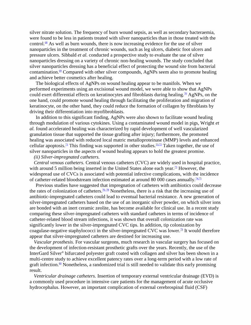
Central venous catheters. Central venous catheters (CVC) are widely used in hospital practice,
with around 5 million being inserted in the United States alone each year.73 However, the
widespread use of CVCs is associated with potential infective complications, with the incidence
of catheter-related bloodstream infection estimated at around 80 000 cases annually.74,75
Previous studies have suggested that impregnation of catheters with antibiotics could decrease
the rates of colonization of catheters.76–78 Nonetheless, there is a risk that the increasing use of
antibiotic-impregnated catheters could lead to eventual bacterial resistance. A new generation of
silver-impregnated catheters based on the use of an inorganic silver powder, on which silver ions
are bonded with an inert ceramic zeolite, has become available for clinical use. In a recent study
comparing these silver-impregnated catheters with standard catheters in terms of incidence of
catheter-related blood stream infections, it was shown that overall colonization rate was
significantly lower in the silver-impregnated CVC tips. In addition, tip colonization by
coagulase-negative staphylococci in the silver-impregnated CVC was lower.79 It would therefore
appear that silver-impregnated catheters are destined for increasing use.
Vascular prosthesis. For vascular surgeons, much research in vascular surgery has focused on
the development of infection-resistant prosthetic grafts over the years. Recently, the use of the
InterGard Silver® bifurcated polyester graft coated with collagen and silver has been shown in a
multi-centre study to achieve excellent patency rates over a long-term period with a low rate of
graft infection.80 Nonetheless, a randomized trial is still needed to validate this early promising
result.
Ventricular drainage catheters. Insertion of temporary external ventricular drainage (EVD) is
a commonly used procedure in intensive care patients for the management of acute occlusive
hydrocephalus. However, an important complication of external cerebrospinal fluid (CSF)
drainage is bacterial colonization of the catheter, resulting in ventriculomeningitis and
encephalitis. The availability of silver-impregnated ventricular catheters since 2004 resulted in a
pilot study addressing their clinical efficacy in neurological and neurosurgical patients requiring
external CSF drainage. The authors found that CSF cultures performed at least three times a
week yielded 25% more positive cultures in the control group compared to 0% in the treatment
group using silver catheters. Furthermore, aseptic meningitis due to inflammation was not seen
in patients with the silver-impregnated biomedical material.81
(iii) Silver in orthopaedics. Artificial joint replacements have become the gold standard
treatment for many arthritic diseases. Like all biomaterials, bone cement based on
polymethylmetacrylate (PMMA) has an elevated risk of infection when implanted into the
human body.82 Indeed, an increasing number of joint infections with multi-resistant bacteria
mean that an adequate prophylaxis against these organisms is necessary. Recent studies have
been carried out to evaluate bone cement loaded with nanosilver.83 Here, nanosilver-loaded bone
cement could be shown to have high antibacterial activity against all tested strains including
methicillin-resistant Staphylococcus aureus (MRSA). Furthermore, the nanoparticles did not
seem to have cytotoxicity to osteoblasts grown in vitro.
As well as bone cement, the use of silver nanoparticles has been studied in artificial joints.
For many years, ultra high molecular weight polyethylene (UHMWPE) has been the material of
choice for fabrication of bearing inserts for joint replacement components. A major problem with
the longevity of UHMWPE is wear and concomitant debris generation, which can activate
macrophages, with subsequent inflammation, and eventual failure of the artificial joints. In one
study, incorporation of silver nanoparticles was demonstrated to lead to both physical and
chemical stabilization of the polymer surface layer toward friction oxidation and degradation.84
This procedure was further shown to significantly decrease the process of polymer/metal
tribochemical debris formation and at the same time enhances UHMWPE biocompatibility and
antimicrobial activity.
Taken together, it would appear that silver nanoparticles could play a significant role in the
next generation of biomaterials in orthopaedics.
(iv) Surgical mesh. For general surgery, surgical implants are often unavoidable. Surgical
meshes are commonly used for bridging large wounds, as well as acting as reinforcements to
tissue repair. However, being foreign material, they do carry a risk of infection. Indeed, it has
been estimated that one million nocosomial infections are seen each year in patients with
implanted prosthetic materials.85 The use of silver nanoparticles polypropylene mesh has been
studied recently. Similar to other studies using silver nanoparticles, the results showed that silver
nanoparticles polypropylene mesh had significant bactericidal efficacy against S. aureus.
Furthermore, it was shown that silver nanoparticles could continue to diffuse off the mesh and
had sustained activity.86 These results clearly warrant further in vivo studies to determine whether
silver nanoparticles-coated polypropylene mesh can decrease the prosthetic infection rate and the
host inflammatory response in the clinical setting.
Are silver nanoparticles harmful?
With the use of silver nanoparticles in medical appliances, exposure to silver in the body is
therefore inevitable and increasing. In order to gain further widespread use in clinical medicine,
the issue of the potential toxicity of AgNPs needs to be fully evaluated.
Although silver is believed traditionally to be relatively non-toxic to mammalian cells, from
previous evidence taken from workers in the silver industry, it is not known if this is still the case
for silver nanoparticles. This is of particular concern because, due to the small size, silver
nanoparticles can gain increasing access to tissues, cells and biological molecules within the
human body.
In this regard, many in vitro studies have been performed. Hsin et al. provided evidence for
molecular mechanism of AgNPs-induced cytotoxicity, showing that AgNPs acted through ROS
and JNK to induce apoptosis via the mitochondrial pathway in NIH3T3 fibroblast cells.87 Park et
al. reported cytotoxicity using AgNPs prepared by dispersing them in fetal bovine serum, as a
biocompatible material, on the cultured RAW264.7 macrophage cell line, which induced cellular
apoptosis.88 Furthermore, AgNPs decreased intracellular glutathione level, increased NO
secretion, increased TNF- in protein and gene levels, and increased the gene expression of
matrix metalloproteinases, such as MMP-3, MMP-11, and MMP-19. Kim et al. demonstrated
cytotoxicity induced by AgNPs in human hepatoma HepG2 cells and indicated that AgNPs
agglomerated in the cytoplasm and nuclei of treated cells, and induced intracellular oxidative
stress and was independent of the toxicity of Ag+ ions.89 On a similar note, Kawata et al. showed
an upregulation of DNA repair-associated genes in hepatoma cells cultured with low dose silver
nanoparticles, suggesting possible DNA damaging effects.90 In HeLa cells, Miura and Shinohara
reported that the expressions of ho-1 and mt-2A, well-known oxidative stress-related genes, were
upregulated by AgNPs treatment, showing that AgNPs had the potential for cytotoxicity in the
case of exposure at high concentrations.91
Despite the findings in these in vitro studies, the overall significance in the in vivo setting, and
also the applicability to humans remain unknown. In the clinic, silver nanoparticle-based wound
dressings are perhaps the most universally used. Since nanosilver wound dressings are applied to
wounded skin where the strict barrier is broken, it is thus expected that the entry of the
nanoparticles into the body would be easier. This, along with the observation that particles in the
skin can be phagocytosed by macrophages and Langerhans cells, might theoretically lead to
perturbations of the immune system. At the same time, nanoparticles entering capillaries could
become circulatory and would soon encounter the liver and expose the liver to a high dose of
silver nanoparticles. Nonetheless, systemic toxicity of ingested silver nanoparticles is scarcely
seen. Supporting this, when we injected silver nanoparticles into experimental mice
intravenously, we did not observe any overt systemic effects, despite the silver nanoparticles
solution used being at a relatively high concentration of 100 mM (unpublished data). At the local
level, although others showed that when cultured keratinocytes were exposed to extracts of
silver-containing dressings, their proliferation was significantly inhibited, we did not observe any
increase in cell death or inhibition of cell growth in our experiments using keratinocytes and
fibroblasts.70,92,93 In contrast, we found that silver nanoparticles increased the growth rate of
keratinocytes. The differences between our data and others' might be attributable to the
difference in laboratory conditions and techniques employed. The concentration of silver used in
experiments might also be an important factor.
Nonetheless, the issue of argyria , the deposition of silver metal causing discoloration of
the tissues, is another concern with chronic ingestion or inhalation of silver preparations.
Although argyria is not a life-threatening condition, it is, however, cosmetically undesirable.94 In
wound care, Wang et al. reported that if silver dressing was topically applied to the porcine deep
dermal partial thickness model, a larger amount of silver would deposit in cutaneous scar tissue
(136 g g−1) than normal skin (less than 0.747 g g−1). The wound would have a slate-gray
appearance.95 Contrary to this finding, Jaya et al. reported that when compared with conventional
silver agents, AgNPs were a safer alternative because of their sustained release dose regime.96
Taking into account the existence of silver nanoparticle-impregnated catheters for clinical
use, hemo-compatibility is another safety concern. Previous reports suggested that nanoparticles
present in blood were associated with thrombosis and activation of immunological reactions.
Studies have provided evidence that exposure to ambient ultrafine particles elicits inflammatory
responses in vascular endothelial cells and blood cells.97,98 For silver, a recent study revealed that
silver nanoparticles could greatly enhance the electron-transfer reactivity of myoglobin.99
Further, the recent identification of the cytotoxicity of silver nanoparticles towards the
spermatogonial stem cell line has aroused great concern over the biosafety of nanomaterials.100
As discussed previously, the liver appears to be an eventual accumulation site of circulatory
silver nanoparticles. Similar patterns of cytotoxicity of silver nanoparticles (decrease of
mitochondria function, LDH leakage and abnormal cell morphologies) were observed in in vitro
studies. Nonetheless, during our other experiments using silver nanoparticles, we routinely
harvested organs (liver, spleen, lung, heart and kidney) and analyzed the silver content using
inductively coupled plasma mass spectroscopy (ICP-MS) after trypsin digestion. Thus far, in
experiments using therapeutic doses of silver nanoparticles, only very low levels of silver (below
0.5 g g−1 of organ) could be detected in the organs of the mice, suggesting that nanosilver was
safe at these low concentrations. Indeed, in clinical situations, wound exudation and systemic
proteins might also contribute towards silver nanoparticles in vivo safety, as the high protein
content probably neutralises nanosilver's tissue toxicity. Taken together, it may be fair to say that
silver nanoparticles would be safe to use clinically at low doses.
Future therapeutic directions
a. Anti-inflammatory agent. The potential anti-inflammatory action of silver nanoparticles
has been suggested in various studies described previously. On the other hand, inflammation has
been noted to play a significant part in the formation of post-operative adhesions. In animal
models, we showed that intra-peritoneal injection of silver nanoparticles significantly reduced
the degree of post-operative fibrous adhesions. The anti-inflammatory effects have also been
substantiated in other inflammatory disease models by others. Taken together, it would suggest
that silver nanoparticles can indeed reduce inflammation and its use in other inflammatory
conditions is eagerly anticipated.
b. Antiviral drug. The antiviral properties of metal nanoparticles are of significant medicinal
interest. With finding a cure for human immunodeficiency virus (HIV) in mind, the post-infected
anti-HIV-1 activities of silver nanoparticles toward Hut/CCR5 cells were evaluated in one
study.101 Here, silver nanoparticles were shown to have dose-dependent anti-retrovirus activities
and exhibited high potency in inhibiting HIV-1 replication. Further, these nanoparticles did not
show acute cytotoxicity to either the Hut/CCR5 cells or to normal peripheral blood mononuclear
cells. It remains to be seen whether silver nanoparticles have activities against other types of
viruses.
c. Anti-platelet agent. Thrombotic disorders have remained a significant problem in clinical
medicine. Results thus far have shown that anticoagulant and thrombolytic therapy may
sometimes lead to serious bleeding complications.102 As platelets play a central role in thrombotic
disorders, the focus has now shifted to regulating and maintaining these cells in an inactive state.
Recently, Shrivastava et al. demonstrated that AgNPs could effectively inhibit integrin-mediated
platelet functional responses like aggregation, secretion, adhesion to immobilized fibrinogen or
collagen and retraction of fibrin clot in a dose-dependent manner.103 Further, in vivo studies using
mouse models also supported the anti-platelet properties of silver nanoparticles. The results,
significant inhibition of platelet functions with a relatively low dose of AgNPs, combined with
the lack of cell lysis, raise the hope for its use as an anti-platelet therapeutic agent.
Future application of silver nanoparticles—effects on stem cells?
The epidermal stem cells, which reside in the dermal layer in the skin, play the most important
roles for repairing the epidermis, regenerating hair and maintaining tissue homeostasis after
injury. Stem cells have the remarkable capacity to both self-perpetuate and also give rise to the
differentiating cells that constitute one or more tissues.104 In recent years, many scientists have
been exploring mysteries underlying their remarkable capacity to perform these feats.105–111 In our
ongoing study, we have also found proliferation of epidermal stem cells in skin promoted by
silver nanoparticles at low concentrations (unpublished data). We are exploring the exact
mechanism of this phenomenon.
Apart from epidermal stem cells, the use of mesenchymal stem cells (MSCs) is one promising
modality for cell-based therapy applications due to their easy isolation and culture as well as the
expansion capacity. Furthermore, MSCs can also provide pleuripotent potential and develop into
various lineages such as skin, bone, tendon, ligament, muscle, fat and blood.112–114 In the
environment of a healing wound, the use of programmed MSCs can thus be an important tool to
compensate for the tissue loss and recovery of function and structure. We are currently studying
the possibility of enhanced proliferation and survival of hMSC by silver nanoparticles, and
results are eagerly awaited.
Conclusion
The advance in nanotechnology has enabled us to utilize particles in the size of the nanoscale.
This has created new therapeutic horizons, and in the case of silver, the currently available data
only reveals the surface of the potential benefits and the wide range of applications. We have yet
to elucidate the exact cellular pathway of silver nanoparticles. Furthermore, it remains to be seen
whether any potential complications for the silver nanoparticles would surface after prolonged
clinical use. Nonetheless, a bright future holds for this precious metal.
References
1 M. A. Albrecht, C. W. Evans and C. L. Raston, Green Chem., 2006, 8, 417–432 [Links].
2 J. D. Aiken and R. G. Finke, J. Mol. Catal. A: Chem., 1999, 145, 1–44 [Links].
3 M. Babincova and P. Babinec, Biomed Pap Med Fac Univ Palacky Olomouc Czech Repub,
2009, 153, 243–50.
4 S. V. Vinogradov, Nanomedicine, 2010, 5, 165–168 [Links].
5 M. Kumar, Y. Ando. Journal of Nanoscience and Nanotechnology. 201, 10, pp. 3739–3758.
6 M. E. Pearce, J. B. Melanko and A. K. Salem, Pharm. Res., 2007, 24(12), 2335–2352
[Links].
7 Y. Xiao and X. Gao, Biomarkers in Medicine, 2010, 4(2), 227–39.
8 M. S. Muthu and B. Wilson, Nanomedicine, 2010, 5(2), 169–71 [Links].
9 Y. Su, S. Qiao and H. Yang, et al., Nanotechnology, 2010, 21(6), 065604 [Links].
10 W. Zhao, H. Cao and C. S. Wan, et al., Di Yi Jun Yi Da Xue Xue Bao, 2002, 22(5), 461–3.
11 R. Bhattacharya and P. Mukherjee, Adv. Drug Delivery Rev., 2008, 60, 1289–1306 [Links].
12 S. M. Hirst, A. S. Karakoti, R. D. Tyler, N. Sriranganathan, S. Seal and C. M. Reilly, Small,
2009, 5, 2848–2856 [Links].
13 S. Hussain and C. Ferguson, Emerg. Med. J., 2006, 23, 929–932.
14 P. Muangman, S. Muangman, S. Opasanon, K. Keorochana and C. Chuntrasakul, Journal of
The Medical Association of Thailand, 2009, 92, 1300–1305.
15 F. Uygur, O. Oncül, R. Evinç, H. Diktas, A. Acar and E. Ulkür, Burns, 2009, 35, 270–273
[Links].
16 D. M. Caruso, K. N. Foster, S. A. Blome-Eberwein, J. A. Twomey, D. N. Herndon and A.
Luterman, Journal of Burn Care & Research, 2006, 27, 298–309.
17 J. Turkevich, P. C. Stevenson and J. Hillier, Discuss. Faraday Soc., 1951, 11, 55–75
[Links].
18 S. Shrivastava, T. Bera and A. Roy, Nanotechnology, 2007, 18, 225103–225111 [Links].
19 Colloidal Silver Products. National Center for Complimentary and Alternative Medicine,
2006, http://nccam.nih.gov/health/silver/.
20 M. C. Fung, M. Weintraub and D. L. Bowen, JAMA, J. Am. Med. Assoc., 1995, 274, 1196–
7.
21 C. N. Lok, C. M. Ho and R. Chen, JBIC, J. Biol. Inorg. Chem., 2007, 12, 527–534 [Links].
22 H. J. Yen, S. H. Hsu and C. L. Tsai, Small, 2009, 5, 1553–1561 [Links].
23 A. Nanda and M. Saravanan, Nanomed.: Nanotechnol., Biol. Med., 2009, 5, 452–456
[Links].
24 A. R. Shahverdi, A. Fakhimi and H. R. Shahverdi, Nanomed.: Nanotechnol., Biol. Med.,
2007, 3, 168–171 [Links].
25 M. Gajbhiye, J. Kesharwani and A. Ingle, Nanomed.: Nanotechnol., Biol. Med., 2009, 5,
382–386 [Links].
26 N. George, J. Faoagali and M. Muller, Burns, 1997, 23, 493–495 [Links].
27 D. J. Leaper, International Wound Journal, 2006, 3, 282–294.
28 E. Barreiro, J. S. Casas and M. D. Couce, Dalton Trans., 2007, 3074–3085 [Links].
29 V. Thomas, M. M. Yallapu and B. Sreedhar, J. Colloid Interface Sci., 2007, 315, 389–395
[Links].
30 S. M. Modak and C. L. Fox, Biochem. Pharmacol., 1973, 22, 2391–2404 [Links].
31 P. D. Bragg and D. J. Rainnie, Can. J. Microbiol., 1974, 20, 883–889 [Links].
32 G. Gravante, R. Caruso and R. Sorge, Ann. Plast. Surg., 2009, 63, 201–205 [Links].
33 D. R. Monteiro, L. F. Gorup and A. S. Takamiya, Int. J. Antimicrob. Agents, 2009, 34, 103–
110 [Links].
34 X. Chen and H. J. Schluesener, Toxicol. Lett., 2008, 176, 1–12 [Links].
35 Q. Li, S. Mahendra and D. Y. Lyon, Water Res., 2008, 42, 4591–4602 [Links].
36 C. S. Chu, A. T. McManus, B. A. Pruitt and A. D. Mason, J. Trauma: Inj. Infect. Crit. Care,
1988, 28, 1488–1492 [Links].
37 E. A. Dritch, A. Marin, V. Malakanov and J. A. Albright, J. Trauma: Inj. Infect. Crit. Care,
1987, 27, 301–304.
38 H. W. Margraff and T. H. Covey, Archives of Surgery, 1977, 112, 699–704.
39 D. Wyatt, D. N. McGowan and M. P. Najarian, J. Trauma: Inj. Infect. Crit. Care, 1990, 30,
857–865 [Links].
40 C. L. Fox and S. M. Modak, Antimicrobial Agents and Chemotherapy, 1974, 5, 582–588.
41 C. L. Fox, Archives of Surgery, 1968, 96, 184–188.
42 V. Edwards-Jones, Lett. Appl. Microbiol., 2009, 49, 147–152 [Links].
43 K. Madhumathi, P. T. Sudheesh Kumar, S. Abhilash, V. Sreeja and H. Tamura, J. Mater.
Sci.: Mater. Med., 2010, 21, 807–13 [Links].
44 M. Rai, A. Yadav and A. Gade, Biotechnol. Adv., 2009, 27, 76–83 [Links].
45 C. N. Lok, C. M. Ho, R. Chen, Q. Y. He, W. Y. Yu, H. Sun, P. K. Tam, J. F. Chiu and C.
M. Che, J. Proteome Res., 2006, 5, 916–24 [Links].
46 Q. L. Feng, J. Wu, G. Q. Chen, F. Z. Cui, T. N. Kim and J. O. Kim, J. Biomed. Mater. Res.,
2000, 52, 662–668 [Links].
47 S. Y. Liau, D. C. Read, W. J. Pugh, J. R. Furr and A. D. Russell, Lett. Appl. Microbiol.,
1997, 25, 279–283 [Links].
48 Y. Matsumura, K. Yoshikata, S. I. Kunisaki and T. Tsuchido, Appl. Environ. Microbiol.,
2003, 69, 4278–4281 [Links].
49 I. Sondi and B. Salopek-Sondi, J. Colloid Interface Sci., 2004, 275, 177–82 [Links].
50 J. R. Morones, J. L. Elechiguerra, A. Camacho and J. T. Ramirez, Nanotechnology, 2005,
16, 2346–2353 [Links].
51 H. Y. Song, K. K. Ko, L. H. Oh and B. T. Lee, European Cell and Materials journal, 2006,
11, 58.
52 J. S. Kim, E. Kuk, K. N. Yu and J. H. Kim, Nanomed.: Nanotechnol., Biol. Med., 2007, 3,
95–101 [Links].
53 J. Tian, K. K. Wong, C. M. Ho, C. N. Lok, W. Y. Yu, C. M. Che, J. F. Chiu and P. K. Tam,
ChemMedChem, 2007, 2, 129–136 [Links].
54 K. K. Wong, S. O. Cheung, L. M. Huang, J. Niu, C. Tao, C. M. Ho, C. M. Che and P. K.
Tam, ChemMedChem, 2009, 4, 1129–1135 [Links].
55 P. Paquet and G. E. Pierard, Int. Arch. Allergy Immunol., 1996, 109, 308–317.
56 D. F. Fiorentino, A. Zlotnik, P. Vieira, T. R. Mosmann, M. Howard, K. W. Moore and A.
O'Garra, The Journal of Immunology, 1991, 146, 3444–3451.
57 R. de Waal Malefyt, J. Abrams, B. Bennett, C. G. Figdor and J. E. de Vries, J. Exp. Med.,
1991, 174, 1209–1220 [Links].
58 M. N. Ajuebor, A. M. Das, L. Virag, C. Szabo and M. Perretti, Biochem. Biophys. Res.
Commun., 1999, 255, 279–282 [Links].
59 P. L. Nadworny, J. F. Wang and E. E. Tredget, Nanomed.: Nanotechnol., Biol. Med., 2008,
4, 241–251 [Links].
60 K. C. Bhol and P. J. Schechter, Dig. Dis. Sci., 2007, 52, 2732–2742 [Links].
61 H. J. Klasen, Burns, 2000, 26, 131–138 [Links].
62 S. Shanmukh, L. Jones, Y. P. Zhao, J. D. Driskell, R. A. Tripp and R. A. Dluhy, Anal.
Bioanal. Chem., 2008, 390, 1551–1555 [Links].
63 Y. F. Huang, H. T. Chang and W. H. Tan, Anal. Chem., 2008, 80, 567–572 [Links].
64 P. Martin, Science, 1997, 276, 75–81 [Links].
65 J. Fong and F. Wood, Int. J. Nanomed., 2006, 1, 441–9 [Links].
66 J. B. Wright, K. Lam and R. E. Burrell, Am. J. Infect. Control, 1998, 26, 572–577 [Links].
67 H. Q. Yin, R. Langford and R. E. Burrell, Journal of Burn Care & Rehabilitation, 1999, 20,
195–200.
68 E. E. Tredget, H. A. Shankowsky, A. Groeneveld and R. Burnell, Journal of Burn Care &
Rehabilitation, 1998, 19, 531–537.
69 R. G. Sibbald, J. Contreras-Ruiz, P. Coutts, M. Fierheller, A. Rothman and K. Woo,
Advances in Skin & Wound Care, 2007, 20, 549–58.
70 X. Liu, P. Y. Lee, C. M. Ho, V. C. Lui, Y. Chen, C. M. Che, P. K. Tam and K. K. Wong,
ChemMedChem, 2010, 5, 468–75 [Links].
71 J. B. Wright, K. Lam, A. G. Buret, M. E. Olson and R. E. Burrell, Wound Repair Regener.,
2002, 10, 141–151.
72 A. B. Lansdown, Journal of Wound Care, 2002, 11, 125–130.
73 I. Raad, Lancet, 1998, 351, 893–8 [Links].
74 D. Pittet, D. Tarara and R. P. Wenzel, JAMA, J. Am. Med. Assoc., 1994, 271, 1598–601.
75 L. A. Mermel, Annals of Internal Medicine., 2000, 132, 391–402 [Links].
76 S. J. George, P. Vuddamalay and M. J. Boscoe, Eur. J. Anaesthesiol., 1997, 14, 428–431
[Links].
77 S. Tennenberg, M. Lieser, B. McCurdy, G. Boomer, E. Howington, C. Newman and I.
Wolf, Archives of Surgery, 1997, 132, 1348–1351.
78 W. H. Sheng, W. J. Ko, J. T. Wang, S. C. Chang, P. R. Hsueh and K. T. Luh, Diagn.
Microbiol. Infect. Dis., 2000, 38, 1–5 [Links].
79 M. D. Khare, S. S. Bukhari, A. Swann, P. Spiers, I. McLaren and J. Myers, J. Infect., 2007,
54, 146–50 [Links].
80 J. B. Ricco, J. Vasc. Surg., 2006, 44, 339–46 [Links].
81 K. Galiano, C. Pleifer, K. Engelhardt, G. Brossner, P. Lackner, C. Huck, C. Lass-Flörl and
A. Obwegeser, Neurol. Res., 2008, 30, 285–7.
82 A. G. Gristina, Science, 1987, 237, 1588–1595 [Links].
83 V. Alt, T. Bechert, P. Steinrücke, M. Wagener, P. Seidel, E. Dingeldein, E. Domann and R.
Schnettler, Biomaterials, 2004, 25, 4383–91 [Links].
84 K. S. Morley, P. B. Webb, N. V. Tokareva, A. P. Krasnov, V. K. Popov, J. Zhang, C. J.
Roberts and S. M. Howdle, Eur. Polym. J., 2007, 43, 307–314 [Links].
85 R. O. Darouiche, N. Engl. J. Med., 2004, 350, 1422–1429 [Links].
86 M. S. Cohen, J. M. Stern, A. J. Vanni, R. S. Kelley, E. Baumgart, D. Field, J. A. Libertino
and I. C. Summerhayes, Surgical Infections, 2007, 8, 397–403.
87 Y. H. Hsin, C. F. Chen, S. Huang, T. S. Shih, P. S. Lai and P. J. Chueh, Toxicol. Lett., 2008,
179, 130–139 [Links].
88 E. J. Park, J. Yi, Y. Kim, K. Choi and K. Park, Toxicol. in Vitro, 2010, 24(3), 872–878
[Links].
89 S. Kim, J. E. Choi, J. Choi, K. H. Chung, K. Park, J. Yi and D. Y. Ryu, Toxicol. in Vitro,
2009, 23, 1076–1084 [Links].
90 K. Kawata, M. Osawa and S. Okabe, Environ. Sci. Technol., 2009, 43, 6046–6051 [Links].
91 N. Miura and Y. Shinohara, Biochem. Biophys. Res. Commun., 2009, 390, 733–737 [Links].
92 J. E. Paddle-Ledinek, Z. Nasa and H. J. Cleland, Plast. Reconstr. Surg., 2006, 117, 110S–
118S.
93 V. K. Poon and A. Burd, Burns, 2004, 30, 140–147 [Links].
94 A. B. Lansdown, Curr. Probl. Dermatol., 2006, 33, 17–34.
95 X. Q. Wang, H. E. Chang and R. Francis, J. Cutaneous Pathol., 2009, 36, 788–792.
96 J. Jain, S. Arora and J. M. Rajwade, Mol. Pharmaceutics, 2009, 6, 1388–1401 [Links].
97 A. Gojova, B. Guo, R. S. Kota, J. C. Rutledge, I. M. Kennedy and A. I. Barakat, Environ.
Health Perspect., 2007, 115, 403–409 [Links].
98 R. Rückerl, R. P. Phipps, A. Schneider, M. Frampton, J. Cyrys, G. Oberdörster, H. E.
Wichmann and A. Peters, Part. Fibre Toxicol., 2007, 4, 1 [Links].
99 X. Gan, T. Liu, J. Zhong, X. Liu and G. Li, ChemBioChem, 2004, 5, 1686–1691 [Links].
100 L. Braydich-Stolle, S. Hussain, J. J. Schlager and M. C. Hofmann, Toxicol. Sci., 2005, 88,
412–419 [Links].
101 R. W. Sun, R. Chen, N. P. Chung, C. M. Ho, C. L. Lin and C. M. Che, Chem. Commun.,
2005, 5059–5061 [Links].
102 A. Khalid, American Family Physician, 2002, 65, 1097–1102.
103 S. Shrivastava, T. Bera and S. K. Singh, ACS Nano, 2009, 3, 1357–64 [Links].
104 E. Fuchs, J. Cell Biol., 2008, 180, 273–284 [Links].
105 G. Han, A. G. Li, Y. Y. Liang, P. Owens, W. He, S. Lu, Y. Yoshimatsu, D. Wang, P. Ten
Dijke, X. Lin and X. J. Wang, Dev. Cell, 2006, 11, 301–312 [Links].
106 K. B. Jensen and F. M. Watt, Proc. Natl. Acad. Sci. U. S. A., 2006, 103, 11958–11963
[Links].
107 V. Horsley, A. O. Aliprantis and L. Polak, Cell, 2008, 132, 299–310 [Links].
108 J. Huelsken, R. Vogel and B. Erdmann, Cell, 2001, 105, 533–545 [Links].
109 V. Horsley, D. O'Carroll and R. Tooze, Cell, 2006, 126, 597–609 [Links].
110 M. Ito, Y. Liu and Z. Yang, Nat. Med., 2005, 11, 1351–1354 [Links].
111 M. Ito, Z. Yang and T. Andl, Nature, 2007, 447, 316–320 [Links].
112 A. I. Caplan, J. Orthop. Res., 1991, 9, 641–650 [Links].
113 A. I. Caplan, Tissue Eng., 2005, 11, 1198–1211 [Links].
114 J. J. Minguell, A. Erices and P. Conget, Experimental Biology and Medicine (Maywood),
2001, 226, 507–520.