Embed Size (px)
Citation preview

HORMONE JITTERS
MANUFACTURING FEARFUL MEMORIES
SLICING PIZZAA PERFECTLY SYMMETRIC
β-PROPELLER PROTEIN
BLACK HOLESPROBING THEIR EVOLUTION
AND INTERACTION WITH GALAXIES
SIMULATION SUPERPOWERTHE K COMPUTER BRINGS UNPRECEDENTED SPEED TO SCIENTIFIC PREDICTION
SPRING 2015
SHOWCASING THE BEST OF RESEARCH AT RIKEN www.riken.jp/en/research/rikenresearch
RIKEN Surface and Interface Science Laboratory Researchers at the laboratory in Wako, Saitama, use scanning tunneling microscopes to explore the properties of material surfaces.
RIKEN, Japan’s flagship research institute, conducts basic and applied experimental research in a wide range of science and
technology fields including physics, chemistry, medical science, biology and engineering.
Initially established as a private research foundation in Tokyo in 1917, RIKEN became an
independent administrative institution in 2003.
RIKEN RESEARCH is an online and print publication that highlights the best research
published by RIKEN. This publication is a selection of the articles published by RIKEN at:
www.riken.jp/en/research/rikenresearchPlease visit the website for recent updates
and related articles. Articles showcase RIKEN’s groundbreaking results and are written for a
non-specialist audience.
For further information on the research presented in this publication or to arrange an interview with a researcher, please contact:
RIKEN Global Relations and Research Coordination Office
2-1, Hirosawa, Wako, Saitama, 351-0198, Japan
Tel: +81 48 462 1225Fax: +81 48 463 3687
E-mail: [email protected]
ISSN 1883-3519
www.riken.jp/en/research/rikenresearch
www.riken.jp/en

PeopleProbing the surface
Yousoo Kim, Surface and Interface Science Laboratory
Stress-relief strategies in plantsRyoung Shin, RIKEN Center for Sustainable Resource Science
EditorialSupercomputers for science
Research highlightsExploratory life sciences6 An inner look at the inner ear The earliest stages of ear development involve a localized signaling cascade
7 Finding the function of long noncoding RNA Mouse experiments suggest that a noncoding RNA can be vital for successful pregnancy
8 Perfect propeller proteins designed by computer The computationally assisted synthesis of a symmetric propeller protein that retraces protein evolution could also be used to develop new protein structures
9 A protein shepherd helps coordinate brain development A single protein activates the machinery needed for axon growth
10 ‘Chatty’ cells help build the brain Genetic cues and neuron–progenitor communication influence cerebral cortex development
11 Proteins feel the pinch An enzyme controls the degradation of defective proteins by opening and closing a pore on its surface
Research highlightsExploratory physical sciences15 Particles find their mass A peculiar state of matter could be used to observe quantum mass acquisition
16 Skyrmions like it hot Pinpoint laser heating creates a maelstrom of magnetic nanotextures
17 Viewing black holes in a different light Satellite studies reveal complex processes of x-ray emission from matter falling into a black hole
18 Super-heavy chemistry Forming a hexacarbonyl complex with the synthetic heavy element seaborgium
19 Electron quantum states set free A vortex of electrons provides unprecedented information on magnetic quantum states in solids
Pizza 2
Pizza 6
Pizza 8 Pizza 9
Pizza 10
1 nm
Pizza 7
Pizza 3 Pizza 4 Pizza 5
8
10
15
Feature highlightThe amazing disappearing mouse
An experimental technique renders mouse tissues transparent and colorless12
43
15
6
12
Contents
SPRING 2015 1

PlacesBuilding neural connectionsBSI Neural Circuit Genetics Research Building, RIKEN Brain Science Institute
Research highlightsApplied physical sciences34 Lining up for molecular memory devices Self-arranging molecules produce ordered arrays for use in efficient optoelectronic memory devices
35 Making iron transparent in the quest for shorter x-ray pulses An intense x-ray beam can make iron foil transparent for just an instant
36 Electrons move in different circles Scanning tunneling microscopy reveals the exotic properties of an unusual type of electron
Research highlightsApplied life sciences22 Beating a bottleneck to regenerate nerves A membrane-mimicking polymer selectively binds to nerve cells and stimulates regeneration
23 Expanding the immune system’s memory A subpopulation of innate immune cells can be ‘primed’ to provide a rapid response against future threats
24 Bacteria and immune cells forge a productive partnership Immune cells act as essential intermediaries between the intestines and ‘friendly’ gut bacteria
25 Evolving the best body clock A mathematical model explores how circadian rhythms that keep track of time have evolved
26 Cutting cholesterol for safer potatoes Identifying a gene responsible for cholesterol production could lead to potatoes with lower toxin levels
27 Learning without conflict A mathematical equation explains how equilibrium is achieved between two forms of synaptic plasticity
28 Locking shut the calcium channel A cellular mechanism that alters calcium regulation could be involved in neurodegenerative disease
29 Implanting fear Two mechanisms work in tandem to form memories of frightening events
30 A genetic twist can lead to infertility The discovery of genital defects in a mutant mouse strain could assist studies of human infertility
22
20
2022
3431
PerspectivesPowering sciencePowerful supercomputers like the K computer developed at the RIKEN Advanced Institute for Computational Science are essential for scientific discovery and prediction
31
2 RIKEN RESEARCH
Contents
Supercomputers for scienceT hank you for taking the time to read the spring issue of
RIKEN RESEARCH. It has been a year since we redesigned the
print magazine and began issuing it as a quarterly. We have
also incorporated the former RIKEN RESEARCH site into RIKEN’s
main institutional website. What do you think about our new look? We
welcome your comments (please e-mail us at [email protected])!
In this issue, our “Perspectives” article features the K computer,
a supercomputer developed at the RIKEN Advanced Institute for
Computational Science. We also introduce RIKEN’s plans to build its
successor by 2020 to help meet more of our scientific, environmental and
social needs. In “Places”, we look at the Neural Circuit Genetics Research
Building at the RIKEN Brain Science Institute; this facility was opened in
2011 and is used to study the neural circuits underpinning behavior.
Our “People” section features two laboratory leaders from Korea:
Yousoo Kim from the Surface and Interface Science Laboratory, who
studies solid surfaces and interfaces at the nanoscale regime; and
Ryoung Shin from the RIKEN Center for Sustainable Resource Science,
who is developing methods to increase crop yields in nutrient-limited
conditions and inhibit the accumulation of radioactive cesium in plants.
In our “Research highlights” sections we focus on some of the hot
topics in science being investigated by our researchers, from galactic
x-ray emissions to a mathematical equation for synaptic plasticity and a
mutant mouse strain that exhibits infertility. In particular, we expect that
you may be surprised by our “Feature highlight” article that describes
a technique for making tissues and even whole organisms transparent,
developed by a team at the RIKEN Quantitative Biology Center.
The last year has been a challenging one for RIKEN, but we must
move forward with our research, taking to heart the lessons we have
learned. We are committed to maintaining the highest standards of
research integrity as we continue to work toward contributing to the
welfare of society.
HORMONE JITTERS
MANUFACTURING FEARFUL MEMORIES
SLICING PIZZAA PERFECTLY SYMMETRIC
β-PROPELLER PROTEIN
BLACK HOLESPROBING THEIR EVOLUTION
AND INTERACTION WITH GALAXIES
SIMULATION SUPERPOWERTHE K COMPUTER BRINGS UNPRECEDENTED SPEED TO SCIENTIFIC PREDICTION
SPRING 2015
SHOWCASING THE BEST OF RESEARCH AT RIKEN www.riken.jp/en/research/rikenresearch
Cover story: The K computer at the RIKEN Advanced Institute for Compu-
tational Science brings unprecedented speed to scientific prediction. Page 31
© 2015 RIKEN
SPRING 2015 3
Editorial
People
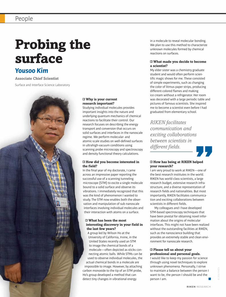
Why is your current research important?Studying individual molecules provides important insights into the nature and underlying quantum mechanics of chemical reactions to facilitate their control. Our research focuses on describing the energy transport and conversion that occurs on solid surfaces and interfaces in the nanoscale regime. We perform molecular- and atomic-scale studies on well-defined surfaces in ultrahigh-vacuum conditions using scanning probe microscopy and spectroscopy and density functional theory calculations.
How did you become interested in the field?In the final year of my doctorate, I came across an impressive paper reporting the successful use of a scanning tunneling microscope (STM) to excite a single molecule bound to a solid surface and observe its vibrations. I immediately recognized that this was the kind of phenomenon I wanted to study. The STM now enables both the obser-vation and manipulation of sub-nanoscale interfaces involving individual molecules and their interaction with atoms on a surface.
What has been the most interesting discovery in your field in the last few years?A group led by Wilson Ho at the University of California, Irvine, in the United States recently used an STM to image the chemical bonds of a molecule—often depicted as sticks con-necting atomic balls. While STMs can be
used to observe individual molecules, the actual chemical bonds in a molecule are
impossible to image. However, by attaching carbon monoxide to the tip of an STM probe, Ho’s group developed a method that can detect tiny changes in vibrational energy
Probing the surfaceYousoo KimAssociate Chief Scientist
Surface and Interface Science Laboratory
in a molecule to reveal molecular bonding. We plan to use this method to characterize unknown molecules formed by chemical reactions on surfaces.
What made you decide to become a scientist?My elder sister was a chemistry graduate student and would often perform scien-tific magic shows for me. These consisted of simple experiments, such as changing the color of litmus paper strips, producing different-colored flames and making ice cream without a refrigerator. Her room was decorated with a large periodic table and pictures of famous scientists. She inspired me to become a scientist even before I had graduated from elementary school.
How has being at RIKEN helped your research?I am very proud to work at RIKEN—one of the best research institutes in the world. RIKEN has world-class scientists, a large research budget, extensive research infra-structure, and a diverse representation of research fields and nationalities. But most importantly, RIKEN facilitates communica-tion and exciting collaborations between scientists in different fields.
My colleagues and I have developed STM-based spectroscopy techniques that have been pivotal for obtaining novel infor-mation about the origins of molecular interfaces. This might not have been realized without the outstanding facilities at RIKEN, such as the nanoscience building that provides an extremely stable and clean envi-ronment for nanoscale research.
Please tell us about your professional and personal goals.I would like to keep my passion for science through using novel techniques to explore unknown phenomena. Personally, I strive to maintain a balance between the person I want to be, the person I should be and the person I am. ©
201
4 RI
KEN
RIKEN facilitates communication and exciting collaborations between scientists in different fields.
4 RIKEN RESEARCH

People
Please describe your role at RIKEN.I have been unit leader of the Regulatory Network Research Unit since 2008. Our unit uses plants to solve environmental and agri-cultural problems. We prioritize research that addresses societal needs and ensure that our findings reach the public.
Specifically, we are looking for ways to increase crop yields in nutrient-limited con-ditions by understanding the mechanisms involved in the uptake and utilization of nutrients in plants. We are also developing efficient methods of removing radioactive cesium in soil and inhibiting its accumu-lation in plants through the potassium uptake and transport system—the mechanism by which radiocesium is absorbed in plants. We expect this research to help decontaminate radiocesium in areas affected by the Fukushima Dai-ichi nuclear power plant accident.
How did you become interested in your current field of research?I am interested in how plants respond to stressful conditions, especially their manage-ment of nutrient deficiency and their ability to avoid absorbing dangerous substances such as nuclear wastes. As a postdoctoral researcher in the United States, I conducted research on the potassium deficiency response and have since expanded my interests to other essential nutrients such as nitrogen and phosphorus.
What excites you the most about your current research?Recently, we isolated several chemicals that can alter the characteristics of cesium uptake by plants. One chemical in particular binds to cesium and converts it into a form that plants cannot absorb from soil. This finding could provide a solution for safe agriculture in the Tohoku region.
Stress-relief strategies in plants Ryoung ShinUnit leader
Regulatory Network Research Unit RIKEN Center for Sustainable Resource Science
© 2
014
RIKE
N
What made you decide to become a scientist?When I was 12 years old, I read an article about genetic engineering that introduced the ‘pomato’—a genetically engineered plant that produces tomatoes above ground and potatoes underground. I found it so impres-sive that I decided to study plant science.
What do you think has been the most interesting discovery in your field in the last few years?A recent study showed that the nitrate transporter AtNRT1.1 also functions as a nitrate sensor in the model plant Arabidopsis thaliana, which means that nitrate needs to bind to AtNRT1.1 to trigger the primary nitrate response. This finding is the first report of a macronutrient sensor in plants. I hope that this discovery will give me useful clues in my research into a similar sensor for potassium in plants.
How has being at RIKEN helped your research?RIKEN provides a wonderful research envi-ronment because of its advanced facilities and strong internal collaborations. The institution offers a unique opportunity to conduct interdisciplinary research, which is one of the most efficient ways to find answers to scientific questions. For example, I am actively collaborating with Kazuki Saito’s
Metabolomics Research Group to analyze metabolic products in plants, which has given me access to the most advanced equipment and resources for studying metabolites. These partnerships have been both enjoyable and rewarding.
Please tell us about your professional and personal goals.I aspire never to lose my scientific curiosity and to be remembered as a good scientist.
Careers at RIKENFor further information, visit our Careers page:Website: www.riken.jp/en/careersE-mail: [email protected]
We prioritize research that addresses societal needs and ensure that our findings reach the public.
SPRING 2015 5

An inner look at the inner earThe earliest stages of ear development involve a localized signaling cascade
The proteins associated with driving the cell shape changes that internalize the embryonic inner ear have been identified by Raj Ladher and colleagues from the RIKEN Center for Developmental Biology1. “Our hope,” says Ladher, “is that by understanding the mor-phogenesis of the inner ear, clinicians will become more aware of what to look for in their diagnosis of otic developmental defects.”
During the earliest stages of embryonic development, the inner ear forms as a thick-ening of cells on the outside of the embryo. This cell mass, known as the otic placode
(see image), is then internalized, or invagi-nated, in two stages: cells first expand at their base to create a small depression, then the tops of the cells constrict to progressively deepen the crater.
Both steps of this invagination process rely on a contractile protein called myosin-II that works in tandem with another protein called actin. Myosin-II breaks up strings of actin during the first, basal expansion phase, whereas the same protein triggers a tighten-ing of the actin filaments in the second, apical constriction phase.
To determine the molecular cues involved in these contrasting activities of myosin-II, Ladher and his colleagues studied otic placode development in chicken embryos. The team showed that the Ras homolog gene family member A (RhoA) protein, which is known to regulate the actin cytoskeleton, had a localized activity pattern that facilitated apical constriction.
The researchers revealed a cascade of signaling proteins behind this process. A protein called Celsr1 was found to accumu-late at the apical cell junctions and determine
6 RIKEN RESEARCH
Research highlights
Invagination of the otic placode (shown) during embryonic development involves a localized signaling cascade that causes the tissue to thicken and then bend and pinch.
© 2
014
Raj L
adhe
r, RI
KEN
Cen
ter f
or D
evel
opm
enta
l Bio
logy
cell polarity. This protein in turn recruited the ArhGEF11 protein, which activated RhoA. In this way, RhoA expression—and thus actin contraction—was restricted to the apical surface.
Ladher notes that this kind of coordinated invagination of an epithelial layer often occurs in development. “So it is likely,” he says, “that this kind of epithelial remodeling could be mechanistically conserved in a number of systems, in particular during neural tube
closure.” The neural tube is the embryo’s precursor to the central nervous system, and abnormal development in this critical organ leads to birth defects such as spina bifida. The invagination cascade found in the inner ear is almost identical to that found previously in the neural tube.
“If a common morphogenetic toolbox is used for many different epithelial shaping processes,” Ladher says, “it might be possible to adapt this research to offer therapies for
neural tube defects, body wall closure defects and wound healing.”
Reference1. Sai, X., Yonemura, S. & Ladher, R. K. Junctionally restricted RhoA activity is necessary for apical constriction during phase 2 inner ear placode invagination. Developmental Biology 394, 206–216 (2014).
Finding the function of long noncoding RNAMouse experiments suggest that a noncoding RNA can be vital for successful pregnancy
The proteins that underlie nearly all bio-logical mechanisms are produced from RNA molecules transcribed from genetic sequences in DNA. However, a large proportion of tran-scribed RNA is not transcoded into proteins and appears to have no significant function. Shinichi Nakagawa from the RIKEN RNA Biology Laboratory and colleagues have now found that one particular long noncoding RNA (lncRNA) is essential for fertility in some circumstances1.
As part of a previous research program, Nakagawa and his team had bred a genetically engineered mouse strain with a ‘knock-out’ mutation on the gene encoding an lncRNA called Neat1. Although most lncRNA knock-outs show no significant effects, the research-ers found high rates of infertility in these Neat1 knock-out females. So they decided to investigate further.
The team found that about half the female knock-out mice had fertility problems, either not becoming pregnant or producing small litters. The effects were surprisingly random, however. Some knock-out mice appeared to have normal pregnancies, others were always infertile, whereas still others were only sometimes infertile. “A particular female might become pregnant normally once, but at the next copula-tion, fail to become pregnant,” says Nakagawa.
Subsequent experiments pinpointed ovary malfunction as the cause of the infertility.
The researchers also found that progester-one levels failed to rise during pregnancy in infertile knock-out mice. These findings implicated the corpus luteum, a structure formed in the ovary after fertilization and which releases progesterone.
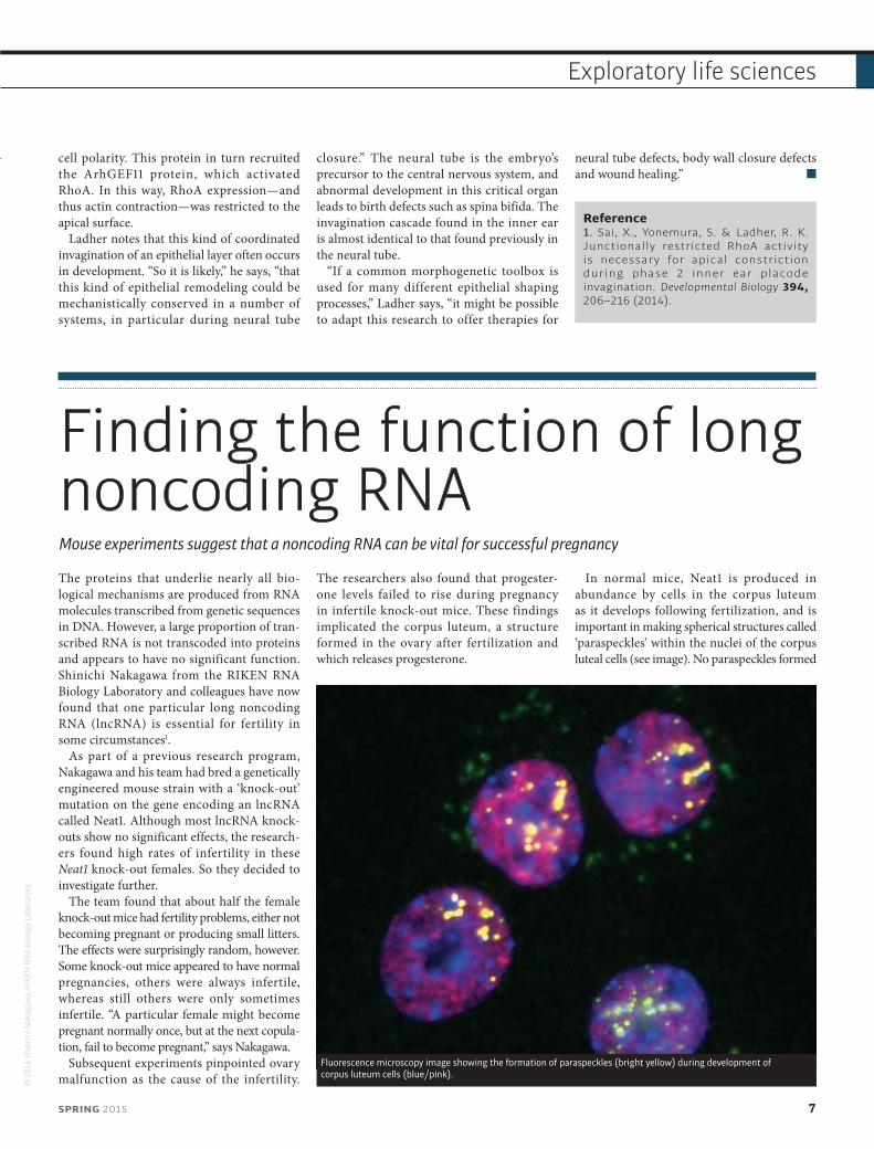
In normal mice, Neat1 is produced in abundance by cells in the corpus luteum as it develops following fertilization, and is important in making spherical structures called ‘paraspeckles’ within the nuclei of the corpus luteal cells (see image). No paraspeckles formed
Fluorescence microscopy image showing the formation of paraspeckles (bright yellow) during development of corpus luteum cells (blue/pink).
SPRING 2015 7
Exploratory life sciences
Research highlights©
201
4 Sh
inic
hi N
akag
awa,
RIK
EN R
NA
Biol
ogy
Labo
rato
ry
in the knock-out mice, but despite the lack of paraspeckles about half of the knock-out mice had seemingly normal pregnancies.
The function of the paraspeckles and their importance in pregnancy therefore remain unclear. “One theory is that they sequester potentially harmful proteins,” says Nakagawa. “We think that Neat1 may fight against certain stresses that disrupt the estab-lishment of pregnancy. If that stress is present,
Neat1 and the paraspeckles are needed, but without that stress, the paraspeckles are not required.”
Nakagawa thinks it is quite possible that some forms of infertility in humans may also be attributable to defects in the Neat1 gene and paraspeckle formation. “We haven’t conducted any tests on humans yet, but defi-nitely that is one of the future directions we would like to take with this research.”
Reference1. Nakagawa, S., Shimada, M., Yanaka, K., Mito, M., Arai, T., Takahashi, E., Fujita, Y., Fujimori, T., Standaert, L., Marine, J.-C. et al. The lncRNA Neat1 is required for corpus luteum formation and the establishment of pregnancy in a subpopulation of mice. Development 141, 4618–4627 (2014).
Perfect propeller proteins designed by computerThe computationally assisted synthesis of a symmetric propeller protein that retraces protein evolution could also be used to develop new protein structures for biotechnology applications
Investigating the structure of protein subunits and how they join together to form larger multi-unit proteins such as β-propellers can yield important insights into the evolution
and activities of these proteins. It could also guide the design of new protein structures for use in biotechnology applications. Research-ers at RIKEN and Yokohama City University
have used a computational technique to deconstruct and re-engineer a family of proteins known as β-propellers1.
The β-propeller proteins are so called because they consist of several charac-teristic flat structures arranged in a pro-peller-like shape. Present-day β-propeller proteins are not symmetric, having slightly different protein sequences in each ‘blade’ of the propeller. This asymmetry is generally believed to have emerged due to the duplica-tion of a single gene, followed by the accumu-lation of mutations that created the variations in individual blades.
“Our goal was to reverse engineer this evolutionary process,” says Voet, one of the members of the research team at the RIKEN Center for Life Science Technologies. “We were trying to find what may be the ancestral sequence that could assemble into a perfectly symmetric β-propeller.” Previous attempts to achieve this perfect symmetry had failed, but a new and rapid computational approach proved successful. The achievement supports the idea that modern β-propellers evolved by duplication and mutation of a common ancestral gene.
The researchers examined the amino acid sequences of many six-bladed β-propeller proteins listed in a protein data bank and used a computational process to predict the most
Pizza 2
Pizza 6
Pizza 8 Pizza 9
Pizza 10
1 nm
Pizza 7
Pizza 3 Pizza 4 Pizza 5
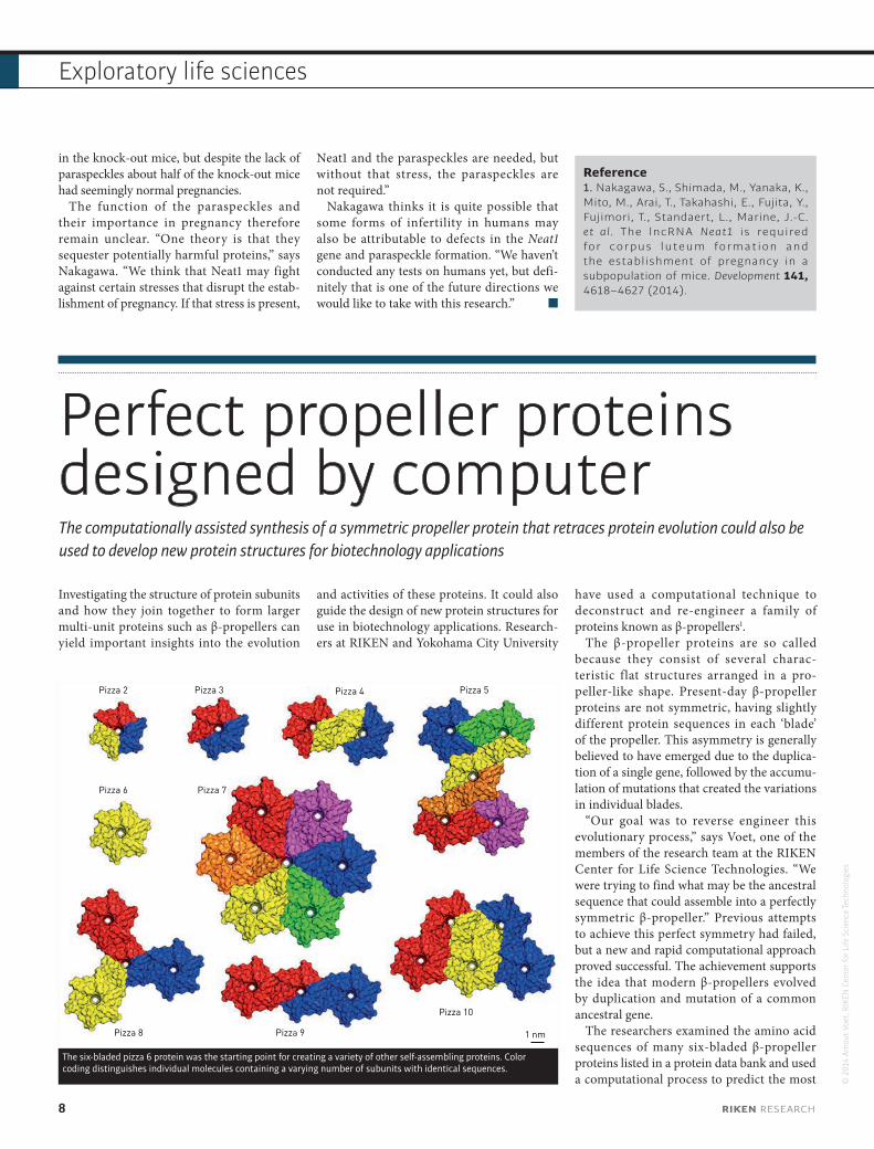
The six-bladed pizza 6 protein was the starting point for creating a variety of other self-assembling proteins. Color coding distinguishes individual molecules containing a varying number of subunits with identical sequences.
© 2
014
Arno
ut V
oet,
RIKE
N C
ente
r for
Life
Sci
ence
Tech
nolo
gies
8 RIKEN RESEARCH
Exploratory life sciences
likely ancestral amino acid sequence for all the proteins. They then used standard genetic engineering methods to produce the possible ancestral protein in Escherichia coli cells. The resulting protein molecules self-assem-bled into a perfectly symmetrical six-bladed propeller structure (see image).
The scientists called this protein pizza 6 due to the resemblance of each blade to a slice of pizza. “To our knowledge, this is the first perfectly symmetric β-propeller protein to be produced synthetically,” explains Voet. Making
and mixing modified versions of the protein blades yielded a variety of related structures with up to 42 blades. Voet likens the approach to snapping together building blocks to build new structures.
In addition to supporting theories of protein evolution, the insights gained through the synthesis and assembly of symmetric β-propeller protein could lead to practical applications. “In future we could use our procedure to make proteins that assemble into bigger shapes for applications such as carrying
drugs or recognizing cancer cells to deliver drug molecules,” says Voet.
Reference1. Voet , A . R . D. , Noguchi , H. , Addy, C., Simoncini, D., Terada, D., Unzai, S., Park, S.-Y., Zhang, K. Y. J. & Tame, J. R. H. Computational design of a self-assembling symmetrical β-propeller protein. Proceedings of the National Academy of Sciences USA 111, 15102–15107 (2014).
A protein shepherd helps coordinate brain developmentA single protein activates the machinery needed for axon growth and holds the axons together for collective extension
During brain development, neurons extend projections called axons to connect with other neurons. Axons from groups of neurons with the same function tend to extend together, but the mechanisms involved in keeping the growing axons in contact for collec-tive extension have been unclear. Masatoshi Takeichi, Shuichi Hayashi and colleagues from the RIKEN Center for Developmental Biology and RIKEN Quantitative Biology Center have now revealed that the protein protocad-herin-17 (Pcdh17) plays a crucial role in this coordinated axon growth and correct devel-opment of the nervous system1.
The protocadherin family of proteins is involved in regulating cell interactions and movement, and some of these proteins have been linked to brain disorders. Pcdh17 is expressed in the amygdala—small areas of the brain that regulate emotion and social behavior.
Takeichi, Hayashi and their colleagues found that when the gene that encodes Pcdh17 was deleted in mice, fewer axons extended from the amygdala, and those that did failed to follow the usual side-by-side path. Too much Pcdh17, however, caused axons from
different groups to interact anomalously. By manipulating the structure of the protein, the researchers showed that Pcdh17 molecules in neighboring axons normally bind to each other to hold extending axons together.
The team also looked for other proteins that interact with Pcdh17 inside cells. This search identified members of the five-protein complex known as WAVE. This complex controls actin polymerization—a process
5 μm
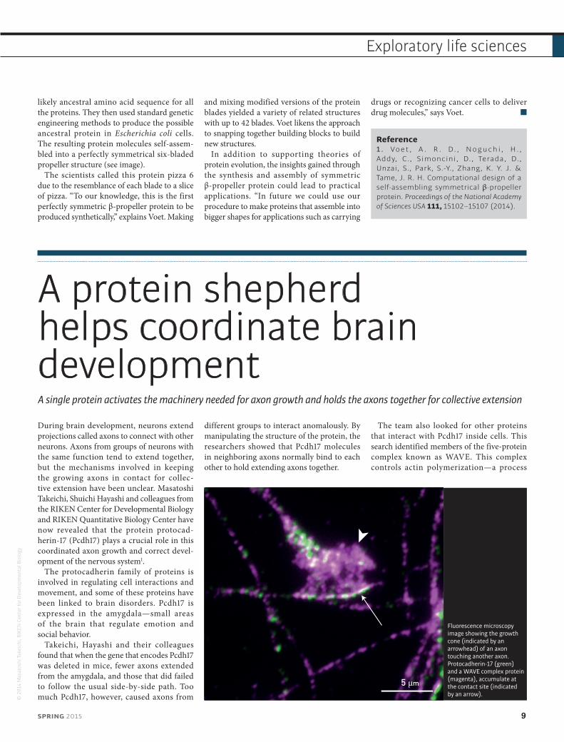
Fluorescence microscopy image showing the growth cone (indicated by an arrowhead) of an axon touching another axon. Protocadherin-17 (green) and a WAVE complex protein (magenta), accumulate at the contact site (indicated by an arrow).
© 2
014
Mas
atos
hi Ta
keic
hi, R
IKEN
Cen
ter f
or D
evel
opm
enta
l Bio
logy
SPRING 2015 9
Exploratory life sciences
in which the protein skeleton of the cell is extended to allow cell migration. The WAVE complex is normally localized to structures called lamellipodia, which are found at cell edges and are required for cell movement.
“On interacting with Pcdh17, the WAVE complex becomes localized to cell-to-cell contacts and converts these sites into motile structures,” explains Takeichi. This locali-zation was observed only at axon-to-axon contacts (see image). “We hypothesize that the motility of growth cones at the leading
ends of extending axons is enhanced by the Pcdh17–WAVE complex.”
The findings show that Pcdh17 plays a role in both holding groups of axons together as they grow, and in recruiting the cellular machinery required for extension.
“We discovered a mechanism by which axons extend together. A deficiency in this process may cause brain defects,” says Takeichi. “Some of the other protocadherin proteins, such as Pcdh19, have been linked to disorders such as epilepsy and mental retardation in
humans. We are now analyzing the cellular and molecular backgrounds of such diseases using mouse models.”
Reference1. Hayashi, S., Inoue, Y., Kiyonari, H., Abe, T., Misaki, K., Moriguchi, H., Tanaka, Y. & Takeichi, M. Protocadherin-17 mediates collective axon extension by recruiting actin regulator complexes to interaxonal contacts. Developmental Cell 30, 673–687 (2014).
‘Chatty’ cells help build the brainDevelopment of the cerebral cortex is influenced by genetic cues and communication between neurons and progenitors
The cerebral cortex, which controls higher processes such as perception, thought and cognition, is the most complex structure in the mammalian central nervous system. Although much is known about the intricate structure of this brain region, the processes governing its formation remain uncertain. Research led by Carina Hanashima from the RIKEN Center for Developmental Biology has now uncovered how feedback between cells, as well as molecular factors, helps shape cortical development during mouse embryogenesis1.
The cortex is made up of layers of intercon-necting cells that are produced in a particu-lar order from progenitor cells. The relatively cell-sparse outer layer is formed first, then the dense deep layer, and finally the tightly packed upper layer. Hanashima and her colleagues were interested to discover exactly how the various layers form, so they created a mouse model that enabled them to control the expres-sion of a particular protein, Foxg1, known to be involved in cortical development.
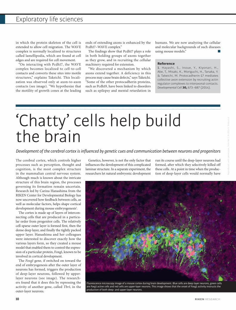
The Foxg1 gene, if switched on toward the end of embryogenesis after the outer layer of neurons has formed, triggers the production of deep-layer neurons, followed by upper-layer neurons (see image). The research-ers found that it does this by repressing the activity of another gene, called Tbr1, in the outer-layer neurons.
Genetics, however, is not the only factor that influences the development of this complicated laminar structure. In a separate experiment, the researchers let natural embryonic development
run its course until the deep-layer neurons had formed, after which they selectively killed off these cells. At a point in time when the produc-tion of deep-layer cells would normally have
Fluorescence microscopy image of a mouse cortex during brain development. Blue cells are deep-layer neurons, green cells are Foxg1 active cells and red cells are upper-layer neurons. This image shows that the onset of Foxg1 activity instructs the production of both deep- and upper-layer neurons.
Repr
oduc
ed, w
ith p
erm
issi
on, f
rom
Ref
. 1 ©
201
4 K.
Tom
a et
al.
10 RIKEN RESEARCH
Exploratory life sciences
ceased, it instead continued. The absence of the ‘production stop’ signal from deep-layer neurons caused the progenitor cells to continue to make deep-layer neurons. “Before this study, there was no evidence for any feedback between post-mitotic neurons and progenitors,” says Hanashima, “but we’ve shown that the two cell types do communicate.”
Extrinsic, cellular factors as well as intrinsic, genetic cues help to guide cortical development. This mechanism allows the developing brain to balance the various different cell types found in the neocortex: it gives the brain flexibility to adjust if too few of one cell type are produced. Although the numbers of cells and embryonic
and gestational periods differ significantly between mice and humans, both species are endowed with almost identical genetic toolkits, and consequently the researchers think it is likely that the human neocortex is generated in much the same way.
Reference1 . To m a , K . , Ku m a m o t o , T. & Hanashima, C. The timing of upper-layer neurogenesis is conferred by sequential derepression and negative feedback from deep-layer neurons. The Journal of Neuroscience 34, 13259–13276 (2014).
Proteins feel the pinchAn enzyme that helps bacteria to adapt to changes in oxygen exposure controls the degradation of defective proteins by opening and closing a pore on its surface
A study by RIKEN researchers has revealed an important coping mechanism employed by enteric bacteria that allows them to thrive in envi-ronments with and without oxygen1. The findings could help develop ways to control both harmful and beneficial bacteria in the human gut.
Most organisms are either oxygen-breath-ing ‘aerobes’ or oxygen-avoiding ‘anaerobes’. The enteric bacteria that dwell within our gut, however, have specialized biological mecha-nisms that allow them to live comfortably in environments with and without oxygen.
The research team, led by Wataru Nishii and Shigeyuki Yokoyama from the RIKEN Structural Biology Laboratory, focused on a protease enzyme called Lon, which maintains cellular health by destroying damaged or defective proteins. Virtually all organisms have a version of Lon, but the enzyme found in enteric bacteria differs slightly from those found in other organisms, possessing a pair of amino acids that can form a strong chemical bond under certain chemical conditions.
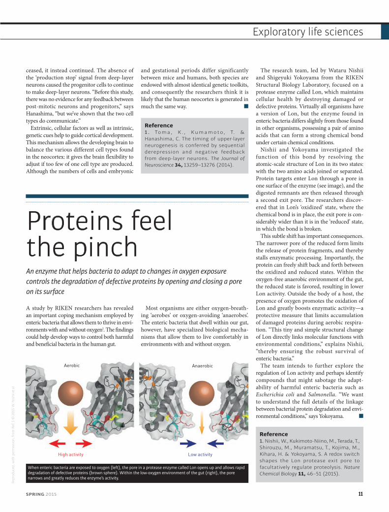
Nishii and Yokoyama investigated the function of this bond by resolving the atomic-scale structure of Lon in its two states: with the two amino acids joined or separated. Protein targets enter Lon through a pore in one surface of the enzyme (see image), and the digested remnants are then released through a second exit pore. The researchers discov-ered that in Lon’s ‘oxidized’ state, where the chemical bond is in place, the exit pore is con-siderably wider than it is in the ‘reduced’ state, in which the bond is broken.
This subtle shift has important consequences. The narrower pore of the reduced form limits the release of protein fragments, and thereby stalls enzymatic processing. Importantly, the protein can freely shift back and forth between the oxidized and reduced states. Within the oxygen-free anaerobic environment of the gut, the reduced state is favored, resulting in lower Lon activity. Outside the body of a host, the presence of oxygen promotes the oxidation of Lon and greatly boosts enzymatic activity—a protective measure that limits accumulation of damaged proteins during aerobic respira-tion. “This tiny and simple structural change of Lon directly links molecular functions with environmental conditions,” explains Nishii, “thereby ensuring the robust survival of enteric bacteria.”
The team intends to further explore the regulation of Lon activity and perhaps identify compounds that might sabotage the adapt-ability of harmful enteric bacteria such as Escherichia coli and Salmonella. “We want to understand the full details of the linkage between bacterial protein degradation and envi-ronmental conditions,” says Yokoyama.
Reference1. Nishii, W., Kukimoto-Niino, M., Terada, T., Shirouzu, M., Muramatsu, T., Kojima, M., Kihara, H. & Yokoyama, S. A redox switch shapes the Lon protease exit pore to facultatively regulate proteolysis. Nature Chemical Biology 11, 46–51 (2015).
Aerobic
High activity Low activity
Anaerobic
When enteric bacteria are exposed to oxygen (left), the pore in a protease enzyme called Lon opens up and allows rapid degradation of defective proteins (brown sphere). Within the low-oxygen environment of the gut (right), the pore narrows and greatly reduces the enzyme’s activity.
Repr
oduc
ed, w
ith p
erm
issi
on, f
rom
Ref
. 1 ©
201
5 W
. Nis
hii e
t al.
SPRING 2015 11
Exploratory life sciences

Biology
The amazing disappearing mouseAn experimental technique that renders mouse tissues transparent and colorless allows scientists to image the cellular-scale effects of disease deep within the body
12 RIKEN RESEARCH
Feature highlight

© 2
015
RIKE
N
In a Petri dish culture, cells and tissue are sufficiently transparent that they can be readily explored using any of a variety of microscopy techniques. Whole organs, however, are opaque under light microscopy,
and it has proved challenging to find a way to clarify tissue to the extent needed to permit microscope observations of individual cells deep inside an animal. A research team led by Hiroki Ueda and Kazuki Tainaka from the Laboratory for Synthetic Biology at the RIKEN Quantitative Biology Center has now developed a remarkably effective tissue-clearing technique that promises to allow direct microscopy studies of deep tissue in organs and even whole animals1.
The research team previously discovered a mixture of certain aminoalcohols that causes brain tissue to become clear and glassy, breaking down the dense lipids that block and scatter light. They combined this chemical treatment with a set of image-pro-cessing tools, and the resulting ‘clear, unobstructed brain imaging cocktails and computational analysis’ (CUBIC) method enabled easy visualization of fluo-rescently labeled proteins and cells deep within the brain.
A number of approaches have been developed to break down lipids in fixed tissues, but most of these techniques are optimized for the lipid-rich brain. Although a promising clearing strategy that works at the whole-body scale has also been reported, its utility is limited by its inability to remove naturally occurring pigments like heme, which gives red blood cells their distinctive color. “As pigments are a major source of light absorption,” says Tainaka, “whole organs treated by this method still appeared opaque.”
A moment of clarityAs Ueda’s team began to experiment with the CUBIC treatment of different organs, they made the unex-pected discovery that these organs began to lose their color over the course of treatment. At the same time, the aminoalcohol mixture bathing these samples changed in color from clear to green, suggesting the presence of iron-laden heme. “This observation led us to hypothesize that the CUBIC cocktail could solubilize and eliminate endogenous heme from blood-infused tissues,” says Tainaka.
Working with colleague Shimpei Kubota from the University of Tokyo, Tainaka subsequently deter-mined that this ‘transparentization’ treatment was broadly applicable to a wide variety of organs. The pair examined nearly a dozen different whole organs, including the heart, liver and lungs, and found that all of these could be effectively clarified and decol-orized after ten days of treatment with the CUBIC cocktail (Fig. 1). Remarkably, with a slightly longer treatment time, an entire infant mouse could be rendered largely transparent—its skeleton could be observed beneath the clarified organs and muscle. The researchers also achieved the same effect in ©
Seb
astia
n Ka
ulitz
ki/H
emer
a/Th
inks
tock
Kazuki Tainaka (center) obtained his PhD in engineering from Kyoto University in 2006. After working as a postdoctoral researcher at RIKEN and Osaka University, he joined Kyoto University as an assistant professor in 2008. In 2010, he joined the RIKEN Quantitative Biological Center. From 2013, he has been a lecturer at the University of Tokyo. His research interests include developing new fundamental technologies for synthetic and systems biology.
Hiroki R. Ueda (left) earned his MD and PhD from the University of Tokyo. He was team leader at the RIKEN Laboratory for Systems Biology and RIKEN Center for Developmental Biology in 2003 and 2004, respectively. He was project leader at the Laboratory for Systems Biology and manager at the Center for Developmental Biology in 2009−2014 and 2004−2013, respectively. He has been head of the Laboratory for Synthetic Biology from 2011 and professor at the University of Tokyo from 2013.
Shimpei I. Kubota (right)graduated from Nagoya University in 2013. He studied the molecular pathogenesis of schizophrenia at Nagoya University Graduate School of Medicine. He is currently enrolled in the Graduate School of Medicine at the University of Tokyo, where he researches sleep. His goals are to design gene networks and make an ‘ironmouse’ that is tough enough to swim, cycle and run.
Feature highlight
SPRING 2015 13
Feature highlight
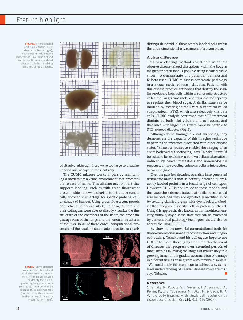
adult mice, although these were too large to visualize under a microscope in their entirety.
The CUBIC mixture works in part by maintain-ing a moderately alkaline environment that promotes the release of heme. This alkaline environment also supports labeling, such as with green fluorescent protein, which allows biologists to introduce geneti-cally encoded visible ‘tags’ for specific proteins, cells or tissues of interest. Using green fluorescent protein and other fluorescent labels, Tainaka, Kubota and their colleagues were able to directly visualize the fine structure of the chambers of the heart, the bronchial passageways of the lungs and the vascular structures of the liver. In all of these cases, computational pro-cessing of the resulting data made it possible to clearly
1 mm
1 mm 1 mm
1 mm
Figure 1: After extended perfusion with the CUBIC chemical mixture (right),
mouse organs including the kidneys (top), liver (middle) and pancreas (bottom) are rendered
clear and colorless, enabling deep microscopic imaging.
Figure 2: Computational analysis of the clarified and
decolorized mouse pancreas (top left) makes it possible
to identify the insulin-producing Langerhans islets
(top right). These can then be mapped three-dimensionally (bottom left) either alone or
in the context of the entire organ (bottom right).
Repr
oduc
ed, w
ith p
erm
issi
on, f
rom
Ref
. 1 ©
201
4 El
sevi
erRe
prod
uced
, with
per
mis
sion
, fro
m R
ef. 1
© 2
014
Els
evie
r
14 RIKEN RESEARCH
Feature highlight
distinguish individual fluorescently labeled cells within the three-dimensional environment of a given organ.
A clear differenceThis new clearing method could help scientists observe disease-related disruptions within the body in far greater detail than is possible using isolated tissue slices. To demonstrate this potential, Tainaka and Kubota used CUBIC to assess pancreatic pathology in a mouse model of type I diabetes. Patients with this disease produce antibodies that destroy the insu-lin-producing beta cells within a pancreatic structure called the Langerhans islets, and thus lose the capacity to regulate their blood sugar. A similar state can be induced by treating animals with a chemical called streptozotocin (STZ), which also selectively kills beta cells. CUBIC analysis confirmed that STZ treatment diminished both islet volume and cell count, and that mice with larger islets were more vulnerable to STZ-induced diabetes (Fig. 2).
Although these findings are not surprising, they demonstrate the capacity of this imaging technique to peer inside mysteries associated with other disease states. “Since our technique enables the imaging of an entire body without sectioning,” says Tainaka, “it would be suitable for exploring unknown cellular aberrations induced by cancer metastasis and immunological response, or for revealing unknown cellular interactions between organs.”
Over the past few decades, scientists have generated transgenic animals that selectively produce fluores-cently labeled proteins in a broad range of cell types. However, CUBIC is not limited to these models, and the researchers demonstrated that similar results could also be obtained with non-genetically modified mice by treating clarified organs with dye-labeled antibod-ies that recognize a specific cellular protein of interest. Using this approach, also known as immunohistochem-istry, virtually any disease state that can be examined by conventional pathology techniques should also be accessible using CUBIC.
By drawing on powerful computational tools for three-dimensional image reconstruction and single-cell tracing, Tainaka and his colleagues hope to use CUBIC to more thoroughly trace the development of diseases that progress over extended periods of time, such as following the stages of malignancy in a growing tumor or the gradual accumulation of damage in different tissues arising from autoimmune disorders. “We could apply this technique to achieve a systems-level understanding of cellular disease mechanisms,” says Tainaka.
Reference1. Tainaka, K., Kubota, S. I., Suyama, T. Q., Susaki, E. A., Perrin, D., Ukai-Tadenuma, M., Ukai, H. & Ueda, H. R. Whole-body imaging with single-cell resolution by tissue decolorization. Cell 159, 911–924 (2014).
Particles find their massSimulations indicate that a peculiar state of matter at close to absolute zero could be used to observe an elusive quantum phenomenon called quantum mass acquisition
A possible means of observing an exotic quantum effect that imparts mass to a normally massless particle has been proposed by researchers from the RIKEN Center for Emergent Matter Science1.
At temperatures close to absolute zero, atoms can start to form a collective state known as a Bose–Einstein condensate (BEC). Scientists have found that this state of matter is useful as a ‘quantum simulator’ for investigating particles that have been predicted to exist by theory but are too difficult to create or observe directly.
“Quantum simulators are very versatile, allowing interactions, particle density and temperature to be tuned,” says Masahito Ueda from the RIKEN research team. “The pressing issue in this field is to look for something very fundamental that can be demonstrated for the first time in such atomic gases.”
Through mathematical modeling, Ueda and his colleagues Nguyen Thanh Phuc from RIKEN and Yuki Kawaguchi from the University of Tokyo showed that a BEC could be used to simulate a so-far-unob-served phenomenon known as quantum mass acquisition. This effect causes a normally massless elementary particle called a quasi-Nambu–Goldstone boson to acquire mass as a result of minute quantum fluc-tuations. Researchers believe that this effect could appear in superfluids, superconductors and some magnetic materials. Yet quantum mass acquisition has never been seen because the effect is too small to be distinguished from other secondary effects.
“Extremely minute quantum phenomena are amplified to a macroscopic level in BECs and therefore made visible,” says
Ueda. The researchers’ analysis shows that the emergent energy gap of the quasi-Nambu–Goldstone boson in a BEC is two orders of magnitude larger than the zero-point energy of the system. This means that the state is much more robust than previ-ously thought, raising the hope that it might be possible to experimentally observe the quasi-Nambu–Goldstone boson and quantum mass acquisition.
The choice of atom in the gas is crucial for observing quantum mass acquisition. Many BECs are made using helium atoms and spin-polarized alkali atoms, which are spinless. Ueda and his team have shown that atoms with spin ‘degrees of freedom’ are required to observe quantum mass acquisition. Such a ‘spinor’ BEC could be created using rubidium atoms.©
ags
andr
ew/i
Stoc
k/Th
inks
tock
SPRING 2015 15
Exploratory physical sciences
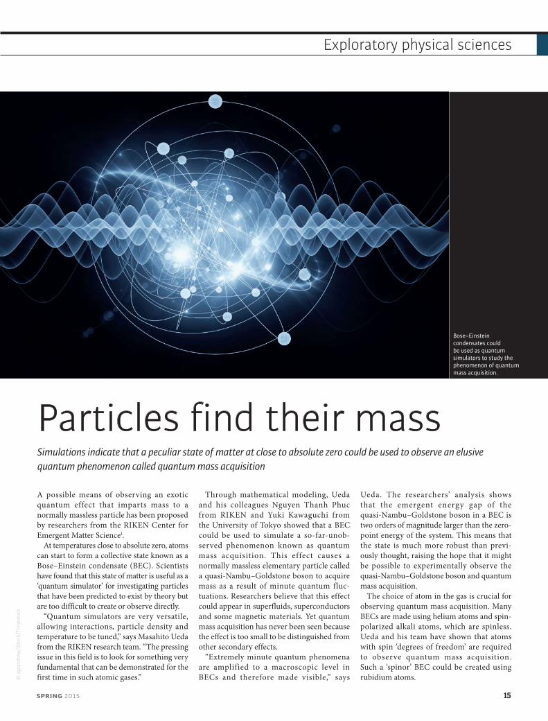
Bose–Einstein condensates could be used as quantum simulators to study the phenomenon of quantum mass acquisition.
“Our work demonstrates that fundamental physical phenomena that can usually only be tested using particle accelerators, can be repro-duced on the tabletop,” says Ueda. “We now plan to explore what other fundamental phenomena can be revealed in atomic BECs.”
Reference1. Phuc, N. T., Kawaguchi, Y. & Ueda, M. Quantum mass acquisition in spinor Bose-Einstein condensates. Physical Review Letters 113, 230401 (2014).
Skyrmions like it hotPinpoint laser heating creates a maelstrom of magnetic nanotextures
A simulation study by researchers from the RIKEN Center for Emergent Matter Science has demonstrated the feasibility of using lasers to create and manipulate nanoscale magnetic vortices1. The ability to create and control these ‘skyrmions’ could lead to the
development of skyrmion-based information storage devices.
The information we consume and work with is encoded in binary form (as ‘1’s or ‘0’s) by switching the characteristics of memory media between two states. As we approach the
performance and capacity limits of conventional memory media, researchers are looking toward exotic physics to develop the next generation of magnetic memories.
One such exotic phenomenon is the skyrmion—a stable, nanoscale whirlpool-like magnetic feature characterized by a constantly rotating magnetic moment. Theoretically, the presence or absence of a skyrmion at any location in a magnetic medium could be used to represent the binary states needed for informa-tion storage. However, researchers have found it challenging to reliably create and annihilate skyrmions experimentally due to the difficulty in probing the mechanics of these processes in any detail. The challenge lies in the incred-ibly short timescale of these processes, which at just a tenth of a nanosecond is up to a billion times shorter than the timescale observable under the Lorentz microscope used to measure magnetic properties.
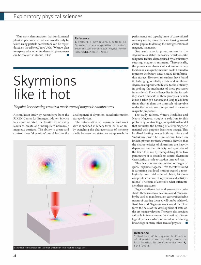
The study authors, Wataru Koshibae and Naoto Nagaosa, sought a solution to this problem by constructing a computational model that simulates the heating of a ferromagnetic material with pinpoint lasers (see image). This localized heating creates both skyrmions and ‘antiskyrmions’. The simulations, based on known physics for these systems, showed that the characteristics of skyrmions are heavily dependent on the intensity and spot size of the laser. Further, by manipulating these two parameters, it is possible to control skyrmion characteristics such as creation time and size.
“Heat leads to random motion of magnetic spins,” explains Nagaosa. “We therefore found it surprising that local heating created a topo-logically nontrivial ordered object, let alone composite structures of skyrmions and antiskyr-mions.” The issue of control is what differenti-ates these structures.
Nagaosa believes that as skyrmions are quite stable, these nanoscale features could conceiva-bly be used as an information carrier if a reliable means of creating them at will can be achieved. Koshibae and Nagaosa’s work could therefore form the basis of the development of state-of-the-art memory devices. The work also provides valuable information on the creation of topo-logical particles, which is crucial for advancing knowledge in many other areas of physics.
Reference1. Koshibae, W. & Nagaosa, N. Creation of skyrmions and antiskyrmions by local heating. Nature Communications 5, 5148 (2014).
Schematic representation of skyrmion creation by local heating using a laser.
© 2
014
Mar
i Ish
ida,
RIK
EN C
ente
r for
Em
erge
nt M
atte
r Sci
ence
(low
er p
art)
; Mod
ified
, with
per
mis
sion
, fro
m R
ef. 1
© 2
014
W. K
oshi
bae
& N
. Nag
aosa
(ins
ets)
16 RIKEN RESEARCH
Exploratory physical sciences

Viewing black holes in a different lightSatellite studies reveal complex processes of x-ray emission from matter falling into the black hole at the center of a galaxy
Most galaxies are assumed to have at their heart a supermassive black hole that draws in vast amounts of surrounding matter. As this matter is sucked in, it releases energy in the form of intense x-ray emissions that in some cases can be more intense than the emission from all the stars in the galaxy combined.
Through detailed study of the x-ray emissions from the center of the galaxy known as NGC 3227, Hirofumi Noda from the RIKEN Nishina Center for Accelerator-Based Science and colleagues have now revealed the multiple processes responsible for these emissions1. “Our results show the presence of multiple
mechanisms of energy conversion of matter near a central black hole,” explains Noda.
The x-ray emissions from the centers of galaxies provide valuable information on the properties of their central black holes, as well as the surrounding matter and ultimately the history of galaxies in general. However, it can be difficult to separate the original radiation emitted by matter close to the black hole from secondary processes caused by other matter around the black hole. The typical spectral analysis applied to images of galactic centers is unable to clearly distinguish the different emission processes. Noda and his colleagues
instead studied the change in emissions over time. By analyzing the different radiation patterns and their correlations, they were able to separate primary emission from the emission spectra arising from secondary processes such as reflection.
Using this approach and wide-range x-ray data obtained over the course of a few weeks by the Japanese research satellite Suzaku, the team identified three distinct spectral compo-nents from NGC 3227—a typical type I Seyfert galaxy that lies about 80 million light years from Earth (see image). The intensity of the source varied by almost an order of magnitude over
© Im
age
cour
tesy
of K
en C
raw
ford
Photograph of galaxy NGC 3227.
SPRING 2015 17
Exploratory physical sciences
the course of the observations, and by analyzing these variations, the researchers could correlate the spectral components with three different mass accretion flows into the galaxy’s black hole.
Noda believes that studying the ultraviolet emissions from matter at a greater distance from the black hole could also provide further insight into the detailed physics of supermassive black holes. X-ray emissions,
however, are still expected to reveal much more information if the spectral resolu-tion of observations can be improved. “The soon-to-be-launched Japanese x-ray satellite ASTRO-H will achieve an unprecedentedly fine energy resolution, which will enable us to understand the properties of black holes, their evolution and their interaction with the galaxy around them.”
Reference1. Noda, H., Makishima, K., Yamada, S., Nakazawa, K., Sakurai, S. & Miyake, K. Suzaku studies of the central engine in the typical type I Seyfert NGC 3227: Detection of multiple primary x-ray continua with distinct properties. The Astrophysical Journal 794, 2 (2014).
Super-heavy chemistryThe formation of a hexacarbonyl complex with the synthetic heavy element seaborgium paves the way for studies of relativistic effects in the chemical properties of the heaviest elements
An international research collaboration involving scientists from the RIKEN Nishina Center for Accelerator-Based Science has for the first time synthesized a carbonyl complex with a super-heavy element at its core1. The technique used to create this exotic molecule, seaborgium hexacarbonyl, promises to advance our knowledge of the chemistry of unstable elements located at the end of the periodic table.
Seaborgium is a synthetic element that can only be produced by nuclear fusion in high-energy particle accelerators. This element has 106 protons in its bloated nucleus, which forces
some of the atom’s electrons to orbit at around 80% of the speed of light. Einstein’s special theory of relativity predicts that electrons become heavier at these velocities, which affects how they form chemical bonds. Studying this chemical bonding behavior, however, is a daunting task. Inorganic compounds of seaborgium have been produced in the past, but the complexing agents are easily destroyed by the heavy ion beam and detection of the compounds is complicated by the creation of byproducts.
Researchers at the RIKEN heavy ion linear accelerator (RILAC), in collaboration with
co-workers from around the world, have now developed an innovative technique for creating and separating super-heavy elements, and also for studying their chemistry. “Using this technique, we have been able to create an organometallic compound of seaborgium, which has never before been achieved for super-heavy elements,” says Hiromitsu Haba, head of the RIKEN Radioisotope Applications Team involved in the research.
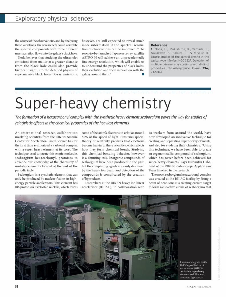
The novel seaborgium hexacarbonyl complex was created at the RILAC facility by firing a beam of neon ions at a rotating curium target to form radioactive atoms of seaborgium that
A series of magnets inside RIKEN’s gas-filled recoil ion separator (GARIS) can isolate super-heavy elements and filter out unwanted byproducts.
© 2
014
RIKE
N N
ishi
na C
ente
r for
Acc
eler
ator
-Bas
ed S
cien
ce
18 RIKEN RESEARCH
Exploratory physical sciences
decayed in a matter of seconds. These atoms were quickly separated from the main ion beam and unwanted byproducts using RIKEN’s gas-filled recoil ion separator (GARIS), and then mixed with carbon monoxide gas as the source of the carbonyl ligand. The products of this reaction then passed over a series of silicon detectors, which identified a grand total of 18 seaborgium atoms.
The researchers conducted similar experi-ments using molybdenum and tungsten, which lie directly above seaborgium in the periodic table and should be chemically similar. They found that seaborgium hexacarbonyl had the same detection profile as hexacarbonyls of molybdenum and tungsten, providing strong
evidence that they had indeed formed the super-heavy hexacarbonyl, Sg(CO)6.
The team now hopes to use GARIS (see image) to study the chemistry of other super-heavy elements, such as hassium (element 108), which may also be affected by strong relativistic effects.
Reference1. Even, J., Yakushev, A., Düllmann, C. E., Haba, H., Asai, M., Sato, T. K., Brand, H., Di Nitto, A., Eichler, R., Fan, F. L. et al. Synthesis and detection of a seaborgium c a r b o ny l comple x . Sc ience 345, 1491–1493 (2014).
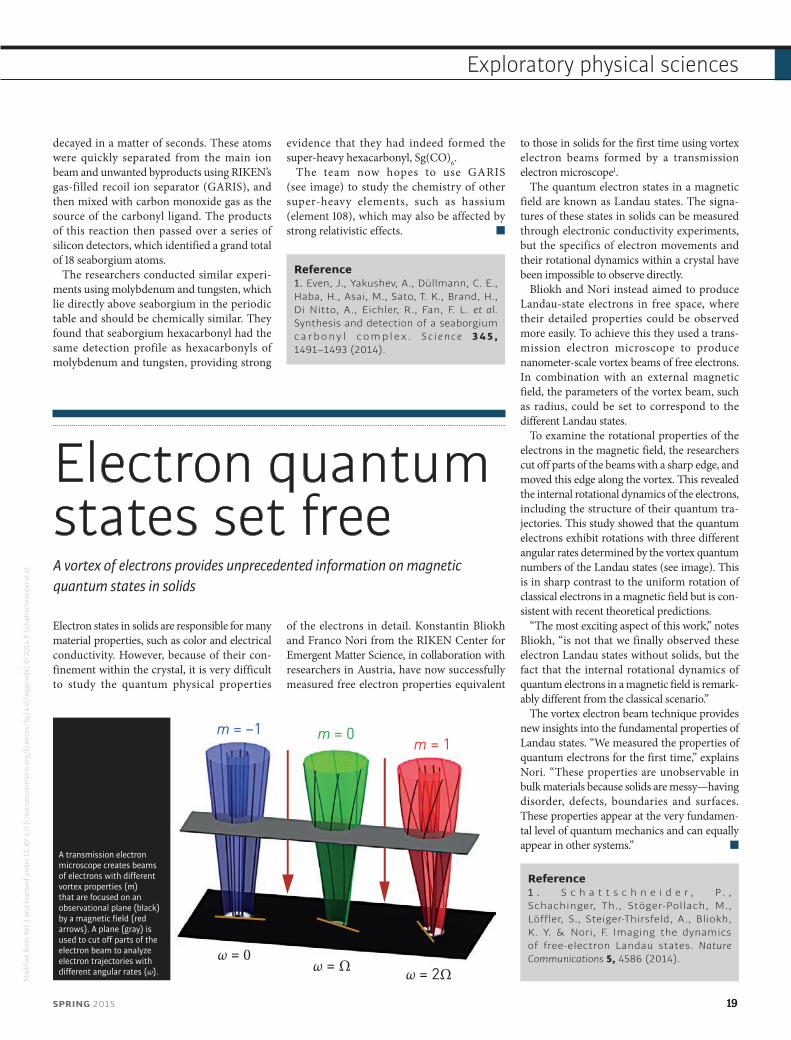
Electron quantum states set freeA vortex of electrons provides unprecedented information on magnetic quantum states in solids
Electron states in solids are responsible for many material properties, such as color and electrical conductivity. However, because of their con-finement within the crystal, it is very difficult to study the quantum physical properties
of the electrons in detail. Konstantin Bliokh and Franco Nori from the RIKEN Center for Emergent Matter Science, in collaboration with researchers in Austria, have now successfully measured free electron properties equivalent
to those in solids for the first time using vortex electron beams formed by a transmission electron microscope1.
The quantum electron states in a magnetic field are known as Landau states. The signa-tures of these states in solids can be measured through electronic conductivity experiments, but the specifics of electron movements and their rotational dynamics within a crystal have been impossible to observe directly.
Bliokh and Nori instead aimed to produce Landau-state electrons in free space, where their detailed properties could be observed more easily. To achieve this they used a trans-mission electron microscope to produce nanometer-scale vortex beams of free electrons. In combination with an external magnetic field, the parameters of the vortex beam, such as radius, could be set to correspond to the different Landau states.
To examine the rotational properties of the electrons in the magnetic field, the researchers cut off parts of the beams with a sharp edge, and moved this edge along the vortex. This revealed the internal rotational dynamics of the electrons, including the structure of their quantum tra-jectories. This study showed that the quantum electrons exhibit rotations with three different angular rates determined by the vortex quantum numbers of the Landau states (see image). This is in sharp contrast to the uniform rotation of classical electrons in a magnetic field but is con-sistent with recent theoretical predictions.
“The most exciting aspect of this work,” notes Bliokh, “is not that we finally observed these electron Landau states without solids, but the fact that the internal rotational dynamics of quantum electrons in a magnetic field is remark-ably different from the classical scenario.”
The vortex electron beam technique provides new insights into the fundamental properties of Landau states. “We measured the properties of quantum electrons for the first time,” explains Nori. “These properties are unobservable in bulk materials because solids are messy—having disorder, defects, boundaries and surfaces. These properties appear at the very fundamen-tal level of quantum mechanics and can equally appear in other systems.”
Reference1 . S c h a t t s c h n e i d e r , P . , Schachinger, Th., Stöger-Pollach, M., Löffler, S., Steiger-Thirsfeld, A., Bliokh, K. Y. & Nori, F. Imaging the dynamics of free-electron Landau states. Nature Communications 5, 4586 (2014).
m = –1
ω = 0ω = Ω ω = 2Ω
m = 0m = 1
A transmission electron microscope creates beams of electrons with different vortex properties (m) that are focused on an observational plane (black) by a magnetic field (red arrows). A plane (gray) is used to cut off parts of the electron beam to analyze electron trajectories with different angular rates (ω).
Mod
ified
from
Ref
. 1 a
nd li
cens
ed u
nder
CC
BY 4
.0 (c
reat
ivec
omm
ons.
org/
licen
ces/
by/4
.0/l
egal
code
) © 2
014
P. Sc
hatt
schn
eide
r et a
l.
SPRING 2015 19
Exploratory physical sciences

© 2
014
RIKE
N
Contact informationWebsite: www.brain.riken.jp/en/E-mail: [email protected]
RIKEN Brain Science Institute
Building neural connectionsThe RIKEN Brain Science Institute has the infrastructure to support interdisciplinary research on the identification and manipulation of the neural circuits underpinning behavior
20 RIKEN RESEARCH
Places
T he sheer complexity of the brain is astonishing—processing that thought alone involves orchestrated interac-tions between numerous elements
in the brain, including neurons and their sup-porting neuroglia. Every feeling, thought and action is processed by a neuronal circuit, each of which sends tens to hundreds of electrical signals a second, sometimes at speeds faster than a race car. The almost hundred billion neurons in the brain fire even during sleep, which enables vision, movement, thought, emotion and memory.
To understand how the brain controls behavior, scientists explore the function of its neural circuits. It is a daunting task to translate a feeling as intangible as depres-sion into the development and function of circuits or, on an even smaller scale, to the expression of single genes in indi-vidual neurons. Advances in the genetic and optogenetic manipulation of model organisms, particularly in mice, combined with state-of-the-art imaging and physio-logical tools have improved our understand-ing of the connections that exist between specific areas of the brain. The RIKEN Brain Science Institute is taking advantage of this convergence—in 2011 it established infra-structure to conduct world-class research on genetic approaches for studying neural circuits and behavior.
Space to thinkThe BSI Neural Circuit Genetics Research Building was constructed to facilitate inter-disciplinary research in this emerging field. With 9,500 square meters of floor space, interactive laboratories, and state-of-the-art shared facilities, the building is designed to bypass the disciplinary divisions that often exist in the life sciences. For example,
laboratories equipped for observing and recording brain activity are closely inte-grated with areas for studying the behavioral expression of such activity, linking cause with consequence. Hence, researchers based at the building cannot help but engage with colleagues in ways resembling the level of integration in the brain itself.
The association between the building and the field of neural circuit genetics is so close that RIKEN inaugurated the building in October 2011 with a symposium entitled “Genes, Circuits and Behavior”. Leaders in the f ield spoke on the role of neural networks in decision-making, reward-based learning, and the memory of past experiences. The RIKEN Brain Science Institute director and Nobel prizewinner, Susumu Tonegawa, described his vision for the building as a site of new discover-ies in molecular and cell biology, electro-physiology, behavior and cognition—with the ultimate goal being the neural circuit
basis of brain dysfunction and restoration of healthy function in brain disease.
Research circuitsThe BSI Neural Circuit Genetics Research Building houses 6 laboratories and approxi-mately 50 researchers, around 30 of whom are from overseas. Two examples of labo-ratories in this building are those of Yukiko Goda and Joshua Johansen. Goda’s labora-tory observes synapses—specialized sites of contact and communication between two neurons. By searching for patterns in the formation of synaptic connections, Goda’s team gains insights into dynamic processes such as how small neural networks encode changes in activity. Johansen’s laboratory studies memory formation by investigating the neural circuits involved in remembering fearful events. Supported by the infrastruc-ture and facilities at the BSI Neural Circuit Genetics Research Building, these teams are making the brain just a little less perplexing.
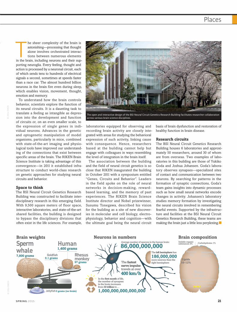
The open and interactive design of the BSI Neural Circuit Genetics Research Building facilitates researcher collaboration across various brain science disciplines.©
201
4 RI
KEN
© 2
014
RIKE
N
Brain weights Brain compositionNeurons in numbers
Jellyfish 0 grams (no brain)
Spermwhale7,800 grams
Human1,400 grams
Rhesusmonkey97 grams
Goldfish0.1 grams
Water77–78%
Lipids10–12%
Inorganicsalts 1%
Proteins8%
Carbohydrates 1%Soluble organicsubstances 2%
On average, working memoryin the human brain canstore seven numbers
In the first month of life,the number of synapsesin the brain increasesfrom 50 trillion to…
1,000,000,000,000,000
Average number of neurons in the human brain:
86,000,000,000
The fastestnerve impulsetravels at over
The left hemisphere has
186,000,000more neurons than the right hemisphere
400 km/h
Places
SPRING 2015 21
Places
Beating a bottleneck to regenerate nervesA membrane-mimicking biocompatible conducting polymer selectively binds to nerve cells and stimulates the changes needed for nerve regeneration
Promoting the guided regeneration of nerve cells could transform the treatment of a vast range of debilitating conditions, including brain injuries, nerve damage and degenerative neurological diseases. Electrically stimulat-ing the outgrowth of ‘neurites’—new projec-tions from nerve cells—is a promising avenue of research, but it has been hampered by immune rejection and scar tissue insulating the electrodes from the targeted cells. Progress has reached a bottleneck because traditional electronic materials, mostly metals and semi-conductors, are unable to provide the biocom-patibility and mechanical strength needed for stable electrical transmission.
Researchers in Japan, China and Taiwan, led by Hsiao-hua Yu of the RIKEN
Responsive Organic Materials Laboratory (now at the Institute of Chemistry, Academia Sinica, Taiwan), have now potentially broken through this bottleneck with the development of a targeted, electrically conducting polymer that mimics the cell membrane1.
The researchers used polyethylenedioxythio-phene polymers assembled from two monomer units, each carrying a chemical component designed to mimic a crucial aspect of cell membranes. One monomer has a peptide group that replicates the selective binding between cells and the extracellular matrix outside cells. The other monomer carries a mixture of hydrophilic and hydrophobic parts mimicking the phospholipid molecules of cell membrane lipid bilayers.
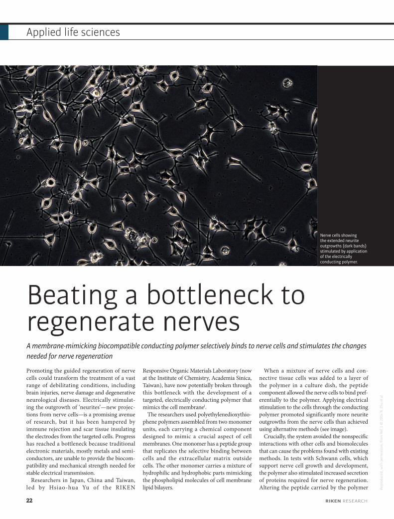
When a mixture of nerve cells and con-nective tissue cells was added to a layer of the polymer in a culture dish, the peptide component allowed the nerve cells to bind pref-erentially to the polymer. Applying electrical stimulation to the cells through the conducting polymer promoted significantly more neurite outgrowths from the nerve cells than achieved using alternative methods (see image).
Crucially, the system avoided the nonspecific interactions with other cells and biomolecules that can cause the problems found with existing methods. In tests with Schwann cells, which support nerve cell growth and development, the polymer also stimulated increased secretion of proteins required for nerve regeneration. Altering the peptide carried by the polymer
Nerve cells showing the extended neurite outgrowths (dark bands) stimulated by application of the electrically conducting polymer.
Repr
oduc
ed, w
ith p
erm
issi
on, f
rom
Ref
. 1 ©
201
4 B.
Zhu
et a
l.
22 RIKEN RESEARCH
Applied life sciences
could allow a variety of cell types to be targeted, greatly extending the range of applications.
“The ultimate goal of our research is to promote tissue regeneration, particularly neuron regeneration, including within the brain,” says Yu. In addition to regenerating cultured cells for grafting into patients, Yu also envisages creating bioimplanted devices
that could stimulate in situ nerve regeneration in a patient. “Our conducting polymer could also be used to coat the materials in neuroprosthetic devices to provide an elec-trical interface capable of more specific interactions toward cells and proteins while preventing the problems of rejection by the patient’s immune system.”
Reference1. Zhu, B., Luo, S.-C., Zhao, H., Lin, H.-A., Sekine, J., Nakao, A., Chen, C., Yamashita, Y. & Yu, H.-h. Large enhancement in neurite outgrowth on a cell membrane-mimicking c o n d u c t i n g p o l y m e r. N a t u r e Communications 5, 4523 (2014).
Expanding the immune system’s memoryA newly identified subpopulation of innate immune cells can be ‘primed’ to provide a rapid response against the emergence of future threats, including tumor growth
The adaptive immune system has the ability to ‘remember’ a given pathogen or cancer cell by producing memory T cells that can mount a rapid counterattack against future threats. The innate immune system, on the other hand, is seen as more of a blunt instru-ment, only capable of launching a broad
defensive response against potential invaders. A research team led by Shin-Ichiro Fujii and colleagues from the RIKEN Center for Inte-grative Medical Sciences has now identified a population of innate immune cells that display the attributes of memory cells and which may help keep tumor growth at bay1.
Natural killer T (NKT) cells are recognized components of the innate immune system that are involved in the rapid response to virally infected cells and tumor formation. Recent studies, however, have hinted that after infection, some of these cells have many of the characteristics of memory cells. Spurred by clinical observations during NKT-cell therapy, Fujii’s team began investigating a subtype called invariant natural killer T (iNKT) cells.
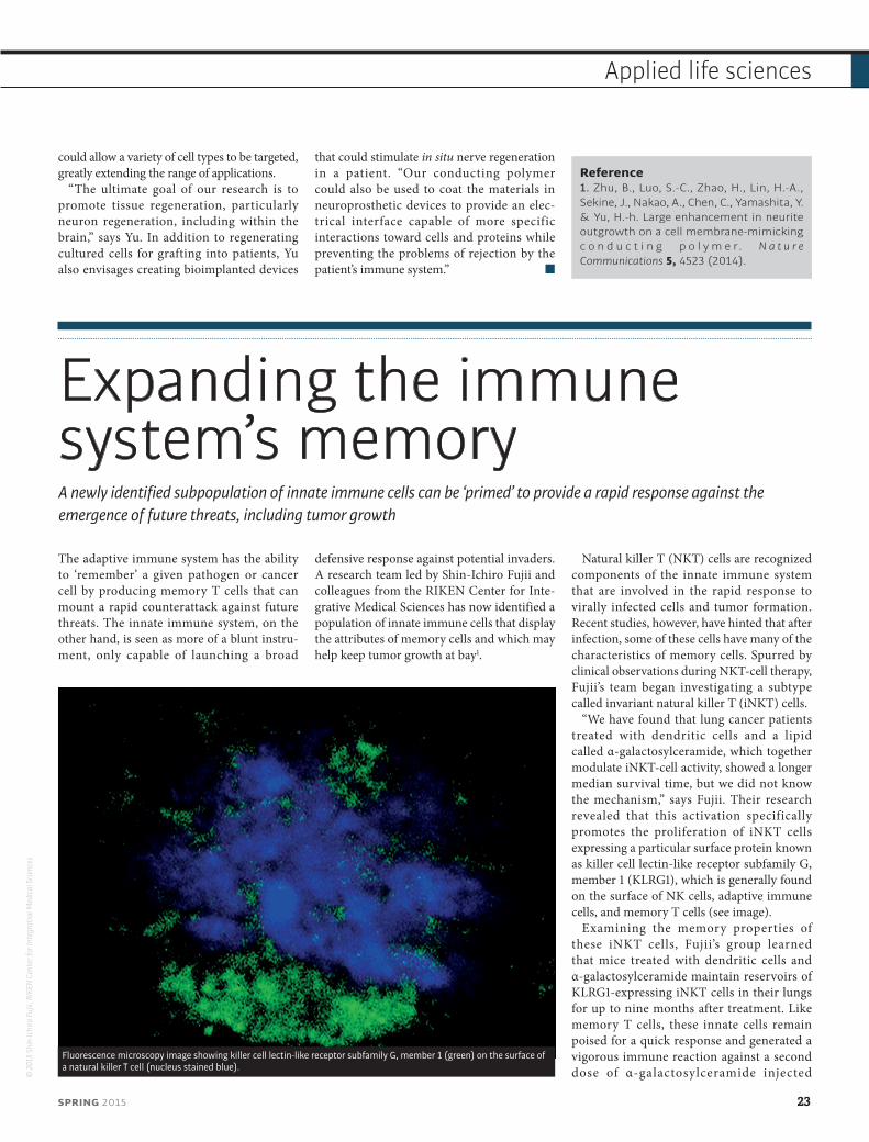
“We have found that lung cancer patients treated with dendritic cells and a lipid called α-galactosylceramide, which together modulate iNKT-cell activity, showed a longer median survival time, but we did not know the mechanism,” says Fujii. Their research revealed that this activation specifically promotes the proliferation of iNKT cells expressing a particular surface protein known as killer cell lectin-like receptor subfamily G, member 1 (KLRG1), which is generally found on the surface of NK cells, adaptive immune cells, and memory T cells (see image).
Examining the memory properties of these iNKT cells, Fujii’s group learned that mice treated with dendritic cells and α-galactosylceramide maintain reservoirs of KLRG1-expressing iNKT cells in their lungs for up to nine months after treatment. Like memory T cells, these innate cells remain poised for a quick response and generated a vigorous immune reaction against a second dose of α-galactosylceramide injected ©
201
5 Sh
in-Ic
hiro
Fujii
, RIK
EN C
ente
r for
Inte
grat
ive M
edica
l Scie
nces
Fluorescence microscopy image showing killer cell lectin-like receptor subfamily G, member 1 (green) on the surface of a natural killer T cell (nucleus stained blue).
SPRING 2015 23
Applied life sciences

several weeks or even months after the initial treatment. Additional experiments showed that the KLRG1-expressing iNKT cells produced by mice inoculated with dendritic cells and α-galactosylceramide could sharply reduce metastatic growth after injection with melanoma cells.
These findings reveal an additional layer of complexity for the innate immune
system. “We have determined that the innate immune system can undergo a memory response,” says Fujii. His group is now exploring whether the same memory-cell population can be identi-fied and selectively stimulated in humans, offering a potential means for bolstering the protective response against cancer and other diseases.
Reference1. Shimizu, K., Sato, Y., Shinga, J., Watanabe, T., Endo, T., Asakura, M., Yamasaki, S., Kawahara, K., Kinjo, Y., Kitamura, H. et al. KLRG+ invariant natural killer T cells are long-lived effectors. Proceedings of the National Academy of Sciences USA 111, 12474–12479 (2014).
Bacteria and immune cells forge a productive partnershipImmune cells act as essential intermediaries between the intestines and ‘friendly’ gut bacteria in the effort to prevent infection
To prevent infection, the intestinal wall relies on the support of its own ‘home-grown’ army of commensal bacteria. These gut microbiota collaborate with and are in turn regulated by their host’s immune system via a variety of mechanisms. As part of a research team led by Hiroshi Kiyono from the University of Tokyo, Mitsuo Sakamoto, Yoshiyuki Goto and colleagues from the RIKEN BioResource Center have now helped to illuminate one mechanism by which the gut microbiota and immune cells collaborate1.
The study began with the discovery of a population of epithelial cells in the intestinal lining that become decorated with the sugar fucose. “Previous reports have shown that epithelial fucose is essential for host–micro-biota interaction,” says Sakamoto. “These data prompted us to identify the mechanism that induces this epithelial fucosylation.”
The evidence to date has suggested that interaction with certain commensal bacteria stimulates this fucosylation directly. The researchers verified that mice entirely lacking gut microbiota have far fewer fucosylated
epithelial cells, and, in particular, that a subset of microbes known as segmented filamentous bacteria (SFB) appear to make a major con-tribution to this process. However, the SFB cannot achieve this fucosylation on their own. The team found that these bacteria must col-laborate with immune cells known as type 3 innate lymphoid cells (ILC3s), which serve as a front-line defense against infection.
The researchers subsequently determined that interaction with SFB and certain other commensal species causes ILC3s to release a signaling factor that stimulates epithelial pro-duction of an enzyme responsible for fuco-sylation. Interestingly, they also uncovered that a microbiota-independent molecule produced by ILC3s functions cooperatively with the microbiota-dependent signaling factor to induce epithelial fucosylation.
Together, these processes contribute to the formation of a fucose layer on the surface of the intestinal epithelia that effectively wards off infection. Mice lacking the primary fucosyla-tion enzyme induced by ILC3s displayed sup-pressed fucosylation (see image) and proved to
Normal mouse intestinal epithelial cells show pervasive fucosylation (top). Selective depletion of immune cells known as type 3 innate lymphoid cells dramatically suppresses fucosylation (bottom).
Repr
oduc
ed, w
ith p
erm
issi
on, f
rom
Ref
. 1 ©
201
4 Y.
Got
o et
al.
24 RIKEN RESEARCH
Applied life sciences
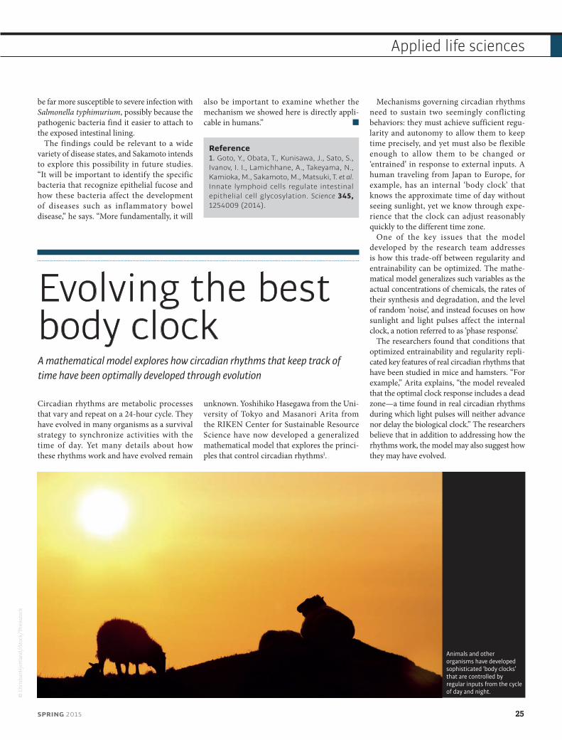
be far more susceptible to severe infection with Salmonella typhimurium, possibly because the pathogenic bacteria find it easier to attach to the exposed intestinal lining.
The findings could be relevant to a wide variety of disease states, and Sakamoto intends to explore this possibility in future studies. “It will be important to identify the specific bacteria that recognize epithelial fucose and how these bacteria affect the development of diseases such as inflammatory bowel disease,” he says. “More fundamentally, it will
also be important to examine whether the mechanism we showed here is directly appli-cable in humans.”
Reference1. Goto, Y., Obata, T., Kunisawa, J., Sato, S., Ivanov, I. I., Lamichhane, A., Takeyama, N., Kamioka, M., Sakamoto, M., Matsuki, T. et al. Innate lymphoid cells regulate intestinal epithelial cell glycosylation. Science 345, 1254009 (2014).
Evolving the best body clockA mathematical model explores how circadian rhythms that keep track of time have been optimally developed through evolution
Circadian rhythms are metabolic processes that vary and repeat on a 24-hour cycle. They have evolved in many organisms as a survival strategy to synchronize activities with the time of day. Yet many details about how these rhythms work and have evolved remain
unknown. Yoshihiko Hasegawa from the Uni-versity of Tokyo and Masanori Arita from the RIKEN Center for Sustainable Resource Science have now developed a generalized mathematical model that explores the princi-ples that control circadian rhythms1.
Mechanisms governing circadian rhythms need to sustain two seemingly conflicting behaviors: they must achieve sufficient regu-larity and autonomy to allow them to keep time precisely, and yet must also be flexible enough to allow them to be changed or ‘entrained’ in response to external inputs. A human traveling from Japan to Europe, for example, has an internal ‘body clock’ that knows the approximate time of day without seeing sunlight, yet we know through expe-rience that the clock can adjust reasonably quickly to the different time zone.
One of the key issues that the model developed by the research team addresses is how this trade-off between regularity and entrainability can be optimized. The mathe-matical model generalizes such variables as the actual concentrations of chemicals, the rates of their synthesis and degradation, and the level of random ‘noise’, and instead focuses on how sunlight and light pulses affect the internal clock, a notion referred to as ‘phase response’.
The researchers found that conditions that optimized entrainability and regularity repli-cated key features of real circadian rhythms that have been studied in mice and hamsters. “For example,” Arita explains, “the model revealed that the optimal clock response includes a dead zone—a time found in real circadian rhythms during which light pulses will neither advance nor delay the biological clock.” The researchers believe that in addition to addressing how the rhythms work, the model may also suggest how they may have evolved.
Animals and other organisms have developed sophisticated ‘body clocks’ that are controlled by regular inputs from the cycle of day and night.
© C
hris
tianH
jort
land
/iSt
ock/
Thin
ksto
ck
SPRING 2015 25
Applied life sciences

Arita hopes that other researchers may be able to test the applicability of the model to humans. He suggests it may also be relevant to cave-dwelling or deep-sea animals, which may only have relics of a body clock since they live in the dark. The work could also help in
the search for key biological components responsible for circadian rhythms. “It should be useful for distinguishing molecules most likely to be involved from the wider range of candidate molecules that other researchers are identifying,” says Arita.
Reference1. Hasegawa, Y. & Arita, M. Optimal implementations for reliable circadian clocks. Physical Review Letters 113, 108101 (2014).
Cutting cholesterol for safer potatoesThe identification of a gene responsible for cholesterol production could lead to potatoes with lower toxin levels
In many parts of the world, potatoes are a reliable dietary staple. However, potato
plants also produce the toxins solanine and chaconine, which can protect growing
sprouts from potential predators such as insects and fungi. These toxins, known as steroidal glycoalkaloids (SGAs), occur at very low levels in the edible tubers, but their levels in green skin and sprouts (see image) can be highly poisonous and even deadly to humans.
Researchers led by Kazuki Saito of the RIKEN Center for Sustainable Resource Science have now identified the mechanism of SGA synthesis in potatoes in an effort to engineer plants that do not produce these toxic compounds1.
S a i to’s g roup p ar t n e re d w i t h an interdisciplinary consortium of researchers from across Japan to unravel the processes surrounding SGA synthesis. They began by searching the potato genome for genes with a likely role in the synthesis of cholesterol, which is known to be a critical intermedi-ate in the process of SGA production. Their search uncovered two candidate genes, sterol side chain reductase 1 and 2 (SSR1 and SSR2), which encode enzymes similar to those involved in cholesterol production in animals.
Careful functional analysis suggested that SSR2 is primarily responsible for converting precursor compounds into the cholesterol that subsequently gives rise to SGA. Indeed, use of a technique called RNA silencing to selectively inhibit SSR2 resulted in significantly lower SGA levels without impairing plant growth. Encouraged by these results, Saito and his col-leagues used customized enzymes known as transcription activator-like effector nucleases (TALENs) to selectively excise the SSR2 gene from the potato genome. The resulting plants showed impaired cholesterol production and
Elevated levels of steroidal glycoalkaloid toxins like solanine occur in the sprouts and green skins of germinating potatoes.
© Y
uriy
S/iS
tock
/Thi
nkst
ock
26 RIKEN RESEARCH
Applied life sciences
contained 90% lower levels of SGA compared with unmodified potatoes.
The biological function of SGAs is poorly defined, and it remains to be determined whether this modification might, for example, render plants more vulnerable to pests. Saito is nevertheless encouraged by these results and foresees commercial potential for this potato strain. “TALEN editing does not eventually leave a ‘footprint’ of trans-genic modification in the genome and can be regarded as similar to classical breeding, which is more acceptable to the public than conven-tional genetically modified organisms,” he says.
The biochemical pathway uncovered in this study could yield other valuable returns as well. For example, cholesterol is a precursor
to a variety of important hormones, and con-trolled cholesterol production could be useful for biotechnology applications. “This gene could be used for the production of cholesterol and its derivatives in plants or even microbes,” says Saito.
Reference1. Sawai, S., Ohyama, K., Yasumoto, S., Seki, H., Sakuma, T., Yamamoto, T., Takebayashi, Y., Kojima, M., Sakakibara, H., Aoki, T. et al. Sterol side chain reductase 2 is a key enzyme in the biosynthesis of cholesterol, the common precursor of toxic steroidal glycoalkaloids in potato. The Plant Cell 26, 3763–3774 (2014).
Learning without conflictA mathematical equation explains how equilibrium is achieved between two forms of synaptic plasticity during brain learning
When the brain learns, individual synapses change in strength. This phenomenon, known as synaptic plasticity, is driven by two fundamental processes: Hebbian plasticity and homeostatic plasticity. Taro Toyoizumi from the RIKEN Brain Science Institute and colleagues have now developed a mathematical equation of synaptic plasticity that describes the interplay between these two processes1.
“Hebbian plasticity is thought to underlie various forms of learning and memory,” explains Toyoizumi. This form of plasticity is characterized by a short timescale—occurring within minutes of an experience—and is specific to a particular set of synapses. In contrast, homeostatic plasticity can be much slower and affect synapses in contact with a post-synaptic cell in a global manner. This plasticity has a scaling effect that brings the neuronal activity within a certain physiological range. “It’s like turning up the brightness on a television,” says Toyoizumi.
University of California neuroscientists Megumi Kaneko and Michael Stryker,
co-authors of the present study, had previously demonstrated that both types of plasticity operate in an experimental protocol known as ocular dominance plasticity (ODP). In the ODP experiment, young mice have one eye blindfolded during a critical developmental period. This manipulation alters the relative representation of information from both eyes in the visual cortex. Hebbian plasticity weakens the efficacy of the closed eye in driving the visual cortex, whereas homeostatic plasticity strengthens that of the open eye.
Working with mathematician Kenneth Miller from Columbia University, Toyoizumi showed that existing mathematical models of synaptic strength were unable to reproduce the ODP data. “The learning outcome becomes unstable if homeostatic plasticity directly competes with but lags Hebbian plasticity,” he says.
Toyoizumi and Miller developed a new model in which synaptic strength is described as a product of biophysical factors for Hebbian and homeostatic plasticity, with each reaching equilibrium independently as learning
During learning, synapses in the brain strengthen and weaken as a result of Hebbian and homeostatic plasticity. An equation describing the interaction of the two forms of plasticity explains experience-dependent reorganization of cortical circuitry during visual learning experiments.
© A
lexa
nder
Shi
roko
v/iS
tock
/Thi
nkst
ock
SPRING 2015 27
Applied life sciences
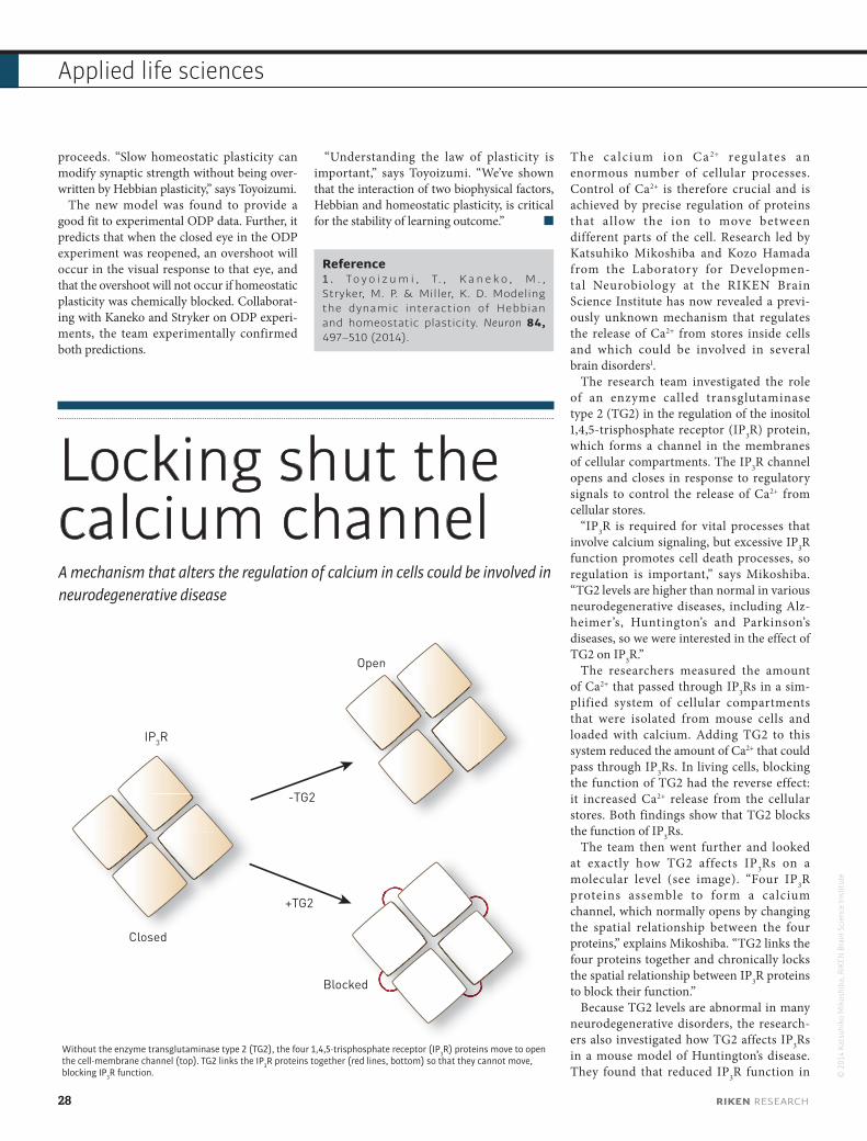
proceeds. “Slow homeostatic plasticity can modify synaptic strength without being over-written by Hebbian plasticity,” says Toyoizumi.
The new model was found to provide a good fit to experimental ODP data. Further, it predicts that when the closed eye in the ODP experiment was reopened, an overshoot will occur in the visual response to that eye, and that the overshoot will not occur if homeostatic plasticity was chemically blocked. Collaborat-ing with Kaneko and Stryker on ODP experi-ments, the team experimentally confirmed both predictions.
“Understanding the law of plasticity is important,” says Toyoizumi. “We’ve shown that the interaction of two biophysical factors, Hebbian and homeostatic plasticity, is critical for the stability of learning outcome.”
Reference1 . To y o i z u m i , T. , K a n e k o , M . , Stryker, M. P. & Miller, K. D. Modeling the dynamic interaction of Hebbian and homeostatic plasticity. Neuron 84, 497–510 (2014).
Locking shut the calcium channelA mechanism that alters the regulation of calcium in cells could be involved in neurodegenerative disease
The ca lc ium ion Ca 2+ regulates an enormous number of cellular processes. Control of Ca2+ is therefore crucial and is achieved by precise regulation of proteins that al low the ion to move between different parts of the cell. Research led by Katsuhiko Mikoshiba and Kozo Hamada from the Laboratory for Developmen-tal Neurobiology at the RIKEN Brain Science Institute has now revealed a previ-ously unknown mechanism that regulates the release of Ca2+ from stores inside cells and which could be involved in several brain disorders1.
The research team investigated the role of an enzyme called transglutaminase type 2 (TG2) in the regulation of the inositol 1,4,5-trisphosphate receptor (IP3R) protein, which forms a channel in the membranes of cellular compartments. The IP3R channel opens and closes in response to regulatory signals to control the release of Ca2+ from cellular stores.
“IP3R is required for vital processes that involve calcium signaling, but excessive IP3R function promotes cell death processes, so regulation is important,” says Mikoshiba. “TG2 levels are higher than normal in various neurodegenerative diseases, including Alz-heimer’s, Huntington’s and Parkinson’s diseases, so we were interested in the effect of TG2 on IP3R.”
The researchers measured the amount of Ca2+ that passed through IP3Rs in a sim-plified system of cellular compartments that were isolated from mouse cells and loaded with calcium. Adding TG2 to this system reduced the amount of Ca2+ that could pass through IP3Rs. In living cells, blocking the function of TG2 had the reverse effect: it increased Ca2+ release from the cellular stores. Both findings show that TG2 blocks the function of IP3Rs.
The team then went further and looked at exactly how TG2 affects IP3Rs on a molecular level (see image). “Four IP3R proteins assemble to form a calcium channel, which normally opens by changing the spatial relationship between the four proteins,” explains Mikoshiba. “TG2 links the four proteins together and chronically locks the spatial relationship between IP3R proteins to block their function.”
Because TG2 levels are abnormal in many neurodegenerative disorders, the research-ers also investigated how TG2 affects IP3Rs in a mouse model of Huntington’s disease. They found that reduced IP3R function in ©
201
4 Ka
tsuh
iko
Mik
oshi
ba, R
IKEN
Bra
in S
cien
ce In
stitu
te
OpenOpen
-TG2
+TG2
Closed
Blocked
IP3R
Without the enzyme transglutaminase type 2 (TG2), the four 1,4,5-trisphosphate receptor (IP3R) proteins move to open the cell-membrane channel (top). TG2 links the IP3R proteins together (red lines, bottom) so that they cannot move, blocking IP3R function.
28 RIKEN RESEARCH
Applied life sciences
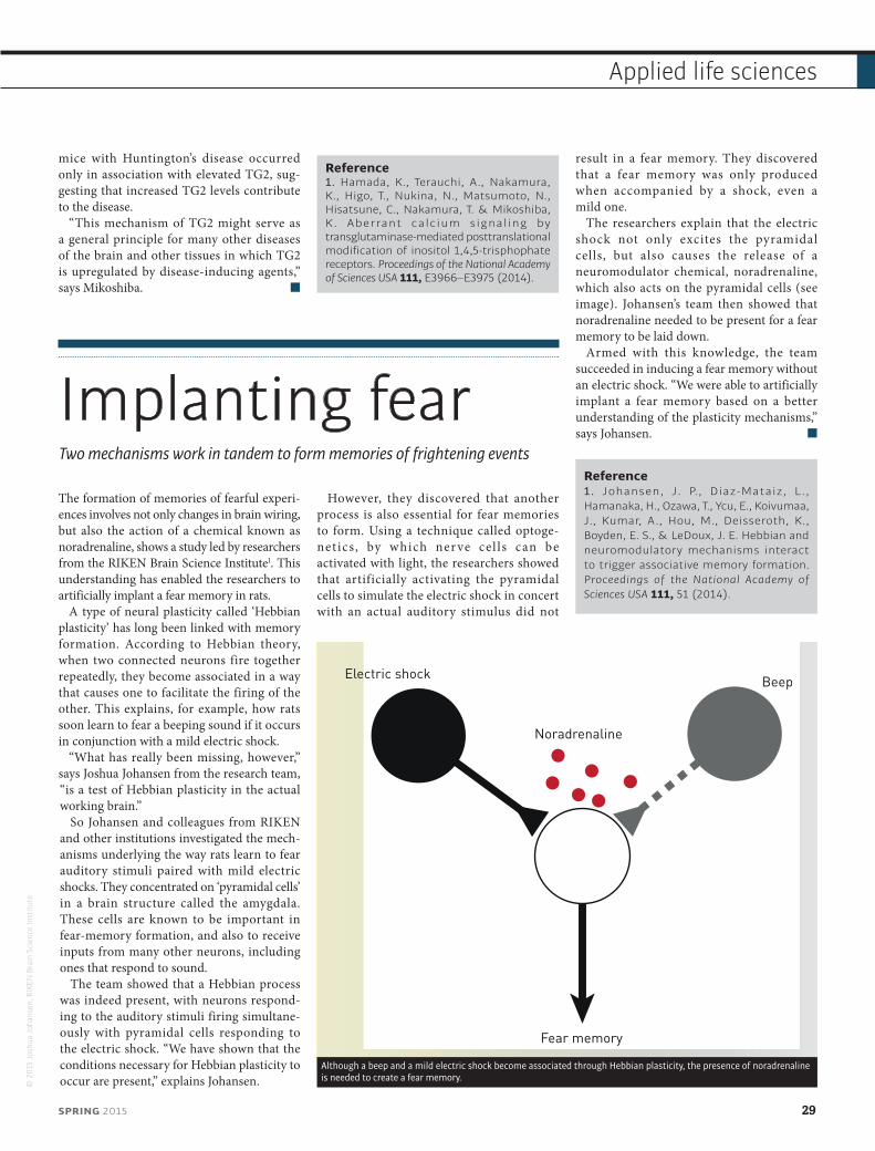
mice with Huntington’s disease occurred only in association with elevated TG2, sug-gesting that increased TG2 levels contribute to the disease.
“This mechanism of TG2 might serve as a general principle for many other diseases of the brain and other tissues in which TG2 is upregulated by disease-inducing agents,” says Mikoshiba.
Reference1. Hamada, K., Terauchi, A., Nakamura, K., Higo, T., Nukina, N., Matsumoto, N., Hisatsune, C., Nakamura, T. & Mikoshiba, K. Aberrant calcium signaling by transglutaminase-mediated posttranslational modification of inositol 1,4,5-trisphophate receptors. Proceedings of the National Academy of Sciences USA 111, E3966–E3975 (2014).
Implanting fearTwo mechanisms work in tandem to form memories of frightening events
The formation of memories of fearful experi-ences involves not only changes in brain wiring, but also the action of a chemical known as noradrenaline, shows a study led by researchers from the RIKEN Brain Science Institute1. This understanding has enabled the researchers to artificially implant a fear memory in rats.
A type of neural plasticity called ‘Hebbian plasticity’ has long been linked with memory formation. According to Hebbian theory, when two connected neurons fire together repeatedly, they become associated in a way that causes one to facilitate the firing of the other. This explains, for example, how rats soon learn to fear a beeping sound if it occurs in conjunction with a mild electric shock.
“What has really been missing, however,” says Joshua Johansen from the research team, “is a test of Hebbian plasticity in the actual working brain.”
So Johansen and colleagues from RIKEN and other institutions investigated the mech-anisms underlying the way rats learn to fear auditory stimuli paired with mild electric shocks. They concentrated on ‘pyramidal cells’ in a brain structure called the amygdala. These cells are known to be important in fear-memory formation, and also to receive inputs from many other neurons, including ones that respond to sound.
The team showed that a Hebbian process was indeed present, with neurons respond-ing to the auditory stimuli firing simultane-ously with pyramidal cells responding to the electric shock. “We have shown that the conditions necessary for Hebbian plasticity to occur are present,” explains Johansen.
However, they discovered that another process is also essential for fear memories to form. Using a technique called optoge-netics , by which ner ve cel ls can be activated with light, the researchers showed that artificially activating the pyramidal cells to simulate the electric shock in concert with an actual auditory stimulus did not
result in a fear memory. They discovered that a fear memory was only produced when accompanied by a shock, even a mild one.
The researchers explain that the electric shock not only excites the pyramidal cells, but also causes the release of a neuromodulator chemical, noradrenaline, which also acts on the pyramidal cells (see image). Johansen’s team then showed that noradrenaline needed to be present for a fear memory to be laid down.
Armed with this knowledge, the team succeeded in inducing a fear memory without an electric shock. “We were able to artificially implant a fear memory based on a better understanding of the plasticity mechanisms,” says Johansen.
Reference1. Johansen, J. P., Diaz-Mataiz, L., Hamanaka, H., Ozawa, T., Ycu, E., Koivumaa, J., Kumar, A., Hou, M., Deisseroth, K., Boyden, E. S., & LeDoux, J. E. Hebbian and neuromodulatory mechanisms interact to trigger associative memory formation. Proceedings of the National Academy of Sciences USA 111, 51 (2014).
© 2
015
Josh
ua J
ohan
sen,
RIK
EN B
rain
Sci
ence
Inst
itute
Noradrenaline
Fear memory
BeepElectric shock
Although a beep and a mild electric shock become associated through Hebbian plasticity, the presence of noradrenaline is needed to create a fear memory.
SPRING 2015 29
Applied life sciences
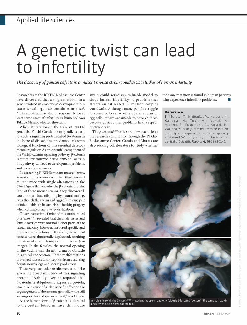
A genetic twist can lead to infertilityThe discovery of genital defects in a mutant mouse strain could assist studies of human infertility
Researchers at the RIKEN BioResource Center have discovered that a single mutation in a gene involved in embryonic development can cause sexual organ abnormalities in mice1. “This mutation may also be responsible for at least some cases of infertility in humans,” says Takuya Murata, who led the study.
When Murata joined the team of RIKEN geneticist Yoichi Gondo, he originally set out to study a signaling protein called β-catenin in the hope of discovering previously unknown biological functions of this essential develop-mental regulator. As an essential component of the Wnt/β-catenin signaling pathway, β-catenin is critical for embryonic development. Faults in this pathway can lead to development problems and disease, even cancer.
By screening RIKEN’s mutant mouse library, Murata and co-workers identified several mutant mice with single alterations in the Ctnnb1 gene that encodes the β-catenin protein. One of these mouse strains, they discovered, could not produce offspring by natural mating, even though the sperm and eggs of a mating pair of mice of this strain gave rise to healthy progeny when combined via in vitro fertilization.
Closer inspection of mice of this strain, called β-cateninC429S, revealed that the male testes and female ovaries were normal. Other parts of the sexual anatomy, however, harbored specific and unusual malformations. In the males, the seminal vesicles were abnormally duplicated, resulting in detoured sperm transportation routes (see image). In the females, the normal opening of the vagina was absent—a major obstacle to natural conception. These malformations prevented successful conception from occurring despite normal egg and sperm production.
These very particular results were a surprise given the broad influence of this signaling protein. “Nobody ever anticipated that β-catenin, a ubiquitously expressed protein, would be a cause of such a specific effect on the organogenesis of the internal genitalia while still leaving oocytes and sperm normal,” says Gondo.
As the human form of β-catenin is identical to the protein found in mice, this mouse
strain could serve as a valuable model to study human infertility—a problem that affects an estimated 50 million couples worldwide. Although many people struggle to conceive because of irregular sperm or egg cells, others are unable to have children because of structural problems in the repro-ductive organs.
The β-cateninC429S mice are now available to the research community through the RIKEN BioResource Center. Gondo and Murata are also seeking collaborators to study whether
the same mutation is found in human patients who experience infertility problems.
Reference1. Murata, T., Ishitsuka, Y., Karouji, K., Kaneda, H. , Toki , H. , Nakai , Y. , Makino, S., Fukumura, R., Kotaki, H., Wakana, S. et al. β-cateninC429S mice exhibit sterility consequent to spatiotemporally sustained Wnt signalling in the internal genitalia. Scientific Reports 4, 6959 (2014).
In male mice with the β-cateninC429S mutation, the sperm pathway (blue) is bifurcated (bottom). The same pathway in a healthy mouse is shown at the top.
Repr
oduc
ed fr
om R
ef. 1
and
lice
nsed
und
er C
C BY
4.0
(htt
p://
crea
tivec
omm
ons.
org/
licen
ses/
by/4
.0/l
egal
code
) © 2
014
T. M
urat
a et
al.
30 RIKEN RESEARCH
Applied life sciences

© 2
015
RIKE
N
Kimihiko Hirao is director of the RIKEN Advanced
Institute for Computational Science (AICS)
Hirao has been director of the AICS since its
establishment in 2010. He has authored more than
300 papers on the subject of theoretical chemistry,
including significant theoretical contributions
to computational quantum chemistry.
Supercomputers
Powering scienceSupercomputers can process information hundreds of thousands times faster than the average home computer, making them essential tools for scientific discovery. As the number crunching ability of these powerful machines approaches speeds in the quadrillions of arithmetic calculations per second, supercomputers will bring unprecedented certainty and speed to scientific prediction.
Powerful supercomputers like the K computer developed at the RIKEN Advanced Institute for Computational Science are essential for scientific discovery and prediction.
SPRING 2015 31
Perspectives
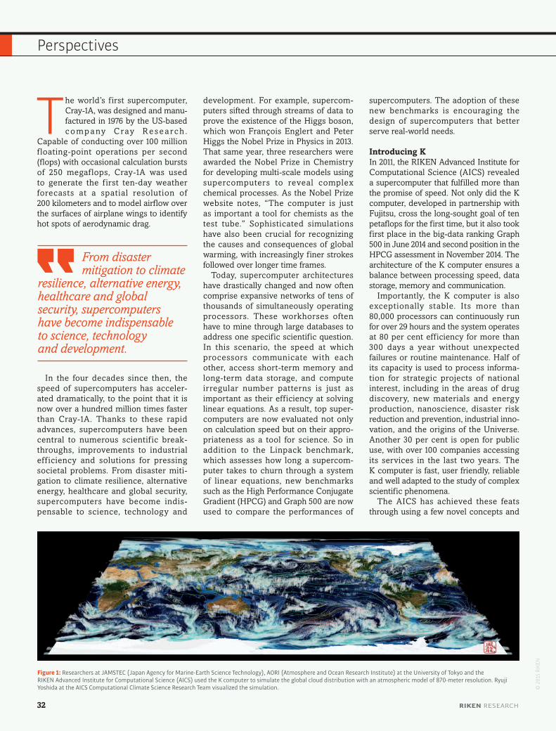
The world’s f irst supercomputer, Cray-1A, was designed and manu-factured in 1976 by the US-based c o m p a ny C ray R e s e a rc h .
Capable of conducting over 100 million f loating-point operations per second (flops) with occasional calculation bursts of 250 megaflops, Cray-1A was used to generate the f irst ten-day weather forecasts at a spatial resolution of 200 kilometers and to model airflow over the surfaces of airplane wings to identify hot spots of aerodynamic drag.
In the four decades since then, the speed of supercomputers has acceler-ated dramatically, to the point that it is now over a hundred million times faster than Cray-1A. Thanks to these rapid advances, supercomputers have been central to numerous scientif ic break-throughs, improvements to industrial eff iciency and solutions for pressing societal problems. From disaster miti-gation to climate resilience, alternative energy, healthcare and global security, supercomputers have become indis-pensable to science, technology and
development. For example, supercom-puters sifted through streams of data to prove the existence of the Higgs boson, which won François Englert and Peter Higgs the Nobel Prize in Physics in 2013. That same year, three researchers were awarded the Nobel Prize in Chemistry for developing multi-scale models using supercomputers to reveal complex chemical processes. As the Nobel Prize website notes, “The computer is just as important a tool for chemists as the test tube.” Sophisticated simulations have also been crucial for recognizing the causes and consequences of global warming, with increasingly finer strokes followed over longer time frames.
Today, supercomputer architectures have drastically changed and now often comprise expansive networks of tens of thousands of simultaneously operating processors. These workhorses often have to mine through large databases to address one specific scientific question. In this scenario, the speed at which processors communicate with each other, access short-term memory and long-term data storage, and compute irregular number patterns is just as important as their eff iciency at solving linear equations. As a result, top super-computers are now evaluated not only on calculation speed but on their appro-priateness as a tool for science. So in addition to the Linpack benchmark, which assesses how long a supercom-puter takes to churn through a system of linear equations, new benchmarks such as the High Performance Conjugate Gradient (HPCG) and Graph 500 are now used to compare the performances of
supercomputers. The adoption of these new benchmarks is encouraging the design of supercomputers that better serve real-world needs.
Introducing KIn 2011, the RIKEN Advanced Institute for Computational Science (AICS) revealed a supercomputer that fulfilled more than the promise of speed. Not only did the K computer, developed in partnership with Fujitsu, cross the long-sought goal of ten petaflops for the first time, but it also took first place in the big-data ranking Graph 500 in June 2014 and second position in the HPCG assessment in November 2014. The architecture of the K computer ensures a balance between processing speed, data storage, memory and communication.
Importantly, the K computer is also exceptionally stable. Its more than 80,000 processors can continuously run for over 29 hours and the system operates at 80 per cent efficiency for more than 300 days a year without unexpected failures or routine maintenance. Half of its capacity is used to process informa-tion for strategic projects of national interest, including in the areas of drug discovery, new materials and energy production, nanoscience, disaster risk reduction and prevention, industrial inno-vation, and the origins of the Universe. Another 30 per cent is open for public use, with over 100 companies accessing its services in the last two years. The K computer is fast, user friendly, reliable and well adapted to the study of complex scientific phenomena.
The AICS has achieved these feats through using a few novel concepts and
Figure 1: Researchers at JAMSTEC (Japan Agency for Marine-Earth Science Technology), AORI (Atmosphere and Ocean Research Institute) at the University of Tokyo and the RIKEN Advanced Institute for Computational Science (AICS) used the K computer to simulate the global cloud distribution with an atmospheric model of 870-meter resolution. Ryuji Yoshida at the AICS Computational Climate Science Research Team visualized the simulation. ©
201
5 RI
KEN
From disaster mitigation to climate
resilience, alternative energy, healthcare and global security, supercomputers have become indispensable to science, technology and development.
32 RIKEN RESEARCH
Perspectives
techniques. Crucial among them was the institutional merging of computer science (the design of actual computers) with computational science (the development of applications that employ advanced computers). At the AICS, researchers in the divisions of computer science and computational science work side by side.
The K computer also uses a custom-ized network called Tofu (Torus fusion), a six-dimensional grid connection through which the over 80,000 processors, or ‘nodes’, communicate with each. Each node is assigned to a project, or ‘job’, based on the most spatially eff icient allocation involving short and quick
exchanges between nodes. Tofu repre-sents a shift away from earlier networks that used hierarchical systems to connect nodes. Furthermore, to improve the stability of the system, the AICS dropped the operating temperature by an astound-ing 50 degrees Celsius by switching from air cooling to a system that employs both air and water.
Zooming inSince its launch, the K computer has been used by many research groups to simulate physical phenomena more accu-rately than previously possible. These projects would have been diff icult, and sometimes even impossible, to achieve without the help of the K computer.
In 2014, a team from Nagoya Univer-sity reconstructed the nanometer-sized protein shell of a poliovirus f loating in water by simulating the movement and interaction of 6.5 million individual atoms1. Later in the year, RIKEN col-laborated with the University of Tokyo and Fujitsu to simulate the pumping mechanism of the heart2. To do this, the K computer took two days to recreate 170,000 tetrahedrons representing the individual muscle fibers of heart tissue, integrating information from physics, engineering, medicine and physiology. The simulation shows how blood flows
from the lungs, through the chambers in the heart, and back out to the body. The program has been used to model the hearts of real patients and predict the outcome of various treatment options to improve surgery success rates.
Another group of scientists studied a new concept of electrolyte for lithium-ion batteries called lithium-salt super-concentrated solution. They used the K computer to elucidate the unusual elec-trochemical stability and ionic transport of the electrolyte at the atomic scale. Such novel electrolytes are expected to enable the use of high-voltage lithium-ion batteries with fast charging time.
And based on a simulation of the three-dimensional structure of three cancer proteins, a further research group has discovered over 10 potential leads for new anticancer drugs that are currently being preclinically tested.
The K computer has also far exceeded the Cray-1A’s weather outlooks. While the older computer could provide projec-tions at a resolution of 200 kilometers, the K computer can zoom in on 870-meter resolution imagery to follow the dense, towering cumulonimbus clouds that bring thunderstorms (Fig. 1), as well as predict the onset and behavior of a massive atmospheric disturbance known as the Madden–Julian Oscillation over a period of a month. The oscillation is respon-sible for heavy rainfall that could lead to cyclones and heat and cold waves in tropical regions.
Tipping pointThe supercomputers of today, however, have not advanced enough to simulate natural phenomena with the speed and accuracy needed to meet all of our scientif ic, environmental and social demands—much faster and more respon-sive machines are needed. The AICS and Fujitsu have partnered once more to construct a computer that will take the science of prediction into uncharted
territories. The major national project is supported by JPY 110 billion in funding from the Japanese government and targeted for completion by 2020.
The post-K supercomputer will be designed to address important unre-solved problems in science and engi-neering. In building its core framework, the AICS plans to use state-of-the-art Japanese technologies and will collabo-rate with international partners to develop standardized software that can be applied across a global spectrum of research for increased user convenience. The super-computer will inherit the K computer’s parallel processing structure and Tofu network to provide a scalable, easy-to-use computational resource.
A system of such capacity will enable researchers to simulate solutions to many existing and emerging societal problems. For example, by processing large volumes of satellite and radar data, it may be possible to warn city residents of imminent outbursts of torrential ‘guerilla rain’, which can lead to flash floods that cause significant loss of life and damage to property.
Scientists now consider simulation to be the third pillar of scientific discovery, after theory and experiment. But to power science toward the tipping point where description turns into prediction, researchers of all breeds—computer, computational and specialized subject scientists—will have to co-design a supercomputer suited to their needs. The AICS is offering them an opportunity to do that.
References1. Yoshii, N., Andoh, A ., Fujimoto, K., Kojima, H., Yamada, A. & Okazaki, S. MODYLAS: A highly parallelized general-purpose molecular dynamics simulation program. International Journal of Quantum Chemistry 115, 342–348 (2015).
2. Washio, T., Okada, J., Takahashi, A., Yoneda, K., Kadooka, Y., Sugiura, S. & Hisada, T. Multiscale heart simulation with cooperative stochastic cross-bridge dynamics and cellular structures. Multiscale Modeling & Simulation 11, 965–999 (2013).
For additional references, visit the online version of this article at:
www.riken.jp/en/research/rikenresearch/perspectives/7625
The AICS is designing the post-K supercomputer to address important unresolved problems in science and engineering through the institutional merger of computer science and computational science.
SPRING 2015 33
Perspectives
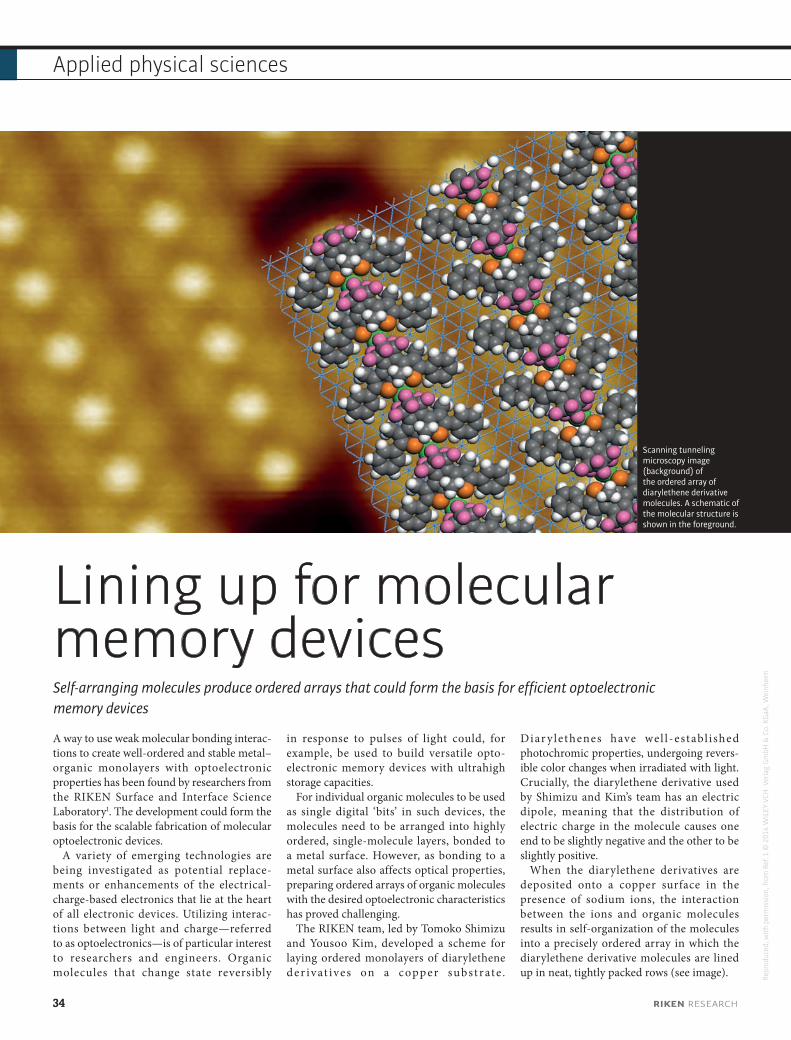
Repr
oduc
ed, w
ith p
erm
issi
on, f
rom
Ref
. 1 ©
201
4 W
ILEY
-VCH
Ver
lag
Gm
bH &
Co.
KG
aA, W
einh
eim
Lining up for molecular memory devicesSelf-arranging molecules produce ordered arrays that could form the basis for efficient optoelectronic memory devices
A way to use weak molecular bonding interac-tions to create well-ordered and stable metal–organic monolayers with optoelectronic properties has been found by researchers from the RIKEN Surface and Interface Science Laboratory1. The development could form the basis for the scalable fabrication of molecular optoelectronic devices.
A variety of emerging technologies are being investigated as potential replace-ments or enhancements of the electrical-charge-based electronics that lie at the heart of all electronic devices. Utilizing interac-tions between light and charge—referred to as optoelectronics—is of particular interest to researchers and engineers. Organic molecules that change state reversibly
in response to pulses of light could, for example, be used to build versatile opto-electronic memory devices with ultrahigh storage capacities.
For individual organic molecules to be used as single digital ‘bits’ in such devices, the molecules need to be arranged into highly ordered, single-molecule layers, bonded to a metal surface. However, as bonding to a metal surface also affects optical properties, preparing ordered arrays of organic molecules with the desired optoelectronic characteristics has proved challenging.
The RIKEN team, led by Tomoko Shimizu and Yousoo Kim, developed a scheme for laying ordered monolayers of diarylethene der ivat ives on a copp er subst rate .
Diar y lethenes have wel l -establ ished photochromic properties, undergoing revers-ible color changes when irradiated with light. Crucially, the diarylethene derivative used by Shimizu and Kim’s team has an electric dipole, meaning that the distribution of electric charge in the molecule causes one end to be slightly negative and the other to be slightly positive.
When the diarylethene derivatives are deposited onto a copper surface in the presence of sodium ions, the interaction between the ions and organic molecules results in self-organization of the molecules into a precisely ordered array in which the diarylethene derivative molecules are lined up in neat, tightly packed rows (see image).
Scanning tunneling microscopy image (background) of the ordered array of diarylethene derivative molecules. A schematic of the molecular structure is shown in the foreground.
34 RIKEN RESEARCH
Applied physical sciences
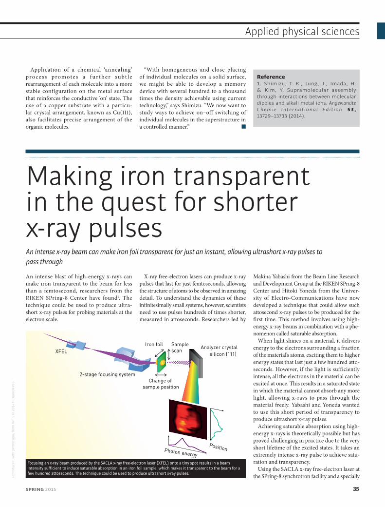
Application of a chemical ‘annealing’ pro cess promotes a f ur t her subt le rearrangement of each molecule into a more stable configuration on the metal surface that reinforces the conductive ‘on’ state. The use of a copper substrate with a particu-lar crystal arrangement, known as Cu(111), also facilitates precise arrangement of the organic molecules.
“With homogeneous and close placing of individual molecules on a solid surface, we might be able to develop a memory device with several hundred to a thousand times the density achievable using current technology,” says Shimizu. “We now want to study ways to achieve on–off switching of individual molecules in the superstructure in a controlled manner.”
Reference1. Shimizu, T. K., Jung, J., Imada, H. & Kim, Y. Supramolecular assembly through interactions between molecular dipoles and alkali metal ions. Angewandte C h e m i e I n t e r n a t i o n a l E d i t i o n 5 3 , 13729–13733 (2014).
Making iron transparent in the quest for shorter x-ray pulsesAn intense x-ray beam can make iron foil transparent for just an instant, allowing ultrashort x-ray pulses to pass through
An intense blast of high-energy x-rays can make iron transparent to the beam for less than a femtosecond, researchers from the RIKEN SPring-8 Center have found1. The technique could be used to produce ultra-short x-ray pulses for probing materials at the electron scale.
X-ray free-electron lasers can produce x-ray pulses that last for just femtoseconds, allowing the structure of atoms to be observed in amazing detail. To understand the dynamics of these infinitesimally small systems, however, scientists need to use pulses hundreds of times shorter, measured in attoseconds. Researchers led by
Makina Yabashi from the Beam Line Research and Development Group at the RIKEN SPring-8 Center and Hitoki Yoneda from the Univer-sity of Electro-Communications have now developed a technique that could allow such attosecond x-ray pulses to be produced for the first time. This method involves using high-energy x-ray beams in combination with a phe-nomenon called saturable absorption.
When light shines on a material, it delivers energy to the electrons surrounding a fraction of the material’s atoms, exciting them to higher energy states that last just a few hundred atto-seconds. However, if the light is sufficiently intense, all the electrons in the material can be excited at once. This results in a saturated state in which the material cannot absorb any more light, allowing x-rays to pass through the material freely. Yabashi and Yoneda wanted to use this short period of transparency to produce ultrashort x-ray pulses.
Achieving saturable absorption using high-energy x-rays is theoretically possible but has proved challenging in practice due to the very short lifetime of the excited states. It takes an extremely intense x-ray pulse to achieve satu-ration and transparency.
Using the SACLA x-ray free-electron laser at the SPring-8 synchrotron facility and a specially
Analyzer crystalsilicon (111)
2-stage focusing system
XFELIron foil Sample
scan
Change of sample position
Photon energy
Position
Focusing an x-ray beam produced by the SACLA x-ray free-electron laser (XFEL) onto a tiny spot results in a beam intensity sufficient to induce saturable absorption in an iron foil sample, which makes it transparent to the beam for a few hundred attoseconds. The technique could be used to produce ultrashort x-ray pulses.
SPRING 2015 35
Applied physical sciences
Repr
oduc
ed, w
ith p
erm
issi
on, f
rom
Ref
. 1 ©
201
4 H
. Yon
eda
et a
l.
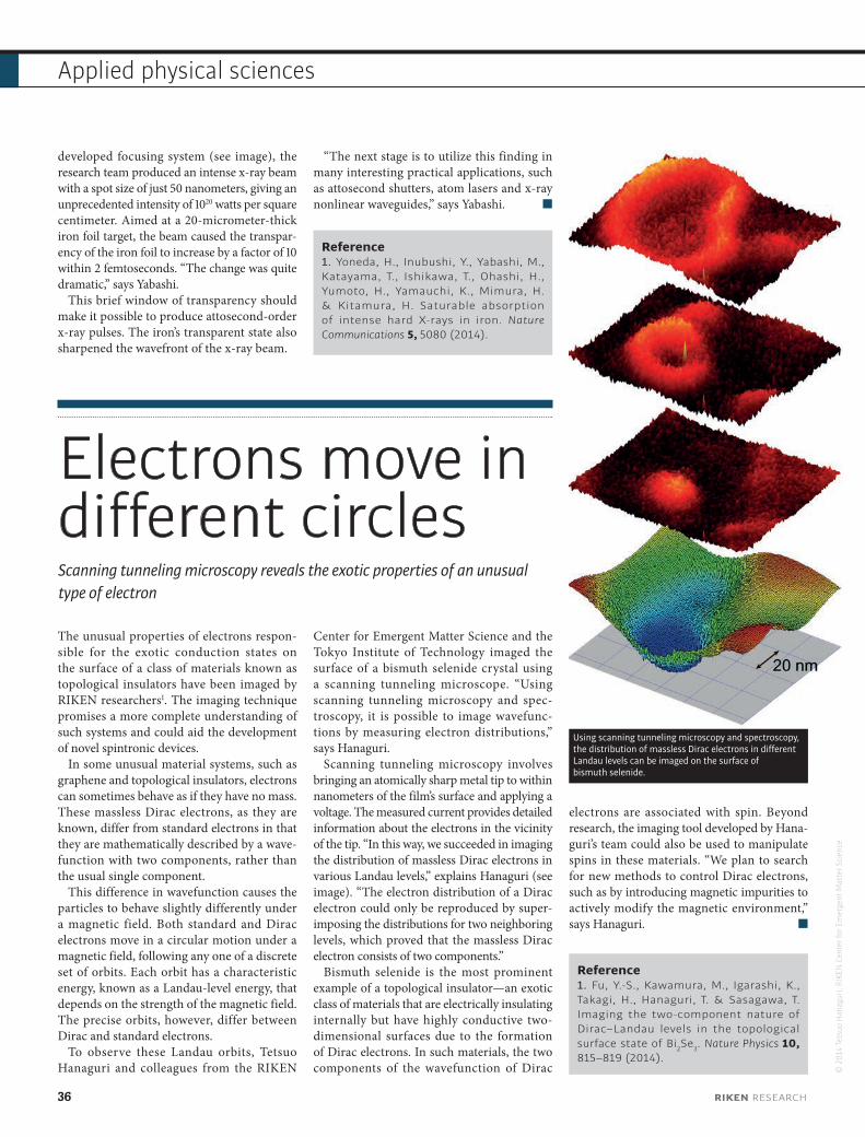
developed focusing system (see image), the research team produced an intense x-ray beam with a spot size of just 50 nanometers, giving an unprecedented intensity of 1020 watts per square centimeter. Aimed at a 20-micrometer-thick iron foil target, the beam caused the transpar-ency of the iron foil to increase by a factor of 10 within 2 femtoseconds. “The change was quite dramatic,” says Yabashi.
This brief window of transparency should make it possible to produce attosecond-order x-ray pulses. The iron’s transparent state also sharpened the wavefront of the x-ray beam.
“The next stage is to utilize this finding in many interesting practical applications, such as attosecond shutters, atom lasers and x-ray nonlinear waveguides,” says Yabashi.
Reference1. Yoneda, H., Inubushi, Y., Yabashi, M., Katayama, T., Ishikawa, T., Ohashi, H., Yumoto, H., Yamauchi, K., Mimura, H. & Kitamura, H. Saturable absorption of intense hard X-rays in iron. Nature Communications 5, 5080 (2014).
Electrons move in different circlesScanning tunneling microscopy reveals the exotic properties of an unusual type of electron
The unusual properties of electrons respon-sible for the exotic conduction states on the surface of a class of materials known as topological insulators have been imaged by RIKEN researchers1. The imaging technique promises a more complete understanding of such systems and could aid the development of novel spintronic devices.
In some unusual material systems, such as graphene and topological insulators, electrons can sometimes behave as if they have no mass. These massless Dirac electrons, as they are known, differ from standard electrons in that they are mathematically described by a wave-function with two components, rather than the usual single component.
This difference in wavefunction causes the particles to behave slightly differently under a magnetic field. Both standard and Dirac electrons move in a circular motion under a magnetic field, following any one of a discrete set of orbits. Each orbit has a characteristic energy, known as a Landau-level energy, that depends on the strength of the magnetic field. The precise orbits, however, differ between Dirac and standard electrons.
To observe these Landau orbits, Tetsuo Hanaguri and colleagues from the RIKEN
Center for Emergent Matter Science and the Tokyo Institute of Technology imaged the surface of a bismuth selenide crystal using a scanning tunneling microscope. “Using scanning tunneling microscopy and spec-troscopy, it is possible to image wavefunc-tions by measuring electron distributions,” says Hanaguri.
Scanning tunneling microscopy involves bringing an atomically sharp metal tip to within nanometers of the film’s surface and applying a voltage. The measured current provides detailed information about the electrons in the vicinity of the tip. “In this way, we succeeded in imaging the distribution of massless Dirac electrons in various Landau levels,” explains Hanaguri (see image). “The electron distribution of a Dirac electron could only be reproduced by super-imposing the distributions for two neighboring levels, which proved that the massless Dirac electron consists of two components.”
Bismuth selenide is the most prominent example of a topological insulator—an exotic class of materials that are electrically insulating internally but have highly conductive two-dimensional surfaces due to the formation of Dirac electrons. In such materials, the two components of the wavefunction of Dirac
electrons are associated with spin. Beyond research, the imaging tool developed by Hana-guri’s team could also be used to manipulate spins in these materials. “We plan to search for new methods to control Dirac electrons, such as by introducing magnetic impurities to actively modify the magnetic environment,” says Hanaguri.
Reference1. Fu, Y.-S., Kawamura, M., Igarashi, K., Takagi, H., Hanaguri, T. & Sasagawa, T. Imaging the two-component nature of Dirac–Landau levels in the topological surface state of Bi2Se3. Nature Physics 10, 815–819 (2014).
Using scanning tunneling microscopy and spectroscopy, the distribution of massless Dirac electrons in different Landau levels can be imaged on the surface of bismuth selenide.
36 RIKEN RESEARCH
Applied physical sciences
© 2
014
Tets
uo H
anag
uri,
RIKE
N C
ente
r for
Em
erge
nt M
atte
r Sci
ence

centers and facilities
across Japan
RIKENWako
Center for Emergent Matter ScienceCenter for Advanced Photonics
Center for Sustainable Resource ScienceBrain Science Institute
Nishina Center for Accelerator-Based ScienceRadioactive Isotope Beam Factory
Advanced Center for Computing and CommunicationResearch Cluster for Innovation
Chief Scientist LaboratoriesAssociate Chief Scientist Laboratories
Distinguished Senior Scientist LaboratoriesInitiative Research UnitsGlobal Research Cluster
TsukubaBioResource Center
YokohamaCenter for Sustainable Resource ScienceCenter for Integrative Medical Sciences
Center for Life Science Technologies
TokyoCenter of Research Network for Infectious Diseases
SendaiRIKEN Sendai Facility
HarimaSPring-8 CenterSPring-8 Synchrotron Radiation FacilitySACLA X-ray Free Electron Laser Facility
KobeCenter for Developmental BiologyHPCI Program for Computational Life SciencesCenter for Life Science TechnologiesAdvanced Institute for Computational ScienceK computer
OsakaQuantitative Biology Center
NagoyaRIKEN Nagoya Facility

RIKEN 2014-001
RIKEN 2015-013
www.riken.jp/en/research/rikenresearch
www.riken.jp/enwww.facebook.com/RIKEN.english www.twitter.com/riken_en@riken_en