Embed Size (px)
Citation preview
Single Atom Substitution for Marking and Motion Tracking of Individual Molecules byScanning Tunneling Microscopy
Guillaume Schull,† Herve Ness,† Ludovic Douillard,*,† Celine Fiorini-Debuisschert,†Fabrice Charra,† Fabrice Mathevet,‡ David Kreher,‡ and Andre-Jean Attias‡
CEA, IRAMIS, SerVice de Physique et Chimie des Surfaces et Interfaces, F-91191 Gif-sur-YVette, France, andUniVersite Pierre et Marie Curie, Laboratoire de Chimie Macromoleculaire, CNRS-UMR 7610,4 Place Jussieu F-75252, Paris cedex 05, France
ReceiVed: April 7, 2008; ReVised Manuscript ReceiVed: July 9, 2008
We report on a simple way to mark and track individual molecules self-assembled on a surface byscanning tunneling microscopy. The tracer mechanism consists in a minimal one-atom chemicalsubstitution. While this substitution leads to significant modifications in the STM signature of themolecules, no substantial changes of the physics of self-assembling are observed when using the modifiedor unmodified molecular building blocks. This allows us to follow the intrinsic dynamical properties ofthe self-assembled molecular patterns.
Introduction
Scanning probe microscopies1 and more especially scanningtunneling microscopy2 (STM) have revolutionized the real spaceimaging of surfaces at the atomic scale level. For the study ofmolecules adsorbed on surfaces, early pioneering works3-6 havemostly been focused on direct imaging of single molecules. Nowa constant trend is to go beyond simple imaging in order toachieve a better discrimination at the molecular level.
One natural way along this line is the use of specific chemicalSTM marking groups.7,8 STM marking groups are functionalgroups exhibiting unusual contrast in the STM images relative tothe rest of the molecule moieties. Experimental investigationscarried on substituted long-chain molecules have identified severalgroups with interesting properties. Some groups can increase thetunneling probability,8-13 such as thiol group -SH, sulfur atomsS, iodine atoms I, amino group -NH2, amide group -OdC-NH2
and so on; some can decrease it,12,14-16 for example, fluorine atomsF, carboxylic group -COOH, cyano group -CN and so forth; orsome can present an alternate contrast depending on the differentmolecular configurations on the surface,17,18 such as bromine atomsBr. Other successful approaches include addition or eliminationof molecular moieties,19 tip functionalization,20-23 and variousspectrometric investigations, for example, scanning tunnelingspectroscopies21,24,25 or inelastic tunneling spectroscopies.26,27
In this work, we present a simple way to single out andtrack individual molecules self-assembled on graphite surfaceby STM applying STM marker concepts in conjunction withfirst principle calculations. The tracer mechanism we usedconsists in labeling a molecule by a minimal one-atomchemical substitution. In this way, while the STM signatureof the substituted molecule exhibits significant intramolecularmodifications, the self-assembling physics remains the samewhen using the modified or unmodified molecular blocks.Application of this singling out method is demonstrated here
by direct observation of the intrinsic dynamics of recentlyobtained nanoporous assemblies acting as 2D molecularsieves.28-31 On a more general basis, the present workrepresents a valuable contribution to studies performed byscanning tunneling microscopy and aiming at the recognitionof specific molecular events that are pivotal to surfacedynamics such as surface diffusion,32,33 domain boundarymotion.34,35 and so forth.
Results and Discussion
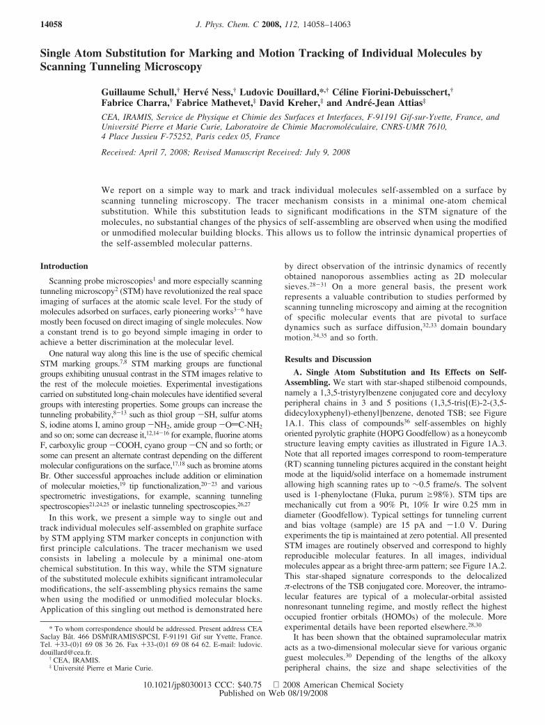
A. Single Atom Substitution and Its Effects on Self-Assembling. We start with star-shaped stilbenoid compounds,namely a 1,3,5-tristyrylbenzene conjugated core and decyloxyperipheral chains in 3 and 5 positions (1,3,5-tris[(E)-2-(3,5-didecyloxyphenyl)-ethenyl]benzene, denoted TSB; see Figure1A.1. This class of compounds36 self-assembles on highlyoriented pyrolytic graphite (HOPG Goodfellow) as a honeycombstructure leaving empty cavities as illustrated in Figure 1A.3.Note that all reported images correspond to room-temperature(RT) scanning tunneling pictures acquired in the constant heightmode at the liquid/solid interface on a homemade instrumentallowing high scanning rates up to ∼0.5 frame/s. The solventused is 1-phenyloctane (Fluka, purum g98%). STM tips aremechanically cut from a 90% Pt, 10% Ir wire 0.25 mm indiameter (Goodfellow). Typical settings for tunneling currentand bias voltage (sample) are 15 pA and -1.0 V. Duringexperiments the tip is maintained at zero potential. All presentedSTM images are routinely observed and correspond to highlyreproducible molecular features. In all images, individualmolecules appear as a bright three-arm pattern; see Figure 1A.2.This star-shaped signature corresponds to the delocalizedπ-electrons of the TSB conjugated core. Moreover, the intramo-lecular features are typical of a molecular-orbital assistednonresonant tunneling regime, and mostly reflect the highestoccupied frontier orbitals (HOMOs) of the molecule. Moreexperimental details have been reported elsewhere.28,30
It has been shown that the obtained supramolecular matrixacts as a two-dimensional molecular sieve for various organicguest molecules.30 Depending of the lengths of the alkoxyperipheral chains, the size and shape selectivities of the
* To whom correspondence should be addressed. Present address CEASaclay Bat. 466 DSM\IRAMIS\SPCSI, F-91191 Gif sur Yvette, France.Tel. +33-(0)1 69 08 36 26. Fax +33-(0)1 69 08 64 62. E-mail: [email protected].
† CEA, IRAMIS.‡ Universite Pierre et Marie Curie.
J. Phys. Chem. C 2008, 112, 14058–1406314058
10.1021/jp8030013 CCC: $40.75 2008 American Chemical SocietyPublished on Web 08/19/2008
supramolecular networks can be tuned to a specific guestmolecule.30,31 Because the sieving properties rely on the intrinsicdynamics of the host matrix any further investigation requiresprecise knowledge of the behavior of each individual molecularblock.
In an attempt to achieve direct visual discrimination betweenindividual TSB molecular units within the honeycomb structure,a minimal change in the skeleton of the TSB molecules hasbeen adopted. The modification consists in the replacement ofone carbon atom of the central conjugated core by a nitrogenatom, namely the inner benzene ring is replaced by a pyridinecore (Figure 1A.1,B.1). Details regarding the synthesis routesleading to both molecular entities are available in the SupportingInformation section. As illustrated in Figure 1B.3, 1,3,5-tristyrylpyridine (TSP) molecules self-assemble on graphite ina way identical to the TSB ones, forming a long-range regularhoneycomb lattice. From a crystallography point of view, thesupramolecular networks obtained using either TSB or TSPmolecules as elementary building blocks are nondiscernible andadopt a p(�241x�241 R3.2°) surface reconstruction.31 However,at the intramolecular level, the individual STM signatures ofboth molecules exhibit subtle differences. In particular, thepyridine inner core appears with an asymmetric shape inthe STM images, as can be seen in Figure 1A.2,B.2. Owing tothe high spatial resolution of STM, the intramolecular featuresof a TSP molecule seem to lack one of its central lobes incomparison to the TSB molecules. Overall, a TSP moleculedeparts from the well-balanced star shape signature of a TSBmolecule to adopt an asymmetric arrowlike configurationpointing to a specific direction. Since both building blocks differby the substitution of only one atom, the STM images suggesta reduction of the occupied density of states at the position ofthe nitrogen atom.
B. First-Principle Calculations. To achieve a better under-standing of the system at the molecular level, quantum me-
chanical calculations of the ground-state electronic structure andtotal energy of model systems have been performed using afirst-principle method. The aim of the calculations is to getinformation about the adsorption properties of a TSB/TSP likemolecule on a graphite substrate and to calculate STM imagesfrom the electronic structure. Previous first-principle calculationshave shown a qualitative correlation between the STM contrastand the energy spacing between the electronic levels of thesurface and of the molecular adsorbates.37 In some cases, theenergy spacing between these levels might be further enhancedby the tip-induced electric field in the tunnel junction.37,38
In our calculations, we have used the ab initio code packageABINIT,39 which is a density-functional theory based code. Inorder to reduce the computing time and the memory spacerequired, the calculations are performed for molecules whosealkoxy peripheral chains are considerably shortened. Although,the length of the alkoxy chains plays an extremely importantrole in the auto-organization of the molecules on graphitesurfaces,31,40 we believed that these chains play a minor roleon the STM contrast observed above the conjugated core ofthe molecule. Indeed, we have checked from calculations onisolated molecules that the electronic density in the conjugatedcore is not strongly modified when the alkoxy chains aremodeled by a single H atom or by a methyl group.
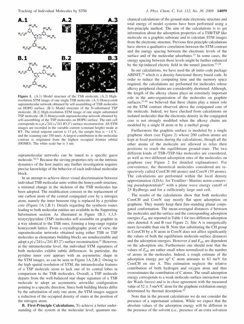
Furthermore the graphite surface is modeled by a singlegraphene sheet (see Figure 2) whose 200 carbon atoms arekept at fixed positions during the calculations, though all theother atoms of the molecule are allowed to relax theirpositions to reach the equilibrium ground-state. The twodifferent kinds of TSB-/TSP-like molecules are considered,as well as two different adsorption sites of the molecules ongraphene (see Figure 2 for detailed explanations). Forconvenience, the theoretical molecules considered are re-spectively called CoreCH (60 atoms) and CoreN (59 atoms).The calculations are performed within the local densityapproximation (LDA), for Troullier-Martins norm-conserv-ing pseudopotentials41 with a plane wave energy cutoff of25 Rydbergs and for a sufficiently large unit cell.
The results of the calculations show that both moleculesCoreCH and CoreN stay mostly flat upon adsorption ongraphene. They mainly keep their free-standing planar conju-gated conformation. The mean-averaged distances d betweenthe molecules and the surface and the corresponding adsorptionenergies Eads are reported in Table 1 for two different adsorptionsites denoted A and B (see Figure 2). Site A is energeticallymore favorable than site B. Note that substituting the CH groupin CoreCH by a N atom in CoreN does not affect significantlythe values of both the equilibrium molecule-surface distancesand the adsorption energies. However d and Eads are dependenton the adsorption site. Furthermore one should note that thevalues of Eads are rather small when normalized to the numberof atoms in the molecules. Indeed, a rough estimate of theadsorption energy per sp2 C atom amounts to 63 meV forCoreCH on site A. This estimation neglects the relativecontribution of both hydrogen and oxygen atom and thusoverestimates the contribution of C atoms. The small adsorptionenergy corresponds to a weak molecule-surface interaction (vander Waals forces) and is in close agreement with the measuredvalue of 52 ( 5 meV/C atom for the graphene exfoliation energydetermined by thermal desorption.42
Note that in the present calculations we do not consider thepresence of a supernatant solution. While we expect that theabsolute values of the adsorption energy will be different inthe presence of the solvent (i.e., presence of an extra solvation
Figure 1. (A.1) Model structure of the TSB molecule. (A.2) High-resolution STM image of one single TSB molecule. (A.3) Honeycombsupramolecular network obtained by self-assembling of TSB moleculeson HOPG surface. (B.1) Model structure of the N-substituted TSPmolecule. (B.2) High-resolution STM image of one single substitutedTSP molecule. (B.3) Honeycomb supramolecular network obtained byself-assembling of the TSP molecules on HOPG surface. The unit cellcorresponds to a p(�241x�241 R3.2°) surface reconstruction. All STMimages are recorded in the variable current (constant height) mode atRT. The initial setpoint current is 15 pA, the sample bias is -1.0 V,and the scanning rate 350 nm/s. A largest contribution in the molecularcontrast is originated from the highest occupied frontier orbital(HOMO). The white scale bar is 3 nm.
Tracking of Individual Molecules by STM J. Phys. Chem. C, Vol. 112, No. 36, 2008 14059
energy) we believe that the effects of the supernatant solutionwill be equivalent for both CoreCH and CoreN molecules andboth adsorption sites. Hence one will probably keep the samerelative difference between adsorption energies in the presenceand in the absence of the solvent. Experimental supports alongthis line are the equivalent self-assembling products of TSBmolecules obtained on graphite either at the liquid-solidinterface or under ultra high vacuum conditions.30
Now that we have shown that changing “one atom” in themolecule (CoreCH T CoreN) does not affect too muchthe adsorption properties of these molecules, we turn on to theeffects of such a substitution on the corresponding electronic
structures. Several HOMO (highest occupied molecular orbitals)and LUMO (lowest unoccupied molecular orbitals) states havebeen computed for both molecules in different sites. Theisodensity plots (see Supporting Information) of these electronicstates around the Fermi energy show significant differences inthe shape when changing “one core atom” in the molecule andfor the adsorption sites A and B. These differences are evenmore noticeable in the calculated STM images.
The STM images are calculated within the Tersoff-Hamann(TH) approximation,43 i.e., the tunneling current is taken to beproportional to the integrated density of states at the positionof the tip. Images at constant (arbitrary) current are shown inFigure 2B.1,B.2 for an applied sample bias corresponding tothe experimental conditions. One obtains a rather good agree-ment with the experiments as far as the overall shape of themolecules is concerned. Most importantly, a strong decreaseof the tunneling current just above the N atom of CoreNmolecule is obtained (with respect to the case of the CoreCHmolecule) as observed experimentally.
It is interesting to note that the observed STM images appearto contain also a nonzero weight from the empty states (seeSupporting Information) although the current flows from thesurface to the tip. This might be due to the fact that the graphitesurface is semimetallic and that the tails of the molecularresonances of the empty states overlap more than expected withthe Fermi level of the surface. Nonperturbative STM calcula-tions44 would be needed to clarify this point.
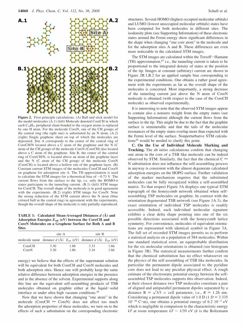
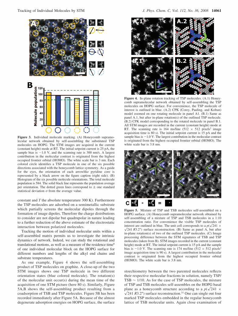
C. On the Use of Individual Molecule Marking andTracking. The ab initio calculations confirm that changingone atom in the core of a TSB like molecule can be indeedobserved by STM. Similarly, the fact that the chemical C TN substitution does not influence the self-assembling processin anyway is consistent with the almost degenerate calculatedadsorption energies on the HOPG surface. Further validationof the marker mechanism requires that the substitutedmolecules can be fully recognized within a self-assembledmatrix. To that respect Figure 3A displays one typical STMtopograph of the honeycomb network obtained when self-assembling TSP molecules on graphite. In contrast with theorientation degenerated TSB network (see Figure 1A.3), theexact orientation of individual TSP molecules is readilyaccessible. Indeed, each individual molecular signatureexhibits a clear delta shape pointing into one of the sixpossible directions associated with the honeycomb latticesymmetry. For convenience, molecules of equivalent orienta-tions are represented with identical symbol in Figure 3A.The full set of recorded STM images permits us to performa statistical analysis on a population of 584 molecules. Withinone standard statistical error, an equiprobable distributionfor the six molecular orientations is obtained (see histogramin Figure 3B). The statistical measurements further confirmthat the chemical substitution has no effect whatsoever onthe physics of the self-assembling of TSB like molecules. Inparticular the permanent dipole associated to the pyridinecore does not lead to any peculiar physical effect. A roughestimate of the electrostatic potential energy between the self-assembled TSP molecules supports this observation. Indeed,at their closest distance two TSP molecules constitute a pairof aligned and antiparallel permanent dipoles separated by adistance R ) �3/3 × cell parameter, i.e., R ) 1.26 nm.Considering a permanent dipole value of 1.0 D (1 D ) 3.33510-30 C ·m), one obtains a potential energy of 6.2 10-4 eVwhich is negligible in comparison to the characteristic energykT at room temperature kT ∼ 1/39 eV (k is the Boltzmann
Figure 2. First-principle calculations. (A) Ball and stick model forthe model molecules (A.1) (left) Molecule denoted CoreCH in whicheach C10H21 peripheral chain bonded to the oxygen atoms is replacedby one H atom. For the molecule CoreN, one of the CH groups ofthe central ring (the right one) is substituted by an N atom. (A.2)(right) Single graphene sheet on top of which the molecules aredeposited. Site A corresponds to the center of the central ring ofCoreCH/N located above a C atom of the graphene and the N (Catom of the CH group) of the molecule CoreN (CoreCH) also locatedabove a C atom of the graphene. Site B, the center of the centralring of CoreCH/N, is located above an atom of the graphene layerand the N (C atom of the CH group) of the molecule CoreN(CoreCH) is located above a hollow site of the graphene layer. (B)Constant current STM images of the molecules CoreCH and CoreNon graphene for adsorption site A. The TH approximation is usedto calculate the STM images for a theoretical bias of -0.73 V. Thecurrent flows from the surface to the tip, i.e. only the HOMO-nstates participate to the tunneling current. (B.1) (left) STM imagefor CoreCH. The overall shape of the molecule is in good agreementwith the experiments. (B.2) (right) STM image for CoreN. Notethe strong reduction of the STM current above the N atom (bluecolored ball in the central ring) in agreement with the experiments,though the overall shape of the molecule is only partially reproduced.
TABLE 1: Calculated Mean-Averaged Distances d (Å) andAdsorption Energies Eads (eV) between the CoreCH andCoreN Molecules on a Graphene Surface for Both A and BSites
site A site B
molecule name distance d (Å) Eads (eV) distance d (Å) Eads (eV)
CoreCH 3.30 1.88 3.33 1.66CoreN 3.30 1.86 3.34 1.66
14060 J. Phys. Chem. C, Vol. 112, No. 36, 2008 Schull et al.
constant and T the absolute temperature 300 K). Furthermorethe TSP molecules are adsorbed on a semimetallic substratewhich partially screens the molecular dipoles through theformation of image dipoles. Therefore the charge distributionsto consider are not dipolar but quadrupolar in nature leadingto a further reduction of the above estimate of the electrostaticinteraction between polarized molecules.
Tracking the motion of individual molecular units within aself-assembled matrix permits us to investigate the intrinsicdynamics of network. Indeed, we can study the rotational andtranslational motions, as well as a measure of the residence time8
of one individual molecular block on the HOPG surface fordifferent numbers and lengths of the alkyl end chains andsubstrate temperatures.
As one example, Figure 4 shows the self-assemblingproduct of TSP molecules on graphite. A close-up of the twoSTM images shows one TSP molecule in two differentorientation states (blue colored molecule). The rotation(s)of the molecular unit occur(s) during the mean time of theacquisition of one STM picture (here 80 s). Similarly, Figure5A,B shows the self-assembling product resulting from acoadsorption of TSB and TSP molecules. Figure 5B has beenrecorded immediately after Figure 5A. Because of the almostdegenerate adsorption energies on HOPG surface, the surface
stoechiometry between the two parented molecules reflectstheir respective molecular fractions in solution, namely TSP/TSB ∼ 1/10. As for the case of TSP molecules, the mixtureof TSP and TSB molecules self-assembles on the HOPG basalplane as a honeycomb structure according to a p(�241 ×�241 R3.2°) surface reconstruction.31 One can single out fourmarked TSP molecules embedded in the regular honeycomblattice of TSB molecular units. Again close examination of
Figure 3. Individual molecule marking. (A) Honeycomb supramo-lecular network obtained by self-assembling the substituted TSPmolecules on HOPG. The STM images are acquired in the current(constant height) mode at RT. The initial setpoint current is 25 pA, thesample bias is -1.0 V, and the scanning rate is 300 nm/s. A largestcontribution in the molecular contrast is originated from the highestoccupied frontier orbital (HOMO). The white scale bar is 3 nm. Eachcolored circle identifies a TSP molecule in one of the six possibledirections associated with the honeycomb lattice symmetry. As a guidefor the eyes, the orientation of each arrowlike pyridine core isrepresented by a black arrow on the figure caption (right side). (B)Histogram of the six possible molecule orientations. The total moleculepopulation is 584. The solid black line represents the population averageper orientation. The dotted green lines correspond to ( one standardstatistical deviation σ from the average value.
Figure 4. In-plane rotation tracking of TSP molecules. (A.1) Honey-comb supramolecular network obtained by self-assembling the TSPmolecules on HOPG surface. For convenience, the TSP molecule ofinterest is outlined in blue. (A.2) CPK (Corey, Pauling, and Koltun)model zoomed on one rotating molecule in panel A1. (B.1) Same aspanel A.1, but after in-plane rotation(s) of the outlined TSP molecule.(B.2) CPK model corresponding to the rotated molecule in panel B.1.All STM images are recorded in the current (constant height) mode atRT. The scanning rate is 164 ms/line (512 × 512 pixels2 imageacquisition time is 80 s). The initial setpoint current is 15 pA and thesample bias is -1.0 V. The largest contribution in the molecular contrastis originated from the highest occupied frontier orbital (HOMO). Thewhite scale bar is 3.8 nm.
Figure 5. Mixture of TSP and TSB molecules self-assembled on aHOPG surface. (A) Honeycomb supramolecular network obtained byself-assembling of a mixture of TSP and TSB molecules in a 1:10concentration ratio. For convenience the visible TSP molecules ofinterest are outlined in blue. The unit cell corresponds to a p(�241 ×�241 R3.2°) surface reconstruction. (B) Same as panel A, but afterin-plane rotation(s) of two of the outlined TSP molecules. (C) Imageprocessing difference between the STM signatures of TSB and TSPmolecules (taken from B). STM images recorded in the current (constantheight) mode at RT. The initial setpoint current is 15 pA and the samplebias is -1.0 V. The scanning rate is 174 ms/line (512 × 512 pixels2
image acquisition time is 90 s). A largest contribution in the molecularcontrast is originated from the highest occupied frontier orbital(HOMO). The white scale bar is 3.8 nm.
Tracking of Individual Molecules by STM J. Phys. Chem. C, Vol. 112, No. 36, 2008 14061
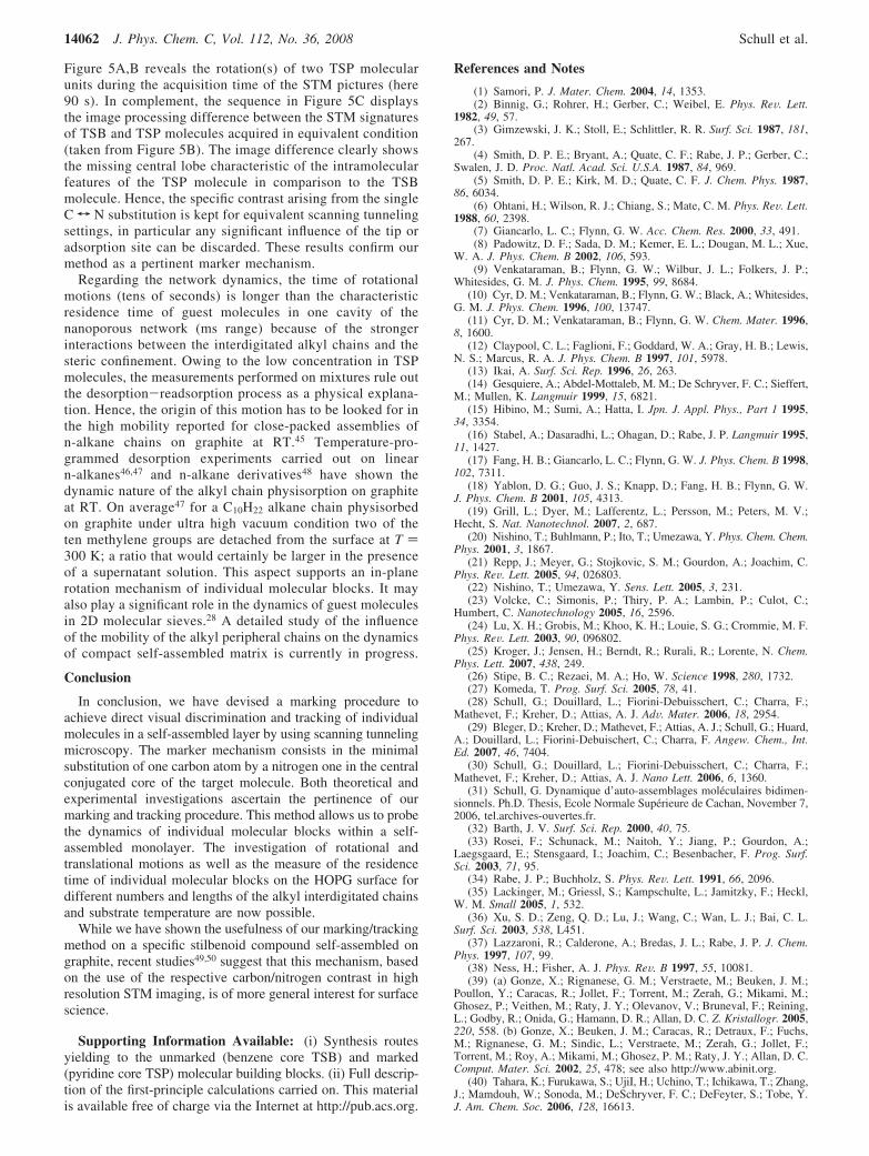
Figure 5A,B reveals the rotation(s) of two TSP molecularunits during the acquisition time of the STM pictures (here90 s). In complement, the sequence in Figure 5C displaysthe image processing difference between the STM signaturesof TSB and TSP molecules acquired in equivalent condition(taken from Figure 5B). The image difference clearly showsthe missing central lobe characteristic of the intramolecularfeatures of the TSP molecule in comparison to the TSBmolecule. Hence, the specific contrast arising from the singleC T N substitution is kept for equivalent scanning tunnelingsettings, in particular any significant influence of the tip oradsorption site can be discarded. These results confirm ourmethod as a pertinent marker mechanism.
Regarding the network dynamics, the time of rotationalmotions (tens of seconds) is longer than the characteristicresidence time of guest molecules in one cavity of thenanoporous network (ms range) because of the strongerinteractions between the interdigitated alkyl chains and thesteric confinement. Owing to the low concentration in TSPmolecules, the measurements performed on mixtures rule outthe desorption-readsorption process as a physical explana-tion. Hence, the origin of this motion has to be looked for inthe high mobility reported for close-packed assemblies ofn-alkane chains on graphite at RT.45 Temperature-pro-grammed desorption experiments carried out on linearn-alkanes46,47 and n-alkane derivatives48 have shown thedynamic nature of the alkyl chain physisorption on graphiteat RT. On average47 for a C10H22 alkane chain physisorbedon graphite under ultra high vacuum condition two of theten methylene groups are detached from the surface at T )300 K; a ratio that would certainly be larger in the presenceof a supernatant solution. This aspect supports an in-planerotation mechanism of individual molecular blocks. It mayalso play a significant role in the dynamics of guest moleculesin 2D molecular sieves.28 A detailed study of the influenceof the mobility of the alkyl peripheral chains on the dynamicsof compact self-assembled matrix is currently in progress.
Conclusion
In conclusion, we have devised a marking procedure toachieve direct visual discrimination and tracking of individualmolecules in a self-assembled layer by using scanning tunnelingmicroscopy. The marker mechanism consists in the minimalsubstitution of one carbon atom by a nitrogen one in the centralconjugated core of the target molecule. Both theoretical andexperimental investigations ascertain the pertinence of ourmarking and tracking procedure. This method allows us to probethe dynamics of individual molecular blocks within a self-assembled monolayer. The investigation of rotational andtranslational motions as well as the measure of the residencetime of individual molecular blocks on the HOPG surface fordifferent numbers and lengths of the alkyl interdigitated chainsand substrate temperature are now possible.
While we have shown the usefulness of our marking/trackingmethod on a specific stilbenoid compound self-assembled ongraphite, recent studies49,50 suggest that this mechanism, basedon the use of the respective carbon/nitrogen contrast in highresolution STM imaging, is of more general interest for surfacescience.
Supporting Information Available: (i) Synthesis routesyielding to the unmarked (benzene core TSB) and marked(pyridine core TSP) molecular building blocks. (ii) Full descrip-tion of the first-principle calculations carried on. This materialis available free of charge via the Internet at http://pub.acs.org.
References and Notes
(1) Samori, P. J. Mater. Chem. 2004, 14, 1353.(2) Binnig, G.; Rohrer, H.; Gerber, C.; Weibel, E. Phys. ReV. Lett.
1982, 49, 57.(3) Gimzewski, J. K.; Stoll, E.; Schlittler, R. R. Surf. Sci. 1987, 181,
267.(4) Smith, D. P. E.; Bryant, A.; Quate, C. F.; Rabe, J. P.; Gerber, C.;
Swalen, J. D. Proc. Natl. Acad. Sci. U.S.A. 1987, 84, 969.(5) Smith, D. P. E.; Kirk, M. D.; Quate, C. F. J. Chem. Phys. 1987,
86, 6034.(6) Ohtani, H.; Wilson, R. J.; Chiang, S.; Mate, C. M. Phys. ReV. Lett.
1988, 60, 2398.(7) Giancarlo, L. C.; Flynn, G. W. Acc. Chem. Res. 2000, 33, 491.(8) Padowitz, D. F.; Sada, D. M.; Kemer, E. L.; Dougan, M. L.; Xue,
W. A. J. Phys. Chem. B 2002, 106, 593.(9) Venkataraman, B.; Flynn, G. W.; Wilbur, J. L.; Folkers, J. P.;
Whitesides, G. M. J. Phys. Chem. 1995, 99, 8684.(10) Cyr, D. M.; Venkataraman, B.; Flynn, G. W.; Black, A.; Whitesides,
G. M. J. Phys. Chem. 1996, 100, 13747.(11) Cyr, D. M.; Venkataraman, B.; Flynn, G. W. Chem. Mater. 1996,
8, 1600.(12) Claypool, C. L.; Faglioni, F.; Goddard, W. A.; Gray, H. B.; Lewis,
N. S.; Marcus, R. A. J. Phys. Chem. B 1997, 101, 5978.(13) Ikai, A. Surf. Sci. Rep. 1996, 26, 263.(14) Gesquiere, A.; Abdel-Mottaleb, M. M.; De Schryver, F. C.; Sieffert,
M.; Mullen, K. Langmuir 1999, 15, 6821.(15) Hibino, M.; Sumi, A.; Hatta, I. Jpn. J. Appl. Phys., Part 1 1995,
34, 3354.(16) Stabel, A.; Dasaradhi, L.; Ohagan, D.; Rabe, J. P. Langmuir 1995,
11, 1427.(17) Fang, H. B.; Giancarlo, L. C.; Flynn, G. W. J. Phys. Chem. B 1998,
102, 7311.(18) Yablon, D. G.; Guo, J. S.; Knapp, D.; Fang, H. B.; Flynn, G. W.
J. Phys. Chem. B 2001, 105, 4313.(19) Grill, L.; Dyer, M.; Lafferentz, L.; Persson, M.; Peters, M. V.;
Hecht, S. Nat. Nanotechnol. 2007, 2, 687.(20) Nishino, T.; Buhlmann, P.; Ito, T.; Umezawa, Y. Phys. Chem. Chem.
Phys. 2001, 3, 1867.(21) Repp, J.; Meyer, G.; Stojkovic, S. M.; Gourdon, A.; Joachim, C.
Phys. ReV. Lett. 2005, 94, 026803.(22) Nishino, T.; Umezawa, Y. Sens. Lett. 2005, 3, 231.(23) Volcke, C.; Simonis, P.; Thiry, P. A.; Lambin, P.; Culot, C.;
Humbert, C. Nanotechnology 2005, 16, 2596.(24) Lu, X. H.; Grobis, M.; Khoo, K. H.; Louie, S. G.; Crommie, M. F.
Phys. ReV. Lett. 2003, 90, 096802.(25) Kroger, J.; Jensen, H.; Berndt, R.; Rurali, R.; Lorente, N. Chem.
Phys. Lett. 2007, 438, 249.(26) Stipe, B. C.; Rezaei, M. A.; Ho, W. Science 1998, 280, 1732.(27) Komeda, T. Prog. Surf. Sci. 2005, 78, 41.(28) Schull, G.; Douillard, L.; Fiorini-Debuisschert, C.; Charra, F.;
Mathevet, F.; Kreher, D.; Attias, A. J. AdV. Mater. 2006, 18, 2954.(29) Bleger, D.; Kreher, D.; Mathevet, F.; Attias, A. J.; Schull, G.; Huard,
A.; Douillard, L.; Fiorini-Debuischert, C.; Charra, F. Angew. Chem., Int.Ed. 2007, 46, 7404.
(30) Schull, G.; Douillard, L.; Fiorini-Debuisschert, C.; Charra, F.;Mathevet, F.; Kreher, D.; Attias, A. J. Nano Lett. 2006, 6, 1360.
(31) Schull, G. Dynamique d’auto-assemblages moleculaires bidimen-sionnels. Ph.D. Thesis, Ecole Normale Superieure de Cachan, November 7,2006, tel.archives-ouvertes.fr.
(32) Barth, J. V. Surf. Sci. Rep. 2000, 40, 75.(33) Rosei, F.; Schunack, M.; Naitoh, Y.; Jiang, P.; Gourdon, A.;
Laegsgaard, E.; Stensgaard, I.; Joachim, C.; Besenbacher, F. Prog. Surf.Sci. 2003, 71, 95.
(34) Rabe, J. P.; Buchholz, S. Phys. ReV. Lett. 1991, 66, 2096.(35) Lackinger, M.; Griessl, S.; Kampschulte, L.; Jamitzky, F.; Heckl,
W. M. Small 2005, 1, 532.(36) Xu, S. D.; Zeng, Q. D.; Lu, J.; Wang, C.; Wan, L. J.; Bai, C. L.
Surf. Sci. 2003, 538, L451.(37) Lazzaroni, R.; Calderone, A.; Bredas, J. L.; Rabe, J. P. J. Chem.
Phys. 1997, 107, 99.(38) Ness, H.; Fisher, A. J. Phys. ReV. B 1997, 55, 10081.(39) (a) Gonze, X.; Rignanese, G. M.; Verstraete, M.; Beuken, J. M.;
Poullon, Y.; Caracas, R.; Jollet, F.; Torrent, M.; Zerah, G.; Mikami, M.;Ghosez, P.; Veithen, M.; Raty, J. Y.; Olevanov, V.; Bruneval, F.; Reining,L.; Godby, R.; Onida, G.; Hamann, D. R.; Allan, D. C. Z. Kristallogr. 2005,220, 558. (b) Gonze, X.; Beuken, J. M.; Caracas, R.; Detraux, F.; Fuchs,M.; Rignanese, G. M.; Sindic, L.; Verstraete, M.; Zerah, G.; Jollet, F.;Torrent, M.; Roy, A.; Mikami, M.; Ghosez, P. M.; Raty, J. Y.; Allan, D. C.Comput. Mater. Sci. 2002, 25, 478; see also http://www.abinit.org.
(40) Tahara, K.; Furukawa, S.; UjiI, H.; Uchino, T.; Ichikawa, T.; Zhang,J.; Mamdouh, W.; Sonoda, M.; DeSchryver, F. C.; DeFeyter, S.; Tobe, Y.J. Am. Chem. Soc. 2006, 128, 16613.
14062 J. Phys. Chem. C, Vol. 112, No. 36, 2008 Schull et al.

(41) Troullier, N.; Martins, J. L. Phys. ReV. B 1991, 43, 1993.(42) Zacharia, R.; Ulbricht, H.; Hertel, T. Phys. ReV. B 2004, 69, 155406.(43) Tersoff, J.; Hamann, D. R. Phys. ReV. Lett. 1983, 50, 1998.(44) Ness, H.; Fisher, A. J. Phys. ReV. B 1997, 56, 12469.(45) Pint, C. L. Surf. Sci. 2006, 600, 921.(46) Paserba, K. R.; Gellman, A. J. J. Chem. Phys. 2001, 115, 6737.(47) Gellman, A. J.; Paserba, K. R. J. Phys. Chem. B 2002, 106,
13231.
(48) Muller, T.; Flynn, G. W.; Mathauser, A. T.; Teplyakov, A. V.Langmuir 2003, 19, 2812.
(49) Abdel-Mottaleb, M. M. S.; Schuurmans, N.; De Feyter, S.; VanEsch, J.; Feringa, B. L.; De Schryver, F. C. Chem. Commun. 2002, 17,1894.
(50) Kampschulte, L.; Griessl, S.; Heckl, W. M.; Lackinger, M. J. Phys.Chem. B 2005, 109, 14074.
JP8030013
Tracking of Individual Molecules by STM J. Phys. Chem. C, Vol. 112, No. 36, 2008 14063