Embed Size (px)
Citation preview
67
Quantum Dots: Inorganic Fluorescent Probes for Single - Molecule Tracking Experiments in Live Cells Maxime Dahan , Paul Alivisatos , and Wolfgang J. Parak
4
4.1 Introduction
Light microscopy is central to the development of modern cell biology. Because it is a sensitive and noninvasive approach, optical imaging provides an invaluable tool to decipher complex cellular processes. Over the past decade, advances in optical instrumentation, chemical probes, cell biology techniques and data process-ing, have enabled ever - more sensitive measurements in live cells; indeed, recent novel approaches have pushed the detection limit down to the ultimate level of single molecules. It has thus become possible to follow, in real time, the motion of individual proteins or nucleic acids in their natural cellular environment. Since they provide direct access to molecular properties in a cellular context, single - molecule experiments promise to elucidate many aspects of cell organization and dynamics. In fact, single - molecule measurements have already been used fruit-fully to address important questions related to molecular diffusion [1 – 4] and transport [5, 6] , membrane compartmentation [7] , gene expression [8, 9] , protein – protein interactions [10] , or cell signaling [11] .
On the basis of its high sensitivity, fl uorescence microscopy is a key technology for optical detection and single - molecule experiments [12] . In general, the molecules of interest must be tagged with a fl uorescent marker in order to be optically detected, which means it is essential that the properties of the tags used in the experiments must also be known. An “ ideal ” fl uorescence marker must fulfi ll several requirements. From an optical point of view, it should be very bright (i.e., have a large absorption cross - section and a high quantum yield), be photostable (i.e., no photobleaching), have a low amount of fl uorescence intermittency (also known as “ blinking ” ), and have a narrow emission spectrum (to provide an easily identifi able optical signature). From a physico - chemical point of view, the marker should neither affect the molecule to which it is attached, nor the molecule ’ s environment. Furthermore, the marker requires suitable biochemical binding sites for its attachment to the molecule that is to be labeled. At present, no such “ ideal ” fl uorescence marker, with optical, chemical, and biochemical properties
Single Particle Tracking and Single Molecule Energy Transfer Edited by Christoph Bräuchle, Don C. Lamb, and Jens MichaelisCopyright © 2010 WILEY-VCH Verlag GmbH & Co. KGaA, WeinheimISBN: 978-3-527-32296-1
68 4 Inorganic Fluorescent Probes for Single-Molecule Tracking Experiments in Live Cells
that satisfy all of the above conditions, is available. However, the brightness and photostability of semiconductor quantum dot s ( QD s) make them very suitable makers for high - sensitivity measurements in general, and for single - molecule tracking experiments in particular.
These advantages will be discussed in this chapter, where several fl uorescent labels, both organic and inorganic, that are used for single - molecule tracking, are fi rst introduced, and their respective merits and limitations briefl y discussed. Attention is then focused on the optical properties of QDs, and details of their chemical synthesis, solubilization and functionalization are presented. Finally, some recent applications of QDs for single - molecule imaging in live cells are reviewed, and the future prospects of these techniques in cell biology discussed.
4.2 Fluorescent Labels for Single - Molecule Tracking in Cells
4.2.1 Organic Fluorophores
Although, traditionally, the fl uorescence markers used for tracing molecules have been organic molecules, today a wide range of dyes is available commercially that spans virtually the whole visible spectrum. Moreover, these dyes can be attached, using well - established conjugation protocols, to a large variety of biomolecules, including proteins, nucleic acids, and sugars [13] . Arguably the biggest advantage of organic fl uorophores is their small size; fl uorescein, for example, has a molecu-lar weight of only 376 Da. Thus, the attachment of such small labels to bigger molecules (e.g., proteins or oligonucleotides) would be expected to have a negligi-ble effect on the biofunctionality of the molecule. Unfortunately, organic fl uoro-phores exhibit limited photostability and, upon repetitive excitation – emission cycles, tend to be rapidly photo - destroyed. This process, which often is referred to as “ photobleaching, ” imposes a severe limitation for tracing applications as it restricts the time interval over which the target molecule can be tracked optically. When investigating a single organic fl uorophore, the observation time is usually limited to a couple of seconds (Table 4.1 ), which often is too short to fully inves-tigate complex cellular processes.
4.2.2 Fluorescent Proteins
Rather than being labeled with organic fl uorophores via bioconjugation chemistry, proteins can be genetically modifi ed to become fl uorescent. For this, nucleotide sequences encoding for fl uorescent protein s ( FP s) can be added to the genome so that, upon protein biosynthesis, a fusion product of the target protein and a fl uo-
4.2 Fluorescent Labels for Single-Molecule Tracking in Cells 69
rescent protein will be expressed by the cell. In many cases, the resultant construct conserves the characteristics of the target protein, while being also fl uorescent due to the genetically added domain. Unquestionably, the advent of genetically encoded fl uorescent markers (for which the Nobel Prize for Chemistry was awarded in 2008) has revolutionized the biochemical methods used to selectively visualize molecules or cellular compartments, and to record their properties in living systems [12, 14] . A multitude of fl uorescent reporters and sensors, such as green fl uorescent protein ( GFP ) and its many variants, can now be prepared and inserted
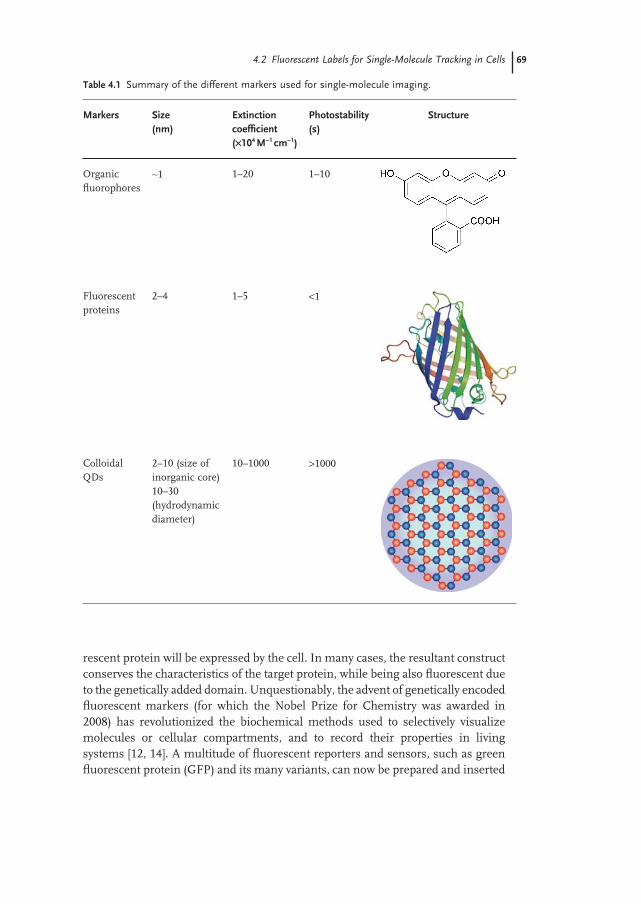
Table 4.1 Summary of the different markers used for single - molecule imaging.
Markers Size (nm)
Extinction coeffi cient ( × 10 4 M − 1 cm − 1 )
Photostability (s)
Structure
Organic fl uorophores
∼ 1 1 – 20 1 – 10
Fluorescent proteins
2 – 4 1 – 5 < 1
Colloidal QDs
2 – 10 (size of inorganic core) 10 – 30 (hydrodynamic diameter)
10 – 1000 > 1000
70 4 Inorganic Fluorescent Probes for Single-Molecule Tracking Experiments in Live Cells
into the genome of various organisms, offering a wide fl exibility to approach complex biological questions. Genetically modifi ed proteins are particularly useful for the investigation of intracellular processes, as the proteins are directly gener-ated through the natural protein expression pathways. In contrast, labeling a target biomolecule with a fl uorophore requires the organic dye to be coupled to a purifi ed version of the biomolecule, outside the cellular environment. As a result, for intracellular applications the labeled proteins must be reintroduced into the cytosol, creating many experimental diffi culties.
Nevertheless, fl uorescent proteins also present their own limitations, notably for single - molecule experiments. First, they are by no means small labels; typically, their molecular weight is close to 30 kDa, which corresponds to a physical dimen-sion of between 2 and 4 nm (Table 4.1 ). More problematic is the fact that, com-pared to organic fl uorophores, fl uorescent proteins tend to absorb light less effi ciently. In fact, their extinction coeffi cients usually vary between 10 4 and 10 5 , a factor of 2 to 10 lower than the best organic dyes. Finally, they are even less pho-tostable than organic fl uorophores and, when imaged at the single - molecule level, usually photobleach in less than 1 s.
4.2.3 Fluorescent Microspheres
Strategies have been developed to circumvent some of the problems (notably the limited photostability) encountered with single organic fl uorophores or fl uores-cent proteins. One approach consists of grouping several fl uorophores within a bead to form a new single particle. For example, polymeric spheres the size of a few tens of nanometers up to a few micrometers, can be impregnated with organic fl uorophores. As each sphere comprises many fl uorophores, its photophysical properties correspond to those of an ensemble of dyes. As a result of this aver-aging, problems associated with individual emitters, such as blinking, can be bypassed. Due to the large number (tens to thousands) of fl uorophores within one particle, lower excitation intensities are required in order to achieve a good detec-tion effi ciency, which in turn means that photobleaching and other intensity - related problems are also reduced. Naturally, fl uorescent spheres are bigger than single organic fl uorophores, and therefore might potentially interfere with the molecular properties of the target molecule. Consequently, the improved photo-physical properties are acquired at the cost of a size increase or reduced colloidal stability. In fact, practical problems encountered with fl uorescent microspheres are such that they are seldom employed for single - molecule experiments in cells.
4.2.4 Colloidal Quantum Dots
The emergence of functional inorganic nanomaterials has recently added new elements to the toolbox with which biological systems can be investigated [15 – 17] . These nanomaterials possess physical properties (optical, electrical, magnetic, etc.)

4.2 Fluorescent Labels for Single-Molecule Tracking in Cells 71
which are often superior to those of their organic counterparts, opening up fasci-nating prospects for advanced sensing and detection schemes in fundamental and applied biomedical research.
Colloidal QDs are probably the most prominent example of nanomaterials used in a biological context [17] . Quantum dots are semiconductor nanocrystals in which the diameter of the optically active inorganic core varies between 2 and 10 nm (Table 4.1 ; Figure 4.1 a), intermediate between organic molecules and
Figure 4.1 (a) Transmission electron microscopy (TEM) image of polymer - coated CdSe/ZnS quantum dots (QDs). Adapted from Ref. [18] . (b) Solutions of QDs of different size (left image). Under illumina-tion with a hand - held UV lamp, the size - dependent fl uorescence of the CdSe QDs can be seen. Adopted from Ref. [19] . (c) Comparison of absorption (solid curve) and fl uorescence (dashed curve) spectra of organic fl uoro phores and colloidal QDs. Upper image: Normalized absorption and fl uorescence spectra in water of three
typical organic fl uorophores (fl uorescein, tetramethy lrhodamine ( TAMRA ), and Cy5). Lower image: Normalized absorption and fl uorescence spectra in water of polymer - coated CdSe/ZnS QDs of different sizes. Adopted from Ref. [16] . (d) Time - dependence of the fl uorescence intensity of silanized QDs and rhodamine 6G. The QDs exhibit a stable emission for at least 4 h, while the organic fl uorophores bleach after 10 min. The data correspond to nanocrystals of four different colors of emission and to rhodamine 6G. Adopted from Ref. [20] .
72 4 Inorganic Fluorescent Probes for Single-Molecule Tracking Experiments in Live Cells
polymeric spheres. Dependent on the material and the size of the particle, QDs emit fl uorescence at different wavelengths in the visible or infrared ( IR ) region (see Section 4.3 ; Figure 4.1 b). Once solubilized and functionalized, QDs can be used as biological fl uorescent probes [21, 22] although, due to their crystalline nature, they have distinct optical properties compared to conventional organic fl uorophores or fl uorescent proteins. In particular, their extinction coeffi cient often exceeds 10 6 in the visible spectrum, which means that they are bright emit-ters that can be detected individually, with a high signal - to - noise ratio ( SNR ). Furthermore, QDs are much more photostable than organic dyes, most likely due to their crystalline structure, which is more robust against photoinduced damage than is that of organic chains. This combination of brightness and photostability makes QDs especially appealing for single - molecule experiments. During the past few years, a rapidly growing number of experiments have shown that the ability to track individual biomolecules over extended periods of time in live cells, opens many prospects for advanced biological assays. However, the development of novel QD - based assays requires not only a good understanding of their optical properties but also a control of their colloidal and biochemical properties. Hence, details of the optical properties, synthesis, solubilization and functionalization of QDs are provided in the following sections.
4.3 Optical Properties of Colloidal Quantum Dots
4.3.1 Absorption and Emission Properties
As the inorganic core of QDs is only a few nanometers in diameter (typically 2 – 10 nm; see Figure 4.1 a), their energy levels are determined by quantum mechanical effects. Charge carriers (electrons and holes) are confi ned to the nanometer - dimension of the colloidal particles, which results in a confi nement energy [23] that adds to the energy levels of the bulk material. Therefore, the energy gap in a semiconductor nanoparticle, which is the energy difference between the valence band (equivalent to the highest occupied level) and the conduction band (equivalent to the lowest unoccupied level), is larger than that for bulk material, and can be fi nely tuned with the size of the QD (see Figure 4.1 b) [23] . Importantly, it is not only the average size of QDs but also their size distribution that can be controlled at the nanoscale, with a relative dispersion in radius equal or inferior to 5%.
The relaxation of light - generated electron hole pairs may take place not only through fl uorescence emission but also through nonradiative pathways. For example, if one of the charge carriers reacts with trap states at the QD surface, then no light will be emitted. In order to raise fl uorescence quantum yields as high as possible, the electron - hole pairs must be kept away from the QD surface, and a general strategy in this direction is to overcoat the QDs with a thin layer of
4.3 Optical Properties of Colloidal Quantum Dots 73
a semiconductor material with a higher band gap. For this, one of the most common systems is CdSe/ZnS, in which CdSe cores are overcoated with a shell of ZnS [24] . As the band gap of ZnS is higher than that of CdSe, light - generated electrons and holes do not possess suffi cient energy to completely enter from the CdSe core into the ZnS shell, and are thus confi ned to the CdSe core. As ZnS can be grown quasiepitaxially on CdSe, there are only few surface states at the CdSe/ZnS interface, and thus the fl uorescence yield is enhanced. This effect is important both for the stability and intensity of the fl uorescence signal. For the best samples, the fl uorescence quantum yield can exceed 50%, comparable to good organic fl uorophores.
4.3.2 QD s as Fluorescent Biological Probes
The interest in QDs for biological imaging stems from a combination of unique photophysical properties [17, 21, 22] . For example, QDs possess a narrow emission spectrum ( < 30 nm, without red tail; see Figure 4.1 c), with a peak position deter-mined by the energy gap. As indicated above, the gap depends on the size of the semiconductor core, and can be precisely adjusted during their synthesis by inor-ganic chemistry. Quantum dots also have a large absorption spectrum, due to the fact that virtually any photons with energy higher than the gap can be absorbed by the nanoparticles (see Figure 4.1 c). As a consequence, QD samples with a distinguishable emission wavelength can be excited with a single laser line [25] , provided that the laser photons have a wavelength lower than the shortest of all emission wavelengths. Because their emission spectra is narrow, there is little crosstalk between emission channels. As QDs are also much less prone to pho-todestruction, they will fl uoresce over much longer durations than organic emit-ters (Figure 4.1 d). For these reasons, QDs appear ideal for multicolor detection, which was the primary motivation when they were introduced into biology [21, 22] . The spectral properties of QDs have also been shown as advantageous for energy - transfer experiments when used as donors [26, 27] .
Of note, QDs have other – sometimes overlooked – interesting optical properties. In particular, their radiative lifetime is ∼ 10 – 20 ns, which is longer than the radia-tive lifetime of the autofl uorescence signal ( ∼ 1 – 5 ns) that limits the detection sensitivity in cells or tissues. As a result, effi cient time - gating can performed, leading to enhanced SNRs in biological imaging [28] . As they are composed of semiconductor materials, QDs are electron - dense and can be visualized using transmission electron microscopy ( TEM ) in biological environments [3, 29] (Figure 4.1 a). This offers many interesting possibilities for so - called “ correlative micros-copy, ” where both optical and electronic imaging are employed to access the structure and dynamics of biological specimens. Finally, the synthesis and engi-neering of QDs with different semiconductor materials and structures have expanded the range of possible emission wavelengths, from the visible to the red and IR regions, which are more appropriate for imaging in tissues and animals [30] .
74 4 Inorganic Fluorescent Probes for Single-Molecule Tracking Experiments in Live Cells
4.3.3 Single Quantum Dot Detection
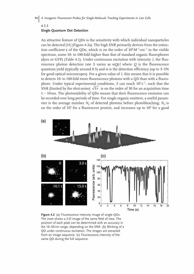
An attractive feature of QDs is the sensitivity with which individual nanoparticles can be detected [31] (Figure 4.2 a). The high SNR primarily derives from the extinc-tion coeffi cient ε of the QDs, which is on the order of 10 6 M − 1 cm − 1 in the visible spectrum, some 10 - to 100 - fold higher than that of standard organic fl uorophores (dyes or GFP) (Table 4.1 ). Under continuous excitation with intensity I , the fl uo-rescence photon detection rate S varies as α Q ε I where Q is the fl uorescence quantum yield (typically around 0.5) and α is the detection effi ciency (up to 3 – 5% for good optical microscopes). For a given value of I , this means that it is possible to detects 10 - to 100 - fold more fl uorescence photons with a QD than with a fl uoro-phore. Under typical experimental conditions, S can reach 10 5 s − 1 , such that the SNR (limited by the shot - noise) S is on the order of 30 for an acquisition time τ ∼ 10 ms. The photostability of QDs means that their fl uorescence emission can be recorded over long periods of time. For single organic emitters, a useful param-eter is the average number N p of detected photons before photobleaching; N p is on the order of 10 5 for a fl uorescent protein, and increases up to 10 6 for a good
Figure 4.2 (a) Fluorescence intensity image of single QDs. The inset shows a 2 - D image of the same fi eld of view. The position of each peak can be determined with an accuracy in the 10 – 50 nm range, depending on the SNR. (b) Blinking of a QD under continuous excitation. The images are extracted from an image sequence. (c) Fluorescence intensity of the same QD during the full sequence.
4.4 Synthesis of Colloidal Fluorescent Quantum Dots 75
organic fl uorophore. In comparison, 10 8 – 10 9 photons can be routinely detected for a single QD. This number does not necessarily refl ect an upper bound imposed by photodestruction but, rather, results from other physical or biological factors limiting the total duration of the experiment.
4.3.4 Fluorescence Intermittency of Individual Quantum Dots
During the past decade, single - molecule optical experiments have amply shown that, when observed individually, fl uorescence emitters exhibit properties that remain hidden at the ensemble level. With regards to the detection of individual QDs, the most striking – and possibly unwelcome – effect is arguably that of blink-ing when, under continuous illumination, the emission randomly alternates between bright ( “ on ” ) and dark ( “ off ” ) periods (Figure 4.2 ) [31] . Of note, similar intermittency effects have been observed with virtually all fl uorescent emitters (dyes, fl uorescent proteins), but such effects are probably more spectacular with QDs due to the long observation times made possible by the photostability of the nanoparticles. Although it is not completely understood, the physical origin of the blinking phenomenon in QDs is commonly attributed to a transient ionization of the nanoparticle [32] . Upon light excitation, an electron - hole pair is created within the QD that can relax through different competing decay channels. Most frequently, the pair recombines radiatively by emitting a fl uorescence photon. However, with a low probability, the electron (or the hole) can tunnel out of the QD toward surface traps. While the QD is in this ionized state, the nonradiative recombination rate is enhanced and the fl uorescence emission dramatically quenched. At a later time, the trapped charge tunnels back into the inorganic core, and the nanoparticle resumes its regular fl uorescence cycles. Both the “ on ” and “ off ” times are distributed according to a power - law [32, 33] , which means that the mechanisms behind the blinking cannot be described simply in terms of Poisson processes. Blinking is certainly a limitation for the use of QDs as a nanoscopic light source, and much effort has been expended in attempts to reduce it. Recently, progress towards nonblinking nanoparticles has been reported, although not yet for water - soluble nanoparticles, and at the cost of an increase in the particle size [34, 35] . In practice, fl uorescence blinking is, nevertheless, a convenient criterion to evaluate whether individual nanoparticles are observed, since it is normally assumed that an intermittent fl uorescent spot is indicative of a single QD emission.
4.4 Synthesis of Colloidal Fluorescent Quantum Dots
Colloidal QDs are fl uorescent crystalline nanoparticles made from semiconductor materials, which are dispersed in a solvent [16, 17, 36] . Although alternative methods exist, most common colloidal QDs are grown using a wet - chemistry
76 4 Inorganic Fluorescent Probes for Single-Molecule Tracking Experiments in Live Cells
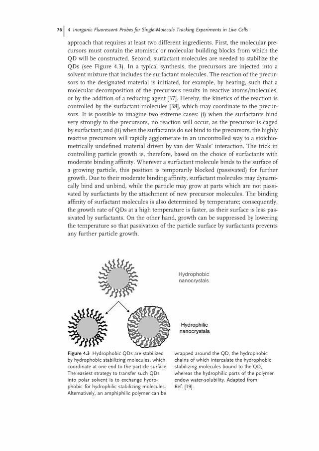
approach that requires at least two different ingredients. First, the molecular pre-cursors must contain the atomistic or molecular building blocks from which the QD will be constructed. Second, surfactant molecules are needed to stabilize the QDs (see Figure 4.3 ). In a typical synthesis, the precursors are injected into a solvent mixture that includes the surfactant molecules. The reaction of the precur-sors to the designated material is initiated, for example, by heating, such that a molecular decomposition of the precursors results in reactive atoms/molecules, or by the addition of a reducing agent [37] . Hereby, the kinetics of the reaction is controlled by the surfactant molecules [38] , which may coordinate to the precur-sors. It is possible to imagine two extreme cases: (i) when the surfactants bind very strongly to the precursors, no reaction will occur, as the precursor is caged by surfactant; and (ii) when the surfactants do not bind to the precursors, the highly reactive precursors will rapidly agglomerate in an uncontrolled way to a stoichio-metrically undefi ned material driven by van der Waals ’ interaction. The trick in controlling particle growth is, therefore, based on the choice of surfactants with moderate binding affi nity. Wherever a surfactant molecule binds to the surface of a growing particle, this position is temporarily blocked (passivated) for further growth. Due to their moderate binding affi nity, surfactant molecules may dynami-cally bind and unbind, while the particle may grow at parts which are not passi-vated by surfactants by the attachment of new precursor molecules. The binding affi nity of surfactant molecules is also determined by temperature; consequently, the growth rate of QDs at a high temperature is faster, as their surface is less pas-sivated by surfactants. On the other hand, growth can be suppressed by lowering the temperature so that passivation of the particle surface by surfactants prevents any further particle growth.
Figure 4.3 Hydrophobic QDs are stabilized by hydrophobic stabilizing molecules, which coordinate at one end to the particle surface. The easiest strategy to transfer such QDs into polar solvent is to exchange hydro-phobic for hydrophilic stabilizing molecules. Alternatively, an amphiphilic polymer can be
wrapped around the QD, the hydrophobic chains of which intercalate the hydrophobic stabilizing molecules bound to the QD, whereas the hydrophilic parts of the polymer endow water - solubility. Adapted from Ref. [19] .
4.5 Surface Chemistry for the Water-Solubilization of Quantum Dots 77
By applying these general principles, QDs can be grown with controlled size from a variety of semiconducting materials, such as CdSe, CdTe, CdS, InP, and InAs. The role of the surfactant allows even for a shape - controlled growth. As each QD is a crystal, and thus possesses different facets, a surfactant can be selected which binds more weakly to one facet than to another. Then, as this particular facet will be less passivated by surfactant molecules, the particle will grow prefer-entially in this direction. This capability would enable, for example, the growth of rod - shaped rather than spherical particles [39] . Different facets of particles allow even for the growth of defi ned hybrid structures, in which one particle comprises two oriented domains made from different materials. Here, the particle composed of the fi rst material will act as a seed for nucleation of the precursors used for growth of the second material. As surfactant molecules may stick with less affi nity to the tips of rod - shaped semiconductor particles than to the surface parallel to the axis, the second material will grow preferentially on the tips rather than along the walls of the semiconductor rods [40] . It could be said that, today, colloidal chemistry allows for the growth of particles with a defi ned size, shape, and com-position for a large variety of materials [39, 41 – 45] .
Traditionally, colloidal semiconductor nanoparticles are grown from materials in Groups II - VI of the Periodic Table, such as CdSe, CdS, and CdTe, due mainly to the fact that these particles are the most easy to synthesize. However, because of their more ionic structure, they grow more slowly during their synthesis than do nanoparticles from Groups IV - IV or III - V, which have a more covalent struc-ture. At present, the most widespread QDs are the CdSe/ZnS nanoparticles, where the ZnS shell around the CdSe core provides improved optical properties, notably a quantum yield exceeding 50% [24] . Also important for biological applications is the fact the fl uorescence of CdSe/ZnS QDs is in the visible spectrum. As their emission wavelengths vary between 500 and 700 nm, these QDs are perfectly compatible with the optical microscopy techniques used in biological laboratories. Unfortunately, working with Cd - based materials raises several problems due to the metal ’ s toxicity [46, 47] ; in particular, cadmium cannot be used in the applica-tion of nanoparticles to diagnostics or therapeutics in humans. Hence, the future trend in QD synthesis will unquestionably move towards more biocompatible QDs, composed of different materials.
4.5 Surface Chemistry for the Water - Solubilization of Quantum Dots
Whilst the fl uorescence of a QD is determined primarily by the material and the size of the semiconductor nanoparticle, its interaction with the environment is mediated by its surface. First, the surface chemistry of the QDs will determine the solubility, or more precisely the colloidal stability, of the particles. Second, the surface chemistry is crucial when linking molecules to the particle, and thus for the design of specifi c interfaces with the biological world, to which the particles are to be connected.
78 4 Inorganic Fluorescent Probes for Single-Molecule Tracking Experiments in Live Cells
4.5.1 General Strategies for Water - Solubilization
With regards to the solubility of QDs, two different lines must be considered. As noted above, the particles are synthesized in a surfactant solution that may be based on either aqueous or organic solvents. In the former case, the particles are capped with hydrophilic surfactant molecules, and thus are automatically soluble in aqueous solution. In the latter case, the particles are capped with hydrophobic surfactant molecules, and thus are soluble only in organic solvents and not in aqueous media (Figure 4.3 ). Before being used for biological tracking applications, the particles must fi rst be rendered hydrophilic, and thus water - soluble. Although, initially, QD syntheses originated via water - based reactions, and these particles are directly soluble in water, there remain certain disadvantages associated with QDs. In contrast to synthesis in organic solvents, a water - based synthesis is limited to a temperature of up to only 100 ° C, and to the use of water - soluble precursors and surfactant molecules. The use of a high - temperature synthesis, coupled with a wide variety of metallo - organic precursors and a huge choice of surfactants, permits QDs to be grown which (in general) have an improved crystallinity, a better size distribution, and more complex architectures. Whilst this statement does not apply to metal particles (e.g., gold) and metal - oxides (e.g., iron oxide), it generally holds true for many semiconductor nanoparticles. Nowadays, virtually all colloidal QDs produced by the major manufacturers are based on CdSe/ZnS grown in organic solvents.
Obviously, these particles must be rendered hydrophilic before they can be used in biological applications, and for this two general strategies are available. First, the original hydrophobic surfactant molecules can be replaced in a ligand - exchange procedure with hydrophilic surfactant molecules (Figure 4.3 ). Second, the origi-nally hydrophobic nanoparticles can be embedded in an amphiphilic shell in the hydrophobic cavity of which is embedded the QD, the hydrophilic outside of which warrants water - solubility. Generally speaking, a ligand - exchange will lead to smaller nanoparticles, whereas an amphiphilic coating will provide more colloi-dally stable particles. Today, both methods are used in parallel. It should also be pointed out that, although the concepts described here appear to be straightfor-ward, they must be regarded only as model systems. Most often, the exact geom-etry and molecular order on the surface of the QDs is not known exactly, since its experimental access may be very complicated. Even the hydrodynamic diameters of QDs, as determined by different methods and different groups, will vary to a signifi cant degree [48] .
As noted above, the presence of surfactant molecules warrants colloidal stability, and in aqueous solution this can be achieved in either of two ways. Quantum dots with similarly - charged surfaces will repel each other electrostatically; in fact, most QDs are stabilized by negatively charged surfactant molecules bound to their surface. Although charge stabilization is very effi cient, it depends critically on the nature of the solvent. Indeed, the charge of the molecules can change with the pH. For example, COO − groups will lose their charge at a low pH - value by protona-
4.5 Surface Chemistry for the Water-Solubilization of Quantum Dots 79
tion to COOH; therefore, negatively charged QDs will be stable at a high pH, but unstable at a low pH. Another problem emerges due to the presence of salt; ions with a charge opposite to that present on the particle surface will be electrostati-cally attracted and form a cloud of counterions around the QDs. These counterions will then screen the charge of the particles and thus reduce the electrostatic repul-sion between the QDs. As a consequence, the charged particles typically become colloidally unstable in electrolytic solutions with a high salt content. Hydrophilic polymers may also be used for particle stabilization in aqueous solution, as their presence on the particle surface will cause steric repulsion between particles because the surface molecules on adjacent particles cannot penetrate each other. Steric repulsion of the fi rst order depends neither on the pH nor the salt content of the solution, and thus is almost universal. Examples of materials which cause particle - stabilization by steric repulsion include poly(ethylene glycol) ( PEG ) [49 – 51] and sugars [52] . Stabilization by charge and stabilization by steric hindrance can also be achieved with ligand exchange, by embedding particles into an addi-tional shell.
4.5.2 Solubilization with Ligand Exchange
Ligand exchange represents a very straightforward approach to solubilization. In this process, QDs capped with the original surfactant are incubated in a solution containing an excess of the new surfactant molecule. As noted above, most sur-factants bind with only fi nite strength to the QD surface, and heating will acceler-ate the continuous process of binding and unbinding. However, by dispersing QDs (which have been capped by surfactant molecules of type A) in a solution that contains a huge excess of surfactant molecules of type B, this will (statistically) eventually lead to QDs becoming capped by surfactant molecules of type B, even in the situation where the type A surfactant molecules would bind more strongly to the QD surface than would type B. In a fi nal step, the surfactant molecules remaining in solution must be removed.
Ligand exchange can be used to exchange hydrophobic surfactant molecules for hydrophilic molecules, thus transferring QDs from an organic solution to the aqueous phase. In aqueous phase, ligand exchange can also be used to exchange one type of hydrophilic surfactant for another hydrophilic type; this may also be applied to QDs that originally were synthesized in aqueous solution.
Although ligand exchange is a straightforward process, it has several disadvan-tages. First, all ligand exchange procedures carry an inherent risk of a reduced colloidal stability, especially if a phase transfer from nonpolar to polar solvents is involved. During ligand exchange, at a point when part of the original molecules have been exchanged and some original molecules are still attached to the particle surface, the QDs will possess a mixed surface chemistry, at which point they are most vulnerable to agglomeration. Occasionally, it is impossible to perform a complete ligand exchange [53] . The exposure of QDs to several subsequent ligand exchange procedures inevitably leads to a growing degree of particle aggregation.
80 4 Inorganic Fluorescent Probes for Single-Molecule Tracking Experiments in Live Cells
As noted above, the ability to perform ligand exchange is based on the fact that the surfactant molecules continuously bind to, and unbind from, the QD surface; such a situation endangers the colloidal stability of particles when a solution is bare of excess surfactants. Hence, when an increasing number of surfactant mol-ecules leave the QD surface with time, and there is not any excess of surfactant molecules available to rebind, then the particles eventually will agglomerate as they lose their colloidal stability. The problem here is that, especially for the surface of the most commonly used CdSe/ZnS QDs, there is no reactive group available that can bind to the surface with high affi nity. Most often, surfactant molecules which contain thiol groups as anchors for the attachment to the QD surface are used in this role. However, as the bond of thiols to ZnS is not very strong (notably with regards to photo - oxidation [54] ), the ligands will typically leave the particle surface within a few days, leading to the formation of particle agglomerates. In order to increase the affi nity of the surfactant molecules to the QD surface, several groups have used multidentate ligands [55 – 57] or ligand molecules which can be crosslinked [20, 58, 59] . Crosslinking, in particular, can lead to colloidally very stable QDs, although such stability tends to occur via a multilayer of surfactant molecules than by a monolayer. On the positive side, monolayer - protected QDs – by defi nition – lead to the smallest possible hydrodynamic particle diameters. The choice of different classes of ligand on the particle surface, such as carboxylic acids [22] , sugars [52] , peptides [60] , PEG [49] , and polymers [61] , allows for an adjust-ment to be made of the particle surface within the biological environment where the QDs are to be used.
4.5.3 Surface Coating with Amphiphilic Molecules
The overcoating of hydrophobically capped QDs with amphiphilic molecules differs fundamentally from ligand exchange. Here, the original hydrophobic sur-factant resulting from the particle synthesis is not removed; rather, an additional layer of amphiphilic molecules is added around the QDs. Whilst the hydrophobic part of the amphiphilic molecules points towards the hydrophobic QD surface, the hydrophilic part points towards the solution and renders the coated particle water - soluble. Whereas, ligand exchange is based on the chemical binding of reactive groups to the particle surface, overcoating with amphiphilic molecules is mediated only by hydrophobic interactions. Moreover, this fundamental interaction is very general and does not depend on the exact chemical nature of the QD surface, nor of the amphiphilic molecule. The amphiphilic shell around the QD can be seen as micelle with a hydrophobic cavity and a hydrophilic surface. Many reports exist of mainly polymeric [18, 25, 62 – 64] and lipid micelles [65 – 67] . In this way, virtually any hydrophobically capped QD can be embedded within a variety of amphiphilic molecules, although chemical rather than geometric arguments come into play here. In the case of too - large micelles, several QDs would become embedded in one micelle, such that the single QD character would be lost. On the other hand, the amphiphilic polymer molecules should not be so small that a signifi cant frac-tion of the entire QD surface would become coated.
4.6 Interfacing Quantum Dots with Biology 81
Amphiphilic micelles represent an almost universal means of solubilizing the originally hydrophobically capped QDs into aqueous solution. Naturally, the micelle adds to the total particle diameter (which can reach 25 – 30 nm in the case of CdSe/ZnS QDs, compared to 2 – 10 nm for the inorganic particle core); however, this loss in performance is often compensated by the excellent colloidal properties provided by this technique. As the stability of the outer shell is not based on the bonds between the individual surfactant molecules and the QD surface, but rather on a collective attachment via hydrophobic interactions, the QDs are colloidally very stable. This technique also allows for different types of QDs to be embedded into shells with the same surface chemistry, a fi nding which may prove to be a major advantage when seeking a unifi ed strategy for bioconjugation [18] .
4.6 Interfacing Quantum Dots with Biology
4.6.1 Conjugation of QD s to Biomolecules
Whilst today, techniques allow for the synthesis of highly colloidally stable QDs, the bioconjugation of these particles remains very much an open subject that is under continuous development. As a huge variety of different functionalization strategies have been reported, this section will provide an overview of the different concepts available rather than detail all of the approaches. At present, two funda-mentally different strategies may be applied for bioconjugation: (i) biological mol-ecules can be attached directly to the QD surface; or (ii) they can be bound to the surfactant shell and its subsequent layers.
The direct attachment of biological molecules to the QD surface is based on ligand exchange, in which a portion of the surfactant molecules are replaced with biological molecules. If the original surfactant molecules bind only very weakly to the QD surface, they can be partly replaced with biological molecules which adsorb nonspecifi cally to the QD surface; however, such constructs are very labile and will be of very limited practical use. This method is more suited to biological molecules bearing a group that binds to the particle surface. In the case of CdSe/ZnS QDs, this is possible for molecules with thiol groups. For example, the amino acid cysteine has one thiol group; hence, peptides with cysteine in their sequence can be bound to the CdSe/ZnS surface simply by replacing the original ligands. Another example of this approach is the attachment of oligonucleotides that have been modifi ed at one end with a thiol group. Unfortunately, as all of the problems relating to limited colloidal stability with regards to ligand exchange also hold true in this case, the direct attachment of biological molecules to QD surfaces would not be the method of choice for preparing stable conjugates.
On most occasions, the biological molecules are attached to the shell of the surfactant molecules and/or to the additional layers around it. The stability of this conjugation is limited, therefore, by the stability of the surfactant layer around the QDs. In the most primitive scenario, biological molecules are attached by
82 4 Inorganic Fluorescent Probes for Single-Molecule Tracking Experiments in Live Cells
nonspecifi c adsorption; however, a greater stability can be achieved by electrostati-cally mediated adsorption – that is, by the attraction of oppositely charged mole-cules to the charged particle surface. One well - established protocol of this type is based on fusing a positively charged zipper domain to proteins, which in turn become stably bound when the zipper domain binds to the surface of negatively charged QDs [57, 68] . The most prominent approach, however is the use of bio-conjugate chemistry, in which functional groups on the QD surface (in particular COOH and NH 2 ) are crosslinked with the functional groups of the biological molecule (notably NH 2 , SH, and COOH). Many crosslinker molecules are available for this purpose, and many different protocols have been developed [69] . Although covalent attachment is, arguably, the most stable method of bioconjugation, it should be noted that QD bioconjugation is by far not yet fully established. This relates to the fact that most bioconjugation protocols have been classically devel-oped for the modifi cation of proteins. Due to their different natures, QDs in general do not behave as proteins, which means that established protocols must often be modifi ed. This is especially true for the limited range of QD concentration and for salt stability, as QDs cannot be reasonably concentrated above several tens of micromolar concentration. The high salt concentrations used to link similarly charged proteins so as to reduce their electrostatic repulsion would also screen the charge of the QDs, causing them to agglomerate. Whilst no salt would warrant for QD stability, proteins with the same sign of charge as the QDs would be repelled, but a high salt content would cause QD agglomeration. In practice, whilst identify-ing the “ window of opportunity ” is often complicated, the covalent attachment of many different types of biological molecule to QDs has been reported. Of particu-lar interest here is the attachment of proteins, whether of antibodies for molecular recognition or enzymes for functional assays.
To some extent, the conjugation process can be controlled to a point where conjugates with a defi ned stoichiometry may be prepared [51, 70] . By using elec-trophoresis in agarose gels, constructs with a QD attached to an exactly defi ned number of molecules (zero, one, two, or more) can be separated and identifi ed [51, 71] , provided that the molecules have a suffi cient size and/or charge. This aspect of QD functionalization, although requiring further development, has been shown to be essential for single - molecule experiments which, ideally, require QD – biomolecule constructs in a 1 : 1 ratio.
Unfortunately, bioconjugation is only as good as the subsequent purifi cation procedure. In order to obtain pure conjugates, any unbound biological molecules remaining in solution must be removed as they would severely interfere with the labeling process. Whilst the free molecules may bind to exactly the same receptors as those molecules conjugated to the QD label, they cannot be visualized because they are unlabelled. Moreover, in the presence of a large excess of unbound mol-ecules, the extent of labeling would be greatly suppressed due to competition between the unbound and conjugated molecules for the designated binding sites. Purifi cation is also crucial for the removal of excess surfactant molecules, which might interfere with cells. The purifi cation of QDs is not straightforward, but due to their size (which is comparable for example with proteins), any purifi cation
4.7 Single Quantum Dot Tracking Experiments in Live Cells 83
methods based on sorting by size (e.g., size - exclusion chromatography) will have only a limited application. Worse still, the analysis of bioconjugation and purifi ca-tion is also complicated, and consequently a number of reports on applied conju-gation and purifi cation strategies have been based on the trust of the authors rather than on any experimental characterization of the fi nal product. To date, no stand-ard protocols have been developed for evaluating the bioconjugation of QDs, and on most occasions sketches regarding the geometry of particle – biomolecule con-jugates are accepted as idealized model. These are in agreement with experimental data and appear plausible, although reality they might involve more complex structures. As noted above, even the absolute hydrodynamic diameters can be determined only with an accuracy of a few nanometers [48] . Clearly, much improve-ment is required in this area before any industrial applications of QDs in biocon-jugation processes can be considered.
4.6.2 Cytotoxicity of Semiconductor QD s
One problem associated with CdSe/ZnS QDs is their potential cytotoxicity since, when the QDs corrode, they release toxic cadmium ions [46, 47] . This may cause major diffi culties in tracking, tracing, and homing applications with whole cells, which must be labeled with as many QDs as possible in order to remain suffi -ciently bright over a period of days, also taking into account any dilution of the QD label as the cells divide [72] . Nonetheless, the toxicity of CdSe/ZnS - based QDs can essentially be neglected for single - molecule tracing applications (as described in this chapter), for two reasons. First, the nature of optical single - molecule obser-vations decrees that the QD labels must be very dilute, such that the effective QD concentrations are low. Second, single - molecule tracing is, at best, extended over several hours, which means that the incubation times are relatively short. Never-theless, future improvements in particle synthesis soon will result in the develop-ment of Cd - free QDs [73, 74] .
4.7 Single Quantum Dot Tracking Experiments in Live Cells
4.7.1 Why Experiments at the Single Molecule Level?
During the past 15 years, a variety of experiments has shown that the ability to image a single emitter represents a powerful approach to probe complex environ-ments at the nanoscale [75, 76] . Although seen initially as a topic of interest in its own right, the detection of individual molecules has evolved to become a major tool for monitoring a multitude of physical or chemical processes. In fact, the fi eld has grown so much that it has become almost impossible to review all of the research directions and possible applications in physics, chemistry, or biology.
84 4 Inorganic Fluorescent Probes for Single-Molecule Tracking Experiments in Live Cells
Here, the aim is not to provide an exhaustive view but rather to highlight the concepts that are important for single - molecule experiments in live cells.
From a general point of view, the fundamental advantage of measurements at the single - molecule level is that they provide access to the full distribution of parameter values, rather than to the mere average values as in conventional ensemble measurements [75 – 77] . In other words, fl uctuations around the mean value can be determined. This is important in two distinct cases. First, when a sample is inhomogeneous, molecular subpopulations can be identifi ed by looking at molecules one at a time; these subpopulations can be considered as “ static ” fl uctuations. Second, single - molecule experiments are also useful to look at “ dynamic fl uctuations ” ; indeed, for a system driven by biochemical reactions, the temporal evolution is not deterministic. Rather, molecules undergo stochastic processes. Unless molecules can be all synchronized at a given time, it is not pos-sible to determine the kinetic parameters that control their dynamics. In most cases, only equilibrium constants are measured when averaging over an ensemble of molecules. In contrast, single - molecule experiments allow for a full description of the stochastic temporal evolution of a molecule, giving access to the transition rates underlying the biochemical reactions.
The two main motivations for single - molecule experiments – namely probing static and dynamic fl uctuations – are of particular interest in live cells. As cells are physical and biochemical systems with a high degree of spatial heterogeneity, it is essential to probe the behavior of molecules as a function of their local environ-ment. Moreover, biochemical reactions can rarely be synchronized in live cells, which means that single - molecule measurements might provide a unique approach to determine in situ the kinetics of these reactions. Importantly, in many situations the cell activity is mediated by a low number of proteins, and should preferably be investigated at the single - molecule level. Indeed, studies that involve the over-expression of a fl uorescently labeled mutant have a risk to largely perturb the biochemical equilibria within the cell, and to signifi cantly shift the kinetics and amplitude of the cellular response.
An important technical aspect of single molecule experiments is the accuracy with which the position of individual emitters can be determined. As the emitter size is much smaller than λ (the wavelength of light; 400 – 800 nm in the visible spectrum), its fl uorescence spot corresponds to the point spread function ( PSF ) of the microscope (Figure 4.3 ). Although this spot has itself a width on the order of λ , its center can be localized with much higher precision. In fact, in the common case of a shot - noise - limited signal, the center position can be determined with an accuracy σ on the order of N (where N is the number of photons detected during the acquisition time) [78, 79] . For a reasonable value of N ∼ 10 4 , σ is as low as 5 – 10 nm, which means that a single molecule can inform on a complex environ-ment (such as a cell membrane) truly at the nanoscale.
In single - molecule tracking measurements, sequences of consecutive images are acquired and individual trajectories extracted from these sequences. When QDs are used as fl uorescent markers, one diffi culty derives from the fl uorescence
4.7 Single Quantum Dot Tracking Experiments in Live Cells 85
intermittency of the nanoparticles. As the markers can transiently disappear from the fl uorescence images, specifi c algorithms are required to track individual QD - tagged molecules [80 – 82] . In brief, the algorithm can be separated into two parts. First, all of the visible spots within a sequence of images are detected using a conventional cross - correlation with a Gaussian model for the PSF of the microscope. Second, these spots are associated to reconstruct spatial trajectories of the individual proteins. For that purpose, a model of the spatial dynamics (diffusive motion, directed transport, etc.) is used to provide an estimate of the possible location of the molecules, either in consecutive images or after an “ off ” period.
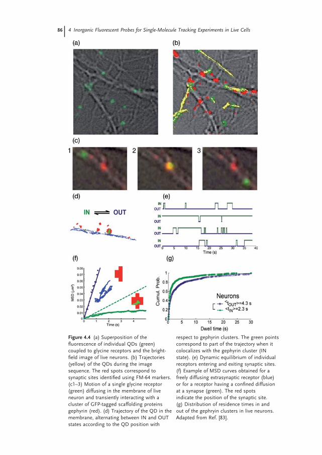
In general, the output of single QD tracking experiments is a set of trajectories, with a time resolution in the range of 1 to 100 ms, and a spatial resolution between 5 and 50 nm (Figure 4.4 a,b). Several methods have been devised to extract statisti-cally meaningful properties from these molecular trajectories. The most common method is based on the computation of the mean square displacement ( MSD ), which provides an estimate of the parameters controlling the diffusive or directed transport of the molecules (Figure 4.4 f ) [84, 85] . More advanced techniques have been later introduced to analyze situations in which a molecule does not exhibit a unique mode of motion throughout its trajectory but, rather, dynamically alter-nates between multiple types of movement [86 – 89] . This is, for instance, the case when a membrane molecule, which initially is diffusing, becomes temporarily confi ned in a microdomain, or when a protein is transported due to transient attachment to a molecular motor.
4.7.2 Tracking Single Membrane Receptor Molecules
The use of QDs for single - molecule tracking experiments in cells was introduced for membrane receptors. The fi rst biological question to be investigated using single QD imaging was the diffusion dynamics of glycine receptors in live cultured neurons (Figure 4.4 ) [3] . Since then, a similar experimental approach has been employed to study a large number of membrane molecules, including (without being exhaustive) other receptors for neurotransmitters [ γ - aminobutyric acid ( GABA ), α - amino - 3 - hydroxy - 5 - methyl - 4 - isoxazolepropionic acid ( AMPA ), N - methyl - D - aspartate ( NMDA )] [90 – 92] , potassium channels [93] , cystic fi brosis transmem-brane conductance regulators [94] , aquaporin - 4 water channels [95] , epidermal growth factor ( EGF ) receptors [81, 96] , lipids, glycosylphosphatidylinositol ( GPI ) - anchored proteins [97] , and immunoglobulin E ( IgE ) receptors [98] .
There are many reasons, both technical and conceptual, why membrane mole-cules have been the subject of such attention. From a technical point of view, it is relatively simple to label membrane molecules by the use of antibodies against an extracellular epitope (Figure 4.4 a – c). In many cases, there exist antibodies with high affi nity ( < 1 nM) that allow binding over many hours. Once combined with a secondary antibody coupled to a QD, this type of affi nity labeling permits a rather
86 4 Inorganic Fluorescent Probes for Single-Molecule Tracking Experiments in Live Cells
Figure 4.4 (a) Superposition of the fl uorescence of individual QDs (green) coupled to glycine receptors and the bright - fi eld image of live neurons. (b) Trajectories (yellow) of the QDs during the image sequence. The red spots correspond to synaptic sites identifi ed using FM - 64 markers. (c1 – 3) Motion of a single glycine receptor (green) diffusing in the membrane of live neuron and transiently interacting with a cluster of GFP - tagged scaffolding proteins gephyrin (red). (d) Trajectory of the QD in the membrane, alternating between IN and OUT states according to the QD position with
respect to gephyrin clusters. The green points correspond to part of the trajectory when it colocalizes with the gephyrin cluster (IN state). (e) Dynamic equilibrium of individual receptors entering and exiting synaptic sites. (f) Example of MSD curves obtained for a freely diffusing extrasynaptic receptor (blue) or for a receptor having a confi ned diffusion at a synapse (green). The red spots indicate the position of the synaptic site. (g) Distribution of residence times in and out of the gephryin clusters in live neurons. Adapted from Ref. [83] .
4.7 Single Quantum Dot Tracking Experiments in Live Cells 87
straightforward access to single - molecule dynamics, with detection possible over extended periods of time (tens of minutes, or more). The main requirement is to use labeling conditions such that the membrane density of tagged molecules is low enough, and individual QDs can be identifi ed. Thanks to the brightness of QDs (see Section 4.3 ), a simple epifl uorescence microscope with a UV lamp is often suffi cient for these experiments, although a laser - based total internal refl ec-tion fl uorescence ( TIRF ) microscope can be useful to achieve a high SNR in the detection of biomolecules in the basal membrane.
From a conceptual point of view, the organization of the plasma membrane raises many fascinating questions. Far from being a homogeneous viscous fl uid, as described in the fl uid - mosaic model [99] , the membrane is a dynamically com-partmentalized two - dimensional ( 2 - D ) system, and this compartmentalization in microdomains seems to play a key role in cellular activity and signaling. Moreover, the domains that coexist in the membrane exhibit a large degree of heterogeneity, with variability in both composition and size. For all of these reasons, experiments at the single - molecule level have been highly desirable in order to understand the molecular and physical mechanisms that lead to the dynamic and plastic organiza-tion of the membrane.
In fact, the fi eld of single - particle tracking predates the advent of QDs. Since the late 1980s, latex beads or gold nanoparticles, detected using differential inter-ference contrast ( DIC ), have been used to follow the motion of membrane mole-cules, often with great success. Nevertheless, the emergence of single - molecule fl uorescence imaging techniques (using organic fl uorophores, FPs, or QDs) has signifi cantly expanded the range of applications of single - particle tracking. One reason for this is that fl uorescence probes are much smaller than either the beads ( ∼ 500 nm) or the gold nanoparticles ( ∼ 40 nm) initially used. Also, fl uorescence measurements are often more sensitive than DIC, and allow the multicolor detec-tion of different species. Lastly, single - particle tracking experiments do not permit access to intracellular targets.
The case of receptors for neurotransmitters provides a clear example of the interest for single - molecule experiments [3, 4] . In neurons, synapses – either inhib-itory or excitatory – can be viewed as specialized membrane subdomains (with a typical size of 1 µ m) where signal transmission occurs between pre - and postsyn-aptic terminals. An open question, but one which is crucial to the plasticity of the nervous system, is to understand how receptors for neurotransmitters (AMPA, NMDA, GABA, glycine, etc.) reach proper synaptic sites, and how their number is regulated at synapses during neuronal activity [100, 101] . Single QD measure-ments have been extremely fruitful to record the membrane traffi cking of these receptors. Experiments at the single - molecule level have revealed a diffusion dynamics that was not anticipated from conventional imaging methods [3, 4] . In extrasynaptic domains, receptors diffuse rapidly and freely (Figure 4.4 c – f), but at synaptic sites – where they are stabilized through interactions with scaffolding proteins – their diffusion is signifi cantly slowed down and they remain temporarily confi ned. More importantly, receptors can exchange dynamically between the extrasynaptic and synaptic domains. After a couple of seconds or minutes, those
88 4 Inorganic Fluorescent Probes for Single-Molecule Tracking Experiments in Live Cells
receptors stabilized at synapses can escape and rapidly explore the extrasynaptic membrane until, by random diffusion, they reach another synaptic site. When using photostable QDs, the kinetic parameters of this process – that is, the residence time in each domain – can be quantifi ed (Figure 4.4 d – g) and related to important cellular properties such as the cytoskeleton organization or the neuronal activity [100] . Altogether, these experiments demonstrate that both dif-fusion and interaction mechanisms can be addressed in the membrane, one molecule (QD) at a time [83] . The ability to study biochemical reactions in situ is particularly interesting, since in vitro studies cannot necessarily take into account parameters such as the 2 - D nature of the membrane, the inhomogeneities of protein concentrations, or interfering interactions with other molecules such as lipids.
One frequently asked question is whether the labeling of a membrane protein with a QD signifi cantly affects its lateral mobility. Although, the additional friction due to the extracellular QD is generally negligible compared to the friction of the viscous membrane [102] , side effects can alter the diffusion dynamics. In particu-lar, the use of divalent antibodies can induce artifi cial crosslinking, making the preparation of monofunctional probes all the more important [70] . Also, the QD size can create steric hindrance when exploring confi ned environment such as a synapse [103] . However, there is no general rule of the QD effect, and this should be tested case by case.
4.7.3 Visualizing Internalization Pathways
Understanding the mechanisms by which membrane proteins are internalized and transported upon activation is crucial to the analysis of signaling pathways. Several experiments have shown that the brightness and photostability of QDs are very useful for the visualization of internalization pathways. Once labeled with a QD, ligands can be detected as they bind to their target receptors. The membrane diffusion and subsequent endocytosis of the ligand/receptor complex can then be recorded [96] . The resultant endosomes can be tracked as they are transported along, possibly using cytoskeleton - dependent mechanisms. One prominent example of this approach concerns the response to EGF, a ligand which initiates signaling cascades involved in many cellular functions [96] . By labeling bioti-nylated EGF, it was possible to decipher the early stages of the signaling process: binding to receptor tyrosine - kinases followed by cellular uptake and active transport.
Following these pioneering experiments, many other measurements have taken advantage of the possibility to monitor QDs (functionalized or not) as they traffi c from the membrane to the cytoplasm through internalization processes [104 – 109] . Of note, whilst some of these experiments do not require single - molecule detection, the number of nanoparticles in an endosome is often limited to a couple of units, which means that measurements are carried out in a regime of few QDs.
4.7 Single Quantum Dot Tracking Experiments in Live Cells 89
4.7.4 Tracking Intracellular Motor Molecules
Moving beyond the membrane and directly targeting intracellular proteins is an important goal, as many biochemical reactions take place directly in the cytoplasm or the nucleus. However, the single QD imaging of intracellular proteins remains a challenge, and experiments in this direction are still in their infancy. Indeed, reaching single - molecule sensitivity in the detection of biomolecules within live cells raises several diffi culties. First, the QDs must enter the cell cytoplasm (without being trapped in endosomal compartments) and reach their molecular target. Second, the fl uorescence of individual QDs must be detected in a noisy environment due to the autofl uorescence of intracellular compartments and organelles. Finally, the motion in the cytosol is likely to be three - dimensional, compared to 2 - D diffusion in the membrane.
Several approaches have been tested to insert QDs in the cytoplasm of living cells [17, 110] . In all these techniques, a major challenge is to effectively release QDs into the cytosol and to avoid sequestering QDs in organelles. The most effi -cient method is probably mechanical microinjection [111] ; for example, QDs were successfully microinjected into Xenopus embryos [65] and used for cell - lineage tracing. However, in this experiment, the number of injected QDs was large (far above the single - molecule regime) and did not permit a careful analysis of the behavior of individual nanoparticles. Despite many advantages, microinjection is a delicate and time - consuming method which does not permit the labeling of a high number of cells, and for these reasons other techniques have been explored to insert objects into the cytosol. Methods based on the transient membrane per-meabilization with electroporation [112] or on transfection agents [113] have been tested, but often seemed to produce QD aggregates within the cells. A more sophisticated approach is based on a chemical modifi cation of the particle surface. By making use of concepts developed for drug delivery [114] , the surface of QDs can be modifi ed in such a way so that, after incorporation by the cell via endocytotic pathways, the vesicular structures in which the QDs are trapped are dissolved so that the QDs are released into the cytosol. In an even more sophisticated approach, the QDs are coated with peptides isolated from viruses (e.g., the TAT peptide), which possibly allow crossing of the cell membrane into the cytosol [115, 116] . In all of the above - described techniques, it seems that the effi ciency of the internaliza-tion depends on the QD charge and surface coating. A general rule here is that a good knowledge and control of the colloidal parameters of the nanoparticles is essential for understanding their intracellular behavior.
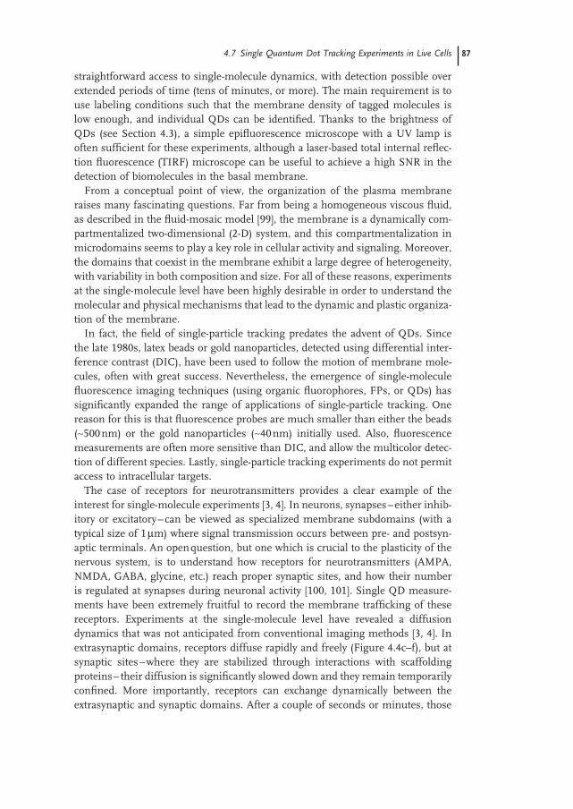
A single QD assay has been recently implemented to probe, for the fi rst time, the transport properties of individual kinesin and myosin V motors in live cells [6, 117] (Figure 4.5 a,b). The properties of these molecular motors have been abun-dantly investigated with single - molecule in vitro experiments [118] . By means of micromanipulation techniques and fl uorescence assays, important parameters such as the velocity, the processivity and the stall force have been determined. However, the intrinsic limitation of these experiments is that the motors are
90 4 Inorganic Fluorescent Probes for Single-Molecule Tracking Experiments in Live Cells
observed out of their natural cellular habitat. In fact, the cell is a complex environ-ment where molecular parameters (pH, ionic strength, ATP concentration, etc.) are actively regulated, and crowding is extreme. All of these aspects can potentially affect the motor motility, and are diffi cult to faithfully reproduce in vitro . To visual-ize single motors in live cells, purifi ed kinesins or myosins V were labeled in vitro , their activity tested in standard motility assays, and subsequently internalized into HeLa cells using a method based on the osmotic release of pynocytic vesicles. An analysis of the movement of individual motors gave access to their velocity and processivity in cells. Interestingly, the results (velocity ∼ 0.5 – 1 µ m s − 1 and proces-sivity ∼ 1 – 2 s) were relatively unchanged compared to their in vitro values. In the case of myosin V, it was also possible to observe individual steps of the motors [117] .
4.7.5 From Single Molecules to Populations
A specifi city of QDs for single - molecule experiments in live cells is that many nanoparticles can be easily detected and tracked in parallel. For instance, when using a UV lamp as the excitation source (rather than a laser) and an intensifi ed CCD, fi eld of views of ∼ 100 × 100 µ m 2 containing tens to hundreds of discernable QDs can be recorded at frame rate of ∼ 10 – 30 images per second. Once analyzed with an appropriate algorithm [80, 81] , these sequences of images yield a large number of individual trajectories. From a practical point of view, this means that experiments have a higher throughput of single - molecule events, and that statisti-cally meaningful results can be more rapidly obtained. The fact that many QDs can be simultaneously tracked has also conceptual implications on the way in which the spatiotemporal dynamics of molecular populations can be investigated.
Figure 4.5 (a) Motion of an individual kinesin motor labeled with a QD in the cytoplasm of a living HeLa cell. Adapted from Ref. [6] . (b) Position along the x - axis versus time, indicating a succession of phases of directed and diffusive motion.
4.8 Conclusions and Perspectives 91
Indeed, the subpopulation of QD - labeled molecules, which appear as individual fl uorescence spots in the images (Figure 4.4 a), corresponds to sampling (presum-ably random) of the whole population. By analyzing the dynamics of these QD spots, the properties of the population can be inferred with a sensitivity that, often, cannot be achieved using ensemble labeling. For instance, the fi rst moments of the spatial distribution, as well as spatiotemporal correlations between individual spots, can be computed with high spatial and temporal resolution. Using QDs as tracers within populations of molecules of interest will in fact reinforce the con-ceptual link between the analysis of molecular systems in cells and the methods used in soft - condensed matter to investigate the dynamics of complex systems.
This aspect of single QD experiments was recently used to probe the dynamics of chemoreceptors in nerve growth cones [90] . When submitted to an external gradient of chemoattractants, the QD - labeled chemoreceptors were redistributed asymmetrically towards the gradient source (this is indicative of an amplifi cation step in gradient sensing) [119] . The analysis of individual receptor trajectories showed that the symmetry - breaking resulted from a microtubule - dependent directed transport. Furthermore, by investigating the mean position of the QD spots, the formation of polarity at the cell surface could be quantitatively described and modeled as a positive - feedback loop involving receptor asymmetric activation and microtubule dynamics [90] .
In general, understanding how cell organization and morphogenesis dynami-cally emerge, and how they are related to molecular events, should greatly benefi t from ultrasensitive QD imaging in live cells. It is anticipated that the approach described above will be fruitful for deciphering the complex processes involved in the self - organization of biological systems [120, 121] . It should be especially useful in order to develop multiscale descriptions of biological systems for which molec-ular - level information (determined by examining single trajectories) are integrated into a view at a systems - level.
4.8 Conclusions and Perspectives
Quantum dots represent a new class of inorganic fl uorophores with unique optical properties. When detected at the single - molecule level, they offer exciting possibili-ties to visualize the real - time motion of individual biomolecules in their cellular environment. Several reported examples have illustrated the interest of single QD experiments in cell biology to decipher the molecular processes that contribute to cell organization and dynamics. However, making QD imaging a standard tool in cell biology still requires a signifi cant effort, in order to improve the colloidal and biochemical properties of the probes.
From the synthesis and bioconjugation points of view, two improvements can be expected for the future. Clearly, experiments would benefi t from QDs with reduced blinking [34] , and the use of elongated rather than spherical QDs might represent a step in this direction [122] . The conjugation of QDs with biological
92 4 Inorganic Fluorescent Probes for Single-Molecule Tracking Experiments in Live Cells
ligands, in particular with proteins, can also be improved. While currently most experiments are performed with an uncontrolled number of ligand molecules per QD, it is today possible to synthesize QDs with an exactly controlled number of binding sites per QD [51, 70] . The way in which ligands are attached to the QD can also be improved. At present, for most of the time, the geometry of the linkage between the ligand and the QD is completely undefi ned. However, if the proteins were to be linked via their amino - groups to the carboxy - groups on the QD surface with crosslinker molecules, the binding geometry would be undefi ned, as each protein can have several amino - groups. Enzymes or antibodies might thus be linked in a way that they are inactivated. However, by fusing specifi c linking domains to proteins, they could be linked in more defi ned ways to the particle surface [57] .
An improved QD technology should permit the investigation of new classes of scientifi c question. So far, single - particle tracking has been dedicated almost exclu-sively to tracing the positions of labeled molecules, although in many cases the orientation of molecules with respect to their binding partners has also played a crucial role. Rotational motion can be observed by measuring polarized fl uores-cence [123] or by defocused microscopy [124] . Although, rod - shaped QD rods have a polarized emission, and thus should be suited to this type of experiment [125] , today most experiments using QDs are based on spherical particles. However, due to their changed blinking behavior and polarized emission, a much wider use of asymmetric QD structures is predicted in the future.
References
1 Schutz , G.J. , Kada , G. , Pastushenko , V.P. , and Schindler , H. ( 2000 ) EMBO J. , 19 , 892 .
2 Harms , G.S. , Cognet , L. , Lommerse , P.H.M. , Blab , G.A. , Kahr , H. , Gamsj ä ger , R. , Spaink , H.P. , Soldatov , N.M. , Romanin , C. , and Schmidt , T. ( 2001 ) Biophys. J. , 81 , 2639 .
3 Dahan , M. , Levi , S. , Luccardini , C. , Rostaing , P. , Riveau , B. , and Triller , A. ( 2003 ) Science , 302 , 442 .
4 Tardin , C. , Cognet , L. , Bats , C. , Lounis , B. , and Choquet , D. ( 2003 ) EMBO J. , 22 , 4656 .
5 Kural , C. , Kim , H. , Syed , S. , Goshima , G. , Gelfand , V.I. , and Selvin , P.R. ( 2005 ) Science , 308 , 1469 .
6 Courty , S. , Luccardini , C. , Bellaiche , Y. , Cappello , G. , and Dahan , M. ( 2006 ) Nano Lett. , 6 , 1491 .
7 Murase , K. , Fujiwara , T. , Umemura , Y. , Suzuki , K. , Iino , R. , Yamashita , H. ,
Saito , M. , Murakoshi , H. , Ritchie , K. , and Kusumi , A. ( 2004 ) Biophys. J. , 86 , 4075 .
8 Yu , J. , Xiao , J. , Ren , X.J. , Lao , K.Q. , and Xie , X.S. ( 2006 ) Science , 311 , 1600 .
9 Shav - Tal , Y. , Darzacq , X. , Shenoy , S.M. , Fusco , D. , Janicki , S.M. , Spector , D.L. , and Singer , R.H. ( 2004 ) Science , 304 , 1797 .
10 Douglass , A.D. , and Vale , R.D. ( 2005 ) Cell , 121 , 937 .
11 Lommerse , P.H.M. , Snaar - Jagaiska , B.E. , Spaink , H.P. , and Schmidt , T. ( 2005 ) J. Cell Sci. , 118 , 1799 .
12 Giepmans , B.N.G. , Adams , S.R. , Ellisman , M.H. , and Tsien , R.Y. ( 2006 ) Science , 312 , 217 .
13 Chen , I. , and Ting , A.Y. ( 2005 ) Curr.
Opin. Biotechnol. , 16 , 35 . 14 Zhang , J. , Campbell , R.E. , Ting , A.Y. ,
and Tsien , R.Y. ( 2002 ) Nat. Rev. Mol. Cell
Biol. , 3 ( 12 ), 906 – 918 .
References 93
15 Alivisatos , P. ( 2004 ) Nat. Biotechnol. , 22 , 47 .
16 Parak , W.J. , Pellegrino , T. , and Plank , C. ( 2005 ) Nanotechnology , 16 , R5 .
17 Michalet , X. , Pinaud , F.F. , Bentolila , L.A. , Tsay , J.M. , Doose , S. , Li , J.J. , Sundaresan , G. , Wu , A.M. , Gambhir , S.S. , and Weiss , S. ( 2005 ) Science , 307 , 538 .
18 Pellegrino , T. , Manna , L. , Kudera , S. , Liedl , T. , Koktysh , D. , Rogach , A.L. , Keller , S. , R ä dler , J. , Natile , G. , and Parak , W.J. ( 2004 ) Nano Lett. , 4 , 703 .
19 Pellegrino , T. , Kudera , S. , Liedl , T. , Javier , A.M. , Manna , L. , and Parak , W.J. ( 2005 ) Small , 1 , 48 .
20 Gerion , D. , Pinaud , F. , Williams , S.C. , Parak , W.J. , Zanchet , D. , Weiss , S. , and Alivisatos , A.P. ( 2001 ) J. Phys. Chem. B , 105 , 8861 .
21 Bruchez , M.J. , Moronne , M. , Gin , P. , Weiss , S. , and Alivisatos , A.P. ( 1998 ) Science , 281 , 2013 .
22 Chan , W.C.W. and Nie , S. ( 1998 ) Science , 281 , 2016 .
23 Parak , W.J. , Manna , L. , and Nann , T. ( 2008 ) Nanotechnology: Volume 1:
Principles and Fundamentals (ed. G. Schmid ), Wiley - VCH Verlag GmbH , Weinheim , p. 73 .
24 Dabbousi , B.O. , Rodriguez - Viejo , J. , Mikulec , F.V. , Heine , J.R. , Mattoussi , H. , Ober , R. , Jensen , K.F. , and Bawendi , M.G. ( 1997 ) J. Phys. Chem. B , 101 , 9463 .
25 Wu , M.X. , Liu , H. , Liu , J. , Haley , K.N. , Treadway , J.A. , Larson , J.P. , Ge , N. , Peale , F. , and Bruchez , M.P. ( 2003 ) Nat.
Biotechnol. , 21 , 41 . 26 Medintz , I.L. , Uyeda , H.T. , Goldman ,
E.R. , and Mattoussi , H. ( 2005 ) Nat.
Mater. , 4 , 635 . 27 Fern á ndez - Arg ü elles , M.T. , Yakovlev , A. ,
Sperling , R.A. , Luccardini , C. , Gaillard , S. , Medel , A.S. , Mallet , J. - M. , Brochon , J. - C. , Feltz , A. , Oheim , M. , and Parak , W.J. ( 2007 ) Nano Lett. , 7 , 2613 .
28 Dahan , M. , Laurence , T. , Pinaud , F. , Chemla , D.S. , Alivisatos , A.P. , Sauer , M. , and Weiss , S. ( 2001 ) Opt. Lett. , 26 , 825 .
29 Giepmans , B.N. , Deerinck , T.J. , Smarr , B.L. , Jones , Y.Z. , and Ellisman , M.H. ( 2005 ) Nat. Methods , 2 , 743 .
30 Kim , S. , Lim , Y.T. , Soltesz , E.G. , De Grand , A.M. , Lee , J. , Nakayama , A. ,
Parker , J.A. , Mihaljevic , T. , Laurence , R.G. , Dor , D.M. , Cohn , L.H. , Bawendi , M.G. , and Frangioni , J.V. ( 2004 ) Nat.
Biotechnol. , 22 , 93 . 31 Nirmal , M. , Dabbousi , B.O. , Bawendi ,
M.G. , Macklin , J.J. , Trautman , J.K. , Harris , T.D. , and Brus , L.E. ( 1996 ) Nature , 383 , 802 .
32 Shimizu , K.T. , Neuhauser , R.G. , Leatherdale , C.A. , Empedocles , S.A. , Woo , W.K. , and Bawendi , M.G. ( 2001 ) Phys. Rev. B , 63 , 205316 .
33 Brokmann , X. , Hermier , J.P. , Messin , G. , Desbiolles , P. , Bouchaud , J.P. , and Dahan , M. ( 2003 ) Phys. Rev. Lett. , 90 , 120601 .
34 Mahler , B. , Spinicelli , P. , Buil , S. , Quelin , X. , Hermier , J. - P. , and Dubertret , B. ( 2008 ) Nat. Mater. , 7 , 659 .
35 Chen , Y. , Vela , J. , Htoon , H. , Casson , J.L. , Werder , D.J. , Bussian , D.A. , Klimov , V.I. , and Hollingsworth , J.A. ( 2008 ) J. Am. Chem. Soc. , 130 , 5026 .
36 Lin , C. - A.J. , Liedl , T. , Sperling , R.A. , Fern á ndez - Arg ü elles , M.T. , Costa - Fern á ndez , J.M. , Pereiro , R. , Sanz - Medel , A. , Chang , W.H. , and Parak , W.J. ( 2007 ) J. Mater. Chem. , 17 , 1343 .
37 Kudera , S. , Carbone , L. , Manna , L. , and Parak , W.J. ( 2008 ) Semiconductor
Nanocrystal Quantum Dots (ed. A.L. Rogach ), Springer , Vienna , p. 1 .
38 Peng , X. , Wickham , J. , and Alivisatos , A.P. ( 1998 ) J. Am. Chem. Soc. , 120 , 5343 .
39 Peng , X. , Manna , L. , Yang , W. , Wickham , J. , Scher , E. , Kadavanich , A. , and Alivisatos , A.P. ( 2000 ) Nature , 404 , 59 .
40 Mokari , T. , Rothenberg , E. , Popov , I. , Costi , R. , and Banin , U. ( 2004 ) Science , 304 , 1787 .
41 Manna , L. , Scher , E.C. , and Alivisatos , A.P. ( 2002 ) J. Cluster Sci. , 13 , 521 .
42 Cozzoli , P.D. , Pellegrino , T. , and Manna , L. ( 2006 ) Chem. Soc. Rev. , 35 , 1195 .
43 Kumar , S. and Nann , T. ( 2006 ) Small , 2 , 316 .
44 Kudera , S. , Carbone , L. , Zanella , M. , Cingolani , R. , Parak , W.J. , and Manna , L. ( 2006 ) Phys. Stat. Sol. (c) , 203 , 1329 .
45 Kudera , S. , Carbone , L. , Carlino , E. , Cingolani , R. , Cozzoli , P.D. , and Manna , L. ( 2007 ) Physica E , 37 , 128 .
94 4 Inorganic Fluorescent Probes for Single-Molecule Tracking Experiments in Live Cells
46 Derfus , A.M. , Chan , W.C.W. , and Bhatia , S.N. ( 2004 ) Nano Lett. , 4 , 11 .
47 Kirchner , C. , Liedl , T. , Kudera , S. , Pellegrino , T. , Javier , A.M. , Gaub , H.E. , St ö lzle , S. , Fertig , N. , and Parak , W.J. ( 2005 ) Nano Lett. , 5 , 331 .
48 Sperling , R.A. , Liedl , T. , Duhr , S. , Kudera , S. , Zanella , M. , Lin , C. - A.J. , Chang , W. , Braun , D. , and Parak , W.J. ( 2007 ) J. Phys. Chem. C , 111 , 11552 .
49 Kanaras , A.G. , Kamounah , F.S. , Schaumburg , K. , Kiely , C.J. , and Brust , M. ( 2002 ) Chem. Commun. , 2002 , 2294 .
50 Ballou , B. , Lagerholm , B.C. , Ernst , L.A. , Bruchez , M.P. , and Waggoner , A.S. ( 2004 ) Bioconjug. Chem. , 15 , 79 .
51 Sperling , R.A. , Pellegrino , T. , Li , J.K. , Chang , W.H. , and Parak , W.J. ( 2006 ) Adv. Funct. Mater. , 16 , 943 .
52 Rojo , J. , Diaz , V. , Fuente , J.M.D.L. , Segura , I. , Barrientos , A.G. , Riese , H.H. , Bernad , A. , and Penades , S. ( 2004 ) ChemBioChem , 5 , 291 .
53 von Holt , B. , Kudera , S. , Weiss , A. , Schrader , T.E. , Manna , L. , Parak , W.J. , and Braun , M. ( 2008 ) J. Mater. Chem. , 18 , 2728 .
54 Aldana , J. , Wang , Y.A. , and Peng , X. ( 2001 ) J. Am. Chem. Soc. , 123 , 8844 .
55 Kim , S. , Kim , S. , Tracy , J. , Jasanoff , A. , and Bawendi , M. ( 2005 ) J. Am. Chem.
Soc. , 127 , 4556 . 56 Li , Z. , Jin , R. , Mirkin , C.A. , and
Letsinger , R.L. ( 2002 ) Nucleic Acids Res. , 30 , 1558 .
57 Mattoussi , H. , Mauro , J.M. , Goldman , E.R. , Anderson , G.P. , Sundar , V.C. , Mikulec , F.V. , and Bawendi , M.G. ( 2000 ) J. Am. Chem. Soc. , 122 , 12142 .
58 Parak , W.J. , Gerion , D. , Zanchet , D. , Woerz , A.S. , Pellegrino , T. , Micheel , C. , Williams , S.C. , Seitz , M. , Bruehl , R.E. , Bryant , Z. , Bustamante , C. , Bertozzi , C.R. , and Alivisatos , A.P. ( 2002 ) Chem.
Mater. , 14 , 2113 . 59 Alejandro - Arellano , M. , Ung , T. , Blanco ,
A. , Mulvaney , P. , and Liz - Marzan , L.M. ( 2000 ) Pure Appl. Chem. , 72 , 257 .
60 Pinaud , F. , King , D. , Moore , H. - P. , and Weiss , S. ( 2004 ) J. Am. Chem. Soc. , 126 , 6115 .
61 Nikolic , M.S. , Krack , M. , Aleksandrovic , V. , Kornowski , A. , Forster , S. , and
Weller , H. ( 2006 ) Angew. Chem., Int. Ed. , 45 , 6577 .
62 Lin , C. - A.J. , Sperling , R.A. , Li , J.K. , Yang , T. - Y. , Li , P. - Y. , Zanella , M. , Chang , W.H. , and Parak , W.J. ( 2008 ) Small , 4 , 334 .
63 Yu , W.W. , Chang , E. , Falkner , J.C. , Zhang , J. , Al - Somali , A.M. , Sayes , C.M. , Johns , J. , Drezek , R. , and Colvin , V.L. ( 2007 ) J. Am. Chem. Soc. , 129 , 2871 .
64 Luccardini , C. , Tribet , C. , Vial , F. , Marchi - Artzner , V. , and Dahan , M. ( 2006 ) Langmuir , 22 , 2304 .
65 Dubertret , B. , Skourides , P. , Norris , D.J. , Noireaux , V. , Brivanlou , A.H. , and Libchaber , A. ( 2002 ) Science , 298 , 1759 .
66 Carion , O. , Mahler , B. , Pons , T. , and Dubertret , B. ( 2007 ) Nat. Protoc. , 2 , 2383 .
67 Boulmedais , F. , Bauchat , P. , Brienne , M.J. , Arnal , I. , Artzner , F. , Gacoin , T. , Dahan , M. , and Marchi - Artzner , V. ( 2006 ) Langmuir , 22 , 9797 .
68 Mattoussi , H. , Mauro , J.M. , Goldman , E.R. , Green , T.M. , Anderson , G.P. , Sundar , V.C. , and Bawendi , M.G. ( 2001 ) Phys. Stat. Sol. B , 224 , 277 .
69 Hermanson , G.T. ( 1996 ) Bioconjugate
Techniques , Academic Press , San Diego . 70 Howarth , M. , Liu , W.H. , Puthenveetil ,
S. , Zheng , Y. , Marshall , L.F. , Schmidt , M.M. , Wittrup , K.D. , Bawendi , M.G. , and Ting , A.Y. ( 2008 ) Nat. Methods , 5 , 397 .
71 Pons , T. , Uyeda , H.T. , Medintz , I.L. , and Mattoussi , H. ( 2006 ) J. Phys. Chem. B , 110 , 20308 .
72 Parak , W.J. , Boudreau , R. , Gros , M.L. , Gerion , D. , Zanchet , D. , Micheel , C.M. , Williams , S.C. , Alivisatos , A.P. , and Larabell , C.A. ( 2002 ) Adv. Mater. , 14 , 882 .
73 Pradhan , N. and Peng , X.G. ( 2007 ) J. Am. Chem. Soc. , 129 , 3339 .
74 Pradhan , N. , Battaglia , D.M. , Liu , Y.C. , and Peng , X.G. ( 2007 ) Nano Lett. , 7 , 312 .
75 Weiss , S. ( 1999 ) Science , 283 , 1676 . 76 Moerner , W.E. and Orrit , M. ( 1999 )
Science , 283 , 1670 . 77 Kulzer , F. , Kummer , S. , Matzke , R. ,
Br ä uchle , C. , and Basche , T. ( 1997 ) Nature , 387 , 688 .
78 Bobroff , N. ( 1986 ) Rev. Sci. Instrum. , 57 , 1152 .
References 95
79 Thompson , R.E. , Larson , D.R. , and Webb , W.W. ( 2002 ) Biophys. J. , 82 , 2775 .
80 Bonneau , S. , Dahan , M. , and Cohen , L.D. ( 2005 ) IEEE Trans. Image Process. , 14 , 1384 .
81 Serg é , A. , Bertaux , N. , Rigneault , H. , and Marguet , D. ( 2008 ) Nat. Methods , 5 , 687 .
82 Jaqaman , K. , Loerke , D. , Mettlen , M. , Kuwata , H. , Grinstein , S. , Schmid , S.L. , and Danuser , G. ( 2008 ) Nat. Methods , 5 , 695 .
83 Ehrensperger , M.V. , Hanus , C. , Vannier , C. , Triller , A. , and Dahan , M. ( 2007 ) Biophys. J. , 92 , 3706 .
84 Qian , H. ( 2000 ) Biophys. J. , 79 , 137 . 85 Saxton , M.J. and Jacobson , K. ( 1997 )
Annu. Rev. Biophys. Biomol. Struct. , 26 , 373 .
86 Bouzigues , C. and Dahan , M. ( 2007 ) Biophys. J. , 92 , 654 .
87 Meilhac , N. , Le Guyader , L. , Salome , L. , and Destainville , N. ( 2006 ) Phys. Rev. E , 73 , 011915 .
88 Simson , R. , Sheets , E.D. , and Jacobson , K. ( 1995 ) Biophys. J. , 69 , 989 .
89 Huet , S. , Karatekin , E. , Tran , V.S. , Fanget , I. , Cribier , S. , and Henry , J.P. ( 2006 ) Biophys. J. , 91 , 3542 .
90 Bouzigues , C. , Morel , M. , Triller , A. , and Dahan , M. ( 2007 ) Proc. Natl Acad. Sci.
USA , 104 , 11251 . 91 Groc , L. , Heine , M. , Cognet , L. , Brickley ,
K. , Stephenson , F.A. , Lounis , B. , and Choquet , D. ( 2004 ) Nat. Neurosci. , 7 , 695 .
92 Groc , L. , Heine , M. , Cousins , S.L. , Stephenson , F.A. , Lounis , B. , Cognet , L. , and Choquet , D. ( 2006 ) Proc. Natl Acad.
Sci. USA , 103 , 18769 . 93 Nechyporuk - Zloy , V. , Stock , C. , Schillers ,
H. , Oberleithner , H. , and Schwab , A. ( 2006 ) Am. J. Physiol. Cell Physiol. , 291 , C266 .
94 Haggie , P.M. , Kim , J.K. , Lukacs , G.L. , and Verkman , A.S. ( 2006 ) Mol. Biol.
Cell , 17 , 4937 . 95 Crane , J.M. , Hoek , A.N.V. , Skach , W.R. ,
and Verkman , A.S. ( 2008 ) Mol. Biol.
Cell , 19 , 3369 . 96 Lidke , D.S. , Nagy , P. , Heintzmann , R. ,
Arndt - Jovin , D.J. , Post , J.N. , Grecco , H.E. , Jares - Erijman , E.A. , and Jovin , T.M. ( 2004 ) Nat. Biotechnol. , 22 , 198 .
97 Pinaud , F. , Michalet , X. , Iyer , G. , Margeat , E. , Moore , H.P. , and Weiss , S. ( 2009 ) Traffi c , 10 , 691 – 712 .
98 Andrews , N.L. , Lidke , K.A. , Pfeiffer , J.R. , Burns , A.R. , Wilson , B.S. , Oliver , J.M. , and Lidke , D.S. ( 2008 ) Nat. Cell Biol. , 10 , 955 .
99 Singer , S.J. and Nicolson , G.L. ( 1972 ) Science , 175 , 720 .
100 Triller , A. and Choquet , D. ( 2008 ) Neuron , 59 , 359 .
101 Choquet , D. and Triller , A. ( 2003 ) Nat.
Rev. Neurosci. , 4 , 251 . 102 Saffman , P.G. and Delbr ü ck , M. ( 1975 )
Proc. Natl Acad. Sci. USA , 72 , 3111 . 103 Groc , L. , Lafourcade , M. , Heine , M. ,
Renner , M. , Racine , V. , Sibarita , J.B. , Lounis , B. , Choquet , D. , and Cognet , L. ( 2007 ) J. Neurosci. , 27 , 12433 .
104 Rajan , S.S. , Liu , H.Y. , and Vu , T.Q. ( 2008 ) ACS Nano , 2 , 1153 .
105 Cui , B.X. , Wu , C.B. , Chen , L. , Ramirez , A. , Bearer , E.L. , Li , W.P. , Mobley , W.C. , and Chu , S. ( 2007 ) Proc. Natl Acad. Sci.
USA , 104 , 13666 . 106 Nabiev , I. , Mitchell , S. , Davies , A. ,
Williams , Y. , Kelleher , D. , Moore , R. , Gun ’ ko , Y.K. , Byrne , S. , Rakovich , Y.P. , Donegan , J.F. , Sukhanova , A. , Conroy , J. , Cottell , D. , Gaponik , N. , Rogach , A. , and Volkov , Y. ( 2007 ) Nano Lett. , 7 , 3452 .
107 Cambi , A. , Lidke , D.S. , Arndt - Jovin , D.J. , Figdor , C.G. , and Jovin , T.M. ( 2007 ) Nano Lett. , 7 , 970 .
108 Ruan , G. , Agrawal , A. , Marcus , A.I. , and Nie , S. ( 2007 ) J. Am. Chem. Soc. , 129 , 14759 .
109 Tekle , C. , van Deurs , B. , Sandvig , K. , and Iversen , T.G. ( 2008 ) Nano Lett. , 8 , 1858 .
110 Luccardini , C. , Yakovlev , A. , Gaillard , S. , Alberola , M.V.T. , Hoff , A.P. , Mallet , J. - M. , Parak , W.J. , Feltz , A. , and Oheim , M. ( 2007 ) J. Biomed. Biotechnol. , 2007 , Article ID, 68963.
111 Tada , H. , Higuchi , H. , Wanatabe , T.M. , and Ohuchi , N. ( 2007 ) Cancer Res. , 67 , 1138 .
112 Derfus , A. , Chan , W. , and Bhatia , S. ( 2004 ) Adv. Mater. , 16 , 961 .
113 Chen , F. and Gerion , D. ( 2004 ) Nano
Lett. , 4 , 1827 .
96 4 Inorganic Fluorescent Probes for Single-Molecule Tracking Experiments in Live Cells
114 Plank , C. , Oberhauser , B. , Mechtler , K. , Koch , C. , and Wagner , E. ( 1994 ) J. Biol.
Chem. , 269 , 12918 . 115 de la Fuente , J.M. and Berry , C.C.
( 2005 ) Bioconjug. Chem. , 16 , 1176 . 116 Lewin , M. , Carlesso , N. , Tung , C.H. ,
Tang , X.W. , Corry , D. , Scadden , D.T. , and Weissleder , R. ( 2000 ) Nat.
Biotechnol. , 18 , 410 . 117 Pierobon , P. , Achouri , S. , Courty , S. ,
Dunn , A.R. , Spudich , J.A. , Dahan , M. , and Cappello , G. ( 2009 ) Biophys. J ., 96 , 4268 – 4275 .
118 Howard , J. ( 2001 ) Mechanics of Motor
Proteins and the Cytoskeleton , Sinauer . 119 Mortimer , D. , Fothergill , T. , Pujic , Z. ,
Richards , L.J. , and Goodhill , G.J. ( 2008 ) Trends Neurosci. , 31 , 90 .
120 Karsenti , E. ( 2008 ) Nat. Rev. Mol. Cell.
Biol. , 9 , 255 . 121 Misteli , T. ( 2001 ) J. Cell Biol. , 155 , 181 . 122 Wang , S. , Querner , C. , Emmons , T. ,
Drndic , M. , and Crouch , C.H. ( 2006 ) J. Phys. Chem. B , 110 , 23221 .
123 Hugel , T. , Michaelis , J. , Hetherington , C.L. , Jardine , P.J. , Grimes , S. , Walter , J.M. , Faik , W. , Anderson , D.L. , and Bustamante , C. ( 2007 ) PLoS Biol. , 5 , 558 .
124 Brokmann , X. , Ehrensperger , M.V. , Hermier , J.P. , Triller , A. , and Dahan , M. ( 2005 ) Chem. Phys. Lett. , 406 , 210 .
125 Hu , J. , Yang , L. - W. , Wang , L. - S. , Li , W. , and Alivisatos , A.P. ( 2002 ) J. Phys. Chem.
B , 106 , 2447 .