Embed Size (px)
Citation preview
304
Int. J. Odontostomat.,5(3):304-308, 2011.
Sinonasal Mucoepidermoid Carcinoma:a Case Report and Literature Review
Carcinoma Mucoepidermoide Nasosinusal: Reporte de Caso y Revisión de la Literatura
Ilson Sepúlveda*; Loreto Spencer** & Enrique Platin***
SEPÚLVEDA, I.; SPENCER, L. & PLATIN, E. Sinonasal Mucoepidermoid Carcinoma: a case report and literature review.Int. J. Odontostomat., 5(3):304-308, 2011.
SUMMARY: We report on a patient who presented at our clinic with a growth of the right palate of twenty yearsduration. A biopsy of the area was performed and the lesion was diagnosed as low-grade mucoepidermoid carcinoma(MEC). Due to the size of the tumor, it was decided to begin treatment with radiotherapy followed by chemotherapy. Once theinitial treatment was completed, the lesion was reevaluated and surgery followed by reconstruction was recommended. ThePatient rejected the recommended treatment and opted to enrolled in a pain management, palliative care program.
KEY WORDS: mucoepidermoid, carcinoma, Computed tomography, Magnetic resonance imaging, tumorsinonasal.
INTRODUCTION
Malignant neoplasms of the salivary glands arevery rare, representing 0.3% of head and neckmalignant tumors (Ghosh-Laskar et al., 2011).Mucoepidermoid Carcinoma (MEC) is the mostcommon malignancy of salivary gland tumors, occurringmost frequently in the major salivary glands,predominantly the parotid gland (Giger et al., 2005).
Stewart, Foote and Becker in 1945 first describedthe histological features of MEC. They reported on 45salivary gland tumor cases, made up of mucosecretorycell, squamous cells and basal cells or intermediatestratum cells. (Gallager et al., 1959). After comparingthe morphological features of MEC with other chronicinflammatory non-neoplastic lesions of the salivaryglands it was theorized that these cells originated inthe ductal system (Foschini et al., 2002).
MEC usually occurs in the head and neck regionbut it has been reported in other anatomical regions;for example, distant glandular structures and unlikelysites such as the anal region, esophagus andtracheobronchial mucosa. The latter is extremely rare,
corresponding to only 0.1- 0.2% of malignant lunglesions. The lesions often present radiographically asa solitary nodule or mass with or without postobstructive pneumonia or atelectasis (Gallager et al.;Magliulo & Appiani, 2010; Kim et al., 1999).
Occurrence of MEC in other areas of the headand neck such as the thyroid gland is extremely rare. It is predominant in females and presents as twoentities: Typical MEC and Sclerotic MEC withEosinophilia. Cases of MEC have been reported inthe middle ear and mastoid region, where the presenceof salivary gland tissues is deemed controversial. Only5 cases of parotid MEC have been reported in the mainexcretory duct, where they can be easily confused withminor salivary glands of the oral mucosa or accessoryparotid gland (Giger et al., 2005; Davies et al., 2005;Gupta & Kaluskar, 2003).
Histologically, the proportion of the 3 cellulartypes helps to define and grade the tumor. High-gradetumors are poorly differentiated, and consistpredominantly of intermediate and squamous cells with
* Oral, Maxillofacial-Head and Neck Radiologist. Department of Ear; Head and Neck Cancer Committe, ENT Service, Hospital Regional deConcepción, Concepción. Professor of Oral and Maxillofacial Radiology, Universidad de Concepción, School of Dentistry, Concepción, Chile.
** Physician Pathologist, Pathology Department, Hospital Regional de Concepción, Chile*** Professor of Oral and Maxillofacial Radiology, University of North Carolina School of Dentistry, Chapel Hill, USA.
305
polymorphism, high mitotic activity and necrosis. Low-grade tumors are highly differentiated, and consistpredominantly of muco-secretory cells, with minimalatypia and cystic formation. Intermediate grade lesionsconsist of a mixture of both cell types (squamous andmuco-secretory) (Magliulo & Appiani; Emerick et al.,2007). The histological subdivision of MEC includes high,intermediate and low-grades, and is highly correlatedto the prognostic outcome (Guzzo et al., 2002).
When an infiltrative neoplasm is suspected, MRI(Magnetic Resonance Imaging) with or withoutintravenous contrast should be performed. MRI has ahigh sensitivity in determining the extent of the tumor’sinfiltration pattern, and perineural invasion to the skullbase and meningeal membranes (Yousem et al., 2000).
Some investigators have suggested that low-grade MEC with favorable clinical staging (T1 or T2;N0) can be treated surgically. Surgery and radiotherapyis recommended for high-grade MEC. The treatmentis controversial for intermediate MEC; therefore, thereis no consensus for treating intermediate MEC. The 5-year survival rate is 92% for low-grade MEC and 63%for high grade MEC (April et al., 1997). Early cases ofMEC involving minor salivary glands have a survivalrate of 78,6% for low-grade and only 50% for high-grade. The recurrence rate in low grade MEC is verylow, unlike for intermediate and high-grade MEC whereit reaches 30% and 70% respectively (Rosdeutscher& Burnette, 2003; Ghosh-Laskar et al.).
CLINICAL CASE
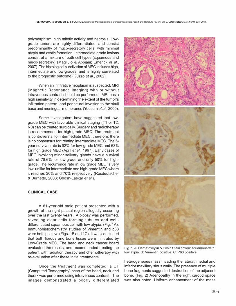
A 61-year-old male patient presented with agrowth of the right palatal region allegedly occurringover the last twenty years. A biopsy was performed,revealing clear cells forming tubules and well-differentiated squamous cell with low atypia. (Fig. 1A).Immunohistochemistry studies of Vimentin and p63were both positive (Figs. 1B and 1C). It was concludedthat both fibrous and bone tissue were infiltrated byLow-Grade MEC. The head and neck cancer boardevaluated the results, and recommended treating thepatient with radiation therapy and chemotherapy withre-evaluation after these initial treatments.
Once the treatment was completed, a CT(Computed Tomography) scan of the head, neck andthorax was performed using intravenous contrast. Theimages demonstrated a poorly differentiated
Fig. 1. A: Hematoxyylin & Eosin Stain tintion: squamous with low atipia. B: Vimentin positive. C: P63 positive.
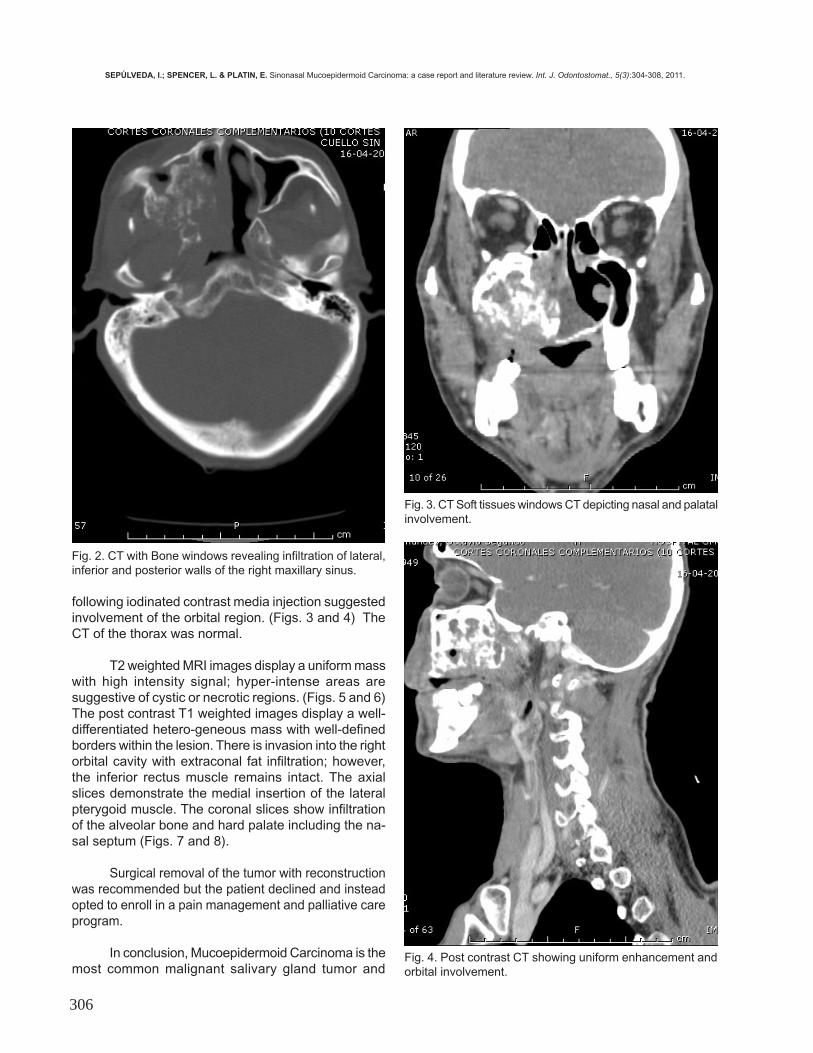
heterogeneous mass invading the lateral, medial andinferior maxillary sinus walls. The presence of multiplebone fragments suggested destruction of the adjacentbone. (Fig. 2) Adenopathy in the right carotid spacewas also noted. Uniform enhancement of the mass
SEPÚLVEDA, I.; SPENCER, L. & PLATIN, E. Sinonasal Mucoepidermoid Carcinoma: a case report and literature review. Int. J. Odontostomat., 5(3):304-308, 2011.
306
following iodinated contrast media injection suggestedinvolvement of the orbital region. (Figs. 3 and 4) TheCT of the thorax was normal.
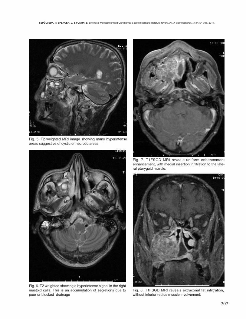
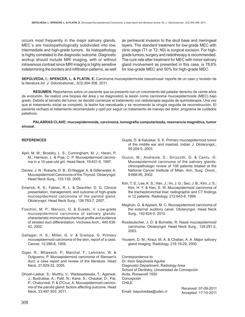
T2 weighted MRI images display a uniform masswith high intensity signal; hyper-intense areas aresuggestive of cystic or necrotic regions. (Figs. 5 and 6)The post contrast T1 weighted images display a well-differentiated hetero-geneous mass with well-definedborders within the lesion. There is invasion into the rightorbital cavity with extraconal fat infiltration; however,the inferior rectus muscle remains intact. The axialslices demonstrate the medial insertion of the lateralpterygoid muscle. The coronal slices show infiltrationof the alveolar bone and hard palate including the na-sal septum (Figs. 7 and 8).
Surgical removal of the tumor with reconstructionwas recommended but the patient declined and insteadopted to enroll in a pain management and palliative careprogram.
In conclusion, Mucoepidermoid Carcinoma is themost common malignant salivary gland tumor and
Fig. 4. Post contrast CT showing uniform enhancement andorbital involvement.
Fig. 2. CT with Bone windows revealing infiltration of lateral,inferior and posterior walls of the right maxillary sinus.
Fig. 3. CT Soft tissues windows CT depicting nasal and palatalinvolvement.
SEPÚLVEDA, I.; SPENCER, L. & PLATIN, E. Sinonasal Mucoepidermoid Carcinoma: a case report and literature review. Int. J. Odontostomat., 5(3):304-308, 2011.
307
Fig. 5. T2 weighted MRI image showing many hyperintenseareas suggestive of cystic or necrotic areas.
Fig. 6. T2 weighted showing a hyperintense signal in the rightmastoid cells. This is an accumulation of secretions due topoor or blocked drainage
Fig. 7. T1FSGD MRI reveals uniform enhancementenhancement, with medial insertion infiltration to the late-ral pterygoid muscle.
Fig. 8. T1FSGD MRI reveals extraconal fat infiltration,without inferior rectus muscle involvement.
SEPÚLVEDA, I.; SPENCER, L. & PLATIN, E. Sinonasal Mucoepidermoid Carcinoma: a case report and literature review. Int. J. Odontostomat., 5(3):304-308, 2011.
308
occurs most frequently in the major salivary glands.MEC´s are hisotopathologically subdivided into low,intermediate and high-grade tumors. Its histopathologyis highly correlated to the diagnostic outcome. Diagnosticworkup should include MRI imaging, with or withoutintravenous contrast since MRI imaging is highly sensitiveindetermining the borders and infiltration patterns, as well
as perineural invasion to the skull base and meningeallayers. The standard treatment for low-grade MEC withclinic stage (T1 or T2; N0) is surgical excision. For high-grade tumors, surgery and radiotherapy is recommended.The cure rate after treatment for MEC with minor salivarygland involvement as presented in this case, is 78,6%for low-grade MEC and 50% for high-grade MEC.
SEPÚLVEDA, I.; SPENCER, L. & PLATIN, E. Carcinoma mucoepidermoide nasosinusal: reporte de un caso y revisión dela literatura.Int. J. Odontostomat., 5(3):304-308, 2011.
RESUMEN: Reportamos sobre un paciente que se presentó con un crecimiento del paladar derecho de veinte añosde evolución. Se realizó una biopsia del área y se diagnosticó la lesión como carcinoma mucoepidermoide (MEC) bajogrado. Debido al tamaño del tumor, se decidió comenzar el tratamiento con radioterapia seguida de quimioterapia. Una vezque el tratamiento inicial se completó, la lesión fue reevaluada y se recomendo la cirugía seguida de reconstrucción. Elpaciente rechazó el tratamiento recomendado y optó por seguir un tratamiento de manejo del dolor, programa de cuidadospaliativos.
PALABRAS CLAVE: mucoepidermoide, carcinoma, tomografía computarizada, resonancia magnética, tumorsinusal.
REFERENCES
April, M. M.; Brodsky, L. S.; Cunningham, M. J.; Harari, P.M.; Harrison, L. & Poje, C. P. Mucoepidermoid carcino-ma in a 10-year-old girl. Head Neck, 19:431-5, 1997.
Davies, J. N.; Roberts, D. B.; El-Naggar, A. & Gillenwater, A.
Mucoepidermoid Carcinoma of the Thyroid. Otolaryngol.Head Neck Surg., 133:139, 2005.
Emerick, K. S.; Fabian, R. L. & Deschler, D. G. Clinical
presentation, management, and outcome of high-grademucoepidermoid carcinoma of the parotid gland.Otolaryngol. Head Neck Surg., 136:783-7, 2007.
Foschini, M. P.; Marucci, G. & Eusebi, V. Low-grade
mucoepidermoid carcinoma of salivary glands:characteristic immunohistochemical profile and evidenceof striated duct differentiation. Virchows Arch., 440:536-42, 2002.
Gallager, H. S.; Miller, G. V. & Grampa, G. Primary
mucoepidermoid carcinoma of the skin; report of a case.Cancer, 12:286-8, 1959.
Giger, R.; Mhawech, P.; Marchal, F.; Lehmann, W. &
Dulguerov, P. Mucoepidermoid carcinoma of Stensen'sduct: a case report and review of the literature. HeadNeck, 27:829-33, 2005.
Ghosh-Laskar, S.; Murthy, V.; Wadasadawala, T.; Agarwal,
J.; Budrukkar, A.; Patil, N.; Kane, S.; Chaukar, D.; Pai,P.; Chaturvedi, P. & D'Cruz, A. Mucoepidermoid carcino-ma of the parotid gland: factors affecting outcome. HeadNeck, 33:497-503, 2011.
Gupta, D. & Kaluskar, S. K. Primary mucoepidermoid tumorof the middle ear and mastoid. Indian J. Otolaryngol.,55:204-5, 2003.
Guzzo, M.; Andreola, S.; Sirizzotti, G. & Cantu, G.
Mucoepidermoid carcinoma of the salivary glands:clinicopathologic review of 108 patients treated at theNational Cancer Institute of Milan. Ann. Surg. Oncol.,9:688-95, 2002.
Kim, T. S.; Lee, K. S.; Han, J.; Im, J. G.; Seo, J. B.; Kim, J. S.;
Kim, H. Y. & Han, S. W. Mucoepidermoid carcinoma ofthe tracheobronchial tree: radiographic and CT findingsin 12 patients. Radiology, 212:643-8, 1999.
Magliulo, G. & Appiani, M. C. Mucoepidermoid carcinoma of
the external auditory canal. Otolaryngol. Head NeckSurg., 142:624-5, 2010.
Rosdeutscher, J. D. & Burnette, R. Nasal mucoepidermoid
carcinoma. Otolaryngol. Head Neck Surg., 129:291-2,2003.
Yousem, D. M.; Kraut, M. A. & Chalian, A. A. Major salivary
gland imaging. Radiology, 216:19-29, 2000.
Correspondence to:Dr. Ilson Sepúlveda AguilarDiagnostic Department, Radiology AreaSchool of Dentistry, Universidad de ConcepciónAvda. Roosevelt 1550ConcepciónCHILE Email: [email protected]
Received: 07-09-2011Accepted: 17-10-2011
SEPÚLVEDA, I.; SPENCER, L. & PLATIN, E. Sinonasal Mucoepidermoid Carcinoma: a case report and literature review. Int. J. Odontostomat., 5(3):304-308, 2011.