Embed Size (px)
Citation preview
Copyright © 2012 Infusion Nurses Society. Unauthorized reproduction of this article is prohibited.
390 Copyright © 2012 Infusion Nurses Society Journal of Infusion Nursing
ABSTRACTInfusion therapy is among the most common health care interventions, with approximately 90% of hospitalized patients receiving vascular access and an estimated 1.3 million home infusion thera-pies delivered annually. Whereas most individuals complete their therapy uneventfully, others experi-ence alterations in skin integrity, some significant enough to disrupt therapy. There are limited pub-lished data on the incidence of skin damage associ-ated with infusion therapy, and the etiology of dam-age has not been previously described in detail. Wound, ostomy, and continence (WOC) nurses have developed a significant understanding of skin-
Skin Damage Associated With Intravenous Therapy
Common Problems and Strategies for Prevention
The Art and Science of Infusion NursingThe Art and Science of Infusion NursingDebra Thayer, MS, RN, CWOCN
Infusion therapy is among the most common health care interventions, with an estimated 90% of hospitalized patients receiving vascular access.1 Patients also receive infusion therapy in other health care settings, including an estimated 1.3
million home infusion therapies delivered annually.2 Whereas most individuals complete their course of therapy uneventfully, others experience alterations in skin integrity, some significant enough to disrupt thera-py. There are limited published data on the incidence of
Author Affiliations: 3M Skin and Wound Care Division, 3M Health Care, St. Paul, Minnesota.Debra Thayer, MS, RN, CWOCN, is a board-certified wound, ostomy, and continence (WOC) nurse with over 20 years of experience in the specialty, working in hospitals, nursing homes, and home health care. She holds a bachelor’s degree in nursing from the University of Wisconsin and a master’s degree in nurs-ing from the University of Minnesota. She was a faculty member of the Abbott Northwestern Hospital WOC Nursing Education program for 9 years and served on the clinical faculty at the University of Minnesota. Prior to joining 3M as a skin and wound care specialist, she had a private nursing practice that included
DOI: 10.1097/NAN.0b013e318270a91e
patient care and industry consultation. In her current role as sen-ior technical service specialist, she has educated nurses and lec-tured on wound- and skin-related topics across the United States, Asia/Pacific, Latin America, and Europe. Ms Thayer is a member of the Wound, Ostomy and Continence Nurses Society. She has participated in research studies and has been published. Skin damage and care and wound infection are areas of special interest.Corresponding Author: Debra Thayer, MS, RN, CWOCN ([email protected]).Ms Thayer is currently employed by 3M Health Care and holds stock in the company.
related problems and effective prevention strate-gies from over 40 years of experience with ostomy patients—another population in which adhesive wear is a constant and localized, superficial skin damage is common. This article will offer a WOC nursing perspective of skin damage and seek to provide a context for understanding and preventing skin damage in the infusion therapy patient.Key words: adhesive application, adhesive removal, adhesive trauma, alcohol-free barrier film, barrier integrity, contact dermatitis, infusion therapy, maceration, moisture-associated skin damage, skin integrity, skin damage, skin strip-ping, tension blisters
skin damage associated with infusion therapy, and the etiology of damage has not been previously described in detail. Wound, ostomy, and continence (WOC) nurses have developed a significant understanding of skin-related problems and effective prevention strategies from over 40 years of experience with ostomy patients—another population in which adhesive wear is a con-stant and localized, superficial skin damage is common. This article will offer a WOC nursing perspective of skin damage and seek to provide a context for
NAN200243.indd 390NAN200243.indd 390 10/28/12 11:20 AM10/28/12 11:20 AM
Copyright © 2012 Infusion Nurses Society. Unauthorized reproduction of this article is prohibited.
VOLUME 35 | NUMBER 6 | NOVEMBER/DECEMBER 2012 Copyright © 2012 Infusion Nurses Society 391
understanding and preventing skin damage in the infu-sion therapy patient.
SKIN STRUCTURE AND FUNCTION
The most critical function that skin performs is that of protection. It provides a physical barrier to irritants and pathogens and is crucial in fluid regulation. Through sensation, it transmits “early warning” information to avoid impending injury. A complex network of nerves, in combination with hair follicles, releases or conserves heat to provide temperature regulation. Skin also has an important role in immune surveillance and identification of foreign substances that can penetrate the epidermis.
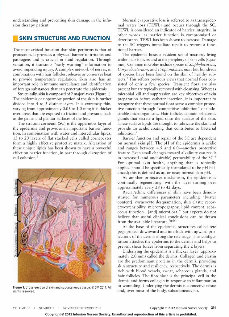
Structurally, skin is composed of 2 major layers (Figure 1). The epidermis or uppermost portion of the skin is further divided into 4 to 5 distinct layers. It is extremely thin, varying from approximately 0.05 to 1.0 mm; it is thicker over areas that are exposed to friction and pressure, such as the palms and plantar surfaces of the feet.
The stratum corneum (SC) is the uppermost layer of the epidermis and provides an important barrier func-tion. In combination with water and intercellular lipids, 15 to 20 layers of flat stacked cells called corneocytes form a highly effective protective matrix. Alteration of these unique lipids has been shown to have a powerful effect on barrier function, in part through disruption of cell cohesion.3
Normal evaporative loss is referred to as transepider-mal water loss (TEWL) and occurs through the SC. TEWL is considered an indicator of barrier integrity; in other words, as barrier function is compromised or deteriorates, TEWL has been shown to increase. Damage to the SC triggers immediate repair to restore a func-tional barrier.
The epidermis hosts a resident set of microbes living within hair follicles and at the periphery of skin cells (squa-mes). Common microbes include species of Staphylococcus, Corynebacterium, and Propionibacterium, but hundreds of species have been found on the skin of healthy sub-jects.4 This refutes previous views that normal flora con-sisted of only a few species. Transient flora are also present but are typically removed with cleansing. Whereas microbial kill and suppression are key objectives of skin preparation before catheter insertion, it is important to recognize that these normal flora serve a complex protec-tive function through “competitive inhibition” of unde-sirable microorganisms. Hair follicles contain sebaceous glands that secrete a lipid onto the surface of the skin. These surface lipids are thought to lubricate the skin and provide an acidic coating that contributes to bacterial inhibition.5
Barrier function and repair of the SC are dependent on normal skin pH. The pH of the epidermis is acidic and ranges between 4.5 and 6.0—another protective feature. Even small changes toward alkalinity can result in increased (and undesirable) permeability of the SC.6 For optimal skin health, anything that is topically applied should be specifically formulated to be pH bal-anced; this is defined as at, or near, normal skin pH.
As another protective mechanism, the epidermis is continually regenerating, with the layer turning over approximately every 28 to 42 days.
Racial/ethnic differences in skin have been demon-strated for numerous parameters including “[water content], corneocyte desquamation, skin elastic recov-ery/extensibility, microtopography, lipid content, seba-ceous function…[and] microflora,” but experts do not believe that useful clinical conclusions can be drawn from the available literature.7(p26)
At the base of the epidermis, structures called rete pegs project downward and interlock with upward pro-jections of the dermis along the rete ridge. This configu-ration attaches the epidermis to the dermis and helps to prevent shear forces from separating the 2 layers.
Underlying the epidermis is a thicker layer (approxi-mately 2.0 mm) called the dermis. Collagen and elastin are the predominant proteins in the dermis, providing skin structure and resiliency, respectively. The dermis is rich with blood vessels, sweat, sebaceous glands, and hair follicles. The fibroblast is the principal cell in the dermis and forms collagen in response to inflammation or wounding. Underlying the dermis is connective tissue and, over most of the body, subcutaneous fat.
Figure 1. Cross-section of skin and subcutaneous tissue. © 3M 2011. All rights reserved.
NAN200243.indd 391NAN200243.indd 391 10/28/12 11:20 AM10/28/12 11:20 AM
Copyright © 2012 Infusion Nurses Society. Unauthorized reproduction of this article is prohibited.
392 Copyright © 2012 Infusion Nurses Society Journal of Infusion Nursing
Although the SC is a highly effective barrier, it is not completely impermeable. Under certain conditions, sub-stances can enter through and between cells and around hair follicles. Permeation is governed by a number of factors including body location and skin characteristics. Chemical characteristics of the substance in question, including molecular size, determine whether a substance will enter the skin. The majority of nonmedicated, topi-cally applied products used in clinical practice are intended to remain on the skin’s surface. In contrast, topically applied medications are specifically formulat-ed to penetrate into deeper layers of the epidermis or the dermis, where they can gain access to the vasculature. Topical antimicrobials are intended to penetrate into the superficial layers of the SC to kill microbes.
Not surprisingly, damaged or diseased skin forms a less effective barrier with greater chance of penetration. The skin’s “microclimate” will also affect permeation of topically applied substances. Microclimate refers to the interaction of skin temperature and humidity. Although primarily used in the domain of pressure ulcer preven-tion, the concept has relevance here. For every 1.5°C increase in body temperature, sweat rate doubles, with wetness increasing skin surface permeability.8
At extremes of age, there are significant non–sun-related structural and functional variations in skin. It is beyond the scope of this paper to review the character-istics of neonatal skin. The reader is referred to the work of Lund and Kuller9 for a comprehensive discus-sion of this topic.
In the elderly, epidermal-dermal junction thinning as well as a decrease in collagen and elastin are seen. These alterations have the net effect of making the skin more susceptible to mechanical injury from friction and adhe-sive use. Additionally, elderly skin has been noted to have diminished perfusion, altered immune response, and reduced response to growth factors necessary to trigger healing. This may result in delayed barrier repair and healing once skin is damaged.10
INTRAVENOUS THERAPY AND THE POTENTIAL TO INDUCE SKIN DAMAGE
From the perspective of skin health, all interventions fundamental to intravenous (IV) catheter insertion and site maintenance have the potential to affect skin integ-rity. The impact of site location on skin condition, espe-cially with regard to central venous catheters (subclavi-an versus internal jugular versus femoral), has not been reported.
Any solvent or detergent has the potential to disrupt intercellular lipids and create barrier damage.11 Antimicrobial preparation is unquestionably necessary for skin antisepsis but by design interferes with normal
flora and likely modifies skin pH to some degree. Despite their accepted safety, alcohol (a solvent), chlo-rhexidine gluconate (CHG), and povidone-iodine have all been shown to cause contact reactions in healthy subjects.12-14 Preparations applied using a scrubbing technique effectively disrupt skin squames to dislodge flora, but they also create frictional forces on the skin surface. The contribution of this effect in potentiating skin damage is unknown but must be considered. After antimicrobial preparation, catheter insertion creates an unavoidable full-thickness wound at the entry site. Securement helps to prevent subsequent vertical and rotational movement of a catheter that can cause addi-tional site trauma.15
Lastly, a reliable method of securement is necessary to provide catheter security and site protection, with adhesive dressings and devices being the standard of care.16 Patients with central lines in place for extended periods, including those with peripherally inserted cen-tral catheters, find themselves in need of repeated appli-cation of antimicrobials in addition to removal and reapplication of adhesives.
Many factors govern whether an adhesive will affix as desired and how it will be tolerated by the individual. It is likely that these factors interact in clinical situations in ways that are not completely understood. Although all individuals possess the same skin structure, there is tremendous diversity in “skin type,” with variability in dryness as well as reactivity to topically applied sub-stances. In a recent survey, 44.6% of adults surveyed reported their skin as “sensitive” or “very sensitive.”17 Not surprisingly, skin that is damaged or diseased is less likely to tolerate and successfully wear adhesives than skin with a healthy, intact SC.
The skin’s microclimate exhibits significant variation between individuals, with skin moisture dependent on environmental humidity and, as previously described, body temperature. Adhesion to dry skin is more consist-ently achievable than adhesion to moist or wet skin.
Adhesives are better tolerated and, not surprisingly, are less likely to fail in areas that are not subject to movement. Sites that routinely move, crease, or fold can distort the adhesive. Excessive hair is another factor that adversely affects adhesion, as well as complicating adhesive removal.
Interestingly, despite the frequent use of adhesive dressings and tapes for patient care, there has been scant research published on the individual or cumula-tive impact of the above factors in clinical settings.
COMMON ETIOLOGIES OF SKIN DAMAGE
For more than 40 years, patients with urinary and fecal diversions (ostomies) have been wearing adhesive
NAN200243.indd 392NAN200243.indd 392 10/28/12 11:20 AM10/28/12 11:20 AM
Copyright © 2012 Infusion Nurses Society. Unauthorized reproduction of this article is prohibited.
VOLUME 35 | NUMBER 6 | NOVEMBER/DECEMBER 2012 Copyright © 2012 Infusion Nurses Society 393
products over extended periods of months to years. Removal and reapplication 2 to 3 times per week is typical, and skin damage is a common complication. WOC nurses’ observations have led to a better under-standing and categorization of the most frequently observed skin problems.18,19 These include (1) moisture-associated skin damage, (2) contact dermatitis, and (3) mechanical trauma induced by adhesives. This knowl-edge may provide a useful construct to better under-stand the likely etiologies of skin damage in the infusion therapy population.
Moisture-Associated Skin Damage
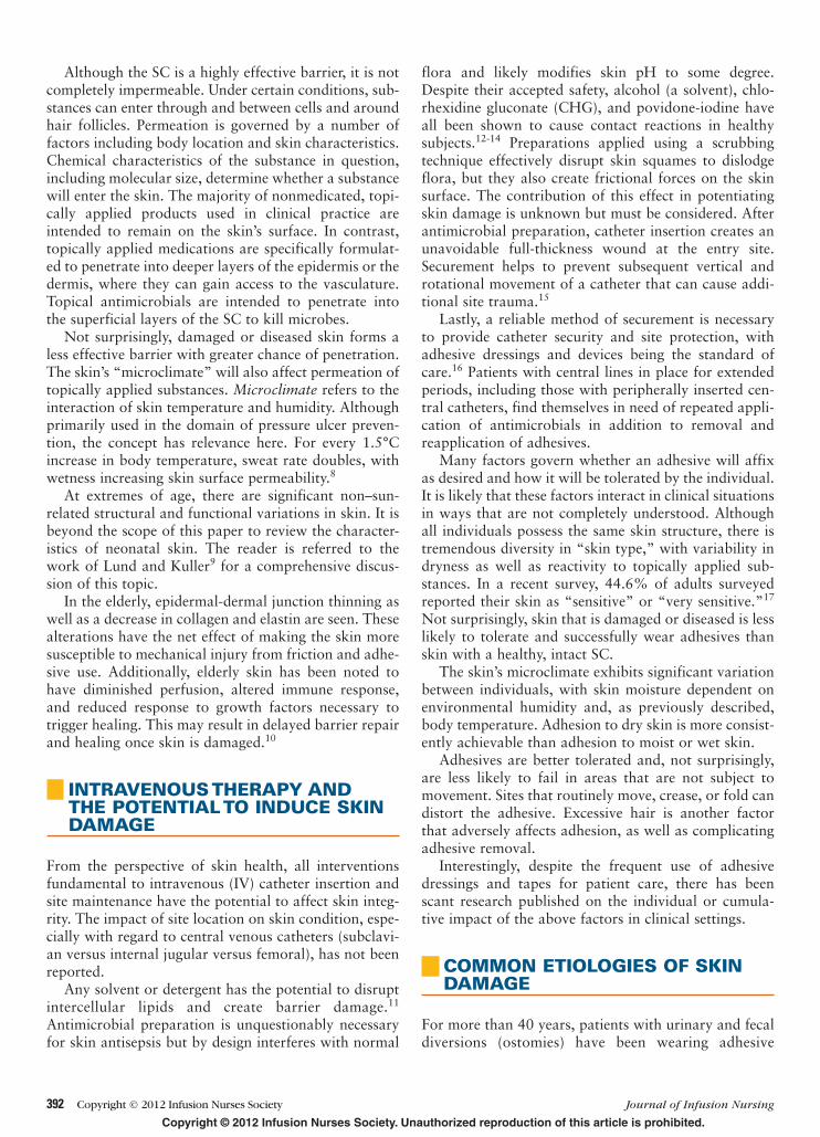
Skin is normally dry, with the capability of transpiring moisture in the form of TEWL. Three primary changes have been observed when skin is exposed to excessive hydration, typically occurring when occlusive or poorly breathable adhesive products are used or the patient experiences significant diaphoresis. First, an increase in permeability of the SC is noted.20-22 This represents a sign of compromised barrier integrity. There is an alka-line shift in skin pH, which also is adverse for barrier health as previously noted. Lastly, inflammation is trig-gered. Clinically, the skin is moist, soft, and, over time, changes in color, becoming white or gray. The texture becomes “soggy,” and with ongoing exposure the skin surface may actually crumble. This type of skin damage has been traditionally described as maceration and is illustrated in Figure 2. More recently, the term mois-ture-associated skin damage has been used.23
Friction is a superficial mechanical force that creates damage without tissue deformation. Frictional forces are increased when moisture is present, making wet skin more likely than dry skin to sustain damage.24 It is plau-sible and likely that minute frictional forces are present under dressings or around devices at infusion sites such as the neck and antecubital space where movement is
inevitable. Partial-thickness ulcerations that develop at the periphery of adhesive dressings are thought to reflect this type of mechanical damage. In a patient with significant diaphoresis, moisture and friction could be key factors contributing to skin injury under a secure-ment dressing.
Contact Dermatitis
When erythema is observed in connection with use of topical products, it is common for clinicians to presume an allergic reaction. In actuality, 2 primary forms of contact dermatitis have been described in the dermatol-ogy and dermatotoxicology literature.
Irritant contact dermatitis (ICD) is a nonallergic inflammatory response triggered by exposure to an irri-tating substance. It comprises a group of skin reactions that are complex and incompletely understood. The threshold for reaction is based on the offending sub-stance and varies from person to person. In the case of caustic alkalinic or acidic industrial chemicals, onset can be immediate and the skin damage severe. Within health care settings, a reaction more typically occurs over hours to days. Erythema, edema, and vesicle for-mation may be seen in addition to other “primary” lesions. A cumulative ICD may be seen as a result of repeated skin exposure and may persist for extended periods even after the initiating substance has been removed.25 Interestingly, a subgroup of individuals within ICD are referred to as “stingers”; they react with discomfort to topically applied substances, yet they do not demonstrate visible signs of inflammation.
By contrast, allergic contact dermatitis (ACD) is an immune-mediated inflammatory response characterized by 2 phases. During sensitization, an allergen (ie, the antigen) in the form of a small molecule called a hapten penetrates the SC. Within the epidermis, these molecules bind with immune cells (Langerhans cells) and are trans-ported to the lymph nodes, where they are presented to T lymphocytes. If the T cell possesses a complementary receptor for the antigen, the now-sensitized T cells pro-liferate and return to skin. In the second phase (termed elicitation), T lymphocytes accumulate at the site of exposure. When the offending substance is presented again by the Langerhans cells, both antigen-specific and nonspecific T cells produce inflammatory mediators including lymphokines and cytokines. Additionally, mast cells and basophils release histamine and other inflam-mation-producing substances. The clinical results of these chemicals are the classic observable signs of ery-thema, edema, and pruritis. In many reactions, primary lesions including macules, papules, vesicles, bullae, and wheals are noted. Some of these cellular chemicals (tumor necrosis factor beta, for example) are capable of exerting a direct cytotoxic effect on epidermal cells lead-ing to lysis and skin damage.
Figure 2. An example of maceration induced by an occlusive adhesive product. © 3M 2011. All rights reserved.
NAN200243.indd 393NAN200243.indd 393 10/28/12 11:20 AM10/28/12 11:20 AM
Copyright © 2012 Infusion Nurses Society. Unauthorized reproduction of this article is prohibited.
394 Copyright © 2012 Infusion Nurses Society Journal of Infusion Nursing
The sensitization phase is generally thought to occur over days to weeks; however, a potent allergen can cause much more rapid response, and the sensitization and elicitation phases can be combined. Not surpris-ingly, sensitization is more easily induced when the bar-rier integrity is compromised but still requires antigen-specific susceptibility. Corticosteroids, both systemic and topical, affect the patient’s ability to mount an aller-gic response. Patients who have experienced long-term topical corticosteroid therapy have reduced density of Langerhans cells in skin and inhibited induction of con-tact sensitization. Systemic corticosteroids may compro-mise T lymphocyte function as well as capability of Langerhans cells.
It is important to understand that it is virtually impossible to distinguish ICD from ACD by clinical (visual) assessment.26,27 The affected area is typically erythemic and characterized by varying shades of pink to red, but it often features macules, papules, and vesi-cles in the form of a diffuse rash. Demarcation or delineation of the affected area has traditionally been offered as a diagnostic differentiator, but there is sub-stantial overlap in the presentation of ICD and ACD, making this clinical feature of limited use for most clini-cians.
The impact of topical antimicrobials on adhesive tol-erance and potential contribution to irritant dermatitis must be considered. As noted by Shelanski et al,28(p138) “Any topically applied chemical substance has the potential to induce an irritant or hypersensitization reaction in any individual at some time.” Most secure-ment dressings are breathable to some degree but also provide some measure of occlusion. It is well accepted that occlusion enhances penetration of topically applied substances into the skin; this is related to the fact that occlusion makes the SC more permeable. It is plausible that the presence of dressings or devices on the skin may enhance penetration of an antimicrobial sufficiently into the skin to trigger an irritant reaction in some patients. It is not known whether a reaction is more likely to occur as a result of the preparation alone (ie, independent of the dressing) or the combination of the preparation and adhesive of the dressing/device.
At the time of this writing, data on incidence of con-tact dermatitis (ICD or ACD) in the infusion therapy population are limited. In a large trial comparing a CHG-impregnated sponge versus standard care with a transparent dressing, Timsit et al29 reported that 8 out of 817 patients (approximately 1%) experienced “severe contact dermatitis” when exposed to the sponge. They distinguished this from “skin allergy,” which presented in 1 patient each in the sponge and control groups, respectively. It was not reported whether patch testing had been performed to establish the diagnosis of allergy.
Estimating dermatitis in the infusion therapy popula-tion based on numbers in the general population is also
difficult. Overall incidence of dermatitis in the general population is estimated to be 14%,30 and 80% of occu-pational dermatitis is reported to be irritant versus allergic.31 Numbers must be interpreted cautiously, however, because patch testing is not always done, and, as previously noted, definitive diagnosis is difficult without it. In the ostomy population, where patch test-ing is done with some frequency, ACD occurred in only 0.7% of one sample.32 Anecdotally, WOC nurses report it to be much less common than ICD. Although it is common for patients to attribute what they presume is allergy to adhesive products (especially tape), skin dam-age is likely mechanical in many of these situations and unrelated to a true immune response.
Adhesive Trauma
Adhesive removal can be expected to detach skin cells,33-35 but in some situations it results in significant disruption to the epidermis. Few studies have been pub-lished that examine the incidence or effects of repeated adhesive application and removal in clinical situations in which adhesives are worn over extended periods and with repeated application. Ostomy clinicians frequently offer anecdotal descriptions of adhesive damage, but published data are limited. Clinical understanding has also not been aided by use of imprecise clinical terms such as tape burn to describe skin injuries associated with adhesive use. Orthopedic studies have evaluated the effects of postoperative dressings in total joint arthroplasty patients but typically have focused on adverse effects after a single adhesive application or limited numbers of dressing changes.36-38 Konya et al39 evaluated skin injury from medical adhesives in a popu-lation aged 65 and older and reported a 20% incidence rate of skin damage at “hyperalimentation” infusion sites.
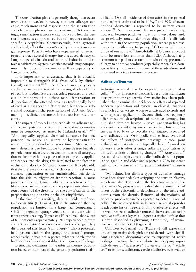
Two related but distinct types of adhesive damage have been described: skin stripping and tension blisters, which are also described in the literature as tape blis-ters. Skin stripping is used to describe delamination of layers of the epidermis or detachment of the entire epi-dermis from the dermis. Removal of the majority of adhesive products can be expected to detach layers of cells. If the recovery time in between removal episodes is adequate for cell regeneration, no adverse effects will be seen. Repeated adhesive removal, however, can easily remove sufficient layers to expose a moist surface that is often described as glistening. Over time, inflamma-tion will also be noted (Figure 3).
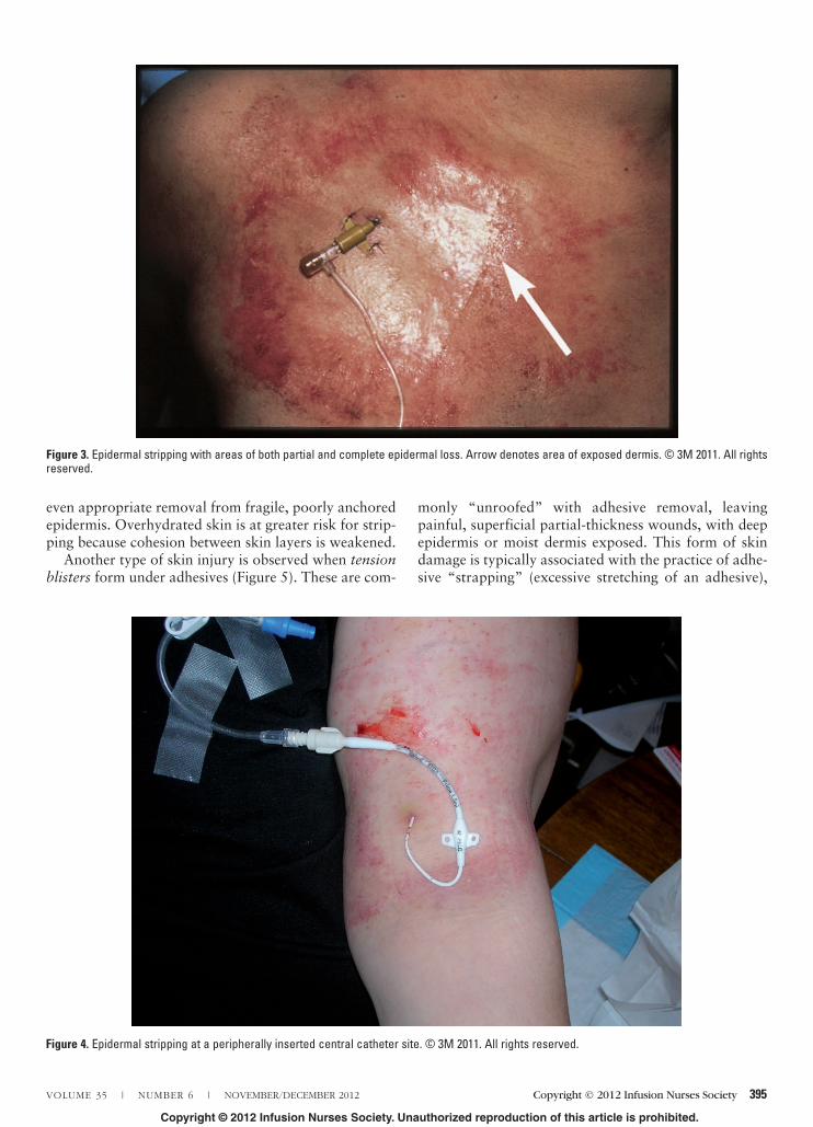
Complete epidermal loss (Figure 4) will expose the underlying moist dark pink or red dermis with signifi-cant associated discomfort due to exposure of nerve endings. Factors that contribute to stripping injury include use of “aggressive” adhesives, use of “tackifi-ers” to enhance adhesion, careless adhesive removal, or
NAN200243.indd 394NAN200243.indd 394 10/28/12 11:20 AM10/28/12 11:20 AM
Copyright © 2012 Infusion Nurses Society. Unauthorized reproduction of this article is prohibited.
VOLUME 35 | NUMBER 6 | NOVEMBER/DECEMBER 2012 Copyright © 2012 Infusion Nurses Society 395
even appropriate removal from fragile, poorly anchored epidermis. Overhydrated skin is at greater risk for strip-ping because cohesion between skin layers is weakened.
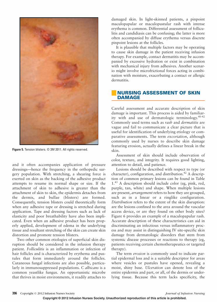
Another type of skin injury is observed when tension blisters form under adhesives (Figure 5). These are com-
monly “unroofed” with adhesive removal, leaving painful, superficial partial-thickness wounds, with deep epidermis or moist dermis exposed. This form of skin damage is typically associated with the practice of adhe-sive “strapping” (excessive stretching of an adhesive),
Figure 3. Epidermal stripping with areas of both partial and complete epidermal loss. Arrow denotes area of exposed dermis. © 3M 2011. All rights reserved.
Figure 4. Epidermal stripping at a peripherally inserted central catheter site. © 3M 2011. All rights reserved.
NAN200243.indd 395NAN200243.indd 395 10/28/12 11:20 AM10/28/12 11:20 AM
Copyright © 2012 Infusion Nurses Society. Unauthorized reproduction of this article is prohibited.
396 Copyright © 2012 Infusion Nurses Society Journal of Infusion Nursing
and it often accompanies application of pressure dressings—hence the frequency in the orthopedic sur-gery population. With stretching, a shearing force is exerted on skin as the backing of the adhesive product attempts to resume its normal shape or size. If the attachment of skin to adhesive is greater than the attachment of skin to skin, the epidermis detaches from the dermis, and bullae (blisters) are formed. Consequently, tension blisters could theoretically form when any adhesive tape or dressing is stretched during application. Tape and dressing factors such as lack of elasticity and poor breathability have also been impli-cated. Even when an adhesive product has been prop-erly applied, development of edema in the underlying tissue and resultant stretching of the skin can create skin distortion and promote tension blisters.
Two other common etiologies of superficial skin dis-ruption should be considered in the infusion therapy patient. Folliculitis is an inflammation or infection of hair follicles and is characterized by erythema and pus-tules that form immediately around the follicles. Cutaneous fungal infections are also possible, particu-larly in immunosuppressed populations. C albicans is a common yeastlike fungus. An opportunistic microbe that thrives in moist environments, it readily attaches to
damaged skin. In light-skinned patients, a pinpoint maculopapular or maculopustular rash with intense erythema is common. Differential assessment of follicu-litis and candidiasis can be confusing; the latter is more often accompanied by diffuse erythema versus discrete pinpoint lesions at the follicles.
It is plausible that multiple factors may be operating to cause skin damage in the patient receiving infusion therapy. For example, contact dermatitis may be accom-panied by excessive hydration or exist in combination with mechanical injury from adhesives. Another scenar-io might involve microfrictional forces acting in combi-nation with moisture, exacerbating a contact or allergic dermatitis.
NURSING ASSESSMENT OF SKIN DAMAGE
Careful assessment and accurate description of skin damage is important. This process is aided by familiar-ity with and use of dermatologic terminology.40-42 Commonly used terms such as rash and dermatitis are vague and fail to communicate a clear picture that is useful for identification of underlying etiology or com-parative assessments. The term excoriation, although commonly used by nurses to describe skin damage featuring erosion, actually defines a linear break in the skin.
Assessment of skin should include observation of color, texture, and integrity. It requires good lighting, attention to detail, and patience.
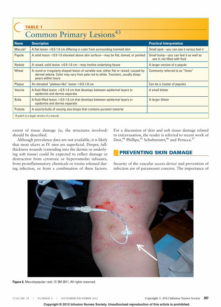
Lesions should be described with respect to type (or character), configuration, and distribution.42 A descrip-tion of common primary lesions can be found in Table 1.43 A description should include color (eg, pink, red, purple, tan, white) and shape. When multiple lesions are present, arrangement refers to how they are grouped, such as in a linear or a ringlike configuration. Distribution refers to the extent of the skin disruption: are the lesions confined to the area around the vascular access device, or are they found on other body sites? Figure 6 provides an example of a maculopapular rash. Accurate description of these characteristics can aid in discriminating an infectious versus inflammatory proc-ess and may assist in distinguishing IV site-specific skin damage from dermatologic disorders that stem from systemic disease processes or reactions to therapy (eg, patients receiving certain chemotherapeutics or targeted therapy).
The term erosion is commonly used to indicate par-tial epidermal loss and is a suitable descriptor for areas where vesicles or pustules have opened, revealing a moist, shiny base. Ulceration can denote loss of the entire epidermis and part, or all, of the dermis or under-lying tissue. Because this term lacks specificity, the
Figure 5. Tension blisters. © 3M 2011. All rights reserved.
NAN200243.indd 396NAN200243.indd 396 10/28/12 11:20 AM10/28/12 11:20 AM
Copyright © 2012 Infusion Nurses Society. Unauthorized reproduction of this article is prohibited.
VOLUME 35 | NUMBER 6 | NOVEMBER/DECEMBER 2012 Copyright © 2012 Infusion Nurses Society 397
extent of tissue damage (ie, the structures involved) should be described.
Although prevalence data are not available, it is likely that most ulcers at IV sites are superficial. Deeper, full-thickness wounds (extending into the dermis or underly-ing soft tissue) could be expected to reflect damage or destruction from cytotoxic or hyperosmolar infusates, from proinflammatory chemicals or toxins released dur-ing infection, or from a combination of these factors.
For a discussion of skin and soft tissue damage related to extravasation, the reader is referred to recent work of Dest,44 Phillips,45 Schulmeister,46 and Perucca.47
PREVENTING SKIN DAMAGE
Security of the vascular access device and prevention of infection are of paramount concern. The importance of
TABLE 1
Common Primary Lesions43
Name Description Practical Interpretation
Maculea A flat lesion �0.5-1.0 cm differing in color from surrounding (normal) skin Small spot—you can see it versus feel it
Papule A solid lesion �0.5-1.0 elevated above skin surface—may be flat, domed, or pointed Small bump—you can feel it as well as see it; not filled with fluid
Nodule A raised, solid lesion �0.5-1.0 cm—may involve underlying tissue A larger version of a papule
Wheal A round or irregularly shaped lesion of variable size; either flat or raised; caused by dermal edema. Color may vary from pale red to white. Transient, usually disap-
pears within hours
Commonly referred to as “hives”
Plaque An elevated “plateau-like” lesion �0.5-1.0 cm Can be a cluster of papules
Vesicle A fluid-filled lesion �0.5-1.0 cm that develops between epidermal layers or epidermis and dermis separate
A small blister
Bulla A fluid-filled lesion �0.5-1.0 cm that develops between epidermal layers or epidermis and dermis separate
A larger blister
Pustule A vesicle/bulla of varying size/shape that contains purulent material
aA patch is a larger version of a macule.
Figure 6. Maculopapular rash. © 3M 2011. All rights reserved.
NAN200243.indd 397NAN200243.indd 397 10/28/12 11:20 AM10/28/12 11:20 AM
Copyright © 2012 Infusion Nurses Society. Unauthorized reproduction of this article is prohibited.
398 Copyright © 2012 Infusion Nurses Society Journal of Infusion Nursing
skin integrity, however, cannot be overemphasized. The critical nature of intact skin as a barrier to pathogens compels the clinician to view skin damage prevention as an infection control strategy. Consequently, all interven-tions for site care should be designed and implemented with maintenance or improvement of skin integrity in mind.
In nonemergent situations, preparation of the surface before antimicrobial application should be considered. Excessive hair harbors bacteria and interferes with adhesion. Hair can be removed with a surgical clipper before application of preparations. From an infection control perspective, clippers have been shown to pro-vide a benefit over razors.48
Trapping wet solutions under an adhesive can increase the risk of irritant dermatitis or moisture-associated skin damage. Antimicrobial preparations should be allowed to dry completely before dressing application. Manufacturers’ instructions should serve as a guide, but variability in skin type as well as environ-mental conditions will dictate additional drying time for some patients.
It is well accepted that adhesive removal results in detachment of superficial SC, and repeated removal results in inflammation and irritation.49 Application of an alcohol-free barrier film may mitigate adhesive strip-ping by forming a protective interface between the epi-dermis and the adhesive. Barrier films are liquids, mini-mally consisting of a polymer and a solvent. The solvent delivers the polymer and then evaporates off, leaving the polymer on the skin as a transparent, breathable protective coating. Although many of these products are similar in appearance, there are significant differences in their composition that can affect their ability to protect the skin. Formulations in which the polymer is poorly dissolved in the solvent will tend to be slow-drying and sticky, making the product difficult to work with and potentially interfering with adhesion. The chemistry of the polymer is also important. The polymer should pro-vide a waterproof coating on the skin and be compati-ble with CHG. Robert Asmus (Division Scientist, 3M HealthCare, personal communication) describes CHG as a “social molecule,” meaning that it is reactive with many other compounds and, in particular, those that are anionic (ie, negatively charged). When CHG comes in contact with an anionic compound, it is converted, wholly or partly, to an insoluble salt, and a loss of anti-bacterial action is to be expected.50,51 Nonionic com-pounds will not react with CHG. Consequently, they are thought not to pose a risk for incompatibility with the antimicrobial.
In barrier films, the presence of an additional ingredi-ent called a plasticizer is advantageous in that it softens the film. This makes it less brittle and more likely to flex and bend without cracking, thus maintaining an intact protective barrier over the surface of the skin. This is
especially important when a barrier film is applied over areas that flex and crease, such as the neck.
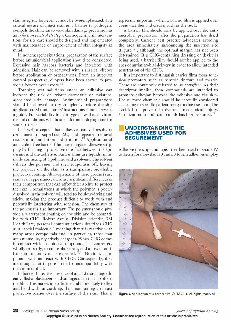
A barrier film should only be applied over the anti-microbial preparation after the preparation has dried completely. Current best practice advocates avoiding the area immediately surrounding the insertion site (Figure 7), although the optimal margin has not been determined. If a CHG-containing dressing or device is being used, a barrier film should not be applied to the area of antimicrobial delivery in order to allow intended penetration of the CHG.
It is important to distinguish barrier films from adhe-sion promoters such as benzoin tincture and mastic. These are commonly referred to as tackifiers. As their descriptor implies, these compounds are intended to promote adhesion between the adhesive and the skin. Use of these chemicals should be carefully considered according to specific patient need; routine use should be avoided to prevent inadvertent mechanical injury. Sensitization to both compounds has been reported.52
UNDERSTANDING THE ADHESIVES USED FOR SECUREMENT
Adhesive dressings and tapes have been used to secure IV catheters for more than 30 years. Modern adhesives employ
Figure 7. Application of a barrier film. © 3M 2011. All rights reserved.
NAN200243.indd 398NAN200243.indd 398 10/28/12 11:20 AM10/28/12 11:20 AM
Copyright © 2012 Infusion Nurses Society. Unauthorized reproduction of this article is prohibited.
VOLUME 35 | NUMBER 6 | NOVEMBER/DECEMBER 2012 Copyright © 2012 Infusion Nurses Society 399
complex chemistry that affects dressing/device performance and skin condition. Understanding adhesive characteristics can enable the clinician to optimize product selection and clinical use to achieve securement and avoid skin injury.
Most adhesive dressings and tapes used for IV therapy incorporate pressure-sensitive adhesives (PSAs). On visual inspection, skin looks like an even surface. Microscopically, however, skin is highly irregular, with a “peak and valley” appearance. When gentle but firm pressure is applied dur-ing placement, adhesives with viscoelastic properties behave like liquids and “flow” into the skin’s contours, promoting optimal adhesion. As a result, cohesion between the dressing (or tape) and the skin is optimized. A well-formulated PSA should possess a “strong holding force,” meaning it will adhere to the surface for the desired period of time, yet it can be removed without leav-ing residue on the skin.51 PSAs vary in how adhesion builds on the skin over time. Removal of an adhesive dur-ing its peak adhesion may increase the possibility of skin trauma. Understanding this characteristic is critical in order to select a product whose performance is consistent with the desired protocol for wear time.
Strong holding force should be distinguished from an aggressive adhesive. Aggressive adhesives may incorpo-rate PSAs but often include rubber-based systems—either natural or synthetic. Aggressive adhesives tend to have thicker adhesive coatings, which create higher adhesion. Moisture-occlusiveness of the adhesive or adhesive back-ing is another factor that can create high adhesion. As the skin overhydrates from a moisture barrier on its surface, cohesive strength between cells is decreased or lost. As a result, removal of the tape can result in skin stripping. For these reasons, routine use of aggressive adhesives should be avoided because of the risk of skin damage.
Breathability of dressings and tapes contributes to the ability to manage the skin’s normal TEWL while maintaining adhesion. Some PSA formulations are designed to perform better on moist surfaces, but medi-cal adhesives do not perform optimally when skin is wet. This characteristic reinforces the need to allow all topicals to dry before adhesive application.
Application and removal of adhesive dressings or tapes are simple yet critical steps in the prevention of skin dam-age. Stretching an adhesive product during application does not improve adhesion and, as previously noted, will result in shearing forces at the skin surface that increase the likelihood of tension blisters and skin trauma. As a result, adhesives should be applied without tension.
In intensive care areas where patients are likely to expe-rience fluid shifts and develop generalized edema, the use of gentle, stretchable adhesives is optimal to better accom-modate stretching of the skin. Staff, however, should be educated not to stretch the adhesive on application and to redress a site to avoid skin injury should edema develop.
Removal is another important consideration. “Peel force” is a key factor associated with skin damage and
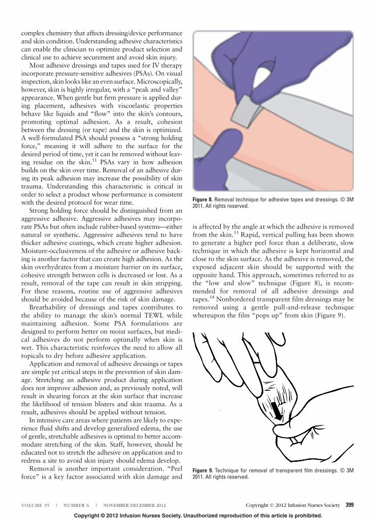
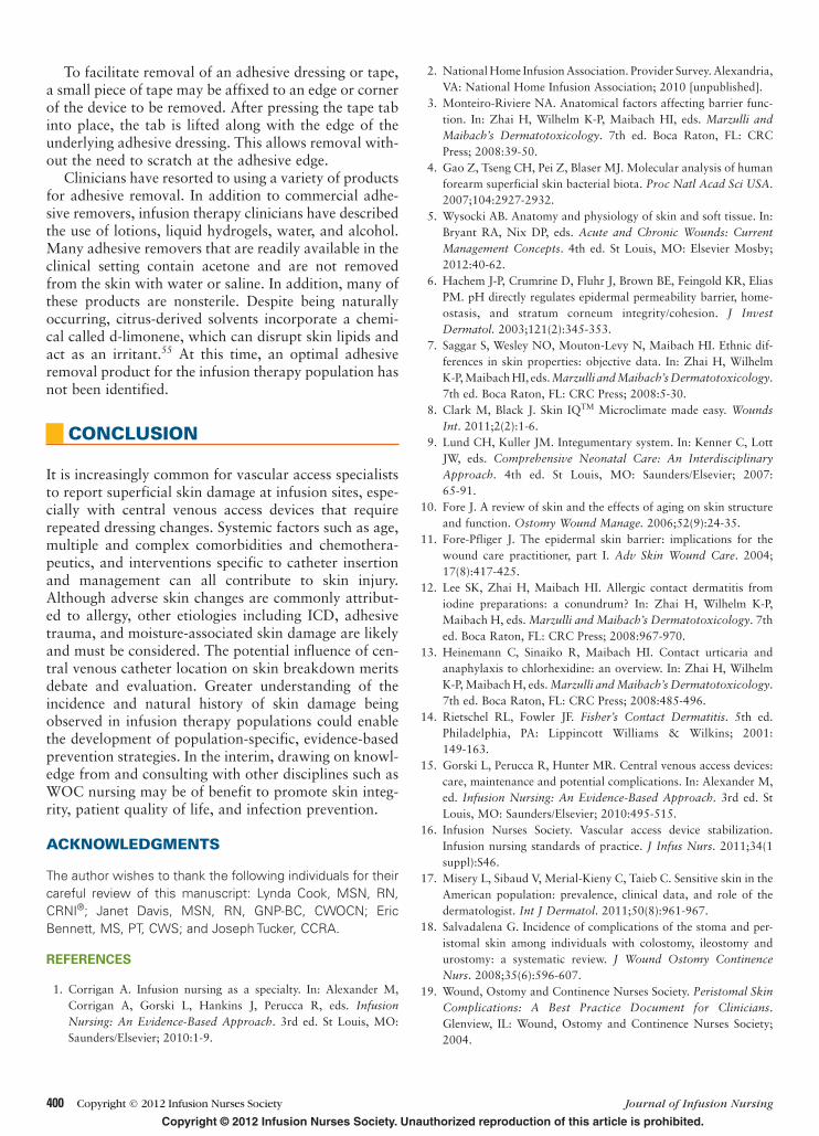
is affected by the angle at which the adhesive is removed from the skin.53 Rapid, vertical pulling has been shown to generate a higher peel force than a deliberate, slow technique in which the adhesive is kept horizontal and close to the skin surface. As the adhesive is removed, the exposed adjacent skin should be supported with the opposite hand. This approach, sometimes referred to as the “low and slow” technique (Figure 8), is recom-mended for removal of all adhesive dressings and tapes.54 Nonbordered transparent film dressings may be removed using a gentle pull-and-release technique whereupon the film “pops up” from skin (Figure 9).
Figure 8. Removal technique for adhesive tapes and dressings. © 3M 2011. All rights reserved.
Figure 9. Technique for removal of transparent film dressings. © 3M 2011. All rights reserved.
NAN200243.indd 399NAN200243.indd 399 10/28/12 11:20 AM10/28/12 11:20 AM
Copyright © 2012 Infusion Nurses Society. Unauthorized reproduction of this article is prohibited.
400 Copyright © 2012 Infusion Nurses Society Journal of Infusion Nursing
To facilitate removal of an adhesive dressing or tape, a small piece of tape may be affixed to an edge or corner of the device to be removed. After pressing the tape tab into place, the tab is lifted along with the edge of the underlying adhesive dressing. This allows removal with-out the need to scratch at the adhesive edge.
Clinicians have resorted to using a variety of products for adhesive removal. In addition to commercial adhe-sive removers, infusion therapy clinicians have described the use of lotions, liquid hydrogels, water, and alcohol. Many adhesive removers that are readily available in the clinical setting contain acetone and are not removed from the skin with water or saline. In addition, many of these products are nonsterile. Despite being naturally occurring, citrus-derived solvents incorporate a chemi-cal called d-limonene, which can disrupt skin lipids and act as an irritant.55 At this time, an optimal adhesive removal product for the infusion therapy population has not been identified.
CONCLUSION
It is increasingly common for vascular access specialists to report superficial skin damage at infusion sites, espe-cially with central venous access devices that require repeated dressing changes. Systemic factors such as age, multiple and complex comorbidities and chemothera-peutics, and interventions specific to catheter insertion and management can all contribute to skin injury. Although adverse skin changes are commonly attribut-ed to allergy, other etiologies including ICD, adhesive trauma, and moisture-associated skin damage are likely and must be considered. The potential influence of cen-tral venous catheter location on skin breakdown merits debate and evaluation. Greater understanding of the incidence and natural history of skin damage being observed in infusion therapy populations could enable the development of population-specific, evidence-based prevention strategies. In the interim, drawing on knowl-edge from and consulting with other disciplines such as WOC nursing may be of benefit to promote skin integ-rity, patient quality of life, and infection prevention.
ACKNOWLEDGMENTS
The author wishes to thank the following individuals for their careful review of this manuscript: Lynda Cook, MSN, RN, CRNI®; Janet Davis, MSN, RN, GNP-BC, CWOCN; Eric Bennett, MS, PT, CWS; and Joseph Tucker, CCRA.
REFERENCES
1. Corrigan A. Infusion nursing as a specialty. In: Alexander M, Corrigan A, Gorski L, Hankins J, Perucca R, eds. Infusion Nursing: An Evidence-Based Approach. 3rd ed. St Louis, MO: Saunders/Elsevier; 2010:1-9.
2. National Home Infusion Association. Provider Survey. Alexandria, VA: National Home Infusion Association; 2010 [unpublished].
3. Monteiro-Riviere NA. Anatomical factors affecting barrier func-tion. In: Zhai H, Wilhelm K-P, Maibach HI, eds. Marzulli and Maibach’s Dermatotoxicology. 7th ed. Boca Raton, FL: CRC Press; 2008:39-50.
4. Gao Z, Tseng CH, Pei Z, Blaser MJ. Molecular analysis of human forearm superficial skin bacterial biota. Proc Natl Acad Sci USA. 2007;104:2927-2932.
5. Wysocki AB. Anatomy and physiology of skin and soft tissue. In: Bryant RA, Nix DP, eds. Acute and Chronic Wounds: Current Management Concepts. 4th ed. St Louis, MO: Elsevier Mosby; 2012:40-62.
6. Hachem J-P, Crumrine D, Fluhr J, Brown BE, Feingold KR, Elias PM. pH directly regulates epidermal permeability barrier, home-ostasis, and stratum corneum integrity/cohesion. J Invest Dermatol. 2003;121(2):345-353.
7. Saggar S, Wesley NO, Mouton-Levy N, Maibach HI. Ethnic dif-ferences in skin properties: objective data. In: Zhai H, Wilhelm K-P, Maibach HI, eds. Marzulli and Maibach’s Dermatotoxicology. 7th ed. Boca Raton, FL: CRC Press; 2008:5-30.
8. Clark M, Black J. Skin IQTM Microclimate made easy. Wounds Int. 2011;2(2):1-6.
9. Lund CH, Kuller JM. Integumentary system. In: Kenner C, Lott JW, eds. Comprehensive Neonatal Care: An Interdisciplinary Approach. 4th ed. St Louis, MO: Saunders/Elsevier; 2007:65-91.
10. Fore J. A review of skin and the effects of aging on skin structure and function. Ostomy Wound Manage. 2006;52(9):24-35.
11. Fore-Pfliger J. The epidermal skin barrier: implications for the wound care practitioner, part I. Adv Skin Wound Care. 2004; 17(8):417-425.
12. Lee SK, Zhai H, Maibach HI. Allergic contact dermatitis from iodine preparations: a conundrum? In: Zhai H, Wilhelm K-P, Maibach H, eds. Marzulli and Maibach’s Dermatotoxicology. 7th ed. Boca Raton, FL: CRC Press; 2008:967-970.
13. Heinemann C, Sinaiko R, Maibach HI. Contact urticaria and anaphylaxis to chlorhexidine: an overview. In: Zhai H, Wilhelm K-P, Maibach H, eds. Marzulli and Maibach’s Dermatotoxicology. 7th ed. Boca Raton, FL: CRC Press; 2008:485-496.
14. Rietschel RL, Fowler JF. Fisher’s Contact Dermatitis. 5th ed. Philadelphia, PA: Lippincott Williams & Wilkins; 2001:149-163.
15. Gorski L, Perucca R, Hunter MR. Central venous access devices: care, maintenance and potential complications. In: Alexander M, ed. Infusion Nursing: An Evidence-Based Approach. 3rd ed. St Louis, MO: Saunders/Elsevier; 2010:495-515.
16. Infusion Nurses Society. Vascular access device stabilization. Infusion nursing standards of practice. J Infus Nurs. 2011;34(1 suppl):S46.
17. Misery L, Sibaud V, Merial-Kieny C, Taieb C. Sensitive skin in the American population: prevalence, clinical data, and role of the dermatologist. Int J Dermatol. 2011;50(8):961-967.
18. Salvadalena G. Incidence of complications of the stoma and per-istomal skin among individuals with colostomy, ileostomy and urostomy: a systematic review. J Wound Ostomy Continence Nurs. 2008;35(6):596-607.
19. Wound, Ostomy and Continence Nurses Society. Peristomal Skin Complications: A Best Practice Document for Clinicians. Glenview, IL: Wound, Ostomy and Continence Nurses Society; 2004.
NAN200243.indd 400NAN200243.indd 400 10/28/12 11:20 AM10/28/12 11:20 AM
Copyright © 2012 Infusion Nurses Society. Unauthorized reproduction of this article is prohibited.
VOLUME 35 | NUMBER 6 | NOVEMBER/DECEMBER 2012 Copyright © 2012 Infusion Nurses Society 401
20. Schäfer P, Bewick-Sonntag C, Capri MG, Berardesca E. Physiological changes in skin barrier function in relation to occlusion level, exposure time and climatic conditions. Skin Pharmacol Appl Skin Physiol. 2002;15:7-19.
21. Mayoritz HN, Sims N. Biophysical effects of water and synthetic urine on skin. Adv Skin Wound Care. 2001;14(6):302-308.
22. Matsumura H, Oka K, Umekage K, et al. Effect of occlusion on human skin. Contact Dermatitis. 1995;33:231-235.
23. Gray M, Black JM, Baharestani MM, et al. Moisture-associated skin damage: overview and pathophysiology. J Wound Ostomy Continence Nurs. 2011;38(3):233-241.
24. Sivamani RK, Goodman J, Gitis NV, Maibach HI. Friction coef-ficient of skin in real-time. Skin Res Technol. 2003;9(3):235-239.
25. Widmer J, Elsner P, Burg G. Skin irritant reactivity following experimental cumulative irritant contact dermatitis. Contact Dermatitis. 1994;199(30):33-39.
26. Belsito DV. The diagnostic evaluation, treatment and prevention of allergic contact dermatitis in the new millennium. J Allergy Clin Immunol. 2000;105(3):409-420.
27. Marzulli FN, Maibach HI. Allergic contact dermatitis. In: Zhai H, Wilhelm K-P, Maibach H, eds. Marzulli and Maibach’s Dermatotoxicology. 7th ed. Boca Raton, FL: CRC Press; 2008: 155-157.
28. Shelanski MV, Phillips SB, Potts CE. Evaluation of cutaneous reactivity to recently marketed dermatologic products. Int J Dermatol. 1996;35(2):137-140.
29. Timsit J-F, Schwebel C, Bouadma L, et al. Chlorhexidine-impregnated sponges and less frequent dressing changes for pre-vention of catheter-related infections in critically ill adults. JAMA. 2009;301(12):1231-1241.
30. Centers for Disease Control and Prevention. QuickStats: percent-age of adults aged 20-59 years who reported dermatitis in the preceding year, by sex and race/ethnicity—National Health and Nutrition Examination Survey, United States, 1999-2004. MMWR Morb Mortal Wkly Rep. 2007;56(11):258. http://www.cdc.gov/mmwr/preview/mmwrhtml/mm5611a8.htm. March 23, 2007. Accessed August 31, 2011.
31. National Institute for Occupational Safety and Health. Skin exposures and effects. http://www.cdc.gov/niosh/topics/skin/. Updated August 26, 2010. Accessed August 31, 2011.
32. Lyon CC, Smith AJ, Griffiths CEM, Beck MH. The spectrum of skin disorders in abdominal stoma patients. Br J Dermatol. 2000;143:1248-1260.
33. Dykes PJ, Heggie R, Hill SA. Effects of adhesive dressings on the stratum corneum of the skin. J Wound Care. 2001;10(2):7-9.
34. Alikahn A, Maibach HI. Biology of the stratum corneum: tape stripping and protein quantification. In: Firage MA, Maibach HI, Miller KW, eds. Textbook of Aging Skin. New York, NY: Springer-Verlag; 2009:401-408.
35. Cutting KF. Impact of adhesive surgical tape and wound dressing on the skin with reference to skin stripping. J Wound Care. 2008;17(4):157-162.
36. Jester R, Russell L, Fell S, Williams S, Prest C. A one hospital study of the effect of wound dressings and other related factors on skin blistering following total hip and knee arthroplasty. J Orthop Nurs. 2000;4:71-77.
37. Koval KJ, Egol KA, Polatsch DB, Baskies MA, Homman JP, Hiebert RN. Tape blisters following hip surgery: a prospective,
randomized study of two types of tape. J Bone Joint Surg Am. 2003;85:1884-1887.
38. Clarke JV, Deakin AH, Dillon JM, Emmerson S, Kinnimonth AWG. A prospective clinical audit of a new dressing design for lower limb arthroplasty wounds. J Wound Care. 2009;18(1):5-11.
39. Konya C, Sanada H, Sugama J, et al. Skin injuries caused by medical adhesive tape in older people and associated factors. J Clin Nurs. 2010;19:1236-1242.
40. Nix D. Skin and wound inspection and assessment. In: Bryant RA, Nix DP, eds. Acute and Chronic Wounds: Current Management Concepts. 4th ed. St Louis, MO: Elsevier Mosby; 2012:108-121.
41. Lawton S. Assessing the patient with a skin condition. World Wide Wounds [serial online]. May 2002. http://www.worldwidewounds.com/2002/may/Lawton/Skin-Assessment-Dermatology-Patient.html. Accessed August 11, 2011.
42. Massey D. The value and role of skin and nail assessment in the critically ill. Nurs Crit Care. 2006;11(2):80-85.
43. Habif TP. Principles of diagnosis and anatomy. In: Clinical Dermatology. 5th ed. Philadelphia, PA: Mosby Elsevier; 2010:3-11.
44. Dest VM. Systemic therapy-induced skin reactions. In: Haas ML, Moore-Higgs GJ, eds. Principles of Skin Care and the Oncology Patient. Pittsburgh, PA: Oncology Nursing Society; 2011:141-166.
45. Phillips LD. Complications of infusion therapy: peripheral and central infusions. In: Phillips LD. Manual of IV Therapeutics. 5th ed. Philadelphia, PA: FA Davis Company; 2010:566-574.
46. Schulmeister L. Antineoplastic therapy. In: Alexander M, Corrigan A, Gorski L, Hankins J, Perucca R, eds. Infusion Nursing: An Evidence-Based Approach. 3rd ed. St Louis, MO: Saunders/Elsevier; 2010:366-367.
47. Perucca R. Peripheral venous access devices. In: Alexander M, Corrigan A, Gorski L, Hankins J, Perucca R, eds. Infusion Nursing: An Evidence-Based Approach. St Louis, MO: Saunders/Elsevier; 2010:471-472.
48. Tanner J, Woodings D, Moncaster K. Perioperative hair removal to reduce surgical site infection. Cochrane Database Syst Rev. 2006;3:CD004122.
49. Tokumura F, Umekage K, Sado M, et al. Skin irritation due to repetitive application of adhesive tape: the influence of adhesive strength and seasonal variability. Skin Res Technol. 2005;11:102-106.
50. Holbrook A. The determination of small quantities of chlorhexi-dine in pharmaceutical preparations. J Pharmacol Pharmacother. 1958;10:370.
51. Senior N. Some observations on the formulation and properties of chlorhexidine. J Soc Cosmet Chem. 1973;24:259-278.
52. Rietschel RL, Fowler JF. Fisher’s Contact Dermatitis. 5th ed. Philadelphia, PA: Lippincott Williams & Wilkins; 2001:137-147.
53. Lund CH, Tucker JA. Adhesion and newborn skin. In: Hoath S, Maibach HI, eds. Neonatal Skin: Structure and Function. New York, NY: Marcel Dekker, Inc; 2003:299-324.
54. LaVelle BE. Tape With Care: A Three Part Continuing Education Program. St Paul, MN: 3M Health Care; 2004.
55. Tornier C, Rosdy M, Maibach HI. In vitro skin irritation testing on SkinEthic™ reconstituted human epidermis: reproducibility for fifty chemicals tested with two protocols. In: Zhai H, Wilhelm K-P, Maibach H, eds. Marzulli and Maibach’s Dermatotoxicology. 7th ed. Boca Raton, FL: CRC Press; 2008:927-944.
NAN200243.indd 401NAN200243.indd 401 10/28/12 11:20 AM10/28/12 11:20 AM