Embed Size (px)
Citation preview
Volume 56, Number IPrinted in the U.S.A.
INTERNATIONAL JOURNAL OF LEPROSY
Soluble Antigen of M. leprae Coupled with LiposomesElicits Both "Early" and "Late" Delayed
Hypersensitivity Skin Reactions'Utpal Sengupta, Sudhir Sinha, Gopal Ramu, Ravinder K. Lavania,
and Chhitar M. Gupta2
Lepromin is a preparation of killed My-cobacterium leprae of human or armadilloorigin, and it is used extensively for the as-sessment of delayed-type hypersensitivity(DTH) against M. leprae. After intradermalinjection of lepromin, two types ofskin DTHresponses are seen in people who are sen-sitized: an "early" (Fernandez) reactionwhich peaks at 24-48 hr and a "late" (Mit-suda) reaction which peaks at 3-4 weeks ('7).The late lepromin reaction has shown astrong correlation with immunity to my-cobacterial infections in both experimentalanimals (2) and humans (5). However, theintegrity of bacterial morphology in a lep-romin preparation appears to be essentialfor the development of a late reaction. Ul-trasonically disrupted lepromin or the sol-uble extract of M. leprae (leprosin) elicit onlythe early reaction (22.23). There are specu-lations that the soluble antigen(s) may alsoproduce a late reaction, provided the anti-gen is presented in the same manner as thatof intact Al. leprae (' 7 22). To investigate thispossibility, we have used soluble antigen(s)of M. leprae (23) coupled with liposome asa skin-test antigen, hoping that such a sys-tem would mimic the antigen presentationby intact bacilli.
MATERIALS AND METHODSAll of the chemicals and reagents used in
this study were of the highest purity avail-able. Egg lecithin was prepared as describedearlier (21). Gangliosidcs were isolated from
' Received for publication on 29 July 1987; acceptedfor publication in revised form on 17 September 1987.
2 U. Sengupta, Ph.D.; S. Sinha, M.Sc.; R. K. Lavania,M.D., Central JALMA Institute for Leprosy, Taj Ganj,Agra 282001, India. G. Ramu, M.D., Sacred HeartHospital, Kumbakonam 612401, India. C. M. Gupta,Ph.D., Division of Membrane Biology, Central DrugResearch Institute, Lucknow 226001, India.
buffalo brain using the known method (13).Cholesterol was purchased from CentronResearch Laboratory, Bombay, India, andcrystallized three times from methanol. So-dium cyanoborohydride was from SigmaChemical Company, St. Louis, Missouri,U.S.A. Sodium ' 25I-iodide (carrier-free) wasbought from Bhabha Atomic ResearchCentre, Trombay, India.
M. leprae soluble antigen(s). Soluble an-tigen(s) of M. leprae, prepared by the de-scribed method (23), was kindly provided byDr. R. J. W. Rees, Clinical Research Centre,Harrow, U.K. Briefly, M. leprae purifiedfrom infected armadillo tissues (25) were ul-trasonically disrupted before ultracentrifu-gation (105,000 x g x 30 min). The su-pernatant, termed "leprosin-A," was usedafter protein estimation (14).
'25I-labeling of the soluble antigen(s). Fif-ty mg protein of the soluble antigen(s) (in 50/21) was labeled with ' 25 I using the "Iodogen"technique (7). More than 95% of the radio-activity was incorporated in the protein peakrecovered after Sephadex G-25 gel filtra-tion.
Preparation of liposomes. Unilamellar li-posomes were prepared from egg lecithin(45 AM), cholesterol (45 MM), and ganglio-sides (9 ktIvI) in 2.0 ml borate-buffered saline(10 mM borate containing 60 mM NaC1,pH 8.4) by probe-type sonication and frac-tionated by ultracentrifugation, as describedearlier (").
Covalent coupling of liposomes with an-tigen(s). The soluble protein of Al. lepraewas covalently coupled to the liposome sur-face according to the method of Heath, etal. (")). Briefly, liposomes were oxidized withsodium periodate (2 lir, 25-30°C, dark). Theexcess reagent was removed by gel filtration,and the liposome fractions, after pooling to-gether, were concentrated to about 1 ml (5—
45
46^ International Journal of Leprosy^ 1988
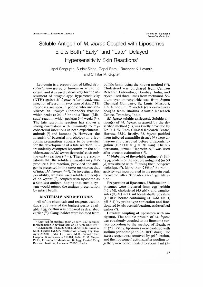
THE TABLE. Early (48 In) and late (3weeks) skin reactions with different antigenpreparations in borderline tuberculoid lep-rosy patients.
Patient Reaction LA L'' LP' L2' L3'• 0'
1 Early 29' 28 30 28 28 0Late 0 10 10 10 0Early 20 25 3/ /6 27 <5Late o 7 9 9 7 0
3 Early 28 22 ND' 46 3/ <5Late 0 0 ND 0 0 0
4 Early 0 0 ND 13 15 <5Late 0 6 ND 9 0 0
5 Early 23 18 ND 27 30 <5Late 0 0 ND 0 0 0
6 Early 12 12 ND 15 13 0Late 0 0 ND 0 0 0
7 Early 15 P ND 14 12 0Late () 5 ND 8 5 0
8 Early /6 24 ND "73 27 0Late 0 12 ND 10 6 0
9 Early 30 22 ND 20 19 0Late 0 5 ND 10 10 0
10 Early 18 16 ND 16 14 0Late 0 5 ND 7 6 0
" LA = leprosin-A (10 pg/ml).L = Dharmendra lepromin (107 bacilli/m1).L1-3 = liposomized leprosin-A at 40, 20, and 10
pg protein/ml.C = control liposomes.
' Indurations in mm.ND = not determined.
7 AM lipid), as described earlier (20). To itwere added the unlabeled Al. leprae anti-gen(s) (about 8 mg), '21-labeled antigen(s)(=50,000 cpm), and sodium cyanoboro-hydride (2 M, 10 pl/m1). The mixture wasincubated at 25-30°C for 14-16 hr. It waschromatographed on a Scpharose 6B col-umn (1.4 x 35 cm) using phosphate-buff-ered saline (10 mM phosphate containing150 M NaC1, pH 7.4; PBS) as the eluant.The liposomes were recovered in the voidvolume. Under these conditions, 18-20%(=1.5 mg unlabeled protein or =10,000cpm) of the protein initially added got cross-linked to the liposome surface. The lipo-some-rich fractions were pooled together,dialyzed against sterile PBS, and filteredthrough a Millipore membrane (0.2 rim). Apreparation of control liposomes was alsosubjected to the above-mentioned antigencoupling steps, in the absence of antigen.
Testing of antigen-coupled liposomes. Theantigen-coupled liposomes were diluted to
10, 20, and 40 pg protein/ml sterile PBSand used for skin testing. Simultaneously,control liposomes (free of antigen), Dhar-mendra lepromin (''), and leprosin-A (23)were also tested. All of the preparations werecoded and tested by a clinician (GR) in adouble-blind manner. Ten borderline tu-berculoid (BT) and five lepromatous (LL)leprosy patients ('s) attending the outpatientdepartment and wards of Central JALMAInstitute of Leprosy (CJIL), Agra, India, wereselected kir the study. The test preparationswere injected intradermally (0.1 ml each)into the back of every patient. The diame-ters of the skin indurations (mean of tworeadings at right angles to each other) wererecorded in millimeters with the help of avernier scale at 48 hr and 21 days. An in-duration of 5 mm or more was regarded aspositive in the case of the 48-hr (early) re-action and that or 4 mm or more was re-garded as positive for the 21-day (late) re-action ("). Biopsy specimens representingthe early (48 hr) and late (21 day) skin re-actions to the liposome-coupled antigenwere fixed in Zenker-formol saline, pro-cessed, and embedded in paraffin. Five-pmsections were stained with hematoxylin andeosin (H&E). The sections were examinedby a pathologist without referring to the casenotes to avoid personal bias.
RESULTSThe Table depicts the skin reactions in
10 BT patients at 48 hr (early) and at 21days (late) elicited by the different antigenpreparations and controls. While leprosin-A(LA) elicited only the early reaction, Dhar-mendra lepromin (L) elicited early, late, orboth early and late reactions. In two patientstested initially, all three concentrations ofliposomized leprosin elicited strong reac-tions. In subsequent patients, the use of thehighest concentration (LI) was omitted. Inall of these patients, liposomized antigenelicited early and late skin reactions akin tothose elicited by Dharmendra lepromin, ex-cept in one patient who was early-reactionnegative with Dharmendra lepromin butpositive with liposomized antigen. Controlliposomes induced insignificant reactions at48 hr in four patients.
The 48-hr skin reaction elicited by lipo-somized leprosin showed a histological pic-
V ^,... % a 14-48111. ‘
1 440‘, 341 ■ uie 0; 4
Ii441, 4 4
6 , .4.1 A 1 6 6I .^AO
56, 1^Sengupta, et al.: M. leprae Antigens in Liposomes^47
FIG. 1. Photomicrograph of 48-hr reaction of a 13Tcase showing lymphocyte and polymorphonuclear in-filtration (H&E x 1200).
FIG. 2. Photomicrograph of 21-day reaction of aBT case showing lymphocytes, epithelioid cells, andgiant cells in the infiltrate (H&E x 1200).
lure of DTH, with a predominant lympho-cytic infiltration. Polymorphonuclear cellswere present to a lesser extent (Fig. 1). The21-day reaction to the liposomized antigenshowed a histological picture resembling atuberculoid granuloma with focal collec-tions of epithelioid and giant cells (Fig. 2).None of the live LL patients showed anypositive skin reaction to these antigens.
DISCUSSIONSeveral studies have shown that the im-
munogenicity of antigens is improved byincorporating them in liposomes C). It hasalso been demonstrated that this action ofliposomes is further enhanced when antigenis covalently attached on their surface (24).
These observations prompted us to cova-lently couple soluble Al. leprae antigen(s)(leprosin) to liposomes, and then to deter-mine whether the liposome-coupled anti-gen(s) could elicit both the early and lateskin DTH responses.
The results of this study support theworking hypothesis that the presentation ofeliciting antigen(s) in a specific orientationis required for the development of late re-
actions. In the method of Heath, et al. (")which we used, protein molecules are co-valently linked with the liposome surfacedue to a reaction between the amino groupsof the proteins and the aldehyde groups ofthe ganglioside via the formation of Schiff'sbases. In the present situation, it seems thatsuch a linkage does not adversely affect theantigenicity of the attached proteins. Ap-parently the liposomes presented surface-bound protein antigens to the host cells inthe specific manner of intact Al. leprae andthus activated the lymphoid cell precursorswhich were responsible for the developmentof the late skin-test reaction.
The cells may not require the continuedpresence of the stimulus for the develop-ment of late reactions, since we know thatliposomes are rapidly disintegrated in vivo('). For the same reason, the adjuvant effectof liposomes may not be operational in thedevelopment of the late reaction. Moreover,in a separate study, when the liposome-en-trapped leprosin was used for skin testingor BT patients it did not produce the latereaction although the early reaction waselicited (data not shown). Both early and
48^ International Journal of Leprosy^ 1988
late reactions to the liposome-coupled an-tigen showed histological pictures corre-sponding to the reactions elicited by integrallepromin (4).
The early reaction elicited by control li-posomes in some of the cases is not signif-icant, since an early reaction of <5 mm isnot regarded as positive (".23).
On the basis of the present study, it is notpossible to define the molecule(s) involvedin the development of the DTH reactions.The decision to use whole (unfractionated)soluble antigen was made because studiesso far have led to diverse conclusions re-garding the physicochemical properties ofthe DTH-eliciting antigens of Al. leprae oreven any other mycobacterium (3). More-over, studies on Al. leprae are limited dueto non-availability of the organisms in suf-ficient quantities. The major source of M.leprae remains the experimentally infectednine-banded armadillo (9. Recently, iso-lation of molecularly defined Al. leprae an-tigens has been made possible with the helpof monoclonal antibodies (6) and gene clon-ing (26). Such preparations would be thoseof choice for future work using liposomes.
There is experimental evidence to suggestthat the late (21 day) DTH reaction is amarker for protective immunity againstmultibacillary mycobacterial infections (2.5).
Since the Al. leprae antigen covalently linkedwith liposomes is capable of eliciting the latereaction, liposome may serve as a carrierand immunopotentiator of activity of mo-lecularly defined, cell-mediated immunity-inducing antigens of Ai. leprae which arepresently being evolved (14. 15).
SUMMARYThe soluble antigen(s) of Mycobacterium
leprac was(were) coupled to liposomes andused for skin testing of leprosy patients,hoping that this mode of antigen presenta-tion would be identical to that of integrallepromin. The liposomized antigen(s) elic-ited both early (24-48 hr) and late (3-4weeks) delayed-type hypersensitivity reac-tions, true to the nature of lepromin, unlikethe soluble antigen(s) alone which elicit(s)only the early reaction.
RESUMENSc incorporaron antigenos solubles de .tiycobucte-
rium leprae a liposomas y Sc usaron para pruebas en
piel en pacientes con lepra esperando que este modode presentaciOn del antigen° pudiera ser identico al dela lepromina integral. Los antigenos incluidos en losliposomas, indujeron tanto las reacciones de hipersen-sibilidad retardada tempranas (24-48 h) como las tar-dias (3-4 semanas), en tanto que los ant igenos solublessolos nada to indujeron la reacciOn temprana.
RESUMEOn a couple l'antigene ou les antigenes de M yco-
bac teri u m leprae n des liposomes pour pratiquer desepreuves cutanees chez des malades de la lepre, dansl'espoir de demontrer que cc mode de presentationantigenique serait identique a celui de la lepromineintegrale. L'antigene ou les antigenes wit entrain& desreactions precoces apres 24 it 48 heures, et des reac-tions tardives du type d'hypersensibilite retardee apres3 et 4 semaines, comme c'est le cas apres injection delepromine, alors que l'antigene on les antigenes so-lublcs administres scuts Wont entrain& qu'une reactionprecoce.
Acknowledgments. We are grateful to Dr. (Ms.) A.Bali. National Institutes of Health, Bethesda, Mary-land, U.S.A., for helping in the standardization of tech-niques. We are also grateful to Dr. K. V. Desikan, CJ IL,Agra, India, for his critical review of the manuscript.Mr. Anil Chopra provided secretarial assistance andMr. H. 0. Agarwal provided the photomicrographs.Part of the work was supported by ad hoc grants fromthe Indian Council of Medical Research (vide scheme1/8/82).
REFERENCES1. BALI, A., DITAWAN, S. and GUPTA, C. M. Stability
of liposomes in circulation is markedly enhancedby structural modification of their phospholipidcomponent. FEBS Lett. 154 (1983) 373-377.
2. CURTIS, J. and TURK, J. L. Mitsuda-type leprom inreaction as a measure of host resistance in My-coltactcTiitni lepraemurium infection. Infect. Im-mun. 24 (1979) 492-500.
3. DANIEL, T. M. and JANICKI, W. Mycobacterial an-tigens: a review of their isolation, chemistry andimmunological properties. Microbiol. Rev. 42(1978) 84-113.
4. DESIKAN, K. V., MUKHERJEE, A., RAMU, G. andTIWARI, V. D. Sequential histological study of lep-romin reaction. Int. J. Lepr. 51 (1983) 473-480.
5. DFIARMENDRA and CHATTERJEE, K. R. Prognosticvalue of the lepromin test in contacts of leprosycases. Lepr. India 27 (1955) 149-153.
6. ENGERS, H. D. Results of a World Health Orga-nization-sponsored workshop on monoclonal an-tibodies to Mycobacterium leprae. Infect. Immun.48 (1985) 603-605.
7. FRAKER, P. J. and SPECK, J. C. Protein and cellmembrane iodinations with a sparingly solublechloramide, 1,3,4,6-tetrachloro 3a, 6a-diphenyl-
56, 1^Sengupta, et al.: M. leprae Antigens in Liposomes^49
glycoluril. Biochem. Biophys. Res. Commun. 80(1978) 849-857.
8. GREGORIADIS, G. Liposomes for drugs and vac-cines. Trends Biotechnol. 3 (1985) 235-241.
9. Gum-A, C. M. and BALI, A. Carbamyl analogs ofphosphatidylcholines: synthesis, interaction withphospholipases and permeability behavior of theirliposomes. Biochim. Biophys. Acta 663 (1981) 506-515.
10. HEATH, T. D., MACHER, B. A. and PAPAHAD-
JOPOULOS, D. Covalent attachment of immuno-globulins to liposomes via glycosphingolipids.Biochim. Biophys. Acta 640 (1981) 66-81.
11. JOPLING, W. H. The lepromin test. In: Handbookof Leprosy. 3rd ed. London: Heineman MedicalBooks Ltd., 1984, pp. 47-50.
12. KIRCHHEIMER, W. F. and STORRS, E. E. Attemptsto establish the armadillo (Da.s'jpu.s. novemcinausLinn) as a model for the study of leprosy. Int. J.Lepr. 39 (1971) 692-702.
13. LAINE, R. A., STELLNER, K. and HAKOMOR1, S. I.
Isolation and characterisation of membrane gly-cosphingolipids. Methods Membrane Biol. 2(1974)214-244.
14. LowRY, 0. H., ROSERROUGH, N. J. and RANDALL,R. J. Protein measurement with Folin phenol re-agent. J. Biol. Chem. 193 (1951) 265-275.
15. MUSTAFA, A. S., GILL, H. K., NERLAND, A.,BRITTON, W. J., MEHRA, V., BLOOM, B. R., YOUNG,
R. A. and GODAL, T. Human T cell clones rec-ognize a major M. /cprae protein antigen expressedin E. coil. Nature 319 (1986) 63-66.
16. OTTENHOFF, T. H. M., KLASTER, P. R., IvANY1, J.,ELFERINK, D. G., DE WIT, M. Y. L. and DE VRIES,
R. R. P. Al. /cpme-specific protein antigens de-fined by cloned human helper T cells. Nature 319(1986) 66-68.
17. REES, R. J. W. The significance of the leprominreaction in man. Progr. Allergy 8 (1964) 224-258.
18. RIDLEY, D. S. and JOPLING, \V. H. Classificationof leprosy according to immunity:: a five-groupsystem. Int. J. Lepr. 34 (1966) 255-273.
19. SENGUPTA, U., RAMU, G. and DES1KAN, K. V. As-sessment of Dhartnendra antigen. II. Standardiza-tion of the antigen. Lepr. India 51 (1979) 316-322.
20. SINGIIAL, A., BALI, A. and GUPTA, C. NI. Anti-body-mediated targeting of liposomes to eryth-rocytes in whole blood. Biochim. Biophys. Acta880 (1986) 72-77.
21. SINGLETON, M. B., GRAY, NI. S., BROWN, M. L.and WHITE, J. L. Chromatographically homoge-neous lecithin from egg phospholipids. J. Am. OilChem. Soc. 42 (1965) 53-56.
22. SINHA, S., SENGUPTA, U., RAMU, G. and DESIKAN,
K. V. Assessment of Dharmendra antigen. III.Comparative study with NIitsuda antigen. Lepr.India 51 (1979) 323-329.
23. SMELT, A. H. M., REES, R. J. W. and LIEW, F. Y.Induction of delayed type hypersensitivity to M.lepme in healthy individuals. Clin. Exp. Immunol.44 (1981) 501-506.
24. SNYDER, S. L. and VANNIER, NV. E. Immunologicresponse to protein immobilized on the surface ofliposomes via covalent azo-bonding. Iliochim.Biophys. Acta 772 (1984) 288-294.
25. WORLD HEALTH ORGANIZATION. Report or the En-larged Steering Committee Meeting of I MMLEP.Protocol 1/79: WHO, 1979, annex 1, p. 4.
26. YOUNG, R. A., MEHRA, V., SWEETSER, D., By-CHANAN, T., CLARK-CURTISS, J., DAVIS, R. W. andBLoont, B. R. Genes for the major protein antigensof the leprosy parasite Mycobacterium leprae. Na-ture 316 (1985) 450-452.