Embed Size (px)
Citation preview
Soluble Monomeric IgG1 Fc*
Received for publication, April 3, 2012, and in revised form, April 18, 2012 Published, JBC Papers in Press, April 19, 2012, DOI 10.1074/jbc.M112.368647
Tianlei Ying1, Weizao Chen, Rui Gong, Yang Feng, and Dimiter S. DimitrovFrom the Protein Interactions Group, Centre for Cancer Research Nanobiology Program, Centre for Cancer Research, FrederickNational Laboratory for Cancer Research, National Institutes of Health, Frederick, Maryland 21702
Background: The Fc region of an antibody is a homodimer of two CH2-CH3 chains.Results:Monomeric IgG1 Fcs (mFcs) were generated by using a novel panning/screening procedure.Conclusion: The mFcs are highly soluble and retain binding to human FcRn comparable with that of Fc.Significance: The mFcs are promising for the development of novel therapeutic antibodies of small size and long half-lives.
Antibody fragments are emerging as promising biopharma-ceuticals because of their relatively small size and other uniqueproperties. However, compared with full-size antibodies, theseantibody fragments lack the ability to bind the neonatal Fcreceptor (FcRn) and have reduced half-lives. Fc engineered tobind antigens but preserve interactions with FcRn and Fc fusedwith monomeric proteins currently are being developed as can-didate therapeutics with prolonged half-lives; in these and othercases, Fc is a dimer of two CH2-CH3 chains. To further reducethe size of Fc but preserve FcRn binding, we generated threehuman soluble monomeric IgG1 Fcs (mFcs) by using a combi-nation of structure-based rational protein design combinedwith multiple screening strategies. These mFcs were highly sol-uble and retained binding to human FcRn comparable with thatof Fc. These results provide direct experimental evidence thatefficient binding to human FcRn does not require human Fcdimerization. The newly identified mFcs are promising for thedevelopment ofmFc fusion proteins and for novel types ofmFc-based therapeutic antibodies of small size and long half-lives.
Therapeutic monoclonal antibodies have been improvedgradually during the past twodecades (1–10).Most of themAbsapproved for clinical use are full-size (7, 9, 10). However, full-size antibodies exhibit poor penetration into tissues, especiallysolid tumors, and also poor or absent binding to regions of someantigens that are occluded and can only be accessed by mole-cules of smaller size (11–13). Engineering of a variety of anti-body fragments of smaller size such as Fab, Fv, scFv, heavy chainvariable domain (VH), and other antibody fragment formats areunder development (9, 10, 14–18). However, to date, theseantibody fragments have been of limited therapeutic applica-tions because they usually display greatly reduced half-livescompared with full-size IgG. One approach to increase half-
lives is by fusion with Fc or by engineering binding sites to theneonatal Fc receptor (FcRn).2
The Fc contributes to the long half-life of IgG through itsunique pH-dependent association with the neonatal Fc recep-tor (FcRn) (19, 20). The IgG Fc can bind to FcRn in the acidicenvironment of the endosome after internalization and then berecycled into the cell surface and released into circulation. Thisprotects IgG from degradation and increases its serum half-life.A technology to produce Fc fusion proteins has been proveneffective in extending half-lives of therapeutic molecules (21,22). A typical Fc fusion protein contains two effector moleculesbecause the Fc fragment of the IgG consists of a tightly packedhomodimer, and each molecule is attached to one chain of theFc dimer. Recently, so-called “monomeric Fc fusion proteins”were generated by fusing a single active protein to dimericwild-type Fc (23–25). Such smaller molecules have been shown topossess extended half-lives compared with the dimeric versionand are promising for therapeutic applications. Despite thisadvancement, the Fc domain in a fusion protein is still dimericand of relatively large size (�50 kDa).It has been debated whether the dimeric Fc binds to two
molecules FcRn or only one andwhether the dimeric state of Fcis required for efficient binding to FcRn (20, 26–30). Severalexperiments showed a 2:1 FcRn/Fc binding stoichiometry (26,28, 29), whereas the 1:1 FcRn-Fc complex was also observed insome studies (27, 29). A better understanding of these interac-tions could be helped by generating and using monomeric Fc(mFc).Here, we describe the identification and characterization of
highly soluble functional mFcs of half the Fc size and preservedbinding to FcRn. Fc dimerization is mediated mainly by a largehydrophobic interface in its CH3 domain, which involves atleast 16 residues in each polypeptide chain that make intermo-lecular interactions (31, 32). Disruption of this large interactioninterfacewould cause exposure of a hydrophobic surface result-ing in poor solubility, instability, and/or aggregation. To solvethis problem, a large phage library was constructed in which�109 human IgG1 Fc individual molecules were displayed withextensive mutations in the CH3 dimer interface. This library* This work was supported by the Intramural Research Program of the
National Institutes of Health, National Cancer Institute, Center for CancerResearch, Frederick National Laboratory for Cancer Research, under Con-tract No. N01-CO-12400.
1 To whom correspondence should be addressed: Protein Interaction Group,Frederick National Laboratory for Cancer Research, NIH, Bldg. 469, Rm. 140,Frederick, MD 21702-1201. Tel.: 301-846-6275; Fax: 301-846-5598; E-mail:[email protected].
2 The abbreviations used are: FcRn, neonatal Fc receptor; mFc, monomeric Fc;VH, heavy chain variable domain; ABTS, 2,2�-azino-bis(3-ethylbenzothiazo-line-6-sulphonic acid); BCIP/NBT, 5-bromo-4-chloro-3-indolyl-phosphate/nitro blue tetrazolium.
THE JOURNAL OF BIOLOGICAL CHEMISTRY VOL. 287, NO. 23, pp. 19399 –19408, June 1, 2012Published in the U.S.A.
JUNE 1, 2012 • VOLUME 287 • NUMBER 23 JOURNAL OF BIOLOGICAL CHEMISTRY 19399
by guest on April 4, 2019
http://ww
w.jbc.org/
Dow
nloaded from
was first selected by protein G to screen for soluble and wellfolded variants, and then functional clones were furtherenriched by panning directly against human FcRn for severalrounds.Using these strategies, we generated three mFc proteins that
are �99% monomeric as indicated by size exclusion chroma-tography. These novel antibody formats are relatively stableand also represent, to the best of our knowledge, the smallestfragments (�27 kDa) of IgG that retain binding to FcRn com-parable with that of Fc. A VH-mFc fusion protein was stillmonomeric and functional, suggesting that using mFc toreplace dimeric wild-type Fc for generating fusion proteins ispromising. Moreover, mFcs could be efficiently expressed inEscherichia coli, with a yield of 15–20 mg of purified proteinsper liter culture. The mFc bound to FcRn similarly to Fc, sug-gesting that dimeric form of Fc is not required for efficientbinding to FcRn. These results also suggest the possibility ofusing monomeric Fc as scaffolds for antibody engineering. Thedesired binders would have small size, long half-lives, and anti-gen-binding ability, which could make them promising candi-date therapeutics and diagnostics for certain purposes.
EXPERIMENTAL PROCEDURES
Library Construction and Selection of Monomeric Fc Clones—A large phage display library (�1.3 � 109 individuals) was con-structed by randomly mutating seven residues located at theCH3 dimer interface of human IgG1 Fc (Leu-351, Thr-366,Leu-368, Pro-395, Phe-405, Tyr-407, and Lys-409). Amplifiedlibraries with 1012 phage-displayed Fc mutants were applied toa pre-equilibrated protein G column (Roche Applied Science).The resins were washed extensively with PBS, and the boundphages were eluted by 0.1 M HCl glycine (pH 2.2). The elutionwas then neutralized with 1 M Tris base, mixed with TG1 cellsfor 1 h at 37 °C, and the phages capable of binding to protein Gwere amplified from the infected cells and used in the biopan-ning against FcRn. Human FcRn, containing both � and �chains in a 1:1 molar ratio, was expressed in mammalian cellsand purified as a soluble protein as described previously (33).Libraries with 1012 phages were mixed with PBS (pH 6.0) andincubated in ELISA wells coated with FcRn for 2 h at 37 °C.After incubation, the wells were washed 10 times for the firstround and 20 times for the later rounds with PBS (pH 6.0)containing 0.05% Tween 20. The bound phages were elutedwith PBS (pH 7.4), amplified by infecting TG1 cells along withhelper phage M13KO7 (Invitrogen). 80 clones were randomlypicked from the fifth selection round, transferred into HB2151cells, inoculated into 3 ml of 2YT medium containing 100�g/ml ampicillin, and incubated for 2 h at 37 °C with shaking at250 rpm. After the addition of 1.5 �l of isopropyl-1-thio-�-D-galactopyranoside, bacteria were grown for 3 additional h, har-vested by centrifugation. The pellet was resuspended in PBSbuffer containing 5microunits of polymixin B (Sigma-Aldrich),incubated at room temperature for 30 min, and centrifuged at10,000 � g for 10 min. The supernatant was separated by non-reducing SDS-PAGE without boiling and then analyzed byWestern blot. The clones that did not show evidence of dimericFc via Western blot (�54-kDa bands) were selected for furthercharacterization.
Expression and Purification of Monomeric Fc Proteins—Theselected clones (mFc.1, mFc.23, and mFc.67) were sequenced,and plasmids extracted from these clones were used for trans-formation of HB2151 cells. A single and freshly transformedcolony was inoculated into 200 ml of SB medium with 100�g/ml ampicillin and incubated at 37 °C with vigorous shakingat 250 rpm. When optical density of the culture at 600 nmreached �0.6, expression was induced by the addition of iso-propyl-1-thio-�-D-galactopyranoside to a final concentrationof 1 mM, and the culture was further incubated at 30 °C for 6 h.Cells were then harvested by centrifugation at 6000 rpm for 15min and resuspended in PBS buffer. Polymixin B (Sigma-Al-drich) (0.5milliunits/ml) was added to the suspension (1:1000).After 30 min of incubation with rotation at 50 rpm at roomtemperature, the culture was centrifuged at 12,000 rpm for 15min at 4 °C. The supernatant was used for further purificationby nickel-nitrilotriacetic acid resin (Qiagen, Valencia, CA)according to the manufacturer’s protocols. Protein puritywas estimated as �95% by SDS-PAGE, and protein concen-tration was measured spectrophotometrically (NanoVue,GE Healthcare).Size Exclusion Chromatography—The molecular composi-
tion of purified proteins was analyzed by size exclusion chro-matography using an FPLC AKTA BASIC pH/C system (GEHealthcare) with a Superdex 75 10/300GL column (GEHealth-care). PBS (pH 7.4) was selected as the running buffer, and aflow rate of 0.5 ml/min was used. Eluting protein was moni-tored at 280 nm. Themolecularmass standards usedwere ribo-nuclease A (13.7 kDa), chymotrypsinogen A (25 kDa), ovalbu-min (44 kDa), bovine serum albumin (67 kDa), and aldolase(158 kDa).Reverse Mutation—The reverse mutation assay was con-
ducted using a selected monomeric Fc clone, mFc.23, as tem-plate, in which the dependent residues were mutated back totheir original counterparts inwild-type Fc. SixmFc.23mutants,S351L, R366T, H368L, R366T/H368L, K395P, and E405F/K407Y/A409K, were all generated by the overlap extensionPCR method. Each mutation was confirmed by automatedDNA sequencing. The reverse mutations were expressed andpurified using a similar procedure as monomeric Fc proteinsdescribed above.Circular Dichroism (CD)—The circular dichroism spectra of
CH2, Fc, and mFc proteins were collected from 190 to 250 nm(0.1-cm path length), with anAVIVmodel 202 spectropolarim-eter (Aviv Biomedical). The protein samples were dissolved inPBS (pH7.4) at a final concentration of 0.4mg/ml. Spectrawerefirst recorded at 25 °C for native structure measurements. Forevaluation of the refolding, the samples were heated slowly to90 °C (1 °C/min), kept at 90 °C for 10 min, and then rapidlycooled down to 25 °C (10 °C/min), and the spectra wererecorded again. For evaluation of thermal stability, CD signalsat 216 nmwere recorded, and the instrument was programmedto acquire spectra at 1 °C intervals over the range 25–90 °C.Spectrofluorometry—Fluorescence spectra were measured
using a Fluoromax-3 spectrofluorometer (HORIBA JobinYvon, Inc., Edison, NJ). For urea denaturation tests, Fc andmFcproteinswere dissolved in urea-containing buffers (50mMTris-Cl, 450 mM NaCl, pH 8.0, 0 to 8 M urea), to give a final protein
Monomeric IgG1 Fc
19400 JOURNAL OF BIOLOGICAL CHEMISTRY VOLUME 287 • NUMBER 23 • JUNE 1, 2012
by guest on April 4, 2019
http://ww
w.jbc.org/
Dow
nloaded from
concentration of 10 �g/ml. The samples were kept overnight at4 °C, and fluorescence measurements were performed with theexcitation wavelength at 280 nm. The emission spectra wererecorded from 320 to 380 nm, and fluorescence intensity at 343nm was used for quantitative evaluation of urea unfolding.Serum Stability Assay—Normal human serum was collected
from healthy human donors approved by the NCI-FrederickResearch Donor Program. Wild-type human Fc (30 �g) andmFc proteins (15 �g) were incubated with normal humanserum in PBS at 37 °C. An aliquot was taken out at each timepoint and immediately stored at �80 °C. Western blot andELISA assays were applied to check the serum stability. ForWestern blot, samples were electrophoresed through SDS-PAGE and transferred onto a 0.2-�mnitrocellulose membrane(Bio-Rad). Themembrane was blocked with 3%milk in PBS for1 h at room temperature and then incubated with anti-His Tagmonoclonal antibody (ABM, Vancouver, Canada) for 1 h.Washing with PBST was followed by incubation with anti-mouse IgG-alkaline phosphatase antibody (Sigma-Aldrich).The BCIP/NBT substrate solution (Sigma-Aldrich) was usedfor detection. For the ELISA test, wellswere coatedwith 50�l ofanti-Fc Fab (Sigma-Aldrich) and then blocked in 100 �l of pro-tein-free blocking buffer (Thermo Scientific) for 1 h at 37 °C.After fivewasheswith PBST,wellswere incubatedwith samplesfor 2 h at 37 °C. Following six washes with PBST, 50 �l of HRP-conjugated anti-FLAG tag antibody (Sigma-Aldrich) wereadded and incubated for 1 h at 37 °C. The assay was developedwith ABTS substrate (Roche Applied Science) and monitoredat 405 nm.Binding ELISA—Antigens were coated on ELISA plate wells
at 50 ng per well in PBS overnight at 4 °C and blocked withprotein-free blocking buffer for 1 h at 37 °C. 3-Fold seriallydiluted protein was added and incubated at 37 °C for 2 h. Theplates were washed with PBST, and HRP-conjugated anti-FLAG tag antibody in PBS was incubated with wells for 1 h at37 °C. After extensive washes with PBST, binding was detectedwith the addition of ABTS substrate, and signals were read at405 nm.Surface PlasmonResonance Experiments—The interaction of
sampleswith immobilized FcRnwasmonitored by surface plas-mon resonance detection using a BIAcore X100 instrument(GE Healthcare). Purified human FcRn was diluted in 10 mM
sodium acetate buffer (pH 5.0) and immobilized on a CM5 bio-sensor chip using an amine coupling kit. The running bufferwas PBSwith 0.005%Tween 20 for binding at pH 7.4 or PBS pH6.0 with 0.005% Tween 20 for binding at pH 6.0. The proteinsdiluted with running buffer were allowed to flow through thecells, at concentrations ranging from 125 nM to 2000 nM. After10 min of dissociation, the chip was regenerated with pH 8.0buffer (100 mM Tris, pH 8.0, 50 mM NaCl). Another test with aprotein concentration of 500 nM was repeated to monitor theregeneration efficiency. As a negative control for the FcRnbinding assay, the Fc I253A/S254A/H435A/Y436Amutant wasconstructed using the overlap extension method.Generation of m36 Fusion Proteins—The following primers
were used: Omp, 5�-AAGACAGCTATCGCGATTGCAG-3�;gIIIF, 5�-ATCACCGGAACCAGAGCCACCAC-3�; m36R, 5�-TGAGGAGACGGTGACCAGGGTGCCCTG-3�; mFcF, 5�-
CTGGTCACCGTCTCCTCAGCACCTGAACTCCTGGG-3�;and mFcCH3F, 5�-CTGGTCACCGTCTCCTCACCCCGAG-AACCACAGGTGTAC-3�. The m36 gene was amplified byPCR (primer, Omp andm36R) with them36-encoding plasmidpCom36 (13) as a template. The mFc.67 gene (primer, mFcFand gIIIF) and mFc.67 CH3 domain gene (primer, mFcCH3Fand gIIIF) were amplified from the mFc.67 plasmid, which wasconstructed in a pComb3x vector. The m36 fragment wasjoined to mFc.67 or the CH3 domain of mFc.67 by overlapextension PCR, and the products were digested with SfiIenzyme and cloned into a pComb3x vector. The fusion proteinswere expressed and purified using a similar procedure as formonomeric Fc proteins described above.
RESULTS
Identification of mFc from Library of Fc Mutants—To iden-tifymFcs, we utilized a combination of structure-based rationalprotein design combined with multiple screening strategies ofFc mutant libraries. Fc dimerization is mainly mediated by alarge (1000 Å buried surface), tightly packed interface betweenthe two CH3 domains (Fig. 1A). This interface is composed ofmultiple regions containing at least 16 residues in each chain,most of which are hydrophobic (31, 32). We identified fourregions in the CH3 domain of human IgG1 contributing to theinterchain interactions with the following critical contact resi-dues: Leu-351, Thr-366, and Thr-368, Pro-395, Phe-405, Tyr-407, and Lys-409 (Fig. 1A). This is in agreement with previousstudies, e.g. significant destabilizing effects were found bymutation of the above seven residues in human IgG1 CH3 (34).Two problems must be solved to generate the soluble Fc mon-omer: disruption of the strong interactions between these res-idues and prevention of protein aggregation due to exposure ofthe hydrophobic residues.We hypothesized that functional and highly soluble Fc
monomers could be produced by panning and screening of alarge Fc library with extensive mutations in the hydrophobicinterface for proper folding, FcRn binding, and solubility. Thus,a phage library was constructed by randomly mutating theabove seven residues (Leu-351, Thr-366, Leu-368, Pro-395,Phe-405, Tyr-407, andLys-409) in human IgG1Fc. Initially, thislibrary was panned directly against human single-chain solubleFcRn (33). Buffer with pH 6.0 was used for washing and bufferwith pH 7.4 was used for elution to select pH-dependent bind-ers. However, enrichment was not observed after two rounds ofpanning. It is likely that functional binders were masked byubiquitousmisfoldedmolecules in the library. Thus, the librarywas first panned against protein G resulting in a libraryenriched for phage-displayed soluble and well folded mFcs.This library was further panned against human FcRn for fiverounds as described above. To select for highly expressedmonomers, 80 clones from the final enriched library wereexpressed in E. coli and screened by non-reducing SDS-PAGEand Western blot. This panning/screening procedure is sche-matically depicted in Fig. 1B.Using this procedure, three mFc proteins, mFc.1, mFc.23, and
mFc.67, were identified (Fig. 1C). The mFc.1 and mFc.23 containsevenmutations in theCH3dimer interface,whereasmFc.67 con-tains six. Although contain different mutations, all of them were
Monomeric IgG1 Fc
JUNE 1, 2012 • VOLUME 287 • NUMBER 23 JOURNAL OF BIOLOGICAL CHEMISTRY 19401
by guest on April 4, 2019
http://ww
w.jbc.org/
Dow
nloaded from
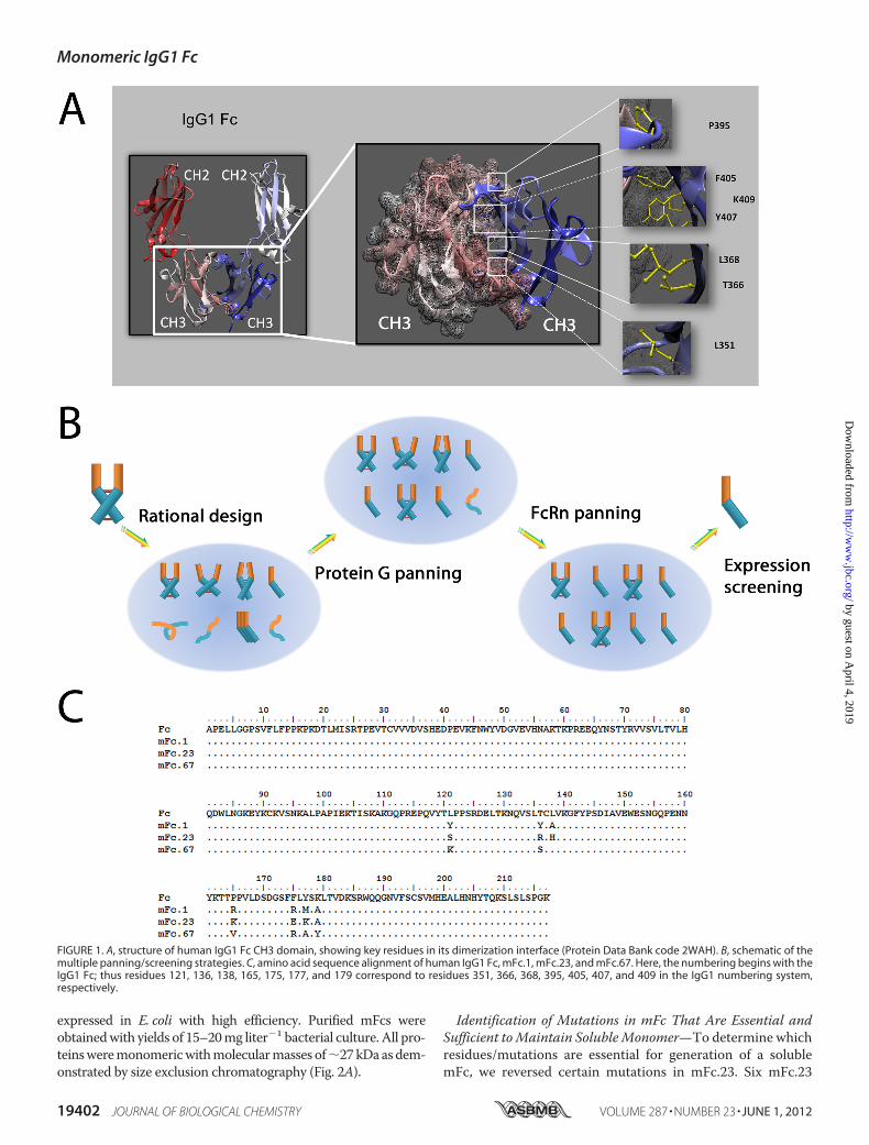
expressed in E. coli with high efficiency. Purified mFcs wereobtainedwith yields of 15–20mg liter�1 bacterial culture. All pro-teinsweremonomericwithmolecularmasses of�27kDaasdem-onstrated by size exclusion chromatography (Fig. 2A).
Identification of Mutations in mFc That Are Essential andSufficient toMaintain SolubleMonomer—To determine whichresidues/mutations are essential for generation of a solublemFc, we reversed certain mutations in mFc.23. Six mFc.23
FIGURE 1. A, structure of human IgG1 Fc CH3 domain, showing key residues in its dimerization interface (Protein Data Bank code 2WAH). B, schematic of themultiple panning/screening strategies. C, amino acid sequence alignment of human IgG1 Fc, mFc.1, mFc.23, and mFc.67. Here, the numbering begins with theIgG1 Fc; thus residues 121, 136, 138, 165, 175, 177, and 179 correspond to residues 351, 366, 368, 395, 405, 407, and 409 in the IgG1 numbering system,respectively.
Monomeric IgG1 Fc
19402 JOURNAL OF BIOLOGICAL CHEMISTRY VOLUME 287 • NUMBER 23 • JUNE 1, 2012
by guest on April 4, 2019
http://ww
w.jbc.org/
Dow
nloaded from
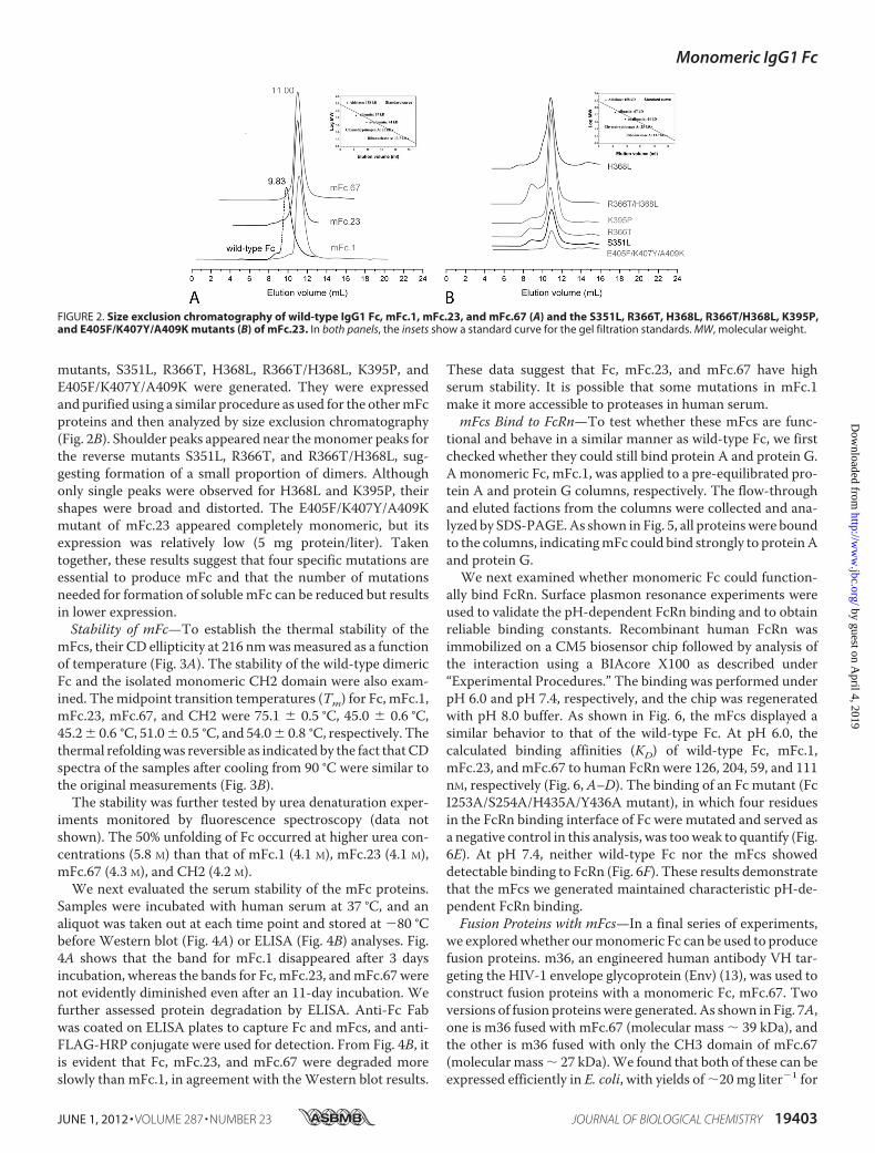
mutants, S351L, R366T, H368L, R366T/H368L, K395P, andE405F/K407Y/A409K were generated. They were expressedand purified using a similar procedure as used for the othermFcproteins and then analyzed by size exclusion chromatography(Fig. 2B). Shoulder peaks appeared near themonomer peaks forthe reverse mutants S351L, R366T, and R366T/H368L, sug-gesting formation of a small proportion of dimers. Althoughonly single peaks were observed for H368L and K395P, theirshapes were broad and distorted. The E405F/K407Y/A409Kmutant of mFc.23 appeared completely monomeric, but itsexpression was relatively low (5 mg protein/liter). Takentogether, these results suggest that four specific mutations areessential to produce mFc and that the number of mutationsneeded for formation of soluble mFc can be reduced but resultsin lower expression.Stability of mFc—To establish the thermal stability of the
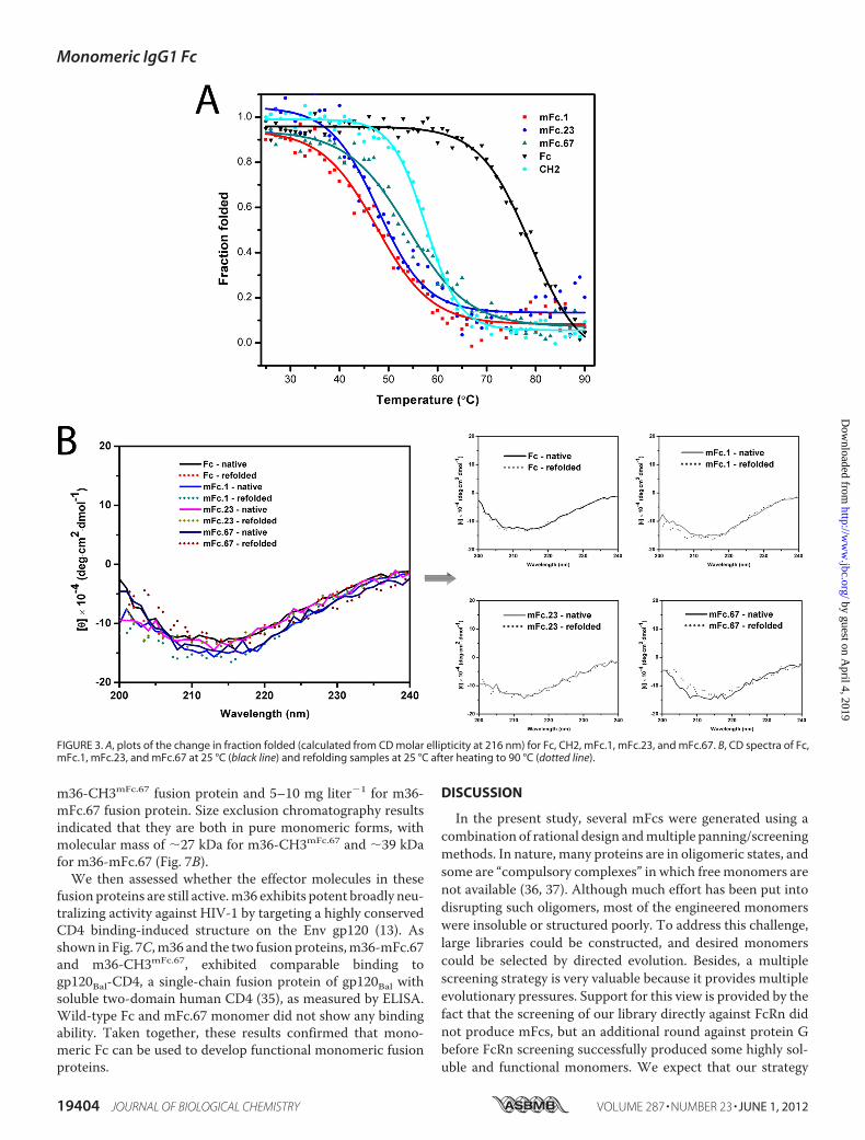
mFcs, their CD ellipticity at 216 nmwasmeasured as a functionof temperature (Fig. 3A). The stability of the wild-type dimericFc and the isolated monomeric CH2 domain were also exam-ined. Themidpoint transition temperatures (Tm) for Fc, mFc.1,mFc.23, mFc.67, and CH2 were 75.1 � 0.5 °C, 45.0 � 0.6 °C,45.2� 0.6 °C, 51.0� 0.5 °C, and 54.0� 0.8 °C, respectively. Thethermal refoldingwas reversible as indicated by the fact thatCDspectra of the samples after cooling from 90 °C were similar tothe original measurements (Fig. 3B).The stability was further tested by urea denaturation exper-
iments monitored by fluorescence spectroscopy (data notshown). The 50% unfolding of Fc occurred at higher urea con-centrations (5.8 M) than that of mFc.1 (4.1 M), mFc.23 (4.1 M),mFc.67 (4.3 M), and CH2 (4.2 M).We next evaluated the serum stability of the mFc proteins.
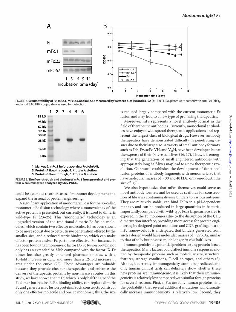
Samples were incubated with human serum at 37 °C, and analiquot was taken out at each time point and stored at �80 °Cbefore Western blot (Fig. 4A) or ELISA (Fig. 4B) analyses. Fig.4A shows that the band for mFc.1 disappeared after 3 daysincubation, whereas the bands for Fc, mFc.23, andmFc.67 werenot evidently diminished even after an 11-day incubation. Wefurther assessed protein degradation by ELISA. Anti-Fc Fabwas coated on ELISA plates to capture Fc and mFcs, and anti-FLAG-HRP conjugate were used for detection. From Fig. 4B, itis evident that Fc, mFc.23, and mFc.67 were degraded moreslowly than mFc.1, in agreement with theWestern blot results.
These data suggest that Fc, mFc.23, and mFc.67 have highserum stability. It is possible that some mutations in mFc.1make it more accessible to proteases in human serum.mFcs Bind to FcRn—To test whether these mFcs are func-
tional and behave in a similar manner as wild-type Fc, we firstchecked whether they could still bind protein A and protein G.Amonomeric Fc, mFc.1, was applied to a pre-equilibrated pro-tein A and protein G columns, respectively. The flow-throughand eluted factions from the columns were collected and ana-lyzed by SDS-PAGE.As shown in Fig. 5, all proteinswere boundto the columns, indicatingmFc could bind strongly to proteinAand protein G.We next examined whether monomeric Fc could function-
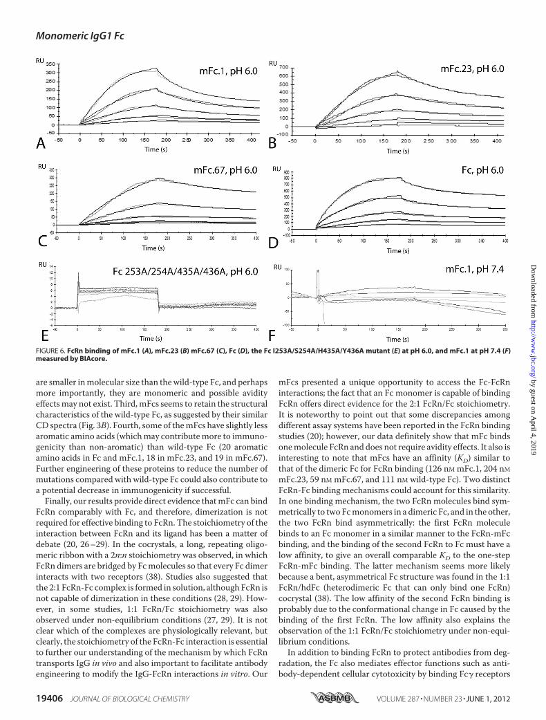
ally bind FcRn. Surface plasmon resonance experiments wereused to validate the pH-dependent FcRn binding and to obtainreliable binding constants. Recombinant human FcRn wasimmobilized on a CM5 biosensor chip followed by analysis ofthe interaction using a BIAcore X100 as described under“Experimental Procedures.” The binding was performed underpH 6.0 and pH 7.4, respectively, and the chip was regeneratedwith pH 8.0 buffer. As shown in Fig. 6, the mFcs displayed asimilar behavior to that of the wild-type Fc. At pH 6.0, thecalculated binding affinities (KD) of wild-type Fc, mFc.1,mFc.23, and mFc.67 to human FcRn were 126, 204, 59, and 111nM, respectively (Fig. 6, A–D). The binding of an Fc mutant (FcI253A/S254A/H435A/Y436A mutant), in which four residuesin the FcRn binding interface of Fc were mutated and served asa negative control in this analysis, was tooweak to quantify (Fig.6E). At pH 7.4, neither wild-type Fc nor the mFcs showeddetectable binding to FcRn (Fig. 6F). These results demonstratethat the mFcs we generated maintained characteristic pH-de-pendent FcRn binding.Fusion Proteins with mFcs—In a final series of experiments,
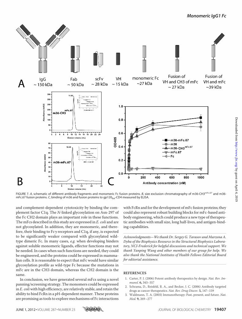
we exploredwhether ourmonomeric Fc can be used to producefusion proteins. m36, an engineered human antibody VH tar-geting the HIV-1 envelope glycoprotein (Env) (13), was used toconstruct fusion proteins with a monomeric Fc, mFc.67. Twoversions of fusion proteinswere generated. As shown in Fig. 7A,one is m36 fused with mFc.67 (molecular mass � 39 kDa), andthe other is m36 fused with only the CH3 domain of mFc.67(molecularmass� 27 kDa).We found that both of these can beexpressed efficiently in E. coli, with yields of �20mg liter�1 for
FIGURE 2. Size exclusion chromatography of wild-type IgG1 Fc, mFc.1, mFc.23, and mFc.67 (A) and the S351L, R366T, H368L, R366T/H368L, K395P,and E405F/K407Y/A409K mutants (B) of mFc.23. In both panels, the insets show a standard curve for the gel filtration standards. MW, molecular weight.
Monomeric IgG1 Fc
JUNE 1, 2012 • VOLUME 287 • NUMBER 23 JOURNAL OF BIOLOGICAL CHEMISTRY 19403
by guest on April 4, 2019
http://ww
w.jbc.org/
Dow
nloaded from
m36-CH3mFc.67 fusion protein and 5–10 mg liter�1 for m36-mFc.67 fusion protein. Size exclusion chromatography resultsindicated that they are both in pure monomeric forms, withmolecular mass of �27 kDa for m36-CH3mFc.67 and �39 kDafor m36-mFc.67 (Fig. 7B).We then assessed whether the effector molecules in these
fusion proteins are still active.m36 exhibits potent broadly neu-tralizing activity against HIV-1 by targeting a highly conservedCD4 binding-induced structure on the Env gp120 (13). Asshown in Fig. 7C,m36 and the two fusion proteins,m36-mFc.67and m36-CH3mFc.67, exhibited comparable binding togp120Bal-CD4, a single-chain fusion protein of gp120Bal withsoluble two-domain human CD4 (35), as measured by ELISA.Wild-type Fc and mFc.67 monomer did not show any bindingability. Taken together, these results confirmed that mono-meric Fc can be used to develop functional monomeric fusionproteins.
DISCUSSION
In the present study, several mFcs were generated using acombination of rational design andmultiple panning/screeningmethods. In nature, many proteins are in oligomeric states, andsome are “compulsory complexes” in which free monomers arenot available (36, 37). Although much effort has been put intodisrupting such oligomers, most of the engineered monomerswere insoluble or structured poorly. To address this challenge,large libraries could be constructed, and desired monomerscould be selected by directed evolution. Besides, a multiplescreening strategy is very valuable because it provides multipleevolutionary pressures. Support for this view is provided by thefact that the screening of our library directly against FcRn didnot produce mFcs, but an additional round against protein Gbefore FcRn screening successfully produced some highly sol-uble and functional monomers. We expect that our strategy
FIGURE 3. A, plots of the change in fraction folded (calculated from CD molar ellipticity at 216 nm) for Fc, CH2, mFc.1, mFc.23, and mFc.67. B, CD spectra of Fc,mFc.1, mFc.23, and mFc.67 at 25 °C (black line) and refolding samples at 25 °C after heating to 90 °C (dotted line).
Monomeric IgG1 Fc
19404 JOURNAL OF BIOLOGICAL CHEMISTRY VOLUME 287 • NUMBER 23 • JUNE 1, 2012
by guest on April 4, 2019
http://ww
w.jbc.org/
Dow
nloaded from
could be extended to other cases ofmonomer development andexpand the arsenal of protein engineering.A significant application ofmonomeric Fc is for the so-called
monomeric Fc fusion technology where a monovalency of theactive protein is presented, but currently, it is fused to dimericwild-type Fc (23–25). This “monomeric” technology is anupgraded version of the traditional dimeric Fc fusion mole-cules, which contain two effector molecules. It has been shownto bemore robust due to better tissue penetration offered by thesmaller size, and a reduced steric hindrance, which can makeeffector protein and/or Fc part more effective. For instance, ithas been found thatmonomeric factor IX-Fc fusion protein notonly has an extended half-life compared with the factor IX-Fcdimer but also greatly enhanced pharmacokinetics, with a10-fold increase in Cmax and more than a 12-fold increase inarea under the curve (25). These advantages are valuablebecause they provide cheaper therapeutics and enhance thedelivery of therapeutic proteins by non-invasive routes. In thisstudy, we have shown thatmFc, which is only half the size of theFc dimer but retains FcRn binding ability, can replace dimericFc and generatemFc fusion proteins. Such constructs consist ofonly one effector molecule and one Fc monomer; thus, the size
is reduced largely compared with the current monomeric Fcfusion and may lead to a new type of promising therapeutics.Moreover, mFc represents a novel antibody format in the
field of therapeutic antibodies. Currently, monoclonal antibod-ies have enjoyed widespread therapeutic applications and rep-resent the largest class of biological drugs. However, antibodytherapeutics have demonstrated difficulty in penetrating tis-sues due to their large size. A variety of small antibody formats,such as Fab, Fv, scFv, VH, andVHH, have been developed but atthe expense of their in vivo half-lives (16, 17). Thus, it is emerg-ing that the generation of small engineered antibodies withappropriately long half-lives may lead to a new therapeutic rev-olution. Our work establishes the development of functionalfusion proteins of antibody fragments with monomeric Fc thathave molecular masses of �30 and 40 kDa, only one-fourth thesize of IgG.We also hypothesize that mFcs themselves could serve as
novel antibody formats and be used as scaffolds for construc-tion of libraries containing diverse binders to various antigens.They are relatively stable, can bind FcRn in a pH-dependentmanner, and can be produced in large quantities in bacteria.Importantly, comparedwithwild-type Fc, a large surface area isexposed in the Fc monomers due to the disruption of the CH3dimerization interface, providing more access for protein engi-neering by designed pointmutations andCDR-grafting onto anmFc framework. It is anticipated that binders generated fromsuch a designwould havemolecularmasses of�27 kDa, similarto that of scFv but possess much longer in vivo half-lives.Immunogenicity is a potential problem for any protein-based
therapeutics.Many factors could affect immune responses elic-ited by therapeutic proteins such as molecular size, structuralfeatures, storage conditions, T-cell epitopes, and others (5).Although currently, immunogenicity cannot be predicted, andonly human clinical trials can definitely show whether thesenew proteins are immunogenic, it is likely that their immuno-genicity is relatively low comparedwith similar foreign proteinsfor several reasons. First, mFcs are fully human proteins, andthe probability that several additional mutations will dramati-cally increase immunogenicity is relatively low. Second, they
FIGURE 4. Serum stability of Fc, mFc.1, mFc.23, and mFc.67 measured by Western blot (A) and ELISA (B). For ELISA, plates were coated with anti-Fc F(ab�)2,and anti-FLAG HRP conjugate was used for detection.
FIGURE 5. The flow-through and elution of mFc.1 from protein A and pro-tein G columns were analyzed by SDS-PAGE.
Monomeric IgG1 Fc
JUNE 1, 2012 • VOLUME 287 • NUMBER 23 JOURNAL OF BIOLOGICAL CHEMISTRY 19405
by guest on April 4, 2019
http://ww
w.jbc.org/
Dow
nloaded from
are smaller inmolecular size than thewild-type Fc, and perhapsmore importantly, they are monomeric and possible avidityeffectsmay not exist. Third,mFcs seems to retain the structuralcharacteristics of the wild-type Fc, as suggested by their similarCD spectra (Fig. 3B). Fourth, some of themFcs have slightly lessaromatic amino acids (whichmay contributemore to immuno-genicity than non-aromatic) than wild-type Fc (20 aromaticamino acids in Fc and mFc.1, 18 in mFc.23, and 19 in mFc.67).Further engineering of these proteins to reduce the number ofmutations compared with wild-type Fc could also contribute toa potential decrease in immunogenicity if successful.Finally, our results provide direct evidence thatmFc can bind
FcRn comparably with Fc, and therefore, dimerization is notrequired for effective binding to FcRn. The stoichiometry of theinteraction between FcRn and its ligand has been a matter ofdebate (20, 26–29). In the cocrystals, a long, repeating oligo-meric ribbon with a 2n:n stoichiometry was observed, in whichFcRn dimers are bridged by Fcmolecules so that every Fc dimerinteracts with two receptors (38). Studies also suggested thatthe 2:1 FcRn-Fc complex is formed in solution, althoughFcRn isnot capable of dimerization in these conditions (28, 29). How-ever, in some studies, 1:1 FcRn/Fc stoichiometry was alsoobserved under non-equilibrium conditions (27, 29). It is notclear which of the complexes are physiologically relevant, butclearly, the stoichiometry of the FcRn-Fc interaction is essentialto further our understanding of the mechanism by which FcRntransports IgG in vivo and also important to facilitate antibodyengineering to modify the IgG-FcRn interactions in vitro. Our
mFcs presented a unique opportunity to access the Fc-FcRninteractions; the fact that an Fc monomer is capable of bindingFcRn offers direct evidence for the 2:1 FcRn/Fc stoichiometry.It is noteworthy to point out that some discrepancies amongdifferent assay systems have been reported in the FcRn bindingstudies (20); however, our data definitely show that mFc bindsonemolecule FcRn and does not require avidity effects. It also isinteresting to note that mFcs have an affinity (KD) similar tothat of the dimeric Fc for FcRn binding (126 nM mFc.1, 204 nMmFc.23, 59 nM mFc.67, and 111 nM wild-type Fc). Two distinctFcRn-Fc binding mechanisms could account for this similarity.In one binding mechanism, the two FcRn molecules bind sym-metrically to twoFcmonomers in a dimeric Fc, and in the other,the two FcRn bind asymmetrically: the first FcRn moleculebinds to an Fc monomer in a similar manner to the FcRn-mFcbinding, and the binding of the second FcRn to Fc must have alow affinity, to give an overall comparable KD to the one-stepFcRn-mFc binding. The latter mechanism seems more likelybecause a bent, asymmetrical Fc structure was found in the 1:1FcRn/hdFc (heterodimeric Fc that can only bind one FcRn)cocrystal (38). The low affinity of the second FcRn binding isprobably due to the conformational change in Fc caused by thebinding of the first FcRn. The low affinity also explains theobservation of the 1:1 FcRn/Fc stoichiometry under non-equi-librium conditions.In addition to binding FcRn to protect antibodies from deg-
radation, the Fc also mediates effector functions such as anti-body-dependent cellular cytotoxicity by binding Fc� receptors
FIGURE 6. FcRn binding of mFc.1 (A), mFc.23 (B) mFc.67 (C), Fc (D), the Fc I253A/S254A/H435A/Y436A mutant (E) at pH 6.0, and mFc.1 at pH 7.4 (F)measured by BIAcore.
Monomeric IgG1 Fc
19406 JOURNAL OF BIOLOGICAL CHEMISTRY VOLUME 287 • NUMBER 23 • JUNE 1, 2012
by guest on April 4, 2019
http://ww
w.jbc.org/
Dow
nloaded from
and complement-dependent cytotoxicity by binding the com-plement factor C1q. The N-linked glycosylation on Asn-297 ofthe Fc CH2 domain plays an important role in these functions.ThemFcs described in this study are expressed inE. coli and arenot glycosylated. In addition, they are monomeric, and there-fore, their binding to Fc� receptors and C1q, if any, is expectedto be significantly weaker compared with glycosylated wild-type dimeric Fc. In many cases, e.g. when developing bindersagainst soluble monomeric ligands, effector functions may notbe needed. In cases when such functions are needed, they couldbe engineered, and the proteins could be expressed inmamma-lian cells. It is reasonable to expect that mFc would have similarglycosylation profile as wild-type Fc because the mutations inmFc are in the CH3 domain, whereas the CH2 domain is thesame.In conclusion, we have generated several mFcs using a novel
panning/screening strategy. Themonomers could be expressedinE. coliwith high efficiency, are relatively stable, and retain theability to bind FcRn in a pH-dependentmanner. These proteinsare promising as tools to exploremechanisms of Fc interactions
with FcRn and for the development ofmFc fusion proteins; theycould also represent robust building blocks formFc-based anti-body engineering, which could produce a new type of therapeu-tic antibodies with small size, long half-lives, and antigen-bind-ing capabilities.
Acknowledgments—We thankDr. Sergey G. Tarasov andMarzena A.Dyba of the Biophysics Resource in the Structural Biophysics Labora-tory, NCI-Frederick for helpful discussions and technical support. Wethank Yanping Wang and other members of our group for help. Wealso thank the National Institutes of Health Fellows Editorial Boardfor editorial assistance.
REFERENCES1. Carter, P. J. (2006) Potent antibody therapeutics by design. Nat. Rev. Im-
munol. 6, 343–3572. Schrama, D., Reisfeld, R. A., and Becker, J. C. (2006) Antibody targeted
drugs as cancer therapeutics. Nat. Rev. Drug Discov. 5, 147–1593. Waldmann, T. A. (2003) Immunotherapy: Past, present, and future. Nat.
Med. 9, 269–277
FIGURE 7. A, schematic of different antibody fragments and monomeric Fc fusion proteins. B, size exclusion chromatography of m36-CH3mFc.67 and m36-mFc.67 fusion proteins. C, binding of m36 and fusion proteins to gp120Bal-CD4 measured by ELISA.
Monomeric IgG1 Fc
JUNE 1, 2012 • VOLUME 287 • NUMBER 23 JOURNAL OF BIOLOGICAL CHEMISTRY 19407
by guest on April 4, 2019
http://ww
w.jbc.org/
Dow
nloaded from
4. Casadevall, A., Dadachova, E., and Pirofski, L. A. (2004) Passive antibodytherapy for infectious diseases. Nat. Rev. Microbiol. 2, 695–703
5. Dimitrov, D. S. (2010) Therapeutic antibodies, vaccines, and anti-bodyomes.MAbs 2, 347–356
6. Leader, B., Baca, Q. J., and Golan, D. E. (2008) Protein therapeutics: Asummary and pharmacological classification. Nat. Rev. Drug Discov. 7,21–39
7. Dimitrov, D. S., and Marks, J. D. (2009) Therapeutic antibodies: Currentstate and future trends–is a paradigm change coming soon?MethodsMol.Biol. 525, 1–27
8. Beck, A., and Reichert, J. M. (2011) Therapeutic Fc-fusion proteins andpeptides as successful alternatives to antibodies.MAbs 3, 415–416
9. Reichert, J. M. (2010) Metrics for antibody therapeutics development.MAbs 2, 695–700
10. Reichert, J. M. (2011) Antibody-based therapeutics to watch in 2011.MAbs 3, 76–99
11. Jain, R. K. (1990) Physiological barriers to delivery ofmonoclonal antibod-ies and other macromolecules in tumors. Cancer Res. 50, 814s-819s
12. Labrijn, A. F., Poignard, P., Raja, A., Zwick, M. B., Delgado, K., Franti, M.,Binley, J., Vivona, V., Grundner, C., Huang, C. C., Venturi, M., Petropou-los, C. J.,Wrin, T., Dimitrov, D. S., Robinson, J., Kwong, P. D.,Wyatt, R. T.,Sodroski, J., and Burton, D. R. (2003) Access of antibody molecules to theconserved coreceptor binding site on glycoprotein gp120 is sterically re-stricted on primary human immunodeficiency virus type 1. J. Virol. 77,10557–10565
13. Chen, W., Zhu, Z., Feng, Y., and Dimitrov, D. S. (2008) Human domainantibodies to conserved sterically restricted regions on gp120 as excep-tionally potent cross-reactive HIV-1 neutralizers. Proc. Natl. Acad. Sci.U.S.A. 105, 17121–17126
14. Holt, L. J., Herring, C., Jespers, L. S., Woolven, B. P., and Tomlinson, I. M.(2003) Domain antibodies: Proteins for therapy. Trends Biotechnol. 21,484–490
15. Saerens, D., Ghassabeh, G.H., andMuyldermans, S. (2008) Single-domainantibodies as building blocks for novel therapeutics. Curr. Opin. Pharma-col. 8, 600–608
16. Dimitrov, D. S. (2009) Engineered CH2 domains (nanoantibodies).MAbs1, 26–28
17. Nelson, A. L., and Reichert, J. M. (2009) Development trends for thera-peutic antibody fragments. Nat. Biotechnol. 27, 331–337
18. Gong, R., Vu, B. K., Feng, Y., Prieto, D. A., Dyba, M. A., Walsh, J. D.,Prabakaran, P., Veenstra, T. D., Tarasov, S. G., Ishima, R., and Dimitrov,D. S. (2009) Engineered human antibody constant domainswith increasedstability. J. Biol. Chem. 284, 14203–14210
19. Simister, N. E., and Mostov, K. E. (1989) An Fc receptor structurally re-lated to MHC class I antigens. Nature 337, 184–187
20. Roopenian, D. C., and Akilesh, S. (2007) FcRn: The neonatal Fc receptorcomes of age. Nat. Rev. Immunol. 7, 715–725
21. Flanagan,M. L., Arias, R. S., Hu, P., Khawli, L. A., and Epstein, A. L. (2007)Soluble Fc fusion proteins for biomedical research. Methods Mol. Biol.378, 33–52
22. Jazayeri, J. A., and Carroll, G. J. (2008) Fc-based cytokines: Prospects forengineering superior therapeutics. BioDrugs 22, 11–26
23. Bitonti, A. J., Dumont, J. A., Low, S. C., Peters, R. T., Kropp, K. E., Palom-bella, V. J., Stattel, J. M., Lu, Y., Tan, C. A., Song, J. J., Garcia, A. M.,Simister, N. E., Spiekermann, G. M., Lencer, W. I., and Blumberg, R. S.(2004) Pulmonary delivery of an erythropoietin Fc fusion protein in non-human primates through an immunoglobulin transport pathway. Proc.Natl. Acad. Sci. U.S.A. 101, 9763–9768
24. Dumont, J. A., Low, S. C., Peters, R. T., and Bitonti, A. J. (2006)MonomericFc fusions: Impact on pharmacokinetic and biological activity of proteintherapeutics. BioDrugs 20, 151–160
25. Peters, R. T., Low, S. C., Kamphaus, G. D., Dumont, J. A., Amari, J. V., Lu,Q., Zarbis-Papastoitsis, G., Reidy, T. J., Merricks, E. P., Nichols, T. C., andBitonti, A. J. (2010) Prolonged activity of factor IX as a monomeric Fcfusion protein. Blood 115, 2057–2064
26. Huber, A. H., Kelley, R. F., Gastinel, L. N., and Bjorkman, P. J. (1993)Crystallization and stoichiometry of binding of a complex between a ratintestinal Fc receptor and Fc. J. Mol. Biol. 230, 1077–1083
27. Popov, S., Hubbard, J. G., Kim, J., Ober, B., Ghetie, V., and Ward, E. S.(1996) The stoichiometry and affinity of the interaction of murine Fcfragments with the MHC class I-related receptor, FcRn. Mol. Immunol.33, 521–530
28. Martin,W. L., and Bjorkman, P. J. (1999) Characterization of the 2:1 com-plex between the class I MHC-related Fc receptor and its Fc ligand insolution. Biochemistry 38, 12639–12647
29. Sánchez, L. M., Penny, D. M., and Bjorkman, P. J. (1999) Stoichiometry ofthe interaction between the major histocompatibility complex-related Fcreceptor and its Fc ligand. Biochemistry 38, 9471–9476
30. Martin, W. L., West, A. P., Jr., Gan, L., and Bjorkman, P. J. (2001) Crystalstructure at 2.8 A of an FcRn-heterodimeric Fc complex: Mechanism ofpH-dependent binding.Mol. Cell 7, 867–877
31. Deisenhofer, J. (1981) Crystallographic refinement and atomicmodels of ahuman Fc fragment and its complex with fragment B of protein A fromStaphylococcus aureus at 2.9- and 2.8-A resolution. Biochemistry 20,2361–2370
32. Miller, S. (1990) Protein-protein recognition and the association of immu-noglobulin constant domains. J. Mol. Biol. 216, 965–973
33. Feng, Y., Gong, R., and Dimitrov, D. S. (2011) Design, expression andcharacterization of a soluble single-chain functional human neonatal Fcreceptor. Protein Expr. Purif. 79, 66–71
34. Dall’Acqua, W., Simon, A. L., Mulkerrin, M. G., and Carter, P. (1998)Contribution of domain interface residues to the stability of antibodyCH3domain homodimers. Biochemistry 37, 9266–9273
35. Fouts, T. R., Tuskan, R., Godfrey, K., Reitz, M., Hone, D., Lewis, G. K., andDeVico, A. L. (2000) Expression and characterization of a single-chainpolypeptide analogue of the human immunodeficiency virus type 1 gp120-CD4 receptor complex. J. Virol. 74, 11427–11436
36. Goodsell, D. S., and Olson, A. J. (2000) Structural symmetry and proteinfunction. Annu. Rev. Biophys. Biomol. Struct. 29, 105–153
37. Brown, J. H. (2006) Breaking symmetry in protein dimers: Designs andfunctions. Protein Sci. 15, 1–13
38. Burmeister, W. P., Gastinel, L. N., Simister, N. E., Blum, M. L., and Bjork-man, P. J. (1994) Crystal structure at 2.2 A resolution of the MHC-relatedneonatal Fc receptor. Nature 372, 336–343
Monomeric IgG1 Fc
19408 JOURNAL OF BIOLOGICAL CHEMISTRY VOLUME 287 • NUMBER 23 • JUNE 1, 2012
by guest on April 4, 2019
http://ww
w.jbc.org/
Dow
nloaded from
Tianlei Ying, Weizao Chen, Rui Gong, Yang Feng and Dimiter S. DimitrovSoluble Monomeric IgG1 Fc
doi: 10.1074/jbc.M112.368647 originally published online April 19, 20122012, 287:19399-19408.J. Biol. Chem.
10.1074/jbc.M112.368647Access the most updated version of this article at doi:
Alerts:
When a correction for this article is posted•
When this article is cited•
to choose from all of JBC's e-mail alertsClick here
http://www.jbc.org/content/287/23/19399.full.html#ref-list-1
This article cites 38 references, 7 of which can be accessed free at
by guest on April 4, 2019
http://ww
w.jbc.org/
Dow
nloaded from