Embed Size (px)
Citation preview
U.P.B. Sci. Bull., Series B, Vol. 73, Iss. 2, 2011 ISSN 1454-2331
SOME ASPECTS CONCERNING THE ISOLATION OF CELLULOSE MICRO- AND NANO- FIBERS
Adriana N. FRONE1, Denis M. PANAITESCU2, Dan DONESCU3
Celuloza este cel mai răspândit biopolimer de pe Pământ. În biosinteză polimerii de celuloză se aglomerează pentru a forma substructuri, microfibre, care, la rândul lor, se aglomerează pentru a forma fibre celulozice. Prin metode eficiente aceste fibre pot fi dezintegrate în substructuri celulozice de dimensiuni micro- sau nanometrice. Acest articol acoperă câteva dintre aspectele privind sursele de obtinere ale micro- şi nano-fibrelor celulozice precum şi cele mai importante metode de izolare a acestora. Una dintre aceste metode, hidroliza acidă, a fost aplicată experimental pentru obţinerea de nano-fibre celulozice. Mǎrind timpul de hidroliză şi menţinând ceilalţi parametri constanţi, au fost obţinute două tipuri de fibre celulozice. Acestea au fost studiate prin difuzia dinamică a luminii (DLS) şi microscopie de forţă atomică (AFM) şi au fost utilizate ca ranforturi intr-o matrice de alcool polivinilic (APV). Proprietăţile mecanice ale compozitelor rezultate au fost semnificativ influenţate de caracteristicile morfologice ale acestor fibre.
Cellulose is the most widespread biopolymer on earth. In biosynthesis, cellulose polymers aggregate to form substructures, microfibrils, which in turn aggregate to form cellulose fibers. By applying effective methods these fibers can be disintegrated into cellulose substructures with micro- or nano-size dimensions. This article covers some aspects related to the sources of cellulose micro- and nano- fibers and the most important methods for their isolation. One of these methods, acid hydrolysis, was experimentally used to obtain cellulose nano-fibers. Increasing the hydrolysis time and keeping other processing parameters constant, two types of cellulose fibers were obtained. They were studied by dynamic light scattering (DLS) and atomic force microscopy (AFM) and were used as reinforcements in a polyvinyl alcohol (PVA) matrix. The mechanical properties of the resulted composites were significantly influenced by the morphological features of these fibers.
Keywords: cellulose nano-fibers, polyvinyl alcohol, polymer composites, renewable resources
1PhD student Eng., Polymer Department, National Research and Development Institute for
Chemistry and Petrochemistry-ICECHIM Bucharest, Romania, e-mail: [email protected]
2 Dr. Eng., Polymer Department, National Research and Development Institute for Chemistry and Petrochemistry-ICECHIM, Bucharest, Romania
3 Prof. Dr. Eng., Polymer Department, National Research and Development Institute for Chemistry and Petrochemistry-ICECHIM, Bucharest, Romania
134 Adriana N. Frone, Denis M. Panaitescu, Dan Donescu
1. Introduction
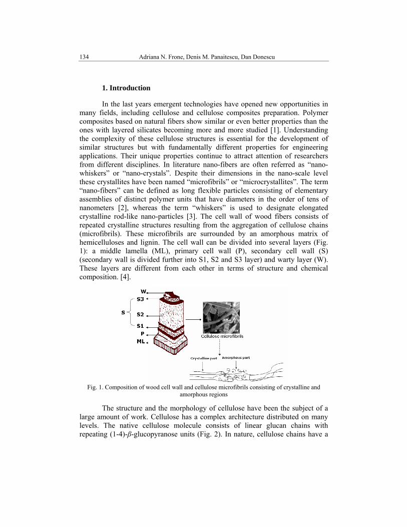
In the last years emergent technologies have opened new opportunities in many fields, including cellulose and cellulose composites preparation. Polymer composites based on natural fibers show similar or even better properties than the ones with layered silicates becoming more and more studied [1]. Understanding the complexity of these cellulose structures is essential for the development of similar structures but with fundamentally different properties for engineering applications. Their unique properties continue to attract attention of researchers from different disciplines. In literature nano-fibers are often referred as “nano-whiskers” or “nano-crystals”. Despite their dimensions in the nano-scale level these crystallites have been named “microfibrils” or “microcrystallites”. The term “nano-fibers” can be defined as long flexible particles consisting of elementary assemblies of distinct polymer units that have diameters in the order of tens of nanometers [2], whereas the term “whiskers” is used to designate elongated crystalline rod-like nano-particles [3]. The cell wall of wood fibers consists of repeated crystalline structures resulting from the aggregation of cellulose chains (microfibrils). These microfibrils are surrounded by an amorphous matrix of hemicelluloses and lignin. The cell wall can be divided into several layers (Fig. 1): a middle lamella (ML), primary cell wall (P), secondary cell wall (S) (secondary wall is divided further into S1, S2 and S3 layer) and warty layer (W). These layers are different from each other in terms of structure and chemical composition. [4].
Fig. 1. Composition of wood cell wall and cellulose microfibrils consisting of crystalline and
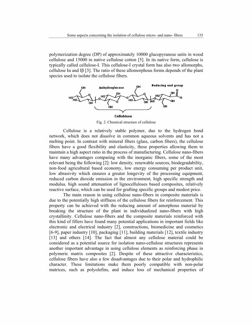
amorphous regions The structure and the morphology of cellulose have been the subject of a large amount of work. Cellulose has a complex architecture distributed on many levels. The native cellulose molecule consists of linear glucan chains with repeating (1-4)-β-glucopyranose units (Fig. 2). In nature, cellulose chains have a
Some aspects concerning the isolation of cellulose micro- and nano- fibers 135
polymerization degree (DP) of approximately 10000 glucopyranose units in wood cellulose and 15000 in native cellulose cotton [5]. In its native form, cellulose is typically called cellulose-I. This cellulose-I crystal form has also two allomorphs, cellulose Iα and Iβ [3]. The ratio of these allomorphous forms depends of the plant species used to isolate the cellulose fibers.
Fig. 2. Chemical structure of cellulose
Cellulose is a relatively stable polymer, due to the hydrogen bond network, which does not dissolve in common aqueous solvents and has not a melting point. In contrast with mineral fibers (glass, carbon fibers), the cellulose fibers have a good flexibility and elasticity, these properties allowing them to maintain a high aspect ratio in the process of manufacturing. Cellulose nano-fibers have many advantages comparing with the inorganic fibers, some of the most relevant being the following [2]: low density, renewable sources, biodegradability, non-food agricultural based economy, low energy consuming per product unit, low abrasivity which ensures a greater longevity of the processing equipment, reduced carbon dioxide emission in the environment, high specific strength and modulus, high sound attenuation of lignocelluloses based composites, relatively reactive surface, which can be used for grafting specific groups and modest price. The main reason in using cellulose nano-fibers in composite materials is due to the potentially high stiffness of the cellulose fibers for reinforcement. This property can be achieved with the reducing amount of amorphous material by breaking the structure of the plant in individualized nano-fibers with high crystallinity. Cellulose nano-fibers and the composite materials reinforced with this kind of fillers have found many potential applications in important fields like electronic and electrical industry [2], constructions, biomedicine and cosmetics [6-9], paper industry [10], packaging [11], building materials [12], textile industry [13] and others [14]. The fact that almost any cellulose material could be considered as a potential source for isolation nano-cellulose structures represents another important advantage in using cellulose elements as reinforcing phase in polymeric matrix composites [2]. Despite of these attractive characteristics, cellulose fibers have also a few disadvantages due to their polar and hydrophilic character. These limitations make them poorly compatible with non-polar matrices, such as polyolefins, and induce loss of mechanical properties of
136 Adriana N. Frone, Denis M. Panaitescu, Dan Donescu
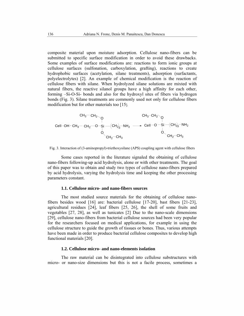
composite material upon moisture adsorption. Cellulose nano-fibers can be submitted to specific surface modification in order to avoid these drawbacks. Some examples of surface modifications are: reactions to form ionic groups at cellulose surfaces (sulfonation, carboxylation, grafting), reactions to create hydrophobic surfaces (acetylation, silane treatments), adsorption (surfactants, polyelectrolytes) [2]. An example of chemical modification is the reaction of cellulose fibers with silane. When hydrolyzed silane solutions are mixted with natural fibers, the reactive silanol groups have a high affinity for each other, forming –Si-O-Si- bonds and also for the hydroxyl sites of fibers via hydrogen bonds (Fig. 3). Silane treatments are commonly used not only for cellulose fibers modification but for other materials too [15].
Si
O
OCH2CH3
CH2 CH3
CH3 CH23Cell OH Cell O Si
OCH2 CH3
CH2 NH23OCH2
CH2CH3 O
NH2
Fig. 3. Interaction of (3-aminopropyl)-triethoxysilane (APS) coupling agent with cellulose fibers
Some cases reported in the literature signaled the obtaining of cellulose nano-fibers following-up acid hydrolysis, alone or with other treatments. The goal of this paper was to obtain and study two types of cellulose nano-fibers prepared by acid hydrolysis, varying the hydrolysis time and keeping the other processing parameters constant.
1.1. Cellulose micro- and nano-fibers sources The most studied source materials for the obtaining of cellulose nano-fibers besides wood [16] are: bacterial cellulose [17-20], bast fibers [21-23], agricultural residues [24], leaf fibers [25, 26], the shell of some fruits and vegetables [27, 28], as well as tunicates [2] Due to the nano-scale dimensions [29], cellulose nano-fibers from bacterial cellulose sources had been very popular for the researchers focused on medical applications, for example in using the cellulose structure to guide the growth of tissues or bones. Thus, various attempts have been made in order to produce bacterial cellulose composites to develop high functional materials [20]. 1.2. Cellulose micro- and nano-elements isolation The raw material can be disintegrated into cellulose substructures with micro- or nano-size dimensions but this is not a facile process, sometimes a
Some aspects concerning the isolation of cellulose micro- and nano- fibers 137
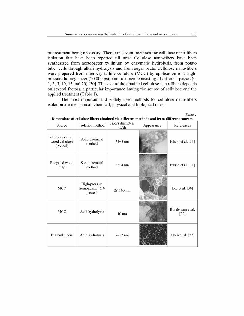
pretreatment being necessary. There are several methods for cellulose nano-fibers isolation that have been reported till now. Cellulose nano-fibers have been synthesized from acetobacter xyllinium by enzymatic hydrolysis, from potato tuber cells through alkali hydrolysis and from sugar beets. Cellulose nano-fibers were prepared from microcrystalline cellulose (MCC) by application of a high-pressure homogenizer (20,000 psi) and treatment consisting of different passes (0, 1, 2, 5, 10, 15 and 20) [30]. The size of the obtained cellulose nano-fibers depends on several factors, a particular importance having the source of cellulose and the applied treatment (Table 1). The most important and widely used methods for cellulose nano-fibers isolation are mechanical, chemical, physical and biological ones.
Table 1 Dimensions of cellulose fibers obtained via different methods and from different sources
Source Isolation method Fibers diameters (L/d) Appearance References
Microcrystalline wood cellulose
(Avicel)
Sono-chemical method 21±5 nm Filson et al. [31]
Recycled wood pulp
Sono-chemical method 23±4 nm Filson et al. [31]
MCC High-pressure
homogenizer (10 passes)
28-100 nm Lee et al. [30]
MCC Acid hydrolysis 10 nm
Bondenson et al. [32]
Pea hull fibers Acid hydrolysis 7–12 nm Chen et al. [27]
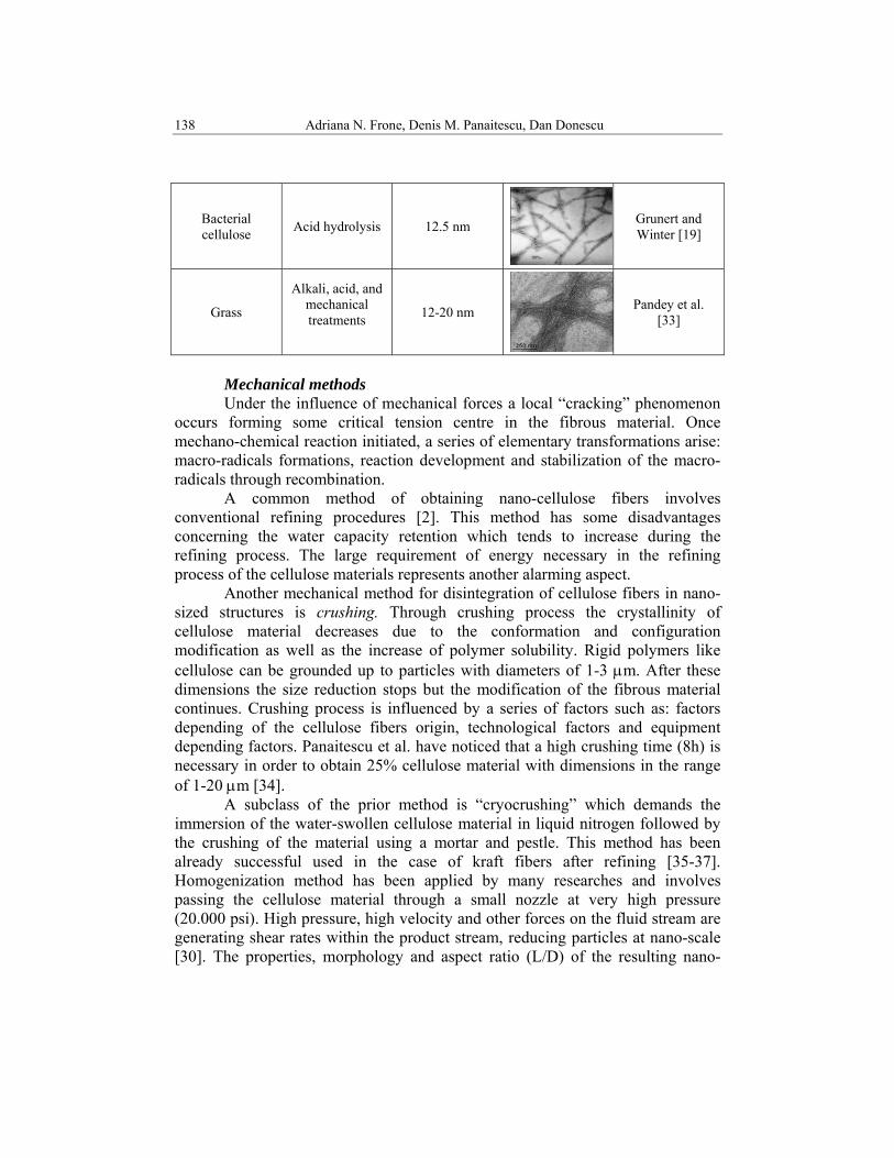
138 Adriana N. Frone, Denis M. Panaitescu, Dan Donescu
Bacterial cellulose Acid hydrolysis 12.5 nm Grunert and
Winter [19]
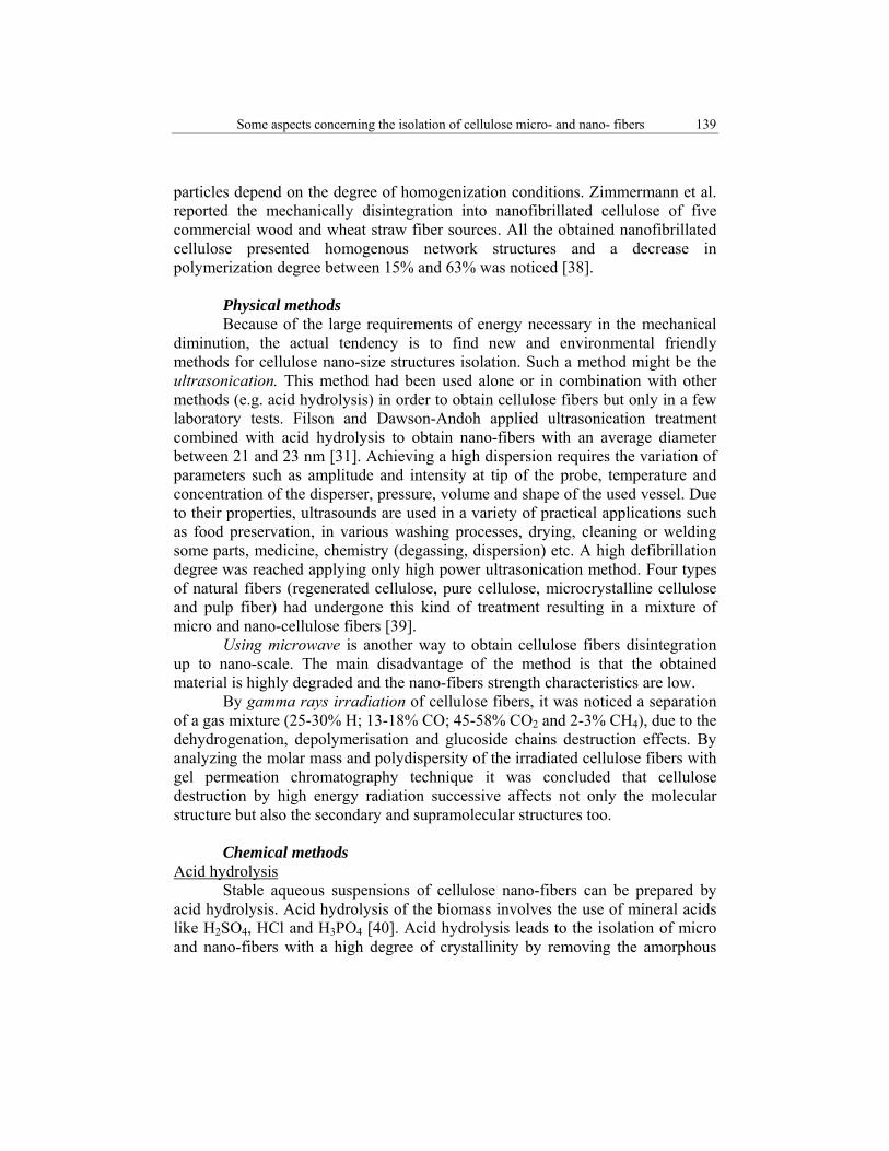
Grass
Alkali, acid, and mechanical treatments
12-20 nm Pandey et al. [33]
Mechanical methods Under the influence of mechanical forces a local “cracking” phenomenon occurs forming some critical tension centre in the fibrous material. Once mechano-chemical reaction initiated, a series of elementary transformations arise: macro-radicals formations, reaction development and stabilization of the macro-radicals through recombination. A common method of obtaining nano-cellulose fibers involves conventional refining procedures [2]. This method has some disadvantages concerning the water capacity retention which tends to increase during the refining process. The large requirement of energy necessary in the refining process of the cellulose materials represents another alarming aspect. Another mechanical method for disintegration of cellulose fibers in nano-sized structures is crushing. Through crushing process the crystallinity of cellulose material decreases due to the conformation and configuration modification as well as the increase of polymer solubility. Rigid polymers like cellulose can be grounded up to particles with diameters of 1-3 μm. After these dimensions the size reduction stops but the modification of the fibrous material continues. Crushing process is influenced by a series of factors such as: factors depending of the cellulose fibers origin, technological factors and equipment depending factors. Panaitescu et al. have noticed that a high crushing time (8h) is necessary in order to obtain 25% cellulose material with dimensions in the range of 1-20 μm [34]. A subclass of the prior method is “cryocrushing” which demands the immersion of the water-swollen cellulose material in liquid nitrogen followed by the crushing of the material using a mortar and pestle. This method has been already successful used in the case of kraft fibers after refining [35-37]. Homogenization method has been applied by many researches and involves passing the cellulose material through a small nozzle at very high pressure (20.000 psi). High pressure, high velocity and other forces on the fluid stream are generating shear rates within the product stream, reducing particles at nano-scale [30]. The properties, morphology and aspect ratio (L/D) of the resulting nano-
Some aspects concerning the isolation of cellulose micro- and nano- fibers 139
particles depend on the degree of homogenization conditions. Zimmermann et al. reported the mechanically disintegration into nanofibrillated cellulose of five commercial wood and wheat straw fiber sources. All the obtained nanofibrillated cellulose presented homogenous network structures and a decrease in polymerization degree between 15% and 63% was noticed [38]. Physical methods Because of the large requirements of energy necessary in the mechanical diminution, the actual tendency is to find new and environmental friendly methods for cellulose nano-size structures isolation. Such a method might be the ultrasonication. This method had been used alone or in combination with other methods (e.g. acid hydrolysis) in order to obtain cellulose fibers but only in a few laboratory tests. Filson and Dawson-Andoh applied ultrasonication treatment combined with acid hydrolysis to obtain nano-fibers with an average diameter between 21 and 23 nm [31]. Achieving a high dispersion requires the variation of parameters such as amplitude and intensity at tip of the probe, temperature and concentration of the disperser, pressure, volume and shape of the used vessel. Due to their properties, ultrasounds are used in a variety of practical applications such as food preservation, in various washing processes, drying, cleaning or welding some parts, medicine, chemistry (degassing, dispersion) etc. A high defibrillation degree was reached applying only high power ultrasonication method. Four types of natural fibers (regenerated cellulose, pure cellulose, microcrystalline cellulose and pulp fiber) had undergone this kind of treatment resulting in a mixture of micro and nano-cellulose fibers [39]. Using microwave is another way to obtain cellulose fibers disintegration up to nano-scale. The main disadvantage of the method is that the obtained material is highly degraded and the nano-fibers strength characteristics are low. By gamma rays irradiation of cellulose fibers, it was noticed a separation of a gas mixture (25-30% H; 13-18% CO; 45-58% CO2 and 2-3% CH4), due to the dehydrogenation, depolymerisation and glucoside chains destruction effects. By analyzing the molar mass and polydispersity of the irradiated cellulose fibers with gel permeation chromatography technique it was concluded that cellulose destruction by high energy radiation successive affects not only the molecular structure but also the secondary and supramolecular structures too. Chemical methods Acid hydrolysis Stable aqueous suspensions of cellulose nano-fibers can be prepared by acid hydrolysis. Acid hydrolysis of the biomass involves the use of mineral acids like H2SO4, HCl and H3PO4 [40]. Acid hydrolysis leads to the isolation of micro and nano-fibers with a high degree of crystallinity by removing the amorphous
140 Adriana N. Frone, Denis M. Panaitescu, Dan Donescu
regions of the raw cellulose material. Applying this method a negatively charged surface of the cellulose fibers can be obtained, through the esterification of hydroxyl groups by the sulfate ions. The time and temperature of hydrolysis reaction as well as acid concentration are the factors that are playing an important role concerning the morphology and the dimensions of the obtained fibers. Many researchers have successfully used this method, alone or in combination with others methods, managing to obtain cellulose structures with nano-scale dimensions starting from different cellulose sources [24, 27, 41, 42]. Bondenson et al treated microcrystalline cellulose (MCC) with sulfuric acid in concentration of 63.5% (w/w) in order to isolate cellulose whiskers with an aspect ratio between 20 and 40 [32]. Indeed, using of sulfuric acid leads to more stable whiskers aqueous suspension than that prepared using hydrochloric acid. The H2SO4 prepared whiskers present a negatively charged surface, whereas the HCl prepared whiskers are not charged [14]. Lee et al. reported the obtaining of nano-cellulose fibers by acid hydrolysis of MCC testing two different concentrations of hydrobromic acid (1.5 M and 2.5 M) [43].The prepared nano-cellulose was characterized by X-ray diffraction, degree of polymerization, molecular weight and scanning electron microscopy. It was pointed out that acid hydrolysis decreased steadily the polymerization degree (DP) and molecular weight (Mw) of MCC. The crystallinity of MCC treated with 1.5 M and 2.5 M HBr showed a significant increase due to the degradation of amorphous domains in cellulose. Higher crystalline cellulose had the higher thermal stability compare to the raw material. The size reduction of MCC particles by acid hydrolysis was confirmed also by the scanning electron microscope (SEM) images. The diameter of MCC after 1.5 M and 2.5 M HBr hydrolysis was similar, the individual crystallites having needle shaped structures. Rosa et al. obtained cellulose whiskers with diameters as low as 5 nm and aspect ratio of up to 60 by sulfuric acid hydrolysis from coconut husk fibers [28]. The raw material had been previously submitted to a delignification process in order to facilitate the isolation of cellulose nano-whiskers. The authors could not observe a correlation between preparation conditions and particle size of obtained cellulose whiskers. They found that a higher residual lignin content increases thermal stability of the nano-whiskers. Alkaline hydrolysis Alkaline hydrolysis determines the partial separation of the cellulose fibers from the cell wall [42, 44-46] and an improvement of the physical and chemical characteristics of cellulose, particularly its reactivity to other chemical agents. These treatments are usually made using diluted solutions of NaOH (1-10%) at low or high temperatures and concentrated NaOH solutions over 10% only at low temperatures. NH4OH and anhydrous NH3 (gas or liquid) are also used to activate the organic materials, providing in some cases an increase of the hydrolytic degradation. Zuluaga et al. [25] reported that by applying four different alkaline
Some aspects concerning the isolation of cellulose micro- and nano- fibers 141
treatments (peroxide alkaline, peroxide alkaline–hydrochloric acid, 5wt% potassium hydroxide and 18wt% potassium hydroxide) cellulose fibers with average diameters between 3 nm to 5 nm were obtained. Organic solvent treatments The first attempts to dissolve cellulose date back to the early 1920s but since then several aqueous and nonaqueous cellulose solvents have been discovered. Usually, all of these solvents suffer either from high environmental toxicity or from insufficient solvation power [47]. In general, the traditional cellulose dissolution processes require relatively harsh conditions and the use of expensive and uncommon solvents, which usually cannot be recovered after the process. Currently, the use of organic solvents is already at the stage of industrial testing. The use of the organic solvents for the isolation of cellulose micro or nano-elements might become the technology of the future given the fact that beside the main product, cellulose, lignin and hemicelluloses can be exploited too. Another major aspect of this technology is the easier recovery of organic solvents by distillation, and the residue absence which gives this treatment an environmental friendly character. Oksman et al. [48] reported the swelling of the cellulose fibers into a solvent system (N,N-dimethylacetamide and lithium chloride) in order to facilitates the cellulose nano-fibers isolation. By electro-spinning techniques the cellulose solution was converted in very thin fibres or filaments. Cellulose has been dissolved in some others solvents such as N-methylmorpholine-N-oxide, trifluoroacetic acid [49, 50], DMSO and DMF [51]. Dogan et al. applied an environmentally friendly microwave heating process in order to achieve a complete dissolution of cellulose in N-methylmorpholine-oxide (NMMO) [52]. It was shown that microwave heating with the power of 210W can be an alternative heating system for dissolution of cellulose in NMMO. Ionic liquid treatments Dissolution of cellulose with ionic liquids allows the comprehensive utilization of cellulose by combining two major green chemistry principles: using environmentally preferable solvents and bio-renewable feed-stocks [53]. Ionic liquids are a new group of organic salts that remain in fluid state at a relatively low temperature (<100o C). These liquids are chemically and thermally stable, present uninflammable and extremely low vapor pressure. Ionic liquids are known as “green solvents” and are industrially used. Some studies have shown that cellulose can be dissolved in some hydrophilic ionic liquids, for example, 1-butyl-3-methylimidazolium chloride (BMIMCl) and 1-allyl-3-methylimidazolium chloride (AMIMCl). Microwave heating significantly accelerates the dissolution process. Cellulose can be easily regenerated from its ionic liquid solutions by addition of water, ethanol or acetone [53]. The ionic liquids can be recovered by various methods like evaporation, ion exchange, and reverse osmosis and they can
142 Adriana N. Frone, Denis M. Panaitescu, Dan Donescu
be reused. Swatloski et al. reported the use of an ionic liquid as solvent for cellulose both for the regeneration of cellulose and for the chemical modification of the polysaccharide [47]. Another attempt dates from 1934 when Graenacher had discovered a solvent system with the ability to dissolve cellulose, but this was thought to be of little practical value at the time [47]. Zhang et al. reported the dissolution and regeneration of cellulose in 1-allyl-3-methylimidazolium chloride (AMIMCl), without any pretreatment [54]. The regenerated cellulose materials had good mechanical properties and the solvent, due to the thermo-stabile character and nonvolatile nature, could be easily recycled. This kind of treatments represents a new ‘‘green’’ way for preparing cellulose derivatives and cellulose composites, which biodegrade faster than the synthetic polymers. Biological treatments Under enzymes action cellulose materials suffer a degradation process. The destruction is influenced by the primary structure features of chemical constituents, since cellulose has a great stability due to its high crystallinity. Lignin acts like a physical barrier that limits the availability of the cellulose material. Degradation of the cellulose substrate occurs in the presence of microorganisms (fungi, bacteria) or, directly, with cellulose enzyme preparations. Enzymatic treatment was performed by Henriksson et al. who reported that such kind of treatment facilitates the obtaining of microfibrillated cellulose nano-fibers [55]. Yan Li et al. reported that removal of non-cellulose components from cellulose fibers by enzyme treatment can increase the crystallinity, thermal stability and the amount of -OH groups of the treated fibers [56]. Combined methods Researchers from University of Toronto were the first group that combined chemical treatment, mechanical refining, homogenization, and crushing of the water-soaked material in the presence of liquid nitrogen in order to obtain cellulose fibers [35]. Through a combination of chemical and mechanical treatments Jonoobi et al. [57] obtained nano-fibers from unbleached and bleached kenaf pulp. The obtained nano-fibers showed higher crystallinity and thermal stability as compared to the raw material. Panaitescu et al. reported the preparation through mechano-chemical methods of cellulose fibers starting from bleached pulp and cellulose I. These cellulose fibers were subsequently used as reinforcements in a PP matrix [58]. A method based on a combination of ball milling, acid hydrolysis and ultrasounds was developed by Qua et al. [59] in order to obtain cellulose nano-fibers starting from flax fibers and microcrystalline cellulose. Roohani et al. [60] reported the preparation of cellulose nano-crystals or whiskers with an average diameter of approximately 14.6 ± 3.9 nm, by

Some aspects concerning the isolation of cellulose micro- and nano- fibers 143
combination of acid hydrolysis and ultrasound using as raw material cotton linters. Disintegration of cellulose wood pulps by enzyme or acid hydrolysis treatments in combination with mechanical shearing was carried out by Henriksson et al. [55]. They found that by using this combination of treatments the isolation of microfibrillated cellulose nano-fibers is possible. Cellulose nano-crystals were successfully produced from microcrystalline wood cellulose and recycled pulp wood using sono-chemical-assisted hydrolysis [31]. Jonoobi et al. [61] developed cellulose nano-fibers with hydrophobic surface characteristics and diameters between 5 and 50 nm using a chemical treatment with acetic anhydride followed by a mechanical disintegration.
2. Experimental section 2.1. Materials Microcrystalline cellulose (MCC) with a mean particle size of 20 μm and an aspect ratio of 2 - 4, from Sigma-Aldrich, and sulphuric acid (H2SO4) 96% from Chimopar Romania were used for the nano-fibers isolation. Poly(vinyl alcohol) (PVA) with a degree of hydrolysis of 99% and a polymerization degree of 1200, purchased from Chemical Enterprise Râsnov (Romania), was used for the nanocomposites preparation. 2.2. Preparation of cellulose fibers Acid hydrolysis of MCC was performed by slowly adding H2SO4 under continuous stirring to a MCC/water suspension placed in an ice water bath, until the final concentration of 60% H2SO4 was reached. The obtained suspension was then heated at 45 °C under continuous stirring for 150 minutes (sample A). A similar suspension was prepared and heated at the same temperature for 300 minutes (sample B). The final mixtures were washed and centrifuged using a Universal 320R Ultracentrifuge at room temperature for 20 minutes with 7000 rpm in order to remove the excess acid, and ultrasonicated for two hours using an Elmasonic S40H ultrasonic bath.
2.3. Preparation of PVA/cellulose fibers composite PVA/cellulose fibers composites with 5% filler content were prepared by mixing the calculated amount of cellulose fibers (samples A and B) with 10% water solution of PVA using a high speed stirrer (500 rpm). The stirring was performed at 800C for 90 minutes. The final mixtures were degassed for 10 minutes in an Elmasonic S40H ultrasonic bath and cast on a PET sheet. The films were kept at room temperature for 2 days until they were completely dried and then removed from the PET plates and placed in a desiccator for two weeks before mechanical characterization.
144 Adriana N. Frone, Denis M. Panaitescu, Dan Donescu
2.4. Characterization The size distribution of cellulose aggregates was estimated by dynamic light scattering (DLS) using a Zetasizer Nano ZS instrument (Malvern, UK). The DLS measurements were performed at an angle of 170° using a He–Ne laser (4 mW) operated at 633 nm, on diluted samples (0.01%). The morphological properties of the obtained cellulose samples were recorded in tapping mode by a MultiMode 8 atomic force microscope equipped with a Nanoscope V converter (Bruker, Santa Barbara, CA). Real time scanning was performed in air at room temperature with scan rates of 1 Hz and scan angle 0o using a silicon tip. Both topographical and phase images (256x256) were captured simultaneously for each sample. Cellulose fibers were suspended in water and a droplet from each sample was placed on a clean glass substrate and allowed to dry under vacuum before AFM analysis. The fibers adhered to the glass surface after the water evaporated and remained tightly bound to this surface. The mechanical properties of the composite films were evaluated using an Instron 3382 Universal Testing Machine with video extensometer. Tensile tests were made at room temperature using rectangular strips with a width of 10 mm and a length of 110 mm cut from 0.03 mm thick composite films. Five specimens were used to characterize each composite sample.
3. Results and discussion
Characterization of cellulose samples by Dynamic light scattering, DLS The particle size and particle size distribution of cellulose fibers depend on the structure of the source, acid concentration, temperature, time of hydrolysis, and the applied mechanical treatment as already specified in Introduction. Suspended in water, cellulose fibers quickly form aggregates with various dimensions. The analysis of the two samples of cellulose fibers by DLS showed different sizes of cellulose fibers aggregates depending on the duration of the applied acid treatment (Table 2). Dynamic light scattering results highlight the important influence of the acid hydrolysis duration on the intensity of MCC defibrillation process.
Table 2 DLS data of cellulose fibers aggregates obtained by acid treatment
Sample Medium diameter of cellulose aggregates, Dm
[nm]
Dm1 peak 1 [nm]
Dm1 peak 2 [nm]
Dm1 peak 3 [nm]
Intensity [%]
Peak 1 Peak 2 Peak 3
A 1080 121 737 5274 15 61.5 23.5 B 677 187 1019 5310 24.3 66.2 9.5
Some aspects concerning the isolation of cellulose micro- and nano- fibers 145
Higher intensity defibrillation was observed in the case of the highest treatment time, the sample B showing smaller medium diameter of cellulose aggregates, Dm as compared to the sample A. Doubling the acid hydrolysis time results in a reduction by ~ 1.6 times of the mean diameter of cellulose aggregates. Moreover, the proportion of cellulose aggregates with larger diameters (peak 3) is smaller in the case of sample B, emphasizing once again the great importance of acid hydrolysis duration on the cellulose fibers dimension.
Determination of morphological properties of cellulose samples by atomic force microscopy, AFM
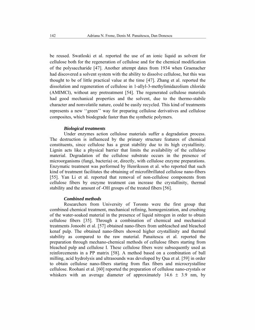
The morphology and size distribution of cellulose fibers dimension were analyzed by AFM, and different scanning areas are shown in Figures 4-7.
Fig. 4. Phase and amplitude AFM image of cellulose sample A: 1.0 μm x 1.0 μm
146 Adriana N. Frone, Denis M. Panaitescu, Dan Donescu
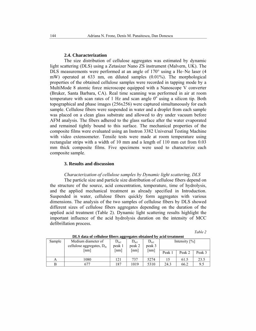
Fig. 5. Phase and amplitude AFM image of cellulose sample B: 1.0 μm x 1.0 μm
Cellulose fibers with different morphological characteristics, depending on
the applied hydrolysis time, can be seen in these images: for sample B, obtained at the highest acid treatment time, nano-fibers with smaller mean diameter (32.6 nm) and higher aspect ratio (4 – 9) as compared to the sample A, with a mean diameter of 37.0 nm and an aspect ratio between 3 and 5.
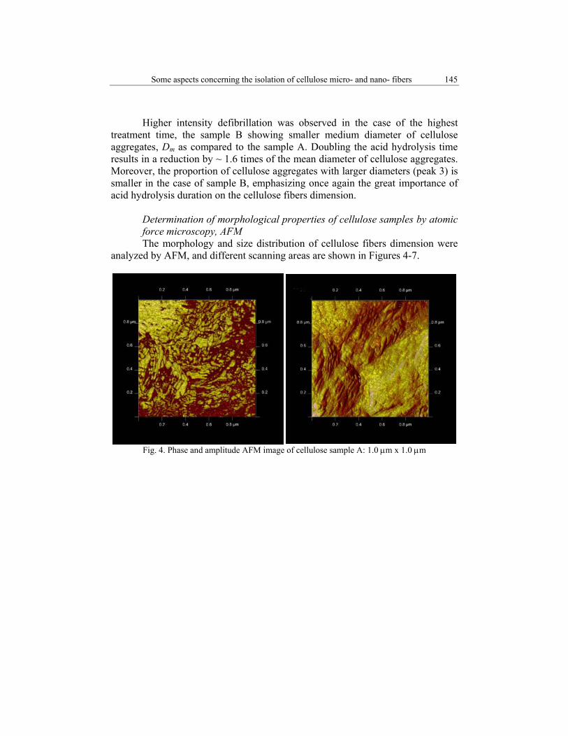
Fig. 6. Phase and amplitude AFM image of cellulose sample A: 500nm x 500 nm
Some aspects concerning the isolation of cellulose micro- and nano- fibers 147
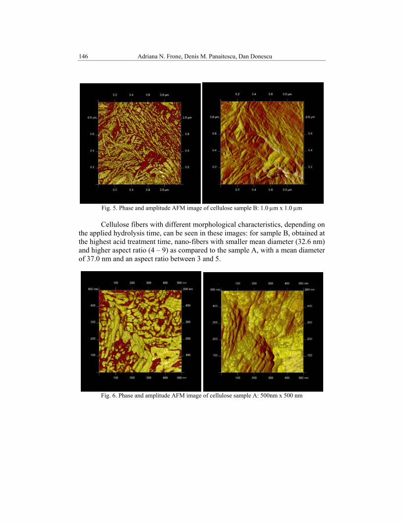
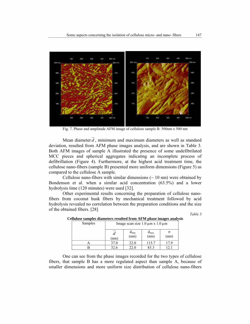
Fig. 7. Phase and amplitude AFM image of cellulose sample B: 500nm x 500 nm
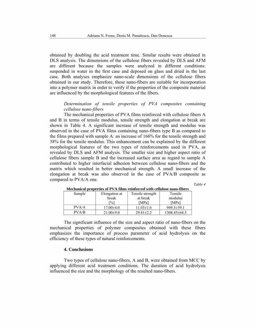
Mean diameter d , minimum and maximum diameters as well as standard
deviation, resulted from AFM phase images analysis, and are shown in Table 3. Both AFM images of sample A illustrated the presence of some undefibrilated MCC pieces and spherical aggregates indicating an incomplete process of defibrillation (Figure 4). Furthermore, at the highest acid treatment time, the cellulose nano-fibers (sample B) presented more uniform dimensions (Figure 5) as compared to the cellulose A sample. Cellulose nano-fibers with similar dimensions (~ 10 nm) were obtained by Bondenson et al. when a similar acid concentration (63.5%) and a lower hydrolysis time (120 minutes) were used [32].
Other experimental results concerning the preparation of cellulose nano-fibers from coconut husk fibers by mechanical treatment followed by acid hydrolysis revealed no correlation between the preparation conditions and the size of the obtained fibers. [28]
Table 3 Cellulose samples diameters resulted from AFM phase images analysis
Samples Image scan size 1.0 μm x 1.0 μm
d (nm)
dmin (nm)
dmax (nm)
σ (nm)
A 37.0 22.0 115.7 17.9 B 32.6 22.0 83.3 12.1
One can see from the phase images recorded for the two types of cellulose fibers, that sample B has a more regulated aspect than sample A, because of smaller dimensions and more uniform size distribution of cellulose nano-fibers
148 Adriana N. Frone, Denis M. Panaitescu, Dan Donescu
obtained by doubling the acid treatment time. Similar results were obtained in DLS analysis. The dimensions of the cellulose fibers revealed by DLS and AFM are different because the samples were analyzed in different conditions: suspended in water in the first case and deposed on glass and dried in the last case. Both analyses emphasize nano-scale dimensions of the cellulose fibers obtained in our study. Therefore, these nano-fibers are suitable for incorporation into a polymer matrix in order to verify if the properties of the composite material are influenced by the morphological features of the fibers.
Determination of tensile properties of PVA composites containing cellulose nano-fibers
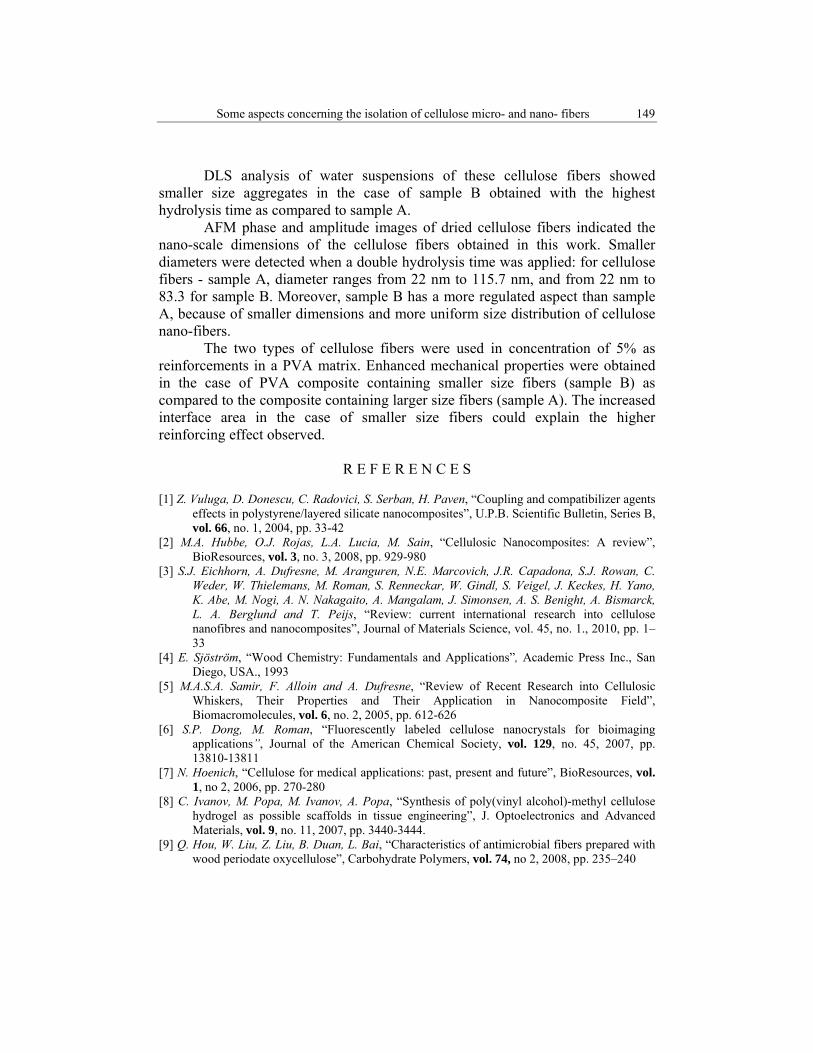
The mechanical properties of PVA films reinforced with cellulose fibers A and B in terms of tensile modulus, tensile strength and elongation at break are shown in Table 4. A significant increase of tensile strength and modulus was observed in the case of PVA films containing nano-fibers type B as compared to the films prepared with sample A: an increase of 166% for the tensile strength and 38% for the tensile modulus. This enhancement can be explained by the different morphological features of the two types of reinforcements used in PVA, as revealed by DLS and AFM analysis. The smaller size and higher aspect ratio of cellulose fibers sample B and the increased surface area as regard to sample A contributed to higher interfacial adhesion between cellulose nano-fibers and the matrix which resulted in better mechanical strength. A small increase of the elongation at break was also observed in the case of PVA/B composite as compared to PVA/A one.
Table 4 Mechanical properties of PVA films reinforced with cellulose nano-fibers
Sample Elongation at break [%]
Tensile strength at break [MPa]
Tensile modulus [MPa]
PVA/A 17.00±4.0 11.03±1.6 949.5±59.1 PVA/B 21.00±9.0 29.41±2.2 1308.45±64.5
The significant influence of the size and aspect ratio of nano-fibers on the
mechanical properties of polymer composites obtained with these fibers emphasizes the importance of process parameter of acid hydrolysis on the efficiency of these types of natural reinforcements.
4. Conclusions
Two types of cellulose nano-fibers, A and B, were obtained from MCC by applying different acid treatment conditions. The duration of acid hydrolysis influenced the size and the morphology of the resulted nano-fibers.
Some aspects concerning the isolation of cellulose micro- and nano- fibers 149
DLS analysis of water suspensions of these cellulose fibers showed smaller size aggregates in the case of sample B obtained with the highest hydrolysis time as compared to sample A. AFM phase and amplitude images of dried cellulose fibers indicated the nano-scale dimensions of the cellulose fibers obtained in this work. Smaller diameters were detected when a double hydrolysis time was applied: for cellulose fibers - sample A, diameter ranges from 22 nm to 115.7 nm, and from 22 nm to 83.3 for sample B. Moreover, sample B has a more regulated aspect than sample A, because of smaller dimensions and more uniform size distribution of cellulose nano-fibers. The two types of cellulose fibers were used in concentration of 5% as reinforcements in a PVA matrix. Enhanced mechanical properties were obtained in the case of PVA composite containing smaller size fibers (sample B) as compared to the composite containing larger size fibers (sample A). The increased interface area in the case of smaller size fibers could explain the higher reinforcing effect observed.
R E F E R E N C E S
[1] Z. Vuluga, D. Donescu, C. Radovici, S. Serban, H. Paven, “Coupling and compatibilizer agents effects in polystyrene/layered silicate nanocomposites”, U.P.B. Scientific Bulletin, Series B, vol. 66, no. 1, 2004, pp. 33-42
[2] M.A. Hubbe, O.J. Rojas, L.A. Lucia, M. Sain, “Cellulosic Nanocomposites: A review”, BioResources, vol. 3, no. 3, 2008, pp. 929-980
[3] S.J. Eichhorn, A. Dufresne, M. Aranguren, N.E. Marcovich, J.R. Capadona, S.J. Rowan, C. Weder, W. Thielemans, M. Roman, S. Renneckar, W. Gindl, S. Veigel, J. Keckes, H. Yano, K. Abe, M. Nogi, A. N. Nakagaito, A. Mangalam, J. Simonsen, A. S. Benight, A. Bismarck, L. A. Berglund and T. Peijs, “Review: current international research into cellulose nanofibres and nanocomposites”, Journal of Materials Science, vol. 45, no. 1., 2010, pp. 1–33
[4] E. Sjöström, “Wood Chemistry: Fundamentals and Applications”, Academic Press Inc., San Diego, USA., 1993
[5] M.A.S.A. Samir, F. Alloin and A. Dufresne, “Review of Recent Research into Cellulosic Whiskers, Their Properties and Their Application in Nanocomposite Field”, Biomacromolecules, vol. 6, no. 2, 2005, pp. 612-626
[6] S.P. Dong, M. Roman, “Fluorescently labeled cellulose nanocrystals for bioimaging applications”, Journal of the American Chemical Society, vol. 129, no. 45, 2007, pp. 13810-13811
[7] N. Hoenich, “Cellulose for medical applications: past, present and future”, BioResources, vol. 1, no 2, 2006, pp. 270-280
[8] C. Ivanov, M. Popa, M. Ivanov, A. Popa, “Synthesis of poly(vinyl alcohol)-methyl cellulose hydrogel as possible scaffolds in tissue engineering”, J. Optoelectronics and Advanced Materials, vol. 9, no. 11, 2007, pp. 3440-3444.
[9] Q. Hou, W. Liu, Z. Liu, B. Duan, L. Bai, “Characteristics of antimicrobial fibers prepared with wood periodate oxycellulose”, Carbohydrate Polymers, vol. 74, no 2, 2008, pp. 235–240
150 Adriana N. Frone, Denis M. Panaitescu, Dan Donescu
[10] P.A.A.P. Marques, T. Trindad, C.P. Neto, “Titanium dioxide/cellulose nanocomposites prepared by a controlled hydrolysis method”, Composites Science and Technology, vol. 66, no. 6-8, 2006, pp. 1038–1044
[11] L. Famá, L. Gerschenson, S. Goyanes, “Starch-vegetable fiber composites to protect food products”, Carbohydrate Polymers, vol. 75, no. 2, 2009, pp. 230–235
[12] T.H. Wegner, J.E. Winandy, M.A. Ritter, “Nanotechnology opportunities in residential and non residential construction”, 2nd International Symposium on Nanotechnology in Construction, Bilbao, Spania 2005
[13] R. Fijan, M. Basile, S. Šostar-Turk, E. Zagar, M. Zigon, R. Lapasin, “A study of rheological and molecular weight properties of recycled polysaccharides used as thickeners in textile printing”, Carbohydrate Polymers, vol. 76, no. 1, 2009, pp .8–16
[14] S. Kamel, “Nanotechnology and its applications in lignocellulosic composites, a mini review”, eXPRESS Polymer Letters, vol. 1, no.9, 2007, pp. 546–575
[15] D. Donescu, R. Somoghi, C. Petcu, M.C. Corobea, R. Ianchis, C.L. Nistor, “Silica hybrid particles synthesized through sol-gel processes”, U.P.B. Scientific Bulletin, Series B, vol. 70, no. 2, 2008, pp. 39-44
[16] W.J. Orts, J. Shey, S.H. Imam, G.M. Glenn, M.E. Guttman, J.F. Revol, “Application of Cellulose Microfibrils in Polymer Nanocomposites”, Journal of Polymers and the Environment, vol. 13, no. 4, 2005, pp. 301-306
[17] M. Laka, S. Chernyavskaya, “Obtaining microcrystalline cellulose from softwood and hard wood pulp”, BioResources, vol. 2, no. 3, 2007, pp. 583-589
[18] A.N. Nakagaito, S. Iwamoto, H. Yano, “Bacterial cellulose: the ultimate nano-scalar cellulose morphology for the production of high-strength composites”, Applied Physics A, vol. 80, no. 1, 2005, pp. 93–97
[19] M. Grunert, W.T. Winter, “Nanocomposites of Cellulose Acetate Butyrate Reinforced with Cellulose Nanocrystals”, Journal of Polymers and the Environment, vol. 10, no. 1-2, 2002, pp. 27-30
[20] S. Yano, H. Maeda, M. Nakajima, T. Hagiwara, T. Sawaguchi, “Preparation and mechanical properties of bacterial cellulose nanocomposites loaded with silica nanoparticles”, Cellulose, vol. 15, no. 1, 2008, pp. 111-120
[21] H.M. Shaikh, K.V. Pandare, G. Nair, A.J. Varma, “Utilization of sugarcane bagasse cellulose for producing cellulose acetates: Novel use of residual hemicellulose as plasticizer”, Carbohydrate Polymers, vol. 76, no. 1, 2009, pp. 23–29
[22] X. Cao, Y. Chen, P.R. Chang, M. Stumborg, M. A. Huneault, “Green Composites Reinforced with Hemp Nanocrystals in Plasticized Starch, Journal of Applied Polymer Science, vol. 109, no. 6, 2008, pp. 3804–3810;
[23] R. Belhassen, J.A. Mendez, S. Boufi, J.P. Lopez, J. Puig, A. Pelach, P. Mutje, “Preparation and Properties of Biocomposites Based on Jute Fibers and Blend of Plasticized Starch and Poly(b-hydroxybutyrate)”, Journal of Applied Polymer Science, vol. 114, no. 1, 2009, pp. 313–321
[24] M. El-Sakhawy, M. L. Hassan, “Physical and mechanical properties of microcrystalline cellulose prepared from agricultural residues”, Carbohydrate Polymers, vol. 67, no. 1, 2007, pp. 1–10
[25] R. Zuluaga, J.L. Putaux, J. Cruz, J. Vélez, I. Mondragon, P. Gañán, “Cellulose microfibrils from banana rachis: Effect of alkaline treatments on structural and morphological features”, Carbohydrate Polymers, vol. 76, no. 1, 2009, pp. 51–59
[26] N.L.G .Rodriguez, W. Thielemans, A. Dufresne,“Sisal cellulose whiskers reinforced polyvinyl acetate nanocomposites”, Cellulose, vol. 13, no. 3, 2006, pp. 261–270
Some aspects concerning the isolation of cellulose micro- and nano- fibers 151
[27] Y. Chen, C. Liu, Peter R. Chang, X. Cao, Debbie P. Anderson “Bionanocomposites based on pea starch and cellulose nanowhiskers hydrolyzed from pea hull fiber: Effect of hydrolysis time”, Carbohydrate Polymers, vol. 76, no. 4, 2009, pp. 607-615
[28] M.F. Rosa, E.S. Medeiros, J.A. Malmonge, K.S. Gregorski, D.F. Wood, L.H.C. Mattoso, G. Glenn,W.J. Orts, S.H. Imam, “Cellulose nanowhiskers from coconut husk fibers: Effect of preparation conditions on their thermal and morphological behavior”, Carbohydrate Polymers, vol. 81, no. 1, 2010, pp. 83–92
[29] L. Zhou, P. Sun, Z. Yang, “Effect of addition of sodium alginate on bacterial cellulose production by Acetobacter xylinum”, Journal of Industrial Microbiology and Biotechnology, vol. 34, no. 7, 2007, pp. 483-489
[30] S. Y. Lee, S. J. Chun, I. A. Kang and J. Y. Park, “Preparation of cellulose nanofibrils by high-pressure homogenizer and cellulose-based composite films”, Journal of Industrial and Engineering Chemistry, vol. 15, no. 1, 2009, pp. 50–55.
[31] P.B. Filson, B.E. Dawson-Andoh, “Sono-chemical preparation of cellulose nanocrystals from lignocellulose derived materials”, Bioresource Technology, vol. 100, 2009, no. 7, pp. 2259–2264.
[32] D. Bondeson, A. Mathew, Kristiina Oksman, “Optimization of the isolation of nanocrystals from microcrystalline cellulose by acid hydrolysis”, Cellulose, vol. 13, no. 2, 2006, pp. 171-180
[33] J.K. Pandey, C.S. Lee, S.H. Ahn, “Preparation and properties of bio-nanoreinforced composites from biodegradable polymer matrix and cellulose whiskers”, Journal of Applied Polymer Science,vol. 115, no. 4, 2010, pp. 2493–2501
[34] D.M. Panaitescu, I. Matasaru, H. Iovu, M. Ghiurea, M.D. Iorga, P. Stanescu, “Compozite termoplastice cu microfibrile celulozice obtinute prin tratamente mecanice”, Materiale Plastice, vol. 44, no. 2, 2007, pp. 144-147
[35] A. Bhatnagar, M. Sain, “Processing of cellulose nanofiber-reinforced composites”, J. of Reinforced Plastics Composites, vol. 24, no. 12, 2005, pp. 1259-1268
[36] A. Alemdar, M. Sain, “Biocomposites from wheat straw nanofibers: Morphology, thermal and mechanical properties”, Composites Science and Technology, vol. 68, no. 2, 2008b, pp. 557-565
[37] A. Chakraborty, M. Sain, M. Kortschot, Cellulose microfibrils: A novel method of preparation using high shear refining and cryocrushing, Holzforschung, vol. 59, no. 1, 2005, pp. 102–107
[38] T. Zimmermann, N. Bordeanu, E. Strub, “Properties of nanofibrillated cellulose from different raw materials and its reinforcement potential”, Carbohydrate Polymers, vol. 79, no. 4, 2010, pp. 1086–1093
[39] S. Wang, Q. Cheng, “A Novel Process to Isolate Fibrils from Cellulose Fibers by High-Intensity Ultrasonication, Part 1: Process Optimization”, Journal of Applied Polymer Science,vol. 113, no. 2, 2009, pp. 1270–1275
[40] Q. Xiang, Y.Y. Lee, P.O. Pettersson, R.W. Torget, “Heterogeneous Aspects of Acid Hydrolysis of a-Cellulose”, Applied Biochemistry and Biotechnology, vol. 105–108, no. 1-3, 2003, pp. 505-514
[41] J. Zhang, T.J. Elder, Y. Pu and A.J. Ragauskas, “Facile synthesis of spherical cellulose nanoparticles”, Carbohydrate Polymers, vol. 69, no.3, 2007, pp. 607–611
[42] J.I. Moran, V.A. Alvarez, V.P. Cyras, A. Vazquez, “Extraction of cellulose and preparation of nanocellulose from sisal fibers”, Cellulose, vol. 15, no. 1, 2008, pp. 149-159
[43] S.Y. Lee, D.J. Mohan, I.A. Kang, G.H. Doh, S. Lee, S.O. Han, “Nanocellulose reinforced PVA composite films: Effects of acid treatment and filler loading,” Fibers and Polymers, vol. 10, no. 1, 2009, pp. 77-82
152 Adriana N. Frone, Denis M. Panaitescu, Dan Donescu
[44] B. Wang, M. Sain, “Isolation of nanofibers from soybean source and their reinforcing capability on synthetic polymers”, Composites Science and Technology, vol. 67, no. 11–12, 2007, pp. 2517–2521
[45] B. Wang, M. Sain, K. Oksman, “Study of structural morphology of hemp fiber from the micro to the nanoscale”, Applied Composite Materials, vol. 14, no. 2, 2007, pp. 89–103
[46] E.S. Abdel-Halim , H.E. Emam, M.H. El-Rafie, “Preparation and characterization of water soluble poly(acrylic acid)–hydroxypropyl cellulose composite”, Carbohydrate Polymers, vol. 74, no. 4, 2008, pp. 783–786
[47] A. Pinkert, K.N. Marsh, S. Pang, M.P. Staiger, “Ionic Liquids and Their Interaction with Cellulose”, Chemical Reviews, vol. 109, no. 12, 2009, pp. 6712–6728
[48] L. Petersson, K. Oksman, “Biopolymer based nanocomposites: Comparing layered silicates and microcrystalline cellulose as nanoreinforcement”, Composites Science and Technology, vol. 66, no.13, 2006, pp. 2187–2196
[49] S. Yokota, T. Kitaoka, J. Sugiyama, H. Wariishi, “Cellulose I nanolayers designed by self-assembly of its thiosemicarbazone on a gold substrate“, Advanced Materials, vol. 19, no. 20, 2007, pp. 3368-3370
[50] S. Gunnars, L. Wågberg, M.A.C. Stuart, “Model films of cellulose: I. Method development and initial results”, Cellulose, vol. 9, no. 3-4, 2002, pp. 239-249
[51] D. Viet, S. Beck-Candanedo, D.G. Gray,“Dispersion of cellulose nanocrystals in polar organic solvents”, Cellulose, vol. 14, no. 2, 2007, pp. 109 –113
[52] H. Dogan, N.D. Hilmioglu, “Dissolution of cellulose with NMMO by microwave heating”, Carbohydrate Polymers, vol. 75, no. 1, 2009, pp. 90–94
[53] S. Zhu, Y. Wu, Q. Chen, Z. Yu, C. Wang, S. Jin, Y. Dinga and G. Wu, “Dissolution of cellulose with ionic liquids and its application: a mini-review”, Green Chemistry, vol. 8, no. 2006, pp. 325–327
[54] H. Zhang, J. Wu, J. Zhang, J. He, “1-Allyl-3-methylimidazolium Chloride Room Temperature Ionic Liquid: A New and Powerful Nonderivatizing Solvent for Cellulose”, Macromolecules, vol. 38, no. 20, 2005, pp. 8272–8277
[55] M. Henriksson, G. Henriksson, L.A. Berglund, T. Lindstrom, “An environmentally friendly method for enzyme-assisted preparation of microfibrillated cellulose (MFC) nanofibers, European Polymer Journal, vol. 43, no. 8, 2007, pp. 3434–3441
[56] Y. Li, K.L. Pickering, “Hemp fibre reinforced composites using chelator and enzyme treatments”, Composites Science and Technology, vol. 68, no. 15-16, 2008, pp. 3293–3298
[57] M. Jonoobi, J. Harun, A. Shakeri, M. Misra, K. Oksman, “Chemical composition, crystallinity, and thermal degradation of bleached and unbleached kenaf bast (Hibiscus cannabinus) pulp and nanofibers,” BioResources, vol. 4, no. 2, 2009, pp. 626-639
[58] D.M. Panaitescu, P. Nechita, H. Iovu, M.D. Iorga, M. Ghiurea, D. Serban, ’’Polypropylene composites with cellulose microfibrils obtained through mechano-chemical treatments”, Materiale Plastice, vol. 44, nr. 3, 2007, pp. 195-198
[59] E.H. Qua, P.R. Hornsby, H.S.S Sharma, G. Lyons, R.D. McCall, “Preparation and Characterization of Poly(vinyl alcohol) Nanocomposites Made from Cellulose Nanofibers”, Journal of Applied Polymer Science,vol. 113, no. 4, 2009, pp. 2238–2247
[60] M. Roohani, Y. Habibi, N.M. Belgacem, G. Ebrahim, A.N. Karimi, A. Dufresne, “Cellulose whiskers reinforced polyvinyl alcohol copolymers nanocomposites”, European Polymer Journal, vol. 44, no. 8, 2008, pp. 2489–2498
[61] M. Jonoobi, J. Harun, A.P. Mathew, M.Z.B. Hussein, K. Oksman, “Preparation of cellulose nanofibers with hydrophobic surface characteristics”, Cellulose, vol. 17, no. 2, 2010, pp. 299–307