Embed Size (px)
Citation preview
Phytopath. 2., 108, 235—241 (1983)© 1983 Verlag Paul Parey, Berlin und HamburgISSN 0031-9481 / InterCode: PHYZA3
Plant Protection Service, Wageningen, Netherlands
Some Factors Influencing Immunofluorescence Microscopyas Applied in Diagnostic Phytobacteriology
with Regards to Erwinia amylovora
By
H. J. MILLER
Received February 14, 1983
Abstract
Zusammenfassung
Einige Faktoren, die die Immunfluoreszente Mikroskopie beeinflussen,die in der diagnostischen Phytobakteriologie verwandt wird,
dargestellt an Erwinia amglovora
Die Praparlungen oder von
g von Erwinia amylovora-\so\\^T\xn^Qn durch unterschicdhche Behand-rsdiiedlidien Urspriingen beeinflui5t die Ergebnisse, die durch immun-
n nidit praktikabel ist, konnen Reihen^:e Anzahlen des zu testenden Bakteriu: s benutzt werden., eine Reduktioi
In order to prevent the establishment and spread of plant diseases whichhave in recent years been facilitated by the ease of transportation as well asthe demand for more and better quality plant material, the need for earlydetection of such diseases has become essential. In the phytobacteriology an
U.S. Copyright Clear itcr Code Statement: 0031-9481/83/0804-0235,102.50/0
236 MILLER
.attempt to fill this need has been made in many laboratories by the applica-tion of immunofluorescence microscopy (IF). With any such method it is notonly the speed and practicability that are so important but also the accuracy.Unfortunately results with IF have until now often proved to be disap-pointing. Probably the haste of getting into a promising new method has led tothe omission of a number of fundamental principles essential in reliablediagnostic practice.
The aim of this paper is to discuss results from recent routine laboratorywork carried out on plant material suspected of being infected with Erwiniaamylovora (Burr.) Winsl. et al., the causative organism of fireblight̂ using amethod similar to that of MILLER (1979). In this way we may gain a betterinsight into the serologica! problems of practical immunofluorescence micros-copy as applied to phytobacteriology and learn within which Umitations we
Materials and Methods
Bacterial cultures
Cultures were obtained from the National Collection of Plant Pathogenic Bacteria(NCPPB), En<;bnd and included: Erivima rhapontici NCPPB 2548 and NCPPB 2549, E. her-hicola NCPPB 2971 (type species) and NCPPB 3009 (atypical E.herbicola — see ROBERTS1980). Sixty cultures of £. amylovora were isolated from 88 plants suspected of fireblight —Table 1.
Culture Media
Sucrose nutrient agar (SNA) was used as described by LELLIOTT (1968) for isolation anddetermination oi levan positive colonies. The presence or fluorescent pseudomonsos was deter-'mined with King's B medium (KING et al. 1954).
Serology
Bacterial suspensions were made als follows: Method A) directly from levan positivecolonies in 0.01 M phosphate buffered saline (PBS) pH 7.2, Method B) as in A but washedthree times in PBS, each time centrifuging at 10,000 g for 10 minuteis and resuspending thepellet in PBS, Method C) suspending bacterial celk in PBS after diffusion from pieces ofsuspected plant tissue and Method D) as in C but washed tKree times in PBS, each time centri-fuging at 10,000 g for 10 minutes and resuspending the pellet in PBS.
The anriserum for E. amylovora as well as the antiserum for Pseudomonas syringae,
used for checking the fluorescent pseudomonads from King's B medium, were produced ac-cording to the method of VRUGGINK and MAAS GE£STHRANUS (1975) and supplied by theInstitute for Plant Protection Research.
used for the primary identification of E. amylovora and P. syringae and 2. Immunofluorescencemicroscopy (indirect IF) as described by MILLER and VRUGGINK (1981). Bacterial raspensions
with suspensions from tissue which frequently yielded lower numbers. The serological endpoint or titre for the IF was considered to be the highest dilution of the antiserum at whidifluorescence occurred before a noticable decrease in imtensity. The reading of the microscope
Some Factors Influencing Immunofluorescence Microscopy 237
Results
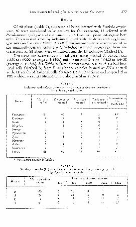
Of 88 plants (Table 1), suspected of being Infected with Erwinia amylo-vora, 60 were considered to be positive for this organism, 12 infected withPseudomonas syringae and the remaining 16 free from plant pathogenic bac-teria. This was confirmed by isolation coupled with the direct slide agglutina-tion test (see LELLIOTT 1968). All 60 E. amylovora isolates were subjected tothe immunofluorescent technique (IF-Method A) and suspensions from thetissue from all 88 plants were examined using the IF technique (Method D).
The titres for E. amylovora in all cases using method A varied from1:800 to 1:3200 (average c. 1:1500) and for method D from 1:1600 to 1:6400(average c. 1:4300). See Table 2. Immunofluorescence results of washed bac-terial cells (Method B) from E. amylovora colonies formed on SNA as wellas the IF results of bacterial cells obtained from plant tissue and suspended inPBS without washing (Method C) are also placed in Table 2.
Table 1
from 88 suspected plaruts
CotoneusterCrataegusMalusMespilusPrunuiPyracanthaPyrusRhamnusSorbusStranvesiaTotals
No. plantstested
5184124213
1288
E. amylovoraisolated
4371103102
60
P. syrmgaeisolated
3130000005
12
Pathogeasnot isolated
E. amylovora
Method D
5 44--0 70 10 12 01 31 11 01 25 2
16 fop'-
D. 575 in Table 3.
Table 2Serological results (IF) using Erwinia amylovora cell suspensions prepared
by four differeat methods
Method
ABCD
No.su^spensions
60171060
400
0060
Recipro
800
1021
20
cal of antiseru
1600 1
48604
3200
202
33
6400
000
23
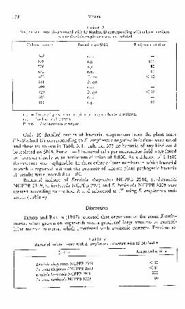
Table 3sues tested with IF-Method D corresponding with culture numbers
Culture number
567575576672673681699729803811
Isolation on SNA
n.p.n.g.n.p.
P. syr.P. syr.n.p.P. syr.n.p.n.p.
1 Reciprocal of titre
_
8008080
< 10.——
< 10—80
n.g. - No bacterial growth.P. syr. = Pseudomonas syringae.
Only 10 detailed results of bacterial suspensions from the plant tissue(IF-Method D) corresponding to E. amylovora negative isolations were savedand these are shown in Table 3. In cult. no. 575 no bacteria of any kind couldbe isolated on SNA, but c. 1—3 bacterial cells per microscope field were foundto fluoresce clearly at an antiserum dilution of 1:800. At a dilution of 1:1600fluorescence was negligeable. In three other culture numbers in which bacterialgrowth is reported without the presence of known plant pathogenic bacteriaIF results were recorded at 1:80.
Bacterial isolates of Erwinia rhapontici NCPPB 2548, E. rhaponticiNCPPB 2549, E. herbicola NCPPB 2971 and E. herhkola NCPPB 3009 weretreated according to method A and subjected to IF using E. amylovora anti-serum (Table 4).
Discussion
ELROD and BRAUN (1947) reported that organisms of the genus Xantho-monas when grown on sugar-rich media produced large amounts of extracel-lular mucoid material which interfered with antigenic patterns. Previous re-
Table 4
ErwiniaErwiniaErwinia
Isc
rhaponrhaponherbkoherbko
la
tictklala
te
i NCPPB 2548;• NCPPB 2549NCPPB 2971NCPPB 3009
Reciprocal of titre
< 10
< 10200
80
oomc Ptictors Intlucncin^ IiTimunoiluorcsccncc iVlicroscopy 239
search in the Erwinia-soh rot group (ELROD 1941) had shown that maltosefermenting and non-maltose fermenting organisms gave large variations in thetitres of agglutination reactions. The reason for the variations obtained withErwinia amylovora using the indirect immunofluorescent technique, in thedifferent methods described in this paper, may well therefore be associatedwith the antigenic properties of extracellular polysaccharides or lipopoly-saccharides which are produced by E. amylovora when grown on artificialmedia (AYERS et al. 1979).
Although washing of E. amylovora cells was reported by MILLER (1979),this had only been done to improve the reading quality of the IF preparations.At the lower antiserum dilutions then in use, 1:20—1:100, the slime present inunwashed cell suspensions obtained from plant material was found to fluoresceand mask observation of the fluorescing cells. Increased sensitivity withwashed cells obtained from infected plant tissue (see Table 2.) is probablycaused by the removal of masking substances such as sugars present in thebacterial slime (see HILDEBRAND 1939, EDEN-GREEN and KNEE 1974). Com-petition for the antiserum or conjugate during the IF staining by these sub-stances would result in a decreased sensitivity.
Results of a number of bacterial suspensions from plant tissue (Table 3.)indicate serological reactions at lower titres than those obtained with thesame method used for known E. amylovora specimens (Table 2). As bacterialgrowth could not be detected on SNA with cult. no. 575 but fluorescing cellswere found in the IF at 1:800, it must be presumed that these cells weredead. The explanation of this occurrence could either be the presence ofcross-reacting cells or that the cells were E. amylovora but, due to a deficiencyin available antigens in the cell wall, reacted at a lower titre than the averagefor method D. Other culture numbers showing low titre reactions may then
In cross-reaction studies carried out with cultures of other Erzvinia spe-cies using E. amylovora antiserum (Table 4.), £. herbicola NCPPB 2971 wasfound to react at 1:200 and E. herbicola NCPPB 3009 at 1:80. Results suchas these explain why ROBERTS (1980) obtained positive results, also withNCPPB 3009, when she used her E. amylovora antiserum at the single dilutionof 1:20, which would seem to be an exceedingly low dilution.
ROBERTS reports only that the titre of the antiserum was determined bythe agglutination method, in this case 1:600, but makes no mention of havingdetermined the titre using the IF technique. This situation has been notedin numerous articles concerning the application of immunofluorescence micros-copy to the detection of plant pathogenic bacteria (e. g. DE BOER and COPE-MAN 1980). It is a requisite to first determine the titre of the antiserum, withthe homologous bacterial isolate if possible, using the same method that is tobe employed for testing purposes as different antigen-antibody interactionsmay be involved. An illustration in our own laboratory was the observance(unpublished results) whereby an antiserum prepared against Corynebacteriiirn
240 MILLER
»iichiga)u'>nc reacted with the homologous bacterial isolate as well as numer-ous other isolates of C. michiganense at a titre of 1:1600 using the indirect IFmethod but failed to react in either the agglutination or immunodiffusionmethods.
In medical serology known antigens of standard concentrations are usedto detect specific antibodies in human sera and the standard practice is todetermine the end point (titre) of the serum by the use of serial dilutions.At various intervals in a patient's disease the lowering or elevation of thistitre is used to follow the stage of the patient's condition. If a titre fallsbelow a certain level, however, it may then be considered insignificant. Withplant pathogenic bacteria, as in the case of E. amylovora, a standard antiserumis used to test the presence of the bacterium. If the number of bacteria beingtested is maintained at a relatively constant level, variations in the amountsof detectable antigens can also be demonstrated by comparing bacterial isolateswith the same antiserum using serial dilutions. This has already been reportedfor Corynebacterium fascians using the indirect IF method (MILLER et al.1980). When using E. amylovora, patterns emerge which illustrate that theorigin and preparation of bacterial cells may have a marked influence on thetitre (Table 2).
It is not always possible to work with highly specific antisera as weknow so little about the antigenic properties of the many saprophytic bac-teria that may be present in or on our suspected plant material. For this rea-son, although often attempted, absorption of undesirable antibodies fromour antiserum becomes impracticable. Yet many bacteria possess commonantigens in varying concentrations. These concentrations are often found to below as can also be deducted from Tables 3 and 4 in which case insufficientdilution of antisera will lead to false positive results (see DE BOER and COPE-MAN 1980). Correct dilution of antisera should in many cases therefore pro-duce such a low concentration of common occurring antibodies that a re-action with the IF method will not be seen but the more specific antibodieswill still be in sufficient concentrations to produce a reaction that may beconsidered positive providing the number of bacteria present in such a re-action is approximately constant. In the IF technique if a certain bacteriumis to be tested, several known isolates should first be examined by a standardi-zed method using serial dilution of the antiserum. When a range has beenestablished, see e. g. Table 2, many cross-reactions will fall away and at thesame time by using a range of antiserum dilutions the chance of a false negativereaction will also decrease. In this laboratory, for example, with E. amylo-vora (Method A) five dilutions, 1:400—1:6400, are done routinely with eadibacterial isolate to be tested with the IF technique. As already shown inTable 2, great care is necessary in the preparation of the bacterial cells to betested.
It is therefore necessary by such means as described in this paper tolearn the limitations of our method. This way we are able to standardize theIF staining procedure according to the bacterial species involved and its source.
Such procedures can only lead to a better understanding and interpretationof the results which in turn makes a more accurate diagnosis possible.
The author wishesM. J. JEGEN.
3 acknowledge the itedinlcal a of Mr. J. JANSE and Mrs
AYERS, A. R., S. B. AYERS, and R. N. GOODMAN, 1979: Extracellular polysaccharide of Erwt-Hid ufnylo'VOVd'. ^ correlation with virulence. Appl. Environ, wlicrobiol- 38̂ 659—666.
DE BOER, S. H . , and R. J. COPEMAN, 1980: Bacterial ringrot testing with the indirect fluores-cent antibody staining procedure. Am. potato J. 57, 457—465.
EDEN-GREEN, S. J., and M. KNEE, 1974: Bacterial polysaccharide and .sorbitol in fireblightexudate. J. Gen. Microbiol. 81, 509—512.
ELROD, R. P., 194t: Serological studies of the Erwiniae. II. Soft-rot group; wkh some bio-chemical considerations. Bot. Gaz. 103, 266—279.
, and A. C. BRAUN, 1947: Serological studies of the genus Xanthomonas. I. Cross-
HiLDEBRAND, E. M., 1939: Studies on fire-blig'k ooze. Phytopathology 29, 142—156.KING, E. O., M. K. WARD, and D. E. RANEY, 1954: Two simple methods for the demonstration
of pyocyanm and fluorescin. J. Lab. & Clin. Med. 44, 301—307.LELLIOTT, R. A., 1968: The diagnosis of fireblight {Erwinia amylovora) and some disesases
caused by Pseudomonas syringae. EPPO Public. Ser. A. 45-E, 27—34.MILLER, H . J., 1979: A review of methods used in the laboratory diagnosis of fireblight. EPPO
Bull. 9, 7—11., H. J., J. D. JANSE, W. KAMERMAN, and P. J. MULLER, 1980: Resent observations on
leafy gall in LiHaceae and some other families. Neth. J. PL Path. 86, 55—68., and H. VRUGGINK, 1981: An assessment of biochemical and serological tests for Agro-
bixct€Tit4TTi Tcjiiiobuct6T suD'Sp. tuTHSjucicns. 1 nytop3.tn. ^ . 102, 292—~"300.ROBERTS, P., 1980: Problems encountered during immunofluorescent diagnosis of fireblight.
PL Path. 29, 93—97.VRUGGINK, H . , and H. P. MAAS GEESTERANUS, 1975: Serological recognition of Era
546—555.: potat
Author's address: Drs. H. J. MILLER, Plant Pro6700 HC Wageningen (Netherlands).
Res. 18,
, P.O. Box 9102,
Phytopath. Z., Bd. 1


![The Phytoalexin Resveratrol Regulates the Initiation of Hypersensitive ... · from Erwinia amylovora, the causative agent of fire blight in Rosaceae [19]. In non-host plants, using](https://img.pdfslide.net/doc/110x75/5edb9a3fad6a402d6665e840/the-phytoalexin-resveratrol-regulates-the-initiation-of-hypersensitive-from.jpg)


