Embed Size (px)
Citation preview
Spine and Spinal Cord Injuries
William Schecter, MD
Anatomy of the Spine
http://education.yahoo.com/reference/gray/fig/387.html
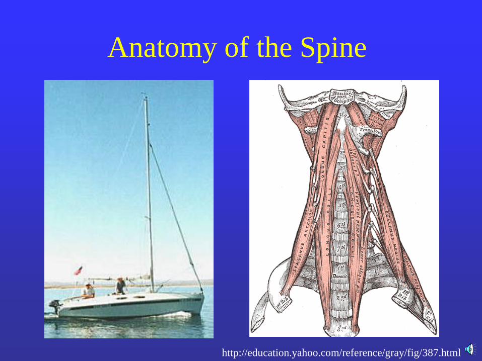
Anatomy of the spine
• 7 cervical vertebrae
• 12 thoracic vertebrae
• 5 lumbar vertebrae
• 5 fused sacral
vertebrae
• 3-4 small bones
comprising the coccyx
http://www.courses.vcu.edu/DANC291-003/unit_3.htm
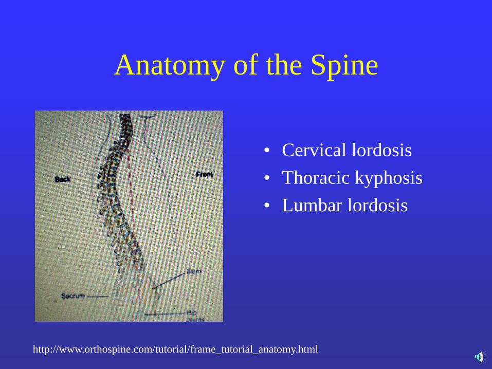
Anatomy of the Spine
• Cervical lordosis
• Thoracic kyphosis
• Lumbar lordosis
http://www.orthospine.com/tutorial/frame_tutorial_anatomy.html
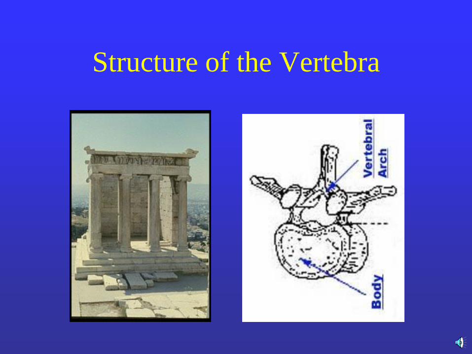
Structure of the Vertebra
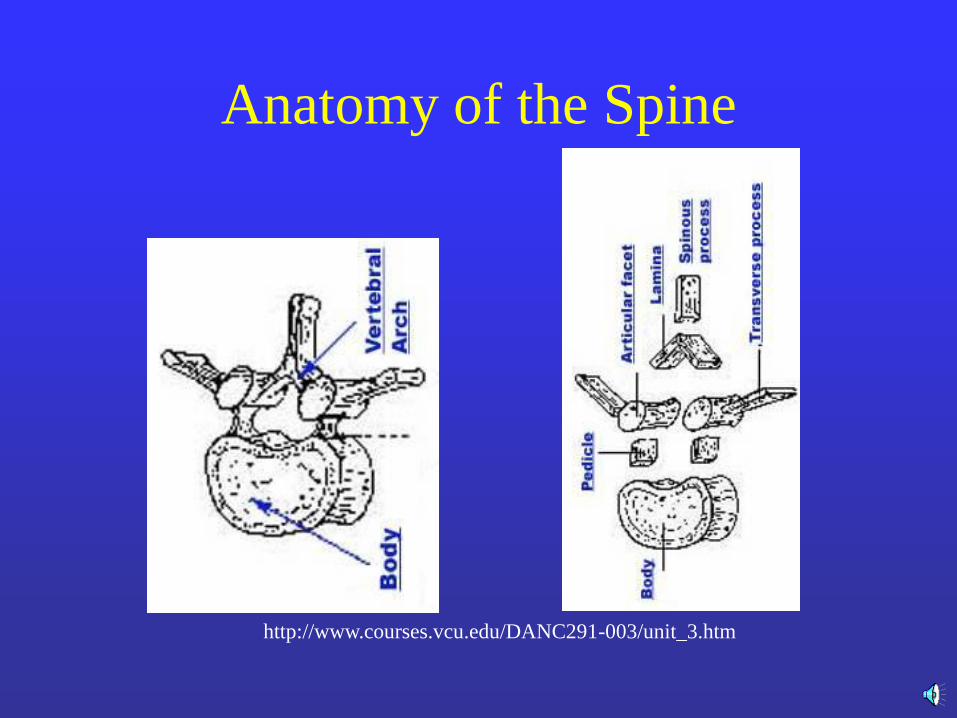
Anatomy of the Spine
http://www.courses.vcu.edu/DANC291-003/unit_3.htm
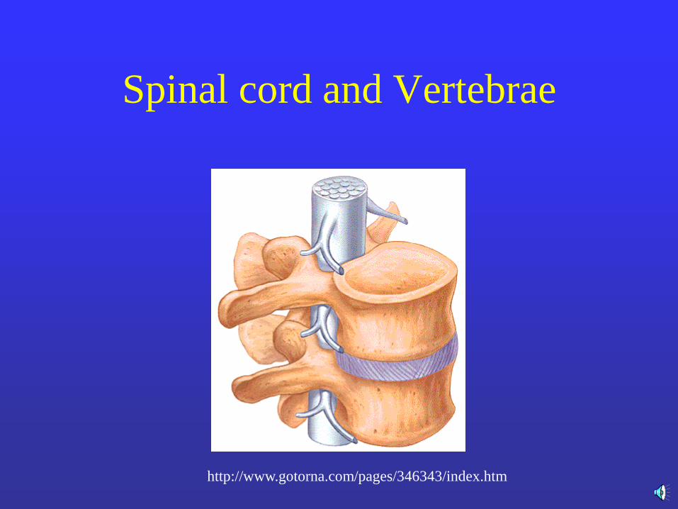
Spinal cord and Vertebrae
http://www.gotorna.com/pages/346343/index.htm
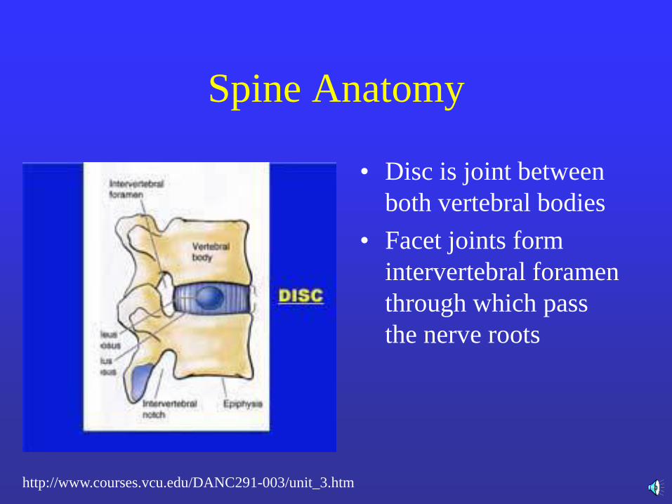
Spine Anatomy
• Disc is joint between
both vertebral bodies
• Facet joints form
intervertebral foramen
through which pass
the nerve roots
http://www.courses.vcu.edu/DANC291-003/unit_3.htm
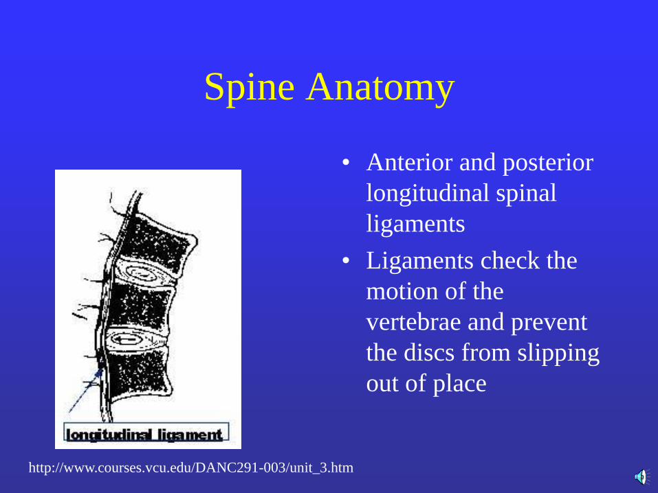
Spine Anatomy
• Anterior and posterior
longitudinal spinal
ligaments
• Ligaments check the
motion of the
vertebrae and prevent
the discs from slipping
out of place
http://www.courses.vcu.edu/DANC291-003/unit_3.htm
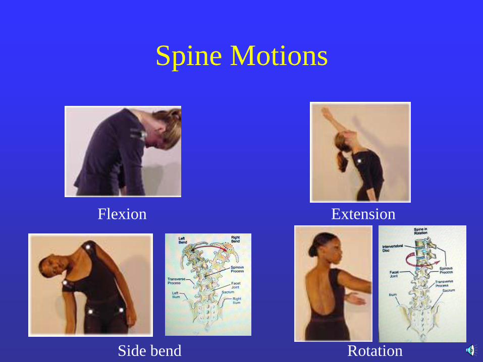
Spine Motions
Flexion Extension
Side bend Rotation
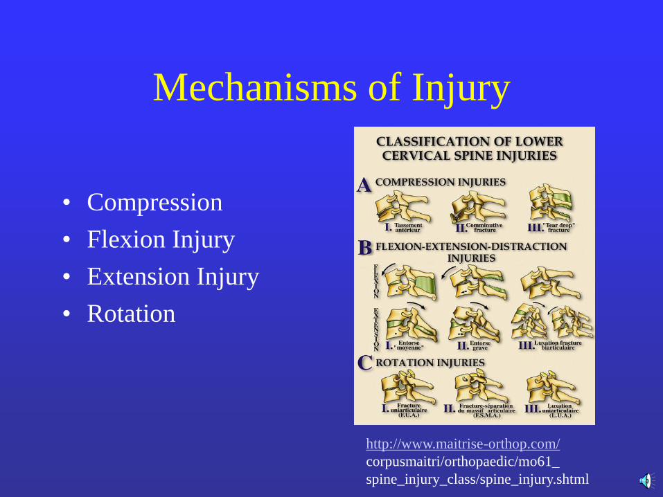
Mechanisms of Injury
• Compression
• Flexion Injury
• Extension Injury
• Rotation
http://www.maitrise-orthop.com/
corpusmaitri/orthopaedic/mo61_
spine_injury_class/spine_injury.shtml
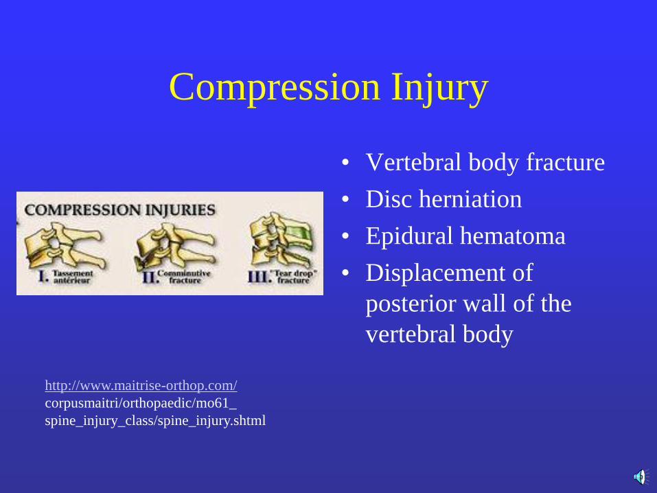
Compression Injury
• Vertebral body fracture
• Disc herniation
• Epidural hematoma
• Displacement of
posterior wall of the
vertebral body
http://www.maitrise-orthop.com/
corpusmaitri/orthopaedic/mo61_
spine_injury_class/spine_injury.shtml
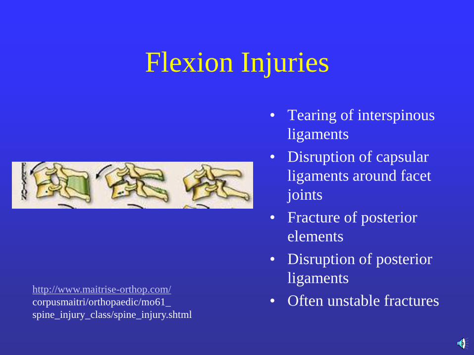
Flexion Injuries
• Tearing of interspinous
ligaments
• Disruption of capsular
ligaments around facet
joints
• Fracture of posterior
elements
• Disruption of posterior
ligaments
• Often unstable fractures http://www.maitrise-orthop.com/
corpusmaitri/orthopaedic/mo61_
spine_injury_class/spine_injury.shtml
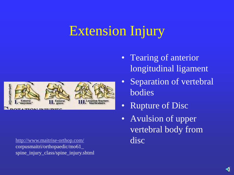
Extension Injury
• Tearing of anterior
longitudinal ligament
• Separation of vertebral
bodies
• Rupture of Disc
• Avulsion of upper
vertebral body from
disc http://www.maitrise-orthop.com/
corpusmaitri/orthopaedic/mo61_
spine_injury_class/spine_injury.shtml
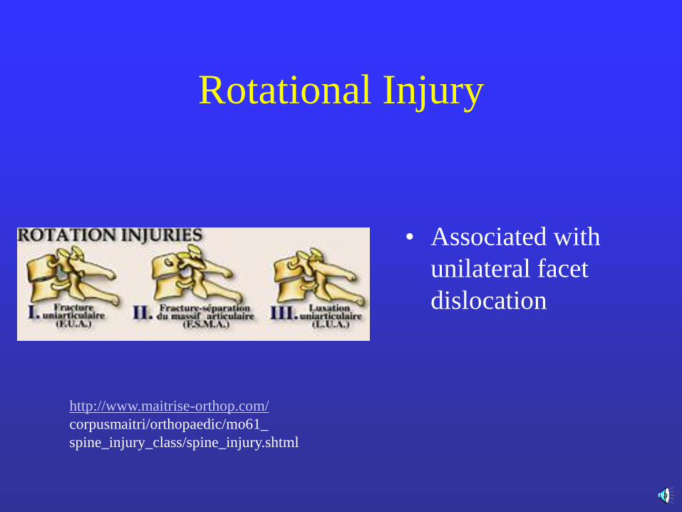
Rotational Injury
• Associated with
unilateral facet
dislocation
http://www.maitrise-orthop.com/
corpusmaitri/orthopaedic/mo61_
spine_injury_class/spine_injury.shtml
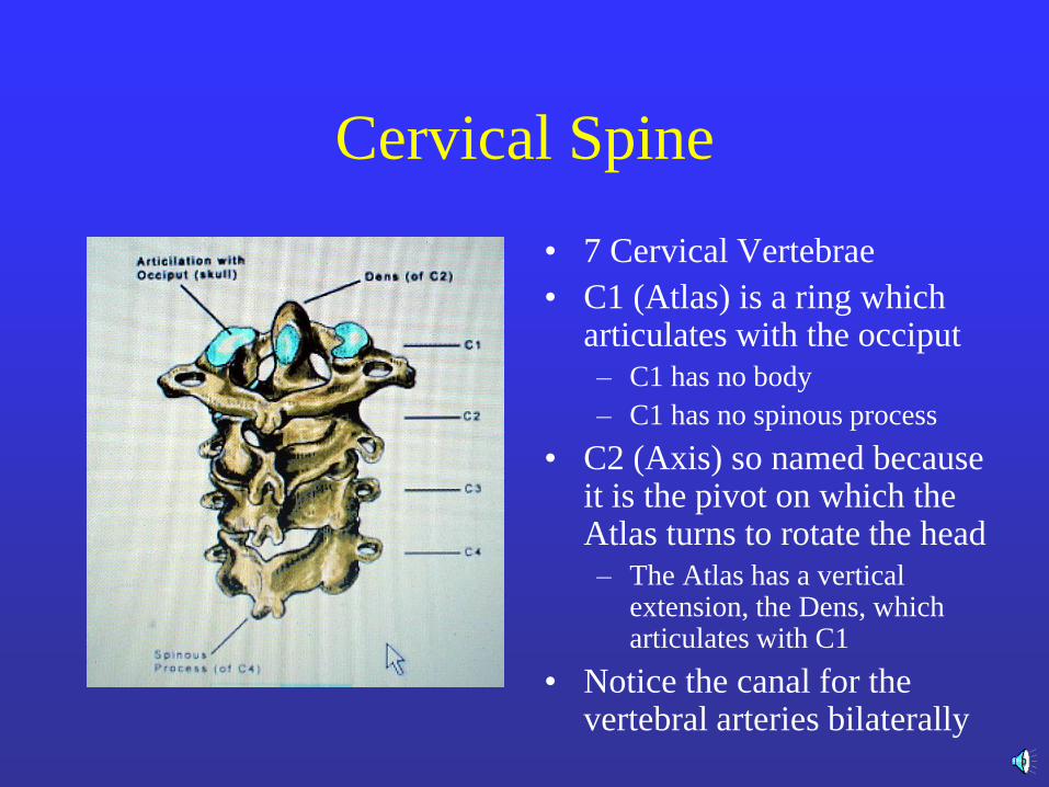
Cervical Spine
• 7 Cervical Vertebrae
• C1 (Atlas) is a ring which articulates with the occiput
– C1 has no body
– C1 has no spinous process
• C2 (Axis) so named because it is the pivot on which the Atlas turns to rotate the head
– The Atlas has a vertical extension, the Dens, which articulates with C1
• Notice the canal for the vertebral arteries bilaterally
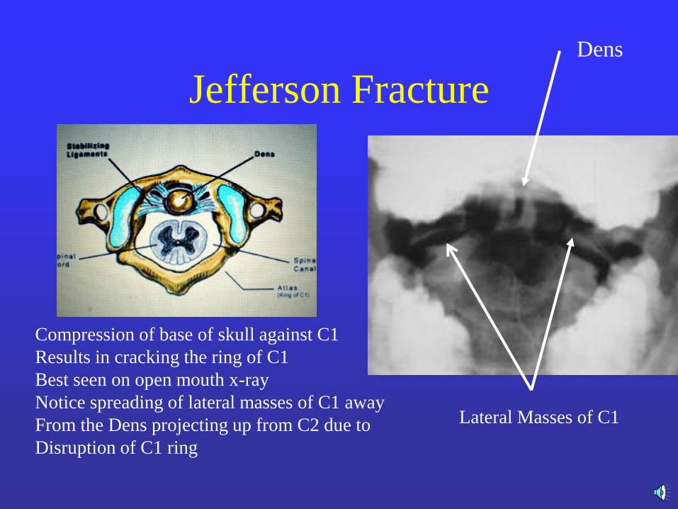
Jefferson Fracture
Compression of base of skull against C1
Results in cracking the ring of C1
Best seen on open mouth x-ray
Notice spreading of lateral masses of C1 away
From the Dens projecting up from C2 due to
Disruption of C1 ring
Lateral Masses of C1
Dens
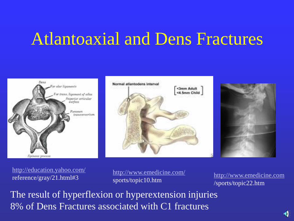
Atlantoaxial and Dens Fractures
The result of hyperflexion or hyperextension injuries
8% of Dens Fractures associated with C1 fractures
http://education.yahoo.com/
reference/gray/21.html#3 http://www.emedicine.com/
sports/topic10.htm http://www.emedicine.com
/sports/topic22.htm
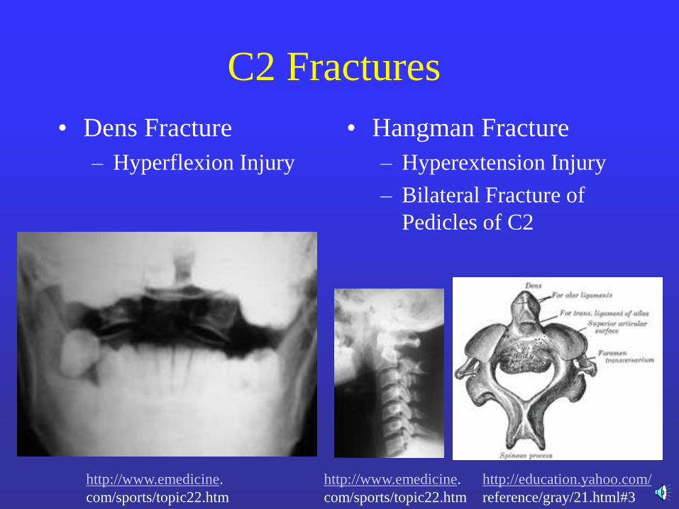
C2 Fractures
• Dens Fracture
– Hyperflexion Injury
• Hangman Fracture
– Hyperextension Injury
– Bilateral Fracture of
Pedicles of C2
http://www.emedicine.
com/sports/topic22.htm
http://education.yahoo.com/
reference/gray/21.html#3
http://www.emedicine.
com/sports/topic22.htm
Fractures above C4
• Associated with Paralysis of muscles of
respiration
• Diaphragm invervated by C3-5
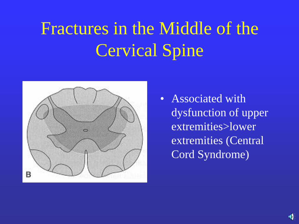
Fractures in the Middle of the
Cervical Spine
• Associated with
dysfunction of upper
extremities>lower
extremities (Central
Cord Syndrome)
Thoracolumbar Trauma
• Mechanism of injury
– Compression
– Distraction
– Rotation
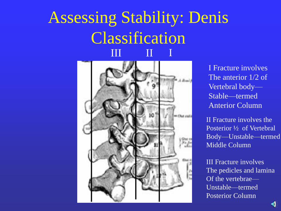
Assessing Stability: Denis
Classification I II III
I Fracture involves
The anterior 1/2 of
Vertebral body—
Stable—termed
Anterior Column
II Fracture involves the
Posterior ½ of Vertebral
Body—Unstable—termed
Middle Column
III Fracture involves
The pedicles and lamina
Of the vertebrae—
Unstable—termed
Posterior Column
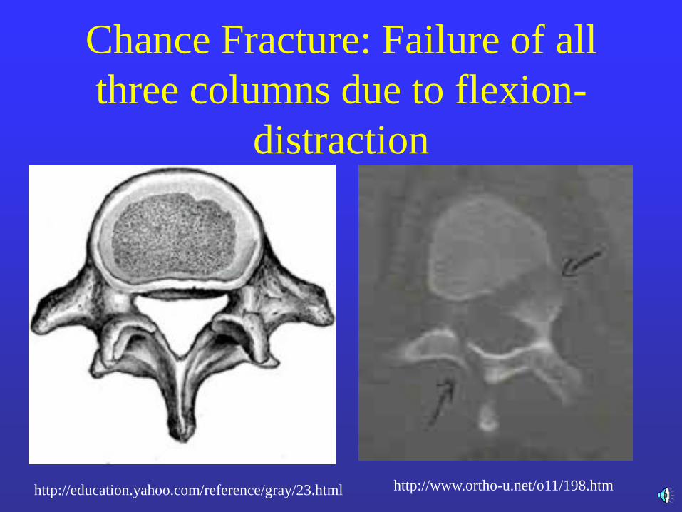
Chance Fracture: Failure of all
three columns due to flexion-
distraction
http://education.yahoo.com/reference/gray/23.html http://www.ortho-u.net/o11/198.htm
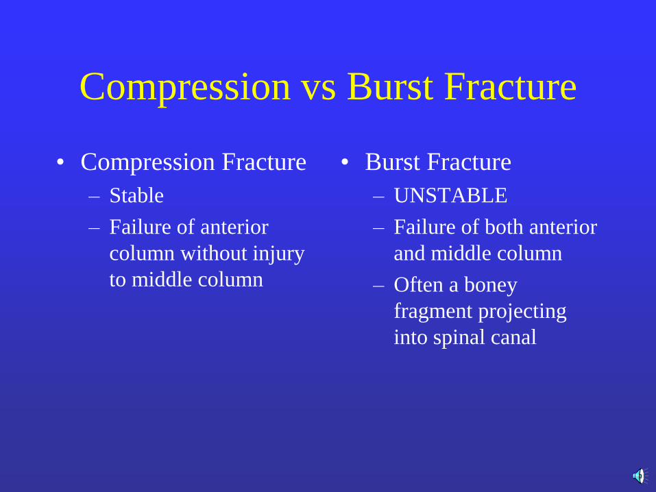
Compression vs Burst Fracture
• Compression Fracture
– Stable
– Failure of anterior
column without injury
to middle column
• Burst Fracture
– UNSTABLE
– Failure of both anterior
and middle column
– Often a boney
fragment projecting
into spinal canal
Indications for Spine Surgery
• Neurologic Deterioration
• Unstable fracture
• Epidural Hematoma
• Narrowing of spinal canal
Goals of Spinal Surgery
• Decompression of Spinal Canal
• Stabilization of Spine
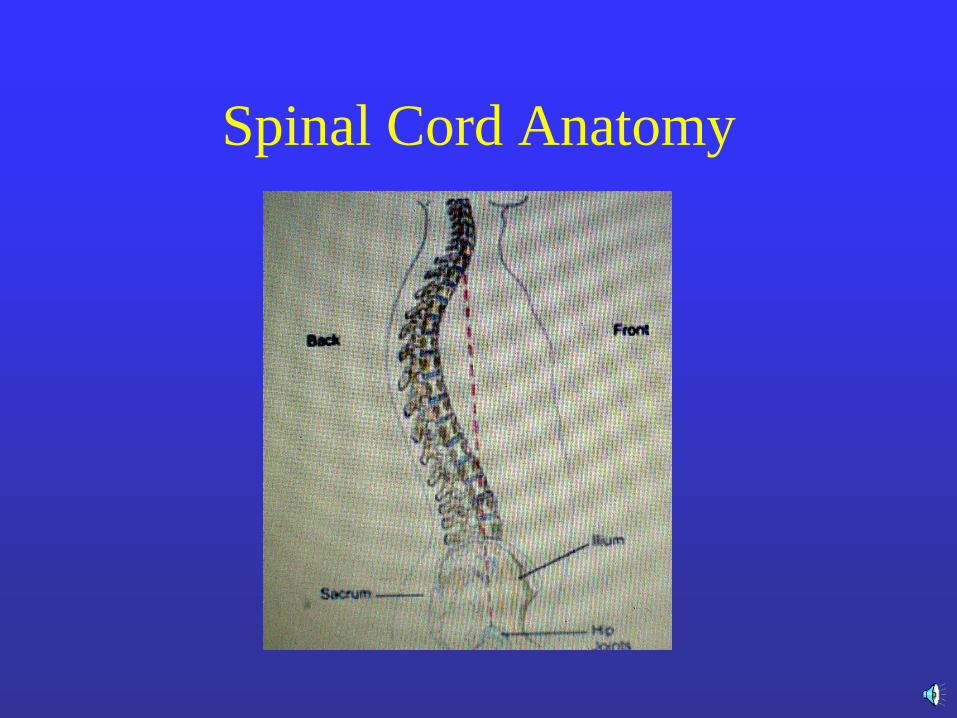
Spinal Cord Anatomy

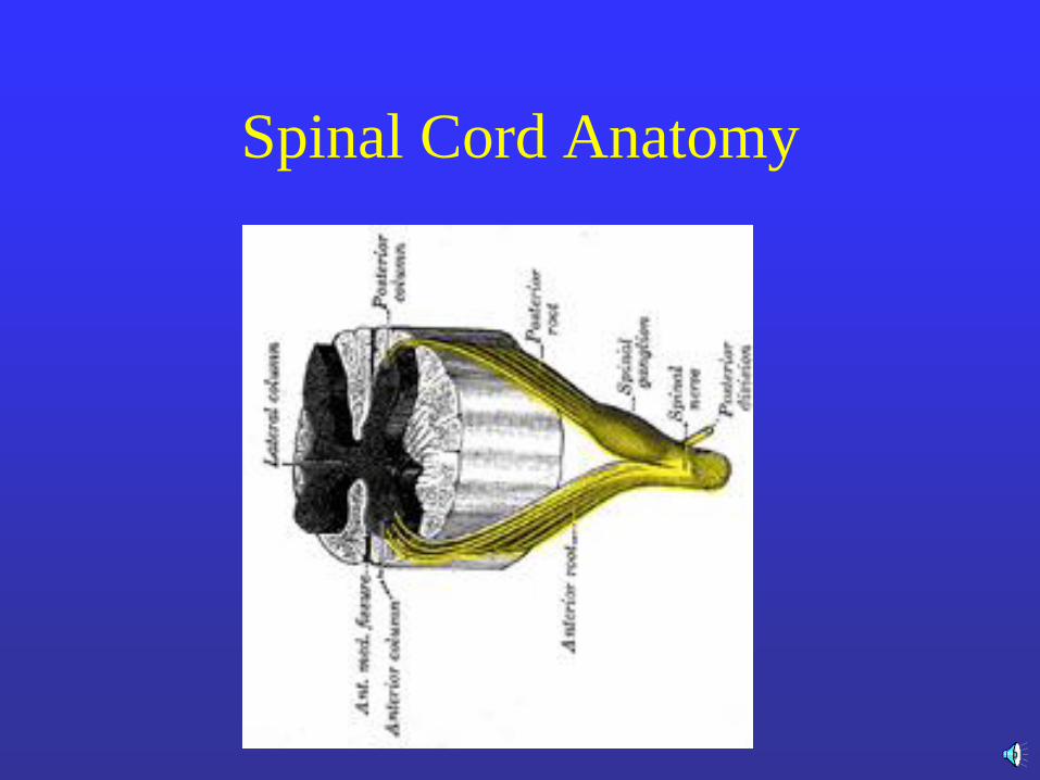
Spinal Cord Anatomy
Spinal Cord Anatomy
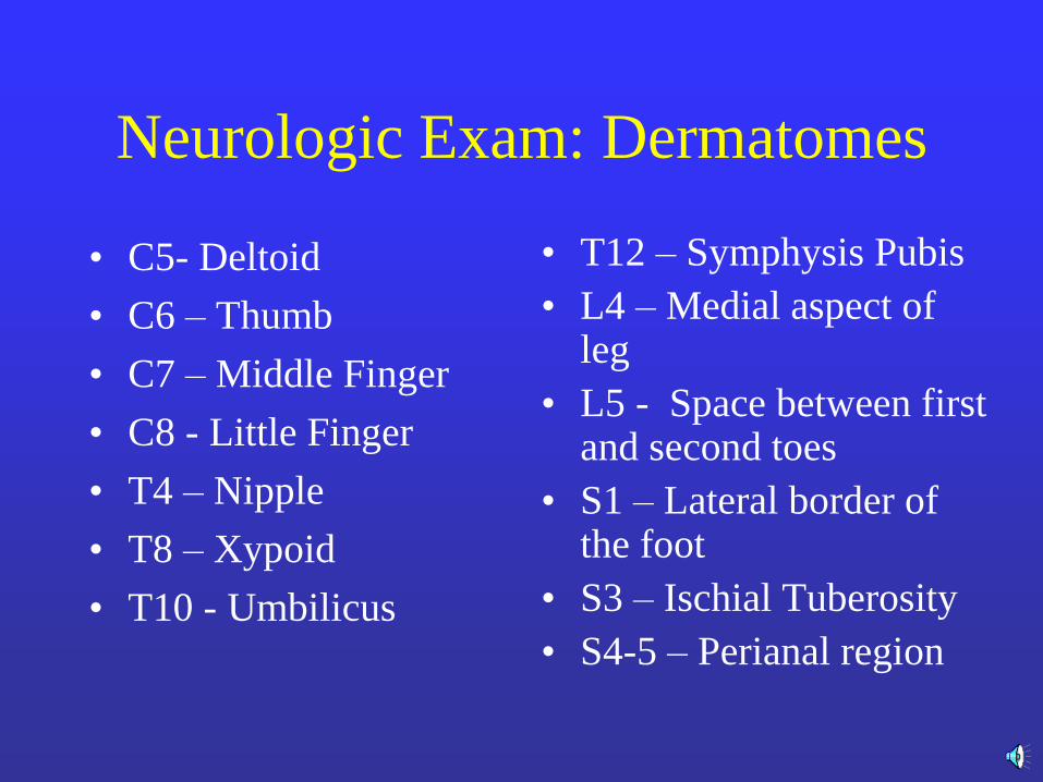
Neurologic Exam: Dermatomes
• C5- Deltoid
• C6 – Thumb
• C7 – Middle Finger
• C8 - Little Finger
• T4 – Nipple
• T8 – Xypoid
• T10 - Umbilicus
• T12 – Symphysis Pubis
• L4 – Medial aspect of leg
• L5 - Space between first and second toes
• S1 – Lateral border of the foot
• S3 – Ischial Tuberosity
• S4-5 – Perianal region
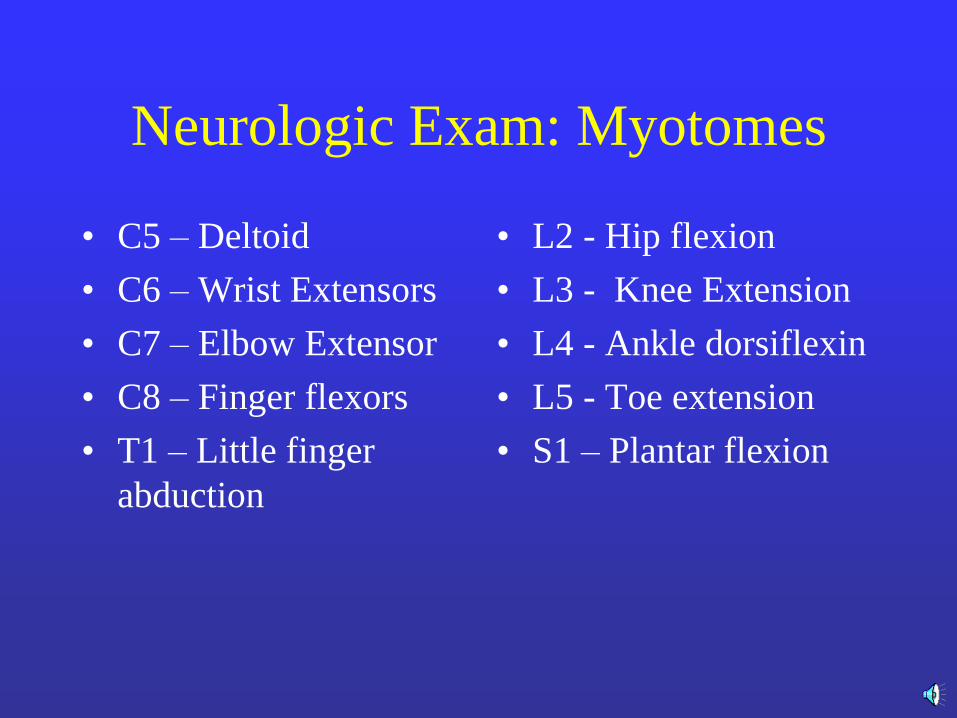
Neurologic Exam: Myotomes
• C5 – Deltoid
• C6 – Wrist Extensors
• C7 – Elbow Extensor
• C8 – Finger flexors
• T1 – Little finger
abduction
• L2 - Hip flexion
• L3 - Knee Extension
• L4 - Ankle dorsiflexin
• L5 - Toe extension
• S1 – Plantar flexion
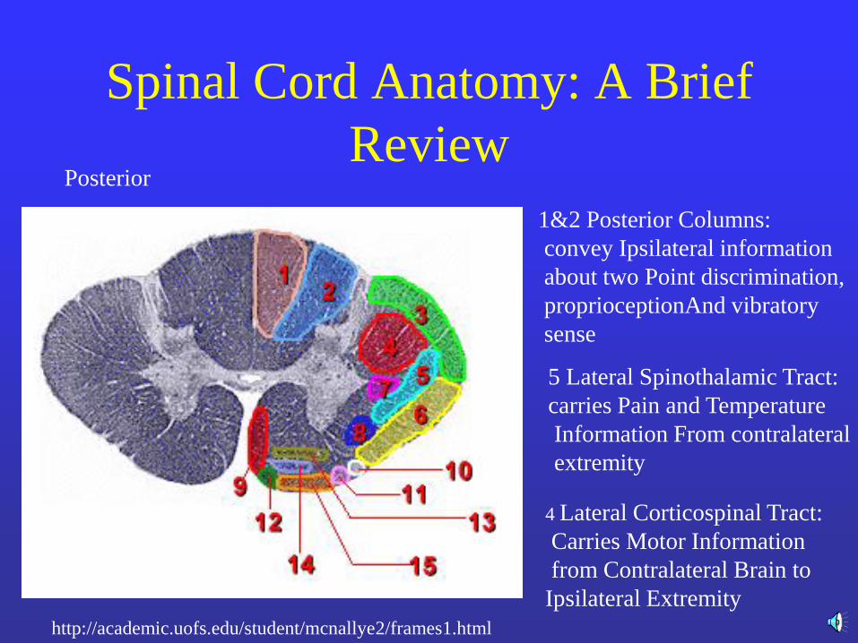
Spinal Cord Anatomy: A Brief
Review
http://academic.uofs.edu/student/mcnallye2/frames1.html
1&2 Posterior Columns:
convey Ipsilateral information
about two Point discrimination,
proprioceptionAnd vibratory
sense
5 Lateral Spinothalamic Tract:
carries Pain and Temperature
Information From contralateral
extremity
4 Lateral Corticospinal Tract:
Carries Motor Information
from Contralateral Brain to
Ipsilateral Extremity
Posterior Posterior
Posterior
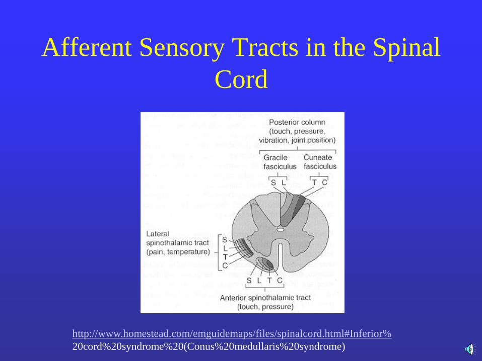
Afferent Sensory Tracts in the Spinal
Cord
http://www.homestead.com/emguidemaps/files/spinalcord.html#Inferior%
20cord%20syndrome%20(Conus%20medullaris%20syndrome)
Clinical Syndromes resulting from
Incomplete Spinal Cord Injury
• Central Cord Syndrome
• Brown-Sequard Syndrome
• Anterior Cord Syndrome
• Conus Medullaris Syndrome
• Cauda Equina Syndrome
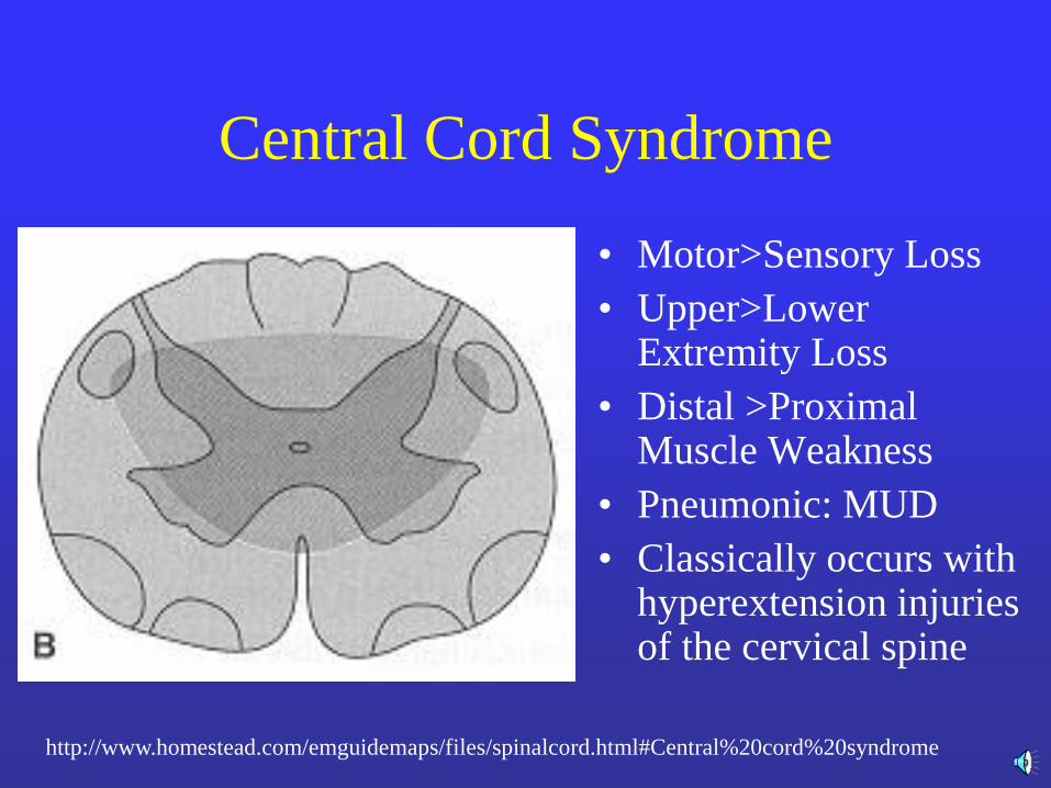
Central Cord Syndrome
• Motor>Sensory Loss
• Upper>Lower Extremity Loss
• Distal >Proximal Muscle Weakness
• Pneumonic: MUD
• Classically occurs with hyperextension injuries of the cervical spine
http://www.homestead.com/emguidemaps/files/spinalcord.html#Central%20cord%20syndrome
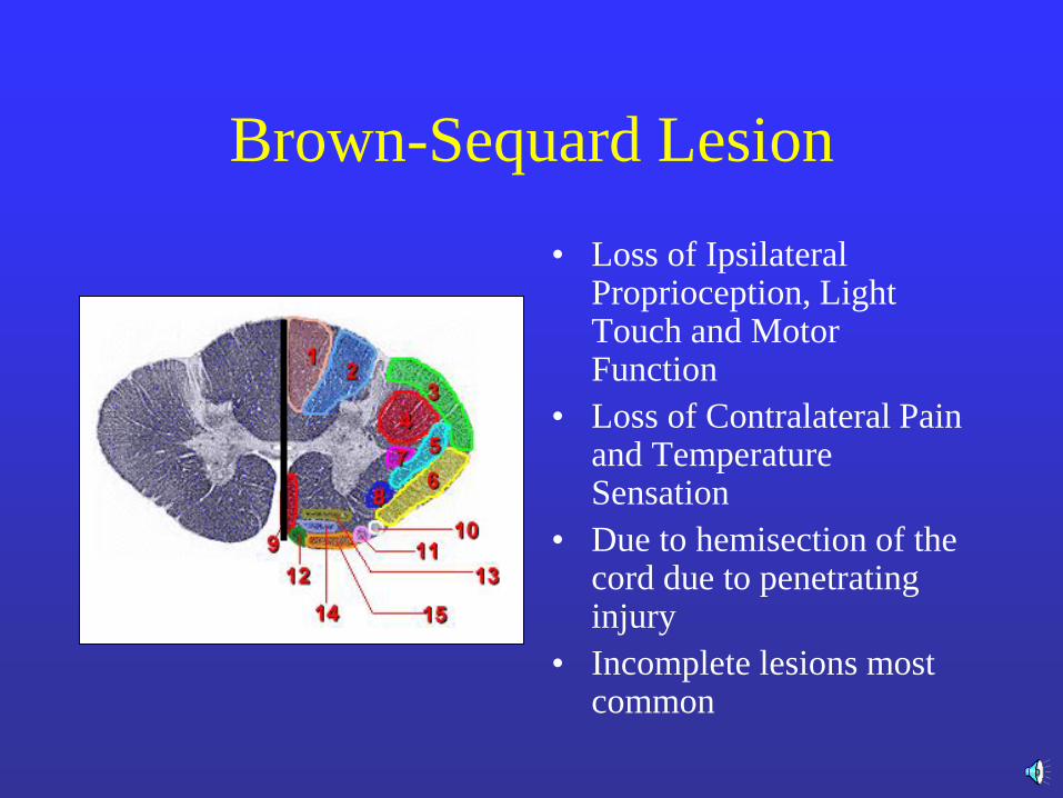
Brown-Sequard Lesion
• Loss of Ipsilateral Proprioception, Light Touch and Motor Function
• Loss of Contralateral Pain and Temperature Sensation
• Due to hemisection of the cord due to penetrating injury
• Incomplete lesions most common
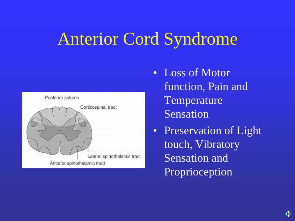
Anterior Cord Syndrome
• Loss of Motor
function, Pain and
Temperature
Sensation
• Preservation of Light
touch, Vibratory
Sensation and
Proprioception
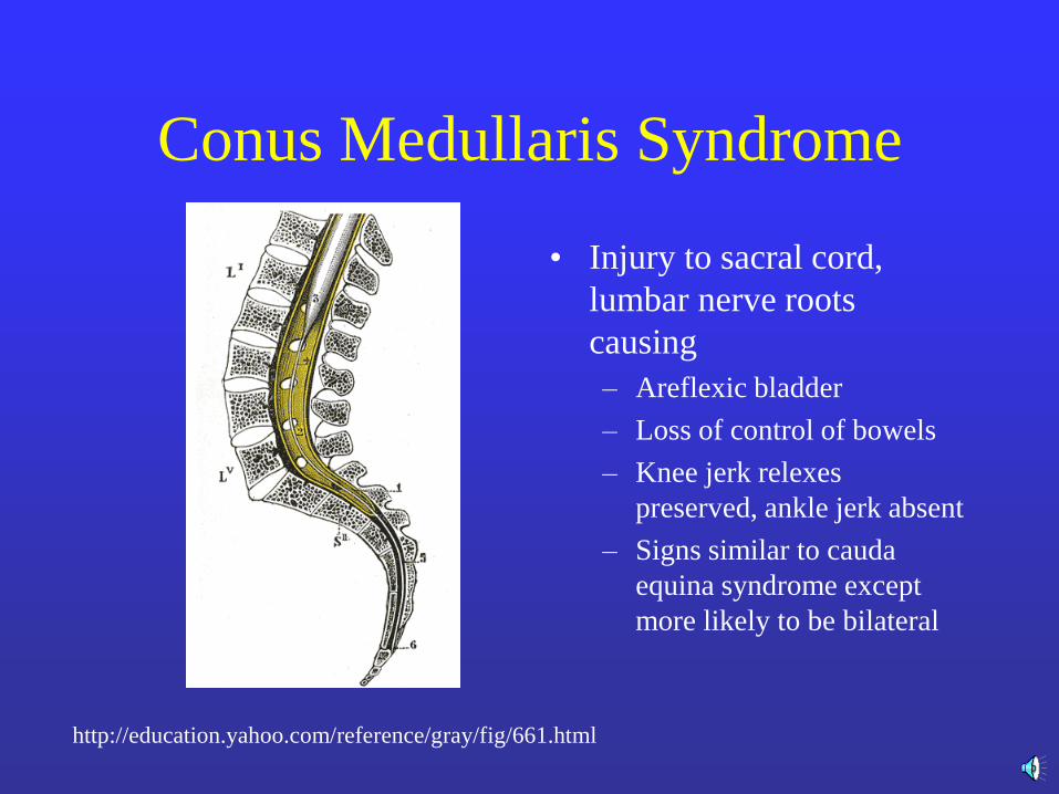
Conus Medullaris Syndrome
• Injury to sacral cord,
lumbar nerve roots
causing
– Areflexic bladder
– Loss of control of bowels
– Knee jerk relexes
preserved, ankle jerk absent
– Signs similar to cauda
equina syndrome except
more likely to be bilateral
http://education.yahoo.com/reference/gray/fig/661.html

Cauda Equina Syndrome
• Injury to nerve roots
and not spinal cord
itself
• Muscle weakness and
decreased sensation
inaffected dermatomes
• Decreased bowel and
bladder control
Treatment of Acute Spinal Cord
Injury
• Methylprednisolone 30mg/kg as soon as
possible (within the first 8 hours after
injury) for proven NON-PENETRATING
spinal cord injury
• 5.4 mg/kg/hr for the next 23 hours
Important Adjunct Measures
• Frequent turning
• Special bed to prevent pressure sores
• Splint extremities to prevent flexion contractures—splints MUST be well padded to protect skin
• Range of motion of joints
• Occupational and Physical Therapy
• Intermittent urinary
catheterization if appropriate
• Skin Care
• Avoid succinylcholine b/o
induced hyperkalemia
• Autonomic hypersensitivity
• Pulmonary Embolus
Prophylaxsis

Principles of Initial Management
• Prevent further damage
• Assume a spine injury until proven
otherwise
Primary Survey
• Airway
• Breathing
• Circulation
• Disability: Moves upper and lower
extremities??
• Exposure
Secondary Survey
Careful Orthopedic and Neurologic
Evaluation takes place in the
Secondary Survey
History
• Pre-injury neurologic status
• Mechanism of injury
• Review Pre-hospital report
• Change in neurologic status?
• DOCUMENT FINDINGS
Cervical Spine Injury
• Cervical Spine poorly protected
• Suspect if:
– Supraclavicular injury
– Maxillofacial trauma
– Head injury
– High speed injury
Clinical Clearance of Cervical
Spine only if:
• Patient awake and fully cooperative
• The neck is pain free without swelling, hematoma,
pain to palpation or boney abnormalities
• No distracting injuries
• The patient has full pain free active range of
motion
• DO NOT PASSIVELY MOVE THE PATIENT’S
HEAD!!!!
Initial Treatment of Possible
Cervical Spine Injury • Immobilization
• Imaging studies
– AP, lateral and open mouth spine films
– Consider CT
– MRI to view ligaments and spinal cord
• Search for occult injury in patient with a neurologic deficit
• DOCUMENT FINDINGS
• Early neurosurgical/orthopedic consultation
Neurological Examination
• Motor examination of upper and lower extremities
• Sensory Examination of upper and lower
extremities
– Examine perianal sensation to pinprick (S3,S4)
– Distinguishes between a complete and incomplete
spinal cord injury
• Reflexes
• DOCUMENT FINDINGS
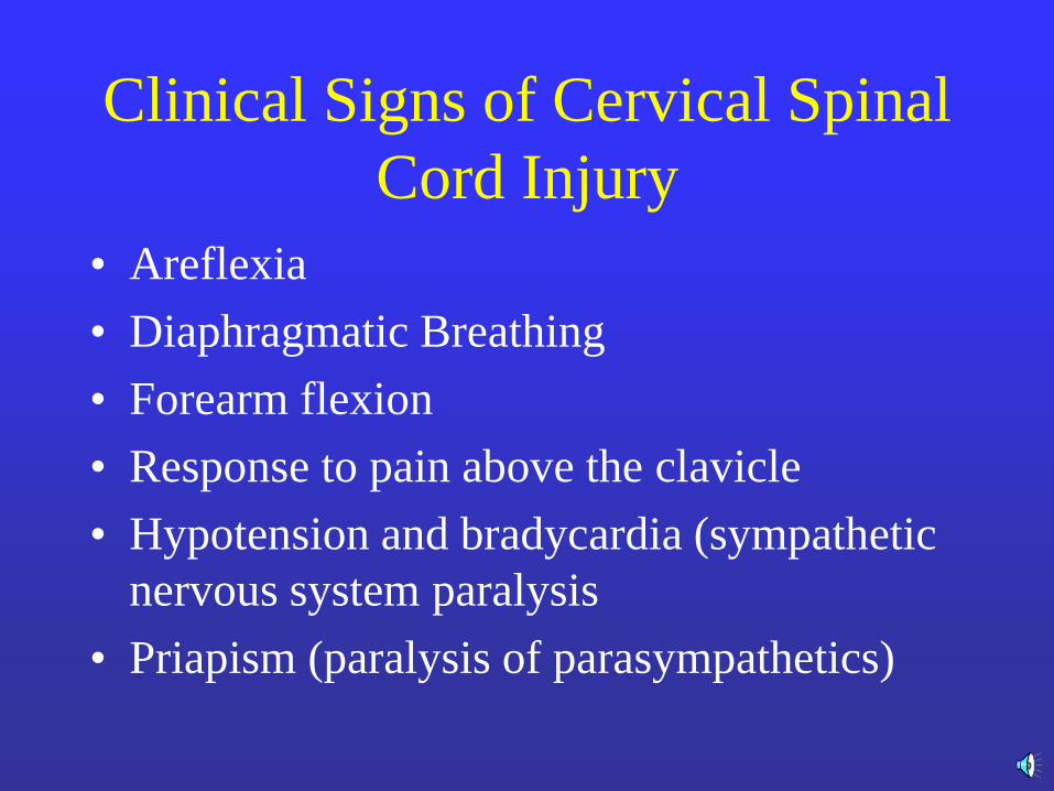
Clinical Signs of Cervical Spinal
Cord Injury
• Areflexia
• Diaphragmatic Breathing
• Forearm flexion
• Response to pain above the clavicle
• Hypotension and bradycardia (sympathetic
nervous system paralysis
• Priapism (paralysis of parasympathetics)
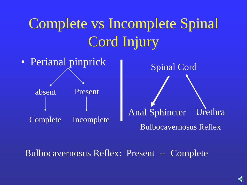
Complete vs Incomplete Spinal
Cord Injury
• Perianal pinprick
absent Present
Complete Incomplete
Bulbocavernosus Reflex: Present -- Complete
Urethra
Spinal Cord
Anal Sphincter
Bulbocavernosus Reflex
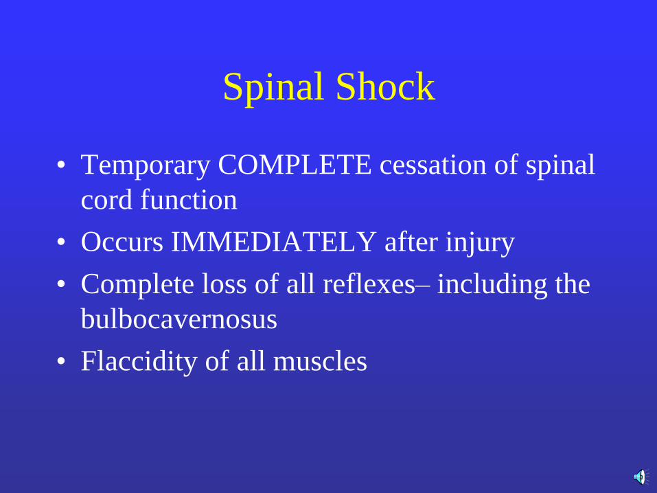
Spinal Shock
• Temporary COMPLETE cessation of spinal
cord function
• Occurs IMMEDIATELY after injury
• Complete loss of all reflexes– including the
bulbocavernosus
• Flaccidity of all muscles
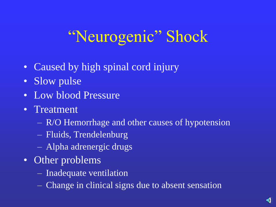
―Neurogenic‖ Shock
• Caused by high spinal cord injury
• Slow pulse
• Low blood Pressure
• Treatment
– R/O Hemorrhage and other causes of hypotension
– Fluids, Trendelenburg
– Alpha adrenergic drugs
• Other problems
– Inadequate ventilation
– Change in clinical signs due to absent sensation
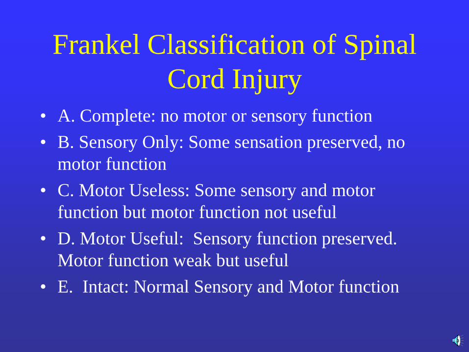
Frankel Classification of Spinal
Cord Injury
• A. Complete: no motor or sensory function
• B. Sensory Only: Some sensation preserved, no
motor function
• C. Motor Useless: Some sensory and motor
function but motor function not useful
• D. Motor Useful: Sensory function preserved.
Motor function weak but useful
• E. Intact: Normal Sensory and Motor function
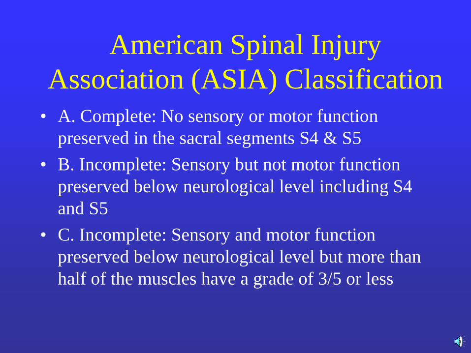
American Spinal Injury
Association (ASIA) Classification
• A. Complete: No sensory or motor function
preserved in the sacral segments S4 & S5
• B. Incomplete: Sensory but not motor function
preserved below neurological level including S4
and S5
• C. Incomplete: Sensory and motor function
preserved below neurological level but more than
half of the muscles have a grade of 3/5 or less
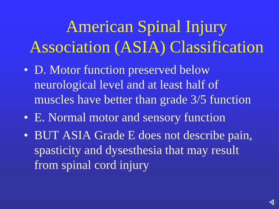
American Spinal Injury
Association (ASIA) Classification
• D. Motor function preserved below
neurological level and at least half of
muscles have better than grade 3/5 function
• E. Normal motor and sensory function
• BUT ASIA Grade E does not describe pain,
spasticity and dysesthesia that may result
from spinal cord injury
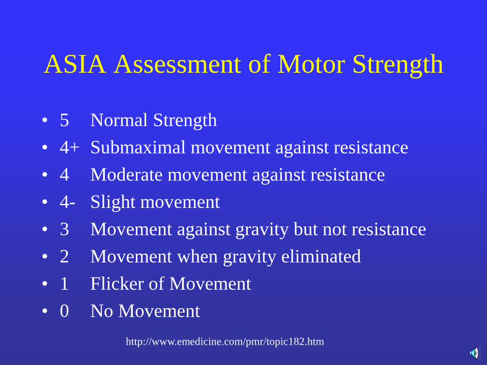
ASIA Assessment of Motor Strength
• 5 Normal Strength
• 4+ Submaximal movement against resistance
• 4 Moderate movement against resistance
• 4- Slight movement
• 3 Movement against gravity but not resistance
• 2 Movement when gravity eliminated
• 1 Flicker of Movement
• 0 No Movement
5 - Normal power
4+ - Submaximal
movement against
resistance
4 - Moderate
movement against
resistance
4- - Slight movement
against resistance
3 - Movement against
gravity but not against
resistance
2 - Movement with
gravity eliminated
1 - Flicker of
movement
0 - No movement
http://www.emedicine.com/pmr/topic182.htm
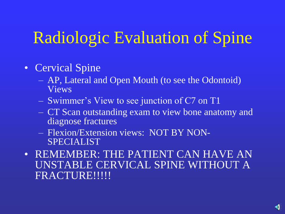
Radiologic Evaluation of Spine
• Cervical Spine – AP, Lateral and Open Mouth (to see the Odontoid)
Views
– Swimmer’s View to see junction of C7 on T1
– CT Scan outstanding exam to view bone anatomy and diagnose fractures
– Flexion/Extension views: NOT BY NON-SPECIALIST
• REMEMBER: THE PATIENT CAN HAVE AN UNSTABLE CERVICAL SPINE WITHOUT A FRACTURE!!!!!
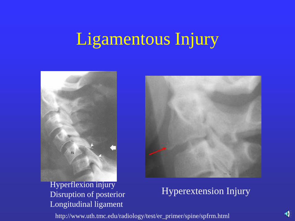
Ligamentous Injury
http://www.uth.tmc.edu/radiology/test/er_primer/spine/spfrm.html
Hyperflexion injury
Disruption of posterior
Longitudinal ligament
Hyperextension Injury
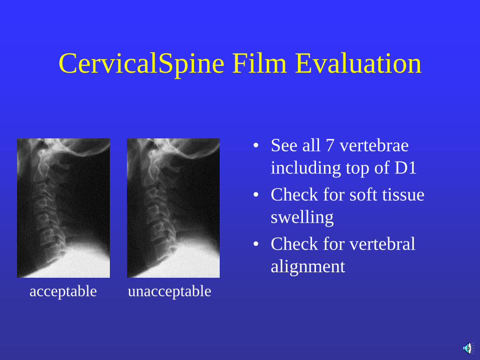
CervicalSpine Film Evaluation
• See all 7 vertebrae
including top of D1
• Check for soft tissue
swelling
• Check for vertebral
alignment
acceptable unacceptable
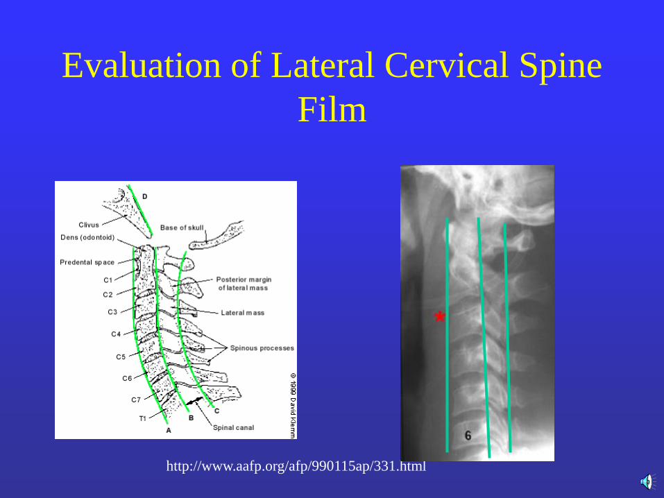
Evaluation of Lateral Cervical Spine
Film
http://www.aafp.org/afp/990115ap/331.html
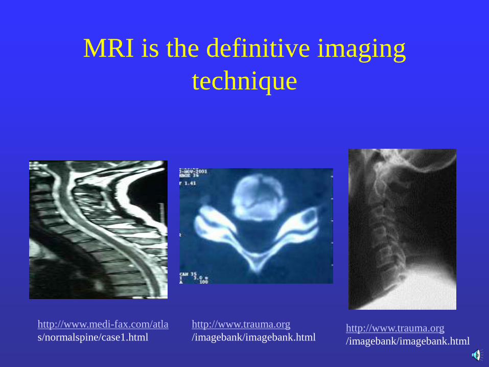
MRI is the definitive imaging
technique
http://www.medi-fax.com/atla
s/normalspine/case1.html http://www.trauma.org
/imagebank/imagebank.html
http://www.trauma.org
/imagebank/imagebank.html
Summary
• Assume a spine injury until proven otherwise in blunt trauma
• X-ray the entire axial skeleton if: (1) appropriate mechanism of injury, (2) patient unable to cooperate with exam, a spine fracture is identified
• Careful DOCUMENTED neurologic, orthopedic, and radiologic evaluation of spine in secondary survey
• Timely orthopedic and neurosurgical consultation