Embed Size (px)
Citation preview
5
Spinocerebellar Ataxia Type 2
Luis Velázquez-Pérez1, Roberto Rodríguez-Labrada1, Hans-Joachim Freund2 and Georg Auburger3
1Centre for the Research and Rehabilitation of Hereditary Ataxias, Holguín, 2International Neuroscience Institute, Hannover,
3Section Experimental Neurology, Dept. Neurology, Goethe University Medical School, Frankfurt am Main,
1Cuba 2,3Germany
1. Introduction
The autosomal dominant cerebellar ataxias (ADCA) are a clinically, pathologically and genetically heterogeneous group of neurodegenerative disorders caused by degeneration of cerebellum and its afferent and efferent connections. The degenerative process may additionally involves the ponto- medullar systems, pyramidal tracts, basal ganglia, cerebral cortex, peripheral nerves (ADCA I) and the retina (ADCA II), or can be limited to the cerebellum (ADCA III) (Harding et al., 1993).
The most common of these dominantly inherited autosomal ataxias, ADCA I, includes many Spinocerebellar Ataxias (SCA) subtypes, some of which are caused by pathological CAG trinucleotide repeat expansion in the coding region on the mutated gene. Such is the case for SCA1, SCA2, SCA3/MJD, SCA6, SCA7, SCA17 and Dentatorubral-pallidoluysian atrophy (DRPLA) (Matilla et al., 2006).
Among the almost 30 SCAs, the variant SCA2 is the second most prevalent subtype
worldwide, only surpassed by SCA3 (Schöls et al., 2004; Matilla et al., 2006; Auburger, 2011).
The disorder was first recognized in India in 1971 by Wadia and Swami, who was intrigued
by the early and marked slowing of saccade movements, associated to the cerebellar
syndrome (Wadia & Swami, 1971). Contemporarily, in Cuba some neurologists were
describing many families coming from the north-east region of the country with the same
distinct clinical picture (Vallés et al., 1978). Subsequent epidemiological surveys in this
Cuban region, Holguín province, focusing on the causes of the highest SCA2 prevalence rate
worldwide found evidence for a founder effect (Orozco et al., 1989; Auburger et al., 1990;
Velázquez-Pérez et al., 2001, 2009a).
2. Epidemiology
The collective worldwide prevalence of SCAs is estimated at about 6 cases per 100,000 people, although much higher figures have been reported in particular populations (Schöls et al., 2004). In the case of SCA2, the global prevalence is unknown because the most of the
www.intechopen.com
Spinocerebellar Ataxia
78
few existing epidemiological studies have been performed in isolated geographical regions with families not large enough for linkage analysis. Nevertheless, large SCA2 families have been found in India, Martinique, Australia, Tunisia, Germany, Italy, Mexico, Poland and especially in Cuba (Klockgether, 2007; Sulek-Pitkowska, et al., 2010, Velázquez-Pérez et al., 2009a).
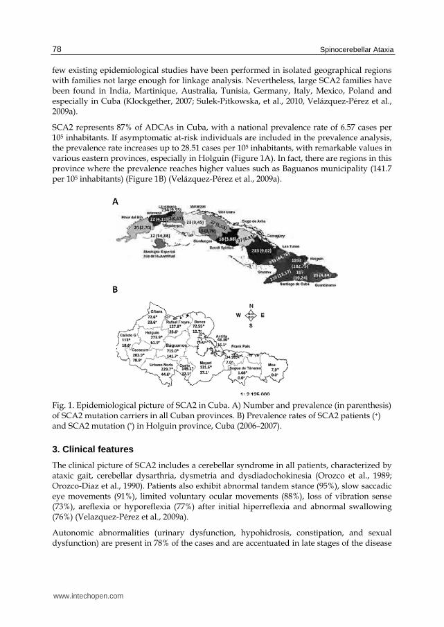
SCA2 represents 87% of ADCAs in Cuba, with a national prevalence rate of 6.57 cases per 105 inhabitants. If asymptomatic at-risk individuals are included in the prevalence analysis, the prevalence rate increases up to 28.51 cases per 105 inhabitants, with remarkable values in various eastern provinces, especially in Holguin (Figure 1A). In fact, there are regions in this province where the prevalence reaches higher values such as Baguanos municipality (141.7 per 105 inhabitants) (Figure 1B) (Velázquez-Pérez et al., 2009a).
Fig. 1. Epidemiological picture of SCA2 in Cuba. A) Number and prevalence (in parenthesis) of SCA2 mutation carriers in all Cuban provinces. B) Prevalence rates of SCA2 patients (+) and SCA2 mutation (*) in Holguin province, Cuba (2006–2007).
3. Clinical features
The clinical picture of SCA2 includes a cerebellar syndrome in all patients, characterized by ataxic gait, cerebellar dysarthria, dysmetria and dysdiadochokinesia (Orozco et al., 1989; Orozco-Diaz et al., 1990). Patients also exhibit abnormal tandem stance (95%), slow saccadic eye movements (91%), limited voluntary ocular movements (88%), loss of vibration sense (73%), areflexia or hyporeflexia (77%) after initial hiperreflexia and abnormal swallowing (76%) (Velazquez-Pérez et al., 2009a).
Autonomic abnormalities (urinary dysfunction, hypohidrosis, constipation, and sexual dysfunction) are present in 78% of the cases and are accentuated in late stages of the disease
www.intechopen.com
Spinocerebellar Ataxia Type 2
79
(Sánchez-Cruz, et al., 2001; Velázquez-Pérez et al., 2009a, Montes et al., 2010), together with dysphagia, ophthalmoplegia and distal amyotrophy. Sleep disturbances are frequent complaints of SCA2 patients and their relatives. The most prominent sleep disorders are restless legs syndrome (Trojano et al., 1998; Schöls et al., 1998; Abele et al., 2001; Irazno et al., 2007), muscle cramps, insomnia and reduced dream recalls (Velázquez-Pérez et al., 2011a).
Other clinical manifestations of SCA2 are the cognitive dysfunctions, which include frontal-executive impairment, verbal short-term memory deficits as well as reduction of attention and concentration (Storey et al., 1999; Reynaldo-Arminan et al., 2002; Bürk et al., 1999a; 2003). Although neuropsychological pattern of cognitive disturbances of SCA2 patients not necessarily resembling dementia, some studies have reported high frequency of demented patients (Durr et al., 1995, Burk et al., 1999a), but in the SCA2 Cuban population this neuropsychological state is rare (Reynaldo-Arminan et al., 2002; Orozco et al., 1989; 1990). Depression/anxiety/suicide attempts are found in a third of cases (Reynaldo-Arminan et al., 2002). In comparison to other SCAs, the frequency of slowed ocular movements, postural and action tremor and hyporeflexia are distinctive features of SCA2 (Schöls et al., 1997).
Extrapyramidal manifestations are common in SCA2 patients. Myoclonuses are reported in 13.7% whereas dystonia is present in 14.2%. Chorea may appear in approximately 7%. These symptoms are accentuated in patients with larger CAG repeats. Parkinsonian signs appear in some patients with low-range expansions containing CAA interruptions (Gwinn-Hardy et al., 2000; Payami et al., 2003; Lu et al., 2004; Charles et el, 2007). Among these manifestations, resting tremor (14,9%) and rigidity (7,9%) are the most common (Schmitz-Hubsch, et al., 2008). Recently it was reported an unusual case of SCA2 presenting as an ataxia-parkinsonism-motor neuron disease syndrome in a 46-year-old Brazilian man with 40 CAG repeats in the SCA2 gene (Braga-Neto et al., 2011).
The age at onset varies from 3 to 79 years (mean 33). Usually, the first symptom of the
disease is the gait ataxia (97%), followed by the cerebellar dysarthria (3%). However some
extracerebellar manifestations may occur a decade or more before the onset of gait
instability or dysarthria, such as painful muscle cramps in the calf, sleep disturbances,
problems with hand writing (Globas et al, 2008), as well as autonomic alterations, consisting
in constipation (19.4%) and pollakiuria (17.7%) (Montes-Brown et al, 2011). In the Cuban
SCA2 population the anticipation of clinical manifestation age in successive generations is
observed in 80% of transmissions, usually upon transmission from an affected father
(Velázquez-Pérez et al., 2009a).
Clinical features develop progressively with an increase in cerebellar syndrome, saccade
slowing, and other features which confine the patients first to a wheelchair and following to
a bed, where they die approximately 15–20 years after the initial symptoms. Nevertheless
patients with larger CAG repeats have earlier age at onset, more saccadic slowing, axial
tremor, pyramidal-dystonic-choreic signs, mental deficit and in general a faster progression
to death (Filla et al 1999, Cancel et al 1997; Schöls et al., 1997; Sasaki et al., 1998; Filla et al.,
1999; Velázquez-Pérez et al., 2009a) and the total disease duration from onset to death may
vary between 6 and 50 years (Klockgether et al., 1998; Maschke et al., 2005). Also, the female
gender is associated with shortened survival (Klockgether et al., 1998). The main cause of
death is bronchopneumonia (63%), followed by bronchial aspiration and cardiovascular
incidents, among others (Velázquez-Pérez et al, 2011b).
www.intechopen.com
Spinocerebellar Ataxia
80
Pediatric-onset SCA2 is associated with large CAG expansions. Infantile phenotype includes rare symptoms such as retinitis pigmentosa, myoclonus-epilepsy, tetraparesis, developmental delay and facial dysmorphism (Babovic-Vuksanovic et al 1998; Rufa et al., 2002; Tan et al., 2004; Di Fabio et al., 2011). Ramocki and coworkers describe a female child who met all developmental milestones until age 3 years, deterioration of expressive language, comprehension, memory, graphomotor skills, and dysarthria. Cranial nerve examination showed bilaterally restricted lateral gaze with oculomotor apraxia (Ramocki, et al., 2008). Abdel-Aleem and Zakiwith reported a male child with progressive extrapyramidal manifestations, developmental delay, slow eye movements and cognitive impairment, trophic changes, vasomotor instability and dysphagia (Aleem and Zakiwith, 2008)
4. Molecular genetics
The underlying mutation of SCA2 consists in the unstable expansion of the trinucleotide repeat (CAG)8CAA(CAG)4CAA(CAG)8 within the ATXN2 gene exon 1 located on chromosome 12q24.1. This repeat encodes a polyglutamine (polyQ) tract in the protein ataxin-2 (Gispert et al., 1993; Pulst et al., 1996; Imbert et al., 1996; Sanpei et al., 1996). In normal individuals, the trinucleotide repeat length varies and contains between 13 and 27 units. Intermediate expansions between 28 and 33 units may predispose the individual to an elevated risk for the motor neuron disease ALS or the Parkinson plus syndrome PSP (Elden et al., 2010; Ross et al., 2011). The prevalence of large normal alleles potentially acting as unstable premutation is particularly high in the Cuban province Holguín (Velázquez-Pérez et al., 2009a). Family planning can be aided by presymptomatic molecular genetic diagnostics, but care has to taken to offer psychological treatment together with the genetic counseling.
Pathological alleles in SCA2 have more than 32 CAG repeats, although the repeats range between 32 and 36 units has incomplete penetrance (Pulst et al., 1996; Cancel et al., 1997; Geschwind et al., 1997). The most frequent expanded allele is 37 (72%). The expanded alleles have lost interrupting CAA-triplets, a factor thought to promote the length instability. Expansions occur in 89% and contractions in 11% of the offspring of affected patients. Paternal transmissions show higher variability in repeat lengths compared with the maternal transmissions. (Velázquez-Pérez et al., 2009a). The presence of CAA interruptions in expanded alleles appears to predispose to a phenotype with Parkinson or with motor neuron disease (Charles et al., 2007; Kim et al., 2007; Modoni et al., 2007; Corrado et al., 2011, Yu et al., 2011), although both CAG and CAA code for glutamine, indicating that the neuronal population affected by the pathogenesis is determined by RNA toxicity rather than protein toxicity.
As in other polyQ diseases, in SCA2 the age at onset and symptom severity correlate inversely with the length of the trinucleotide repeat, which accounts for ~80% of variance, whereas the remaining variability suggests the existence of modifier genes, genetic polymorphisms, epigenetic factors and unknown environmental determinants modulating age of onset (Velázquez-Pérez et al., 2009a). Supporting the above mentioned, long normal CAG repeats in the CACNA1A (Pulst et al., 2005) and RAI1 genes (Hayes et al., 2000) as well as the 10398G polymorphism in the mitochondrial complex I gene (Simon et al., 2007) are associated with earlier manifestation age, also in the Cuban SCA2 population.
www.intechopen.com

Spinocerebellar Ataxia Type 2
81
4.1 The physiological role of ataxin-2 in cell biology
The ataxin-2 protein (ATXN2) is a polypeptide containing 1312 amino acids encoded by 25 exons of the SCA2/ATXN2 gene encompassed within 130 kiloBases of genomic DNA (Sahba et al., 1998), with at least five human isoforms produced by allelic splicing (Nechiporuk et al 1998; Affaitati et al., 2001; Lastres-Becker et al., 2008a) and an expression in many organs, but only selected neurons of the brain (Huynh et al., 1999). It is phosphorylated, but not glycosylated (Turnbull et al., 2004). Currently, the function of ATXN2 is not clear, but several lines of evidence evoke its involvement in RNA metabolism. For example, the protein have sequence motifs related to mRNA processing, most of ATXN2 is associated to polyribosomes, at the rough endoplasmic reticulum (Satterfield and Pallanck, 2006; van de Loo et al., 2009), and this polypeptide interacts with RNA binding proteins such as A2BP1 and PABPC1 (Shibata et al., 2000; Ralser et al., 2005a; Satterfield and Pallanck, 2006).
Interestingly, ATXN2 and its orthologues in other organisms relocalize during periods of cellular stress to mRNP granules where mRNA is stored during translation repression, promote the formation of these stress granules and inhibit cell growth (Swisher and Parker, 2010; Nonhoff et al., 2007). Furthermore, the expression of ATXN2 is induced by specific stressors (Klinkenberg et al., submitted) and ATXN2 levels increase with old age (Huynh et al., 1999). The indirect effects of ATXN2 on RNAs appear to be mediated partially by its interactor DDX6, a RNA helicase (Nonhoff et al., 2007). Also the formation of P-bodies, mRNP granules implicated in RNA degradation, appears to depend on ATXN2, which may localize to these structures and influence the microRNA-mediated deadenylation of silenced RNAs (Nonhoff et al., 2007; Kozlov et al., 2010). There is preliminary evidence that ATXN2 co-sediments and co-localizes with neuronal mRNPs which are responsible for the transport of mRNAs to synaptic sites of local protein synthesis, and indeed ATXN2 is thought to modulate mRNA translation similar to its yeast orthologue Pbp1 (Siddiqui et al., 2007). Thus, ATXN2 might be important for stimulus-dependent local mRNA translation and influence in this way both synaptic strength and long-term potentiation, an electrophysiological finding which was indeed detected in ATXN2-knock-out mice in the amygdala, but not in the hippocampus (Huynh et al., 2009).
Some ATXN2 is also demonstrable at the plasma membrane, and within its protein sequence several proline-rich domains are able to interact with SH3-motif containing proteins. Such an interaction was demonstrated for endophilin A, CIN85 and Src, three components of the endocytosis complex that modulates trophic factor signaling through receptor tyrosine kinases (Ralser et al., 2005b; Nonis et al 2008). In these reports, ATXN2 was found to antagonize the internalization of the receptor for Epidermal Growth Factor. Interestingly, two other neurodegenerative disease proteins are also interactors of this complex, namely Huntingtin and Parkin, which was shown to ubiquitinate ATXN2 directly and to rescue ATXN2-toxicity (Ralser et al., 2005b; Huynh et al., 2007). Furthermore, the deficiency of ATXN2 in knock-out mice was observed to modulate the levels of insulin receptor, resulting in insulin resistance, altered fat metabolism and obesity (Kiehl et al., 2006; Lastres Becker et al., 2008b). Interestingly, the protein family A2D which shares sequence homology with ATXN2 also shows interaction with the cytoplasmic domain of the thrombopoietin and the erythropoietin membrane receptors which lack intrinsic tyrosine kinase activity, but is also internalized to modulate downstream events of cytokine signaling (Meunier et al., 2002). Of
www.intechopen.com
Spinocerebellar Ataxia
82
course, this physiological influence of ATXN2 on trophic signaling may be important for neural atrophy in SCA2. Finally, recent evidence suggests a localization and role of ATXN2 in the nucleus, acting as interactor of the transcriptional regulator ZBRK1 (Hallen et al., 2011).
4.2 ATXN2 role for different diseases
SCA2 is thought to be caused by a toxic gain-of-function of the ATXN2 protein, but it is not clear to which degree the physiological function of ATXN2 is enhanced and to which degree unspecific toxic effects such as the aggregation of polyQ domain proteins dominate in the pathogenesis. Since polyQ expansions in different disease proteins affect different neuronal populations, and since the overexpression of wild-type ATXN2 and its orthologues in lower species, which lack the polyQ domain completely, is neurotoxic, the specific properties of ATXN2 regarding expression, subcellular localization and interactors seem to be relevant in disease. Intermediate-length expansions of the ATXN2 trinucleotide repeat below the threshold of SCA2 manifestation were shown to have a pathogenic role, increasing the individual risk to manifest the motor neuron degeneration disease ALS (Amyotrophic Lateral Sclerosis) and the basal ganglia degeneration disease within the Parkinson-plus group of disorders PSP (Progressive Supranuclear Palsy) (Elden et al., 2010; Daoud et al., 2011; Ross et al., 2011; Sorarù et al., 2011; Lee et al., 2011; van Damme et al., 2011). The RNA metabolism function of ATXN2 may explain this phenomenon, since ALS pathogenesis appears to be mediated mainly by altered mRNA processing (Lagier-Tourenne et al., 2010). ATXN2 gain-of-function also potentiates toxicity of ATXN1 and ATXN3 (the SCA1 and SCA3 disease proteins, respectively) and even toxicity of Tau (the frontotemporal lobar degeneration disease protein) in the fly model (Shulman and Feany, 2003; Al-Ramahi et al., 2007; Lessing and Bonini, 2008; Elden et al., 2010). Conversely, reducing ATXN2 levels is sufficient to mitigate the neurotoxicity triggered by TDP-43, ATXN1 and ATXN3 (Al-Ramahi et al., 2007; Lessing et al., 2008; Elden et al., 2010) in yeast and flies, indicating that these effects are mediated by the physiological function of ATXN2, but not by the polyQ domain which characterizes human ATXN2 and is not conserved until mouse.
Large expansions of ATXN2 were reported to exert a profound effect on intracellular calcium levels through specific binding to the carboxy-terminal region of the type 1 inositol 1,4,5-trisphosphate receptor (IP(3)R1), an intracellular Ca(2+) release channel (Liu et al., 2009), an effect mediated by ATXN2 at its major localization in the cytoplasm.
Several lines of evidence suggest that other alterations of the physiological ATXN2 function influence additional neuron populations and diseases. In neuroblastoma tumors, an upregulation of ATXN2 was found to be a decisive factor to induce apoptosis of the aberrant cells and spontaneous tumor remission (Wiedemeyer et al., 2003). In individuals who reached an age over 100 years, a single nucleotide polymorphism within ATXN2 intron 1 contributes to the genetic signature of exceptional longevity. Moreover, in the general human population the same ATXN2 intron 1 polymorphism determines high blood pressure levels (Levy et al., 2009; Newton-Cheh et al., 2009; Sebastiani et al., 2010).
4.3 Animal models
Animal models have been useful tools to study the polyQ expansion diseases, in particular the brain tissue of early stage pathology. Specifically ATXN2 orthologues are highly
www.intechopen.com
Spinocerebellar Ataxia Type 2
83
conserved until Saccharomyces cerevisiae, permitting high-throughput genetic screens into the function of ATXN2 and revealing the role of ATXN2 as a risk factor for TDP-43 toxicity and motor neuron degeneration (Elden et al., 2010). Again, Drosophila melanogaster studies demonstrated the association of dATX2 with PABP and with polysomes (Satterfield and Pallanck, 2006). The use of RNA interference in Caenorhabditis elegans demonstrated an essential role of the atx-2 gene for early embryonic development (Kiehl et al., 2000).
Taking advantage of the mouse as an organism with genetic versatility and with similarity to man in brain structure, two transgenic models of SCA2 have been generated to date. The first one was produced by Huynh et al., 2000, who reported the use of the murine PcP2 (L7) promoter to direct a strong overexpression of the human ATXN2 gene with an expanded allele of 58 CAG repeats specifically to the cerebellar Purkinje neurons. Using the rotarod test, they found that the animals became ataxic at 26 and 16 weeks for the heterozygous and homozygous transgenic mice, respectively. Also, they described progressive incoordination and morphological alterations of Purkinje cells in this animal model. In 2005, Aguiar and coworkers (Aguiar et al., 2006) generated transgenic mouse lines overexpressing the full-length human ATXN2 gene with 75 CAG units under the control of the human self promoter. A neurological phenotype was reported after 12 weeks for heterozygous and 6 weeks for homozygous mice.
5. Imaging
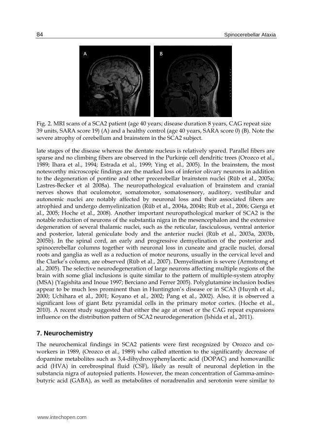
Magnetic resonance imaging shows early cerebellar and brainstem atrophy (Figure 2) with marked involvement of the cerebellar cortex and the pons/inferior olive region in SCA2, in excellent agreement with the traditional neuropathological nomenclature of olivopontocerebellar atrophy (OPCA). Also, frontotemporal atrophy is observed in advanced disease (Bürk et al., 1996; Giuffrida et al., 1999). Voxel-based morphometry studies have revealed the atrophy of the cerebellar and brainstem white matter as well as the symmetric loss of gray matter in the cerebellar vermis (Brenneis et al., 2003; Brenneis et al., 2005; Della Nave et al., 2008a, b, Goel et al., 2011). Positron emission tomography (PET) studies showed a reduced regional glucose metabolism in the cerebellum, brainstem and parietal cortex, which may occur years before the clinical onset of SCA2 (Inagaki et al., 2005). PET analyses also revealed the loss of striatal dopamine transporter function with nigrostriatal atrophy, similar to the pattern observed in idiopathic Parkinson’s disease (Boesch et al., 2004; Wüllner et al 2005; Inagaki et al., 2005). Imaging by proton magnetic resonance spectroscopy demonstrated the loss of choline-containing compounds in SCA2 cerebella, suggesting the decreased production and/or the loss of cell membranes as well as the reduced synthesis of precursors of acetylcholine. The same study demonstrated the increase of lactate levels in the cerebellum suggesting an impairment of glycolysis and mitochondrial function (Boesch et al., 2001).
6. Neuropathology
The macroscopic examination of nervous structures in post-mortem samples of SCA2 patients shows a significant atrophy of the cerebellum, brainstem, frontal lobe, as well as pallor of the midbrain substantia nigra and a reduction of the cerebral and cerebellar white matter. Microscopically, the cerebellum is characterized by an early and marked neuronal loss in Purkinje cell layer with reduction in the number of dendritic arborizations and torpedo-like deformations of their axons. The number of granular neurons is diminished, usually toward
www.intechopen.com
Spinocerebellar Ataxia
84
Fig. 2. MRI scans of a SCA2 patient (age 40 years; disease duration 8 years, CAG repeat size 39 units, SARA score 19) (A) and a healthy control (age 40 years, SARA score 0) (B). Note the severe atrophy of cerebellum and brainstem in the SCA2 subject.
late stages of the disease whereas the dentate nucleus is relatively spared. Parallel fibers are sparse and no climbing fibers are observed in the Purkinje cell dendritic trees (Orozco et al., 1989; Ihara et al., 1994; Estrada et al., 1999; Ying et al., 2005). In the brainstem, the most noteworthy microscopic findings are the marked loss of inferior olivary neurons in addition to the degeneration of pontine and other precerebellar brainstem nuclei (Rüb et al., 2005a; Lastres-Becker et al 2008a). The neuropathological evaluation of brainstem and cranial nerves shows that oculomotor, somatomotor, somatosensory, auditory, vestibular and autonomic nuclei are notably affected by neuronal loss and their associated fibers are atrophied and undergo demyelinization (Rüb et al., 2004a, 2004b; Rüb et al., 2006; Gierga et al., 2005; Hoche et al., 2008). Another important neuropathological marker of SCA2 is the notable reduction of neurons of the substantia nigra in the mesencephalon and the extensive degeneration of several thalamic nuclei, such as the reticular, fasciculosus, ventral anterior and posterior, lateral geniculate body and the anterior nuclei (Rüb et al., 2003a, 2003b, 2005b). In the spinal cord, an early and progressive demyelination of the posterior and spinocerebellar columns together with neuronal loss in cuneate and gracile nuclei, dorsal roots and ganglia as well as a reduction of motor neurons, usually in the cervical level and the Clarke’s column, are observed (Rüb et al., 2007). Demyelination is severe (Armstrong et al., 2005). The selective neurodegeneration of large neurons affecting multiple regions of the brain with some glial inclusions is quite similar to the pattern of multiple-system atrophy (MSA) (Yagishita and Inoue 1997; Berciano and Ferrer 2005). Polyglutamine inclusion bodies appear to be much less prominent than in Huntington’s disease or in SCA3 (Huynh et al., 2000; Uchihara et al., 2001; Koyano et al., 2002; Pang et al., 2002). Also, it is observed a significant loss of giant Betz pyramidal cells in the primary motor cortex. (Hoche et al., 2010). A recent study suggested that either the age at onset or the CAG repeat expansions influence on the distribution pattern of SCA2 neurodegeneration (Ishida et al., 2011).
7. Neurochemistry
The neurochemical findings in SCA2 patients were first recognized by Orozco and co-workers in 1989, (Orozco et al., 1989) who called attention to the significantly decrease of dopamine metabolites such as 3,4-dihydroxyphenylacetic acid (DOPAC) and homovanillic acid (HVA) in cerebrospinal fluid (CSF), likely as result of neuronal depletion in the substancia nigra of autopsied patients. However, the mean concentration of Gamma-amino-butyric acid (GABA), as well as metabolites of noradrenalin and serotonin were similar to
www.intechopen.com
Spinocerebellar Ataxia Type 2
85
normal subjects. Additionally, N-acetyl-aspartate and glutamate are markedly reduced in these patients (Oz et al., 2010).
A pathologically relevant biochemical finding is the significant reduction of zinc, iron and copper levels in the CSF and serum of Cuban SCA2 patients. The reduction of zinc levels could be associated with phenotypic features such as nerve conduction slowing, cognitive dysfunction, and immune-depression at final stages of the disease and could accentuate the dysfunction of cerebellar circuits, based on the important role of this element in the control of synapses in the cerebellum (González et al., 2006). Furthermore, most biomarkers of the antioxidant-prooxidant balance are significantly modified in Cuban SCA2 patients with an increase in malondialdehyde (MDA) as evidence of lipid peroxidation, as well as signs of oxidative damage to protein and DNA and significant reduction of the reduced glutathione (GSH). Also, the activity of glutathione S-transferase (GST), superoxide dismutase (SOD) and catalase (CAT) are depressed in these patients with a disruption of the balance CAT/SOD (Velázquez-Pérez et al, 2003; Almaguer, et al., 2005). A third interesting finding is the decrease of erythropoietin levels in the CSF with a compensatory increase of this molecule in the serum of Cuban SCA2 patients, suggesting the existence of reduced capabilities of neuroprotection in the nervous system (Velazquez-Pérez et al., 2011b). We believe that these biochemical features may contribute to the high phenotypic variability of SCA2 and that they could constitute potential therapeutical targets to design future clinical trials.
8. Neurophysiology
8.1 Nerve conduction and electromyography studies
The most common electrophysiological finding in SCA2 patients is a predominantly sensory axonal neuropathy, expressed by the early and progressive reduction of sensory amplitudes, suggestive of dorsal root ganglionopathy. These alterations are associated with slowing of nerve conduction as sign of demyelination. The progression rate of sensory axonal neuropathy is notably accentuated in patients with large CAG expansion sizes. Motor nerve conduction parameters are usually normal, but in patients with 10-15 years of disease duration it is possible to observe a reduction of motor amplitudes (Kubis et al., 1999; van de Warrenburg et al., 2004; Velázquez-Pérez et al., 2007, 2010). Electromyographical findings reveal motor unit potentials (MUP) with light polyphasic alterations, increased amplitudes and isolated contraction pattern in the first stage of the evolution. In advanced stages of the disease signs of denervation can appear (fibrillations and fasciculations) and the contraction pattern becomes simple oscillations, indicating the loss of motor neurons in the anterior horn of the spinal cord (Velázquez-Pérez et al, 2009b).
8.2 Somatosensory evoked potentials (SSEP)
Tibial nerve SSEPs are characterized by a marked prolongation of the P40 component and central conduction time latencies. In the median nerve SSEP there is a latency prolongation of N20 and N13 components in addition to a reduction of amplitude of Erb potentials. In almost all cases, the SSEPs show abnormal morphology and reduced reproducibility. These alterations get worse quickly in patients with larger CAG repeat number and may be detected even in presymptomatic subjects (Velázquez-Pérez et al., 2007, 2008).
www.intechopen.com
Spinocerebellar Ataxia
86
8.3 Brain Stem Auditory Evoked Potentials (BSAEP)
BSAEPs have poor reproducibility and unstable morphology in 95% of the patients, in addition to the increase of latency of the waves III and V and the prolongation of the I–III interpeak interval. These abnormalities are common in patients with disease duration above 10 years but the abnormal reproducibility and morphology can be detected since preclinical stage (Velázquez-Pérez et al., 2007, 2008).
8.4 Visual Evoked Potentials (VEP)
VEP are frequently normal in SCA2 patients, but some patients in advances stages of the disease have prolonged P100 latencies with normal amplitudes. These findings reflect the integrity of the visual pathway in Cuban SCA2 patients, allowing us to distinguish SCA2 from other spinocerebellar ataxias such as SCA1, SCA3 and in particular SCA7 (Velázquez-Pérez et al., 2007, 2008).
8.5 Event-related evoked potentials (ERPs)
ERPs revealed prolongation of visual P300 latencies in 40% of cases with a significant correlation of this variable with the disease duration and clinical affectation (Kremlacek et al., 2011).
8.6 Motor evoked potentials
The study of the corticospinal tract by transcranial magnetic stimulation in SCA2 patients reveals an increase of central motor conduction time and motor threshold. Also, intracortical facilitation may be reduced and the induced cortical silent period prolonged. The progression of these abnormalities is dependent on the disease duration and ataxia severity. They probably reflect the reduced excitability of the motor cortex, disturbed conduction along the pyramidal tract and the loss of facilitatory influences of the cerebellum on the primary motor cortex (Yokota et al., 1998; Restivo et al., 2000, 2004; Schwenkreis et al., 2002)
8.7 Electrooculography
The main oculomotor abnormality in SCA2 is the slowing of horizontal saccadic movements, which is probably the result of early pontine brainstem degeneration. This feature is electrooculographically detectable in 99% of the patients and in several presymptomatic subjects. The maximal saccade velocity is negatively correlated with the polyQ expansion and the ataxia score, but is not significantly influenced by the disease duration. (Rivaud-Pechoux et al., 1998; Bürk et al., 1999b; Velázquez-Pérez et al., 2004, 2008, 2009c). The prolongation of saccadic latency is observed in 46% of the cases, reflecting the cortical/subcortical involvement in SCA2. Although this saccadic feature is not directly influenced by the CAG repeats or the disease duration it is close related with the frontal-executive dysfunctions, identifying it as a promising cognitive biomarker (Rodríguez-Labrada et al., 2011a). Additionally, SCA2 patients showed saccadic dysmetria reflecting the cerebellar involvement (Velázquez-Pérez et al., 2008) although saccades made for short target amplitudes are usually accurate due to the visual feedback might be continuously available during the slow movements (Federighi et al., 2011). Furthermore, gain measurements in smooth pursuit movements and horizontal optokinetic nystagmus are
www.intechopen.com
Spinocerebellar Ataxia Type 2
87
slightly reduced in SCA2 patients, whereas the vestibulo-ocular reflex is normal (Buttner et al., 1998).
8.8 Videopolysomnography and electroencephalography
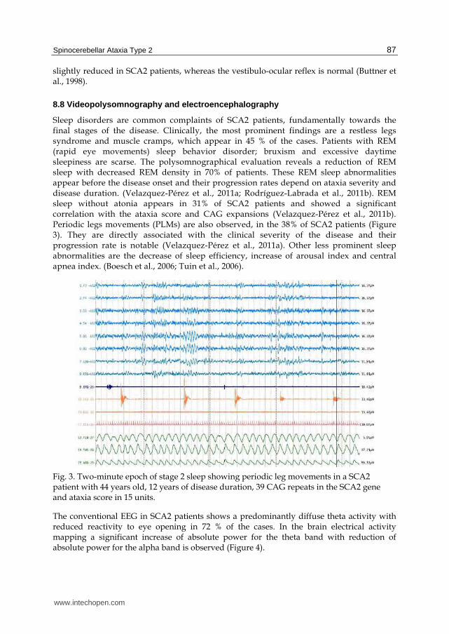
Sleep disorders are common complaints of SCA2 patients, fundamentally towards the final stages of the disease. Clinically, the most prominent findings are a restless legs syndrome and muscle cramps, which appear in 45 % of the cases. Patients with REM (rapid eye movements) sleep behavior disorder; bruxism and excessive daytime sleepiness are scarse. The polysomnographical evaluation reveals a reduction of REM sleep with decreased REM density in 70% of patients. These REM sleep abnormalities appear before the disease onset and their progression rates depend on ataxia severity and disease duration. (Velazquez-Pérez et al., 2011a; Rodríguez-Labrada et al., 2011b). REM sleep without atonia appears in 31% of SCA2 patients and showed a significant correlation with the ataxia score and CAG expansions (Velazquez-Pérez et al., 2011b). Periodic legs movements (PLMs) are also observed, in the 38% of SCA2 patients (Figure 3). They are directly associated with the clinical severity of the disease and their progression rate is notable (Velazquez-Pérez et al., 2011a). Other less prominent sleep abnormalities are the decrease of sleep efficiency, increase of arousal index and central apnea index. (Boesch et al., 2006; Tuin et al., 2006).
Fig. 3. Two-minute epoch of stage 2 sleep showing periodic leg movements in a SCA2 patient with 44 years old, 12 years of disease duration, 39 CAG repeats in the SCA2 gene and ataxia score in 15 units.
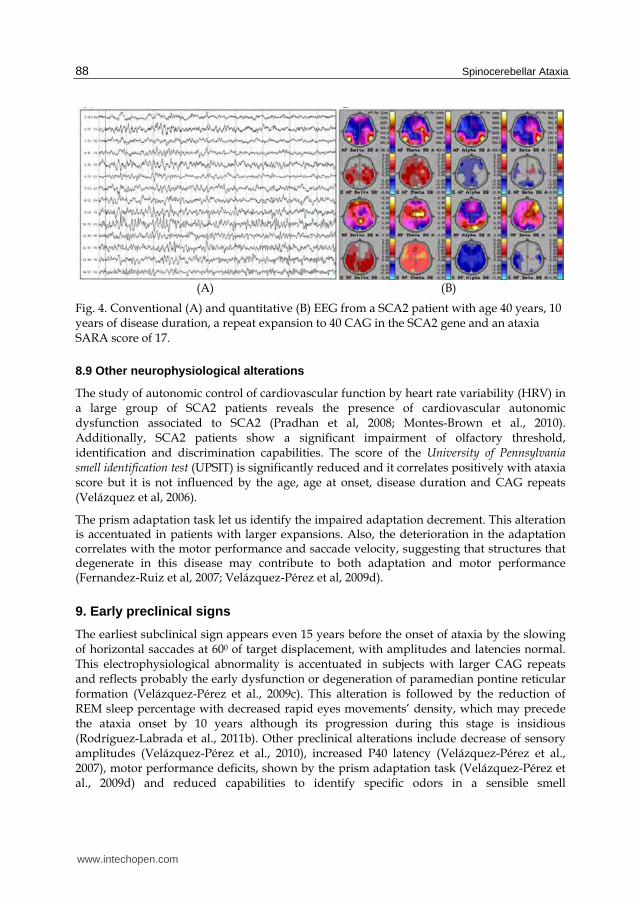
The conventional EEG in SCA2 patients shows a predominantly diffuse theta activity with reduced reactivity to eye opening in 72 % of the cases. In the brain electrical activity mapping a significant increase of absolute power for the theta band with reduction of absolute power for the alpha band is observed (Figure 4).
www.intechopen.com
Spinocerebellar Ataxia
88
(A) (B)
Fig. 4. Conventional (A) and quantitative (B) EEG from a SCA2 patient with age 40 years, 10 years of disease duration, a repeat expansion to 40 CAG in the SCA2 gene and an ataxia SARA score of 17.
8.9 Other neurophysiological alterations
The study of autonomic control of cardiovascular function by heart rate variability (HRV) in a large group of SCA2 patients reveals the presence of cardiovascular autonomic dysfunction associated to SCA2 (Pradhan et al, 2008; Montes-Brown et al., 2010). Additionally, SCA2 patients show a significant impairment of olfactory threshold, identification and discrimination capabilities. The score of the University of Pennsylvania smell identification test (UPSIT) is significantly reduced and it correlates positively with ataxia score but it is not influenced by the age, age at onset, disease duration and CAG repeats (Velázquez et al, 2006).
The prism adaptation task let us identify the impaired adaptation decrement. This alteration is accentuated in patients with larger expansions. Also, the deterioration in the adaptation correlates with the motor performance and saccade velocity, suggesting that structures that degenerate in this disease may contribute to both adaptation and motor performance (Fernandez-Ruiz et al, 2007; Velázquez-Pérez et al, 2009d).
9. Early preclinical signs
The earliest subclinical sign appears even 15 years before the onset of ataxia by the slowing of horizontal saccades at 600 of target displacement, with amplitudes and latencies normal. This electrophysiological abnormality is accentuated in subjects with larger CAG repeats and reflects probably the early dysfunction or degeneration of paramedian pontine reticular formation (Velázquez-Pérez et al., 2009c). This alteration is followed by the reduction of REM sleep percentage with decreased rapid eyes movements’ density, which may precede the ataxia onset by 10 years although its progression during this stage is insidious (Rodríguez-Labrada et al., 2011b). Other preclinical alterations include decrease of sensory amplitudes (Velázquez-Pérez et al., 2010), increased P40 latency (Velázquez-Pérez et al., 2007), motor performance deficits, shown by the prism adaptation task (Velázquez-Pérez et al., 2009d) and reduced capabilities to identify specific odors in a sensible smell
www.intechopen.com
Spinocerebellar Ataxia Type 2
89
identification test (UPSIT). The comprehensive analysis of these early signs in SCA2 suggests the necessity for revisit the current criteria to define the disease onset delineating the boundaries between presymptomatics and symptomatic states.
10. Therapeutical options
Till now, there is no specific treatment for SCA2. Physiotherapy and neuropsychological rehabilitation have palliative effects on motor and cognitive symptoms. Therefore, Cuban SCA2 patients receive a specialized neurorehabilitation program (Pérez-Avila et al., 2004) since 1998, which has been applied to more than 400 patients and has allowed some recovery of motor, cognitive and antioxidant functions in about 75% of the treated patients (Rodríguez et al., 2008).
Regarding clinical trials, few studies have been conducted. For example, muscle cramps are successfully treated with magnesium and levodopa treatment alleviates the parkinsonian signs in SCA2 patients (Lastres-becker, 2008a), whereas severe myoclonus at advanced stage could be dramatically improved by piracetam (De Rosa et al., 2006). Recently, a randomized, double-blind, placebo-controlled pilot trial using riluzole resulted effective to SCA2 and other subjects with cerebellar ataxias (Ristori et al., 2010). Additionally, a double-blinded and placebo-controlled clinical trial with 50 mg zinc sulphate in 36 Cuban SCA2 patients was effective in increasing the zinc levels in serum and CSF of treated subjects and some benefit of this treatment for the cerebellar syndrome, the peripheral neuropathy and the restoration of antioxidant functions was apparent (Velázquez-Pérez et al., 2011c).
Deep brain stimulation with novel patterned low-frequency stimulation (PLFS) was effective in localizing the tremor generator at a subthalamic-thalamic electrode position, suppressing a coarse postural tremor for several postoperative years in one case (Freund et al., 2007; Barnikol et al., 2008).
11. Conclusions
In conclusion, although we have learnt much since SCA2 was described as a distinct clinical entity (Wadia and Swami, 1971) and since its cause was identified and genetic counseling became available (Imbert et al 1996; Pulst et al., 1996; Sanpei et al 1996), until today we have only taken the first steps towards understanding the pathogenic mechanisms and validating neuroprotective therapies.
12. Acknowledgements
We thank all patients and their family members for the observations and the tissues they contributed to aid to understand this devastating disease. The work was supported by the Cuban Ministry of Health the Deutsche Akademische Austauschdienst, the Deutsche Forschungsgemeinschaft over many years (AU 96/1-1, 1-2, 1-3, 4-1, 9-1 und 9-2, 11-1, 13-1), the Alexander von Humboldt Foundation, and a European Union framework (EuroSCA) (MINSAP). The authors thank Dr. Suzana Gispert for her continuous assistance and Dipl. Biol. Jessica Drost for proofreading the manuscript.
www.intechopen.com
Spinocerebellar Ataxia
90
13. References
Abdel-Aleem A, Zaki MS. (2008) Spinocerebellar ataxia type 2 (SCA2) in an Egyptian family presenting with polyphagia and marked CAG expansion in infancy. Journal of Neurology, Vol.255(3), pp. 413–419.
Abele M, Bürk K, Laccone F, Dichgans J & Klockgether T. (2001). Restless legs syndrome in spinocerebellar ataxia types 1, 2, and 3. Journal of Neurology, Vol.248, pp. 311-314, ISSN 0340-5354.
Affaitati A, de Cristofaro T, Feliciello A & Varrone S. (2001). Identification of alternative splicing of spinocerebellar ataxia type 2 gene. Gene, Vol.267, pp 89–93, ISSN 1879-0038.
Aguiar J, Fernández J, Aguilar A, Mendoza Y, Vázquez M, et al. (2006). Ubiquitous expression of human SCA2 gene under the regulation of the SCA2 self promoter cause specific Purkinje cell degeneration in transgenic mice. Neuroscience Letter, Vol.392, pp. 202-206, ISSN 0304-3940.
Almaguer L, Almaguer D, Gonzáles Y, Martínez E & Valcárcel P. (2005). Capacidad antioxidante total de en pacientes cubanos con ataxia Espinocerebelosa tipo 2. Revista Mexicana de Neurociencias, Vol.6, pp. 201-206.
Al-Ramahi I, Pérez AM, Lim J, Zhang M, Sorensen R, et al. (2007). dAtaxin-2 mediates expanded Ataxin-1-induced neurodegeneration in a Drosophila model of SCA1. PLoS Genetic, Vol.3, No.12, pp. e234, ISSN 1553-7404.
Armstrong J, Bonaventura I, Rojo A, Gonzalez G, Corral J, et al. (2005). Spinocerebellar ataxia type 2 (SCA2) with white matter involvement. Neuroscience Letter, Vol.381, pp. 247–51, ISSN 0304-3940.
Auburger G, Díaz GO, Capote RF, Sánchez SG, Pérez MP, et al. (1990). Autosomal dominant ataxia: genetic evidence for locus heterogeneity from a Cuban founder-effect population. American Journal of Human Genetic, Vol.46, pp. 1163-77, ISSN 1537-6605.
Auburger, G. (2011). Spinocerebellar Ataxia type 2, In: Handbook of Clinical Neurology, Third
Series. Aminoff MJ, Boller F, Swaab DF (eds). Subramony SH and Dürr A (volume eds). Amsterdam, Elsevier: Vol 103, chapter 29.
Babovic-Vuksanovic D, Snow K, Patterson MC & Michels VV. (1998). Spinocerebellar ataxia type 2 (SCA 2) in an infant with extreme CAG repeat expansion. American Journal of
Human Genetic Vol.79, pp. 383–387, ISSN 1537-6605. Barnikol UB, Popovych OV, Hauptmann C, Sturm V, Freund HJ & Tass PA. (2008). Tremor
entrainment by patterned low-frequency stimulation. Philos. Transact. A Math. Phys.
Eng. Sci, Vol.366, pp. 3545-73. Berciano J & Ferrer I. (2005) Glial cell cytoplasmic inclusions in SCA2 do not express alpha-
synuclein. Journal of Neurology, Vol.252, pp. 742–44, ISSN 0340-5354. Boesch SM, Donnemiller E, Müller J, Seppi K, Weirich-Schwaiger H, et al. (2004).
Abnormalities of dopaminergic neurotransmission in SCA2: A combined 123 I-ßCIT and 123 I-IBZM SPECT study. Movement Disorders, Vol.19, pp. 1320–1325, ISSN 1531-8257.
Boesch SM, Frauscher B, Brandauer E, Wenning GK, Hogl B & Poewe W. (2006). Disturbance of rapid eye movement sleep in spinocerebellar ataxia type 2. Movement Disorders, Vol.21, pp. 1751-1754, ISSN 1531-8257.
www.intechopen.com
Spinocerebellar Ataxia Type 2
91
Boesch SM, Schocke M, Bürk K, Hollosi P, Fornai F, et al. (2001). Proton magnetic resonance spectroscopic imaging reveals differences in Spinocerebellar Ataxia Types 2 and 6. Journal of Magnetic Resonance Imaging, Vol.13, pp. 553–59, ISSN 1522-2586.
Braga-Neto P, Pedroso JL, Carvalho A, Abrahao A, Alemida L, Escorcio ML, et al. (2011) SCA2 presenting as an ataxia-parkinsonism-motor neuron disease syndrome. Arquives of Neuropsiquiatry, Vol.69, pp. 405, ISSN 0004-282X.
Brenneis C, Boesch SM, Egger KE, Seppi K, Scherfler C, et al. (2005). Cortical atrophy in the cerebellar variant of multiple system atrophy: A voxel-based morphometry study. Movement Disorders, Vol.21, pp. 159–65, ISSN 1531-8257.
Brenneis C, Bosch SM, Schocke M, Wenning GK & Poewe W. (2003). Atrophy pattern in SCA2 determined by voxelbased morphometry. Neuroreport Vol.14, pp. 1799–802, ISSN 0959-4965.
Bürk K, Abele M, Fetter M, Dichgans J, Skalej M, et al. (1996). Autosomal dominant cerebellar ataxia type I clinical features and MRI in families with SCA1, SCA2 and SCA3. Brain, Vol.119, pp. 1497–1505, ISSN 1460-2156.
Bürk K, Globas C, Bosch S, Graber S, Abele M, Brice A, et al. (1999a) Cognitive deficits in spinocerebellar ataxia 2. Brain, Vol.122, pp. 769-777, ISSN 1460-2156.
Bürk K, Fetter M, Abele M, Laccone F, Brice A, et al. (1999b). Autosomal dominant cerebellar ataxia type I: Oculomotor abnormalities in families with SCA1, SCA2, and SCA3. Journal of Neurology, Vol.246, pp. 789–797, ISSN 0340-5354.
Bürk K, Globas C, Bösch S, Klockgether T, Zühlke C, et al. (2003). Cognitive deficits in spinocerebellar ataxia type 1, 2, and 3. Journal of Neurology, Vol.250, pp. 207-211, ISSN 0340-5354.
Buttner N, Geschwind D, Jen JC, Perlman S, Pulst SM & Baloh RW. (1998). Oculomotor phenotypes in autosomal dominant ataxias. Archives of Neurology, Vol.55, pp. 1353-1357, ISSN 1538-3687.
Cancel G, Dürr A, Didierjean O, Imbert G, Bürk K, et al. (1997). Molecular and clinical correlations in spinocerebellar ataxia 2: A study of 32 families. Human Molecular
Genetics, Vol.6, pp. 709–715, ISSN 1460-2083. Charles P, Camuzat A, Benammar N, Sellal F, Destée A, et al. (2007). Are interrupted SCA2
CAG repeat expansions responsible for parkinsonism? Neurology, Vol.69, pp. 1970–1975, ISSN 0028-3878.
Corrado L, Mazzini L, Oggioni GD, Luciano B, Godi M, et al. (2011). ATXN-2 CAG repeat expansions are interrupted in ALS patients. Human Genetics, (May 3), [Epub ahead of print], ISSN 1432-1203.
Daoud H, Belzil V, Martins S, Sabbagh M, Provencher P, et al. (2011). Association of long ATXN2 CAG repeat sizes with increased risk of Amyotrophic Lateral Sclerosis. Archives of Neurology, Vol.68, pp. 739-42, ISSN 1538-3687.
De Rosa A, Striano P, Barbieri F, de Falco A, Rinaldi C, et al. (2006) Suppression of myoclonus in SCA2 by piracetam. Movement Disorders, Vol.21 pp. 116-8, ISSN 1531-8257.
Della Nave R, Ginestroni A, Tessa C, Cosottini M, Giannelli M, et al. (2008a). Structural Damage in Spinocerebellar Ataxia Type 2. A Voxel-Based Morphometry Study. Movement Disorders, Vol.23, pp. 899-903, ISSN 1531-8257.
www.intechopen.com
Spinocerebellar Ataxia
92
Della Nave R, Ginestroni A, Tessa C, Salvatore E, De Grandis D, et al. (2008b). Brain white matter damage in SCA1 and SCA2. An in vivo study using voxel-based morphometry, histogram analysis of mean diffusivity and tract-based spatial statistics. Neuroimage, Vol. 43, pp. 10-9, ISSN 1095-9572.
Di Fabio R, Santorelli F, Bertini E, Balestri M, Cursi L, Tessa A, et al. (2011) Infantile Childhood Onset of Spinocerebellar Ataxia Type 2. The cerebellum. In press DOI 10.1007/s12311-011-0315-9. ISSN 1473-4230.
Durr A, Smadja D, Cancel G, Lezin A, Stevanin G, Mikol J, et al. (1995) Autosomal dominant cerebellar ataxia type I in Martinique (French West Indies). Clinical and neuropathological analysis of 53 patients from three unrelated SCA2 families. Brain, Vol.118, pp. 1573-1581, ISSN 1460-2156.
Elden AC, Kim HJ, Hart MP, Chen-Plotkin AS, Johnson BS, et al. (2010). Ataxin-2 intermediate-length polyglutamine expansions are associated with increased risk for ALS. Nature, Vol.466, pp. 1069-75, ISSN 1476-4687.
Estrada R, Galarraga J, Orozco G, Nodarse A & Auburger G. (1999) Spinocerebellar ataxia 2 (SCA2): morphometric analyses in 11 autopsies characterize it as an olivo-ponto-cerebellar atrophy (OPCA) plus. Acta Neuropathologica, Vol.97, pp. 306 –310, ISSN 1432-0533.
Federighi, P., Cevenini, G., Dotti, M. T., Rosini, F., Pretegiani, E., Federico, A. & Rufa, A. (2011). Differences in saccade dynamics between spinocerebellar ataxia 2 and late-onset cerebellar ataxias. Brain, Vol.134, pp. 879-91, ISSN 1460-2156.
Fernández-Ruiz J, Velásquez-Pérez L, Díaz R, Drucker-Colín R, Pérez-González R, et al. (2007). Prism adaptation in spinocerebellar ataxia type 2. Neuropsychologia, Vol.45, pp. 2692-98, ISSN 0028-3932.
Filla A, De Michele G, Santoro L, Calabrese O, Castaldo I, et al. (1999). Spinocerebellar ataxia type 2 in southern Italy: a clinical and molecular study of 30 families. Journal of
Neurology, Vol.246, pp. 467–71, ISSN 0340-5354. Freund H-J, Barnikol UB, Nolte D, Treuer H, Auburger G, et al. (2007). Subthalamic-
thalamic DBS in a case with spinocerebellar ataxia type 2 and severe tremor – an unusual clinical benefit. Movement Disorders, Vol.22, pp. 732–735, ISSN 1531-8257.
Geschwind DH, Perlman S, Figueroa CP, Treiman LJ & Pulst SM. (1997). The prevalence and wide clinical spectrum of the spinocerebellar ataxia type 2 trinucleotide repeat in patients with autosomal dominant cerebellar ataxia. American Journal of Human
Genetic, Vol.60, pp. 842–850, ISSN 1537-6605. Gierga K, Bürk K, Bauer M, Orozco-Díaz G, Auburger G, et al. (2005). Involvement of the
cranial nerves and their nuclei in spinocerebellar ataxia type 2 (SCA2). Acta
Neuropathologica (Berl.), Vol.109, pp. 617-631, ISSN 1432-0533. Gispert S, Twells R, Orozco G, Brice A, Weber J, et al. (1993). Chromosomal assignment of
the second locus for autosomal dominant cerebellar ataxia (SCA2) to chromosome 12q23-24.1. Nature Genetics, Vol.4, pp. 295-299, ISSN 1061-4036.
Giuffrida S, Saponara R, Restivo DA, Trovato Salinaro A, Tomarchio L, et al. (1999). Supratentorial atrophy in spinocerebellar ataxia type 2: MRI study of 20 patients. Journal of Neurology, Vol.246, pp. 383–378, ISSN 0340-5354.
www.intechopen.com
Spinocerebellar Ataxia Type 2
93
Globas C, du Montcel ST, Baliko L, Boesch S, Depondt C, DiDonato S, et al. (2008) Early symptoms in spinocerebellar ataxia type 1, 2, 3, and 6. Movement Disorders, Vol.23, pp. 2232-2238, ISSN 1531-8257.
Goel G, Kumar Pal P, Ravishankar S, Venkatasubramanian G, Jayakumar PN, Krishna N, et al. (2011). Gray matter volume deficits in spinocerebellar ataxia: An optimized voxel based morphometric study. Parkisonism relat disord. In press. ISSN 1353-8020.
González C, Sánchez G, González-Quevedo A, et al. (2005). Serum and Cerebrospinal fluid levels of copper, iron and zinc in patients with Ataxia type SCA-2 from the province of Holguín in Cuba. Therapeutic Basic. Dialogues in Clinical Neuroscience, Vol.13, pp. 12-16.
Gwinn-Hardy K, Chen JY, Liu HC, Liu TY, Boss M, et al. (2000). Spinocerebellar ataxia type 2 with parkinsonism in ethnic Chinese. Neurology, Vol.55, pp. 800-5, ISSN 0028-3878.
Hallen L, Klein H, Stoschek C, Wehrmeyer S, Nonhoff U, et al. (2011). The KRAB-containing zinc-finger transcriptional regulator ZBRK1 activates SCA2 gene transcription through direct interaction with its gene product, ataxin-2. Human Molecular
Genetics, Vol.20, pp. 104-14, ISSN 1460-2083. Harding AE. (1993). Clinical features and classification of inherited ataxias. In: Inherited
ataxias, AE Harding, T Deufel, (eds), 1–14, Raven, New York, USA. Hayes S, Turecki G, Brisebois K, Lopes-Cendes-I, Gaspar-C, et al. (2000). CAG repeat length
in RAI1 is associated with age at onset variability in spinocerebellar ataxia type 2 (SCA2). Human Molecular Genetics, Vol.9, pp. 1753-58, ISSN 1460-2083.
Hoche F, Balikó L, den Dunnen W, Steinecker K, Bartos L, Sáfrány E, et al. (2010). Spinocerebellar Ataxia Type 2 (SCA2): Identification of Early Brain Degeneration in One Monozygous Twin in the Initial Disease Stage. The cerebellum. In press, ISSN 1473-4230.
Hoche F, Seidel K, Brunt ER, Auburger G, Schöls L, et al. (2008). Involvement of the auditory brainstem system in spinocerebellar ataxia type 2 (SCA2), type 3 (SCA3) and type 7 (SCA7). Neuropathology and Applied Neurobiology, Vol.34, pp. 479-91, ISSN 1365-2990.
Huynh DP, Del Bigio MR, Ho DH & Pulst SM. (1999). Expression of ataxin-2 in brains from normal individuals and patients with Alzheimer's disease and spinocerebellar ataxia 2. Annals of Neurology, Vol.45, pp. 232-41, ISSN 1531-8249.
Huynh DP, Figueroa K, Hoang N & Pulst SM. (2000). Nuclear localization or inclusion body formation of ataxin-2 are not necessary for SCA2 pathogenesis in mouse or human. Nature Genetics, Vol.26, pp. 44–50, ISSN 1061-4036.
Huynh DP, Maalouf M, Silva AJ, Schweizer FE & Pulst SM. (2009). Dissociated fear and spatial learning in mice with deficiency of ataxin-2. PLoS One. Vol.4, pp. e6235, ISSN 1932-6203.
Huynh DP, Nguyen DT, Pulst-Korenberg JB, Brice A & Pulst SM. (2007). Parkin is an E3 ubiquitin-ligase for normal and mutant ataxin-2 and prevents ataxin-2-induced cell death. Experimental Neurology, Vol.203, pp. 531-41, ISSN 1090-2430.
Ihara T, Sasaki H, Wakisaka A, Takada A, Yoshiki T, et al. (1994). Genetic heterogeneity of dominantly inherited olivopontocerebellar atrophy (OPCA) in the Japanese:
www.intechopen.com
Spinocerebellar Ataxia
94
linkage study of two pedigrees and evidence for the disease locus on chromosome 12q (SCA2). Japanese Journal of Human Genetics, Vol.39, pp. 305-13.
Imbert G, Saudou F, Yvert G, Devys D, Trottier Y, et al. (1996). Cloning of the gene for spinocerebellar ataxia 2 reveals a locus with high sensitivity to expanded CAG/ glutamine repeats. Nature Genetics, Vol.14, pp. 285-291, ISSN 1061-4036.
Inagaki A, Iida A, Matsubara M, Inagaki H. (2005). Positron emission tomography and magnetic resonance imaging in spinocerebellar ataxia type 2: a study of symptomatic and asymptomatic individuals. European Journal of Neurology, Vol.12, pp. 725–728, ISSN 1468-1331.
Irazno A, Comella CL, Santamaria J, & Oertel W. (2007). Restless legs syndrome in Parkinson's disease and other neurodegenerative diseases of the central nervous system. Movement Disorders, Vol.22, pp. S424-S430, ISSN 1531-8257.
Ishida C, Komai K, Yonezawa K, Sakajiri KI, Nitta E, Kawashima A & Yamada M. (2010). An autopsy case of an aged patient with spinocerebellar ataxia type 2. Neuropathology. In press, ISSN 1440-1789.
Kiehl TR, Nechiporuk A, Figueroa KP, Keating MT, Huynh DP, et al. (2006). Generation and characterization of Sca2 (ataxin-2) knockout mice. Biochemical and Biophysical
Research Communications, Vol.339, pp. 17–24, ISSN 1090-2104. Kiehl TR, Shibata H & Pulst SM. (2000). The ortholog of human ataxin-2 is essential for early
embryonic patterning in C. elegans. Journal of Molecular Neurosciences, Vol.15, pp. 231–241, ISSN 0895-8696.
Kim JM, Hong S, Kim GP, Choi YJ, Kim YK, et al. (2007). Importance of low-range CAG expansion and CAA interruption in SCA2 Parkinsonism. Archives of Neurology, Vol.64, pp. 1510-18, ISSN 1538-3687.
Klockgether T, Ludtke R, Kramer B, Abele M, Bürk K, Schöls L, et al. (1998). The natural history of degenerative ataxia: a retrospective study in 466 patients. Brain, Vol.121, pp. 589–600, ISSN 1460-2156.
Klockgether T. (2007). Ataxias. In: Textbook of clinical neurology, Goetz CG, (Ed.), 741-757, Saunder.
Koyano S, Iwabuchi K, Yagishita S, Kuroiwa Y & Uchihara T. (2002). Paradoxical absence of nuclear inclusion in cerebellar Purkinje cells of hereditary ataxias linked to CAG expansion. Journal of Neurology Neurosurgery and Psychiatry, Vol.73, pp. 450–52, ISSN 1468-330X.
Kozlov G, Safaee N, Rosenauer A & Gehring K. (2010) Structural basis of binding of P-body-associated proteins GW182 and ataxin-2 by the Mlle domain of poly(A)-binding protein. Journal of Biological Chemistry, Vol.285, pp. 13599-606. ISSN 1083-351X.
Kremlacek J, Valis M, Masopust J, Talab R, Kuba M, Kobova Z, et al. (2011). An Electrophysiological Study of Visual Processing in Spinocerebellar Ataxia Type 2 (SCA2). The cerebellum, Vol.10, pp. 32–42, ISSN 1473-4230.
Kubis N, Dürr A, Gugenheim M, Chneiweiss H, Mazzetti P, et al. (1999). Polyneuropathy in autosomal dominant cerebellar ataxias: Phenotype-genotype correlation. Muscle &
Nerve. Vol.22, pp. 712–7, ISSN 1097-4598.
www.intechopen.com
Spinocerebellar Ataxia Type 2
95
Lagier-Tourenne C, Polymenidou M & Cleveland DW. (2010). TDP-43 and FUS/TLS: emerging roles in RNA processing and neurodegeneration. Human Molecular
Genetics, Vol.19, pp. R46-64, ISSN 1460-2083. Lastres-Becker I, Brodesser S, Lütjohann D, Azizov M, Buchmann J, et al. (2008b) Insulin
receptor and lipid metabolism pathology in ataxin-2 knock-out mice. Human
Molecular Genetics, Vol.17, pp. 1465–1481, ISSN 1460-2083. Lastres-Becker I, Rüb U & Auburger G. (2008a). Spinocerebellar ataxia (SCA2). The
cerebellum, Vol.2, No.2, pp. 115-124, ISSN 1473-4230. Lee T, Li YR, Ingre C, Weber M, Grehl T, et al. (2011). Ataxin-2 intermediate-length
polyglutamine expansions in European ALS patients. Human Molecular Genetics,
Vol. 20, pp. 1697-700, ISSN 1460-2083. Lessing D & Bonini NM. (2008). Polyglutamine genes interact to modulate the severity and
progression of neurodegeneration in Drosophila. PLoS Biology, Vol.6 pp. e29, ISSN 1545-7885.
Levy D, Ehret GB, Rice K, Verwoert GC, Launer LJ, et al. (2009). Genome-wide association study of blood pressure and hypertension. Nature Genetics, Vol.41, pp. 677-87, ISSN 1061-4036.
Liu J, Tang TS, Tu H, Nelson O, Herndon E, et al. (2009). Deranged calcium signaling and neurodegeneration in spinocerebellar ataxia type 2. Journal of Neuroscience, Vol.29, pp. 9148-62, ISSN 1529-2401.
Lu CS, Wu Chou YH, Kuo PC, Chang HC & Weng YH. (2004). The Parkinsonian Phenotype of Spinocerebellar Ataxia Type 2. Archives of Neurology, Vol. 61, pp. 35-38, ISSN 1538-3687.
Maschke M, Oehlert G, Xie TD, Perlman S, Subramony SH, et al. (2005). Clinical feature profile of spinocerebellar ataxia type 1-8 predicts genetically defined subtypes. Movement Disorders, Vol.20, pp. 1405–12, ISSN 1531-8257.
Matilla A, Goold R & Giunti P. (2006). Molecular pathogenesis of spinocerebellar ataxias. Brain, Vol.129, pp. 1357–1370, ISSN 1460-2156.
Medrano J, Velázquez L, Canales N, Rodríguez R & González Y. (2009). Estudio electrofisiológico de pares craneales en enfermos portadores asintomáticos de la SCA2. Revista de Neurología, Vol.49, pp. 278-279, ISSN 1576-6578.
Meunier C, Bordereaux D, Porteu F, Gisselbrecht S, Chrétien S & Courtois G. (2002). Cloning and characterization of a family of proteins associated with Mpl. Journal of Biological
Chemistry, Vol.277, pp. 9139-47, ISSN 1083-351X. Modoni A, Contarino MF, Bentivoglio AR, Tabolacci E, Santoro M, et al. (2007). Prevalence
of spinocerebellar ataxia type 2 mutation among Italian Parkinsonian patients. Movement Disorders, Vol.22, pp. 324-27, ISSN 1531-8257.
Montes-Brown J, Estévez BM & Almaguer MLE. (2011). [Dysautonomic features in presymptomatic subjects and patients with spinocerebellar ataxia type 2]. Revista
Mexicana de Neurociencias, Vol.12 No.2, pp. 76-81. Montes-Brown J, Gilberto MB, Andrés MG, Mario FB & Luis VP. (2010). Heart rate
variability in type 2 spinocerebellar ataxia. Acta Neurologica Scandinavica, Vol.122, pp. 329-35, ISSN 1600-0404.
www.intechopen.com
Spinocerebellar Ataxia
96
Nechiporuk T, Huynh DP, Figueroa K, Sahba S, Nechiporuk A & Pulst SM. (1998). The mouse SCA2 gene: cDNA sequence, alternative splicing and protein expression. Human Molecular Genetics, Vol.7, pp. 1301–9, ISSN 1460-2083.
Newton-Cheh C, Johnson T, Gateva V, Tobin MD, Bochud M, et al. (2009). Genome-wide association study identifies eight loci associated with blood pressure. Nature
Genetics, Vol.41, pp. 666-76, ISSN 1061-4036. Nonhoff U, Ralser M, Welzel F, Piccini I & Balzereit D. (2007). Ataxin-2 interacts with the
DEAD/H-box RNA helicase DDX6 and interferes with P-bodies and stress granules. Molecular Biology of the Cell, Vol.18, pp. 1385-96, ISSN 1059-1524.
Nonis D, Schmidt MH, van de Loo S, Eich F, Dikic I, et al. (2008). Ataxin-2 associates with the endocytosis complex and affects EGF receptor trafficking. Cell Signal. Vol.20, pp. 1725-39, ISSN 0898-6568.
Orozco DG, Estrada R, Perry T, Araña J & Fernández R. (1989). Dominantly inherited olivopontocerebellar atrophy from eastern Cuba. Clinical, neuropathological and biochemical findings. Journal of the Neurological Sciences, Vol.93, pp. 37-50, ISSN 1300-1817.
Orozco-Díaz G, Nodarse-Fleites A, Cordovés-Sagaz R, Auburger G. (1990). Autosomal dominant cerebellar ataxia: clinical analysis of 263 patients from a homogeneous population in Holguín, Cuba. Neurology, Vol.40, pp. 1369-75, ISSN 0028-3878.
Oz G, Iltis I, Hutter D, Thomas W, Bushara KO, Gomez CM. (2010) Distinct Neurochemical Profiles of Spinocerebellar Ataxias 1, 2, 6, and Cerebellar Multiple System Atrophy. Cerebellum DOI 10.1007/s12311-010-0213-6, ISSN 1473-4230.
Pang JT, Giunti P, Chamberlain S, An SF, Vitaliani R, et al. (2002). Neuronal intranuclear inclusions in SCA2: A genetic, morphological and immunohistochemical study of two cases. Brain, Vol.125, pp. 656–63, ISSN 1460-2156.
Payami H, Nutt J, Gancher S, Bird T, McNeal MG, et al. (2003). SCA2 may present as levodopa-responsive parkinsonism. Movement Disorders, Vol.18, pp. 425-29, ISSN 1531-8257.
Pérez-Ávila I, Fernández-Vieitez JA, Martínez-Góngora E, Ochoa-Mastrapa R & Velázquez-Manresa MG. (2004). Efectos de un programa de ejercicios físicos sobre variables neurológicas cuantitativas en pacientes con ataxia espinocerebelosa tipo 2 en estadio leve. Revista de Neurología, Vol.39, pp. 907-10, ISSN 1576-6578.
Pradhan C, Yashavantha BS, Pal PK & Sathyaprabha TN. (2008). Spinocerebellar ataxias type 1, 2 and 3: a study of heart rate variability. Acta Neurologica Scandinavica, Vol.117, pp. 337-42, ISSN 1600-0404.
Pulst SM, Nechiporuk A, Nechiporuk T, Gispert S, Chen XN, et al. (1996). Moderate expansion of a normally biallelic trinucleotide repeat in spinocerebellar ataxia type 2. Nature Genetics, Vol.14, pp. 269-76, ISSN 1061-4036.
Pulst SM, Santos N, Wang D; Yang H, Huynh D, et al. (2005). Spinocerebellar Ataxia type 2: PolyQ Repeat Variation in the CACNA1A Channel Modifies Age of Onset. Brain, Vol.128, pp. 2297-303, ISSN 1460-2156.
Ralser M, Albrecht M, Nonhoff U, Lengauer T, Lehrach H & Krobitsch S. (2005a). An integrative approach to gain insights into the cellular function of human ataxin-2. Journal of Molecular Biology, Vol.346, pp. 203-14, ISSN 0022-2836.
www.intechopen.com
Spinocerebellar Ataxia Type 2
97
Ralser M, Nonhoff U, Albrecht M, Lengauer T, Wanker EE, Lehrach H & Krobitsch S. (2005b). Ataxin-2 and huntingtin interact with endophilin-A complexes to function in plastin-associated pathways. Human Molecular Genetics, Vol.14, pp. 2893-909, ISSN 1460-2083.
Ramocki MB, Chapieski L, McDonald RO, Fernandez F, Malphrus AD. (2008) Spinocerebellar ataxia type 2 presenting with cognitive regression in childhood. Journal of Child Neurology, Vol.23, pp. 999–1001.
Restivo DA, Giuffrida S & Rapisarda G (2000). Central motor conduction to lower limb after transcranial magnetic stimulation in spinocerebellar ataxia type 2 (SCA2). Clinical
Neurophysiology, Vol.111, pp. 630-635, ISSN 1388-2457. Restivo DA, Lanza S, Giuffrida S, Antonuzzo A, Saponara R, et al. (2004). Cortical silent
period prolongation in spinocerebellar ataxia type 2 (SCA2). Functional Neurology, Vol.19, pp. 37–41, ISSN 0393-5264
Reynaldo-Arminan RD, Reynaldo-Hernández R, Paneque-Herrera M, Prieto-Avila L & Pérez-Ruiz E. (2002). Mental disorders in patients with spinocerebellar ataxia type 2 in Cuba. Revista de Neurología, Vol.35, pp. 818-21, ISSN 1576-6578.
Ristori, G; Romano, S; Visconti, A; Cannoni, S; Spadaro, M; Frontali, M, et al. (2010). Riluzole in cerebellar ataxia: A randomized, double-blind, placebo-controlled pilot trial (CME) (LOE Classification). Neurology. Vol.74, No.10, pp. 839-45, ISSN 0028-3878.
Rivaud-Pechoux S, Durr A, Gaymard B, Cancel G, Ploner CJ, et al. (1998). Eye movement abnormalities correlate with genotype in autosomal dominant cerebellar ataxia type I. Annals of Neurology, Vol.43, pp. 297–302, ISSN 1531-8249.
Rodríguez JC, Velázquez L, Sánchez G, Almaguer L, Almaguer D, García JC, et al. (2008). Evaluación de la restauración neurológica en pacientes con ataxia SCA2 cubana. Plasticidad & Restauración Neurológica, Vol.7, No.1, pp. 13-18.
Rodríguez-Labrada R, Velázquez-Pérez L, Canales Ochoa N, et al. (2011b). Subtle Rapid Eye Movement sleep abnormalities in presymptomatic Spinocerebellar Ataxia type 2 gene carriers. Movement Disorders, Vol.26, pp. 347-50, ISSN 1531-8257.
Rodríguez-Labrada R; Velázquez-Pérez L; Seigfried C; Canales N, Auburger G, Medrano J, et al. (2011a). Saccadic latency is prolonged in Spinocerebellar Ataxia type 2 and correlates with the frontal-executive dysfunctions. Journal of the Neurological
Sciences, Vol.306, pp. 103-07, ISSN 1300-1817. Ross OA, Rutherford NJ, Baker M, Soto-Ortolaza AI, Carrasquillo MM, et al. (2011). Ataxin-2
repeat-length variation and neurodegeneration. Human Molecular Genetics, In press, ISSN 1460-2083.
Rüb U, Brunt ER, de Vos RA, Del Turco D, Del Tredici K, et al. (2004a). Degeneration of the central vestibular system in spinocerebellar ataxia type 3 (SCA3) patients and its possible clinical significance. Neuropathology and Applied Neurobiology, Vol.30, pp. 402-14, ISSN 1365-2990.
Rüb U, Brunt ER, Petrasch-Parwez E, Schöls L, Theegarten D, et al. (2006). Degeneration of ingestion-related brainstem nuclei in spinocerebellar ataxia type 2, 3, 6 and 7. Neuropathology and Applied Neurobiology, Vol.32, pp. 635-49, ISSN 1365-2990.
www.intechopen.com
Spinocerebellar Ataxia
98
Rüb U, Bürk K, Schöls L, Brunt ER, de Vos RA, et al. (2004b). Damage to the reticulotegmental nucleus of the pons in spinocerebellar ataxia type 1, 2, and 3. Neurology, Vol.63, pp. 1258-63, ISSN 0028-3878.
Rüb U, Del Turco D, Bürk K, Díaz GO, Auburger G, et al. (2005b). Extended pathoanatomical studies point to a consistent affection of the thalamus in spinocerebellar ataxia type 2. Neuropathology and Applied Neurobiology, Vol.31, pp. 127–40, ISSN 1365-2990.
Rüb U, Del Turco D, Del Tredici K, de Vos RA, Brunt ER, et al. (2003b). Thalamic involvement in a spinocerebellar ataxia type 2 (SCA2) and spinocerebellar type 3 (SCA3) patient and its clinical relevance. Brain, Vol.126, pp. 1–16, ISSN 1460-2156.
Rüb U, Gierga K, Brunt ER, de Vos RA, Bauer M, et al. (2005a). Spinocerebellar ataxias types 2 and 3: degeneration of the precerebellar nuclei isolates the three phylogenetically defined regions of the cerebellum. Journal of Neural Transmission, Vol.112, pp. 1523–45, ISSN 0303-6995.
Rüb U, Schultz C, Del Tredici K, Gierga K, Reifenberger G, de Vos RA, et al. (2003a). Anatomically based guidelines for systematic investigation of the central somatosensory system and their application to a spinocerebellar ataxia type 2 (SCA2) patient. Neuropathology and Applied Neurobiology, Vol.29, pp. 418–33, ISSN 1365-2990.
Rüb U, Seidel K, Ozerden I, Gierga K, Brunt ER, et al. (2007). Consistent affection of the central somatosensory system in spinocerebellar ataxia type 2 and type 3 and its significance for clinical symptoms and rehabilitative therapy. Brain Research
Reviews, Vol.53, pp. 235-49, ISSN 0165-0173. Rufa A, Dotti MT, Galli L, Orrico A, Sicurelli F & Federico A. (2002). Spinocerebellar ataxia
type 2 (SCA2) associated with retinal pigmentary degeneration. European Neurology, Vol.47, pp. 128–29, ISSN 1421-9913.
Sahba S, Nechiporuk A, Figueroa KP, Nechiporuk T & Pulst SM. (1998). Genomic structure of the human gene for spinocerebellar ataxia type 2 (SCA2) on chromosome 12q24.1. Genomics, Vol.47, pp. 359–64, ISSN 1089-8646.
Sanchez-Cruz G, Velazquez-Perez L, Gomez-Pena L, Martinez-Gongora E, Castellano-Sanchez G & Santos-Falcon N. (2001). Dysautonomic features in patients with Cuban type 2 spinocerebellar ataxia. Revista de Neurología, Vol.33, No.5, pp. 428-34, ISSN 1576-6578.
Sanpei K, Takano H, Igarashi S, Sato T, Oyake M, et al. (1996). Identification of the spinocerebellar ataxia type 2 gene using a direct identification of repeat expansion and cloning technique, DIRECT. Nature Genetics, Vol.14, pp. 277-84, ISSN 1061-4036.
Sasaki H, Wakisaka A, Sanpei K, Takano H, Igarashi S, et al. (1998). Phenotype variation correlates with CAG repeat length in SCA2 – a study of 28 Japanese patients. Journal of the Neurological Sciences, Vol.159, pp. 202-08, ISSN 1300-1817.
Satterfield TF & Pallanck LJ. (2006). Ataxin-2 and its Drosophila homolog, ATX2, physically assemble with polyribosomes. Human Molecular Genetics, Vol.15, pp. 2523-32, ISSN 1460-2083.
www.intechopen.com
Spinocerebellar Ataxia Type 2
99
Schmitz-Hubsch T, Fimmers R, Rakowicz M, Rola R, Zdzienicka E, Fancellu R, et al. (2006) Responsiveness of different rating instruments in spinocerebellar ataxia patients. Neurology. Vol.74, pp. 678-g84. ISSN 0028-3878.
Schmitz-Hubsch T, Coudert M, Bauer P, Giunti P, Globas C, Baliko L, et al. (2008) Spinocerebellar ataxia types 1, 2, 3, and 6: disease severity and nonataxia symptoms. Neurology, Vol.71, pp.982-989, ISSN 0028-3878.
Schöls L, Haan J, Riess O, Amoiridis G & Przuntek H. (1998). Sleep disturbance in spinocerebellar ataxias: is the SCA3 mutation a cause of restless legs syndrome? Neurology. Vol.51, pp. 1603–07, ISSN 0028-3878.
Schöls L, Bauer P, Schmidt T, Schulte T & Riess O. (2004). Autosomal dominant cerebellar ataxias: clinical features, genetics, and pathogenesis. The Lancet, Vol. 3, pp. 291-304, ISSN 1474-547X.
Schöls L, Gispert S, Vorgerd M, Menezes Vieira-Saecker AM, Blanke P, et al. (1997). Spinocerebellar ataxia type 2. Genotype and phenotype in German kindreds. Archives of Neurology, Vol.54, pp. 1073-80, ISSN 1538-3687.
Schwenkreis P, Tegenthoff M, Witscher K, Börnke C, Przuntek H, et al. (2002). Motor cortex activation by transcranial magnetic stimulation in ataxia patients depends on the genetic defect. Brain, Vol.125, pp. 301-05, ISSN 1460-2156.
Sebastiani P, Solovieff N, Puca A, Hartley SW, Melista E, et al. (2010). Genetic Signatures of Exceptional Longevity in Humans. Science, (Nov 11), ISSN 0036-8075.
Shibata H, Huynh DP & Pulst SM. (2000). A novel protein with RNA-binding motifs interacts with ataxin-2. Human Molecular Genetics, Vol.9, pp. 1303-13, ISSN 1460-2083.
Shulman JM & Feany MB. (2003). Genetic modifiers of tauopathy in Drosophila. Genetics, Vol.165, pp. 1233-42, ISSN 0016-6731.
Siddiqui N, Mangus DA, Chang TC, Palermino JM, Shyu AB & Gehring K. (2007) Poly(A) nuclease interacts with the C-terminal domain of polyadenylate-binding protein domain from poly(A)-binding protein. Journal of Biological Chemistry, Vol.282, pp. 25067-75, ISSN 1083-351X.
Simon DK, Zheng K, Velázquez L, Figueroa KP, Falcón N, Almaguer LE & Pulst SM. (2007). Mitochondrial complex I gene variant associated with early age of onset in SCA2. Archives of Neurology, Vol.64, pp. 1042–44, ISSN 1538-3687.
Sorarù G, Clementi M, Forzan M, Orsetti V, D'Ascenzo C, et al. (2011). ALS risk but not phenotype is affected by ataxin-2 intermediate length polyglutamine expansion. Neurology, Vol.76, pp. 2030-31, ISSN 0028-3878.
Storey E, Forrest SM, Shaw JH, Mitchell P & Gardner RJ. (1999) Spinocerebellar ataxia type 2: clinical features of a pedigree displaying prominent frontal-executive dysfunction. Archives of Neurology, Vol.56, pp. 43-50, ISSN 1538-3687.
Sulek-Pitkowska A, Zdzienicka E, Raczyñska-Rakowicz M, Krysa W, Rajkiewicz M , Szirkowiec W, et al. (2010). The occurrence of spinocerebellar ataxias in Poland. Neurologia i Neurochirurgia Polska, Vol.44, No.3, pp. 238-45.
Swisher KD & Parker R. (2010). Localization to, and effects of Pbp1, Pbp4, Lsm12, Dhh1, and Pab1 on stress granules in Saccharomyces cerevisiae. PLoS One. Vol.5, pp. e10006, ISSN 1932-6203.
www.intechopen.com
Spinocerebellar Ataxia
100
Tan NC, Zhou Y, Tan AS, Chong SS & Lee WL. (2004). Spinocerebellar ataxia type 2 with focal epilepsy–an unusual association. Annals of the Academy of Medicine Singapore, Vol.33, pp. 103–06, ISSN 0304-4602.
Trojano L, Chiacchio L, Grossi D, Pisacreta AI, Calabrese O, Castaldo I, et al. (1998). Determinants of cognitive disorders in Autosomal Dominant Cerebellar Ataxia type 1. Journal of the Neurological Sciences, Vol.157, pp. 162-67, ISSN 1300-1817.
Tuin I, Voss U, Kang JS, Kessler K, Rüb U, et al. (2006). Stages of sleep pathology in spinocerebellar ataxia type 2 (SCA2). Neurology, Vol.67, pp. 1966-72, ISSN 0028-3878.
Turnbull VJ, Storey E, Tarlac V, Walsh R, Stefani D, et al. (2004). Different ataxin-2 antibodies display different immunoreactive profiles. Brain Research, Vol.1027, pp. 103–16, ISSN 0006-8993.
Uchihara T, Fujigasaki H, Koyano S, Nakamura A, Yagishita S & Iwabuchi K. (2001). Non-expanded polyglutamine proteins in intranuclear inclusions of hereditary ataxias – triple-labeling immunofluorescence study. Acta Neuropathologica, Vol.102, pp. 149–52, ISSN 1432-0533.
Vallés L, Estrada GL & Bastecherrea SL. (1978). Algunas formas de heredoataxia en una región de Cuba. Revista de Neurología (Cubana), Vol.27, pp. 163-76.
Van Damme P, Veldink JH, van Blitterswijk M, Corveleyn A, van Vught PW, et al. (2011). Expanded ATXN2 CAG repeat size in ALS identifies genetic overlap between ALS and SCA2. Neurology, Vol.76, pp. 2066-72, ISSN 0028-3878.
van de Loo S, Eich F, Nonis D, Auburger G & Nowock J. (2009). Ataxin-2 associates with rough endoplasmic reticulum. Experimental Neurology. Vol.215, pp. 110-18, ISSN 1090-2430.
van de Warrenburg BP, Notermans NC, Schelhaas HJ, van Alfen N, Sinke RJ, et al. (2004). Peripheral nerve involvement in spinocerebellar ataxias. Archives of Neurology, Vol. 61, pp. 257–61, ISSN 1538-3687.
Velázquez PL, Fernández-Ruiz J, Díaz R, González RP, Ochoa NC, et al. (2006) Spinocerebellar ataxia type 2 olfactory impairment shows a pattern similar to other major neurodegenerative diseases. Journal of Neurology, Vol.253, No.9, pp. 1165-69, ISSN 0340-5354.
Velázquez-Perez L, Díaz R, Pérez R, Canales N, Rodríguez-Labrada R, et al. (2009d). Motor Decline in Clinically Presymptomatic Spinocerebellar Ataxia Type 2 Gene Carriers. Plos One, Vol.4, pp. 5398-5402, ISSN 1932-6203.
Velázquez-Pérez L, Rodríguez-Chanfrau J, García-Rodríguez JC, Sánchez-Cruz G, Aguilera-Rodríguez R, et al. (2011c). Oral Zinc Sulphate Supplementation for Six Months in SCA2 Patients: A Randomized, Double-Blind, Placebo-Controlled Trial. Neurochemical Research, In press, ISSN 1573-6903.
Velázquez-Pérez L, Rodríguez-Labrada R, Canales-Ochoa N, Sánchez-Cruz G, Fernández-Ruiz J, et al. (2010) Progression markers of Spinocerebellar Ataxia 2. A twenty years neurophysiological follow up study. Journal of the Neurological Sciences, Vol.290, pp. 22-6, ISSN 1300-1817.
Velázquez-Pérez L, Rodríguez-Labrada R, García-Rodríguez JC, Almaguer-Mederos LE, Cruz-Mariño T, Laffita-Mesa JM. (2011b). A Comprehensive Review of
www.intechopen.com
Spinocerebellar Ataxia Type 2
101
Spinocerebellar Ataxia Type 2 in Cuba. The Cerebellum, Vol.10, pp. 184–98, ISSN 1473-4230.
Velázquez-Pérez L, Rodríguez-Labrada R, Medrano-Montero J, Sánchez-Cruz G, Canales-Ochoa N, et al. (2009b). Patrón electromiográfico en enfermos y portadores asintomáticos de la mutación SCA2. Revista de Neurología, Vol.49, No.1, pp. 55-6, ISSN 1576-6578.
Velázquez-Pérez L, Sánchez-Cruz G, Canales-Ochoa N, Rodríguez-Labrada R, Rodríguez-Díaz J, et al. (2007). Electrophysiological features in patients and presymptomatic relatives with spinocerebellar ataxia type 2. Journal of the Neurological Sciences, Vol.263, No.1-2, pp. 158-64, ISSN 1300-1817.
Velázquez-Pérez L, Sánchez-Cruz G, Santos-Falcón N, Enrique Almaguer-Mederos L, Escalona-Batallán K, et al. (2009a). Molecular epidemiology of spinocerebellar ataxias in Cuba: Insights into SCA2 founder effect in Holguín. Neuroscience Letters, Vol.454, pp. 157-60, ISSN 0304-3940.
Velázquez-Pérez L, Santos FN, García R, Paneque HM & Hechavarría PR. (2001). Epidemiología de la Ataxia Cubana. Revista de Neurología, Vol.32, pp. 606-11, ISSN 1576-6578.
Velázquez-Perez L, Seifried C, Santos-Falcón N, Abele M, Ziemann U, et al. (2004). Saccade velocity is controlled by polyglutamine size in spinocerebellar ataxia 2. Annals of
Neurology, Vol.56, No.3, pp. 444-47, ISSN 1531-8249. Velázquez-Pérez L, Seifried C, Abele M, Wirjatijasa F, Rodríguez-Labrada R, et al. (2009c).
Saccade velocity is reduced in presymptomatic spinocerebellar ataxia type 2. Clinical Neurophysiology, Vol.120, pp. 632-35, ISSN 1388-2457.
Velázquez-Pérez L, Voss U, Rodríguez-Labrada R, Auburger G, Canales Ochoa N, Sánchez Cruz G, Galicia Polo L, et al. (2011a). Sleep Disorders in Spinocerebellar Ataxia Type 2 Patients. Neurodegenerative Diseases. In press, ISSN 1660-2862.
Velázquez-Pérez L. (2008). Spinocerebellar ataxia type 2. Main neurophysiological aspects into the
diagnosis, prognosis and disease evolution (2nd Ed), Ediciones Holguín, ISBN 959-221-202-3, Holguín, Cuba.
Velázquez L, Sánchez G, García JC, Delgado R, Márquez L, Martínez E, Net al. (2003) Spinocerebellar ataxia type 2 (SCA-2) in Cuba. A study of the clinical electrophysiological and REDOX system variations and its correlation with CAG repeats. Restorative Neurology and Neurosciences; 277; 20:(6).
Wadia NH & Swami RK. (1971). A new form of heredo-familial spinocerebellar degeneration with slow eye movements (nine families). Brain, Vol.94, pp. 359–74, ISSN 1460-2156.
Wiedemeyer R, Westermann F, Wittke I, Nowock J & Schwab M. (2003). Ataxin-2 promotes apoptosis of human neuroblastoma cells. Oncogene, Vol.22, pp. 401-11, ISSN 0950-9232.
Wüllner U, Reimold M, Abele M, Bürk K, Minnerop M, et al. (2005). Dopamine Transporter Positron Emission Tomography in Spinocerebellar Ataxias Type 1, 2, 3, and 6. Archives of Neurology, Vol.62, pp. 1280-85, ISSN 1538-3687.
www.intechopen.com
Spinocerebellar Ataxia
102
Yagishita S & Inoue M. (1997). Clinicopathology of spinocerebellar degeneration: Its correlation to the unstable CAG repeat of the affected gene. Pathology International, Vol.47, pp. 1–15, ISSN 1440-1827.
Ying SH, Choi SI, Lee M, Perlman SL, Baloh RW, et al. (2005). Relative atrophy of the flocculus and ocular motor dysfunction in SCA2 and SCA6. Annals of the New York
Academy of Sciences, Vol.1039, pp. 430–5, ISSN 0077-8923. Yokota T, Sasaki H, Iwabuchi K, Shiojiri T, Yoshino A, et al. (1998). Electrophysiological
features of central motor conduction in spinocerebellar atrophy type 1, type 2, and Machado-Joseph disease. Journal of Neurology Neurosurgery and Psychiatry, Vol.65, pp. 530–34, ISSN 1468-330X.
Yu Z, Zhu Y, Chen-Plotkin AS, Clay-Falcone D, McCluskey L, et al. (2011). PolyQ repeat expansions in ATXN2 associated with ALS are CAA interrupted repeats. PLoS One, Vol.6, No.3, pp. e17951, ISSN 1932-6203.
www.intechopen.com
Spinocerebellar AtaxiaEdited by Dr. José Gazulla
ISBN 978-953-51-0542-8Hard cover, 198 pagesPublisher InTechPublished online 18, April, 2012Published in print edition April, 2012
InTech EuropeUniversity Campus STeP Ri Slavka Krautzeka 83/A 51000 Rijeka, Croatia Phone: +385 (51) 770 447 Fax: +385 (51) 686 166www.intechopen.com
InTech ChinaUnit 405, Office Block, Hotel Equatorial Shanghai No.65, Yan An Road (West), Shanghai, 200040, China
Phone: +86-21-62489820 Fax: +86-21-62489821
The purpose of this book has been to depict as many biochemical, genetic and molecular advances aspossible, in the vast field of the spinocerebellar ataxias.
How to referenceIn order to correctly reference this scholarly work, feel free to copy and paste the following:
Luis Velázquez-Pérez, Roberto Rodríguez-Labrada, Hans-Joachim Freund and Georg Auburger (2012).Spinocerebellar Ataxia Type 2, Spinocerebellar Ataxia, Dr. José Gazulla (Ed.), ISBN: 978-953-51-0542-8,InTech, Available from: http://www.intechopen.com/books/spinocerebellar-ataxia/spinocerebellar-ataxia-type-2-sca2-

© 2012 The Author(s). Licensee IntechOpen. This is an open access articledistributed under the terms of the Creative Commons Attribution 3.0License, which permits unrestricted use, distribution, and reproduction inany medium, provided the original work is properly cited.


![Spinocerebellar ataxia: an update · ataxia with pigmentary macular degeneration and con-sists of only SCA 7 [20]. ADCA type 3 refers to ‘pure’ cerebellar ataxia, which includes](https://img.pdfslide.net/doc/110x75/5f60a23d2190f22226185a55/spinocerebellar-ataxia-an-update-ataxia-with-pigmentary-macular-degeneration-and.jpg)