Embed Size (px)
Citation preview
ORIGINAL PAPER
Spinocerebellar Ataxias in Brazil—Frequenciesand Modulating Effects of Related Genes
Raphael Machado de Castilhos & Gabriel Vasata Furtado & Tailise Conte Gheno &
Paola Schaeffer & Aline Russo & Orlando Barsottini & José Luiz Pedroso & Diego Z. Salarini &Fernando Regla Vargas & Maria Angélica de Faria Domingues de Lima & Clécio Godeiro &
Luiz Carlos Santana-da-Silva & Maria Betânia Pereira Toralles & Silvana Santos &Hélio van der Linden Jr & Hector Yuri Wanderley & Paula Frassineti Vanconcelos de Medeiros &Eliana Ternes Pereira & Erlane Ribeiro & Maria Luiza Saraiva-Pereira &
Laura Bannach Jardim & on behalf of Rede Neurogenetica
Published online: 14 August 2013# Springer Science+Business Media New York 2013
Abstract This study describes the frequency ofspinocerebellar ataxias and of CAG repeats range in differentgeographical regions of Brazil, and explores the hypotheticalrole of normal CAG repeats at ATXN1, ATXN2, ATXN3,CACNA1A, and ATXN7 genes on age at onset and on neuro-logical findings. Patients with symptoms and family historycompatible with a SCA were recruited in 11 cities of thecountry; clinical data and DNA samples were collected. Capil-lary electrophoresis was performed to detect CAG lengths atSCA1, SCA2, SCA3/MJD, SCA6, SCA7, SCA12, SCA17,and DRPLA associated genes, and a repeat primed PCR wasused to detect ATTCT expansions at SCA10 gene. Fivehundred forty-four patients (359 families) were included.
There were 214 SCA3/MJD families (59.6 %), 28 SCA2(7.8 %), 20 SCA7 (5.6 %), 15 SCA1 (4.2 %), 12SCA10 (3.3 %), 5 SCA6 (1.4 %), and 65 familieswithout a molecular diagnosis (18.1 %). Divergent ratesof SCA3/MJD, SCA2, and SCA7 were seen in regionswith different ethnic backgrounds. 64.7 % of ourSCA10 patients presented seizures. Among SCA2 pa-tients, longer ATXN3 CAG alleles were associated withearlier ages at onset (p<0.036, linear regression). Aportrait of SCAs in Brazil was obtained, where variationin frequencies seemed to parallel ethnic differences.New potential interactions between some SCA-relatedgenes were presented.
Electronic supplementary material The online version of this article(doi:10.1007/s12311-013-0510-y) contains supplementary material,which is available to authorized users.
R. M. de Castilhos : P. Schaeffer :A. RussoMedical Genetics Service, Hospital de Clínicas de Porto Alegre,Rua Ramiro Barcelos 2350, 90.035-903 Porto Alegre,Rio Grande do Sul, Brazil
G. V. Furtado : T. C. GhenoLaboratory of Genetic Identification, Hospital de Clínicas de PortoAlegre, Rua Ramiro Barcelos 2350, 90.035-903 Porto Alegre, RioGrande do Sul, Brazil
O. Barsottini : J. L. PedrosoSetor de Neurologia Geral e Ataxias. Disciplina de NeurologiaClínica da UNIFESP-Escola Paulista de Medicina, UniversidadeFederal de São Paulo, São Paulo, Brazil
D. Z. SalariniAmbulatório de Distúrbios do Movimento e Neurogenética,Disciplina de Neurologia da Santa Casa de São Paulo, São Paulo,Brazil
F. R. VargasUniversidade Federal do Estado do Rio de Janeiro, Rio de Janeiro,Brazil
F. R. Vargas :M. A. F. D. LimaPost-Graduation Program, Universidade Federal do Rio de Janeiro,Rio de Janeiro, Brazil
F. R. Vargas :M. A. F. D. LimaGenetic Counseling Program, Instituto Nacional do Câncer,Rio de Janeiro, Brazil
C. GodeiroUniversidade Federal do Rio Grande do Norte, Natal, Brazil
Cerebellum (2014) 13:17–28DOI 10.1007/s12311-013-0510-y
Keywords Spinocerebellar ataxias . SCA3/MJD . SCA2 .
SCA7 . SCA10 .Modifier genesIntroduction
The spinocerebellar ataxias (SCAs) are neurodegenerativedisorders that share a complex neurological presentation ofan ataxia of adult onset and an autosomal dominant inheri-tance. To date, at least 41 genetic loci have been related to agiven SCA [1, 2]. Many of them result from nucleotide repeatexpansions, and seem to be the most prevalent ones. SevenSCAs (SCA1, SCA2, SCA3/MJD, SCA6, SCA7, SCA17,and dentatorubro-pallidoluysian atrophy, DRPLA) are causedby a CAG repeat expansion within the coding region, produc-ing an extended polyglutamine tract in the mutant protein. InSCA12, the disease is also the result of an expanded (CAG)n,but in the 5′ of the PPP2R2B gene. In SCA8, there is anexpanded CTG-CAG repeat, located in both an untranslatedregion of the ATXN8OS gene and a short ORF in theoverlapping ATXN8 gene. A pentanucleotide expansion isimplicated in SCA10, caused by a tract of hundreds of ATTCTrepeats in an intron of the ATXN10 gene [1, 2]. Other intronicexpansions have been reported in the BEAN gene in SCA31and in the NOP56 gene in SCA36 [3, 4].
Each type of SCA is individually rare worldwide, withlargely variable frequencies among populations (for a review,see [5]). There are few data on the prevalence of individualtypes of SCAs. In contrast, relative frequencies of each formamong large diagnostic series of patients ascertained throughdiagnostic centers are available to be compared. The mostfrequent SCA worldwide is Machado–Joseph disease (SCA3/MJD), followed by SCA2, SCA1, and SCA6. SCA10 has beendiagnosed only in North and South America so far—namely, inMexico [6], Brazil [7–9], Argentina [10], and Venezuela [11].SCA36 corresponds to 6 % of SCAs in Galicia [12], whereasSCA31 could be the fourth most common in some regions ofJapan [13]. Other SCAs may be rare everywhere.
The search for relative frequencies of SCAs in Brazilianseries of cases has been limited to Southern and Southeastregions. The largest series published to date included 114 and104 families from Rio Grande do Sul [9] and Parana [14],where 84 and 48% of them carried MJD. However, Brazil is ahuge country, and these figures probably do not portrait cor-rectly the epidemiology of SCAs in Brazil.
Each individual SCA presents phenotypic variations in ageat onset (AO), neurological manifestations, and progressionrate. In the case of SCAs due to expanded (CAG)n, the(CAG)n repeat length is responsible for 45–80 % of AOvariation ([1, 15–21] among others). Other explanations forthe clinical heterogeneity of SCAs have been sought forinstance in intrafamilial factors [15, 22], candidate genes[18, 23, 24], and methylation patterns [25], with interestingbut partial success.
The aims of this study were to describe the relative fre-quencies of SCA1, SCA2, SCA3/MD, SCA6, SCA7, SCA10,SCA12, SCA17, and DRPLA on different regions of Brazil, to
L. C. Santana-da-SilvaLaboratório de Erros Inatos do Metabolismo, Instituto de CiênciasBiológicas, Universidade Federal do Pará, Belém do Pará, Brazil
M. B. P. TorallesUniversidade Federal da Bahia, Salvador, Brazil
S. SantosUniversidade Estadual da Paraíba, Campina Grande, Brazil
H. van der Linden JrCentro de Reabilitação Dr. Henrique Santillo, Goiânia, Goiás, Brazil
H. Y. WanderleyAPAE de Vitória, Espírito Santo, Brazil
P. F. V. de MedeirosUniversidade Federal de Campina Grande, Paraíba, Brazil
E. T. PereiraUniversidade Federal de Santa Catarina, Florianópolis, Brazil
E. RibeiroAssociação Cearense de Doenças Genéticas, Fortaleza, Brazil
M. L. Saraiva-Pereira : L. B. Jardim (*)Medical Genetics Service and Laboratory of Genetic Identification,Hospital de Clínicas de Porto Alegre, Rua Ramiro Barcelos 2350,90.035-903 Porto Alegre, Rio Grande do Sul, Brazile-mail: [email protected]
M. L. Saraiva-PereiraDepartment of Biochemistry, Universidade Federal do Rio Grande doSul, Porto Alegre, Brazil
L. B. JardimDepartment of Internal Medicine, Universidade Federal do RioGrande do Sul, Porto Alegre, Brazil
L. B. JardimPost-Graduation Program in Medical Sciences, Universidade Federaldo Rio Grande do Sul, Porto Alegre, Brazil
R. M. de Castilhos :G. V. Furtado : T. C. Gheno :M. L. Saraiva-Pereira : L. B. JardimPost-Graduation Program in Genetics and Molecular Biology,Universidade Federal do Rio Grande do Sul, Porto Alegre, Brazil
R. M. de Castilhos : L. C. Santana-da-Silva : S. Santos :M. L. Saraiva-Pereira : L. B. JardimInstituto Nacional de Genética Médica Populacional (INAGEMP),Porto Alegre, Brazil
18 Cerebellum (2014) 13:17–28
test the possible relationship of these diagnoses with normalCAG repeats range found in each geographical region, and toexplore the hypothetical role of ATXN1, ATXN2, ATXN3,CACNA1A, and ATXN7 normal alleles on AO and neurolog-ical findings of SCA2, SCA3/MJD, and SCA7 patients.
Methods
Ethics Statement
The present work has been approved by the Ethics Com-mittee from the institution at which the work wasperformed—Comissão de Ética em Pesquisa do Hospitalde Clínicas de Porto Alegre, which follows the Code ofEthics of the World Medical Association (Declaration ofHelsinki) and the standards established by the author’sInstitutional Review Board and granting agency. We haveobtained written informed consent from all participantsinvolved in the study.
Patients
Patients’ recruitment was based on local referrals to centersspecialized in Neurogenetics (mainly Universitary) for widecatchment in 11 urban centers of Brazil (Fig. 1). In each center,a neurologist (OB, JLP, DZS, CG, HLJ, RMC, and LBJ) orclinical geneticist with experience in neurogenetics (FRV,MAFDL, MBPT, HYW, PFVM, ETP, and ER) evaluated casesand their families, and invited them to participate in this study.The inclusion criteria were the presence of ataxia of adult onset,with or without other neurological signs and symptoms, and ofan autosomal dominant pattern of inheritance. Since both inclu-sion criteria should be met, no exclusion criteria were used. Alldatawere collected prospectively. Age at onset (AO)was definedas that at which patient or a close relative noticed the beginningof first symptom (usually gait unbalance). Data such as age,gender, AO, disease duration, presence or absence of severalneurological findings obtained from a conventional neurologicalexamination (ataxia, nystagmus, ophthalmoparesis in general,dysarthria, dysphagia, fasciculations, pyramidal findings, absentmuscular reflexes, sensory losses, visual loss, rigidity, dystonic
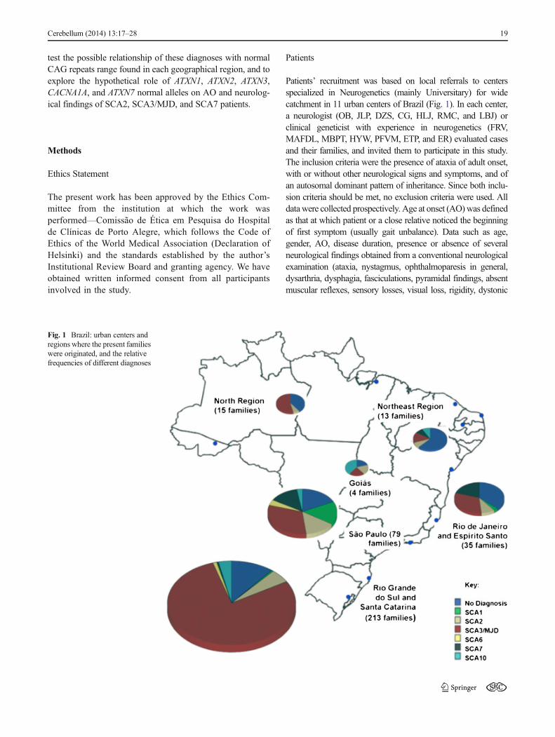
Fig. 1 Brazil: urban centers andregions where the present familieswere originated, and the relativefrequencies of different diagnoses
Cerebellum (2014) 13:17–28 19
movements, bradykinesia, tremor, seizures, and cognitive losses),as well as geographical origin, and family history, were collectedthrough an online digital form. In each 1 of the 11 hospitals, theassistant physician collected demographic and neurological in-formation and entered them into the electronic database, in ourwebsite. After consent, a blood sample was collected and thensent from the site of origin to the central laboratory, where themolecular analyses were performed.
Methods
Genomic DNA was obtained from peripheral blood by thesalting out method [26]. Two different multiplex PCR wereperformed using fluorescence-labeled primers flanking the re-spective repeats. Following amplification, PCR products wereseparated on an ABI3130xl Genetic Analyzer. Expanded allelein the ATXN10 gene was detected by the repeat primed-PCR(TP-PCR) methodology as previously described [27]. Lengthof the expanded repeats at ATXN10 was not determined.
All patients diagnosed up to 2009 were studied by a gene-by-gene (step-by-step) approach—at first the ATXN3 gene wasstudied, then, if no expansion was observed, ATXN2 gene weretested, and so on. Since 2010, all DNA samples were tested by athree-panel approach based on fluorescent multiplex-PCR. Thefirst panel included the simultaneous study of regions of interestat ATXN1, ATXN2, ATXN3, CACNA1A, and ATXN7 genes. Ifno expansion was detected, samples were tested by a secondpanel, that included the regions of interest at PPP2R2B, TBP,and ATN1 genes. Finally, if no expansion was detected by bothpanels, the ATXN10 gene was then tested.
Patient characteristics are given as mean ± SD, range, and95 % CI, when applicable. Categorical variables such as diag-nosis, region of origin and presence or absence of neurologicalfindings, were compared through Fisher exact test and logisticregression. CAG repeats in most loci, and disease duration inseveral diagnostic categories did not show a normal distributionon Shapiro-Wilk test. The remaining continuous variables werenormal. Comparisons of ages at onset between diagnostic cat-egories were performed through ANOVA test with contrasts.Normal CAG repeats distribution from different geographicalorigins of subjects were compared by Kruskal–Wallis test.Correlations between age at onset and the CAG repeats of theexpanded and the normal alleles were tested with Spearmancorrelation test followed by linear regression model.
In those SCA2, SCA3/MJD, and SCA7 patients diagnosedfrom 2010 onwards, the presence or absence of a given neurolog-ical finding was tested according to the sets of the normal, CAGrepeat alleles atATXN1,ATXN2,ATXN3,CACNA1A, andATXN7genes. Variables such as age, disease duration, and the causalexpanded CAG repeat at each disease were also included in themodel because all of them could influence the present phenotype.A logistic regression (binary logistic) was utilized to control for allthese potential confounding factors, potentially related to the
outcomes under study. CAG repeats at each given locus wassubdivided into larger and shorter alleles. Given that shorter allelesdid not show any association with dependent variables, they wereexcluded from the model. Ages at onset were also not included inthis set, given its covariance with each of the expanded CAGrepeats under study. Statistical significance was defined asp<0.05. All statistical tests were performed in PASW 18.
Results
Proportion of Diagnoses and Range of Normal RepeatsPer Subject’s Geographical Origin
As of March 2012, 544 patients from 359 SCA families wereinvestigated: 354 (213 families) from Rio Grande do Sul andSanta Catarina States (in the South Region), 94 (79 families)from São Paulo State, 38 (35 families) from Rio de Janeiroand Espírito Santo States, 23 (15 families) fromNorth Region,30 (13 families) from Northeast Region, and 5 (4 families)from Central-Western Region. The first 270 earlier patientswere tested in a gene-by-gene approach; the former 274 werestudied by the three-panels approach, described in the“Methods” section.
SCA3/MJD was the most common SCA, accounting for214 families (59.6 %) and for 337 patients (62.5 %) recruitedin the present case series. The obtained diagnoses are de-scribed in Fig. 1 and in Table 1. Diagnoses described hereinclude former families reported by our group (114 families in[9] and three additional families in [28]) [9, 28].
Sixty-five Brazilian families with SCA (18.2 %) remainedwithout diagnosis. In these families, SCA1, SCA2, SCA3/MJD,SCA6, SCA7, SCA10, SCA12, SCA17, and DRPLA have beenexcluded (“no diagnosis”, or ND families). ND families wereeven more common in Northeast (61.5 %, Table 1) and North(40 %) regions, and in Rio de Janeiro state (37.1 %; p<0.05,exact Fisher test).
Normal CAG and ATTCT repeat lengths at these loci wereobtained from each region of Brazil; the majority of these datawere obtained by the three-panels approach, described in the“Methods” section. The repeat ranges in each Brazilian regiondid not show any peculiar pattern, nor were related to specificproportions of SCA diagnoses (Table 2).
Associations Between Clinical Characteristics and MolecularResults
General clinical data such as age at onset and disease durationwere obtained in 518 out of the 544 patients of the presentseries, and are presented in Table 3. A detailed neurologicaldescription was obtained in only 389 out of the original 544patients and is presented in Table 4. It is worth to remind that
20 Cerebellum (2014) 13:17–28
this analysis included only a subset of patients, and that wecannot rule out an ascertainment bias.
Ages at Onset
Ages at onset, disease duration and number of expandedrepeats for each condition are shown in Table 3. SCA6 pa-tients showed an older age at onset, whereas SCA2 and SCA7patients showed an earlier age at onset than the overall group;and ND patients showed longer disease duration than the otherdiagnostic categories (p<0.05, ANOVAwith contrasts).
Ages at onset were inversely related to length of the expand-ed repeats in those SCAs where this comparison was feasible—SCA1, SCA2, SCA3/MJD, SCA6, and SCA7 (Electronic Sup-plementary Material (ESM) Fig. 1). In SCA10, the effect of theexpanded ATTCT repeat on the AO cannot be analyzed, sincelength of these repeats was not determined.
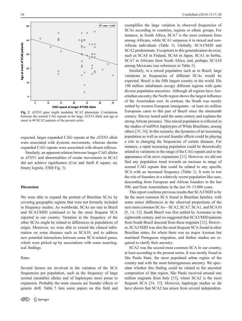
The hypothetical influence of CAG tracts in the normalrange found at ATXN1, ATXN2, ATXN3, CACNA1A, andATXN7 loci on AO in 49 patients with SCA2, 102 patientswith SCA3/MJD, and 32 patients with SCA7 were tested bylinear regression test. Normal CAG tract found at the largerATXN3 allele was associated with AO in SCA2 patients(r=−0.31, p<0.036, linear regression, Fig. 2). In SCA3/MJDand SCA7, only the expanded repeat has influenced AO of the
specific diagnostic category (as shown in Table 3 and ESMFig. 1).
Neurological Findings
The proportions of several neurological findings were com-pared between the different categories: SCA1, SCA2, SCA3/MJD, SCA6, SCA7, SCA10, and ND patients. This analysiswas done in the subset of 389 patients where the data wereavailable.
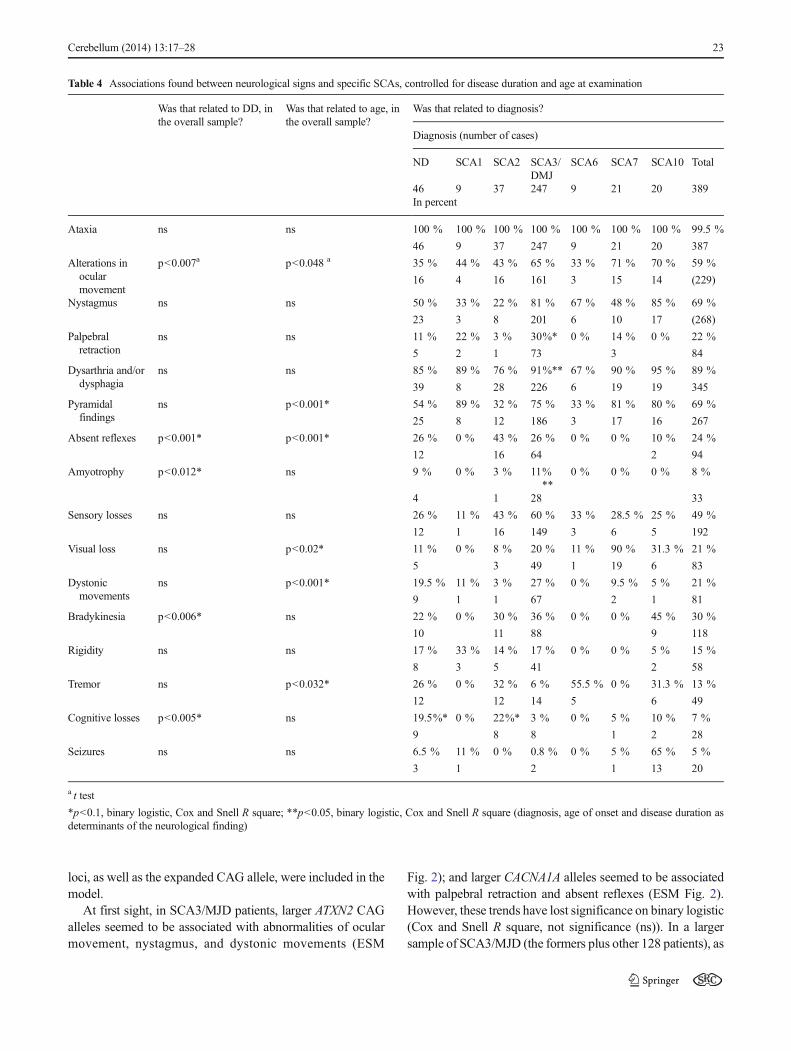
Binary logistic regression was performed, with age anddisease duration included in the model. Cox and Snell Rsquare was presented when a trend (p<0.1) or an association(p<0.05) was found. Table 4 shows that dysarthria–dysphagiaand amyotrophies were significantly more frequent in SCA3/MJD than in other SCAs (p<0.05).
Normal CAGn Repeats As Possible Modifiers of the SCAPhenotypes
A preliminary search for a possible modifier role of the normalCAG repeats at diverse loci on the neurological findings wasperformed in a subgroup of 183 patients. Diseases under studywere SCA3/MJD (including 102 patients), SCA2 (49 pa-tients), and SCA7 patients (32 patients), in the present cohort.
Table 1 Total number and percent of SCA families found, according to diagnoses and to geographical origin in Brazil
Region of Origin Total
South Region (Rio Grandedo Sul and Santa CatarinaStates)
SãoPaulo
Rio de Janeiroand EspíritoSanto
North Region(Pará and AcreStates)
Northeast Region (Rio Grande doNorte, Paraíba, Bahia and CearáStates)
Central-WesternRegion (GoiásState)
Nodiag-nosis(ND)
23 14 13 6 8 1 65
10.8 % 17.7 % 37.1% * 40% * 61.5% * 25 % 18.1%
SCA1 1 13 1 0 0 0 15
0.5 % 16.5%* 2.9 % 4.2%
SCA2 11 11 * 3 1 1 1 28
5.2 % 13.9% 8.6 % 6.7 % 7.7 % 25 % 7.8%
SCA3/MJD
167 25 11 8 2 1 214
78.4% * 31.6 % 31.4 % 53.3 % 15.4 % 25 % 59.6%
SCA6 2 3 0 0 0 0 5
0.9 % 3.8% * 1.4%
SCA7 1 11 7 0 1 0 20
0.5 % 13.9%*
20 %* 7.7 % 5.6
SCA10 8 2 0 0 1 1 12
3.7 % 2.5 % 7.7 % 25% * 3.3%
Total 213 79 35 15 13 4 359
100 % 100 % 100 % 100 % 100 % 100 % 100 %
*p<0.05, Fisher exact test, adjusted standardized residuals
Cerebellum (2014) 13:17–28 21
These patients were included in the three-panels approach,described in the “Methods” section, and therefore were testedfor the CAG repeats of interest in this section. In other words,this subset included only those individuals investigated after
2010. For this reason, we cannot rule out an ascertainmentbias related to recent years.
Disease duration, chronological age, the large normal CAGallele at ATXN1, ATXN2, ATXN3, CACNA1A, and ATXN7
Table 2 Number of CAG repeats found in each locus, and classified as normal, expanded and uncertain alleles. CAG repeats in normal alleles were alsoshown in specific regions, where the related SCAwas more common than in other geographical areas
ATXN1(SCA1)
ATXN2(SCA2)
ATXN3(SCA3/MJD)
CACNA1A(SCA6)
ATXN7(SCA7)
PPP2R2B(SCA12)
TBP(SCA17)
ATN1(DRPLA)
ATXN10(SCA10)
CAG repeats ATTCTrepeats
Number of individuals(alleles) studied
273 (546) 285 (570) 481 (962) 275 (550) 280 (560) 88 (176) 88 (176) 88 (176) 88 (176)
Expanded repeats,m (range)
46.7 (39–60) 42.1 (34–67) 75.45(65–89)
24.3 (22–26) 50.4 (37–73) – – – NM
n 20 51 344 11 23
CAG repeats ofuncertain interpretationor reduced penetrance
One allelewith 36repeats a
Four alleleswith 33repeatsb
None One allelewith 19repeatsc
None. None. None. NA NA
Normal repeats,m (range)
30 (19–35) 22.08 (11–30) 22.27 (7–38) 11 (4–17) 10 (7–14) 11.64(9–19)
34.6(27–38)
13 (2–19) 14(7–20)
n 526 516 618 539 528 176 176 176 153
Normal CAG repeatson the subpopulationwith the highest numberof affected families,m (range)
29.6 (20–35) 22.19 (17–30) 21.85(7–38)
11.41(6–17)
10 (7–14) – – – –
n 170 176 362 182 66
São Paulo São Paulo Rio Grandedo Sul
São Paulo Rio deJaneiro
NA not applicable, NM not measureda One ATXN1 allele with 36 CAG repeats was found in a SCA3/MJD patient (with 30/36 repeats at ATXN1 and 27/79 repeats at ATXN3)b Four siblings from Rio Grande do Sul State with SCA2 and parkinsonian phenotype had their short ATXN2 alleles with 33 repeats (their ATXN2genotypes were 33/34, 33/34, 33/34, and 33/43). They have been previously reported [62]c One CACNA1A allele with 19 CAG repeats was found in a SCA6 patient of Japanese ancestry (with 19/23 repeats) who inherited the allele with 23repeats from her father (13/23 repeats).` His mother was already deceased and his maternal family has no history of ataxic symptoms
Table 3 Age at onset, diseaseduration and number of expandedrepeats for each diagnosticcategory
*p<0.05, ANOVAwith Tukey;**p<0.05, Spearman correlationtest
N Disease duration Age at onset ExpandedCAGrepeat
Correlationbetween age atonset andexpandedCAG repeat
Mean±SD (95 % CI) Mean(range)
R2 **
No diagnosis(ND)
58 10.8±7.3 (8.9–12.8)* 36.6±15.4 (32.5–40.7) – –
SCA 1 19 6.1±4.7 (3.8–8.4) 37.1±11.2 (31.7–42.5) 47 (41–60) 0.70
SCA 2 49 10.1±7.7 (7.9–12.3) 29.7 ±10.6 (26.7–32.8)* 41 (34–67) 0.65
SCA 3/ MJD 328 8.21±6 (7.5–8.8)* 34±11.2 (32.8–35.2) 75 (67–89) 0.55
SCA 6 11 10.5±6.5 (6.2–14.9) 45.4±12.6 (36.9–53.8)* 24 (22–26) 0.66
SCA 7 30 7.7±5.2 (5.7–9.6) 25.5±11.4 (21.2–29.7)* 50 (38–73) 0.80
SCA 10 23 12.9±7.8 (9.4–16.4) 33.8±9.8 (29.5–38) – –
Total 518 8.8±6.4 (8.2–9.3) 33.7±12 (32.7–34.8) – –
22 Cerebellum (2014) 13:17–28
loci, as well as the expanded CAG allele, were included in themodel.
At first sight, in SCA3/MJD patients, larger ATXN2 CAGalleles seemed to be associated with abnormalities of ocularmovement, nystagmus, and dystonic movements (ESM
Fig. 2); and larger CACNA1A alleles seemed to be associatedwith palpebral retraction and absent reflexes (ESM Fig. 2).However, these trends have lost significance on binary logistic(Cox and Snell R square, not significance (ns)). In a largersample of SCA3/MJD (the formers plus other 128 patients), as
Table 4 Associations found between neurological signs and specific SCAs, controlled for disease duration and age at examination
Was that related to DD, inthe overall sample?
Was that related to age, inthe overall sample?
Was that related to diagnosis?
Diagnosis (number of cases)
ND SCA1 SCA2 SCA3/DMJ
SCA6 SCA7 SCA10 Total
46 9 37 247 9 21 20 389In percent
Ataxia ns ns 100 % 100 % 100 % 100 % 100 % 100 % 100 % 99.5 %
46 9 37 247 9 21 20 387
Alterations inocularmovement
p<0.007a p<0.048 a 35 % 44 % 43 % 65 % 33 % 71 % 70 % 59 %
16 4 16 161 3 15 14 (229)
Nystagmus ns ns 50 % 33 % 22 % 81 % 67 % 48 % 85 % 69 %
23 3 8 201 6 10 17 (268)
Palpebralretraction
ns ns 11 % 22 % 3 % 30%* 0 % 14 % 0 % 22 %
5 2 1 73 3 84
Dysarthria and/ordysphagia
ns ns 85 % 89 % 76 % 91%** 67 % 90 % 95 % 89 %
39 8 28 226 6 19 19 345
Pyramidalfindings
ns p<0.001* 54 % 89 % 32 % 75 % 33 % 81 % 80 % 69 %
25 8 12 186 3 17 16 267
Absent reflexes p<0.001* p<0.001* 26 % 0 % 43 % 26 % 0 % 0 % 10 % 24 %
12 16 64 2 94
Amyotrophy p<0.012* ns 9 % 0 % 3 % 11%**
0 % 0 % 0 % 8 %
4 1 28 33
Sensory losses ns ns 26 % 11 % 43 % 60 % 33 % 28.5 % 25 % 49 %
12 1 16 149 3 6 5 192
Visual loss ns p<0.02* 11 % 0 % 8 % 20 % 11 % 90 % 31.3 % 21 %
5 3 49 1 19 6 83
Dystonicmovements
ns p<0.001* 19.5 % 11 % 3 % 27 % 0 % 9.5 % 5 % 21 %
9 1 1 67 2 1 81
Bradykinesia p<0.006* ns 22 % 0 % 30 % 36 % 0 % 0 % 45 % 30 %
10 11 88 9 118
Rigidity ns ns 17 % 33 % 14 % 17 % 0 % 0 % 5 % 15 %
8 3 5 41 2 58
Tremor ns p<0.032* 26 % 0 % 32 % 6 % 55.5 % 0 % 31.3 % 13 %
12 12 14 5 6 49
Cognitive losses p<0.005* ns 19.5%* 0 % 22%* 3 % 0 % 5 % 10 % 7 %
9 8 8 1 2 28
Seizures ns ns 6.5 % 11 % 0 % 0.8 % 0 % 5 % 65 % 5 %
3 1 2 1 13 20
a t test
*p<0.1, binary logistic, Cox and Snell R square; **p<0.05, binary logistic, Cox and Snell R square (diagnosis, age of onset and disease duration asdeterminants of the neurological finding)
Cerebellum (2014) 13:17–28 23
expected, larger expanded CAG repeats at the ATXN3 allelewere associated with dystonic movements, whereas shorterexpanded CAG repeats were associated with absent reflexes.
Similarly, an apparent relation between longer CAG allelesat ATXN1 and abnormalities of ocular movements in SCA2did not achieve significance (Cox and Snell R square, ns;binary logistic, ESM Fig. 3).
Discussion
We were able to expand the portrait of Brazilian SCAs bycovering geographic regions that were not formerly includedin frequency studies. As worldwide, SCAs are rare in Braziland SCA3/MJD continued to be the most frequent SCAreported in our country. Variation in the frequency of theother SCAs might be related to differences in populations oforigin. Moreover, we were able to extend the clinical infor-mation on some diseases such as SCA10, and to addressnew potential interactions between some SCA-related genes,which were picked up by associations with some neurolog-ical findings.
Rates
Several factors are involved in the variation of the SCAfrequencies per population, such as the frequency of largenormal (unstable) alleles and of haplotypes more prone toexpansion. Probably the main reasons are founder effects orgenetic drift. Table 5 lists some papers on this field and
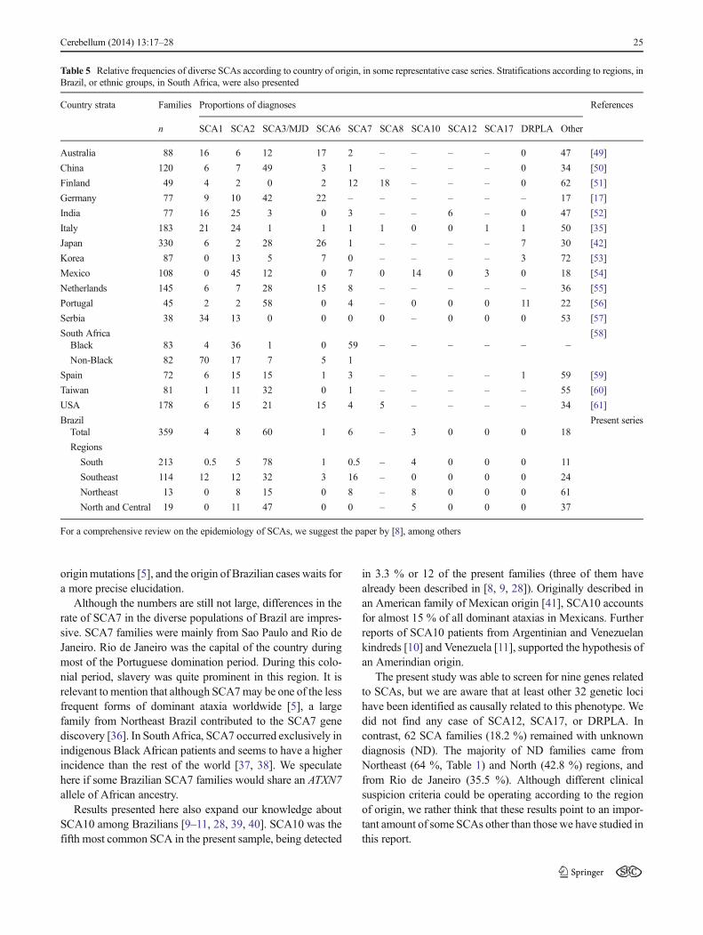
exemplifies the large variation in observed frequencies ofSCAs according to countries, regions or ethnic groups. Forinstance, in South Africa, SCA7 is the most common formamong Africans, while SCA1 surpasses it in mixed and non-African individuals (Table 5). Globally, SCA3/MJD andSCA2 predominate. Exceptions to this generalization do exist,such as SCA8 in Finland, SCA6 in Japan, SCA1 in Serbia,SCA7 in Africans from South Africa, and, perhaps, SCA10among Mexicans (see references in Table 5).
Similarly, in a mixed population such as in Brazil, largevariations in frequencies of different SCAs would beexpected. Brazil is the fifth largest country in the world. His190 million inhabitants occupy different regions with quitediverse population ancestries. Although all regions have Am-erindian ancestry, the North region shows the largest influenceof the Amerindian root. In contrast, the South was mostlysettled by western European immigrants—at least six millionEuropeans came to this part of Brazil since the nineteenthcentury. Slavery lasted until the same century and explains thestrong African presence. This mixed population is reflected inthe studies of mtDNA haplotypes of White Brazilians, amongothers [29, 30]. In this scenario, the dynamics of an increasingpopulation as well as several founder effects could be playinga role in changing the frequencies of certain diseases. Forinstance, a rapid increasing population could be theoreticallyrelated to variations in the range of the CAG repeats and to theappearance of de novo expansions [31]. However, we did notfind any population trend towards an increase in range ofnormal CAG repeats that could be related to any specificSCA with an increased frequency (Table 2). It rests to testthe role of founders in a relatively recent population like ours,descending from European and African founders in the last500, and from Amerindians in the last 10–15,000 years.
This report confirms previous results that SCA3/MJD is byfar the most common SCA found in Brazilian families, withsome minor differences in the observed proportions of thenext most common SCAs—SCA2, SCA7, SCA1, and SCA10[9, 14, 32]. South Brazil was first settled by Azoreans in theeighteenth century, andwe suggested that SCA3/MJD patientsfrom South Brazil descend from these migrants [32]. Howev-er, SCA3/MJDwas also the most frequent SCA found in otherBrazilian states, for where there was no major Azorean butmainland Portuguese migration, and further studies are re-quired to clarify their ancestry.
SCA2 was the second most common SCA in our country,at least according to the present series. It was mostly found inSão Paulo State, the most populated urban region of thecountry and with the most heterogeneous ancestry. We spec-ulate whether this finding could be related to the ancestralcomposition of that region. São Paulo received around onemillion migrants from Italy [33], where SCA2 is the mostfrequent SCA [34, 35]. However, haplotype studies so farhave shown that SCA2 has arisen from several independent-
Fig. 2 ATXN3 gene might modulate SCA2 phenotype. Correlationsbetween the normal CAG repeats at the large ATXN3 allele and age atonset in 49 SCA2 patients of the present series
24 Cerebellum (2014) 13:17–28
originmutations [5], and the origin of Brazilian cases waits fora more precise elucidation.
Although the numbers are still not large, differences in therate of SCA7 in the diverse populations of Brazil are impres-sive. SCA7 families were mainly from Sao Paulo and Rio deJaneiro. Rio de Janeiro was the capital of the country duringmost of the Portuguese domination period. During this colo-nial period, slavery was quite prominent in this region. It isrelevant to mention that although SCA7may be one of the lessfrequent forms of dominant ataxia worldwide [5], a largefamily from Northeast Brazil contributed to the SCA7 genediscovery [36]. In South Africa, SCA7 occurred exclusively inindigenous Black African patients and seems to have a higherincidence than the rest of the world [37, 38]. We speculatehere if some Brazilian SCA7 families would share an ATXN7allele of African ancestry.
Results presented here also expand our knowledge aboutSCA10 among Brazilians [9–11, 28, 39, 40]. SCA10 was thefifth most common SCA in the present sample, being detected
in 3.3 % or 12 of the present families (three of them havealready been described in [8, 9, 28]). Originally described inan American family of Mexican origin [41], SCA10 accountsfor almost 15 % of all dominant ataxias in Mexicans. Furtherreports of SCA10 patients from Argentinian and Venezuelankindreds [10] and Venezuela [11], supported the hypothesis ofan Amerindian origin.
The present study was able to screen for nine genes relatedto SCAs, but we are aware that at least other 32 genetic locihave been identified as causally related to this phenotype. Wedid not find any case of SCA12, SCA17, or DRPLA. Incontrast, 62 SCA families (18.2 %) remained with unknowndiagnosis (ND). The majority of ND families came fromNortheast (64 %, Table 1) and North (42.8 %) regions, andfrom Rio de Janeiro (35.5 %). Although different clinicalsuspicion criteria could be operating according to the regionof origin, we rather think that these results point to an impor-tant amount of some SCAs other than those we have studied inthis report.
Table 5 Relative frequencies of diverse SCAs according to country of origin, in some representative case series. Stratifications according to regions, inBrazil, or ethnic groups, in South Africa, were also presented
Country strata Families Proportions of diagnoses References
n SCA1 SCA2 SCA3/MJD SCA6 SCA7 SCA8 SCA10 SCA12 SCA17 DRPLA Other
Australia 88 16 6 12 17 2 – – – – 0 47 [49]
China 120 6 7 49 3 1 – – – – 0 34 [50]
Finland 49 4 2 0 2 12 18 – – – 0 62 [51]
Germany 77 9 10 42 22 – – – – – – 17 [17]
India 77 16 25 3 0 3 – – 6 – 0 47 [52]
Italy 183 21 24 1 1 1 1 0 0 1 1 50 [35]
Japan 330 6 2 28 26 1 – – – – 7 30 [42]
Korea 87 0 13 5 7 0 – – – – 3 72 [53]
Mexico 108 0 45 12 0 7 0 14 0 3 0 18 [54]
Netherlands 145 6 7 28 15 8 – – – – – 36 [55]
Portugal 45 2 2 58 0 4 – 0 0 0 11 22 [56]
Serbia 38 34 13 0 0 0 0 – 0 0 0 53 [57]
South Africa [58]Black 83 4 36 1 0 59 – – – – – –
Non-Black 82 70 17 7 5 1
Spain 72 6 15 15 1 3 – – – – 1 59 [59]
Taiwan 81 1 11 32 0 1 – – – – – 55 [60]
USA 178 6 15 21 15 4 5 – – – – 34 [61]
Brazil Present seriesTotal 359 4 8 60 1 6 – 3 0 0 0 18
Regions
South 213 0.5 5 78 1 0.5 – 4 0 0 0 11
Southeast 114 12 12 32 3 16 – 0 0 0 0 24
Northeast 13 0 8 15 0 8 – 8 0 0 0 61
North and Central 19 0 11 47 0 0 – 5 0 0 0 37
For a comprehensive review on the epidemiology of SCAs, we suggest the paper by [8], among others
Cerebellum (2014) 13:17–28 25
Clinical Findings and Expanded Repeats
Several findings confirmed many previous observations onSCAs—namely, age at onset of SCA1, SCA2, SCA3/MJD,and SCA6. Correlations between ages at onset and CAGrepeats also confirmed early, classical findings, the exceptionbeing probably SCA7, where the obtained R2 of 0.77 washigher than in other populations [20, 21].
Among our SCA10 patients, epilepsy was far more com-mon than in former Brazilian cases [39]; actually, our ratesresembled those found in Mexican patients [6]. The presentseries included 23 individuals with a DD of 12.9 years andliving up to 3,500 km away from each other. Data on neuro-logical examination were obtained in 20 of them, and in 13(65 %) individuals, generalized tonic–clonic seizures or com-binations of myoclonic, complex partial, and generalized ton-ic–clonic seizures occurred. In contrast, in a large series of 80SCA10 patients living near Curitiba (in Santa Catarina State),epilepsy was found only in 3.75 % of the cases with a meanDD of 15 years [39]. Our series included only one case fromSanta Catarina State: unfortunately, the patient was lost offollow-up and a detailed neurological information was notobtained. Seizures were present in six and absent in three ofour families; in the other three families, this information waslacking. Our positive families included epileptic and non-epileptic individuals (there was intrafamilial phenotypic het-erogeneity). The present six families with epilepsy were foundin all Brazilian regions under study. The three “epilepsy-free”families belonged to Rio Grande do Sul and São Paulo States,where the majority of families under study were living. Norelation between disease duration and seizures was found.
Former case series have presented the phenotypic contrastsbetween different SCAs [17, 42, 43]. An attempt to improveour results was made by controlling confounding differencessuch as chronological ages and disease duration—both inde-pendent variables that could partially determine the neurolog-ical manifestations. By doing so, some differences in theproportions of neurological findings were obtained (Table 4).However, we are aware that prospective studies on naturalhistory are the best design to test these differences [44–46].
Normal CAG Repeats As Candidates to Modify SCAPhenotypes
In the present study, logistic regression has detected somerelationships between CAG repeats at some loci and clinicalmanifestations in some SCAs.
In SCA3/MJD patients, a direct relationship between theexpanded CAG repeat at ATXN3 and dystonic movementswas shown while an inverse correlation was seen with absentreflexes. These results are in agreement with classical findingsof SCA3/MJD, and might confirm the quality of our data andthe rationale of the present approach. The novelty was the
suggested relationships of the normal CAG repeat length atATXN3 gene with the age at onset of SCA2.
Potential relationship between ATXN2 and ATXN3 genes(or their proteins ataxin-2 and ataxin-3) has long been studied.For instance, normal ataxin-2 can be detected in the patho-genic inclusions of SCA3/MJD patients, and, likewise, normalataxin-3 localizes to the inclusions formed in SCA2 patients[47]. In a Drosophila model, toxicity and neurodegenerationinduced by pathogenic forms of ATXN3 depend on the normalactivity of the wild ataxin-2 of the fly [48]. Curiously, we wereunable to reproduce here our previous finding that fascicula-tions in SCA3/MJD correlate with the CAG repeat in ATXN2normal alleles [23]. Differences between our previous and thepresent observations might be due to differences betweenclinical protocols, to sample size, or by chance.
In summary, SCA3/MJD was confirmed to be the mostfrequent SCA in Brazilians, followed by SCA2, SCA7, andSCA10. Variations of the CAG repeats, either in the normal aswell as in the expanded range, were similar to those foundelsewhere. Ages at onset and neurological manifestationswere all expected, with the possible exception of the presenceof seizures among Brazilian patients with SCA10. And al-though the CAG expansion in a single allele of the responsiblegene is sufficient for the determination of each SCA, ourobservations suggest that some interaction of the ATXN3 geneon AO in SCA2 might occur. These results deserve confirma-tion in a second population, and deeper studies in cellular andanimal models.
Acknowledgments We would like to thank the patients and theirfamilies for taking part in this study. We would also like to thank ThaisSanta Rita for her technical assistance; Vanessa Erichsen Emmel andAlexis Trott for their contribution in early stages of this project; andPedro Braga-Neto, Isabel CristinaNeves de Souza, and RaimundaHelenaFeio for their contribution in recruiting some patients. This study wassupported by FAPERGS, CNPq, CAPES, INAGEMP, and FIPE-HCPA.Castilhos RM was supported by INAGEMP. Furtado GV was supportedby CAPES. Gheno TC, Russo A, Saraiva-Pereira ML, and Jardim LBwere supported by CNPq.
Conflict of Interest The authors state that there is no potential conflictof interest
References
1. Schöls L, Bauer P, Schmidt T, Schulte T, Riess O. Autosomaldominant cerebellar ataxia: clinical features, genetics, and pathogen-esis. Lancet Neurol. 2004;3(5):291–304.
2. Bird TD. Hereditary Ataxia Overview. 1998 Oct 28 [Updated 2012May 31]. In: Pagon RA, Bird TD, Dolan CR, et al., editors.GeneReviews™ [Internet]. Seattle (WA): University of Washington,Seattle; 1993-. http://www.ncbi.nlm.nih.gov/books/NBK1138/.
3. Sato N, Amino T, Kobayashi K, et al. Spinocerebellar ataxia type 31is associated with “inserted” penta-nucleotide repeats containing(TGGAA)n. Am J Hum Genet. 2009;85:544–57.
26 Cerebellum (2014) 13:17–28
4. Kobayashi H, Abe K, Matsuura T, et al. Expansion of intronicGGCCTG hexanucleotide repeat in NOP56 causes SCA36, a typeof spinocerebellar ataxia accompanied by motor neuron involvement.Am J Hum Genet. 2011;89:121–30.
5. Sequeiros J, Martins S, Silveira I. Epidemiology and popula-tion genetics of degenerative ataxias. Handb Clin Neurol.2012;103:227–51.
6. Rasmussen A, Matsuura T, Ruano L, et al. Clinical and geneticanalysis of four Mexican families with spinocerebellar ataxia type10. Ann Neurol. 2001;50(2):234–9.
7. Teive HA, Roa BB, Raskin S, et al. Clinical phenotype of Brazilianfamilies with spinocerebellar ataxia 10. Neurology. 2004;63(8):1509–12.
8. Alonso I, Jardim LB, Artigalas O, et al. Reduced penetrance ofintermediate size alleles in spinocerebellar ataxia type 10. Neurology.2006;66(10):1602–4.
9. Trott A, Jardim LB, Ludwig HT, et al. Spinocerebellar ataxias in 114Brazilian families: clinical and molecular findings. Clin Genet.2006;70(2):173–6.
10. Gatto EM, Gao R, White MC, et al. Ethnic origin and extrapyramidalsigns in an Argentinean spinocerebellar ataxia type 10 family. Neu-rology. 2007;69:216–8.
11. Teive HA, Munhoz RP, Arruda WO, Raskin S, Werneck LC,Ashizawa T. Spinocerebellar ataxia type 10—a review. ParkinsonismRelat Disord. 2011;17(9):655–61.
12. García-Murias M, Quintáns B, Arias M, et al. Costa da Morte’ ataxiais spinocerebellar ataxia 36: clinical and genetic characterization.Brain. 2012;135(Pt 5):1423–35.
13. Basri R, Yabe I, Soma H, Sasaki H. Spectrum and prevalence ofautosomal dominant spinocerebellar ataxia in Hokkaido, the northernisland of Japan: a study of 113 Japanese families. J Hum Genet.2007;52:848–55.
14. Teive HA, Munhoz RP, Arruda WO, et al. Spinocerebellar ataxias:genotype-phenotype correlations in 104 Brazilian families. Clinics.2012;67(5):443–9.
15. Ranum LPW, Chung MY, Banfi S, Bryer A, Schut LJ, Ramesar R,et al. Molecular and clinical correlations in spinocerebellar ataxiatype I: evidence for familial effects on the age at onset. Am J HumGenet. 1994;55(2):244–52.
16. Maciel P, Gaspar C, DeStefano AL, Silveira I, Coutinho P, RadvanyJ, et al. Correlation between CAG repeat length and clinical featuresin Machado–Joseph disease. Am J Hum Genet. 1995;57(1):54–61.
17. Schöls L, Amoiridis G, Büttner T, Przuntek H, Epplen JT, Riess O.Autosomal dominant cerebellar ataxia: phenotypic differences ingenetically defined subtypes? Ann Neurol. 1997;42(6):924–32.
18. Pulst SM, Santos N, Wang D, Yang H, Huynh D, et al. Spinocerebellarataxia type 2: polyQ repeat variation in the CACNA1A calciumchannel modifies age of onset. Brain. 2005;128:2297–303.
19. Gomez CM. Spinocerebellar Ataxia Type 6. 1998 Oct 23 [Updated2008 Jun 16]. In: Pagon RA, Bird TD, Dolan CR, et al., editors.GeneReviews™ [Internet]. Seattle (WA): University of Washington,Seattle; 1993-. http://www.ncbi.nlm.nih.gov/books/NBK1140/.
20. Michalik A, Martin J-J, Van Broeckhoven C. Spinocerebellar ataxiatype 7 associated with pigmentary retinal dystrophy. Eur J HumGenet. 2004;12(1):2–15.
21. Han Y, Yu L, Zheng HM, Guan YT. Clinical and genetic study ofspinocerebellar ataxia type 7 in East Asian population. Chin Med J(Engl). 2010;123(16):2274–8.
22. DeStefano AL, Cupples LA,Maciel P, Gaspar C, Radvany J, DawsonDM, Sudarsky L, Corwin L, Coutinho P, MacLeod P, Sequeiros J,RouleauGA, Farrer LA.A familial factor independent of CAG repeatlength influences age at onset ofMachado-Joseph disease. Am JHumGenet. 1996; 59(1):119–127.
23. Jardim L, Silveira I, Pereira ML, et al. Searching for modulatingeffects of SCA2, SCA6 and DRPLA CAG tracts on the Machado-Joseph disease (SCA3) phenotype. Acta Neurol Scand.2003;107(3):211–4.
24. Bettencourt C, Raposo M, Kazachkova N, Cymbron T, SantosC, Kay T, et al. The APOE ε2 allele increases the risk ofearlier age at onset in Machado-Joseph disease. Arch Neurol.2011;68(12):1580–3.
25. Emmel VE, Alonso I, Jardim LB, Saraiva-Pereira ML, Sequeiros J.Does DNA methylation in the promoter region of the ATXN3 genemodify age at onset in MJD (SCA3) patients? Clin Genet.2011;79(1):100–2.
26. Miller SA, Dykes DD, Polesky HF. A simple salting-out procedurefor extracting DNA from human nucleated cells. Nucleic Acids Res.1988;16(3):1215.
27. Cagnoli C, Michielotto C, Matsuura T, Ashizawa T, Margolis RL,Holmes SE, et al. Detection of large pathogenic expansions inFRDA1, SCA10, and SCA12 genes using a simple fluorescentrepeat-primed PCR assay. J Mol Diagn. 2004;6(2):96–100.
28. Almeida T, Alonso I, Martins S, et al. Ancestral origin of the ATTCTrepeat expansion in spinocerebellar ataxia type 10 (SCA10). PLoSOne. 2009;4(2):e4553
29. Parra FC, Amado RC, Lambertucci JR, Rocha J, AntunesCM, Pena SDJ. Color and genomic ancestry in Brazilians.PNAS. 2003;100:177–82.
30. Pena SDJ, Pietro GD, Fuchshuber-Moraes M, et al. (2013) TheGenomic Ancestry of Individuals from Different Geographical Re-gions of Brazil Is More Uniform Than Expected. PLoS One6(2):e17063. doi:10.1371/journal.pone.0017063.
31. Andrés AM, Lao O, Soldevila M, Calafell F, Bertranpetit J. Dynam-ics of CAG repeat loci revealed by the analysis of their variability.Hum Mutat. 2002;21:61–70.
32. Jardim LB, Silveira I, Pereira ML, et al. A survey of spinocerebellarataxia in South Brazil—66 new cases with Machado-Joseph disease,SCA7, SCA8, or unidentified disease-causing mutations. J Neurol.2001;248(10):870–6.
33. Trento A. Do outro lado do Atlântico—Um século de ImigraçãoItaliana no Brasil. São Paulo: Nobel; 1988.
34. Filla A, Mariotti C, Caruso G, et al. Relative frequencies of CAGexpansions in spinocerebellar ataxia and dentatorubropallidoluysianatrophy in 116 Italian families. Eur Neurol. 2000;44(1):31–6.
35. Brusco A, Gellera C, Cagnoli C, et al. Molecular genetics of hered-itary spinocerebellar ataxia: mutation analysis of spinocerebellarataxia genes and CAG/CTG repeat expansion detection in 225 Italianfamilies. Arch Neurol. 2004;61(5):727–33.
36. David G, Giunti P, Abbas N, et al. The gene for autosomal dominatcerebellar ataxia type II is located in a 5-cM region in 3p12-p13:genetic and physical mapping of the SCA7 locus. Am J Hum Genet.1996;59(6):1328–36.
37. Bryer A, Krause A, Bill P, et al. The hereditary adult-onset ataxias inSouth Africa. J Neurol Sci. 2003;216(1):47–54.
38. Greenberg J, Solomon GAE, Vorster AA, Heckmann J, Bryer A.Origin of the SCA7 gene mutation in South Africa: implications formolecular diagnostics. Clin Genet. 2006;70:415–7.
39. Teive HA, Munhoz RP, Raskin S, et al. Spinocerebellar ataxia type10: frequency of epilpesy in a large sample of Brazilian patients. MovDisord. 2010;25(16):2875–8.
40. Raskin S, Ashizawa T, Teive HA, et al. Reduced penetrance in aBrazilian family with spinocerebellar ataxia type 10. Arch Neurol.2007;64(4):591–4.
41. Matsuura T, Yamagata T, Burgess DL, et al. Large expansion of theATTCT pentanucleotide repeat in spinocerebellar ataxia type 10. NatGenet. 2000;26(2):191–4.
42. Maruyama H, Izumi Y, Morino H, et al. Difference in disease-freesurvival curve and regional distribution according to subtype ofspinocerebellar ataxia: a study of 1,286 Japanese patients. Am J MedGenet. 2002;114(5):578–83.
43. Schmitz-Hübsch T, Coudert M, Bauer P, et al. Spinocerebellar ataxiatypes 1, 2, 3, and 6: disease severity and nonataxia symptoms. Neurol-ogy. 2008;71(13):982–9.
Cerebellum (2014) 13:17–28 27
44. Jardim LB, Hauser L, Kieling C, et al. Progression rate of neurolog-ical deficits in a 10-year cohort of SCA3 patients. Cerebellum.2010;9(3):419–28.
45. Jacobi H, Bauer P, Giunti P, et al. The natural history of spinocerebellarataxia type 1, 2, 3, and 6: a 2-year follow-up study. Neurology.2011;77(11):1035–41.
46. duMontcel TS, Charles P, Goizet C, et al. Factors influencing diseaseprogression in autosomal dominant cerebellar ataxia and spasticparaplegia. Arch Neurol. 2012;69(4):500–8.
47. Uchihara T, Fujigasaki H, Koyano S, Nakamura A, Yagishita S, et al.Non-expanded polyglutamine proteins in intranuclear inclusions ofhereditary ataxias—triple-labeling immunofluorescence study. ActaNeuropathol. 2001;102:149–52.
48. Lessing D, Bonini NM. Polyglutamine genes interact to modulate theseverity and progression of neurodegeneration in Drosophila. PLoSBiol. 2008;6(2):e29.
49. Storey E, du Sart D, Shaw JH, Lorentzos P, Kelly L, McKinleyGardner RJ, et al. Frequency of spinocerebellar ataxia types 1, 2, 3,6, and 7 in Australian patients with spinocerebellar ataxia. Am J MedGenet. 2000;95(4):351–7.
50. Jiang H, Tang B, Xu B, Zhao GH, Shen L, Tang JG, et al. Frequencyanalysis of autosomal dominant spinocerebellar ataxias in Han pop-ulation in the Chinese mainland and clinical and molecular charac-terization of spinocerebellar ataxia type 6. Zhonghua Yi Xue YiChuan Xue Za Zhi. 2005;22(1):1–4.
51. Juvonen V, Hietala M, Kairisto V, Savontaus ML. The occurrence ofdominant spinocerebellar ataxias among 251 Finnish ataxia patientsand the role of predisposing large normal alleles in a geneticallyisolated population. Acta Neurol Scand. 2005;111(3):154–62.
52. Srivastava AK, Choudhry S, Gopinath MS, Roy S, Tripathi M,Brahmachari SK, et al. Molecular and clinical correlation in fiveIndian families with spinocerebellar ataxia 12. Ann Neurol.2001;50(6):796–800.
53. Jin DK, Oh MR, Song SM, Koh SW, Lee M, Kim GM, et al.Frequency of spinocerebellar ataxia types 1,2,3,6,7 and dentatorubral
pallidoluysian atrophy mutations in Korean patients withspinocerebellar ataxia. J Neurol. 1999;246(3):207–10.
54. Alonso E,Martínez-Ruano L, De Biase I, Mader C, Ochoa A, YescasP, et al. Distinct distribution of autosomal dominant spinocerebellarataxia in the Mexican population. Mov Disord. 2007;22(7):1050–3.
55. van de Warrenburg BP, Sinke RJ, Verschuuren-Bemelmans CC,Scheffer H, Brunt ER, Ippel PF, et al. Spinocerebellar ataxias in theNetherlands: prevalence and age at onset variance analysis. Neurol-ogy. 2002;58(5):702–8.
56. Vale J, Bugalho P, Silveira I, Sequeiros J, Guimarães J, Coutinho P.Autosomal dominant cerebellar ataxia: frequency analysis and clinicalcharacterization of 45 families from Portugal. Eur J Neurol.2010;17(1):124–8.
57. Dragasević NT, Culjković B, Klein C, Ristić A, Keckarević M,Topisirović I, et al. Frequency analysis and clinical characterizationof different types of spinocerebellar ataxia in Serbian patients. MovDisord. 2006;21(2):187–91.
58. Smith DC, Bryer A, Watson LM, Greenberg LJ. Inheritedpolyglutamine spinocerebellar ataxias in South Africa. S Afr MedJ. 2012;102(8):683–6.
59. Pujana MA, Corral J, Gratacòs M, Combarros O, Berciano J, GenísD, et al. Spinocerebellar ataxias in Spanish patients: genetic analysisof familial and sporadic cases. Hum Genet. 1999;104(6):516–22.
60. Tsai HF, Liu CS, Leu TM,Wen FC, Lin SJ, Liu CC, et al. Analysis oftrinucleotide repeats in different SCA loci in spinocerebellar ataxiapatients and in normal population of Taiwan. Acta Neurol Scand.2004;109(5):355–60.
61. Moseley ML, Benzow KA, Schut LJ, Bird TD, Gomez CM,Barkhaus PE, et al. Incidence of dominant spinocerebellar andFriedreich triplet repeats among 361 ataxia families. Neurology.1998;51(6):1666–71.
62. Socal M, Emmel V, Rieder C, Hilbig A, Saraiva-Pereira M, Jardim LIntrafamilial variability of Parkinson phenotype in SCAs: Novelcases due to SCA2 and SCA3 expansions. Parkinsonism RelatDisord. 2009:15(5):374–8.
28 Cerebellum (2014) 13:17–28