Embed Size (px)
Citation preview

SpotlightDangers of Missing an Epidural Abscess: Multiple Visits and Delayed Diagnosis with a Severely Negative Outcome
Source and Credits
• This presentation is based on the June 2021 AHRQ WebM&MSpotlight Caseo See the full article at https://psnet.ahrq.gov/webmmo CME credit is available
o Commentary by: Linnea Lantz, DO, Joseph Yoon, MD and David Barnes, MD
o AHRQ WebM&M Editors in Chief: Patrick Romano, MD, MPH and Debra Bakerjian, PhD, APRN, RNo Spotlight Editors: Ulfat Shaikh, MD, and Patrick Romano, MDo Managing Editor: Meghan Weyrich, MPH
2
Objectives
At the conclusion of this educational activity, participants should be able to:
• List risk factors, symptoms, and exam findings associated with spinal epidural abscess
• Evaluate for spinal epidural abscess with laboratory testing and imaging
• Recognize pitfalls leading to erroneous interpretation of the tests for spinal epidural abscess
• Identify multiple visits for the same complaint as a red flag that warrants broadening of differentials and avoidance of premature closure
3
DANGERS OF MISSING AN EPIDURAL ABSCESS: MULTIPLE VISITS AND DELAYED
DIAGNOSIS WITH A DEVASTATING OUTCOME
A case describing how multiple preventable errors over several clinical visits culminated in a devastating
outcome
4
Case Details (1)
• 44-year-old man presented to his primary care physician (PCP) with complaints of new onset headache, photophobia, and upper respiratory tract infection symptoms.– Patient had recent history of interferon for Hepatitis C and a remote history
of cervical spine surgery requiring permanent spinal hardware • On physical examination, his neck was tender but no neurologic
abnormalities. Laboratory testing showed only abnormal transaminases.
• He was sent home from clinic with advice to take over-the-counter analgesics.
5
Case Details (2)
• Three days later, he presented to the emergency department (ED) with worsened headache and neck pain– He was discharged without imaging or evaluation of cerebrospinal fluid
(CSF)
• Four days later, he returned to his PCP with worsening headache, nausea, vomiting and photophobia– He was given oxycodone for pain
6
Case Details (3)• He was next seen in the hospital’s urgent care clinic with a fever of
101.4 F, hallucinations and neck stiffness• He was sent to the ED by ambulance for suspected meningitis
– CSF obtained by lumbar puncture was cloudy with 692 white blood cells per microliter (80% neutrophils), low glucose, and high protein, but no visible organisms on Gram staining.
– Cultures of blood and CSF were obtained but imaging of the cervical spine was not ordered.
– He was diagnosed with viral meningitis and sent home with antiemetics and long-acting oral morphine sulfate. He was not treated with antibiotics.
• The next morning, two blood cultures returned positive for Staphylococcus aureus while CSF culture was negative.
• After being called at home, the patient returned to the hospital and was admitted with a presumptive diagnosis of Staphylococcal meningitis.
7
Case Details (4)
• Despite initiation of intravenous antibiotic therapy, the patient developed worsening neurologic symptoms on his third hospital day including weakness, urinary incontinence, and urinary retention. He was unable to stand or walk.
• Cervical magnetic resonance imaging (MRI) showed a spinal epidural abscess (SEA) adjacent to his surgical hardware.
• At that point, he was transferred to a tertiary care center where decompressive surgery was performed.
• Despite surgical intervention, the patient remained quadriplegic. – He was unable to resume employment and required full-time home care
after discharge. – The patient committed suicide several years later.
8
DANGERS OF MISSING AN EPIDURAL ABSCESS: MULTIPLE VISITS AND DELAYED
DIAGNOSIS WITH A DEVASTATING OUTCOME
THE COMMENTARYBy Linnea Lantz, DO, Joseph Yoon, MD,
and David Barnes, MD
9
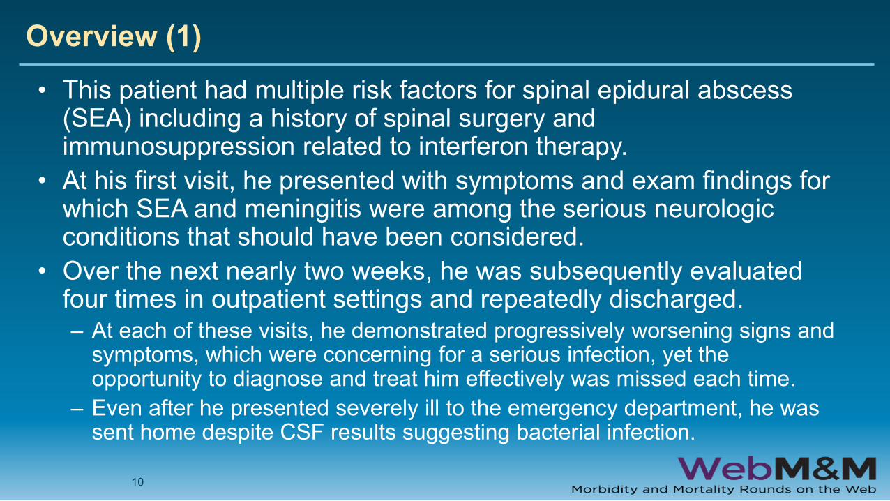
Overview (1)• This patient had multiple risk factors for spinal epidural abscess
(SEA) including a history of spinal surgery and immunosuppression related to interferon therapy.
• At his first visit, he presented with symptoms and exam findings for which SEA and meningitis were among the serious neurologic conditions that should have been considered.
• Over the next nearly two weeks, he was subsequently evaluated four times in outpatient settings and repeatedly discharged. – At each of these visits, he demonstrated progressively worsening signs and
symptoms, which were concerning for a serious infection, yet the opportunity to diagnose and treat him effectively was missed each time.
– Even after he presented severely ill to the emergency department, he was sent home despite CSF results suggesting bacterial infection.
10
Overview (2)
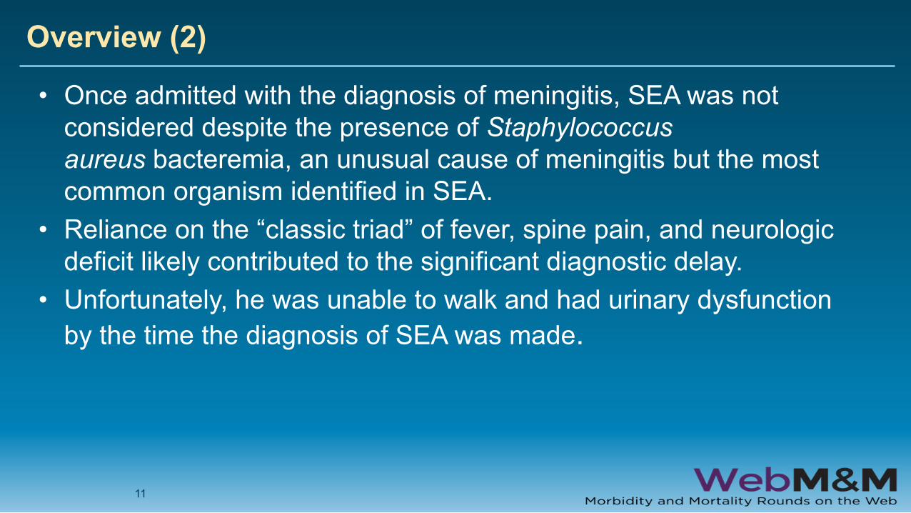
• Once admitted with the diagnosis of meningitis, SEA was not considered despite the presence of Staphylococcus aureus bacteremia, an unusual cause of meningitis but the most common organism identified in SEA.
• Reliance on the “classic triad” of fever, spine pain, and neurologic deficit likely contributed to the significant diagnostic delay.
• Unfortunately, he was unable to walk and had urinary dysfunction by the time the diagnosis of SEA was made.
11
Overview (3)
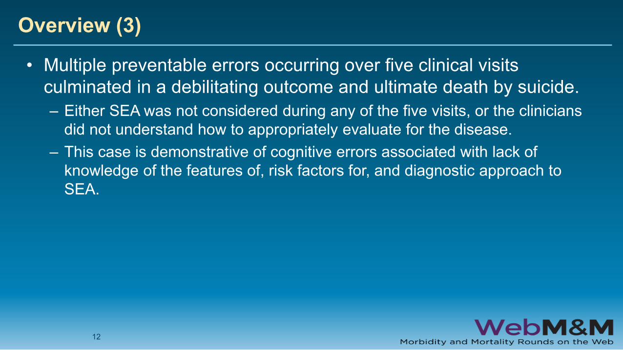
• Multiple preventable errors occurring over five clinical visits culminated in a debilitating outcome and ultimate death by suicide.– Either SEA was not considered during any of the five visits, or the clinicians
did not understand how to appropriately evaluate for the disease. – This case is demonstrative of cognitive errors associated with lack of
knowledge of the features of, risk factors for, and diagnostic approach to SEA.
12

Background
13
Background (1)• Neck and back pain are common chief complaints and account for more
health care spending in the United States than other conditions • While most cases of neck and back pain are due to benign
musculoskeletal causes, some are related to potentially debilitating and life-threatening etiologies such as spinal epidural abscess (SEA).– SEA is a collection of purulent material caused by a bacterial infection in the
epidural space of the vertebral canal. – It can be caused by hematogenous spread of bacteria from distal infection or
intravenous drug use (IVDU) and can be spread from a contiguous site of infection or by direct inoculation from spinal.
– Sources of distal infection most commonly include the skin and soft tissue, urinary tract, respiratory tract, and dental sources, as well as bacteremia of unknown origin.
– The purulent fluid collection associated with SEA leads to pain and neurologic symptoms via direct spinal cord compression as well as by compression of neighboring vascular structures leading to thrombosis and cord ischemia.
14
Background (2)
• The incidence of SEA has increased from 0.5-2 per 10,000 hospital admissions in 1970-1990 to 2-8 per 10,000 hospital admissions in 2000-2015.– This increase is multifactorial and related to an aging population with
increasing medical comorbidities, increased use of spinal instrumentation, rising rates of intravenous drug use, and improvements in imaging to facilitate diagnosis.
• Despite increasing incidence, SEA remains a relatively rare and challenging diagnosis due to its low incidence and association with non-specific early symptoms.
15
Background (3)
• Diagnosis is delayed in approximately 75% of patients and most patients present multiple times to an ED or primary care setting for their complaint prior to diagnosis.
• Inpatient delays are also common: many patients with SEA are admitted with an incorrect initial diagnosis, most often meningitis, herniated intervertebral disc, or a presumed infection unrelated to the spine.
• The diagnosis is often not apparent until neurologic deficits develop. • Mortality in patients with SEA is approximately 16%, while 15% of
survivors have permanent paresis or paralysis and 27% have other permanent neurologic deficits. Patients who present with neurologic deficits and those whose diagnosis is delayed have the worst outcomes.
16

Risk Factors
17
Risk Factors• 98% of patients diagnosed with SEA have at least one risk factor
predisposing them to the disease. – These include diabetes mellitus, distal site of infection, a history
of undergoing a procedure using spinal instrumentation, blunt spinal trauma, intravenous drug use, vertebral column abnormalities, immunosuppression, malignancy, alcoholism, human immunodeficiency virus (HIV) and acquired immunodeficiency syndrome (AIDS), chronic renal failure, and the presence of an indwelling vascular catheter.
– Spinal procedures that convey risk include invasive spinal surgical procedures, epidural and spinal anesthetic procedures, and vertebral or paravertebral steroid injections. Blunt spinal trauma, such as a motor vehicle collision or fall, can lead to disruption of normal anatomic barriers and is a significant risk factor that is often overlooked.
– Spinal abnormalities such as degenerative disc disease, scoliosis, and other chronic bony spinal conditions increase vulnerability to infection at those sites.
18
Clinical Presentation
19
Clinical Presentation (1)
• SEA Staging– Stage I is defined by back or neck pain, fever and tenderness. – Stage II progresses to include stabbing or shooting radicular pain,
nuchal rigidity/neck stiffness, and changes in deep tendon reflexes. – Stage III includes the development of sensory abnormalities, motor
weakness, and bowel or bladder dysfunction. – Stage IV is defined by paralysis.
• SEA does not necessarily always progress in this manner– Not all symptoms may be present at each stage. – Rate of progression is highly variable. The time from symptom onset to first
clinical contact can span days to weeks. Severe neurologic deterioration can take hours to days.
20
Clinical Presentation (2)• Neck or back pain is overwhelmingly the most common and
earliest symptom of SEA.– Pain is typically localized to the affected spinal level but may radiate to the chest,
abdomen, pelvis, or extremities and has been known to mimic pancreatitis, aortic dissection, nephrolithiasis, urinary tract infection, and other thoracic or abdominal pathology.
– Localized pain may be the only presenting symptom early in the course of the disease when the prognosis is best – It is critical to consider SEA for any patient with neck or back pain.
• Every patient who presents with neck or back pain should be asked for a complete history with attention to the presence of any SEA risk factors. Patients should also undergo a complete physical examination. – At minimum, this should include complete vital signs, direct visualization of the
skin, palpation of painful areas, and thorough neurologic assessment including reflexes, grading of muscle activity, sensory assessment, and gait. In patients with severe symptoms or risk factors, a post-void residual bladder volume and assessment of anal sphincter tone should also be performed.
21
Clinical Presentation (3)
• The presence of fever in any patient with neck or back pain should raise suspicion for SEA. – However, only 48% of patients with SEA present with fever. Therefore,
absence of fever does not rule out the disease. In fact, it is common for patients to present with normal vital signs.
– Like many other “classic” triads, the triad of fever, spine pain, and neurologic deficit has been found to be present in only 8% of patients with SEA who presented to an ED.
• Including SEA in the differential diagnosis of a patient with normal vital signs is important to avoid missing or delaying the diagnosis.
22
Clinical Presentation (4)• Physical examination of patients with SEA may reveal midline
spinal tenderness, paraspinal tenderness, or objective neurologic deficits. – Pain or tenderness are the most common exam findings and are present in
more than half of patients at their initial presentation. – Early neurologic deficits include muscle weakness, urinary incontinence or
retention, and sensory deficits. – Advanced neurologic deficits may include paraparesis and quadriparesis,
but these are late-stage findings that, once present, portend poor prognosis.
– A substantial proportion of patients with SEA who present with neurologic deficits do not recover function despite operative intervention. The presence of preoperative neurologic deficits is associated with worse outcomes ranging from permanent neurologic dysfunction to death.
23

Laboratory Studies, Microbiology and Imaging
24

Laboratory Studies (1)
• The interpretation of laboratory studies in cases of suspected SEA is challenging. – A complete blood count (CBC) is insensitive and non-specific. – Leukocytosis is present in only two thirds of cases and does not
differentiate SEA from other infectious processes. • The most sensitive serologic studies are inflammatory markers
- erythrocyte sedimentation rate (ESR) and C-reactive protein (CRP) – In patients with at least one SEA risk factor, ESR has been shown to
be 100% sensitive and 67% specific for identifying SEA (CRP shows similar sensitivity, but is less well studied)
– ESR and CRP should be obtained for all patients in whom SEA is suspected.
25
Laboratory Studies (2)
• Lumbar puncture (LP) is commonly used to evaluate headache, neck pain, and fever but should not be performed to evaluate specifically for SEA. – CSF unreliable in the diagnosis of SEA, as CSF studies may be normal or
consistent with meningeal inflammation– LP sampling also carries the risk of spreading infection into the intrathecal
space. – Meningitis is the most common incorrect admission diagnosis in patients
who are ultimately diagnosed with SEA. – While it can mimic meningitis, SEA should be considered in patients with
risk factors and historical features, and in those who fail to respond to treatment for bacterial meningitis.
26
Microbiology (1)
• Blood cultures are commonly obtained from patients with SEA when they present with fever or other features of sepsis. – Approximately two-thirds of these cultures return positive.
• Because bacteremia is frequently associated with SEA—either as the causative etiology or the result of the disease—positive blood cultures can be useful in guiding antibiotic therapy.
• Blood culture results are highly concordant with SEA fluid culture results.
• Positive blood cultures in a patient complaining of neck or back pain should raise suspicion for SEA.
27
Imaging (1)
• MRI of the entire spinal column with and without contrast is the imaging study of choice in a patient with suspected SEA. – Whole-spine imaging is recommended because abscesses often span
multiple levels, and multiple separate lesions are present in 10% of cases – The use of gadolinium contrast ensures visualization of SEA; non-contrast
studies have poor sensitivity and can miss SEA.
28
Imaging (2)
• Historically, CT myelography was performed to aid in identifying SEA prior to the widespread availability of MRI.
• CT myelography is a more invasive procedure requiring dural puncture, which carries the risk of spreading infection into the intrathecal space.
• MRI has replaced CT myelography as the preferred imaging study due to its higher sensitivity, superior specificity, excellent delineation and extent of abscess collection, and ability to identify concomitant vertebral osteomyelitis or discitis.
• However, CT myelography continues to have utility in cases where MRI is contraindicated such as for patients with MRI-incompatible implanted devices
29
Imaging (3)
• Diagnostic x-rays have little to no utility in the evaluation for SEA. – Abscess is not visible on diagnostic radiographs and only 30-50% of
patents with confirmed SEA have any abnormality on x-ray. – When abnormalities are present, they are non-specific, most commonly
bony end-plate erosion, degenerative changes, or disk space narrowing .• Prompt imaging is critical to diagnosing SEA.
– If appropriate imaging studies cannot be obtained in a timely manner, the patient should be transferred to a capable facility with the necessary resources
30
Management
31
Management (1)
• Once diagnosed by imaging, emergent spine surgery consultation is required because the primary treatment for the vast majority of patients with SEA is urgent surgical decompression.
• The goals of surgical management are to preserve neurologic function and to control the source of the infection.
• Operative management is followed by 4-6 weeks of parenteral antibiotic therapy tailored to culture results.
• If a surgical specialist is unavailable, patients should be transferred to a referral center with specialist availability.
32

Management (2)
• Antibiotics should be administered after cultures have been obtained from blood, urine, and the abscess itself. – If the patient has features of sepsis or neurologic deficits, or if operative
intervention will be delayed, antibiotics should be administered first. – In practice, it is appropriate to administer antibiotics early if bacterial
meningitis is suspected. Empiric intravenous antibiotic therapy should be broad-spectrum and should cover Staphylococci species, Streptococci, and gram-negative bacilli.
– The combination of vancomycin plus ceftriaxone or cefepime is one example of appropriate empiric antibiotic coverage.
33
Management (3)
• In certain circumstances, conservative therapy with antibiotics alone for SEA may comprise reasonable intervention based on factors such as declining neurologic status, hemodynamic instability, or poor surgical candidacy.
• However, medical management alone is controversial and is a current area of study in the field of spinal surgery. – Medical management is best carried out at a facility with expert staff
capable of performing frequent serial neurologic exams, laboratory studies, and repeat imaging, and where surgical intervention can be performed at the earliest sign of deterioration.
34
Approach to Improving Safety
35
Approach to Improving Safety (1)
• This case demonstrates how delays in diagnosis and a series of errors over several visits culminated in a devastating outcome.
• Diagnostic error, defined as a diagnosis that is wrong, delayed, or missed, contributes to substantial patient harm in the United States. – Diagnostic errors can lead to death or disability. – Among malpractice claims, those involving diagnostic errors are both the
most common and costly, and result in more claims-associated death and disability than any other category.
36
Approach to Improving Safety (2)
• There are three types of diagnostic errors.– No-fault errors are outside the control of the clinician or health system,
such as atypical and rare presentations, malingering or misleading patients, a patient with altered mental status who is unable to communicate a history, or a patient unwilling to undergo testing or evaluation necessary for diagnosis (Kassirer 1989).
– Systems errors relate to systemic and organizational inefficiencies, technical failures, equipment problems, and communication or teamwork issues.
– Cognitive errors relate to a clinician’s decision-making, including inadequate knowledge or memory, bias, inattention, inadequate data gathering, and faulty information synthesis.
37
Approach to Improving Safety (3)
• Multiple diagnostic errors resulted in a delay in diagnosis, which led to progression of the disease and quadriplegia: – Failure to include SEA in the differential diagnosis at each visit, which may
exemplify availability bias when the clinician has little prior experience with a diagnosis and it is unavailable in memory
– Failure to broaden the differential diagnosis at each subsequent visit despite worsening signs and symptoms
– Failure to recognize multiple risk factors for SEA including previous spinal instrumentation and immunosuppression
– Failure to order laboratory tests for inflammatory markers on a patient with neck pain, neck tenderness, and multiple SEA risk factors
38
Approach to Improving Safety (4)– Premature diagnostic closure (on the diagnosis of bacterial
meningitis) leading to failure to obtain spinal imaging, which may exemplify anchoring bias when clinicians become “anchored” to an incorrect diagnosis
– Failure to recognize Staphylococcus aureus as an uncommon cause of bacterial meningitis, and the most common cause of SEA
– Failure to admit and start intravenous antibiotics on a febrile patient with CSF concerning for bacterial meningitis
– Over-reliance on the classic triad of fever, spine pain, and neurologic deficit to diagnose SEA, which may exemplify a common cognitive shortcut known as “representativeness heuristic” when the clinician compares an individual patient with a prototypical example from memory or literature
39
Approach to Improving Safety (5)• This patient underwent several evaluations for the same or similar
symptoms over a period of days. A patient with multiple visits to an ED or primary care setting for the same or similar complaint—particularly when symptoms have progressed—should lead the next clinician to pause, expand their differential diagnosis, and lower their threshold for diagnostic testing. – Returning patients predispose clinicians to cognitive errors such as premature closure
caused by diagnostic momentum from the previous visit. – Admission rates for patients with unscheduled return visits (within 72 hours with
problems related to their initial complaint) are higher than for patients presenting with a first complaint. These admissions are typically related to diagnostic error or natural progression of disease that was not clinically apparent at the first visit.
– Therefore, a deliberate mental pause, to broaden the differential diagnosis and consider alternative diagnoses that could have been missed, is necessary in “repeat visit” cases. Further testing to evaluate for less common diseases or uncommon presentations of a disease is often advisable.
40
Approach to Improving Safety (6)
• Most patients with SEA are evaluated multiple times before being diagnosed, and the diagnosis is often not made until the patient develops late-stage findings of neurologic deficit.– The patient in this case was evaluated and discharged four separate times
for the incorrect diagnosis of meningitis.– Each of this patient’s visits represented a missed opportunity to recognize
the risk factors, signs, and symptoms of SEA.
41
Approach to Improving Safety (7)• The most significant improvement to safety for patients with SEA
is to reduce the time to diagnosis. – This requires clinicians to lower the testing threshold and follow evidence-
based recommendations for testing those with risk factors or symptoms that suggest SEA.
– Recent studies have proposed guidelines for screening and risk-stratifying patients with back pain to shorten the time to diagnosis of SEA. These guidelines suggest that any patient with back or neck pain and progressive neurologic deficits or fever should undergo emergent evaluation with MRI.
– If a patient with risk factors for SEA presents with severe neck or back pain but has a normal neurologic exam, we recommend obtaining ESR and CRP. If these markers are normal, other diagnoses should be considered and discharge with follow-up may be appropriate. If inflammatory markers are elevated, MRI of the spine, with and without contrast, should be carried out.
42
Approach to Improving Safety (8)
• Larger studies are needed to establish a validated decision rule for diagnosing SEA. – Well-designed clinical decision rules consider systemic issues that affect
the diagnosis of SEA, such as lack of availability of MRI in some settings, the time-consuming nature and high cost of MRI, and the reality that admitted patients are often “boarded” and awaiting inpatient beds for many hours in the ED
43

TAKE HOME POINTS
44
Take-Home Points (1)
45
• Early diagnosis and timely intervention are necessary to achieve good clinical outcomes for patients with SEA.
• The “classic triad” of spinal pain, fever, and neurologic deficit is unreliable for diagnosing patients with SEA, with a sensitivity of only 8%. It relies on the presence of neurologic deficit, which is a late-stage finding in cases of SEA and associated with poor prognosis.
• Patients with neck or back pain plus neurologic deficit or fever should undergo emergent whole-spine MRI, with and without contrast. Patients with neck or back pain without neurologic deficit or fever, but with risk factors or concerning features for SEA, should be tested for inflammatory markers (ESR and CRP) followed by imaging if positive.
Take-Home Points (2)
46
• Lumbar puncture should not be performed when evaluating specifically for SEA because CSF studies can differentiate bacterial meningitis from aseptic meningitis but are not helpful in diagnosing SEA.
• Patients who present multiple times for the same or similar complaint should be deemed high-risk patients, and additional testing should be considered for them.
• Beware of premature diagnostic closure. Take time to consider uncommon diagnoses or atypical presentations of a serious illness that may have been overlooked, and lower thresholds for performing further tests and evaluations when SEA remains a possibility.

REFERENCES
47
References
48
1. Dieleman JL, Cao J, Chapin A, et al. US Health Care Spending by Payer and Health Condition, 1996-2016. Jama 2020;323(9):863–84.
2. Sendi P, Bregenzer T, Zimmerli W. Spinal epidural abscess in clinical practice. Qjm Int J Medicine 2008;101(1):1–12.3. Tompkins M, Panuncialman I, Lucas P, Palumbo M. Spinal Epidural Abscess. J Emerg Medicine 2010;39(3):384–90.4. Sharfman ZT, Gelfand Y, Shah P, et al. Spinal Epidural Abscess: A Review of Presentation, Management, and Medicolegal
Implications. Asian Spine J 2020;14(5):742–59.5. Peterson JA, Paris P, Williams AC. Acute epidural abscess. Am J Emerg Medicine 1987;5(4):287–90.6. Danner RL, Hartman BJ. Update of Spinal Epidural Abscess: 35 Cases and Review of the Literature. Clin Infect Dis
1987;9(2):265–74.7. Sampath P, Rigamonti D. Spinal Epidural Abscess. J Spinal Disord 1999;12(2):89–93.8. Farber SH, Murphy KR, Suryadevara CM, et al. Comparing outcomes of early, late, and non-surgical management of intraspinal
abscess. J Clin Neurosci 2017;36:64–71.9. Darouiche RO. Spinal Epidural Abscess. New Engl J Medicine 2006;355(19):2012–20.10. DiGiorgio AM, Stein R, Morrow KD, Robichaux JM, Crutcher CL, Tender GC. The increasing frequency of intravenous drug abuse–
associated spinal epidural abscesses: a case series. Neurosurg Focus 2019;46(1):E4.11. Strauss I, Carmi-Oren N, Hassner A, Shapiro M, Giladi M, Lidar Z. Spinal epidural abscess: in search of reasons for an increased
incidence. Israel Medical Assoc J Imaj 2013;15(9):493–6.12. Davis DP, Wold RM, Patel RJ, et al. The clinical presentation and impact of diagnostic delays on emergency department patients
with spinal epidural abscess. J Emerg Medicine 2004;26(3):285–91.13. Maslen DR, Jones SR, Crislip MA, Bracis R, Dworkin RJ, Flemming JE. Spinal Epidural Abscess: Optimizing Patient Care. Arch
Intern Med 1993;153(14):1713–21.14. Reihsaus E, Waldbaur H, Seeling W. Spinal epidural abscess: a meta-analysis of 915 patients. Neurosurg Rev 2000;23(4):175–
204.15. Soehle M, Wallenfang T. Spinal Epidural Abscesses: Clinical Manifestations, Prognostic Factors, and Outcomes. Neurosurgery
2002;51(1):79.
References
49
16. Hlavin ML, Kaminski HJ, Ross JS, Ganz E. Spinal Epidural Abscess: A Ten-Year Perspective. Neurosurgery 1990;27(2):177–84.17. Nussbaum ES, Rigamonti D, Standiford H, Numaguchi Y, Wolf AL, Robinson WL. Spinal epidural abscess: A report of 40 cases
and review. Surg Neurol 1992;38(3):225–31.18. Rigamonti D, Liem L, Sampath P, et al. Spinal epidural abscess: contemporary trends in etiology, evaluation, and management.
Surg Neurol 1999;52(2):189–97.19. Heusner AP. Nontuberculous Spinal Epidural Infections. New Engl J Medicine 1948;239(23):845–54.20. Kaufman DM, Kaplan JG, Litman N. Infectious agents in spinal epidural abscesses. Neurology 1980;30(8):844–844.21. Darouiche RO, Hamill RJ, Greenberg SB, Weathers SW, Musher DM. Bacterial Spinal Epidural Abscess. Medicine
1992;71(6):369–85.22. Khanna RK, Malik GM, Rock JP, Rosenblum ML. Spinal Epidural Abscess: Evaluation of Factors Influencing Outcome.
Neurosurgery 1996;39(5):958–64.23. Curry WT, Hoh BL, Amin-Hanjani S, Eskandar EN. Spinal epidural abscess: clinical presentation, management, and outcome.
Surg Neurol 2005;63(4):364–71.24. Vakili M, Crum-Cianflone NF. Spinal Epidural Abscess: A Series of 101 Cases. Am J Medicine 2017;130(12):1458–63.25. Tang H-J, Lin H-J, Liu Y-C, Li C-M. Spinal Epidural Abscess—Experience with 46 Patients and Evaluation of Prognostic Factors. J
Infection 2002;45(2):76–81.26. Davis DP, Salazar A, Chan TC, Vilke GM. Prospective evaluation of a clinical decision guideline to diagnose spinal epidural
abscess in patients who present to the emergency department with spine pain: Clinical article. J Neurosurg Spine 2011;14(6):765–70.
27. Manickam A, Marshman LAG, Korah IP. Pan-regional (cervico-thoraco-lumbo-sacral) spinal epidural abscess with multi-level discitis, vertebral body osteomyelitis and facet joint septic arthritis: complete resolution with non-operative management. Interdiscip Neurosurg 2014;1(4):69–72.
28. Chima-Melton C, Pearl M, Scheiner M. Diagnosis of spinal epidural abscess: a case report and literature review. Spinal Cord Ser Cases 2017;3(1):17013.
29. Angtuaco EJC, McConnell JR, Chadduck WM, Flanigan S. MR Imaging of Spinal Epidural Sepsis. AJR 1987;149(6):1249–53.30. Berns DH, Blaser SI, Modic MT. Magnetic Resonance Imaging of the Spine. Clin Orthop Relat R 1989;244(NA;):78–100.
References
50
31. Leape LL, Berwick DM, Bates DW. In Reply. NEJM 2020;288(19):2405.32. Leape LL, Brennan TA, Laird N, et al. The Nature of Adverse Events in Hospitalized Patients. New Engl J Medicine
1991;324(6):377–84.33. Tehrani ASS, Lee H, Mathews SC, et al. 25-Year summary of US malpractice claims for diagnostic errors 1986–2010: an analysis
from the National Practitioner Data Bank. Bmj Qual Saf 2013;22(8):672.34. Kassirer JP, Kopelman RI. Cognitive errors in diagnosis: Instantiation, classification, and consequences. Am J Medicine
1989;86(4):433–41.35. Graber ML, Franklin N, Gordon R. Diagnostic Error in Internal Medicine. Arch Intern Med 2005;165(13):1493–9.36. Wong TW, Lam KW. Reattendance audit in an inner-city emergency department. J Accid Emerg Med 1994;11(4):213.37. Soh CHW, Lin Z, Pan DST, et al. Risk Factors for Emergency Department Unscheduled Return Visits. Medicina 2019;55(8):457.38. Tetsuka S, Suzuki T, Ogawa T, Hashimoto R, Kato H. Spinal Epidural Abscess: A Review Highlighting Early Diagnosis and
Management. Jma J 2019;3(1):29–40.39. Sayed ME, Witting MD. Low yield of ED magnetic resonance imaging for suspected epidural abscess. Am J Emerg Medicine
2011;29(9):978–82.