Embed Size (px)
Citation preview
Case ReportStaphylococcal Scalded Skin Syndrome in Neonate
K. Kouakou,1 M. E. Dainguy,1 and K. Kassi2
1Department of Pediatrics, Training and Research Unit of Medical Sciences,Felix Houphouet Boigny University of Abidjan, Cote d’Ivoire2Department of Dermatology and Infectiology, Training and Research Unit of Medical Sciences,Felix Houphouet Boigny University of Abidjan, BP 5151, Abidjan 21, Cote d’Ivoire
Correspondence should be addressed to K. Kassi; [email protected]
Received 3 April 2015; Accepted 24 May 2015
Academic Editor: Gerald E. Pierard
Copyright © 2015 K. Kouakou et al. This is an open access article distributed under the Creative Commons Attribution License,which permits unrestricted use, distribution, and reproduction in any medium, provided the original work is properly cited.
We described a case of Staphylococcal Scalded Skin Syndrome in infant age of 21 days by discussing clinical and managementissues. This newborn presented large erythematous, eroded, and oozing areas covered by epidermal skin flap. The average surfaceof cutaneous unsticking on admission was 31.35% of body surface area corresponding to lesions of superficial second-degree burns.An important biological inflammatory syndrome including positive C-reactive protein was found. Under treatment, erythrodermadecreased within 7 to 10 days and the newborn was completely healed after 3 weeks of followup, with the disappearance of theinflammatory syndrome and total body surface restored.This clinical case report showed that SSSS remains amajor dermatologicalproblem in neonates.Therefore, its diagnosis should bemade without doubt and its care should start earlier in a neonate emergencyunit in order to have good prognosis. And the rigorous “search and destroy” policy based on screening of staff and patients andisolation of identified patients advocated in the United Kingdom should be applied in neonate units in Cote d’Ivoire.
1. Introduction
Staphylococcal Scalded Skin Syndrome (SSSS) or acutestaphylococcal epidermolysis is an exfoliative skin diseaseand a toxin mediated staphylococcal infections affectingmostly neonates and adolescents and it is rare in adults [1, 2].Currently, the incidence of this disease is increasing in allages. Its resistance to conventional antibiotic treatment is alsoa new reality. Prognosis is mostly favourable and skin lesionshealed without scarring [3]. We describe a case of a newbornof 21 days of age with SSSS and discuss relevant pathology,clinical issue, and management.
2. Case Report
A newborn was hospitalized for erythroderma. The diseasestartedwith a sore throat and conjunctivitis.Within 48 hours,the newborn developed a fever and tender erythema whichprogresses to generalized erythematous skin lesions mostlyseen in the axillary and groin areas. It was associated withformation of large fragile-roofed superficial blisters which
rupture on the slightest pressure leading to extended areas ofdenuded and eroded skin. The Nikolski (easy separation ofskin layers upon application of horizontal, tangential pressureto the skin) sign was present.
Themedical history showed that hermother got pregnanttwo times. Her blood group was AB positive. The clinicalexamination during mother’s pregnancy was normal out ofthe hemoglobin type which was abnormal (type AC). HIVtest was negative. Vaccination for tetanus and hepatitis B wasup to date. Her mother had not any blistering disease history.Drugs taken during mother’s pregnancy were “Tanakan,Folifer, and Fansidar tablets,” used at the 26th and the 32ndweeks of pregnancy according to the national program formalaria control in Cote d’Ivoire.
We found a history of traditional medicines use from the3rdmonth of pregnancy to vaginal delivery.This delivery wasnormal under epidural anesthesia with marcaine at the 40thweek of pregnancy.Therewas no family history of similar skinlesions.
In the birth, it was a female newborn weighing 3.7kilograms (Kg) and of the size 45 centimeters (cm). Cranial
Hindawi Publishing CorporationCase Reports in Dermatological MedicineVolume 2015, Article ID 901968, 4 pageshttp://dx.doi.org/10.1155/2015/901968
2 Case Reports in Dermatological Medicine
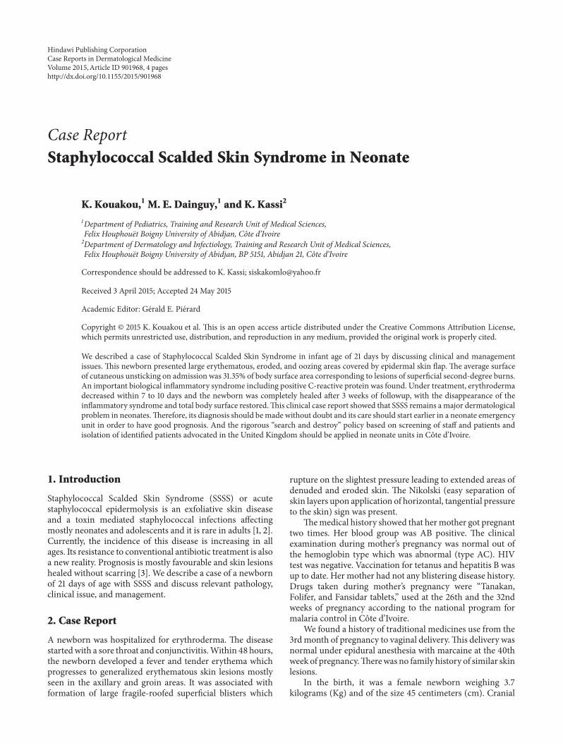
Figure 1: Staphylococcal Scalded Skin Syndrome in a newborn withgeneralized bullous epidermolysis.
perimeter was estimated to be 33 cm and the APGAR scorewas estimated to be 8 at the first minute and 9 at the 5thminute.
Its clinical examination on admission revealed no feverwith 36,2∘C of temperature, and the respiratory frequencywas 45 cycles/minute.Theheart rate was 120 beatings/minute,and integuments were colored.
The examination of the skin and the mucous membraneshighlighted the skin peeling and widespread blisters prevail-ing in the anterior and posterior parts of the lower limbrelying on an erythematous basis. The epidermal necrolysishas quickly extended to the bottom and to the trunk duringhospitalization. There appear large erythematous, eroded,and oozing areas covered by epidermal skin flap (Figure 1).Mucous membranes were intact. The genital examinationshowed unsticking lesions on the big and small lips of thevagina which bled when in contact. Other body systemexaminations were normal. The average surface of cutaneousunsticking on admission was 31.35% of the body surface areacorresponding to the lesions of superficial second-degreeburns.
We found an important biological inflammatory syn-drome including positive C-reactive protein.
Three differential diagnoses were evoked with these clini-cal manifestations: (1) toxic epidermal necrosis drug induced(Lyell syndrome), (2) Staphylococcal Scalded Skin Syndrome(SSSS), and (3) Staphylococcal Shock.
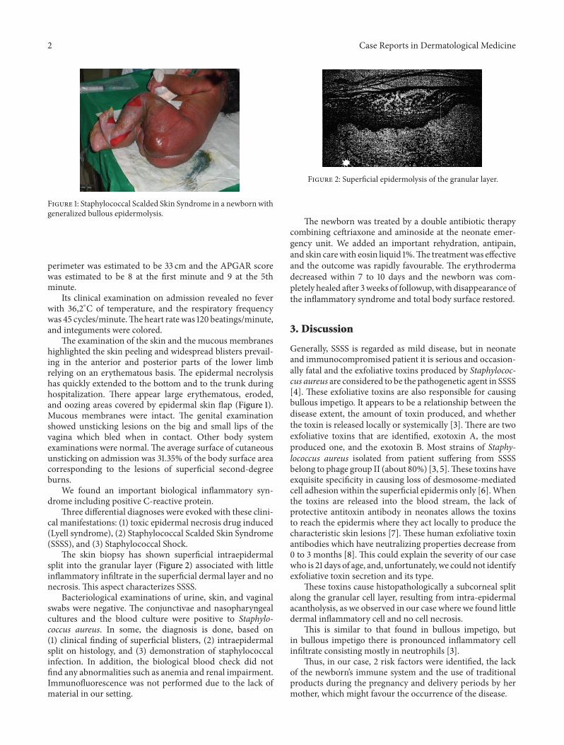
The skin biopsy has shown superficial intraepidermalsplit into the granular layer (Figure 2) associated with littleinflammatory infiltrate in the superficial dermal layer and nonecrosis. This aspect characterizes SSSS.
Bacteriological examinations of urine, skin, and vaginalswabs were negative. The conjunctivae and nasopharyngealcultures and the blood culture were positive to Staphylo-coccus aureus. In some, the diagnosis is done, based on(1) clinical finding of superficial blisters, (2) intraepidermalsplit on histology, and (3) demonstration of staphylococcalinfection. In addition, the biological blood check did notfind any abnormalities such as anemia and renal impairment.Immunofluorescence was not performed due to the lack ofmaterial in our setting.
Figure 2: Superficial epidermolysis of the granular layer.
The newborn was treated by a double antibiotic therapycombining ceftriaxone and aminoside at the neonate emer-gency unit. We added an important rehydration, antipain,and skin carewith eosin liquid 1%.The treatmentwas effectiveand the outcome was rapidly favourable. The erythrodermadecreased within 7 to 10 days and the newborn was com-pletely healed after 3weeks of followup,with disappearance ofthe inflammatory syndrome and total body surface restored.
3. Discussion
Generally, SSSS is regarded as mild disease, but in neonateand immunocompromised patient it is serious and occasion-ally fatal and the exfoliative toxins produced by Staphylococ-cus aureus are considered to be the pathogenetic agent in SSSS[4]. These exfoliative toxins are also responsible for causingbullous impetigo. It appears to be a relationship between thedisease extent, the amount of toxin produced, and whetherthe toxin is released locally or systemically [3]. There are twoexfoliative toxins that are identified, exotoxin A, the mostproduced one, and the exotoxin B. Most strains of Staphy-lococcus aureus isolated from patient suffering from SSSSbelong to phage group II (about 80%) [3, 5].These toxins haveexquisite specificity in causing loss of desmosome-mediatedcell adhesion within the superficial epidermis only [6].Whenthe toxins are released into the blood stream, the lack ofprotective antitoxin antibody in neonates allows the toxinsto reach the epidermis where they act locally to produce thecharacteristic skin lesions [7]. These human exfoliative toxinantibodies which have neutralizing properties decrease from0 to 3 months [8]. This could explain the severity of our casewho is 21 days of age, and, unfortunately, we could not identifyexfoliative toxin secretion and its type.
These toxins cause histopathologically a subcorneal splitalong the granular cell layer, resulting from intra-epidermalacantholysis, as we observed in our case where we found littledermal inflammatory cell and no cell necrosis.
This is similar to that found in bullous impetigo, butin bullous impetigo there is pronounced inflammatory cellinfiltrate consisting mostly in neutrophils [3].
Thus, in our case, 2 risk factors were identified, the lackof the newborn’s immune system and the use of traditionalproducts during the pregnancy and delivery periods by hermother, which might favour the occurrence of the disease.
Case Reports in Dermatological Medicine 3
All these pathogenesis characteristics allow us to under-stand the clinical manifestations of SSSS particularly inneonate.
SSSS has usually a swift onset of painful, tender, and redskin accentuated in flexural and periorificial areas. After 24 to48 hours, flaccid blisters and erosions develop and large areasof the overlying epidermis loosen and peel like a scald whichcan be extended [9]. In our case, the disease starts with theinflammation of the conjunctivae (conjunctivitis) which is aStaphylococcus commensal site like umbilicus and axilla.
The diagnosis is usually made on clinical ground [9];it relies mainly on the recognition of the characteristicappearance of the rash with fever. But it is important toswab the skin, the orificial areas, and the mucus membranesfor bacterial confirmation and to identify the primary focusinfection and screening for Staphylococcus aureus carriage,as we performed in our case. The skin biopsy often showsa superficial intraepidermal split into the granular layerassociated with little inflammatory infiltrate in the superficialdermal layer without necrosis as we found in our case. Thisdiagnosis is made in our case based on (1) clinical finding ofsuperficial blisters, (2) intraepidermal split on histology, and(3) demonstration of staphylococcal infection.
3.1. Treatment. Antistaphylococcal antibiotics, temperatureregulation, maintaining fluid and electrolyte balance, nutri-tional management, and skin care form the basics of treat-ment [3].
These antibiotics represent one of themain pillars of SSSStreatment, but the growing concerns of the resistant strainsof staphylococci anti-ETA and anti-ETB might be the futurechallenges.
In fact, resistance was observed for some antibiotics:5% for gentamicin, 7% for tetracycline, and 2% for chlo-ramphenicol, whereas there were no strains resistant tomethicillin, cephalothin, cephalexin, and vancomycin [4].
In practice, blisters should be left intact because it helpsto reduce further trauma to the skin. Topical antibioticsor antiseptic eye ointment is also helpful to manage theconjunctivitis. In the best case, patients should be managedin the pediatric intensive care unit and consideration needsto be given to mattress requirement, pain management, tem-perature regulation, fluid management (rehydration), nutri-tion, and skin care. Corticosteroids are contraindicated withthe worsening of the disease [3]. Appropriate intravenousantibiotics against penicillin-resistant staphylococci shouldbe used such as methicillin and flucloxacillin and the useof intravenous fluid management and analgesia in case oforal intake is reduced because of the perioral lesions [7].In our case, we used a double antibiotic therapy combiningceftriaxone and aminoside at the neonate emergency unit.We added important rehydration, antipain, and skin carewith eosin liquid of 1%. The treatment was effective and thenewborn was completely healed after 3 weeks of followup,with the disappearance of the inflammatory syndrome andtotal body surface restored.
3.2. Followup. The prognosis of SSSS in childhood is mostlyfavourable. The mortality rate is approximately 4% and it
is associated with extensive skin involvement [3]. In ourcase, under treatment, erythroderma decreased within 7 to10 days and the newborn was completely healed after 3 weeksof followup, with the disappearance of the inflammatorysyndrome and total body surface restored.
3.3. Prevention. As asymptomatic nasal carriage of staphylo-cocci aureus is an important source of infection in neonates,strict control measures should be taken such as isolationof infected patients, barrier nursing, and antiseptic handwashing by both staff and visitor to the unit. The rigorous“search and destroy” policy based on screening of staff andpatients and isolation of identified patients that is now beingincreasingly advocated in United Kingdom [9] should beapplied in neonate units in Cote d’Ivoire.
4. Conclusion
While most cases of SSSS are easily treated, it remains anemergency case and a potential fatal condition in neonate. Itsdiagnosis should be made without doubt and its care shouldstart early in neonate emergency unit in order to have goodprognosis.
Conflict of Interests
The authors declare that there is no conflict of interestsregarding the publication of this paper.
References
[1] J. C. Coleman and N. R. Dobson, “Diagnostic dilemma:extremely low birth weight baby with staphylococcal scalded-skin syndrome or toxic epidermal necrolysis,” Journal of Peri-natology, vol. 26, no. 11, pp. 714–716, 2006.
[2] S. Kadam, A. Tagare, J. Deodhar, Y. Tawade, and A. Pandit,“Staphylococcal scalded skin syndrome in a neonate,” IndianJournal of Pediatrics, vol. 76, no. 10, p. 1074, 2009.
[3] G. K. Patel and A. Y. Finlay, “Staphylococcal scalded skinsyndrome: diagnosis and management,” The American Journalof Clinical Dermatology, vol. 4, no. 3, pp. 165–175, 2003.
[4] K. Murono, K. Fujita, and H. Yoshioka, “Microbiologic charac-teristics of exfoliative toxin-producing Staphylococcus aureus,”ThePediatric Infectious Disease Journal, vol. 7, no. 5, pp. 313–315,1988.
[5] E. Rieger-Fackeldey, L. R. W. Plano, A. Kramer, and A. Schulze,“Staphylococcal scalded skin syndrome related to an exfoliativetoxin A- and B-producing strain in preterm infants,” EuropeanJournal of Pediatrics, vol. 161, no. 12, pp. 649–652, 2002.
[6] C. B. Lillibridge, M. E. Melish, and L. A. Glasgow, “Site ofaction of exfoliative toxin in the staphylococcal scalded skinsyndrome,” Pediatrics, vol. 50, pp. 728–738, 1972.
[7] S. Ladhani, “Understanding the mechanism of action of theexfoliative toxins of Staphylococcus aureus,” FEMS Immunologyand Medical Microbiology, vol. 39, no. 2, pp. 181–189, 2003.
[8] T. Hubiche, M. Bes, L. Roudiere, F. Langlaude, J. Etienne, andP. del Giudice, “Mild staphylococcal scalded skin syndrome: an
4 Case Reports in Dermatological Medicine
underdiagnosed clinical disorder,” British Journal of Dermatol-ogy, vol. 166, no. 1, pp. 213–215, 2012.
[9] G. A. Johnston, “Treatment of bullous impetigo and the staphy-lococcal scalded skin syndrome in infants,” Expert Review ofAnti-Infective Therapy, vol. 2, no. 3, pp. 439–446, 2004.