Embed Size (px)
Citation preview
Technology Assessment Program
Report No. 7
Stereotactic Radiosurgery forMetastases to the Brain:
A Systematic Review of PublishedStudies of Effectiveness
Authors: Diana Anderson, R.N., M.P.H., Research AnalystKaren Flynn, D.D.S., M.S., Manager
Contributors: Elizabeth Adams, R.R.T., M.P.H., Management & Program AnalystElaine Alligood, M.L.S., MDRC Information Center Librarian
Report Date: December 1997
MTA95-002-01

The Health Services Research and Development Service (HSR&D) is a programwithin the Veterans Health Administration's Office of Research an Development.HSR&D provides expertise in health services research, a field that examines theeffects of organization, financing and management on a wide range of problems inhealth care delivery, quality of care, access, cost and patient outcomes. Itsprograms span the continuum of health care research and delivery, from basicresearch to the dissemination of research results, and ultimately to the applicationof these findings to clinical, managerial and policy decisions.
Technology Assessment ProgramManagement Decision and Research Center (152M)Health Services Research and Development Service
Office of Research and DevelopmentVA Medical Center
150 South Huntington AvenueBoston, MA 02130
Tel: (617) 278-4469 FTS: (700) 839-4469 Fax: (617) [email protected]
Released March 1998
December 1997
MTA95-002-01 MDRC Technology Assessment Program - Radiosurgery Report
ACKNOWLEDGEMENTS
The contributions of the following reviewers are gratefully acknowledged. The MDRC takes fullresponsibility for the views expressed herein. Participation as a reviewer does not implyendorsement.
Cynthia Mulrow, M.D., M.Sc. Professor of MedicineDirector, VA Cochrane Center at San AntonioAudie L. Murphy Veterans Memorial Hospital
Joseph Ransohoff, M.D. Chief of NeurosurgeryJames A. Haley Veterans HospitalTampa, Florida
The MDRC Technology Assessment Program wishes to thank Jennifer Cheslog, Stephanie Piper,Kevin Rys, and the staff of the Information Dissemination Program for their help with the report.

December 1997
MTA95-002-01 MDRC Technology Assessment Program - Radiosurgery Report - Page i
Stereotactic Radiosurgery for theTreatment of Metastases to the Brain
EXECUTIVE SUMMARY
PurposeThis report was written by the Management Decision and Research Center (MDRC) TechnologyAssessment (TA) Program in response to requests for information about the effectiveness ofstereotactic radiosurgery for the treatment of metastases to the brain.
BackgroundMetastases to the brain (herein referred to as “brain metastases”) are a major cause of disabilityand death in cancer patients. Treatment options are limited, particularly for patients withsurgically inaccessible or recurrent brain metastases.
Stereotactic radiosurgery (SRS) is a specialized form of radiation therapy that delivers preciselyfocused beams of radiation to a targeted lesion in the brain. The intent is to destroy the lesion, orcontrol its growth, without harming nearby healthy tissue. This technology has been used to treata variety of functional and benign brain abnormalities, and indications for its use are now beingexpanded to include metastases to the brain.
Key Findings
Cost and ReimbursementSRS uses fewer resources than open cranial surgery. Relative to surgery, radiosurgery reducesthe length of hospital stay by nearly 7 days, to approximately 3 to 4 days. SRS can often be doneas an outpatient procedure. Medicare has reimbursed SRS at the same rate as open cranialsurgery, but a 30% reduction in reimbursement for SRS was proposed for 1998. This was basedon the HCFA estimate of reduced procedure costs.
RegulationCommercially available SRS units are approved for therapeutic use by the Food and DrugAdministration (FDA). Facilities must comply with federal standards and guidelines forradiation safety and quality control.
Evidence of effectivenessThe best published evidence is from case series, a relatively weak study design that does notprovide strong evidence of effectiveness. The studies treated different types of patients andreported outcomes in non-comparable ways. Studies frequently combined outcomes fromdifferent patient or treatment groups, making effectiveness data difficult to interpret. While the
December 1997
MTA95-002-01 MDRC Technology Assessment Program - Radiosurgery Report - Page ii
findings about the effectiveness of SRS were fairly encouraging and consistent for comparablegroups of patients, they should be considered preliminary and interpreted with caution:
• Evidence suggested that SRS was a relatively safe and effective technology for the definitivetreatment of newly diagnosed and recurrent metastases to the brain in selected patients. Mostof the reported side effects of SRS were mild, temporary, and could be relieved bymedication. Treatment resulted in very few major complications and very rare deaths.
• All of the types of cancers treated responded to therapy. The spread of melanomas, breast
cancers, and kidney cancers was controlled for a longer period of time than for other cancers.For breast cancers, this improved tumor control was accompanied by an increased length ofsurvival.
• Median survival after SRS ranged from 26 to 56 weeks. This compared favorably with
outcomes from other treatments. • Patients with limited numbers of relatively small tumors, and who had well-controlled
systemic cancer, may have gained the greatest benefits from treatment. SRS treatment wasequally effective for patients with two metastases as for patients with solitary metastases;SRS treatment was as effective for recurrent metastases as for initially untreated metastases.The effectiveness of SRS treatment and survival benefit in patients with three or moremetastases remain undetermined. The absence of active systemic cancer was stronglyassociated with survival, as was good baseline functional status.
• • Valid comparisons of the relative effectiveness of treatment options are not possible using
existing research. Available data suggested that for patients with smaller solitary metastases,SRS (alone or + radiotherapy) appeared to be as effective as surgery + radiotherapy inprolonging survival. SRS may have been more effective than surgery in postponingrecurrences, and SRS caused fewer complications. Outcomes from both surgery and SRSwere considerably better than those from radiotherapy alone. Optimal management ofpatients with multiple metastases is yet to be determined.
• It is too early to draw definite conclusions about optimal treatment parameters (including
radiation dose, the use of whole brain radiotherapy in addition to SRS, and maximumtreatable tumor size). The Radiation Therapy Oncology Group (RTOG) will continue tostudy the optimal dose planning and maximal possible treatable tumor volume in ongoing orplanned RTOG radiosurgery trials.
ConclusionsIn the absence of data from high quality studies, uncertainty remains about the true effectivenessof SRS for the treatment of metastases to the brain. One randomized clinical trial is in progress(See Table 7, page 15), and further trials are needed, to address the many unanswered questionsabout the use of SRS for this application. Such trials will provide stronger evidence on which tobase clinical and policy decisions.

December 1997
MTA95-002-01 MDRC Technology Assessment Program - Radiosurgery Report - Page iii
TABLE OF CONTENTS
I. INTRODUCTION AND BACKGROUND ..................................................................... 1A. Purpose.................................................................................................................. 1B. Metastases to the Brain ......................................................................................... 1C. Standard Treatments ............................................................................................. 1
II. DESCRIPTION OF STEREOTACTIC RADIOSURGERY ........................................... 2 III. REGULATION AND REIMBURSEMENT.................................................................... 3 IV. METHODS FOR THE SYSTEMATIC REVIEW........................................................... 3 V. APPRAISAL OF THE LITERATURE ............................................................................ 5
A. Application of the Inclusion Criteria .................................................................... 5B. Data Synthesis....................................................................................................... 5
VI. PUBLISHED FINDINGS................................................................................................. 6A. Description of Patients, Treatments, and Outcomes ............................................. 6B. Reported Outcomes of Stereotactic Radiosurgery ................................................ 7C. Factors Associated with Improved Treatment Outcomes ..................................... 9
VII. DISCUSSION................................................................................................................. 11A. Effectiveness of Stereotactic Radiosurgery for Brain Metastases ...................... 11B. Effectiveness of Stereotactic Radiosurgery vs. Traditional Treatments ............. 13
VIII. CONCLUSIONS ............................................................................................................ 14 IX. ONGOING TRIALS AND CLINICAL TRIAL RESOURCES..................................... 15 X. REFERENCES ............................................................................................................ R-1
LIST OF TABLES AND FIGURESFigure 1 Inclusion Criteria for the Systematic Review .............................................................................4Figure 2 Study Designs to Assess Effectiveness.......................................................................................4Table 1 Eligible Studies, Effectiveness of SRS for Brain Metastases .....................................................5Figure 3 Definitions of Outcome Measures for Cancer Treatment...........................................................7Table 2 Summary of the Evidence: Effectiveness of Stereotactic Radiosurgery (SRS) for the
Treatment of Brain Metastases ...................................................................................................8Table 3 Summary of Outcomes Data, based on Case Series Reports in Table 2.....................................9Table 4 Potential Predictive Factors Associated with Local Control of Tumor Growth .......................10Table 5 Potential Predictive Factors Associated with Improved Survival.............................................10Table 6 Radiotherapy, Radiosurgery, and Surgery for Solitary Brain Metastases ................................13Table 7 Clinical Trial of Stereotactic Radiosurgery in Adults for Brain Metastases.............................15
December 1997
MTA95-002-01 MDRC Technology Assessment Program - Radiosurgery Report - Page 1
I. INTRODUCTION AND BACKGROUND
A. PurposeThis report was written by the Management Decision and Research Center (MDRC)Technology Assessment (TA) Program in response to requests for information about theeffectiveness of stereotactic radiosurgery for the treatment of metastases to the brain.
B. Metastases to the BrainMetastases to the brain (herein referred to as “brain metastases”) are a significant andincreasing health problem in the aging population (Sperduto et al. 1996; Loeffler et al.1993). About 100,000 cancer patients developed brain metastases in 1997 (Parker et al.1997). Any primary cancer can spread to the brain, but the most common aremelanomas, and cancers of the lung, breast, colorectum, or kidney (Kihlstrom et al.1993; Posner 1993). Nearly half of the patients diagnosed with brain metastases have onelesion, and 20% have two metastases (DeAngelis 1994).
As many as half of patients dying from cancer are found to have brain metastases onautopsy (Flickinger et al. 1994). Despite improvements in the treatment of systemiccancers, brain metastases remain a major cause of disability and death in cancer patients,including the estimated 170,000 American veterans who have cancer (Kizer 1997).
C. Standard TreatmentsUntreated, patients with brain metastases usually survive for 1 to 2 months (Posner 1974).Most die from the uncontrolled systemic spread of their primary cancer, but one-quarterto one-half die from complications associated with the spread of their brain metastases(Borgelt et al. 1980).
Steroids were found to reduce symptoms and prolong survival by about 1 month(Weissman 1988). With few exceptions, chemotherapy has a limited role in the treatmentof brain metastases (NCI September 1997).
Whole brain radiotherapy (WBRT) reduces symptoms and prolongs life more effectivelythan steroids alone. Median survival in randomized clinical trials of WBRT ranged from15 to 18 weeks (Borgelt et al. 1980). WBRT retreatment for recurrent brain metastases iscontroversial, but additional survival benefits have been reported in case series studies(Kurup et al. 1980; Hazuka et al. 1988). For most patients, re-irradiation seldomproduces clinically significant benefits, and it is known to cause damage to healthy braintissue (DeAngelis 1994; Arbit et al. 1995).
For selected patients with a solitary brain metastasis, adding open cranial surgery towhole brain radiotherapy greatly prolongs life. In two randomized clinical trials, mediansurvival for patients treated with surgery + WBRT was 40-43 weeks, compared to 15-26

December 1997
MTA95-002-01 MDRC Technology Assessment Program - Radiosurgery Report - Page 2
weeks for WBRT alone. Quality of life was significantly improved in the surgical groups(Patchell et al. 1990; Noordijk et al. 1994). No randomized clinical trials have been doneto assess the effectiveness of surgery for the removal of 2 or more metastases, or for thesurgical retreatment of recurrent metastases. Surgery for these patients is controversial,and infrequently done. Case series data from surgical studies for selected patients withmultiple or recurrent lesions are promising (Lang et al. 1996).
Only about 30% of patients with brain metastases are eligible for surgery (DeAngelis,1994). Definitive treatment options are limited for patients who are not surgicalcandidates, and for those with recurrent brain metastases. If these patients have well-controlled systemic cancer, death from neurological causes is likely.
Treatment of brain tumors remains an active area of research. Radiosurgery has beenused to treat a variety of functional and benign brain abnormalities, and its effectivenessfor the treatment of brain metastases is now being investigated (Flickinger et al. 1994).
II. DESCRIPTION OF STEREOTACTIC RADIOSURGERY
Stereotactic radiosurgery (SRS) is a specialized form of radiation therapy that delivers precise,small, and focused beams of radiation to a targeted lesion in the brain. The intent is to destroythe lesion, or control its growth, without harming nearby healthy tissue. Treatment is minimallyinvasive. It requires no surgical incision, causes little discomfort, and can be performed on anoutpatient basis, or with a very brief inpatient admission.
Originally developed in 1951, SRS has since incorporated advanced medical imaging andcomputerized localization systems to refine the technology (Phillips et al. 1994; De Salles et al.1993).
Three different systems can be used to perform SRS: Gamma Knife; heavy charged particlesfrom a cyclotron or synchrotron; or a modified linear accelerator. There are over 200stereotactic radiosurgery facilities in the United States (Shrieve et al. 1995), including 27 GammaKnife centers (Elekta 1997), and 2 cyclotrons or synchrotrons (Pakuris 1996; Sisterson 1997).Most of the remaining centers perform radiosurgery using specialized add-on equipment withlinear accelerators (LINACS). LINACS are used to deliver conventional radiotherapy treatmentto cancer patients, and are available at many major health care facilities. A few centers haveLINACS that are only used for SRS.
The Tampa VA Medical Center recently purchased a LINAC-based system called Peacock. Itcan be used to perform SRS on brain tumors or prostate tumors. It can also be used to performspecialized forms of radiotherapy for brain, lung, or prostate cancers (DVA 1997). This systemhas the unique ability to fractionate treatments.
December 1997
MTA95-002-01 MDRC Technology Assessment Program - Radiosurgery Report - Page 3
III. REGULATION AND REIMBURSEMENT
Commercially available SRS units are approved for therapeutic use by the Food and DrugAdministration (Jones et al. 1995; FDA 1997). Radiosurgery facilities must comply withNuclear Regulatory Commission standards and guidelines for radiation safety and qualitycontrol. Cyclotron and synchrotron facilities must comply with Department of Energy qualityassurance protocols.
No formal training guidelines exist for neurosurgeons or radiotherapists performing SRS (Joneset al. 1995). However, professional organizations have promoted strict guidelines and qualityassurance measures (Larson et al. 1994).
SRS is reimbursed by Medicare. Although reimbursement is presently set at the same rate asopen cranial surgery, the Health Care Financing Administration (HCFA) proposed a 30%reduction in SRS reimbursement for 1998. This is based on a recent HCFA analysis whichconcluded that, when compared to open cranial surgery, SRS used fewer resources, and reducedthe length of stay from 7-10 days (for open cranial surgery) to 1-3 days (for SRS). SRS can oftenbe done on an outpatient basis. HCFA recommended no national coverage policy forradiosurgery and indicated that payment decisions are at the discretion of local carriers (DHHS1997).
The Veterans Health Administration reimbursement policy has been to pay for all medicallynecessary procedures that have FDA approval or come under an approved clinical protocol.Patients requiring radiosurgery can be referred to the Tampa VAMC, the only facility presentlyhousing SRS equipment, or can be referred to academic affiliates or other health care facilities.
IV. METHODS FOR THE SYSTEMATIC REVIEW
Information about the effectiveness of SRS for the treatment of metastases to the brain wasobtained by conducting a systematic review of the published literature. A systematic review usesa scientific approach to limit bias and to improve the accuracy of conclusions based on theavailable data (Guyatt 1995).
A search of the English-language medical literature was performed using the following computerdatabases for the years 1991 through July, 1997: MEDLINE, PREMEDLINE, HEALTHPlanning & Administration, HealthSTAR1, and EMBASE2, and Current Contents3 (All Sections,1994 to July, 1997). An updated search of PREMEDLINE was conducted in November, 1997.Terms for the search included: radiosurgery or stereotactic radiosurgery or Gamma Knife.These were combined with brain neoplasm, controlled clinical trials, meta-analysis, multi-center
1 Computer databases maintained by the National Library of Medicine.2 Elsevier Science Publishers.3 Institute for Scientific Information.

December 1997
MTA95-002-01 MDRC Technology Assessment Program - Radiosurgery Report - Page 4
studies or practice guidelines. End references from retrieved articles and listings of Englishlanguage, public domain technology assessments were also searched.
Titles and abstracts of 748 references were screened. 90 references were determined to berelevant, and their full text articles were reviewed for potential inclusion in the systematicreview. Additional articles were retrieved to provide background materials about stereotacticradiosurgery and brain tumor therapy. References were reviewed and were included in thereview if they met the criteria in Figure 1.
Figure 1: Inclusion Criteria for the Systematic Review
• Studies evaluating the effectiveness of SRS for brain metastases• English language journal articles reporting primary data obtained in a clinical setting, or analyses of primary data maintained in registries or institutional databases• Study design and methods clearly described• Case series including ≥ 10 patients, or studies with a more powerful design• Study not superseded by a later publication, with the same purpose, by the same group• Published 1990 or later, to reflect the current status of diagnostic and treatment technologies
Studies were selected for inclusion if they provided the strongest available evidence ofeffectiveness. The strength of the evidence is based on how well bias and confounding factorsare controlled in the design and conduct of a study. Attributes that strengthen the validity offindings include: randomized (vs. nonrandomized), controlled (vs. uncontrolled), blinded (vs.unblinded), prospective (vs. retrospective), large (vs. small), multi-site (vs. single site), andcontemporaneous controls (vs. historical). Based on these attributes, common study designs arelisted in order, from the most to the least rigorous, for internal validity and strength of evidence(Figure 2).
Figure 2: Study Designs to Assess EffectivenessRanked according to decreasing strength of evidence provided
• Large randomized controlled trial, systematic reviews of RCTs• Small randomized controlled trial• Nonrandomized trial with contemporaneous controls• Nonrandomized trial with historical controls• Surveillance (database or register)• Case series, multi-site• Case series, single site• Case report, anecdote
Sources: Adapted from Ibrahim 1985, and Goodman 1993.
December 1997
MTA95-002-01 MDRC Technology Assessment Program - Radiosurgery Report - Page 5
V. APPRAISAL OF THE LITERATURE
No direct comparisons of the effectiveness of different SRS systems were identified. Publishedreviews based on comparisons of case series data for the different systems suggest that theirtreatment outcomes are similar. Lacking strong data to the contrary, the MDRC combinedstudies using different SRS systems in this report.
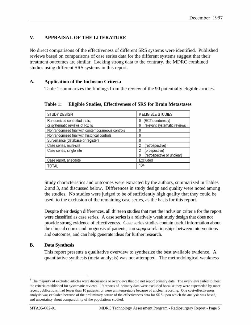
A. Application of the Inclusion CriteriaTable 1 summarizes the findings from the review of the 90 potentially eligible articles.
Table 1: Eligible Studies, Effectiveness of SRS for Brain Metastases
STUDY DESIGN # ELIGIBLE STUDIESRandomized controlled trials,or systematic reviews of RCTs
0 (RCTs underway)0 relevant systematic reviews
Nonrandomized trial with contemporaneous controls 0Nonrandomized trial with historical controls 0Surveillance (database or register) 0Case series, multi-site 2 (retrospective)Case series, single site 2 (prospective)
9 (retrospective or unclear)Case report, anecdote ExcludedTOTAL 134
Study characteristics and outcomes were extracted by the authors, summarized in Tables2 and 3, and discussed below. Differences in study design and quality were noted amongthe studies. No studies were judged to be of sufficiently high quality that they could beused, to the exclusion of the remaining case series, as the basis for this report.
Despite their design differences, all thirteen studies that met the inclusion criteria for the reportwere classified as case series. A case series is a relatively weak study design that does notprovide strong evidence of effectiveness. Case series studies contain useful information aboutthe clinical course and prognosis of patients, can suggest relationships between interventionsand outcomes, and can help generate ideas for further research.
B. Data SynthesisThis report presents a qualitative overview to synthesize the best available evidence. Aquantitative synthesis (meta-analysis) was not attempted. The methodological weakness
4 The majority of excluded articles were discussions or overviews that did not report primary data. The overviews failed to meetthe criteria established for systematic reviews. 19 reports of primary data were excluded because they were superseded by morerecent publications, had fewer than 10 patients, or were uninterpretable because of unclear reporting. One cost-effectivenessanalysis was excluded because of the preliminary nature of the effectiveness data for SRS upon which the analysis was based,and uncertainty about comparability of the populations studied.
December 1997
MTA95-002-01 MDRC Technology Assessment Program - Radiosurgery Report - Page 6
of case series, combined with present differences in design and analysis among theeligible studies, argued against the validity and usefulness of pooling study results(Eysenck 1994).
Two eligible studies (Flickinger et al. 1994, and Auchter et al. 1996) did synthesize someof the case series data. Using explicit inclusion criteria, both studies identifiedradiosurgery patients from multiple institutions, and conducted multivariate analyses toidentify patient and treatment factors that appeared to be associated with improvedoutcomes.
VI. PUBLISHED FINDINGS
The synthesis of evidence from eligible studies includes a summary of patient and treatmentcharacteristics and an overview of treatment outcomes. Eight studies performed statisticalanalyses to identify patient and treatment characteristics that appeared to be associated withimproved outcomes. Their findings are included in this report.
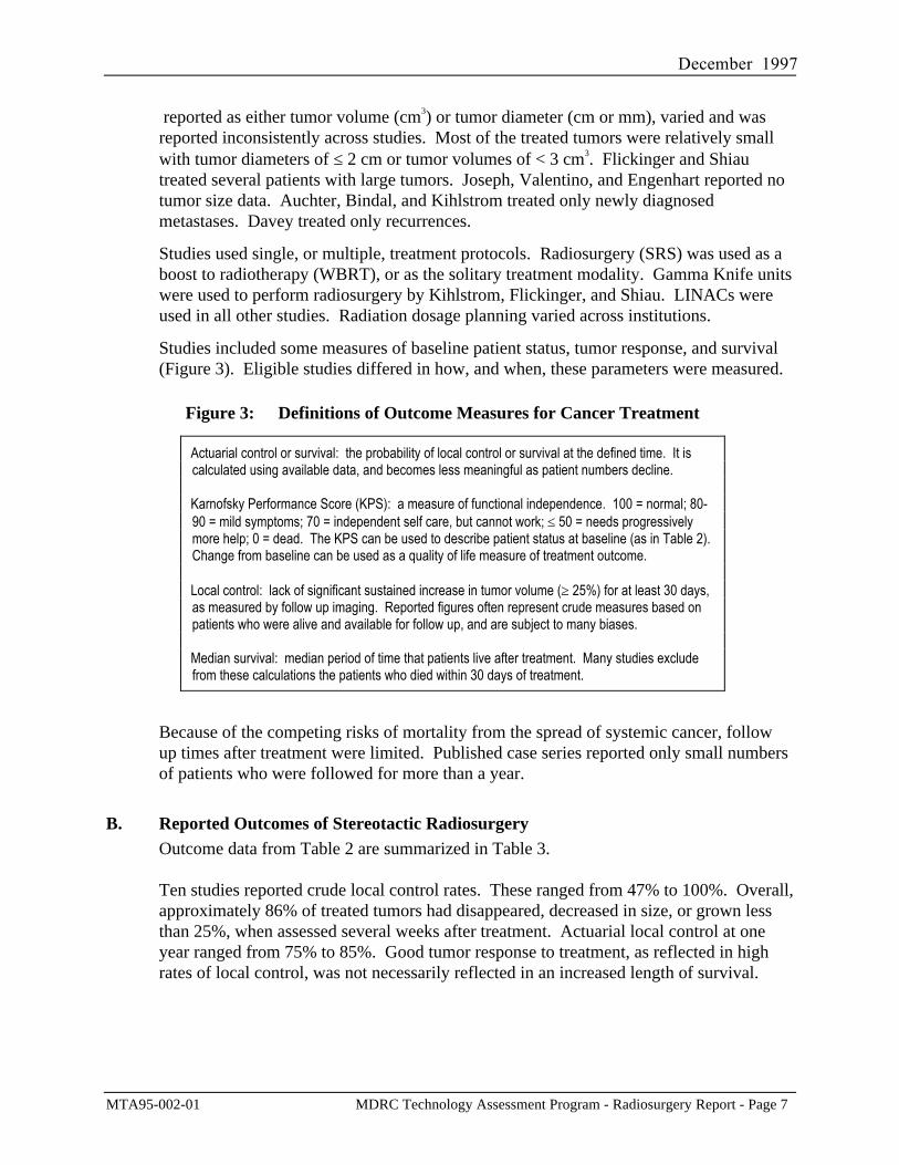
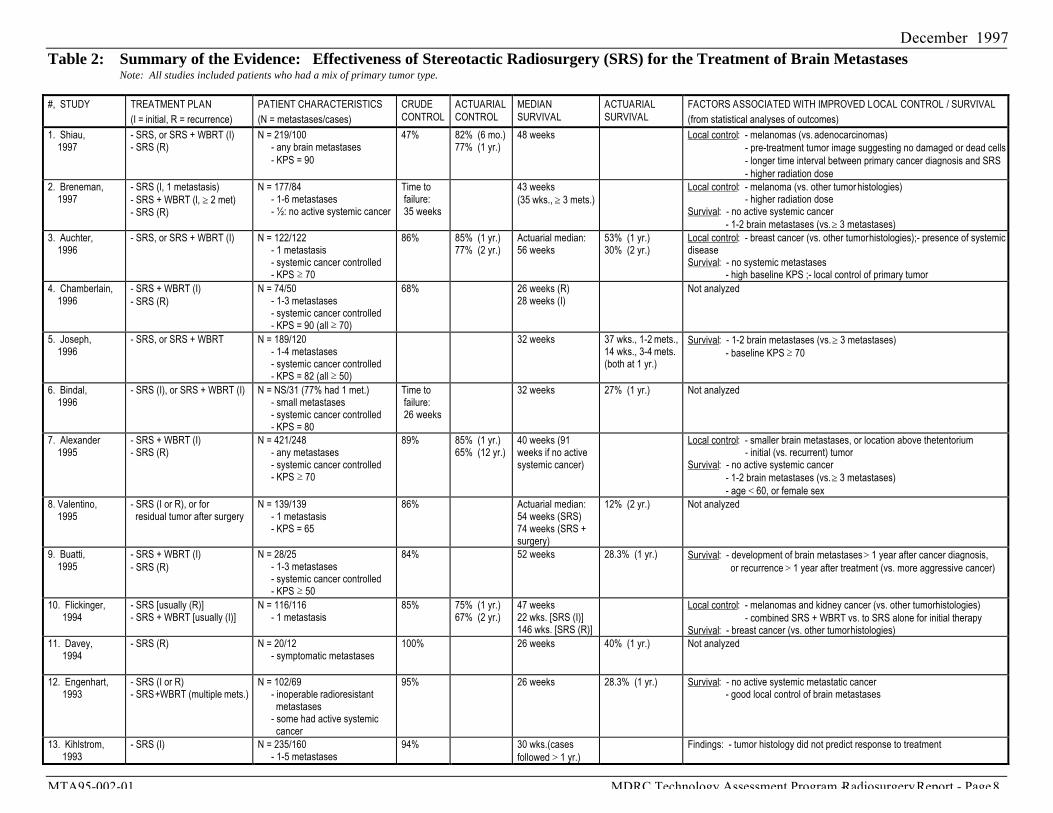
A. Description of Patients, Treatments, and OutcomesThe 13 case series that met the inclusion criteria for this report are summarized in Table25. Additional details relevant to the studies follow. Note that patient characteristics,incompletely described in some studies, may have had a large impact in determiningpatient response to treatment.
Patients ranged in age from 14 to 87, with a median age in the mid-50s (no data reportedby Kihlstrom). Men and women were approximately equally represented in all but theEngenhart study (72% male).
The baseline health status of patients was sometimes difficult to assess because ofincomplete reporting. Data suggested that most studies were fairly selective, andincluded highly functional patients. Kihlstrom, Engenhart, Valentino, and Brenemanappeared to have been less selective, and to have included some patients with moreadvanced disease.
The characteristics of metastases varied among studies. All of the case series included amix of primary tumor types; the histology of treated tumors generally included thosemost frequently found in brain metastases. Auchter and Engenhart treated onlyradioresistant tumors. Auchter, Valentino, and Flickinger included only patients withsolitary metastases. Chamberlain and Buatti included only patients with 1-3 metastases.All other studies included patients with solitary or multiple metastases. Tumor size,
5 Table 2 includes only radiosurgery patient data from the Bindal study (1996). The study also reported outcomes from a
selected series of surgical patients.
December 1997
MTA95-002-01 MDRC Technology Assessment Program - Radiosurgery Report - Page 7
reported as either tumor volume (cm3) or tumor diameter (cm or mm), varied and wasreported inconsistently across studies. Most of the treated tumors were relatively smallwith tumor diameters of ≤ 2 cm or tumor volumes of < 3 cm3. Flickinger and Shiautreated several patients with large tumors. Joseph, Valentino, and Engenhart reported notumor size data. Auchter, Bindal, and Kihlstrom treated only newly diagnosedmetastases. Davey treated only recurrences.
Studies used single, or multiple, treatment protocols. Radiosurgery (SRS) was used as aboost to radiotherapy (WBRT), or as the solitary treatment modality. Gamma Knife unitswere used to perform radiosurgery by Kihlstrom, Flickinger, and Shiau. LINACs wereused in all other studies. Radiation dosage planning varied across institutions.
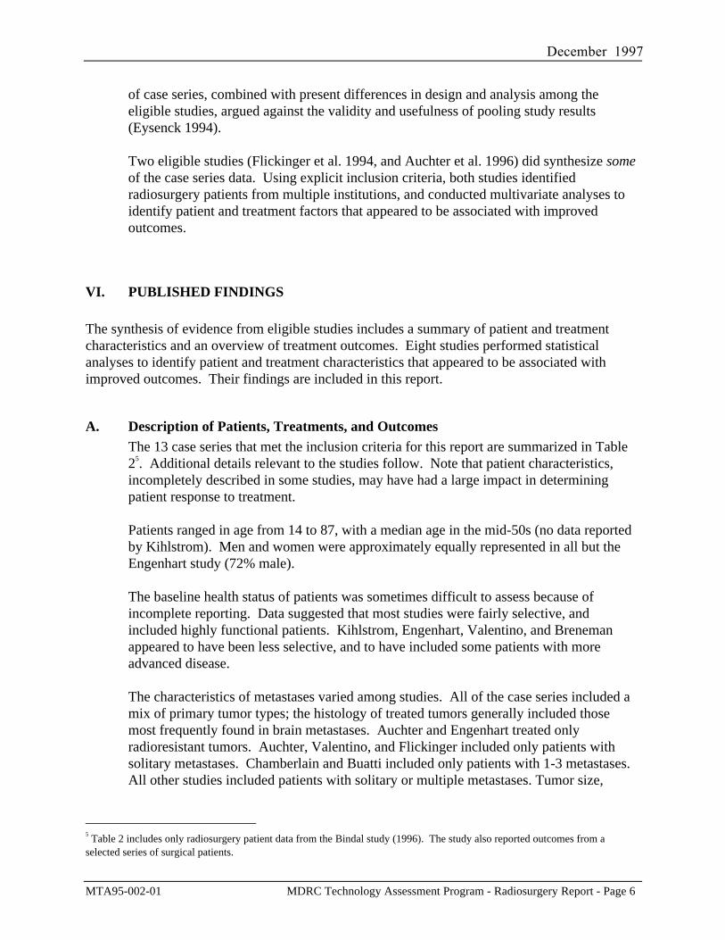
Studies included some measures of baseline patient status, tumor response, and survival(Figure 3). Eligible studies differed in how, and when, these parameters were measured.
Figure 3: Definitions of Outcome Measures for Cancer Treatment
Actuarial control or survival: the probability of local control or survival at the defined time. It iscalculated using available data, and becomes less meaningful as patient numbers decline.
Karnofsky Performance Score (KPS): a measure of functional independence. 100 = normal; 80-90 = mild symptoms; 70 = independent self care, but cannot work; ≤ 50 = needs progressivelymore help; 0 = dead. The KPS can be used to describe patient status at baseline (as in Table 2).Change from baseline can be used as a quality of life measure of treatment outcome.
Local control: lack of significant sustained increase in tumor volume (≥ 25%) for at least 30 days,as measured by follow up imaging. Reported figures often represent crude measures based onpatients who were alive and available for follow up, and are subject to many biases.
Median survival: median period of time that patients live after treatment. Many studies excludefrom these calculations the patients who died within 30 days of treatment.
Because of the competing risks of mortality from the spread of systemic cancer, followup times after treatment were limited. Published case series reported only small numbersof patients who were followed for more than a year.
B. Reported Outcomes of Stereotactic RadiosurgeryOutcome data from Table 2 are summarized in Table 3.
Ten studies reported crude local control rates. These ranged from 47% to 100%. Overall,approximately 86% of treated tumors had disappeared, decreased in size, or grown lessthan 25%, when assessed several weeks after treatment. Actuarial local control at oneyear ranged from 75% to 85%. Good tumor response to treatment, as reflected in highrates of local control, was not necessarily reflected in an increased length of survival.
December 1997
MTA95-002-01 MDRC Technology Assessment Program - Radiosurgery Report - Page 8
Table 2: Summary of the Evidence: Effectiveness of Stereotactic Radiosurgery (SRS) for the Treatment of Brain MetastasesNote: All studies included patients who had a mix of primary tumor type.
#, STUDY TREATMENT PLAN(I = initial, R = recurrence)
PATIENT CHARACTERISTICS(N = metastases/cases)
CRUDECONTROL
ACTUARIALCONTROL
MEDIANSURVIVAL
ACTUARIALSURVIVAL
FACTORS ASSOCIATED WITH IMPROVED LOCAL CONTROL / SURVIVAL(from statistical analyses of outcomes)
1. Shiau, 1997
- SRS, or SRS + WBRT (I)- SRS (R)
N = 219/100- any brain metastases
- KPS = 90
47% 82% (6 mo.)77% (1 yr.)
48 weeks Local control: - melanomas (vs. adenocarcinomas) - pre-treatment tumor image suggesting no damaged or dead cells - longer time interval between primary cancer diagnosis and SRS - higher radiation dose
2. Breneman, 1997
- SRS (I, 1 metastasis)- SRS + WBRT (I, ≥ 2 met)- SRS (R)
N = 177/84- 1-6 metastases- ½: no active systemic cancer
Time to failure: 35 weeks
43 weeks(35 wks., ≥ 3 mets.)
Local control: - melanoma (vs. other tumor histologies) - higher radiation dose
Survival: - no active systemic cancer - 1-2 brain metastases (vs. ≥ 3 metastases)
3. Auchter, 1996
- SRS, or SRS + WBRT (I) N = 122/122- 1 metastasis- systemic cancer controlled- KPS 70
86% 85% (1 yr.)77% (2 yr.)
Actuarial median:56 weeks
53% (1 yr.)30% (2 yr.)
Local control: - breast cancer (vs. other tumor histologies);- presence of systemicdiseaseSurvival: - no systemic metastases
- high baseline KPS ;- local control of primary tumor4. Chamberlain, 1996
- SRS + WBRT (I) - SRS (R)
N = 74/50- 1-3 metastases- systemic cancer controlled- KPS = 90 (all 70)
68% 26 weeks (R)28 weeks (I)
Not analyzed
5. Joseph, 1996
- SRS, or SRS + WBRT N = 189/120- 1-4 metastases- systemic cancer controlled- KPS = 82 (all 50)
32 weeks 37 wks., 1-2 mets.,14 wks., 3-4 mets.(both at 1 yr.)
Survival: - 1-2 brain metastases (vs. ≥ 3 metastases) - baseline KPS 70
6. Bindal, 1996
- SRS (I), or SRS + WBRT (I) N = NS/31 (77% had 1 met.)- small metastases- systemic cancer controlled- KPS = 80
Time to failure: 26 weeks
32 weeks 27% (1 yr.) Not analyzed
7. Alexander 1995
- SRS + WBRT (I)- SRS (R)
N = 421/248- any metastases- systemic cancer controlled- KPS 70
89% 85% (1 yr.)65% (12 yr.)
40 weeks (91weeks if no activesystemic cancer)
Local control: - smaller brain metastases, or location above the tentorium - initial (vs. recurrent) tumor
Survival: - no active systemic cancer - 1-2 brain metastases (vs. ≥ 3 metastases) - age < 60, or female sex
8. Valentino, 1995
- SRS (I or R), or for residual tumor after surgery
N = 139/139- 1 metastasis- KPS = 65
86% Actuarial median:54 weeks (SRS)74 weeks (SRS +surgery)
12% (2 yr.) Not analyzed
9. Buatti, 1995
- SRS + WBRT (I) - SRS (R)
N = 28/25- 1-3 metastases- systemic cancer controlled- KPS 50
84% 52 weeks 28.3% (1 yr.) Survival: - development of brain metastases > 1 year after cancer diagnosis, or recurrence > 1 year after treatment (vs. more aggressive cancer)
10. Flickinger, 1994
- SRS [usually (R)]- SRS + WBRT [usually (I)]
N = 116/116- 1 metastasis
85% 75% (1 yr.)67% (2 yr.)
47 weeks22 wks. [SRS (I)]146 wks. [SRS (R)]
Local control: - melanomas and kidney cancer (vs. other tumor histologies) - combined SRS + WBRT vs. to SRS alone for initial therapy
Survival: - breast cancer (vs. other tumor histologies)11. Davey, 1994
- SRS (R) N = 20/12- symptomatic metastases
100% 26 weeks 40% (1 yr.) Not analyzed
12. Engenhart, 1993
- SRS (I or R)- SRS + WBRT (multiple mets.)
N = 102/69- inoperable radioresistant metastases- some had active systemic cancer
95% 26 weeks 28.3% (1 yr.) Survival: - no active systemic metastatic cancer - good local control of brain metastases
13. Kihlstrom, 1993
- SRS (I) N = 235/160- 1-5 metastases
94% 30 wks.(casesfollowed > 1 yr.)
Findings: - tumor histology did not predict response to treatment

December 1997
MTA95-002-01 MDRC Technology Assessment Program - Radiosurgery Report - Page 9
The median length of survival after treatment with SRS (with or without WBRT) rangedfrom 26 weeks to 56 weeks. For studies that only treated patients with solitarymetastases, median survival ranged from 47 to 56 weeks. Actuarial survival at one yearranged from 28.3% (initial and recurrent tumor treatment, 1-3 metastases) to 53% (initialtumor treatment, 1 metastasis).
Treatment-related morbidity and mortality were uncommon. Most of the adverse effectsreported in these studies were related to radiation-induced injury, and many werereversible with medication. These included increased intracranial pressures, seizures,transient headaches, nausea and vomiting, increased confusion, hemiparesis, anddysphagia. Alopecia, tumor hemorrhage, and discomfort with the head frame were alsoreported. Radiation necrosis, a serious form of delayed brain tissue damage, had a medianrate of 3% (range: 0%-17%). The median rate of deaths occurring within 30 days oftreatment was 1.3% (range: 0%-8%).
Table 3: Summary of Outcomes Data, based on Case Series Reports in Table 2Studies 3 and 10 (highlighted columns) analyzed data collected in several case series that were conducted atdifferent sites. All other studies were single-site case series.
Solitary Metastases Solitary or Multiple Metastases Treated in the Study
Multi-site
Study # from Table 2 8 3 10 1 2 4 5 6 7 9 11 12 13
Median Survival (weeks)2 54 56 47 48 43 26-28 32 32 40 52 26 26 30
% 1-year Actuarial Survival 53 37; 141 27 28.3 40 28.3
% Local Control or Weeksto Failure of Control
86 86 85 47 35weeks
68 26weeks
89 84 100 95 94
% 1-year Actuarial Control 85 75 77 85
1
The two figures represent findings for patients with 1-2 metastases, and 3-4 metastases, respectively.2 Expected survival of patients with untreated brain metastases is 4 to 8 weeks. Expected survival with the use of steroids increases to about 12weeks. Median survival of WBRT ranged from 15-18 weeks. Median survival of WBRT+ surgery ranged from 40-43 weeks (See Page 4.)
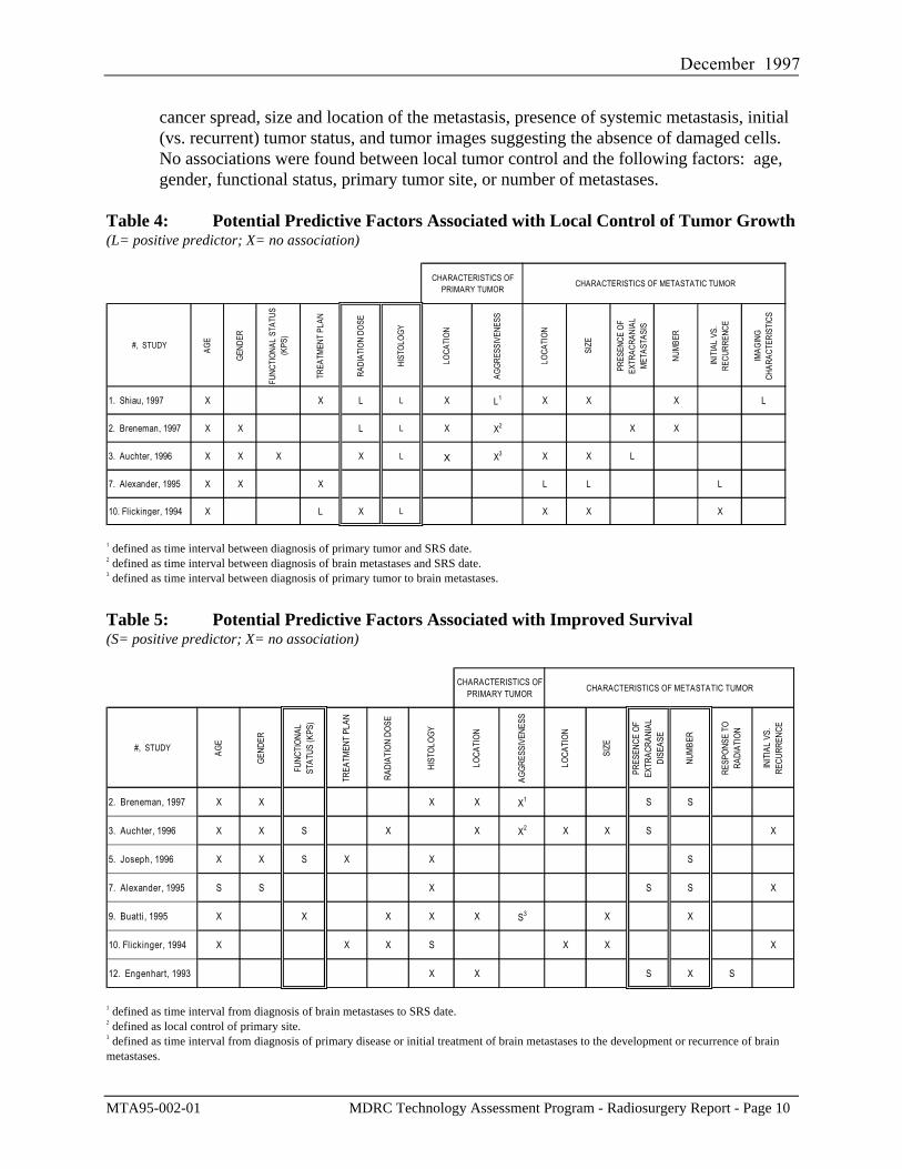
C. Factors Associated with Improved Treatment OutcomesEight retrospective studies, including the two multi-site studies, used statistical analysesto interpret their findings and to determine associations with improved treatmentoutcomes. Results from these analyses are presented in Tables 4 and 5.
Local control of tumor growth was sustained longer for metastases of melanomas (3studies), breast cancer (1 study), and kidney cancer (1 study included with melanomas)compared to other tumor histologies. The use of higher radiation doses (a factorinversely associated with tumor size) was associated with longer local control (2 studies).
An association between local control and one of several other factors was reported by fiveof the studies. However, the following reported associations were not corroborated by otherstudies in the group: WBRT + SRS vs. SRS alone, supratentorial location, indolent
December 1997
MTA95-002-01 MDRC Technology Assessment Program - Radiosurgery Report - Page 10
cancer spread, size and location of the metastasis, presence of systemic metastasis, initial(vs. recurrent) tumor status, and tumor images suggesting the absence of damaged cells.No associations were found between local tumor control and the following factors: age,gender, functional status, primary tumor site, or number of metastases.
Table 4: Potential Predictive Factors Associated with Local Control of Tumor Growth(L= positive predictor; X= no association)
CHARACTERISTICS OF PRIMARY TUMOR
CHARACTERISTICS OF METASTATIC TUMOR
#, STUDY AGE
GEN
DER
FUNC
TIO
NAL
STAT
US
(KPS
)
TREA
TMEN
T PL
AN
RADI
ATIO
N DO
SE
HIST
OLO
GY
LOCA
TIO
N
AGG
RESS
IVEN
ESS
LOCA
TIO
N
SIZE
PRES
ENCE
OF
EXTR
ACRA
NIAL
M
ETAS
TASI
S
NUM
BER
INIT
IAL
VS.
RECU
RREN
CE
IMAG
ING
CH
ARAC
TERI
STIC
S
1. Shiau, 1997 X X L L X L1 X X X L
2. Breneman, 1997 X X L L X X2 X X
3. Auchter, 1996 X X X X L X X3 X X L
7. Alexander, 1995 X X X L L L
10. Flickinger, 1994 X L X L X X X
1 defined as time interval between diagnosis of primary tumor and SRS date.2 defined as time interval between diagnosis of brain metastases and SRS date.3 defined as time interval between diagnosis of primary tumor to brain metastases.
Table 5: Potential Predictive Factors Associated with Improved Survival(S= positive predictor; X= no association)
CHARACTERISTICS OF PRIMARY TUMOR
CHARACTERISTICS OF METASTATIC TUMOR
#, STUDY AGE
GEN
DER
FUNC
TIO
NAL
STAT
US (K
PS)
TREA
TMEN
T PL
AN
RADI
ATIO
N DO
SE
HIST
OLO
GY
LOCA
TIO
N
AGG
RESS
IVEN
ESS
LOCA
TIO
N
SIZE
PRES
ENCE
OF
EXTR
ACRA
NIAL
DI
SEAS
E
NUM
BER
RESP
ONS
E TO
RA
DIAT
ION
INIT
IAL
VS.
RECU
RREN
CE
2. Breneman, 1997 X X X X X1 S S
3. Auchter, 1996 X X S X X X2 X X S X
5. Joseph, 1996 X X S X X S
7. Alexander, 1995 S S X S S X
9. Buatti, 1995 X X X X X S3 X X
10. Flickinger, 1994 X X X S X X X
12. Engenhart, 1993 X X S X S
1 defined as time interval from diagnosis of brain metastases to SRS date.2 defined as local control of primary site.3 defined as time interval from diagnosis of primary disease or initial treatment of brain metastases to the development or recurrence of brainmetastases.
December 1997
MTA95-002-01 MDRC Technology Assessment Program - Radiosurgery Report - Page 11
The strongest predictors of improved survival were the absence of active systemic cancer(4 studies), patient functional status before treatment (2 studies), and the presence of alimited number of metastases (1 or 2 vs. 3 metastases) (3 studies). An associationbetween increased length of survival and one of several other factors was reported by oneof the seven studies. However, the reported associations were not corroborated by otherstudies in the group. Factors included: younger age, female, breast cancer vs. otherhistologies, indolent cancer spread, and good local control of metastases. No associationswere found between increased length of survival and the following factors: the use ofWBRT (in addition to SRS), treatment dose, tumor size, location, or initial (vs. recurrent)tumor status.
Most of the factors associated with improved local control were not associated withincreased survival. Breast cancer histology and lack of aggressiveness of tumor spreadwere the only factors associated with both improved local control and prolonged survival.
VII. DISCUSSION
A. Effectiveness of Stereotactic Radiosurgery for Brain MetastasesAs more effective methods of controlling systemic cancers are becoming available,definitive treatments for brain metastases are being sought. Whole brain radiotherapy(WBRT) offers only modest benefits to patients. The addition of cranial surgery canincrease the duration of survival two to three-fold (relative to radiotherapy) in selectedpatients. However, surgery + WBRT does not completely eradicate cancers, and about20% of treated tumors recur at the same location (Patchell et al. 1990). Not all patientsare good surgical candidates, not all tumors are surgically accessible, and the use ofrepeat surgery for recurrences remains controversial. Treatments that are applicable to abroad range of patients, and that provide better outcomes, are still needed.
The lack of randomized clinical trials and the heterogeneity in the case series studiesthat provide the only available data preclude definitive assessment of theeffectiveness of stereotactic radiosurgery and prevent generalization of the availablefindings to the larger oncology population.
However, the overall trends reflected in case series data were encouraging. Findingssuggested that selected patient groups derived considerable benefit from treatment withstereotactic radiosurgery. Local control of tumor growth was achieved for most patients,at least for a period of time. This can be expected to reduce the symptoms of intracranialdisease, improve the quality of life of the patient, and possibly forestall death fromneurological causes. Most side-effects were mild, temporary, and treatable. The medianlength of survival after SRS treatment ranged from 26 weeks to 56 weeks. This comparesquite favorably with outcomes from other treatments.
December 1997
MTA95-002-01 MDRC Technology Assessment Program - Radiosurgery Report - Page 12
SRS was sometimes used for patients who were surgical candidates. A more commonuse of SRS has been for patients who were not surgical candidates, and for whomradiotherapy or palliative care were the available options.
A review of the findings from the multivariate analyses conducted by eight studiesincluded in this report provides some guidance about significant prognostic factors.
• Findings did not support limiting the use of SRS to patients with solitary metastases.Available data reported no difference in outcomes for patients treated for a solitarymetastasis or for two metastases.
Relatively few patients with 3 or more metastases were treated in the case series includedin this report. Treatment outcomes were conflicting and provided little guidance forclinical decision-making. Findings suggested that this group derived significantly lessbenefit from radiosurgery than patients with 1-2 metastases, and it is uncertain if theyderived any clinically important benefits.
• Patients treated for recurrences of brain metastases appeared to derive as large asurvival benefit from SRS as did those for whom radiosurgery was used as the initialtreatment for metastases. This finding is particularly promising, since patients withrecurrences are not often candidates for definitive therapies.
• Breast cancer histology (vs. other tumor types) was associated with both improved local
control and prolonged survival after treatment with stereotactic radiosurgery.
• The absence of active systemic cancer, along with good baseline functional status andyounger age, were associated with improved survival for patients treated with SRS.These findings are mirrored in an analysis of radiotherapy outcomes (Gaspar et al. 1997),as well as in analyses of surgical outcomes (Patchell et al. 1990; Noordijk et al. 1994.)Note that all surgical patients had a good baseline functional status.
The presence of cancer that spread rapidly was associated with poor survival in a
radiosurgery case series and also in a surgical trial (Patchell et al. 1990).
These findings strongly suggest that severely debilitated patients, or those with activelyprogressing systemic cancers, are unlikely to derive significant survival benefit fromdefinitive treatment of their brain metastases.
• Radiation dose calculations were often based on multiple factors that were difficult toseparate in analyses of case series data. The Radiation Therapy Oncology Group(RTOG) is evaluating the optimal dose planning and maximal possible treatable tumorvolume in ongoing or planned RTOG radiosurgery trials (Shaw et al. 1996).
December 1997
MTA95-002-01 MDRC Technology Assessment Program - Radiosurgery Report - Page 13
B. Effectiveness of Stereotactic Radiosurgery vs. Traditional TreatmentsThe weakness of the data also precludes any definitive assessment of the effectiveness ofstereotactic radiosurgery relative to other treatment options.
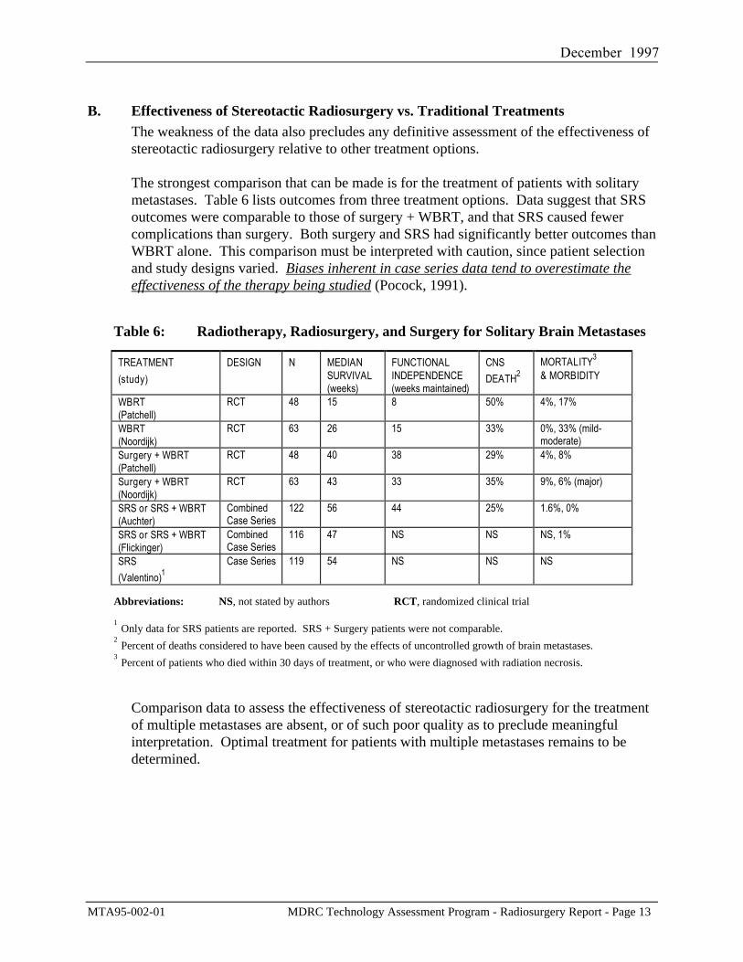
The strongest comparison that can be made is for the treatment of patients with solitarymetastases. Table 6 lists outcomes from three treatment options. Data suggest that SRSoutcomes were comparable to those of surgery + WBRT, and that SRS caused fewercomplications than surgery. Both surgery and SRS had significantly better outcomes thanWBRT alone. This comparison must be interpreted with caution, since patient selectionand study designs varied. Biases inherent in case series data tend to overestimate theeffectiveness of the therapy being studied (Pocock, 1991).
Table 6: Radiotherapy, Radiosurgery, and Surgery for Solitary Brain Metastases
TREATMENT(study)
DESIGN N MEDIANSURVIVAL(weeks)
FUNCTIONALINDEPENDENCE(weeks maintained)
CNSDEATH2
MORTALITY3
& MORBIDITY
WBRT(Patchell)
RCT 48 15 8 50% 4%, 17%
WBRT(Noordijk)
RCT 63 26 15 33% 0%, 33% (mild-moderate)
Surgery + WBRT(Patchell)
RCT 48 40 38 29% 4%, 8%
Surgery + WBRT(Noordijk)
RCT 63 43 33 35% 9%, 6% (major)
SRS or SRS + WBRT(Auchter)
CombinedCase Series
122 56 44 25% 1.6%, 0%
SRS or SRS + WBRT(Flickinger)
CombinedCase Series
116 47 NS NS NS, 1%
SRS(Valentino)1
Case Series 119 54 NS NS NS
Abbreviations: NS, not stated by authors RCT, randomized clinical trial
1 Only data for SRS patients are reported. SRS + Surgery patients were not comparable.2 Percent of deaths considered to have been caused by the effects of uncontrolled growth of brain metastases.3 Percent of patients who died within 30 days of treatment, or who were diagnosed with radiation necrosis.
Comparison data to assess the effectiveness of stereotactic radiosurgery for the treatmentof multiple metastases are absent, or of such poor quality as to preclude meaningfulinterpretation. Optimal treatment for patients with multiple metastases remains to bedetermined.
December 1997
MTA95-002-01 MDRC Technology Assessment Program - Radiosurgery Report - Page 14
VIII. CONCLUSIONS
Lack of data from high quality studies precludes any definitive assessment of the relativeeffectiveness of SRS to standard treatment for brain metastases. It also preludes any definitiveassessment of optimal equipment selection, treatment parameters, or patient selection criteria.
The available data from case series reports suggest that SRS is a relatively safe and effectivetechnology for definitive treatment of brain metastases in selected patients. It appears to offerconsiderably greater survival benefits than traditional whole brain radiotherapy. SRS may becomparable to surgery plus radiation therapy for the treatment of patients with smaller solitarymetastases. SRS can be used to treat patients whose metastases recur after traditional therapies, agroup for whom definitive treatment options are frequently unavailable. As with other definitivetherapies for patients with brain metastases, highly functional patients with well-controlledsystemic cancers derive the greatest benefit from treatment.
Uncertainty remains about the true effectiveness of this technology and the appropriateindications for its use in patients with metastatic brain tumors. A randomized clinical trial is inprogress (see next page), and further trials are needed, to address the many of the unansweredquestions about SRS and about the treatment of brain metastases. Such trials will providestronger evidence on which to base health care treatment and policy decisions.
December 1997
MTA95-002-01 MDRC Technology Assessment Program - Radiosurgery Report - Page 15
IX. ONGOING TRIALS AND CLINICAL TRIAL RESOURCES
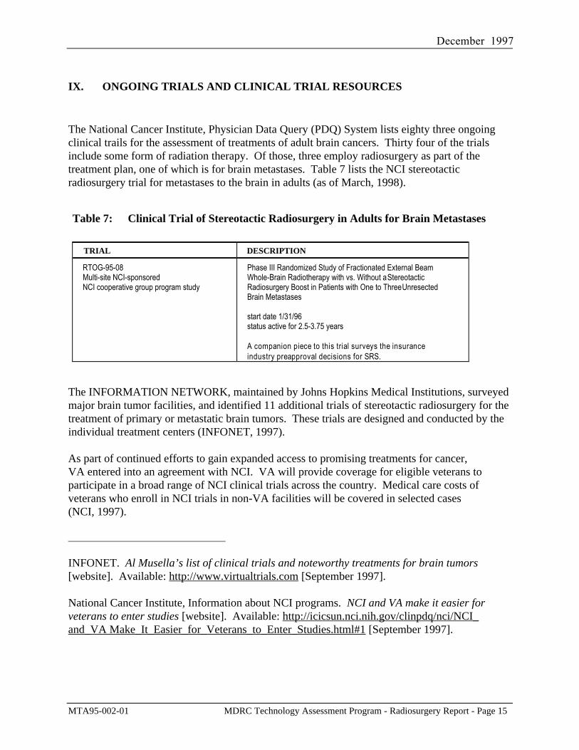
The National Cancer Institute, Physician Data Query (PDQ) System lists eighty three ongoingclinical trails for the assessment of treatments of adult brain cancers. Thirty four of the trialsinclude some form of radiation therapy. Of those, three employ radiosurgery as part of thetreatment plan, one of which is for brain metastases. Table 7 lists the NCI stereotacticradiosurgery trial for metastases to the brain in adults (as of March, 1998).
Table 7: Clinical Trial of Stereotactic Radiosurgery in Adults for Brain Metastases
TRIAL DESCRIPTION
RTOG-95-08Multi-site NCI-sponsoredNCI cooperative group program study
Phase III Randomized Study of Fractionated External BeamWhole-Brain Radiotherapy with vs. Without a StereotacticRadiosurgery Boost in Patients with One to Three UnresectedBrain Metastases
start date 1/31/96status active for 2.5-3.75 years
A companion piece to this trial surveys the insuranceindustry preapproval decisions for SRS.
The INFORMATION NETWORK, maintained by Johns Hopkins Medical Institutions, surveyedmajor brain tumor facilities, and identified 11 additional trials of stereotactic radiosurgery for thetreatment of primary or metastatic brain tumors. These trials are designed and conducted by theindividual treatment centers (INFONET, 1997).
As part of continued efforts to gain expanded access to promising treatments for cancer,VA entered into an agreement with NCI. VA will provide coverage for eligible veterans toparticipate in a broad range of NCI clinical trials across the country. Medical care costs ofveterans who enroll in NCI trials in non-VA facilities will be covered in selected cases(NCI, 1997).
_________________________________
INFONET. Al Musella’s list of clinical trials and noteworthy treatments for brain tumors[website]. Available: http://www.virtualtrials.com [September 1997].
National Cancer Institute, Information about NCI programs. NCI and VA make it easier forveterans to enter studies [website]. Available: http://icicsun.nci.nih.gov/clinpdq/nci/NCI_and_VA Make_It_Easier_for_Veterans_to_Enter_Studies.html#1 [September 1997].

REFERENCES

December 1997
MTA95-002-01 MDRC Technology Assessment Program - Radiosurgery Report - Page R - 1
X. REFERENCES
STUDIES INCLUDED IN THIS REVIEW
Alexander E, III., Moriarty TM, Davis RB, Wen PY, Fine HA, Black PM, et al. Stereotacticradiosurgery for the definitive, noninvasive treatment of brain metastases. Journal of theNational Cancer Institute 1995; 87(1): 34-40.
Auchter RM, Lamond JP, Alexander E, Buatti JM, Chappell R, Friedman WA, et al. A multi-institutional outcome and prognostic factor analysis of radiosurgery for resectable single brainmetastasis. International Journal of Radiation Oncology, Biology, Physics 1996; 35(1): 27-35.
Bindal AK, Bindal RK, Hess KR, Shiu A, Hassenbusch SJ, Shi WM, et al. Surgery versusradiosurgery in the treatment of brain metastasis. Journal of Neurosurgery 1996; 84(5): 748-754.
Breneman JC, Warnick RE, Albright RE, Jr., Kukiatinant N, Shaw J, Armin D, et al.Stereotactic radiosurgery for the treatment of brain metastases. Results of a single institutionseries. Cancer 1997; 79(3): 551-557.
Buatti JM, Friedman WA, Bova FJ, Mendenhall WM. Treatment selection factors forstereotactic radiosurgery of intracranial metastases. International Journal of RadiationOncology, Biology, Physics 1995; 32(4): 1161-1166.
Chamberlain MC, Kormanik P, Barba D, Fuller BG, Smith DE, Shea WMC. Stereotacticradiosurgery for metastatic brain tumors. International Journal of Oncology 1996; 8(3): 617-624.
Davey P, O'Brien PF, Schwartz ML, Cooper PW. A phase I/II study of salvage radiosurgery inthe treatment of recurrent brain metastases. British Journal of Neurosurgery 1994; 8(6): 717-723.
Engenhart R, Kimmig BN, Hover KH, Wowra B, Romahn J, Lorenz WJ, et al. Long-termfollow-up for brain metastases treated by percutaneous stereotactic single high-dose irradiation.Cancer 1993; 71(4): 1353-1361.
Flickinger JC, Kondziolka D, Lunsford LD, Coffey RJ, Goodman ML, Shaw EG, et al. A multi-institutional experience with stereotactic radiosurgery for solitary brain metastasis. InternationalJournal of Radiation Oncology, Biology, Physics 1994; 28(4): 797-802.
Joseph J, Adler JR, Cox RS, Hancock SL. Linear accelerator-based stereotaxic radiosurgery forbrain metastases: the influence of number of lesions on survival. Journal of Clinical Oncology1996; 14(4): 1085-1092.

December 1997
MTA95-002-01 MDRC Technology Assessment Program - Radiosurgery Report - Page R - 2
Kihlstrom L, Karlsson B, Lindquist C. Gamma Knife surgery for cerebral metastases.Implications for survival based on 16 years experience. Stereotactic & Functional Neurosurgery1993; 61(Suppl 1): 45-50.
Shiau CY, Sneed PK, Shu HK, Lamborn KR, McDermott MW, Chang S, et al. Radiosurgery forbrain metastases: relationship of dose and pattern of enhancement to local control. InternationalJournal of Radiation Oncology, Biology, Physics 1997; 37(2): 375-383.
Valentino V. The results of radiosurgical management of 139 single cerebral metastases. ActaNeurochirurgica Supplementum 1995; 63: 95-100.

December 1997
MTA95-002-01 MDRC Technology Assessment Program - Radiosurgery Report - Page R - 3
STUDIES EXCLUDED FROM THIS REVIEW
Adler JR, Cox RS, Kaplan I, Martin DP. Stereotactic radiosurgical treatment of brainmetastases. Journal of Neurosurgery 1992; 76(3): 444-449.
Brada M, Laing R. Radiosurgery/stereotactic external beam radiotherapy for malignant braintumours: the Royal Marsden Hospital experience. Recent Results in Cancer Research 1994;135: 91-104.
Caron JL, Souhami L, Podgorsak EB. Dynamic stereotactic radiosurgery in the palliativetreatment of cerebral metastatic tumors. Journal of Neuro-oncology 1992; 12(2): 173-179.
Coffey RJ, Flickinger JC, Bissonette DJ, Lunsford LD. Radiosurgery for solitary brainmetastases using the cobalt-60 gamma unit: methods and results in 24 patients. InternationalJournal of Radiation Oncology, Biology, Physics 1991; 20(6): 1287-1295.
Coffey RJ, Flickinger JC, Lunsford LD, Bissonette DJ. Solitary brain metastasis: radiosurgeryin lieu of microsurgery in 32 patients. Acta Neurochirurgica Supplementum 1991; 52: 90-92.
Coffey RJ, Lunsford LD, Flickinger JC. The role of radiosurgery in the treatment of malignantbrain tumors. Neurosurgery Clinics of North America 1992; 3(1): 231-244.
De Salles AA, Hariz M, Bajada CL, Goetsch S, Bergenheim T, Selch M, et al. Comparisonbetween radiosurgery and stereotactic fractionated radiation for the treatment of brain metastases.Acta Neurochirurgica Supplementum 1993; 58(8): 115-118.
Flickinger JC, Loeffler JS, Larson DA. Stereotactic radiosurgery for intracranial malignancies.Oncology 1994; 8(1): 81-86; discussion 86, 94, 97-88.
Fukuoka S, Seo Y, Takanashi M, Takahashi S, Suematsu K, Nakamura J. Radiosurgery of brainmetastases with the Gamma Knife. Stereotactic & Functional Neurosurgery 1996; 66(1): 193-200.
Gerosa M, Nicolato A, Severi F, Ferraresi P, Masotto B, Barone G, et al. Gamma Kniferadiosurgery for intracranial metastases: from local tumor control to increased survival.Stereotactic & Functional Neurosurgery 1996; 66(1): 184-192.
Gerosa MA, Nicolato A, Berlucchi S, Piovan E, Zampieri PG, Pasoli A, et al. Gamma kniferadiosurgery of primary and metastatic malignant brain tumors, a preliminary report.Stereotactic & Functional Neurosurgery 1995; 64(Suppl 1): 56-66.
Jokura H, Takahashi K, Kayama T, Yoshimoto T. Gamma knife radiosurgery of a series of onlyminimally selected metastatic brain tumours. Acta Neurochirurgica Supplmentum 1994; 62: 77-82.

December 1997
MTA95-002-01 MDRC Technology Assessment Program - Radiosurgery Report - Page R - 4
Kihlstrom L, Karlsson B, Lindquist C, Noren G, Rahn T. Gamma knife surgery for cerebralmetastasis. Acta Neurochirurgica Supplementum 1991; 52(9): 87-89.
Loeffler JS, Alexander Ed. The role of stereotactic radiosurgery in the management ofintracranial tumors. Oncology 1990; 4(3): 21-41.
Loeffler JS, Kooy HM, Wen PY, Fine HA, Cheng CW, Mannarino EG, et al. The treatment ofrecurrent brain metastases with stereotactic radiosurgery. Journal of Clinical Oncology 1990;8(4): 576-582.
Martens F, Verbeke L. Stereotactic radiosurgery of cerebral metastases: preliminary results.Acta Clinica Belgica 1993; 48(4): 228-233.
Rutigliano MJ, Lunsford LD, Kondziolka D, Strauss MJ, Khanna V, Green M, et al. The costeffectiveness of stereotactic radiosurgery versus surgical resection in the treatment of solitarymetastatic brain tumors. Neurosurgery 1995; 37(3): 445-455.
Shirato H, Takamura A, Tomita M, Suzuki K, Nishioka T, Isu T, et al. Stereotactic irradiationwithout whole-brain irradiation for single brain metastasis. International Journal Of RadiationOncology Biology Physics 1997; 37(2): 385-391.
Simpson JR, Rich KM, Drzymala RE, Wasserman TH, Klein EE, Michaletz-Lorenz M, et al.Stereotactic external beam irradiation using a linear accelerator: the Washington Universityexperience. Missouri Medicine 1995; 92(4): 188-192.
Sturm V, Kimmig B, Engenhardt R, Schlegel W, Pastyr O, Treuer H, et al. Radiosurgicaltreatment of cerebral metastases. Method, indications and results. Stereotactic & FunctionalNeurosurgery 1991; 57(1-2): 7-10.
Valentino V, Mirri MA, Schinaia G, Dalle Ore G. Linear accelerator and Greitz-Bergstrom'shead fixation system in radiosurgery of single cerebral metastases. A report of 86 cases. ActaNeurochirurgica 1993; 121(3-4): 140-145.
Voges J, Treuer H, Erdmann J, Schlegel W, Pastyr O, Muller RP, et al. Linac radiosurgery inbrain metastases. Acta Neurochirurgica Supplementum 1994; 62(6): 72-76.

December 1997
MTA95-002-01 MDRC Technology Assessment Program - Radiosurgery Report - Page R - 5
ADDITIONAL REFERENCES CITED IN THE TEXT (Background and Methodology)
(1997). State-of-the-art information on adult brain tumor prognosis and treatment. [Onlinedatabase]. Available: CancerNet, [July 1997].
(1997). Description and Treatment Trial Protocols for brain metastases. [Online database].Available: CancerNet, [September 1997].
Alexander E, Moriarty TM, Loeffler JS. Radiosurgery for metastases. Journal Of NeuroOncology 1996; 27(3): 279-285.
Arbit E, Wronski M, Burt M, Galicich JH. The treatment of patients with recurrent brainmetastases - a retrospective analysis of 109 patients with nonsmall cell lung cancer. Cancer1995; 76(5): 765-773.
Borgelt B, Gelber R, Kramer S, Brady LW, Chang CH, Davis LW, et al. The palliation of brainmetastases: final results of the first two studies by the Radiation Therapy Oncology Group.International Journal of Radiation Oncology, Biology Physics 1980; 6(1): 1-9.
Cook DJ, Guyatt GH, Laupacis A, Sackett DL. Rules of evidence and clinical recommendationson the use of antithrombotic agents. Chest 1992; 102(4 Suppl): 305s-311s.
De Salles A, Goetsch S, Shaker L. Radiosurgery. Integration of radiation therapy, surgery &radiology. Administrative Radiology 1993; 12(1): 33-34.
DeAngelis LM. Management of brain metastases. Cancer Investigation 1994; 12(2): 156-165.
Department of Health & Human Services. Health Care Financing Administration. Medicareprogram; changes in the hospital inpatient prospective payment systems and fiscal year 1998rates. Federal Register 1997; 62(168): Part IV:45966.
Department of Veterans Affairs. Medical centers pioneer new equipment. New radiotherapystruts its stuff. VAnguard 1997; XLIII(3): 8.
Elekta Home Page. (1996). Gamma Knife worldwide locations as of January 1996. [Website].Available: http://www.elekta.com/usasites.html, [October 2, 1997].
Eysenck HJ. Meta-analysis and its problems. BMJ 1994; 309(6957): 789-792.
Flickinger JC, Loeffler JS, Larson DA. Stereotactic radiosurgery for intracranial malignancies.Oncology 1994; 8(1): 81-86; discussion 86, 94, 97-88.
Food and Drug Administration. Center for Devices and Radiological Health. (1997).Information on releasable 510(k)s. [Website]. Available:http://www.fda.gov/cdrh/510khome.html, [September 3, 1997].

December 1997
MTA95-002-01 MDRC Technology Assessment Program - Radiosurgery Report - Page R - 6
Gaspar L, Scott C, Rotman M, Asbell S, Phillips T, Wasserman T, et al. Recursive partitioninganalysis (RPA) of prognostic factors in three Radiation Therapy Oncology Group (RTOG) brainmetastases trials. International Journal of Radiation Oncology, Biology, Physics 1997; 37(4):745-751.
Goodman C. Literature searching and evidence interpretation for assessing health carepractices. Stockholm: SBU. Swedish Council on Technology Assessment in Health Care; 1993.
Guyatt GH, Sackett DL, Sinclair JC, Hayward R, Cook DJ, Cook RJ. Users' guides to themedical literature. IX. A method for grading health care recommendations. Evidence-BasedMedicine Working Group. JAMA 1995; 274(22): 1800-1804.
Hazuka MB, Kinzie JJ. Brain metastases: results and effects of re-irradiation. InternationalJournal of Radiation Oncology, Biology, Physics 1988; 15(2): 433-437.
Ibrahim MA. Epidemiology and health policy. Rockville, Md: Aspen Systems Corporation;1985.
Jones ED, Banks WW, Fischer LE. Quality assurance for gamma knives. NUCREG/CR-6324.1995, Lawrence Livermore National Laboratory for the United States Nuclear RegulatoryCommission: Washington, DC.
Kihlstrom L, Karlsson B, Lindquist C. Gamma Knife surgery for cerebral metastases.Implications for survival based on 16 years experience. Stereotactic & Functional Neurosurgery1993; 61(Suppl 1): 45-50.
Kizer KW. National cancer strategy. VHA Directive 97-050. 1997, Department of VeteransAffairs. Veterans Health Administration: Washington, DC.
Kurup P, Reddy S, Hendrickson FR. Results of re-irradiation for cerebral metastases. Cancer1980; 46(12): 2587-2589.
Lang FF, Sawaya R. Surgical management of cerebral metastases. Neurosurgical Clinics ofNorth America 1996; 7(3): 459-484.
Larson DA, Bova F, Eisert D, Kline R, Loeffler J, Lutz W, et al. Current radiosurgery practice:results of an ASTRO survey. Task Force on Stereotactic Radiosurgery, American Society forTherapeutic Radiology and Oncology. International Journal of Radiation Oncology, Biology,Physics 1994; 28(2): 523-526.
Laws ER, Jr., Thapar K. Brain tumors. CA: Cancer Journal for Clinicians 1993; 43(5): 263-271.
Loeffler JS, Alexander E. Radiosurgery for the treatment of intracranial metastases. In:Alexander E, Loeffler JS, Lunsford LD, Eds. Stereotactic Radiosurgery. New York: McGraw-Hill; 1997.

December 1997
MTA95-002-01 MDRC Technology Assessment Program - Radiosurgery Report - Page R - 7
Luxton G, Petrovich Z, Jozsef G, Nedzi LA, Apuzzo ML. Stereotactic radiosurgery: principlesand comparison of treatment methods. Neurosurgery 1993; 32(2): 241-259; discussion 259.
Noordijk EM, Vecht CJ, Haaxma-Reiche H, Padberg GW, Voormolen JH, Hoekstra FH, et al.The choice of treatment of single brain metastasis should be based on extracranial tumor activityand age. International Journal of Radiation Oncology, Biology, Physics 1994; 29(4): 711-717.
Pakuris E. (1996). Clinical trials updates: Proton Radiation Oncology Group (PROG).Particles NewsLetter, Number 18. [Website]. Available:http://neurosurgery.mgh.harvard.edu/hcl/ptles.htm, [July 1996].
Parker SL, Tong T, Bolden S, Wingo PA. Cancer statistics, 1997. CA: Cancer Journal forClinicians 1997; 47(1): 5-27.
Patchell RA, Tibbs PA, Walsh JW, Dempsey RJ, Maruyama Y, Kryscio RJ, et al. A randomizedtrial of surgery in the treatment of single metastases to the brain. New England Journal ofMedicine 1990; 322(8): 494-500.
Phillips MH, Stelzer KJ, Griffin TW, Mayberg MR, Winn HR. Stereotactic radiosurgery: areview and comparison of methods. Journal of Clinical Oncology 1994; 12(5): 1085-1099.
Posner JB. Diagnosis and treatment of metastases to the brain. Clinical Bulletin 1974; 4(2): 47-57.
Posner JB. Brain tumors. CA: Cancer Journal for Clinicians 1993; 43(5): 261-262.
Shaw E, Scott C, Souhami L, Dinapoli R, Bahary JP, Kline R, et al. Radiosurgery for thetreatment of previously irradiated recurrent primary brain tumors and brain metastases - initialreport of radiation therapy oncology group protocol 90-05. International Journal Of RadiationOncology Biology Physics 1996; 34(3): 647-654.
Shrieve DC, Loeffler JS. Advances in radiation therapy for brain tumors. Neurologic Clinics1995; 13(4): 773-793.
Sisterson J. (1997). World wide charged particle patient totals, January 1997. ParticlesNewsLetter, Number 20. [Website]. Available:http://neurosurgery.mgh.harvard.edu/hcl/ptles.htm, [July 1997].
Sperduto PW, Hall WA. Radiosurgery, cost-effectiveness, gold standards, the scientific method,cavalier cowboys, and the cost of hope. International Journal of Radiation Oncology, Biology,Physics 1996; 36(2): 511-513.
Weissman DE. Glucocorticoid treatment for brain metastases and epidural spinal cordcompression: a review. Journal of Clinical Oncology 1988; 6(3): 543-551.