Embed Size (px)
Citation preview
Sticky fingers: zinc-fingers asprotein-recognition motifsRoland Gamsjaeger*, Chu Kong Liew*, Fionna E. Loughlin, Merlin Crossley andJoel P. Mackay
School of Molecular and Microbial Biosciences, University of Sydney, NSW 2006, Australia
Review TRENDS in Biochemical Sciences Vol.32 No.2
Zinc-fingers (ZnFs) are extremely abundant in highereukaryotes. Once considered to function exclusively assequence-specific DNA-binding motifs, ZnFs are nowknown to have additional activities such as therecognition of RNA and other proteins. Here we discussrecent advances in our understanding of ZnFs as specificmodules for protein recognition. Structural studies ofZnF complexes reveal considerable diversity in terms ofprotein partners, binding modes and affinities, andhighlight the often underestimated versatility of ZnFstructure and function. An appreciation of the structuralfeatures of ZnF–protein interactions will contribute toour ability to engineer and to use ZnFs with tailoredprotein-binding properties.
IntroductionIn the 20 years since zinc-binding repeats were firstidentified in a protein (TFIIIA [1]), the DNA-bindingproperties of zinc-fingers (ZnFs) have been explored inexquisite detail [2], and it is now even possible to generateor indeed to purchase ‘designer’ ZnF proteins that willtarget specific genomic sites. These TFIIIA-type or ‘classi-cal’ ZnFs have proved to be common in complex organisms:in humans, >15 000 such domains are predicted to exist in�1000 different proteins [3]. More than 20 additionalclasses of structurally distinct modules, also termed ZnFs,have been identified [4], but it should be noted thatmany ofthese are related to classical ZnFs only by the existence ofthe structural zinc atom or atoms. Nevertheless, roles innucleic acid recognition are almost invariably postulatedwhen these domains are first described. In some cases,these predictions have been borne out: both GATA- andGal4-type ZnFs recognize DNA in a sequence-specificmanner [5,6], and several ZnF domains can bind to RNA(reviewed in Refs [7–10]).
It has also emerged, however, that ZnFs can mediateprotein–protein interactions [11]. In the past 3–5 years,detailed structural and functional data have been reportedfor several ZnF–protein interactions (Table 1), showingunequivocally that several classes of ZnFs (including clas-sical ZnFs) can function as protein recognitionmotifs. Herewe review the current understanding of ZnFs as proteinrecognition motifs with a strong emphasis on those ZnFclasses for which the structural basis has been determined.
Corresponding author: Mackay, J.P. ([email protected]).*Authors contributed equally.Available online 8 January 2007.
www.sciencedirect.com 0968-0004/$ – see front matter � 2006 Elsevier Ltd. All rights reserve
One-zinc wonders: classical, GATA, RanBP and A20ZnFsIn the following sections, zinc-binding domains have beengrouped according to their zinc ligation topology. We firstconsider domains that coordinate a single zinc atom. Sev-eral of these classes of ZnFs are well known as DNArecognition motifs and, notably, the interface used by someof these domains to contact DNA often differs from thatinvolved in protein–protein interactions.
Classical ZnFs
Classical ZnFs comprise a short b hairpin and an a helixwith a zinc atom coordinated in either a Cys-Cys-His-His ora Cys-Cys-His-Cys manner (Figure 1a). The transcrip-tional regulator FOG-1 (‘Friend of GATA-1’) contains nineclassical ZnF domains (Figure 1a), at least five of which canmediate protein–protein interactions [12,13]. Remarkably,four of these ZnFs can independently bind the N-terminalGATA-type ZnF of GATA-1 (termed the ‘N-finger’;Figure 1a), and the recruitment of FOG-1 to gene promo-ters by GATA-1 gives rise to changes in gene expressionthat are essential for normal erythro- and megakaryopoi-esis in mammals [14]. The recent structure of the GATA-1N-finger in complex with a FOG-type finger (from theorthologous Drosophila protein U-shaped; Figure 2a)showed for the first time the molecular details of a proteininteraction mediated by classical and GATA-type ZnFs[15].
The GATA-1 N-finger also recognizes GATC sequencesin DNA [16] and the GATA–FOG-finger structure showsthat the predicted DNA recognition surface of the N-fingeris distinct from the FOG-binding surface (Figure 2a). Pull-down data confirm that the N-finger can interact with bothDNA and FOG-1 simultaneously [15] – a surprisinglydexterous feat for a single ZnF. By contrast, the surfaceused by FOG ZnFs to contact GATA-1 (Figure 2b) overlapsconsiderably with the highly conserved DNA recognitionface of classical ZnFs (Figure 2c). This observation, incombination with the fact that the GATA-binding ZnFsin FOG-1 are widely spaced in the protein (i.e. they are notpresent in the tandem arrays that are associated withDNA-binding activity), supports the possibility that theseFOG fingers are not typical DNA recognition modules butinstead have a primary role in protein recognition.
A tandem array of three classical ZnFs, termed ZnF2,ZnF3 and ZnF4, exists in FOG-1. Despite considerableefforts from more than one laboratory, however, no
d. doi:10.1016/j.tibs.2006.12.007

Table 1. Structures of ZnF–protein complexes with coordinates in the Protein Data Bank
ZnF Protein Partner protein Function Refs PDB code ID Kd (M)
GATA GATA-1 FOG Transcription regulation [15] 1Y0J 10�5
ZnF UBP IsoT Ubiquitin Deubiquitination [30] 2G43 10�6
RanBP Npl4 Ubiquitin Ubiquitination (proteolysis) [35] 1Q5W 10�4
A20 Rabex5 Ubiquitin Ubiquitination (proteolysis) [40] 2FID 10�5
LIM LMO2 (N terminus) Ldb1 Transcription regulation [78] 1J2O NA
LIM LMO4 (N terminus) Ldb1 Transcription regulation [78] 1M3V NA
LIM LMO4 Ldb1 Transcription regulation [46] 1RUT 10�9
LIM PINCH Nck2 (SH3 domain) Cell adhesion and migration [49] 1U5S 10�2
RING c-Cbl UbcH7 Ubiquitination (proteolysis) [60] 1FBV NA
RING Rbx1 Cul1 Ubiquitination (proteolysis) [61] 1LDJ NA
RING Cnot4 Ubch5B Ubiquitination (proteolysis) [79] 1UR6 NA
RING Bmi1 Ring1B Ubiquitination (proteolysis) [80,81] 2CKL, 2H0D NA
PHD ING2 H3K4me3 Chromatin regulation [65] 2G6Q 10�6
PHD BPTF H3K4me3 Chromatin regulation [66] 2FUU 10�6
TAZ1 CBP/p300 HIF-1a Cellular hypoxic response [73] 1L8C 10�7
TAZ1 CBP/p300 CITED2 Cellular hypoxic response [75,82] 1P4Q, 1R8U 10�8
Abbreviations: Kd, dissociation constant; NA, not applicable.
64 Review TRENDS in Biochemical Sciences Vol.32 No.2
DNA-binding activity has been demonstrated for this array(e.g. see Ref. [17]). Instead, ZnF3 is both necessary andsufficient to mediate an interaction with the coiled-coildomain of Transforming acidic coiled-coil protein 3(TACC3) [13]. The functional implications of this inter-action are not well defined, although cellular data suggestthat TACC3 might inhibit the nuclear localization of FOG-1 [18]. Structural and mutagenesis studies have localizedthe TACC3-bindingmotif to the a helix of FOGZnF3, againoverlapping with the traditional DNA-binding surface ofclassical ZnFs (Figure 2d).
Figure 1. Topology and structures of ZnF domains. The grouping of the ZnF domains ref
GATA (1GAT), ZnF UBP (2G43), RanBP (1Q5W), A20 (2FID). (b) LIM (1A7I) and MYND (
www.sciencedirect.com
Transcription factors of the Ikaros family, which haveessential roles in lymphoid development, contain twoclusters of classical ZnFs: an N-terminal cluster of 3–4ZnFs that binds DNA, and a C-terminal cluster of twoclassical ZnFs that mediates homo- and heterotypic inter-actions with other family members [19]. Self-associationis important for high-affinity DNA binding by Ikaros, andits C-terminal ZnFs behave as a dimer in gel-filtrationstudies [20]. By contrast, the C-terminal ZnFs of anotherfamily member, Eos, seem to form an oligomer containingapproximately nine monomers [21], although detailed
lects the order in which they are described in the text. (a) Classical (PDB code 1FU9),
1FV6). (c) RING (1CHC) and PHD (1XWH). (d) TAZ (1R8U).

Figure 2. Protein-binding surfaces of classical, GATA-1, RanBP and A20 ZnF domains. The zinc atoms and ligating side chains are shown in dark blue. (a) The GATA-1 N-
finger (light blue) in complex with the classical ZnF from Drosophila U-shaped (red; PDB code 1Y0J). FOG-binding residues of the GATA-1 N-finger are shown in green and
DNA-binding residues in yellow. (b) First ZnF of Drosophila U-shaped (1Y0J) with GATA-1-binding residues shown in green. (c) First ZnF of Zif268 (1AAY) with DNA-binding
residues shown in green. (d) Third ZnF of FOG-1 (1SRK) with TACC3-binding residues shown in green. (e) RanBP ZnF of Npl4 (light blue) in complex with ubiquitin (red; PDB
1Q5W). Ubiquitin-binding residues of Npl4 ZnF are shown in green. (f) A20 ZnF of Rabex-5 (light blue) in complex with ubiquitin (red; 2FID). Ubiquitin-binding residues of
the Rabex-5 ZnF are shown in green.
Review TRENDS in Biochemical Sciences Vol.32 No.2 65
structural information has been elusive for these proteinsbecause of solubility and aggregation issues. The structureof a single monomeric C-terminal ZnF from Eos is highlydynamic [22] andmight be stabilized on oligomerization. Amutagenic analysis [22] has shown that both ZnFs of Eosare required for binding and that, again, residues in the a
helix are important for the interaction.Like members of the Ikaros family, the transcriptional
regulator Roaz uses its N-terminal classical ZnFs to bindDNA and its C-terminal fingers to mediate homo- andheterodimerization [23]. Classical ZnFs in the Wilms’tumour protein WT1 seem to mediate interactions withbothDNAand theHMGbox of the transcription factor SRY[24]; however, no structural data are available for either ofthese interactions.
GATA-type ZnFs
The C-terminal ZnF or ‘C-finger’ of GATA-1 (which is theprimary DNA-binding domain of GATA-1) has also beenreported to mediate interactions with several transcrip-tional regulators, including CBP, Fli-1, Sp1, EKLF andPU.1 [25–28]. However, many of these interactions haveproved to be difficult to analyze in detail, and structuraldata are available only for the interaction between PU.1and GATA-1 [29]. PU.1 drives the development of myeloidcells, whereas GATA-1 regulates erythroid gene expres-sion. The two proteins each antagonize the activity of theother, and it is thought that their direct interactioncontributes to lineage determination [28]. The GATA-1C-finger interacts with the Ets domain of PU.1, itself a
www.sciencedirect.com
well-characterized DNA-binding domain [28]. Thisinteraction has an unexpectedly low affinity (with anassociation constant of �103 M�1), and a combination ofmutagenesis and NMR titration data has implicated boththe a helix and a C-terminal, disordered region of the C-finger in mediating the association [29]. This contrastswith the FOG-binding surface of the GATA-1 N-finger,which comprises residues from the b strands on the oppo-site side of the domain. Notably, some of the PU.1-bindingresidues of the GATA-1 C-finger are also used to contactDNA. Inspection of these residues in the complex betweenthe GATA-1 C-finger and DNA [6] shows that theyare relatively accessible, raising the possibility that theC-finger binds simultaneously to both PU.1 andDNA. Suchbinding, however, has not been demonstrated.
Interestingly, the deubiquitinating enzyme IsoTcontains a ubiquitin-binding domain, termed ZnF UBP,that is a remarkable elaboration of the GATA-type ZnFstructure, containing a 13-residue insertion between thethird and fourth zinc ligands, and considerable additionalstructure (Figure 1a). This domain, which is found inseveral other protein families, recognizes the C-terminaldiglycine of ubiquitin and contributes to the specificity ofIsoT activity in the recycling of polyubiquitin chains [30].
RanBP ZnFs
RanBP ZnFs are found in �17 human proteins, each ofwhich contains between one and eight RanBP ZnFdomains [31]. These domains are named for their presencein the nuclear export protein RanBP2, although little is
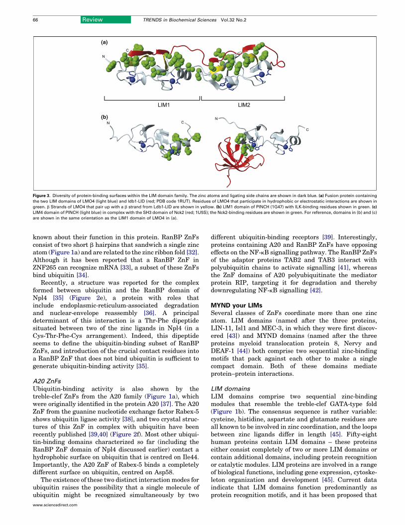
Figure 3. Diversity of protein-binding surfaces within the LIM domain family. The zinc atoms and ligating side chains are shown in dark blue. (a) Fusion protein containing
the two LIM domains of LMO4 (light blue) and ldb1-LID (red; PDB code 1RUT). Residues of LMO4 that participate in hydrophobic or electrostatic interactions are shown in
green. b Strands of LMO4 that pair up with a b strand from Ldb1-LID are shown in yellow. (b) LIM1 domain of PINCH (1G47) with ILK-binding residues shown in green. (c)
LIM4 domain of PINCH (light blue) in complex with the SH3 domain of Nck2 (red; 1U5S); the Nck2-binding residues are shown in green. For reference, domains in (b) and (c)
are shown in the same orientation as the LIM1 domain of LMO4 in (a).
66 Review TRENDS in Biochemical Sciences Vol.32 No.2
known about their function in this protein. RanBP ZnFsconsist of two short b hairpins that sandwich a single zincatom (Figure 1a) and are related to the zinc ribbon fold [32].Although it has been reported that a RanBP ZnF inZNF265 can recognize mRNA [33], a subset of these ZnFsbind ubiquitin [34].
Recently, a structure was reported for the complexformed between ubiquitin and the RanBP domain ofNpl4 [35] (Figure 2e), a protein with roles thatinclude endoplasmic-reticulum-associated degradationand nuclear-envelope reassembly [36]. A principaldeterminant of this interaction is a Thr-Phe dipeptidesituated between two of the zinc ligands in Npl4 (in aCys-Thr-Phe-Cys arrangement). Indeed, this dipeptideseems to define the ubiquitin-binding subset of RanBPZnFs, and introduction of the crucial contact residues intoa RanBP ZnF that does not bind ubiquitin is sufficient togenerate ubiquitin-binding activity [35].
A20 ZnFs
Ubiquitin-binding activity is also shown by thetreble-clef ZnFs from the A20 family (Figure 1a), whichwere originally identified in the protein A20 [37]. The A20ZnF from the guanine nucleotide exchange factor Rabex-5shows ubiquitin ligase activity [38], and two crystal struc-tures of this ZnF in complex with ubiquitin have beenrecently published [39,40] (Figure 2f). Most other ubiqui-tin-binding domains characterized so far (including theRanBP ZnF domain of Npl4 discussed earlier) contact ahydrophobic surface on ubiquitin that is centred on Ile44.Importantly, the A20 ZnF of Rabex-5 binds a completelydifferent surface on ubiquitin, centred on Asp58.
The existence of these two distinct interaction modes forubiquitin raises the possibility that a single molecule ofubiquitin might be recognized simultaneously by two
www.sciencedirect.com
different ubiquitin-binding receptors [39]. Interestingly,proteins containing A20 and RanBP ZnFs have opposingeffects on the NF-kB signalling pathway. The RanBP ZnFsof the adaptor proteins TAB2 and TAB3 interact withpolyubiquitin chains to activate signalling [41], whereasthe ZnF domains of A20 polyubiquitinate the mediatorprotein RIP, targeting it for degradation and therebydownregulating NF-kB signalling [42].
MYND your LIMsSeveral classes of ZnFs coordinate more than one zincatom. LIM domains (named after the three proteins,LIN-11, Isl1 and MEC-3, in which they were first discov-ered [43]) and MYND domains (named after the threeproteins myeloid translocation protein 8, Nervy andDEAF-1 [44]) both comprise two sequential zinc-bindingmotifs that pack against each other to make a singlecompact domain. Both of these domains mediateprotein–protein interactions.
LIM domains
LIM domains comprise two sequential zinc-bindingmodules that resemble the treble-clef GATA-type fold(Figure 1b). The consensus sequence is rather variable:cysteine, histidine, aspartate and glutamate residues areall known to be involved in zinc coordination, and the loopsbetween zinc ligands differ in length [45]. Fifty-eighthuman proteins contain LIM domains – these proteinseither consist completely of two or more LIM domains orcontain additional domains, including protein recognitionor catalytic modules. LIM proteins are involved in a rangeof biological functions, including gene expression, cytoske-leton organization and development [45]. Current dataindicate that LIM domains function predominantly asprotein recognition motifs, and it has been proposed that

Figure 4. RING and PHD domains. (a) Overall view of the ternary complex of c-Cbl
(E3), UbcH7 (E2) and phosphorylated ZAP-70 peptide (the substrate for
ubiquitination by E3; PDB code 1FBV). The c-Cbl domain, which includes the
RING domain, is shown in light blue, zinc atoms and ligating residues are in dark
blue, the UbcH7 domain in red and the ZAP-70 peptide in yellow. In the enlarged
image of the RING domain, E2-binding residues are shown in green. (b) PHD
domains of ING2 (2G6Q) and BPTF (2F6J) interacting with a six-residue H3K4me3
peptide. PHD domains are shown in light blue, the H3K4me3 peptide in red and the
methylated lysine in stick representation. Zinc atoms and ligating residues are dark
blue. The BPTF structure also has a bromodomain (C-terminal to the PHD domain)
that is not shown here.
Review TRENDS in Biochemical Sciences Vol.32 No.2 67
proteins that contain multiple LIM domains mightfunction as molecular bridges [45].
The LIM-only protein LMO4 and its binding partnerldb1 are both negative regulators of mammary epithelialdifferentiation. Their interaction is mediated by the twotandem LIM domains of LMO4 and the LIM interactiondomain (LID) from ldb1. The crystal structure of theLMO4–ldb1-LID complex [46] shows that ldb1-LID bindsin an extended form to LMO4, spanning thewhole length ofthe two LIM domains and forming a tight complex. A seriesof backbone hydrogen bonds is formed, whereby ldb1 effec-tively adds an additional b strand to four of the eight b
hairpins that make up the two LIM domains (Figure 3a).The nature of the side chain interactions between ldb1-LIDand LMO4 differ, however, for each of the two LIMdomains: the LIM1 interface is a mixture of ionic andhydrophobic interactions, whereas the LIM2 interface isprimarily hydrophobic.
The adaptor protein PINCH consists solely of five LIMdomains and has roles in cell adhesion, growth and differ-entiation [47]. PINCH interacts with the integrin-likekinase (ILK) and Nck2, and both interactions are essentialfor integrin signalling. Specifically, the LIM1 domain ofPINCH interacts with the ankyrin repeat domain of ILK,and this interaction is localized to the second zinc-bindingmodule of PINCH-LIM1. Chemical shift perturbation stu-dies have identified a hydrophobic surface patch, made upof residues from the C-terminal helix, as the likely bindingsite for ILK [48] (Figure 3b). PINCH-LIM4 mediates theinteraction with the third Src homology 3 (SH3) domain ofNck2 [49] and does so through a different binding mode. Inthis case, it is the first b hairpin of LIM4 that contacts theSH3 domain of Nck2 (Figure 3c), forming a weak complex(dissociation constant, Kd �3 � 10�3 M) in which two argi-nines from LIM4 are responsible for the formation of saltbridges and hydrophobic interactions with Nck2.
MYND domains
MYND domains also ligate two zinc atoms in a sequentialmanner (Figure 1b), but they are smaller than LIMdomains (�40 residues versus �55 residues for LIMdomains). The first zinc-binding module contains only ashort b hairpin, whereas the second zinc-binding moduleforms two short a helices [50]. Like LIM domains, MYNDdomains function primarily as protein-binding domainsand are present mainly in transcriptional regulators[51,52]. No structural information is available onMYND-mediated interactions, although a few MYNDdomains recognize a Pro-X-Leu-X-Pro (PXLXP) motif intheir partner proteins. For example, the MYND domain inthe transcriptional co-repressor BS69 interacts with theviral oncoproteins E1A and EBNA2 in addition to thetranscription factor MGA, all of which contain a PXLXPmotif [52]. Mutational data implicate a large, positivelycharged surface patch, which is conserved in PXLXP-bind-ing MYND domains [50].
Cross-brace yourself for PHD and RING actionLike LIM and MYND domains, RING (‘really interestingnew gene’ [53]) and PHD (‘plant homeodomain’; named forthe class of proteins in which it was first recognized [54])
www.sciencedirect.com
domains also coordinate two zinc atoms. For RING andPHD domains, however, the two sets of zinc ligands are notsequential, but instead are interdigitated (Figure 1c) or‘cross-braced’.
RING domains
Both RING and PHD domains function primarily asprotein–protein interaction motifs and are found in manyhuman proteins (�200 for RING and �70 for PHD) [31].Until recently, confusion has existed in the classification ofthese domains owing to their similar consensus sequencesand zinc-ligation topology, and several RING domainshave been mistakenly labelled as PHD domains [55].Now, with more sequence and structural information athand, the distinction is clearer. PHD domains contain acrucial tryptophan residue, located two residues N-term-inal to the seventh metal ligand, that is involved in thehydrophobic core; in RING domains, by contrast, the equiv-alent residue is solvent exposed [56]. Furthermore, RINGdomains seem to participate in a wider range of functions.For example, RING domains in KAP-1 and PML mediateinteractions with the transcription factor KOX-1 [57] andthe mRNA cap-binding protein eIF4E [58], respectively,thereby regulating gene repression and RNA transport.
Many RING domains are present in E3 ubiquitinligases, which catalyze the final step in the protein ubiqui-tination pathway. Ubiquitination targets proteins for pro-teosomal degradation and has been shown to function as asignalling module for the destruction of plasma membrane
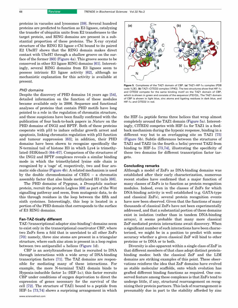
Figure 5. Complexes of the TAZ1 domain of CBP. (a) TAZ1–HIF-1a complex (PDB
code 1L3E). (b) TAZ1–CITED2 complex (1P4Q). The two structures show that HIF-1a
and CITED2 compete for the same binding motif on the TAZ1 domain of CBP,
which is shown in green and consists of the sequence LP(E/Q)L. The TAZ1 domain
of CBP is shown in light blue, zinc atoms and ligating residues in dark blue, and
HIF-1a and CITED2 in red.
68 Review TRENDS in Biochemical Sciences Vol.32 No.2
proteins in vacuoles and lysosomes [59]. Several hundredproteins are predicted to function as E3 ligases, catalyzingthe transfer of ubiquitin units from E2 transferases to thetarget protein, and RING domains are present in a sub-stantial proportion of these proteins. The X-ray crystalstructure of the RING E3 ligase c-Cbl bound to its pairedE2 UbcH7 shows that the RING domain makes directcontact with UbcH7 through a shallow groove on the sur-face of the former [60] (Figure 4a). This groove seems to beconserved in other E3 ligase RING domains [61]. Interest-ingly, several RING domains from E3 ligases seem topossess intrinsic E3 ligase activity [62], although nomechanistic explanation for this activity is available atpresent.
PHD domains
Despite the discovery of PHD domains 14 years ago [54],detailed information on the function of these modulesbecame available only in 2006. Sequence and functionalanalyses of proteins that contain PHD motifs have longpointed to a role in the regulation of chromatin structure,and these suspicions have been finally confirmed with thepublication of four back-to-back papers in Nature on thePHD domains of ING2 and BPTF. Both of these proteinscooperate with p53 to induce cellular growth arrest andapoptosis, linking chromatin regulation with p53 functionand tumour suppression [63]; in addition, their PHDdomains have been shown to recognize specifically theN-terminal tail of histone H3 in which Lys4 is trimethy-lated (H3K4me3) [64–67]. Comparison of the structures ofthe ING2 and BPTF complexes reveals a similar bindingmode in which the trimethylated lysine side chain isrecognized by a ‘cage’ of, respectively, two and four aro-matic side chains (Figure 4b). A related mechanism is usedby the double chromodomains of CHD1 – a chromatinassembly factor that also binds methylated H3 tails [68].
The PHD domains of Pygopus, a Drosophila nuclearprotein, recruit the protein Legless [69] as part of the Wntsignalling pathway and seem, from mutagenic data, to dothis through residues in the loop between the fifth andsixth cysteines. Interestingly, this loop is located in aportion of the PHD domain that corresponds to the surfaceof E3 RING domains.
Fan-TAZ-tically differentTAZ (‘transcriptional adaptor zinc-binding’) domains seemto exist only in the transcriptional coactivator CBP, wheretwo ZnFs form a fold that is unrelated to all other ZnFs[70]: namely, three zinc atoms are present in a triangularstructure, where each zinc atom is present in a loop regionbetween two antiparallel a helices (Figure 1d).
CBP is an acetyltransferase that is recruited to DNAthrough interactions with a wide array of DNA-bindingtranscription factors [71]. The TAZ domains are respon-sible for mediating many of these interactions. Forexample, the more N-terminal TAZ1 domain binds toHypoxia-inducible factor 1a (HIF-1a); this factor recruitsCBP under conditions of oxygen deprivation to direct theexpression of genes necessary for the survival of thecell [72]. The structure of TAZ1 bound to a peptide fromHIF-1a [73,74] shows a surprising arrangement in which
www.sciencedirect.com
the HIF-1a peptide forms three helices that wrap almostcompletely around the TAZ1 domain (Figure 5a). Interest-ingly, CITED2 competes with HIF-1a for TAZ1 in a feed-back mechanism during the hypoxic response, binding in adifferent way but to an overlapping site on TAZ1 [75](Figure 5b). Subtle differences between the structures ofTAZ1 and TAZ2 (in the fourth a helix) prevent TAZ2 frombinding to HIF-1a [73,74], illustrating the specificity ofthese two domains for different transcription factor tar-gets.
Concluding remarksAlthough a model of ZnFs as DNA-binding domains wasestablished after their early characterization, numerousrecent studies have confirmed that a major function ofmany classes of ZnFs is to function as protein recognitionmodules. Indeed, even in the classes of ZnFs for whichDNA-binding activity is well established (e.g. GATA-typeand classical ZnFs), several protein recognition eventshave now been observed. Given that the functions of manythousands of classical ZnFs have not been experimentallyaddressed, and that a substantial portion of these domainsexist in isolation (rather than in tandem DNA-bindingarrays), it seems probable that many more classicalZnF-mediated protein interactions await discovery. Whena significant number of such interactions have been charac-terized, we might be in a position to predict with someaccuracy whether a given classical ZnF will bind to otherproteins or to DNA or to both.
Diversity is also apparent within a single class of ZnF inthat different members of that class adopt distinct protein-binding modes: both the classical ZnF and the LIMdomains are striking examples of this point. These obser-vations reinforce the idea that ZnF domains have evolvedas stable molecular scaffolds, onto which evolution hasgrafted different binding functions as required. One con-served feature among these complexes is that ZnFs tend toundergo little, if any, structural rearrangement on recog-nizing their protein partners. This lack of rearrangement ispresumably due in part to the stability afforded by zinc
Review TRENDS in Biochemical Sciences Vol.32 No.2 69
ligation. Another notable feature is that several of theseinteractions are modular in nature, similar to the mode ofinteraction observed for classical ZnFs binding to DNA. Forexample, the two LIM domains of LMO proteins are inde-pendently folded units that contact adjacent parts of theldb1-LID peptide. Similarly, it is possible that the tandemPHD domains of proteins such as Mi2b [76] will be shownto bind simultaneously to different methylated lysine resi-dues in the same (or different) histone tails. It will beinteresting to see whether other proteins with multipleZnFs adopt a related mechanism for recognizing extendedsubstrates.
As shown in Table 1, ZnF domains mediate interactionswitharangeofdifferentaffinities.These interactions tend tobe of moderate to weak affinity, which is perhaps not sur-prising considering the relatively small size of many ZnFdomains.Nevertheless, the biological importance ofmany ofthe weaker interactions (such as GATA-1–FOG-1, Npl4–ubiquitin and PINCH–Nck2) is indisputable, and despitetheir low affinity the interactions are also highly specific.These data emphasize the prevalence of weak interactionsin regulating cellular processes; it is likely that as structuralmethods become more sensitive we will see an increasingnumber of weak complexes characterized in moleculardetail. It is also possible that post-translational modifi-cations, such as lysine methylation, might increase theaffinity of some of these weak interactions in vivo.
Remarkable progress has been recently made in design-ing tandem arrays of ZnFs with tailored DNA-bindingspecificities, with the ultimate goal of using them as thera-peutics [77]. Although designing protein-binding ZnFswith tailored specificities also holds considerable thera-peutic potential, it is clearly a considerably more difficulttask. The greater variability and complexity of protein-binding interfaces, in addition to the small number ofstructural studies on protein-binding ZnFs, are currentlysignificant impediments. As we have highlighted here,however, progress is being made to address what was,until recently, a substantial deficiency in our understand-ing and, as more ZnF–protein interactions are discovered,our knowledge of such interactions and our potential tomanipulate them can only increase.
AcknowledgementsR.G. is supported by a fellowship from the Austrian Science Fund (J2474).C.K.L. is a C.J. Martin Fellow of the National Health and MedicalResearch Council (NHMRC) and F.E.L. holds an Australian PostgraduateAward. J.P.M. is an NHMRC Senior Research Fellow. This work wassupported by an NHMRC Program Grant to J.P.M. and M.C.
References1 Miller, J. et al. (1985) Repetitive zinc-binding domains in the protein
transcription factor IIIA from Xenopus oocytes. EMBO J. 4, 1609–16142 Wolfe, S.A. et al. (2000) DNA recognition by Cys2His2 zinc finger
proteins. Annu. Rev. Biophys. Biomol. Struct. 29, 183–2123 Rubin, G.M. et al. (2000) Comparative genomics of the eukaryotes.
Science 287, 2204–22154 Krishna, S.S. et al. (2003) Structural classification of zinc fingers:
survey and summary. Nucleic Acids Res. 31, 532–5505 Marmorstein, R. et al. (1992) DNA recognition by GAL4: structure of a
protein–DNA complex. Nature 356, 408–4146 Omichinski, J.G. et al. (1993) NMR structure of a specific DNA complex
of Zn-containing DNA binding domain of GATA-1. Science 261, 438–446
www.sciencedirect.com
7 Brown, R.S. (2005) Zinc finger proteins: getting a grip on RNA. Curr.Opin. Struct. Biol. 15, 94–98
8 Hall, T.M. (2005) Multiple modes of RNA recognition by zinc fingerproteins. Curr. Opin. Struct. Biol. 15, 367–373
9 Lee, B.M. et al. (2006) Induced fit and ‘lock and key’ recognition of 5SRNA by zinc fingers of transcription factor IIIA. J. Mol. Biol. 357, 275–291
10 Lu, D. et al. (2003) Crystal structure of a zinc-finger–RNA complexreveals two modes of molecular recognition. Nature 426, 96–100
11 Mackay, J.P. and Crossley, M. (1998) Zinc fingers are sticking together.Trends Biochem. Sci. 23, 1–4
12 Fox, A.H. et al. (1999) Transcriptional cofactors of the FOG familyinteract with GATA proteins by means of multiple zinc fingers. EMBOJ. 18, 2812–2822
13 Simpson, R.J. et al. (2004) A classic zinc finger from friend of GATAmediates an interaction with the coiled-coil of transforming acidiccoiled-coil 3. J. Biol. Chem. 279, 39789–39797
14 Tsang, A.P. et al. (1998) Failure of megakaryopoiesis and arrestederythropoiesis in mice lacking the GATA-1 transcriptional cofactorFOG. Genes Dev. 12, 1176–1188
15 Liew, C.K. et al. (2005) Zinc fingers as protein recognition motifs:structural basis for the GATA-1/friend of GATA interaction. Proc.Natl. Acad. Sci. U. S. A. 102, 583–588
16 Newton, A. et al. (2001) The N-terminal zinc finger of the erythroidtranscription factor GATA-1 binds GATCmotifs in DNA. J. Biol. Chem.276, 35794–35801
17 Kwan, A.H. et al. (2003) Pentaprobe: a comprehensive sequence for theone-step detection of DNA-binding activities. Nucleic Acids Res. 31,e124
18 Garriga-Canut, M. and Orkin, S.H. (2004) Transforming acidic coiled-coil protein 3 (TACC3) controls friend of GATA-1 (FOG-1) subcellularlocalization and regulates the association between GATA-1 and FOG-1during hematopoiesis. J. Biol. Chem. 279, 23597–23605
19 Kelley, C.M. et al. (1998) Helios, a novel dimerization partner of Ikarosexpressed in the earliest hematopoietic progenitors. Curr. Biol. 8, 508–515
20 McCarty, A.S. et al. (2003) Selective dimerization of a C2H2 zinc fingersubfamily. Mol. Cell 11, 459–470
21 Westman, B.J. et al. (2003) The C-terminal domain of Eos forms a highorder complex in solution. J. Biol. Chem. 278, 42419–42426
22 Westman, B.J. et al. (2004) Structural studies on a protein-bindingzinc-finger domain of Eos reveal both similarities and differences toclassical zinc fingers. Biochemistry 43, 13318–13327
23 Tsai, R.Y. and Reed, R.R. (1998) Identification of DNA recognitionsequences and protein interaction domains of the multiple-Zn-fingerprotein Roaz. Mol. Cell. Biol. 18, 6447–6456
24 Matsuzawa-Watanabe, Y. et al. (2003) Transcriptional activity oftestis-determining factor SRY is modulated by the Wilms’ tumor 1gene product, WT1. Oncogene 22, 7900–7904
25 Blobel, G.A. et al. (1998) CREB-binding protein cooperates withtranscription factor GATA-1 and is required for erythroiddifferentiation. Proc. Natl. Acad. Sci. U. S. A. 95, 2061–2066
26 Eisbacher, M. et al. (2003) Protein–protein interaction between Fli-1and GATA-1 mediates synergistic expression of megakaryocyte-specific genes through cooperative DNA binding. Mol. Cell. Biol. 23,3427–3441
27 Merika, M. and Orkin, S.H. (1995) Functional synergy and physicalinteractions of the erythroid transcription factor GATA-1 with theKruppel family proteins Sp1 and EKLF.Mol. Cell. Biol. 15, 2437–2447
28 Nerlov, C. et al. (2000) GATA-1 interacts with the myeloid PU.1transcription factor and represses PU.1-dependent transcription.Blood 95, 2543–2551
29 Liew, C.W. et al. (2006) Molecular analysis of the interaction betweenthe hematopoietic master transcription factors GATA-1 and PU.1. J.Biol. Chem. 281, 28296–28306
30 Reyes-Turcu, F.E. et al. (2006) The ubiquitin binding domain ZnF UBPrecognizes the C-terminal diglycine motif of unanchored ubiquitin.Cell124, 1197–1208
31 Letunic, I. et al. (2006) SMART 5: domains in the context of genomesand networks. Nucleic Acids Res. 34, D257–D260
32 Wang, B. et al. (2003) Structure and ubiquitin interactions ofthe conserved zinc finger domain of Npl4. J. Biol. Chem. 278,20225–20234
70 Review TRENDS in Biochemical Sciences Vol.32 No.2
33 Plambeck, C.A. et al. (2003) The structure of the zinc finger domain fromhuman splicing factor ZNF265 fold. J. Biol. Chem. 278, 22805–22811
34 Meyer, H.H. et al. (2002) Direct binding of ubiquitin conjugates by themammalian p97 adaptor complexes, p47 and Ufd1–Npl4. EMBO J. 21,5645–5652
35 Alam, S.L. et al. (2004) Ubiquitin interactions of NZnF zinc fingers.EMBO J. 23, 1411–1421
36 Tsai, B. et al. (2002) Retro-translocation of proteins from theendoplasmic reticulum into the cytosol. Nat. Rev. Mol. Cell Biol. 3,246–255
37 Opipari, A.W., Jr et al. (1990) The A20 cDNA induced by tumor necrosisfactor a encodes a novel type of zinc finger protein. J. Biol. Chem. 265,14705–14708
38 Mattera, R. et al. (2006) The Rab5 guanine nucleotide exchange factorRabex-5 binds ubiquitin (Ub) and functions as a Ub ligase through anatypical Ub-interacting motif and a zinc finger domain. J. Biol. Chem.281, 6874–6883
39 Penengo, L. et al. (2006) Crystal structure of the ubiquitin bindingdomains of Rabex-5 reveals two modes of interaction with ubiquitin.Cell 124, 1183–1195
40 Lee, S. et al. (2006) Structural basis for ubiquitin recognition andautoubiquitination by Rabex-5. Nat. Struct. Mol. Biol. 13, 264–271
41 Kanayama, A. et al. (2004) TAB2 andTAB3 activate theNF-kBpathwaythrough binding to polyubiquitin chains. Mol. Cell 15, 535–548
42 Wertz, I.E. et al. (2004) De-ubiquitination and ubiquitin ligase domainsof A20 downregulate NF-kB signalling. Nature 430, 694–699
43 Freyd, G. et al. (1990) Novel cysteine-rich motif and homeodomain inthe product of the Caenorhabditis elegans cell lineage gene lin-11.Nature 344, 876–879
44 Gross, C.T. and McGinnis, W. (1996) DEAF-1, a novel protein thatbinds an essential region in a Deformed response element. EMBO J.15, 1961–1970
45 Kadrmas, J.L. and Beckerle, M.C. (2004) The LIM domain: from thecytoskeleton to the nucleus. Nat. Rev. Mol. Cell Biol. 5, 920–931
46 Deane, J.E. et al. (2004) Tandem LIM domains provide synergisticbinding in the LMO4:Ldb1 complex. EMBO J. 23, 3589–3598
47 Xu, Z. et al. (2005) Molecular dissection of PINCH-1 reveals amechanism of coupling and uncoupling of cell shape modulation andsurvival. J. Biol. Chem. 280, 27631–27637
48 Velyvis, A. et al. (2001) Solution structure of the focal adhesion adaptorPINCH LIM1 domain and characterization of its interaction with theintegrin-linked kinase ankyrin repeat domain. J. Biol. Chem. 276,4932–4939
49 Vaynberg, J. et al. (2005) Structure of an ultraweak protein–proteincomplex and its crucial role in regulation of cell morphology andmotility. Mol. Cell 17, 513–523
50 Spadaccini, R. et al. (2006) Structure and functional analysis of theMYND domain. J. Mol. Biol. 358, 498–508
51 Sims, R.J., 3rd et al. (2002) m-Bop, a repressor protein essential forcardiogenesis, interacts with skNAC, a heart- and muscle-specifictranscription factor. J. Biol. Chem. 277, 26524–26529
52 Ansieau, S. and Leutz, A. (2002) The conserved Mynd domain of BS69binds cellular and oncoviral proteins through a common PXLXP motif.J. Biol. Chem. 277, 4906–4910
53 Lovering, R. et al. (1993) Identification and preliminarycharacterization of a protein motif related to the zinc finger. Proc.Natl. Acad. Sci. U. S. A. 90, 2112–2116
54 Schindler, U. et al. (1993) HAT3.1, a novel Arabidopsis homeodomainprotein containing a conserved cysteine-rich region. Plant J. 4, 137–150
55 Aravind, L. et al. (2003) Scores of RINGS but no PHDs in ubiquitinsignaling. Cell Cycle 2, 123–126
56 Dodd, R.B. et al. (2004) Solution structure of the Kaposi’s sarcoma-associated herpesvirus K3 N-terminal domain reveals a novel E2-binding C4HC3-type RING domain. J. Biol. Chem. 279, 53840–53847
www.sciencedirect.com
57 Peng, H. et al. (2000) Reconstitution of the KRAB–KAP-1 repressorcomplex: a model system for defining the molecular anatomy of RING-B box–coiled-coil domain-mediated protein–protein interactions. J.Mol. Biol. 295, 1139–1162
58 Cohen, N. et al. (2001) PML RING suppresses oncogenictransformation by reducing the affinity of eIF4E for mRNA. EMBOJ. 20, 4547–4559
59 Weissman, A.M. (2001) Themes and variations on ubiquitylation. Nat.Rev. Mol. Cell Biol. 2, 169–178
60 Zheng, N. et al. (2000) Structure of a c-Cbl–UbcH7 complex: RINGdomain function in ubiquitin-protein ligases. Cell 102, 533–539
61 Zheng, N. et al. (2002) Structure of the Cul1–Rbx1–Skp1–F-boxSkp2
SCF ubiquitin ligase complex. Nature 416, 703–70962 Albert, T.K. et al. (2002) Identification of a ubiquitin-protein ligase
subunit within the CCR4–NOT transcription repressor complex.EMBO J. 21, 355–364
63 Garkavtsev, I. et al. (1998) The candidate tumour suppressor p33ING1
cooperates with p53 in cell growth control. Nature 391, 295–29864 Shi, X. et al. (2006) ING2 PHD domain links histone H3 lysine 4
methylation to active gene repression. Nature 442, 96–9965 Pena, P.V. et al. (2006) Molecular mechanism of histone H3K4me3
recognition by plant homeodomain of ING2. Nature 442, 100–10366 Li, H. et al. (2006) Molecular basis for site-specific read-out of histone
H3K4me3 by the BPTF PHD finger of NURF. Nature 442, 91–9567 Wysocka, J. et al. (2006) A PHD finger of NURF couples histone H3
lysine 4 trimethylation with chromatin remodelling.Nature 442, 86–9068 Flanagan, J.F. et al. (2005) Double chromodomains cooperate to
recognize the methylated histone H3 tail. Nature 438, 1181–118569 Townsley, F.M. et al. (2004) Pygopus residues required for its binding
to Legless are critical for transcription and development. J. Biol. Chem.279, 5177–5183
70 Ponting, C.P. et al. (1996) ZZ and TAZ: new putative zinc fingers indystrophin and other proteins. Trends Biochem. Sci. 21, 11–13
71 Blobel, G.A. (2000) CREB-binding protein and p300: molecularintegrators of hematopoietic transcription. Blood 95, 745–755
72 Arany, Z. et al. (1996) An essential role for p300/CBP in the cellularresponse to hypoxia. Proc. Natl. Acad. Sci. U. S. A. 93, 12969–12973
73 Dames, S.A. et al. (2002) Structural basis for Hif-1a/CBP recognition inthe cellular hypoxic response. Proc. Natl. Acad. Sci. U. S. A. 99, 5271–5276
74 Freedman, S.J. et al. (2002) Structural basis for recruitment of CBP/p300 by hypoxia-inducible factor-1a. Proc. Natl. Acad. Sci. U. S. A. 99,5367–5372
75 Freedman, S.J. et al. (2003) Structural basis for negative regulation ofhypoxia-inducible factor-1a by CITED2. Nat. Struct. Biol. 10, 504–512
76 Kwan, A.H. et al. (2003) Engineering a protein scaffold from a PHDfinger. Structure. 11, 803–813
77 Bibikova, M. et al. (2003) Enhancing gene targeting with designed zincfinger nucleases. Science 300, 764
78 Deane, J.E. et al. (2003) Structural basis for the recognition of ldb1 bythe N-terminal LIM domains of LMO2 and LMO4. EMBO J. 22, 2224–2233
79 Dominguez, C. et al. (2004) Structural model of the UbcH5B/CNOT4complex revealed by combining NMR, mutagenesis, and dockingapproaches. Structure. 12, 633–644
80 Buchwald, G. et al. (2006) Structure and E3-ligase activity of the Ring–Ring complex of polycomb proteins Bmi1 and Ring1b. EMBO J. 25,2465–2474
81 Li, Z. et al. (2006) Structure of a Bmi-1–Ring1B polycomb groupubiquitin ligase complex. J. Biol. Chem. 281, 20643–20649
82 De Guzman, R.N. et al. (2004) Interaction of the TAZ1 domain of theCREB-binding protein with the activation domain of CITED2:regulation by competition between intrinsically unstructuredligands for non-identical binding sites. J. Biol. Chem. 279, 3042–3049