Embed Size (px)
Citation preview
Egypt. J. Comp. Path. & Clinic. Path. Vol. 22 No. 3 (July) 2009; 77 - 95
77
Studies On Feline Hepatic Lipidosis
By
Kelany, W. M.a ; Sherein, S. A. Elgayed b ; Mahran, K. M. A.c and Dardery, M. A.d
a Dept. of Int. Med. and Infectious diseases, Faculty of Vet. Med., Cairo Univ., b Dept. of Pathology, Faculty of Vet. Med., Cairo Univ.,
c Dept. of clinical pathology, Faculty of Vet., Med., Cairo Univ. and d Dept. of Pathology, Animal Health Research Institute.
SUMMARY
T his study was performed on 40 cats, divided on the basis of history and clinical presentation into two equal groups; a group of appar-
ently healthy cats and a group of clinically diseased cats. All cats were subjected to clinical evaluation, ultrasonographic and clinicopathological examinations. The livers of three dead cats from the second group were examined histopathologically. The clinical signs of clinically diseased cats included anorexia, vomiting, diarrhea, lethargy, hepato-encephalopathic signs (muscle fasciculation, tremors and convulsions) with hemorrhagic diathesis and ascites in 2 cats. Palpation revealed enlarged liver extended beyond the last right rib while fluid thrill was detected by the tactile percussion in ascetic cats. The hematological pro-file of clinically diseased cats showed a normocytic normochromic ane-mia associated with stress leukogram. Moreover, the biochemical pro-files of these cats showed significant increases in hepatic enzymes (AST, ALT, ALP and GGT), bilirubin, triglycerides, total cholesterol, HDL, LDL and VLDL in comparison to control cats. Serum glucose, BUN, total proteins, albumin and A/G ratio were significantly decreased. Se-rum creatinine and globulins were not significantly changed. The ultra-sonographic findings included diffuse hyperechogenicity of hepatic pa-renchyma, decreased visualization of the intrahepatic blood vessels and enlargement of liver. The liver parenchyma was isoechoic or hypere-choic, compared with falciform or omental fats. The liver was also hy-perechoic with respect to the spleen and renal cortices. Also, there was increased attenuation of ultrasound waves by liver parenchyma. Micro-scopic examination of the livers of dead cats revealed different degrees of lipidosis. From the research, it was concluded that ultrasonogaphy and serum chemistry panel were complementary to each other. It can also be confirmed by autopsy and histopathology of dead cats. The serum chem-istry panel could detect the hepatic dysfunction while ultrasonography defined the nature of lesions.
Egypt. J. Comp. Path. & Clinic. Path. Vol. 22 No. 3 (July) 2009; 77 - 95
78
Referred byReferred by
Prof. Dr. Sohir Sokkar Professor of Pathology, Fac. Vet. Med., Cairo University
Prof. Dr. Safaa Ramadan Professor of Clinical Pathology, Fac. Vet. Med., Cairo University
INTRODUCTION
H epatic lipidosis in cats is a commonly diagnosed hepato-
biliary disease of unknown cause (De-Maria et al., 1998 and Brown et al., 2000). The condition seems to have no age, breed, or gender predilection (Thornburg et al., 1982; Center, 1986 and Jacobs et al., 1989). Hepatic lipidosis is di-agnosed in obese or formerly obese cats with anorexia, hepa-tomegally, jaundice and muscle wasting (Burrows et al., 1981; Thornburg, 1982; Center, 1986; Jacobs et al., 1989; Bennette et al., 2007 and Posner et al., 2008). Abnormal laboratory parameters (serum liver enzymes, bilirubin and bile acid concentrations,… etc) are rapid and accurate meth-ods for diagnosis of feline hepatic disorders (Burrows et al., 1981; Thornburg et al., 1982; Center, 1986; Jacobs et al., 1989 and Na-kamura et al., 2005). Liver biopsy was found to be the most reliable diagnostic procedure to detect he-patic lipidosis (De-Maria et al., 1998 and Willard et al., 1999).
Nowadays, use of ultrasono-graphy enhanced the detection of fatty infiltration in liver with high sensitivity and specificity (Scatar-ige et al., 1984; Nakamura et al., 2005 and Posner et al., 2008).
The purpose of this study is to compare the reliability of ultra-sonography versus laboratory diag-nosis and histopathological studies in the diagnosis of feline hepatic lipidosis.
MATERIAL AND METHODS Animals:
A total number of 40 cats (1.6-10.0 years-old, Persian, Sia-mese and Egyptian Mau) admitted to the clinic of Faculty of Veteri-nary Medicine, Cairo University and to a private veterinary clinic in Giza governorate in the period from September, 2007 to August, 2008 were included in the study. On the basis of history and clinical presentation, the cats were divided into 2 groups (each composed of 20 cats); group (1); a group of ap-parently healthy cats and group (2); a group of clinically diseased cats.
Clinical evaluation:
Age, gender, breed, respira-tory rate, pulse rate, rectal tem-perature of the cats of the study were recorded. All cats were thor-oughly investigated and clinically examined by abdominal palpation and tactile percussion according to the method described by Kelly (1984).
Egypt. J. Comp. Path. & Clinic. Path. Vol. 22 No. 3 (July) 2009; 77 - 95
79
Ultrasonography: Ultrasonography was per-
formed after 24 hours fasting. The examined cats were positioned in dorsal recumbency. Cranial ventral abdomen were cl ipped and sheaved then covered with cou-pling gel. Transverse and longitu-dinal scans were taken using Pie-Medical Scanner (Maastricht, Netherlands) and sector transducer with alternating frequency of 5.0-7.5 MHz according to the method described by Nyland et al., (1989).
Collection of blood samples:
Blood samples were collected from all animals of both groups after 12 hours fasting from anterior median vein. Blood samples were divided into two portions. A por-tion was mixed with potassium salt of EDTA as anticoagulant for he-matological tests and the other por-tion was left to clot in clean and dry test tube and then centrifuged at 3000 rpm for 10 minutes. The clear supernatant serum was then frozen at -20OC for the biochemi-cal studies. Clinicopathological studies:
Complete blood count (CBC) was performed for all blood sam-ples with the standard techniques described by Feldman et al., (2000). The CBC included red blood cells (RBCs) count, hemo-globin (Hb) concentration, packed cell volume (PCV), red cells indi-
ces; mean corpuscular hemoglobin concentration (MCHC), mean cor-puscular volume (MCV) as well as total (TLC) and differential leuko-cyte count (DLC).
Serum liver function tests
were estimated using the commer-cial diagnostic kits supplied by Sentinel (Milan, Italy). The aspar-tate aminotransferase (AST) and alanine aminotransferase (ALT) were estimated according to the method described by Reitman and Frankel (1957), γ-glutamyltransf-erase (GGT) as described by Szasz (1976), alkaline phosphatase (ALP) as described by Roy (1970), total and direct bilirubin as de-scribed by Doumas et al., (1973), glucose as described by Trinder (1959), blood urea nitrogen (BUN) as described by Tabacco et al., (1979), creatinine as described by Fabiny and Ertingshausen (1971), triglycerides as described by Wahlefeld (1974), total choles-terols as described by Allain et al., (1974), high density lipoproteins (HDL) as described by Warnick et al., (1983), low density lipopro-teins (LDL) and very low density lipoproteins (VLDL) were calcu-lated according to the equations of Friedewald et al., (1972), albumin as described by Doumas et al., (1971) and total proteins as de-scribed by Gornall et al., (1949) while serum globulins and A/G ra-tio were calculated.
Egypt. J. Comp. Path. & Clinic. Path. Vol. 22 No. 3 (July) 2009; 77 - 95
80
Histopathological studies: Postmortem examination was
performed, on three dead cats from the clinically diseased group, for gross and histopathological exami-nation. Samples were collected from liver of investigated cases and were fixed in 10% neutral buffered formalin for preparing paraffin tissue sections at 4-6 µ thickness, then stained with Hema-toxylin and Eosin according to the method described by Bancroft and Stevens (1996). Special stains as Masson's trichrome and Prus-siun blue stains were used accord-ing to the methods described by Masson (1929) and Perls (1867), respectively.
Statistical analysis: Analysis of the data was per-
formed by ANOVA (Analysis Of Variance) test using the computer software Statistical Package for Social Sciences (SPSS) for Win-dows (version 10.0) according to the method described by Irwan (1996).
RESULTS Results of clinical evaluation
The clinically healthy cats were characterized by absence of any clinical signs of hepatobiliary disorders and were taken as a model of normal ultrasonography of the liver and as a control nega-tive group for clinicopathological evaluation.
Although, the clinically dis-eased cats showed no significant changes in the parameters of gen-eral clinical examinations (table 1) except icteric color of mucous membranes, there was a panorama of clinical signs. The clinical signs were including anorexia, vomiting, d ia r rhea , le thargy, hepato-encephalopathic signs (muscle fas-ciculation, tremors and convul-sions) with hemorrhagic diathesis and ascites in 2 cats (Photo 1-4). Palpation revealed enlarged liver extended beyond the last right rib while fluid thrill was detected by the tactile percussion in ascetic cats. Three cats died and were sub-jected to histopathological exami-nation (photo 1:5).
Egypt. J. Comp. Path. & Clinic. Path. Vol. 22 No. 3 (July) 2009; 77 - 95
81
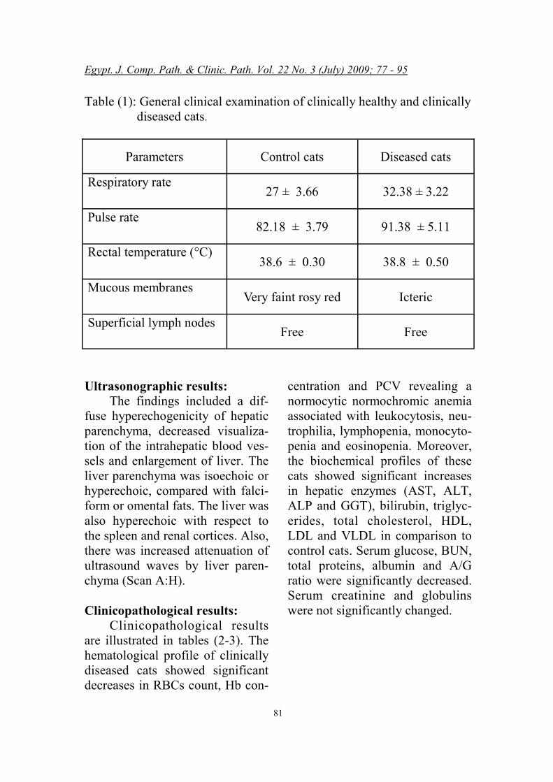
Table (1): General clinical examination of clinically healthy and clinically diseased cats.
Parameters Control cats Diseased cats
Respiratory rate 27 ± 3.66 32.38 ± 3.22
Pulse rate 82.18 ± 3.79 91.38 ± 5.11
Rectal temperature (°C) 38.6 ± 0.30 38.8 ± 0.50
Mucous membranes Very faint rosy red Icteric
Superficial lymph nodes Free Free
Ultrasonographic results: The findings included a dif-
fuse hyperechogenicity of hepatic parenchyma, decreased visualiza-tion of the intrahepatic blood ves-sels and enlargement of liver. The liver parenchyma was isoechoic or hyperechoic, compared with falci-form or omental fats. The liver was also hyperechoic with respect to the spleen and renal cortices. Also, there was increased attenuation of ultrasound waves by liver paren-chyma (Scan A:H). Clinicopathological results:
Clinicopathological results are illustrated in tables (2-3). The hematological profile of clinically diseased cats showed significant decreases in RBCs count, Hb con-
centration and PCV revealing a normocytic normochromic anemia associated with leukocytosis, neu-trophilia, lymphopenia, monocyto-penia and eosinopenia. Moreover, the biochemical profiles of these cats showed significant increases in hepatic enzymes (AST, ALT, ALP and GGT), bilirubin, triglyc-erides, total cholesterol, HDL, LDL and VLDL in comparison to control cats. Serum glucose, BUN, total proteins, albumin and A/G ratio were significantly decreased. Serum creatinine and globulins were not significantly changed.
Egypt. J. Comp. Path. & Clinic. Path. Vol. 22 No. 3 (July) 2009; 77 - 95
82
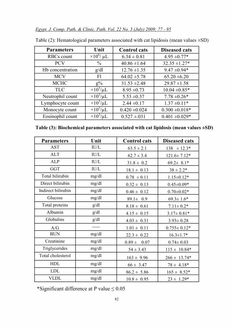
Table (2): Hematological parameters associated with cat lipidosis (mean values ±SD)
Parameters Unit Control cats Diseased cats
RBCs count ×106/ µL 6.34 ± 0.81 4.95 ±0.77* PCV % 40.86 ±1.64 32.35 ±1.27*
Hb concentration g/dl 12.76 ±1.35 9.47 ±0.94* MCV Fl 64.02 ±5.78 65.20 ±6.20
MCHC g% 31.53 ±2.48 29.87 ±1.58 TLC ×103/µL 8.95 ±0.73 10.04 ±0.85*
Neutrophil count ×103/µL 5.53 ±0.37 7.78 ±0.26* Lymphocyte count ×103/µL 2.44 ±0.17 1.37 ±0.11*
Monocyte count ×103/µL 0.420 ±0.024 0.300 ±0.018* Eosinophil count ×103/µL 0.527 ±.031 0.401 ±0.029*
*Significant difference at P value ≤ 0.05
Table (3): Biochemical parameters associated with cat lipidosis (mean values ±SD)
Parameters Unit Control cats Diseased cats AST IU/L 63.5 ± 2.1 138 ± 12.3* ALT IU/L 62.7 ± 3.4 121.6± 7.12* ALP IU/L 31.8 ± 0.2 69.2± 8.1* GGT IU/L 18.1 ± 0.13 38 ± 2.2*
Total bilirubin mg/dl 0.78 ± 0.11 1.15±0.12* Direct bilirubin mg/dl 0.32 ± 0.13 0.45±0.09*
Indirect bilirubin mg/dl 0.46 ± 0.12 0.70±0.02* Glucose mg/dl 89.1± 0.9 69.3± 1.6*
Total proteins g/dl 8.18 ± 0.61 7.11± 0.2* Albumin g/dl 4.15 ± 0.15 3.17± 0.81* Globulins g/dl 4.03 ± 0.31 3.93± 0.28
A/G ----- 1.01 ± 0.11 0.755± 0.12* BUN mg/dl 22.3 ± 0.22 16.3±1.7*
Creatinine mg/dl 0.89 ± 0.07 0.74± 0.03 Triglycerides mg/dl 54 ± 3.43 115 ± 10.84*
Total cholesterol mg/dl 163 ± 9.96 266 ± 13.74* HDL mg/dl 66 ± 3.47 78 ± 4.18* LDL mg/dl 86.2 ± 5.86 165 ± 8.52*
VLDL mg/dl 10.8 ± 0.95 23 ± 1.29*
Egypt. J. Comp. Path. & Clinic. Path. Vol. 22 No. 3 (July) 2009; 77 - 95
83
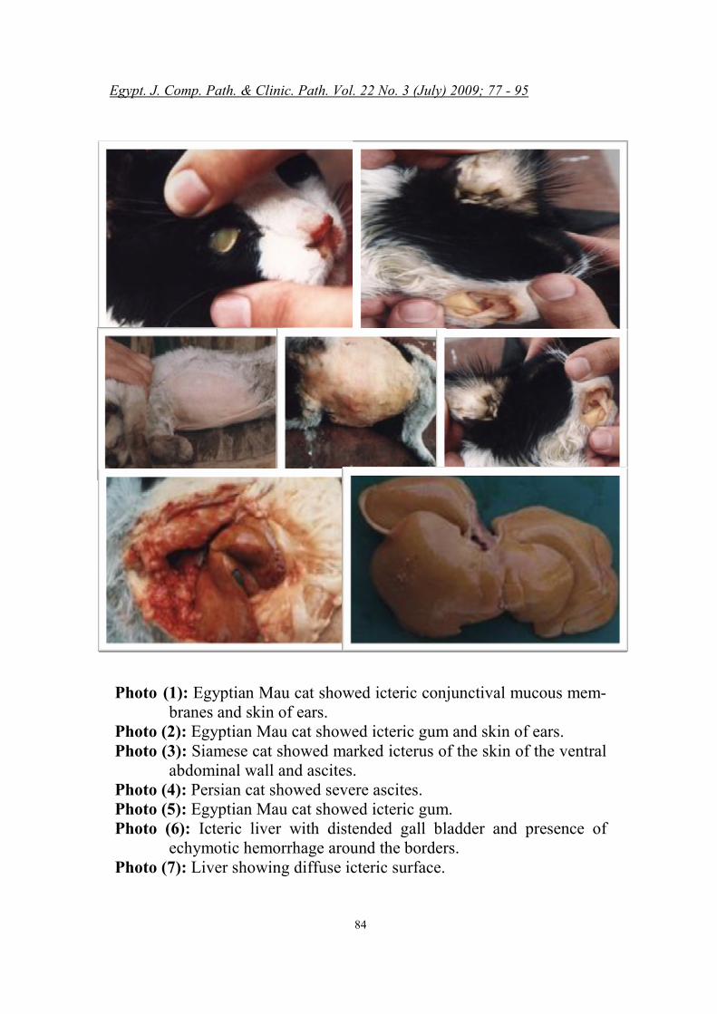
Pathological Results: Gross appearance:
Examined livers showed dif-fuse icteric surface (Photo 6), dis-tended gall bladder and presence of echymotic hemorrhages around the borders (Photo 7).
Microscopical examination:
Microscopic examination of livers from dead cats which have hepatic lipidosis as diagnosed clinically and ultrasonography re-vealed different degrees of lipido-sis as following; it may affect just one hepatic lobule particularly around the central vein as mono-lobular periacinar fatty degen-eration. The rest of the hepatic cells showed different degrees of degenerative changes (Photomic-rograph, 1 & 2).
Hepatic lipidosis may also
progressively extend to invade the whole hepatic lobule and also infil-trating many other lobules as mul-tilobular hepatic lipidosis, (Phot-omicrograph, 3 & 4). Moreover, Fibrosis appeared surrounding the hepatic lobules as perilobular fi-brosis, (Photomicrograph, 5 & 7). That appeared greenish in color with Masson's trichome stain (Photomicrograph, 8). Also, there was congestion with infiltration of hemosidren pigment (hemosider-osis) in-between the perilobular fibrosis. Hemosidren pigments ap-peared as bluish pigment with
Prussiun blue stain, (Photo-micrograph, 6). Furthermore, the perilobular fibrosis infiltrating the entire of the lobules as intralobu-lar fibrosis, (Photomicrograph, 9 & 11) that appeared greenish in color with Masson's trichome stain (Photomicrograph 10). For the por-tal areas, there were bile duct hy-perplasia, congestion and fibrosis as Sclerosing cholangitis, (Photo-micrograph, 12).
Egypt. J. Comp. Path. & Clinic. Path. Vol. 22 No. 3 (July) 2009; 77 - 95
84
Photo (1): Egyptian Mau cat showed icteric conjunctival mucous mem-branes and skin of ears.
Photo (2): Egyptian Mau cat showed icteric gum and skin of ears. Photo (3): Siamese cat showed marked icterus of the skin of the ventral
abdominal wall and ascites. Photo (4): Persian cat showed severe ascites. Photo (5): Egyptian Mau cat showed icteric gum. Photo (6): Icteric liver with distended gall bladder and presence of
echymotic hemorrhage around the borders. Photo (7): Liver showing diffuse icteric surface.
Egypt. J. Comp. Path. & Clinic. Path. Vol. 22 No. 3 (July) 2009; 77 - 95
85
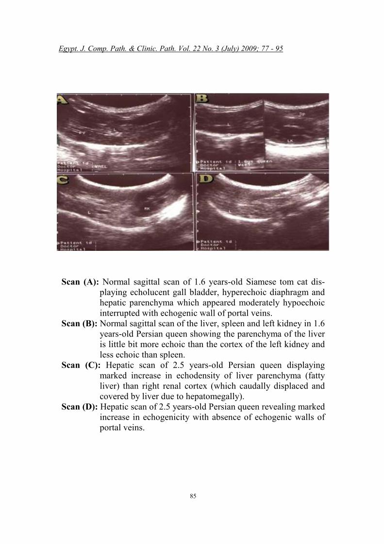
Scan (A): Normal sagittal scan of 1.6 years-old Siamese tom cat dis-playing echolucent gall bladder, hyperechoic diaphragm and hepatic parenchyma which appeared moderately hypoechoic interrupted with echogenic wall of portal veins.
Scan (B): Normal sagittal scan of the liver, spleen and left kidney in 1.6 years-old Persian queen showing the parenchyma of the liver is little bit more echoic than the cortex of the left kidney and less echoic than spleen.
Scan (C): Hepatic scan of 2.5 years-old Persian queen displaying marked increase in echodensity of liver parenchyma (fatty liver) than right renal cortex (which caudally displaced and covered by liver due to hepatomegally).
Scan (D): Hepatic scan of 2.5 years-old Persian queen revealing marked increase in echogenicity with absence of echogenic walls of portal veins.
Egypt. J. Comp. Path. & Clinic. Path. Vol. 22 No. 3 (July) 2009; 77 - 95
86
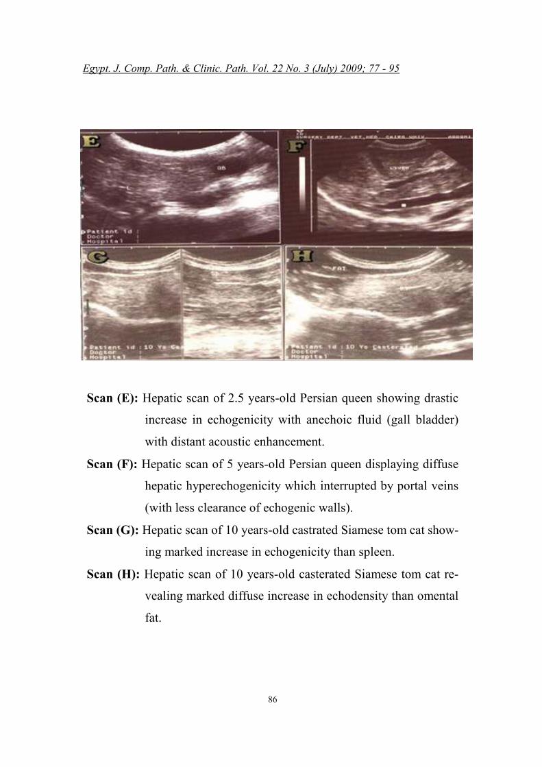
Scan (E): Hepatic scan of 2.5 years-old Persian queen showing drastic
increase in echogenicity with anechoic fluid (gall bladder)
with distant acoustic enhancement.
Scan (F): Hepatic scan of 5 years-old Persian queen displaying diffuse
hepatic hyperechogenicity which interrupted by portal veins
(with less clearance of echogenic walls).
Scan (G): Hepatic scan of 10 years-old castrated Siamese tom cat show-
ing marked increase in echogenicity than spleen.
Scan (H): Hepatic scan of 10 years-old casterated Siamese tom cat re-
vealing marked diffuse increase in echodensity than omental
fat.
Egypt. J. Comp. Path. & Clinic. Path. Vol. 22 No. 3 (July) 2009; 77 - 95
87
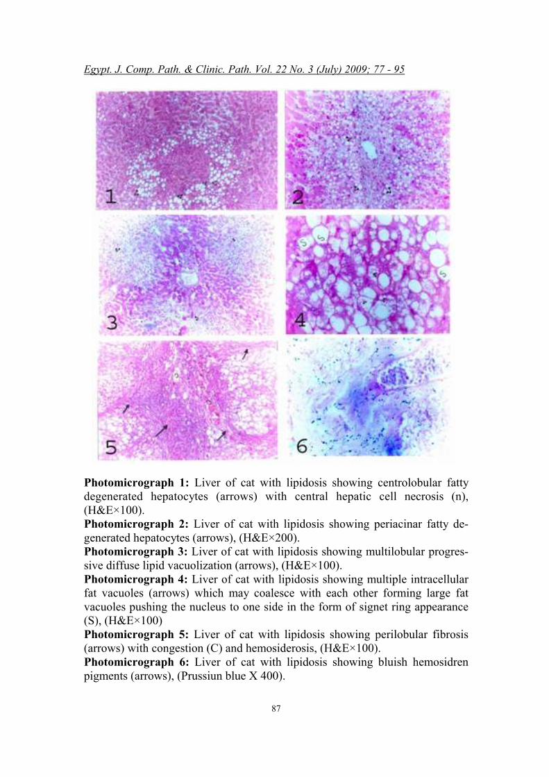
Photomicrograph 1: Liver of cat with lipidosis showing centrolobular fatty degenerated hepatocytes (arrows) with central hepatic cell necrosis (n), (H&E×100). Photomicrograph 2: Liver of cat with lipidosis showing periacinar fatty de-generated hepatocytes (arrows), (H&E×200). Photomicrograph 3: Liver of cat with lipidosis showing multilobular progres-sive diffuse lipid vacuolization (arrows), (H&E×100). Photomicrograph 4: Liver of cat with lipidosis showing multiple intracellular fat vacuoles (arrows) which may coalesce with each other forming large fat vacuoles pushing the nucleus to one side in the form of signet ring appearance (S), (H&E×100) Photomicrograph 5: Liver of cat with lipidosis showing perilobular fibrosis (arrows) with congestion (C) and hemosiderosis, (H&E×100). Photomicrograph 6: Liver of cat with lipidosis showing bluish hemosidren pigments (arrows), (Prussiun blue X 400).
Egypt. J. Comp. Path. & Clinic. Path. Vol. 22 No. 3 (July) 2009; 77 - 95
88
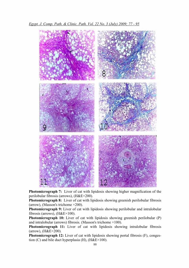
Photomicrograph 7: Liver of cat with lipidosis showing higher magnification of the perilobular fibrosis (arrows), (H&E×200). Photomicrograph 8: Liver of cat with lipidosis showing greenish perilobular fibrosis (arrow), (Masson's trichome ×200). Photomicrograph 9: Liver of cat with lipidosis showing perilobular and intralobular fibrosis (arrows), (H&E×100). Photomicrograph 10: Liver of cat with lipidosis showing greenish perilobular (P) and intralobular (arrows) fibrosis. (Masson's trichome ×100). Photomicrograph 11: Liver of cat with lipidosis showing intralobular fibrosis (arrow), (H&E×200). Photomicrograph 12: Liver of cat with lipidosis showing portal fibrosis (F), conges-tion (C) and bile duct hyperplasia (H), (H&E×100).
Egypt. J. Comp. Path. & Clinic. Path. Vol. 22 No. 3 (July) 2009; 77 - 95
89
DISCUSSION
O n the basis of our investiga-tions; no predisposition of
age, gender and breed of cats were found in feline hepatic lipidosis as other hepatobiliary disorders. These findings agreed with the re-sults reported by Bennette et al., (2007) and Posner et al. (2008).
The most common clinical
symptoms, which may indicate he-patic parenchyma degeneration are anorexia, apathy, vomiting, diar-rhea, polyuria, polydipsia, jaun-dice, coagulopathy, weight loss, dehydration, discoloring of feces and anemia (Blaxter, 1991; Barsanti et al., 1997 and Weiss et al., 1997). Clinical diagnosis of feline hepatic diseases is relatively difficult, because of the substantial functional reserve of the liver. Often pathological signs appear when the function of 70 – 80% of hepatic parenchyma is compromis-ed. Rothuizen and Meyer (2000) ascertain that the above mentioned clinical signs, appear in multiple diseases of different pathogenesis and thus additional diagnostic methods are essential to confirm primary hepatic dysfunction. Basic diagnostic methods (hematology, biochemical blood parameters and ultrasonography) used in the identification of hepatic and bile tract diseases are auxiliary in nature and should be interpreted in connection with anamnesis and
clinical examination (hepatome-gally on palpation and fluid thrill with tactile percussion in ascetic cats).
The hematological profile of
clinically diseased cats showed significant decreases in RBCs count, Hb concentration and PCV revealing a normocytic normo-chromic anemia which seemed to be nutritional anemia and could be attributed to the anorexia which was a consistent clinical finding in all examined cases. Moreover, stress leukogram was obvious and characterized by leukocytosis, neu-trophilia, lymphopenia, monocyto-penia and eosinopenia (Feldman et al., 2000).
The significant increases in
AST, ALT, bilirubin, triglycerides, total cholesterol, HDL, LDL and VLDL with the significant de-crease of BUN, total proteins, al-bumin and A/G indicate hepatocel-lular damage while the significant increases of ALP and GGT indi-cate billary obstruction (Blancha-rd et al., 2002 and Blanchard et al., 2004).
Both types of liver damage
were indicative of severe intra-hepatic cholestasis. So in the pre-sent study, serum hepatic chemis-try panel detected the alterations in hepatic functions but failed to de-fine the nature of hepatobiliary dis-
Egypt. J. Comp. Path. & Clinic. Path. Vol. 22 No. 3 (July) 2009; 77 - 95
90
order. The findings of ultrasonogra-
phy included a diffuse hyperecho-genicity of hepatic parenchyma, decreased visualization of the por-tal blood vessels and enlargement of liver which was observed by caudal displacement of right kid-ney. The liver parenchyma was isoechoic or hyperechoic, com-pared with falciform or omental fats (normally, the liver is less echoic than falciform or omental fats). The liver was also hypere-choic with respect to the spleen and renal cortices (normally, the liver is less echoic than spleen and more echoic than renal cortices). Also, there was increased attenua-tion of ultrasound waves by liver parenchyma. Ultrasonography is useful in evaluating liver paren-chymal structure as increased liver echogenicity and attenuation of sound are associated primarily with cirrhosis and fatty infiltration (Gosink et al., 1979; Joseph et al., 1979; Foster et al., 1980 and Nakamura et al., 2005). The ex-cellent ability of ultrasonography to identify severe hepatic lipidosis in cats correctly was expected be-cause fatty tissues, in general, are hyperechoic and attenuate sound more rapidly than other soft tissues (Freese and Lyons, 1977; Behan and Kazam, 1978; Willard et al., 1999 and Nakamura et al., 2005). The ultrasonographic criteria that
we studied were based on these 2 properties and they were similar to the criteria used to accurately de-tect fatty infiltration in human liver (Gosink et al., 1979; Scatarige et al., 1984 and Brown et al., 2000).
Ultrasonography which is a quick, non-invasive method of im-aging the abdominal viscera was an accurate method of diagnosing severe hepatic lipidosis in the group of cats studied. In clinical practice, ultrasonographic exami-nation of the liver, prior to/or in the absence of liver biopsy, should be a useful, reliable diagnostic test for detection of severe hepatic lipi-dosis in cat with hepatobiliary dis-ease and these results confirmed with those of histopathology which revealed different degrees of lipi-dosis starting with monolobular periacinar fatty degenerations, then multilobular hepatic lipidosis. These results come in accordance with those of Jones (1989). More-over, perilobular and intralobular fibrosis together with congestion and hemosiderosis were formed and confirmed with Masson's trichome stain and Prussiun blue stain, (Hitt, 1989).
Our histopathological results
of portal tract changes agreed with those reported by Thornburg et al., (1982) and Edwards et al., (1983) and revealed Sclerosing cholangitis.
Egypt. J. Comp. Path. & Clinic. Path. Vol. 22 No. 3 (July) 2009; 77 - 95
91
CONCLUSION
F rom the research, it was con-cluded that ultrasonogaphy
and serum chemistry panel were complementary to each other. It can also be confirmed by autopsy and histopathology of dead cats. The serum chemistry panel could detect the hepatic dysfunction while ultrasonography defined the nature of lesions. Further diagnos-tic evaluation is recommended which includes guided needle bi-opsy and computed tomography to determine whether the liver is cul-prit and rule out diseases of other organ system as well as to extend the diagnostic value of ultrasono-graphy.
REFERENCES Allain, C.C., Poon, L.S., Chan,
C.S., Richmond, W. and Fu, P.C. (1974): "Enzymatic de-termination of total serum cholesterol." Clin. Chem., 20: 470-475.
Alpers, D.H. and Sabesin, S.M. (1987): "Fatty liver : Bio-chemical and clinical as-pects." In: Schiff L, Schiff ER, Disease of the liver, 6thed. Philadelphia: Lippincott Co.; 949-978.
Bancroft, J.D. and Stevens, A. (1996): Theory and practice of histological technique. 4th ed., Churchill, Livingston, New York.
Barsanti, J.A.; Jones, B.D. and Spano, J.S. (1997): Pro-longed anorexia associated with hepatic lipidosis in three cats." Feline Pract.; 7: 52- 57.
Behan, M. and Kazam, E. (1978): "The echogenic char-acteristics of fatty tissues and tumors." Radiol.; 129: 143-151.
Bennette, S.L.; Milne, M., Slo-combe, R.F. and Landon, B.P. (2007): "Gall-bladder mucocele and concurrent he-patic lipidosis in a cat." Aust. Vet. J.; 85(10): 397-400.
Blanchard, G.; Paragon, B.M.; Milliat, F. and Lutton, C. (2002): "Dietary L-carnitine supplementation in obese cats alters carnitine metabolism and decreases ketosis during fasting and induced hepatic lipidosis." J. Nutr.; 132(2): 204- 210.
Blanchard, G.; Paragon, B.M.; Serougne, C.; Ferezou, J.; Milliat, F. and Lutton, C. (2004): "Plasma lipids, lipo-protein composition and pro-file during induction and treatment of hepatic lipidosis in cats and the metabolic ef-fect of one of daily meal in healthy cats." J. Anim. Physiol. Anim. Nutr.; 88(3-4): 73-87.
Blaxter, A. (1991): "Diagnosis
Egypt. J. Comp. Path. & Clinic. Path. Vol. 22 No. 3 (July) 2009; 77 - 95
92
and Mangement of Hepatic Disorders." Feline Practice. London , 17-138.
Brown, B.; Mauldin, G.E.; Arm-strong, J.; Moroff, S.D. and Mauldin, G. N. (2000): "Metabolic and hormonal al-terations in cats with hepatic lipidosis." J. Vet. Intern. Med.; 14 (1): 20-26.
Burrows, C.F.; Chiapella, A.M. and Jezyk, P. (1981): "Idiopathic feline hepatic lipi-dosis:The syndrome and speculation on its pathogene-sis." Florida Vet. J.Winter;18-20.
Center, S.A. (1986): "Hepatic lipidosis in the cat." In the Proceedings of the 4th Ameri-can College of Veterinary In-ternal Medicine Forum; 13: 71-79.
De-Maria, R.; Divari, S.; Bo, S.; Sonnio, S.; Lotti, D.; Capuc-chio, M.T. and Castagnaro, M. (1998): "Beta-galacto-sidase deficiency in a Korat cat: a new form of feline GM1-gangliosidosis." Acta Neuropathol.; 96(3): 307-314.
Doumas, B.T., Perry, B.W., Sasse, E.A. and Straum-fjord, J.V. (1973): "Standard-ization in bilirubin assays: Evaluation of selected meth-ods and stability of bilirubin solutions." Clin. Chem., 19:
984-993.
Doumas, B.T., Watson W.A. and Biggs H.G. (1971): "Albumin standards and the measure-ment of serum albumin with bromcresol green." Clin. Chim. Acta; 31:87-96.
Edwards, D.F., McCracken, M.D. and Richardson, D.C. (1983): "Sclerosing cholangi-tis in a cat." J. Am. Vet. Med. Assoc.; 182:710.
Fabiny, D.L. and Ertingshausen, G. (1971): "Automated Reac-tion-Rate Method for Deter-mination of Serum Creatinine with the Centrifi." Chem. Clin. Chem., 17: 696-700.
Feldman, B.F., Zinkl, J.G. and Jain, N.C. (2000): "Schalm’s Veterinary Hematology." 5th (ed.), Lippincott Williams & Wilkins, Philadelphia, USA.
Foster, K.J.; Dewbury, K.C. and Griffith, A.H. (1980): "The accuracy of ultrasound in the detection of fatty infiltration of the liver." Br. J. Radiol.; 53: 440-442.
Freese, M. and Lyons, E.A. (1977): "Ultrasonic back scat-ter from liver tissue: its de-pendence on frequency and protein/lipid composition." J. Clin. Ultrasound; 5: 307-312.
Friedewald, W.T., Levy, R.I. and Fredrickson, D.S. (1972): "Estimation of the concentra-
Egypt. J. Comp. Path. & Clinic. Path. Vol. 22 No. 3 (July) 2009; 77 - 95
93
tion of LDL cholesterol in plasma, without use of the preparative ultracentrifuge." Clin. Chem., 18: 499-504.
Gornall, A.G.; Bardwill, C.J. and David, M.M. (1949): "Determination of serum pro-teins by means of the Biuret reaction." J. Biol. Chem.; 177: 75.
Gosink, B.B.; Lemon, S.K. and Scheible, W. (1979): "Accur-acy of ultrasonography in di-agnosis of hepatocellular dis-ease." A.J.R.; 133: 19-23
Hitt, M.E. (1989): "Feline hepatic lipidosis." In the proceedings of 56th Annual AAHA Meet-ing, St Louis, Mo; April; 7-14; 374-377.
Irwan, T.M. (1996): In "Applied Linear Statistical models", Neter, Kutner, Neachtsheim Wasserman. 4th (ed.).
Jacobs, G.; Cornelius, L. and Al-len, S. (1989): "Treatment of idiopathic hepatic lipidosis in cats." J. Am. Vet. Med. Assoc.; 195: 635-638.
Jones, B.R. (1989): "Feline liver disease." Aust. Vet. Pract.; 19:28.
Joseph, A.E.A.; Dewbury, K.C. and McGuire, P.G. (1979): "Ultrasound in the detection of chronic liver disease (the bright liver)." Br. J. Radiol.; 52: 184-188.
Kelly, W.R. (1984): "Examination of abdomen in small animals." Textbook of Veterinary Clini-cal Diagnosis, 3rd (ed.); 26-46.
Masson, P. (1929): "Trichrome stainings and their prelimi-nary technique." J. Techn. Meth.; 12, 75-90.
Nakamura, M.; Chen, H.M.; Momoi, Y. and Iwasaki, T. (2005): "Clinical application of computed tomography for the diagnosis of feline hepatic lipidosis." J. Vet. Med. Sci.; 67(11): 1163- 1165.
Nyland, T.G.; Hager, D.A. and Herr i ng , D .S . (1 9 8 9 ) : "Sonography of the liver, gall bladder and spleen." Seminars in Veterinary Medicine and Surgery (Small Animal); 4: 13 –31.
Perls, N. (1867): Virchows Arch. Path. Anat., 39: 42.
Posner, L.P.; Asakawa, M. and Erb, H.N. (2008): "Use of propofol for anesthesia in cats with primary hepatic lipido-sis." J. Am. Vet. Med. Assoc.; 232(12): 1841-1843.
Reitman, S. and Frankel, S. (1957): "Colorimeteric meth-od for aspartate and alanine aminotransferases." Am. J. Clin. Path.; 28: 56.
Rogers, Q.R. (1994): "Experim-ental induction of hepatic lipi-dosis in cats." Am. J. Vet.
Egypt. J. Comp. Path. & Clinic. Path. Vol. 22 No. 3 (July) 2009; 77 - 95
94
Res.; 55 (9): 1291- 1302.
Rothuizen, J. and Meyer, H.P (2000): "History, physical ex-amination and signs of liver disease. Textbook of Veterin-ary Internal Medicine." W.B. Saunders comp., Philadelphia.
Roy, A.V. (1970): "Rapid method for determining alkaline phos-phatase activity in serum with thymolphthalein monophos-phate." Clin. Chem., 16: 431.
Scatarige, J.C.; Scott, W.W. and Donovan, P.H. (1984): "Fatty infiltration of the liver: ultra-sonographic and computed tomographic correlation." J. Ultrasound Med.; 3(1): 9-14.
Szasz, G. (1976): "Determination of serum GGT." Clin. Chem.; 22: 2051.
Tabacco, A., Meiattini, F., Moda, E. and Tarli, E. (1979): "Simplified enzymic/color-imetric serum urea nitrogen determination". Clin. Chem., 25: 336-337.
Thornburg, L.P.; Simpson, S. and Digilio, K. (1982): "Fatty liver syndrome in cats." J. Am. Anim. Hosp. Assoc.; 18: 397-400.
Trinder, P. (1959): "Determin-ation of blood glucose using 4-Amino-phenazone". J. Clin. Pathol., 22: 246.
Wahlefeld (1974): In: "Methods
of Enzymatic Analysis", Vol. 5, Bergmeyer, H.U., Aca-demic Press, New York, pp. 1831-1835.
Warnick, G.R., Benderson, V. and Albers, N. (1983): "Sele-cted methods." Clin. Chem., 10: 91-99.
Weiss, D.j., Armstrong, P.J. and Gagne, J. (1997): "Inflamm-atory liver disease." Am Small Anim., 12: 22-27.
Willard, M.D.; Weeks, B.R. and Johnson, M. (1999): "Fine-needle aspirate cytology sug-gesting hepatic lipidosis in four cats with infiltrative he-patic disease." J. Feline Med. Surg.; 1 (4): 215-220.
Yeager, A.E. and Mohammed, H. (1992): "Accuracy of ul-trasonography in the detection of severe hepatic lipidosis in cats", Am. J. Vet. Res.; 53 (4): 597-599.
Egypt. J. Comp. Path. & Clinic. Path. Vol. 22 No. 3 (July) 2009; 77 - 95
95
FGHIا KL ىNOPIا QRSTIا UVW تYZدرا
****NpRn دردjiى٠د، *** jmn NIYoان٠د،** NOW hij`k ا٠N`gIد، * واbc آ`_^٠Uد*TUVWXYرس ا^[\اض اa]-ى\cdXYا ecYا Tdfه\ة-آWjYا Tk]Wl
**WdlmYmnWXYرس اa]-ى\cdXYا ecYا Tdfه\ة-آWjYا Tk]Wl ***TdodUdfا^آ WdlmYmnWXYرس اa]-ى\cdXYا ecYا Tdfه\ة-آWjYا Tk]Wl
****WdlmYmnWXYا pqr-انmdtYا Ttu ثmtw axk]-yraYا
UqjrIا stVpIا zf{ T|راaYا }~ه\ة ���٤٠ هW� �d���Y T�f���Yا ��W|mYا �dw Tر�Wj�Yف اaxw Tcr
�cjYا z� يaXoYا pt��Yر ا^}\اض . اmx� eq� yf{ �cjYا TdYWtYا T|راaYا ��qr �] T�mo] آ� �d�{m��] zYإ T�\�\qY٢٠اTcr٠ T�dfqYا TcwW�Yا T{m���Yون ( اaw
T�\�\| ا}\اض ( Wxdf{ ت\x� y�Yي واaXoYا pt��YWw TwW��Yا T�\�\qYا T{m���Yو اا^}\اض اT�\�\qY [�� ا�u\ار ا^��Td اTdVW��Y و���p اaXoY وا�|�Wjqء
���t آ� اmdtYا�Wت adr اaYرا|T ٠واxY£ال وأ}\اض }�TdX وr¡ وإ|Wxل aXoYا ¤�Wرات و�WX�¥اء ا\lإ pم و�aYرة اmu ��t� Wآ� Td�m�Yق اm� تWlm�YWw aXoY Tdt�\��Yا T��Yوا ylmYmnWwm�qdxYا �t�Yاء ا\lإ pو� Wم آ�aYم ا\dqY Td�Wd�doYا
T�\�\qYا T{m���Yا �] Tj�WUYت ا�WtYد . اmlو Td�m�Yق اm� تWlm�YWw �t�Yا \xا�وar أو»�t درا|T اaYم وmlد ا�W{ Wd�dد�T اTªX�Y . اpt��Y اaXoYي m�wرة ©a�aة
Wآ� �dو�\�dUYا W��¥ دةWز� �{ T��W� ءW�dXYد آ\ات اa{ y� دةWز� ] p�tYواأو»�t درا|d| T\م اaYم ار��W��£�®w W�mtf] W{Wت اaXoY وار��W{W وا»��w Wt\اء TdYWkYاT�W�oYذات ا TdUهaYت اWUdو�\XYوا yfoYول ا\d�qdYmoYوا Tdn��Yن اmهaYم واaYا �d]mdXY^ى اm�q] ضWض [ ا���W���^ة اa�a© T�W�oYوذات ا T���U�Yا T�W�oYوذات ا Tاو�\��Yرة اW�kYاد [�\ى اaqا� yf{ وة�{ aXoYا W��¥ y� ¤f� وثa� yf{ TYد� .
°df{ T�£�^د اmlوو aXoYا ±c| ار\�uا �] Tj�WUYا �cjfY Tdt�\��Yا T��Y² اYذ aوأآ ] aXoYا W���w ز\oU� ت\xا� z�Yوا TdlmYmnWwm�qxYت اWumt�Yا zYا T�W«^Ww
W���Yل ه~{ اm� T{WU�Yا W��¥ دmlوو TdUت دهW{Wj� دmlو . T|راaYا ��f¥ ²Y~wو� Wlm�YWwت �mق اaU{ Td�m�Y ا�©�WX{ أو �mxر t�fY \oX�Yام اa��|ورة ا�\« zYا �] a³آ�Yوا Tdf�k�Yت اWumt�Yاء ا\lوا �cjYا z� يaXoYا pt��Yه\ة اW´Y أي }\ض
TdlmYmnWwm�qxYت اWumt�Yوا Tdt�\��Yا T��Yا ��kw ²Yذ .
:اm�ot�Yن
Tk]Wl اWjYه\ة –آTdf اecY اcdXY\ى –أ|�Wذ اo| \dx| WdlmYmnWXY\. د.أ
T¶¶k]Wl –آT¶¶df اe¶¶cY اcdXY¶¶\ى –أ|¶¶�Wذ اW¶¶dlmYmnWXY ا�آW�u T¶¶dodUdfء �µ ر[�Wن. د.أ اWjYه\ة


![LIPIDOSE HEPÁTICA FELINAarquivo.fmu.br/prodisc/medvet/lcfb.pdf · Hepatic Lipidosis Feline [ Lipidose Hepática Felina], São Paulo,2008, 31 páginas ( Trabalho de Conclusão de](https://img.pdfslide.net/doc/110x75/5eaa331c64216f6988776c62/lipidose-heptica-hepatic-lipidosis-feline-lipidose-heptica-felina-so-paulo2008.jpg)