Embed Size (px)
Citation preview
Studies on Pigment Production by Microorganisms Using Raw
Materials of Agro-industrial Origin
Thesis Submitted by
TARANGINI KORUMILLI (509CH107)
In partial fulfillment for the award of the Degree of
DOCTOR OF PHILOSOPHY
IN
CHEMICAL ENGINEERING
Under the guidance of
Dr. Mrs. SUSMITA MISHRA
Department of Chemical Engineering
NATIONAL INSTITUTE OF TECHNOLOGY
Rourkela 769 008, Orissa, India
June-2014
i
Dr. (Mrs.) Susmita Mishra
Associate Professor
Department of Chemical Engineering
E-mail: [email protected], [email protected]
CERTIFICATE
This is to certify that the thesis entitled “Studies on Pigment Production by Microorganisms
Using Raw Materials of Agro-industrial Origin” submitted by Tarangini Korumilli (Roll No-
509CH107) to National Institute of Technology, Rourkela in partial fulfillment of the
requirements for the completion of the Ph. D. degree in Chemical Engineering, is an authentic
work carried out by her under my supervision and guidance.
Dr. (Mrs.) Susmita Mishra
ii
Acknowledgements
I express my heartfelt gratitude to Prof. (Mrs.) Susmita Mishra, my supervisor, Department of
Chemical Engineering, NIT, Rourkela for her constant encouragement, invaluable advice and
guidance throughout the course of my research work. I shall remain ever grateful for her care,
concern and sincere interest in my prosperity. I must mention that without her timely help in
writing and correction, this thesis could not have been submitted in time.
I am thankful to Prof. R. K. Singh, HOD, Department of Chemical Engineering NIT, Rourkela
for his help and support during the course of my tenure. I owe a depth of gratitude to Prof. G. K.
Roy, Prof. K. C. Biswal, Prof. S. K. Agarwal and Prof. P. Rath, Chemical Engineering
Department, NIT Rourkela, for their support and encouragement.
A special thanks to Prof. (Mrs.) Madhushree Kundu, Prof. Santanu Paria, Prof. Basudeb munshi
for their for their constant support throughout my programme. I am also thankful to Prof. (Mrs.)
Abanti Sahoo, Prof. Hara Mohan Jena, Prof. Sujit Sen and Prof. Arvind kumar, Department of
Chemical Engineering for their valuable advices and moral support.
I would like to thank all the faculty members and non-teaching staff of Chemical Engineering
department for their constant support during my course work. I am thankful to Prof.(Mrs). A.
Mandal and Prof.(Ms.) Usharani Subuddhi, Department of Chemistry for their analytical support.
My heartfelt thanks to my labmates Ramkrishna, Balaji Patro, Adya das,Vijiya Jha, Shilpi for
their joyous company and for helping me in several ways. Special thank to my friend Sateesh
Sagiri and thanks to all my friends of the department for their company and support.
And it goes without saying, that I am indebted to my parents, my husband and brothers, whose
patience, support and endurance made completion of my course a reality.
iii
Abstract
The recent awareness in human safety and environmental conservation has created fresh
enthusiasm for natural sources of pigments. Compared to synthetic pigments, microbial pigments
shows better biodegradability and higher compatibility with the environment, and have numerous
applications from food to cosmetics. Identification of new microbial sources, utilization of low
cost substrates and optimization of process parameters are the areas under focus towards
economical pigment production. The present study aimed at screening and identification of
microbial isolates from soil and water, which are having pigment producing ability. Efforts have
been made to cultivate them on numerous cheaper and inexpensive substrates with no special
conditions and supplements for effective pigment production. Furthermore, two carotenoid
producing strains were also exploited on numerous inexpensive substrates at ambient conditions
for pigment metabolites. While analyzing pigment metabolites in all cases, key parameters
influencing pigment production by respective strains were optimized by utilizing statistical
techniques like Taguchi method and response surface approaches wherever needed. The
conditions for enhanced pigment production were established employing microbial isolates and
purchase strains individually. The melanin producing Pseudomonas guinea, (bacterial strain) was
isolated from marine water sample and was employed on vegetable waste for effective pigment
production. Another strain of Bacillus safensis was isolated from garden soil and showed its
ability to produce melanin on fruit waste extract (FWE). It is noteworthy that both melanins
produced from marine and soil isolates showed antioxidant, photoprotective and metal ion
chelation activities. Addressing garden soil, a new carotenoid producing bacterial strain Bacillus
clausii was screened and cultivated on FWE for high yield pigment production. The pigment
produced by this strain was observed to be a β-carotenoid type and its stability towards thermal
treatment was also evaluated. Eying on the significance of carotenoids, microorganisms
(Rhodotorula rubra, Xanthophyllomyces dendrorhous) in their developmental stage were
purchased and studied for pigment production on various residues as sole substrates. The
obtained yeasts showed improved carotenoids yield i.e. torularhodin and astaxanthin respectively
on FWE. In a nut shell we could conclude that there is a huge scope for industrial scale
production of Melanin and Carotinoid using easily available agro-industrial raw materials such
as rice powder and fruit waste extract (FWE).
iv
CONTENTS
Particulars Page no.
Certificate i
Acknowledgements ii
Abstract iii
List of Figures x
List of Tables xxiii
Chapter 1 Introduction 1
1.1 Motivation and scope 5
1.2 Organization of thesis 7
References 9
Chapter 2 Literature Review 13
2.1 Outline 13
2.2 History of colorants 14
2.3 Dyes and pigments – their implications 15
2.4 Biological pigments- different classes 17
2.4.1 Biological pigments 17
2.4.2 Plant pigments 18
2.4.3 Animal pigments 19
2.4.4 Microbial pigments 20
2.5 Significance of microbial pigments as natural colorants 23
2.6 Classification of microbial pigments 25
2.7 Microbial pigments of commercial importance 29
2.8 Benefits and Applications of Microbial pigments 33
2.9 Novel practices of microbial pigment production 35
2.9.1 Strain improvement 35
2.9.2 Fermentation 36
2.9.3 Low-cost substrates 38
v
2.10 Objectives of the work 39
References 40
Chapter 3 Screening of inexpensive substrates for pigments production by 55
isolated and known microbial strains - Bridge to Part I and II
of Chapter 4
3.1 Melanins and carotenoids - pigments of marketable importance 55
3.2 Sampling 58
3.3 Screening of pigment producing microorganisms 59
3.4 Different substrates and Cultivation of isolates/purchased strains 59
3.5 Strain/substrate selection and further experimentation 60
3.6 Summary 63
References 63
Chapter 4 Approach and investigations
Part I - Isolation of Pigment Producing Bacteria and the Production of 66
Pigments on Cheaper Substrates
4.1 Melanin from Isolated Marine Pseudomonas sp. using Vegetable 66
waste
4.1.1 Introduction 67
4.1.2 Materials and methods 69
4.1.2.1 Chemicals used 69
4.1.2.2 Screening and isolation of the melanin producing strain 69
4.1.2.3 Pigment production, extraction and purification 69
4.1.2.4 DPPH/SPF/metal chelating assay 70
4.1.2.5 Analytical methods 74
4.1.3 Results and Discussion 74
4.1.3.1 Strain selection and characterization of the microorganism 74
4.1.3.2 Pigment production and characteristics of the produced 78
melanin
vi
4.1.3.3 Spectroscopy, SEM/EDX and IR analysis of melanin 81
4.1.3.4 DPPH assay 85
4.1.3.5 XRD analysis 86
4.1.3.6 SPF values determination 88
4.1.3.7 The metal binding capacity of melanin 89
4.1.4 Conclusions 90
References 90
4.2 Melanin by Soil Microbial Isolate on Fruit Waste Extract: - 93
Optimization of Key Production Parameters
4.2.1 Introduction 93
4.2.2 Materials and methods 95
4.2.2.1 Chemicals and microorganism 95
4.2.2.2 Substrate preparation 96
4.2.2.3 Production and purification of melanin 97
4.2.2.4 Parameters optimization using Taguchi and CCD design 97
4.2.2.5 SPF/DPPH/metal chelating assay 98
4.2.2.6 Analytical methods 101
4.2.3 Results and discussion 101
4.2.3.1 Strain selection and Pigment production 101
4.2.3.2 Taguchi design for screening critical factors 102
4.2.3.3 Optimization by response surface methodology 105
(CCD design)
4.2.3.4 Interaction effects of variables and validation of the 106
model
4.2.3.5 Spectroscopy, IR and XRD analysis of melanin 108
4.2.3.6 SPF values determination 111
4.2.3.7 DPPH assay 112
4.2.3.8 The metal binding capacity of melanin 113
4.2.3 Conclusions 114
References 114
vii
4.3 Carotenoid by Bacillus clausi Using Rice Powder as the Sole 117
Substrate: Pigment Analyses and Optimization of Key -
Production Parameters
4.3.1 Introduction 117
4.3.2 Materials and methods 119
4.3.2.1 Sampling, microscopy and strain characterization 119
4.3.2.2 Pigment production 119
4.3.2.3 Purification and stability of the pigment 120
4.3.2.4 Optimization of key parameters 121
4.3.3 Results and discussion 121
4.3.3.1 Colony Screening, pigment production 121
4.3.3.2 Microscopy and microbial characterization 123
4.3.3.3 UV-Visible/fluorescence spectrophotometry and stability 125
of the pigment
4.3.3.4 FTIR analysis 127
4.3.3.5 Optimization of key production parameters 129
4.3.4 Conclusions 132
References 132
Part II - Facile Synthesis of Commercial Pigments by Purchased Strains on 138
Cheaper Substrates
4.4 Carotenoid by Rhodotorula sp. on Fruit Waste Extract as a Sole 138
Carbon Source and Optimization of key production parameters
4.4.1 Introduction 138
4.4.2 Experimental section 141
4.4.2.1 Reagents and chemicals 141
4.4.2.2 Microorganism and its maintenance 141
4.4.2.3 Substrate preparation 141
4.4.2.4 Pigment production and extraction 142
viii
4.4.2.5 DPPH assay 143
4.4.2.6 Analysis 143
4.4.2.7 Experimental design 143
4.4.3 Results and discussion 145
4.4.3.1 Effect of pH on biomass and carotenoid yield 145
4.4.3.2 Biomass growth, pigment yield and glucose utilization 146
4.4.3.3 Box–Behnken design 149
4.4.3.4 DPPH radical scavenging activity 155
4.4.4 Conclusions 156
References 157
4.5 Astaxanthin by Xanthophyllomyces dendrorhous Using Fruit Waste 161
Extract as Sole Source of Energy: Optimization of Culture -
Conditions by Taguchi method for Improved Pigment Production
4.5.1 Introduction 161
4.5.2 Materials and methods 163
4.5.2.1 Chemicals and microorganism 163
4.5.2.2 Substrate preparation 163
4.5.2.3 Culture conditions 163
4.5.2.4 Pigment production and extraction/ Sugar determination 164
4.5.2.5 Taguchi design and statistical analysis 164
4.5.2.6 Determination of free radical scavenging activity 165
4.5.2.7 Analytical methods 166
4.5.3 Results and discussion 167
4.5.3.1 Experimental design by Taguchi method 168
4.5.3.2 Validation of the model 168
4.5.3.3 Pigment yield, biomass trend and glucose utilization 170
4.5.3.4 UV analysis 172
4.5.3.5 FTIR analysis 172
4.5.3.6 DPPH radical scavenging activity 172
4.5.4 Conclusions 173

ix
References 174
Chapter 5 Conclusions and future prospectives 178
Publications from this work 183
Curriculum vitae 184
x
List of Figures
Figure No. Title Page No.
Fig. 2.1. Some food grade pigments and their structures from microorganisms. 32
Fig. 4.1.1. Screening of microbial strains obtained from various parts of a
seashore. A) near stones, B) near shore and C) 10 m away from sea.
75
Fig. 4.1.2. Low (a) and high (b) magnification SEM images of the isolated
microorganism with black colonies on agar plates.
76
Fig. 4.1.3. Melanin production on various media: marine broth, blend (marine
broth and vegetable waste - (10:90; 20:80 and 30:70 in v/v %), and
diluted marine broth 30 % (a). M indicates melanin in (a). Dark
brownish black colonies due to melanin production in sole marine
broth (b) and in marine broth – vegetable waste blend medium (c).
80
Fig. 4.1.4. Acid treated (a) and purified melanin (b) after centrifugation. 81
Fig. 4.1.5. UV-visible spectral properties of melanin pigment obtained from
marine broth (a) and marine broth –vegetable waste medium (b).
83
Fig. 4.1.6. SEM images of purified bacterial melanin from a) marine broth and b)
marine broth – vegetable waste medium.
83
Fig. 4.1.7. EDX analysis of elemental composition of melanin from a) marine
broth and b) marine broth – vegetable waste medium.
84
Fig. 4.1.8. The FTIR spectrum of the melanin pigment obtained from a) marine
broth and b) marine broth – vegetable waste medium.
84
Fig. 4.1.9. DPPH radical scavenging activity of synthesized melanin pigment with
various doses (a) and UV-vis absorption spectrum of melanin (44.7
µg/mL) - DPPH at different days along with control containing no
melanin (b). Dose dependent scavenging activity of the synthesized
melanin (c). Insert of (a) shows the melanin doses 0, 14.9, 29.8 and
44.7 µg/mL to 0.1 mM DPPH from left to right.
86
xi
Fig. 4.1.10. (a) X-ray diffractograms of the produced melanin (M-marine broth,
M-blend) and purchased melanin (M-synthetic). (b) Interlayer spacing
(d-value) and crystallite sizes and (c) % crystallinity of the different
mealnins.
87
Fig. 4.1.11. Metal ion chelation effect of the melanin (produced from vegetable
waste) (a) monitored via complete spectrum and (b). at a fixed
absorption maximum.
89
Fig. 4.2.1. Steps involved in substrate preparation along with the prepared FWE
as figure insert.
96
Fig. 4.2.2. (a) FWE before (left) and after melanin production (right) by the
garden soil microbial isolate, (b) colonies with diffusible melanin on
NA plates, and (c) SEM image of the microorganism. (d) Phylogenetic
tree showing the position of the isolate ZJHD1-43 with reference to
related strains.
102
Fig. 4.2.3. Main effects of factors or average of obtained results (mg/mL) in
which each factor is at a given level. For description of ‘levels’ refer to
Table 4.2.1.
104
Fig. 4.2.4. Three dimensional response surface curves with surface plot (a) and
contour plot (b) showing the effect of interactions of pH and
temperature on melanin yield. (c) melanin yield at optimum conditions
with respect to time.
108
Fig. 4.2.5. (a) UV-visible spectrum of melanin pigment obtained from FWE. (b)
FTIR spectra of standard melanin (upper) and bacterial melanin
(lower). (c) X-ray diffractograms of the obtained melanin (upper) and
purchased melanin (lower) and % crystallinity of both are also shown
as indicated by arrow.
111
Fig. 4.2.6. (a) Dose dependent scavenging activity of the synthesized melanin
from FWE and ascorbic acid as a control. (b) Metal ion chelation effect
of the produced melanin in different doses (monitored spectrally) and
(c) At a fixed absorption maximum.
113
Fig. 4.3.1. Microbial isolate with orange – red spotted colonies on nutrient agar 122
xii
slant (a), and orange pigment by the isolated soil microorganism on
rice powder (b). Phase separation of the obtained pigment was shown
in (c). Resultant separated pigment concentrate in liquid (d insert) and
in solid form after vacuum drying was also given in (d).
Fig. 4.3.2. Orange pigments by the isolated soil microorganism on rice powder
plates at pH - 6 (a) and 8 (b) respectively.
123
Fig 4.3.3. Pigment producing bacterial species with an orange pigment (insert)
magnified by an optical microscope at 400 X magnification (a). SEM
images of the identified pigment producing bacteria with (b) 5000 X,
(c) 9000 X magnifications and (d) Phylogenetic tree showing the
position of the isolate XJU-3 with reference to related strains.
124
Fig. 4.3.4. (a) UV- visible spectrum of the extracted orange pigment in ethanol,
(b) UV absorption of the oxidized pigment with narrow and wide
(insert of (b)) spectral regions, and (c) Fluorescence emission spectra
of the oxidized pigment at 382 nm.
126
Fig. 4.3.5. ATR-FTIR analysis of the extracted and purified orange pigment
before (a) and after (b) oxidation process.
128
Fig. 4.3.6. Main effects of factors or average of obtained results (pigment per 3
gm rice powder) in which each factor is at a given level. For detail
about ‘levels’ refer to Table 4.3.1.
130
Fig. 4.4.1. (a) Growth of biomass and carotenoid yield at various pHs and at
room temperature (30 0C). Prepared fruit waste extract (FWE) – left,
and FWE with R. rubra growth with intracellular carotenoid (right)
upon incubation (b). CHNS analysis of FWE before and after
fermentation; upon separation of R. rubra by centrifugation (c). SEM
image of the used yeast.
146
Fig. 4.4.2. Carotenoid production (mg/l) and biomass yield (mg/ml) along with
glucose utilization (mg/ml) given in [A]. The picture insert shows the
colored biomass from 1 to 6 days. UV-visible spectrum of the purified
carotenoid in methanol medium [B]. FTIR spectra of the synthesized
148
xiii
carotenoid (a) and the purchased β-carotene (b) pigment.
Fig. 4.4.3. (a) Cell mass concentration calculated from regression model equation
versus the corresponding experimentally obtained values. (b)
Carotenoid production calculated from the regression model equation
versus the corresponding experimentally obtained values.
154
Fig. 4.4.4. Response surface and contour plots obtained from Equation (2) and
(3) showing the effect of the temperature, agitation and their mutual
interaction on biomass concentration (a), (b); while it is effect of pH
and temperature and their mutual interaction on caroteniod pigment
concentration (c), (d). The displayed units of all the graphs are in
natural units.
155
Fig. 4.4.5. Free radical scavenging property of the carotenoid by R. rubra. Dose
dependent scavenging activity of the pigment compared with ascorbic
acid (control).
156
Fig. 4.5.1. Pigmentation of FWE (a) by before X. dendrorhous yeast with lower
(b) and higher (c) magnification SEM images.
167
Fig. 4.5.2. Main effects of factors or average of obtained results as mg/g biomass
in which each factor is at a given level. For description of ‘levels’ refer
to Table 4.5.1.
169
Fig. 4.5.3. (a) Time course of the growth and production of astaxanathin by X.
dendrorhous in FWE. Experiment was carried out at optimum
conditions i.e. pH (5), temperature (20 oC) and agitation (300 rpm). (b)
UV-Visible and FTIR spectrum (c) of the produced pigment in
methanol. (d) Antioxidant activity (DPPH radical scavenging) of
astaxanthin and ascorbic acid.
171
xiv
List of Tables
Table No. Title Page No
Table 2.1. Pigment producing microorganisms based on color and
appearance.
27
Table 2.2. Pigments from various microorganisms which are already in use as
natural food colorants.
30
Table 2.3. Different microorganisms and various inexpensive substrates used
for pigments production.
39
Table 4.1.1. Colony characteristics of the isolated melanin producing
bacterium.
76
Table 4.1.2. Normalized product function used in the calculation of SPF by
Mansur equation.
88
Table 4.1.3. SPF values of different melanins. 88
Table 4.2.1. Factors and their levels which were studied by Taguchi approach. 100
Table 4.2.2. Levels of three different factors, applied in each of 18 trials with
the obtained results.
100
Table 4.2.3. Analysis of variance of main effects of factors. 103
Table 4.2.4. Optimum conditions suggested by statistical calculations after
performing the tests
105
Table 4.2.5. Experimental design matrix for the central composite design. 106
Table 4.2.6. Estimated regression coefficients from the model equation. 107
Table 4.3.1. Factors and their levels studied by Taguchi method. 130
Table 4.3.2. Levels of three different factors in each of sixteen trails and
obtained results.
131
Table 4.3.3. Analysis of variance of main effects of factors. SS/MS indicate
sum/mean of squares in the table.
131
Table 4.3.4. Optimum conditions suggested by statistical analysis after
performing the tests.
132
Table 4.4.1. Coded and actual levels of the three variables. 144
Table 4.4.2. Experimental design matrix for the Box-Behnken design. 144

xv
Table 4.4.3. Statistical significance obtained for the regression coefficients in
Eq’s .(1), (2).
151
Table 4.4.4. ANOVA results for biomass and pigment yields. 151
Table 4.5.1. Factors and their levels studied by Taguchi method. 166
Table 4.5.2. Levels of three different factors applied in each of nine trials, with
observed results.
166
Table 4.5.3. Analysis of variance of main effects of factors. 169
Table 4.5.4. Optimum conditions suggested by statistical calculations after
performing the experiments.
170
1
Chapter 1 Introduction
Pigments are the chemical substances that absorb the light of visible region. The produced
color is because of the chromophore, a molecule specific structure which captures the sun
energy and causes an excitation of electron from external orbital to higher orbital, where the
non-absorbed energy is refracted or reflected to be captured by eye [1]. The modern meaning
related to the word pigment has its origin in the twentieth century, meaning a substance
constituted of small particles which is practically insoluble in the applied medium and is used
due to its colorant, protective or other properties. Pigments are compounds with uniqueness
of importance to many industries. In the food industry they are used as additives,
antioxidants, color intensifiers, etc. Pigments come in a wide selection of colors, some of
which are water-soluble. The terms pigment and color are generally applied for the food
coloring matters, sometimes indistinctly [2-3]. Until the mid-19th
century all colorants were
attained from plant or animal extracts. The textile industry used natural pigments, such as
cochineal, wood madder, turmeric, or henna. In 1856, H. Perkin established the first factory
of organic synthetic colors to produce mauve. A few years later the discovery of diazotization
and a coupling reaction by Peter Griess was the next major step forward for development of
the color industry. In the 19th
century, synthetic organic dyes were developed, creating a more
2
economical and broader range of colorants. Since then their quality has been enhanced due to
extensive research and development [4-5]. The economic consequence of the color industry is
clearly reflected in the large number of synthesized compounds; as many as 700 colorants are
currently available. They have widely been used in foodstuff, dyestuff, cosmetic and
pharmaceutical manufacturing processes; encompass various hazardous effects [6]. All
synthetic food components suffered severe criticism, including synthetic additives and
predominantly food pigments. Today, all food color additives are cautiously regulated by
federal authorities to ensure that foods are safe to eat and accurately labeled [3-5]. Pigments
produced from natural sources are of worldwide interest and is gaining significance. These
are looked upon for their safe use as a natural food dye in substitute of synthetic ones in spite
of having of undesirable market [4]. It is therefore, essential to explore various natural
sources of food grade pigments and their potentials [5]. The utilization of natural pigments in
foodstuff, dyestuff, cosmetic and pharmaceutical manufacturing processes has been mounting
in recent years [7-8-9]. Natural colorants or dyes derived from flora and fauna are believed to
be secure because of non-toxic, non-carcinogenic and biodegradable in nature. Natural
pigments are attained from ores, insects, plants and microbes [10-11].
Among all, microbial pigments are dominant sources. The microbial production of
carotenoids, pigments from vegetables or chemical synthesis, have problems of seasonal and
geographic variability in the production and marketing [21].The microbial production of
carotenoids, They are of great interest owing to the stability of the pigments produced and the
accessibility of cultivation technology [2-3, 7-8, 12-13]. The advantages of pigment
production from microorganisms comprise easy and fast growth in the cheap culture medium,
independence from weather conditions and colors of different shades. The economic
3
advantages of microbial pigments include growth on natural substrates such as red rice wine,
red bean curd as carbohydrate source [21-22]. Microbial colorants are in use in the fish
industry already, for example to improve the pink color of farmed salmon [12-15] In nature,
color rich and pigment producing microorganisms (fungi, yeasts, and bacteria) are fairly
common. Microorganisms produce various pigments like carotenoids, melanins, quinones,
flavins, prodigiosins and more specifically monascins, violacein or indigo [7-8, 16-17].
Carotenoids such as β-carotene and xanthophylls like astaxanthin play central roles in
the metabolism of the eye's macula and retina and in retaining healthy vision. β-carotene play
a constructive role in the prevention of cancer and as chemo-protectives [7] [18-19]. In
addition, it, also act as neutraceutical that avert carcinogenesis through anti-oxidative, anti-
free radical or other mechanisms [20].
The production of microbial pigments is very much affected by the temperature of
incubation, depending upon the type of microorganism [23-24]. The growth of Monascus sp.
entails 25-28 °C for the production of pigment, whereas Pseudomonas requires 35-36 °C for
its growth and pigment production [25]. pH of the medium is another parameter that affect
the growth and kind of pigment produced by the in which microorganisms are grown [25].
The yield of astaxanthin from Phaffia rhodozyma was 325 to 212 µg/g astaxanhin at a pH of
6.5 to 3.5 [26]. Pigment production is also affected by carbon source like glucose, fructose,
lactose, maltose, galactose, etc [27-26] and nitrogen source depending upon the
microorganism [28]. Minerals also has significant role in pigment production [29]. Zn (2x10⁻³
M and 3x10⁻³
M) inhibited the growth in liquid medium whereas in solid medium vigorous
growth and pigmentation was observed.
4
The optimization of growth conditions of microorganisms, particularly physical and
nutritional parameters are of prime importance in the development of any pigment production
process owing to their impact on the economy and practicability of the process. Medium
optimization and physical conditions have been customarily performed using one-factor-at-a-
time method. The disadvantages of such a classical method are that it is time consuming,
laborious and expensive; in addition, it fails to resolve the combined effect of different
factors [30-31]. Eyeing on maximizing the pigment yield, productivity and minimizing the
production costs, most of the recent optimization efforts have relied on statistical
experimental design and response surface analysis [32] and, to a smaller extent, artificial
intelligence techniques such as genetic algorithms [33]. Statistical design is a potent tool that
can be used to account for the main as well as interactive influences of fermentation
parameters on process performance. It is an efficient way to generate useful information with
limited experimentation, thereby limiting the process development time and cost [34].
Therefore, researchers are encouraged to apply statistical experimental approaches such as
Taguchi method [35] and response surface methodology (RSM), which provide a great
amount of information based on only a small number of experiments [31].
On summing up, the growing apprehension over the eventual harmful effects of
synthetic colorants on both the consumer and the environment has raised preferential interest
in natural coloring alternatives [5]. Among all, microbial colorants popularly known as
pigments have some advantages over plant and animal based colorants Extensive studies
proved that microbes are known to produce a large amount of stable pigments. [8, 36]. Large
amounts of agro-industrial and domestic residues are generated from diverse economic
activities; utilization of these residues as inexpensive substrates to support the growth of
5
microorganisms to generate value-added products like pigments are of biotechnological
interest in recent years [8, 37]. Several processes and methodologies have been developed
and developing that utilizes a variety of cheaper substrates and wastes as alternative
substrates for the production of microbial pigments [38]. The utilization of several wastes as
or raw materials notably helps in solving pollution problems, while their disposal may
otherwise cause. In addition to the above, screening of different resources, establishing
simple methodologies, and exploring the microbial synthesis of pigments on inexpensive
substrates is an attractive option to develop commercial scale production [2, 37-38]. Aiming
at natural pigments on readily available agro-industrial materials, this study mainly focus on
1) Isolation of pigment producing microorganisms and the production of pigments (especially
carotenoids and melanins), 2) Simple production of commercial pigments in higher amounts
by purchased strains (which are at developmental stage) and 3) Optimization of key
parameters influencing pigment production where ever necessary
1.1 Motivation and scope
The use of synthetic organic colors has been acknowledged for many years as the most
reliable and economical method of restoring some of the food’s original shade to the
processed product. Synthetic colors are superior to natural pigments in tinctorial power, ease
of application, stability, and cost effectiveness. However, from the health safety viewpoint
they are not accepted by consumers, so over the past years growing interest in natural food
colorants has been observed [5].
The utilization of natural pigments in foodstuff, dyestuff, cosmetic and
pharmaceutical manufacturing practices has been increasing in recent years [24]. Natural
6
pigments can be obtained from three major sources i.e. animals, plants [25] and
microorganisms [13]. The accessible authorized natural pigments from animals and plants
have numerous drawbacks such as limited range, volatility against light, heat or adverse pH,
low water solubility and are often non-availability throughout the year. Moreover microbial
pigments are of great interest owing to the stability of the pigments produced and the
availability of cultivation technology [17], [26]. The benefits of pigment production from
microorganisms include easy and fast growth in the cheap culture medium, independence
from weather conditions and colors of different shades. Hence, microbial pigment production
is now one of the promising and emerging fields of research to reveal its potential for various
industrial applications [13], [27], [28].
From an industrial point of view, there is a necessity to develop a high throughput and
cost-effective approaches for large scale production of various microbial pigments.
Conventional media used for the biosynthesis of microbial pigments are rich in a variety of
nutrients. Microorganisms vary in their needs to carbon sources according to their nutrient
nature; the use of pure carbon sources e.g. (glucose, sucrose, and fructose) is expensive from
cost-effective casing, so the industrial processes try to use contemptible carbon sources
especially industrial wastes, variety of plant seed oils etc. have also been used as carbon
substances for obtaining different pigments. From an industrial point of view it is essential to
obtain a suitable medium to simultaneously improve the growth of organism and the pigment
production [17], [29], [30].
Thus, there is an urgent need for alternative colorants that are natural, cost effective
and easily degradable and without production of recalcitrant intermediates when they enter
the environment. There is an increasing interest involving microorganisms as a possible
7
alternate source of colorants used in food, cosmetic and pharma industries. In this direction,
the exploration of several wastes as substrates for the production of microbial pigments could
make huge cut-off in the production costs of these natural biocolorants and makes the
approach promising and worthwhile.
1.2 Organization of thesis
The prime objective of the work presented in this thesis was to select potent pigment
producing microorganisms from natural sources like marine water and soil. Selections of
suitable cheaper substrates for economical productivity of commercial pigments,
development of process by optimization of key parameters are subsequent accomplishments.
Along with above utilization of cheaper substrates for improved pigment production by
carotenoid producing yeast strains is also investigated.
The thesis has been organized into five chapters. Chapters 1 and 2 represent introduction to
the topic and relevant literature review regarding microbial colorants. History of colorants,
significance of dyes and pigments, various classes of pigments, scope of microbial pigments,
available technologies and practices etc., are provided in these chapters. The extensive
summary and descriptions of Chapters 1 and 2 provided ample motivation and facilitated in
framing the research objectives of the work.
Chapter 3 addresses the key role of melanins and carotenoids which are at research project
and development stage pigments and connects the coming chapters as a bridge by justifying
the objectives of the work. This chapter deals with the key issues like sampling, screening,
isolation of pigment producing microbes, synthesis of commercial pigments by using
purchased strains, microbial cultivation strategies, selection of suitable substrates etc.
8
Chapter 4 deal with approach and investigations of the work and is mainly divided into two
parts part I and part II. Studies pertaining to the isolation of pigment producing bacteria and
the production of pigments on cheaper substrates were encompassed in Part I with sub-
chapters of 4.1, 4.2 and 4.3. Sub-chapters 4.1 and 4.2 deals with the isolation of melanin
producing strains from natural resources (like marine water and a soil sample), pigment
production on cheaper substrates like vegetable waste and fruit waste extract, optimization of
key production parameters, analysis of the obtained melanins, evaluating their efficiency as
photoprotective, radical scavenging and metal chelating agents. Additionally, carotenoid
producing activity on rice powder as a sole substrate was described in sub-chapter 4.3 by a
novel garden soil isolate. The pigment was subjected to various analysis and the influential
production parameters were optimized using a simple Taguchi approach.
Part II of Chapter 4 detail facile synthesis of commercial pigments by purchased strains on
cheaper substrates with sub-chapters 4.4 and 4.5. Sub-chapter of 4.4 illustrates the carotenoid
production by the obtained yeast strain Rhodotorula rubra and its ability to utilize the FWE
as sole substrate for pigment production. This study employs a simple two step optimization
of key parameters involved in production. Furthermore, the sub-chapter 4.5 presents
production of the carotenoid astaxanthin by yeast Xanthophyllomyces dendrorhous and the
process of optimization of key parameters using FWE. Antioxidant assay of the obtained
carotenoids by both the yeasts were also evaluated in respective studies.
Finally, Chapter 5 summarises, major findings of all the chapters and suggestions for further
work in the arena of commercial scale pigments production on cheaper substrates using
microorganisms in particular.
9
References
[1] F. Delgado-Vargas, et al., "Natural pigments: carotenoids, anthocyanins, and
betalains—characteristics, biosynthesis, processing, and stability," Critical reviews in
food science and nutrition, vol. 40, pp. 173-289, 2000.
[2] S. Babitha, "Microbial pigments," in Biotechnology for agro-industrial residues
utilisation, ed: Springer, 2009, pp. 147-162.
[3] K. Malik, et al., "Microbial pigments: A review," Int. J. Microbial. Resour. Technol,
vol. 1, pp. 361-365, 2012.
[4] Z. E. Sikorski, Chemical and functional properties of food components: CRC Press,
2006.
[5] C. Socaciu, Food colorants: chemical and functional properties: CRC Press, 2007.
[6] N. Durán, et al., "Ecological-friendly pigments from fungi," Critical reviews in food
science and nutrition, vol. 42, pp. 53-66, 2002.
[7] A. Mortensen, "Carotenoids and other pigments as natural colorants," Pure and
Applied Chemistry, vol. 78, pp. 1477-1491, 2006.
[8] C. K. Venil, et al., "Bacterial pigments and their applications," Process Biochemistry,
vol. 48, pp. 1065-1079, 2013.
[9] D. Cristea and G. Vilarem, "Improving light fastness of natural dyes on cotton yarn,"
Dyes and Pigments, vol. 70, pp. 238-245, 2006.
[10] T. Bechtold and R. Mussak, Handbook of natural colorants: John Wiley & Sons,
2009.
[11] F. Delgado-Vargas and O. Paredes-López, Natural colorants for food and
nutraceutical uses: CRC Press, 2002.
10
[12] N. Nagpal, et al., "Microbial pigments with health benefits-A mini review," Trends in
Biosciences, vol. 4, pp. 157-160, 2011.
[13] L. Dufossé, "Microbial Production of Food Grade Pigments," Food Technology &
Biotechnology, vol. 44, 2006.
[14] L. Dufossé, et al., "Microorganisms and microalgae as sources of pigments for food
use: a scientific oddity or an industrial reality?," Trends in Food Science &
Technology, vol. 16, pp. 389-406, 2005.
[15] T. Storebakken, et al., "Carotenoids in diets for salmonids: IV. Pigmentation of
Atlantic salmon with astaxanthin, astaxanthin dipalmitate and canthaxanthin,"
Aquaculture, vol. 65, pp. 279-292, 1987.
[16] V. Vasanthabharathi, et al., "Melanin production from marine Streptomyces," African
Journal of Biotechnology, vol. 10, pp. 11224-11234, 2013.
[17] F. Pantanella, et al., "Violacein and biofilm production in Janthinobacterium
lividum," Journal of Applied Microbiology, vol. 102, pp. 992-999, 2007.
[18] A. Domínguez-Bocanegra and J. Torres-Muñoz, "Astaxanthin hyperproduction by
Phaffia rhodozyma (now Xanthophyllomyces dendrorhous) with raw coconut milk as
sole source of energy," Applied Microbiology and Biotechnology, vol. 66, pp. 249-
252, 2004.
[19] I. Schmidt, et al., "Biotechnological production of astaxanthin with Phaffia
rhodozyma/Xanthophyllomyces dendrorhous," Applied Microbiology and
Biotechnology, vol. 89, pp. 555-571, 2011.
[20] C. K. Venil and P. Lakshmanaperumalsamy, "An insightful overview on microbial
pigment, prodigiosin," Electronic Journal of Biology, vol. 5, pp. 49-61, 2009.
11
[21] L. C. Mata-Gómez, et al., "Biotechnological production of carotenoids by yeasts: an
overview," Microbial Cell Factories, vol. 13, p. 12, 2014.
[22] H. Mohan Kumari, et al., "Safety evaluation of Monascus purpureus red mould rice in
albino rats," Food and Chemical Toxicology, vol. 47, pp. 1739-1746, 2009.
[23] J. Tinoi, et al., "Simplex optimization of carotenoid production by Rhodotorula
glutinis using hydrolyzed mung bean waste flour as substrate," Process Biochemistry,
vol. 40, pp. 2551-2557, 2005.
[24] K. Sanjay, et al., "Optimization of carotenoid production by Aspergillus carbonarius
in submerged fermentation using a response surface methodology," International
Journal of Food Engineering, vol. 3, 2007.
[25] V. Joshi, et al., "Microbial pigments," Indian Journal of Biotechnology, vol. 2, pp.
362-369, 2003.
[26] E. A. Johnson and M. J. Lewis, "Astaxanthin formation by the yeast Phaffia
rhodozyma," Journal of General Microbiology, vol. 115, pp. 173-183, 1979.
[27] K. Boonyapranai, et al., "Optimization of submerged culture for the production of
naphthoquinones pigment by Fusarium verticillioides," Chiang Mai Journal of
Science, vol. 35, pp. 457-466, 2008.
[28] L. K. Chintapenta, et al., "Culture conditions for growth and pigment production of a
mangrove Penicillium species." MalaysianJournal of Scientifc Researchvol. 1, pp. 29-
35, 2014.
[29] H. Manonmani and K. Sreektaniah, "Pigment production by a strain of Aspergillus
sp," Journal of Food Science and Technology, vol. 21, pp. 195-197, 1984.
12
[30] S. N. Surwase, et al., "Optimization of melanin production by Brevundimonas sp. SGJ
using response surface methodology," 3 Biotech, pp. 1-8, 2012.
[31] M. Aghaie-Khouzani, et al., "Decolorization of some synthetic dyes using optimized
culture broth of laccase producing ascomycete Paraconiothyrium variabile,"
Biochemical Engineering Journal, vol. 60, pp. 9-15, 2012.
[32] P. D. Haaland, Experimental design in biotechnology vol. 105: CRC press, 1989.
[33] D. Weuster-Botz, "Experimental design for fermentation media development:
statistical design or global random search?," Journal of bioscience and
bioengineering, vol. 90, pp. 473-483, 2000.
[34] R. H. Myers and C. M. Anderson-Cook, Response surface methodology: process and
product optimization using designed experiments vol. 705: John Wiley & Sons, 2009.
[35] M. Azin, et al., "Production of xylanase by Trichoderma longibrachiatum on a
mixture of wheat bran and wheat straw: Optimization of culture condition by Taguchi
method," Enzyme and microbial technology, vol. 40, pp. 801-805, 2007.
[36] W. A. Ahmad, et al., Application of Bacterial Pigments as Colorant: Springer, 2012.
[37] S. I. Mussatto, et al., "Use of Agro-industrial wastes in solid-state fermentation
processes," Industrial waste. Croatia: InTech, pp. 121-40, 2012.
[38] P. Buzzini and A. Martini, "Production of carotenoids by strains of Rhodotorula
glutinis cultured in raw materials of agro-industrial origin," Bioresource Technology,
vol. 71, pp. 41-44, 2000.
13
Chapter 2 Literature Review
2.1 Outline
Pigments are the colors that we observe at each step of our lives, because pigments are
present in all the organisms in the world, where plants are the principal producers. Pigments
are present in leaves, fruits, vegetables, and flowers; also, they are also found in skin, eyes,
and other animal structures; and in bacteria and fungi. Natural and synthetic pigments are
used in medicines, foods, clothes, furniture, cosmetics, and in other products. However,
natural pigments have important functions besides imparting beauty, such as photosynthesis
by chlorophylls and carotenoids. Respiration in animals by hemoglobin under stress
conditions plan synthesizes flavonoids; the quinones play very important role in the
conversion of light into chemical energy. The melanins act as a protective screen in humans
and other vertebrates, and in some fungi melanins are essential for vital cycles. Pigments
have a well-known pharmacological activity such as anticancer and effective against
cardiovascular diseases [1-2].
In the recent years, pigments produced from natural sources are of worldwide interest
and is gaining importance. The demand for natural source of pigments is increasing day by
14
day because of the consciousness of positive health benefits out of natural compounds [1]. It
is therefore, necessary to explore various natural sources of colorants and their potentials [2].
Though many natural colors are available from ores, insects, plants and microbes; microbial
colorants play a significant role as food coloring agent, because of its production and easy
down streaming process [3-4].
2.2 History of colorants
Until the mid-19th
century all dyes were obtained from animal or plant extracts. The textile
industry used natural pigments, such as turmeric, cochineal, wood madder, or henna. In 1856,
H. Perkin established the first industrial unit of organic synthetic dyes to produce mauve. A
few years later the discovery of diazotization and a coupling reaction by Peter Griess was the
next major advance for development of the color industry [5]. In the 19th
century, synthetic
organic dyes were developed, creating a more inexpensive and wider range of colorants.
Since then their quality has been enhanced due to extensive research and developments. The
economic significance of the color industry is clearly reflected in the large number of
synthesized compounds; as many as 700 colorants are currently available [4].
Toward the end of the 19th
century, when synthetic colors were first adopted for use
on a large scale, they were hailed as a considerable technological breakthrough. The term
'synthetic' was associated with the idea of progress and synthetic colorants were actually
considered safer in food than the naturals, as they were tinctorially much stronger and
consequently a smaller quantity was needed to achieve a specific colored effect [5].
Synthetic colorants were used in foods, medicines, and cosmetics, but through the
years their importance reduced. This cutback of synthetic colorants started about five decades
15
ago. All synthetic food components suffered severe criticism, including synthetic additives
and mostly food pigments. Color additives were one of the first man-made (synthetic)
products regulated by law. Today, all food color additives are cautiously regulated by federal
authorities to ensure that foods are safe to eat and accurately labeled [6].
2.3 Dyes and pigments – their implications
Color is an important marker of food quality. The consumer links food color with good
processing and safety. However, color cannot be studied without taking into account the
human sensory system. Perception of color is associated to three factors: spectral composition
of the light source, physical object characteristics, and eye sensitivity [7]
Coloring agents can be divided primarily into two classes i.e. pigments and dyes.
Dyes are water soluble substances and have at least one salt-forming group. The most
common is the sulfonic acid group; however carboxylic acid residues can also be used. These
dyes are generally isolated as sodium salts. They have colored anions and are well-known as
anionic dyes. The other dyes containing basic groups, like –NH2, -NH-CH3, or –N(CH3)2,
from water –soluble salts with acids. These are the cationic dyes and have positively charged
colored ion. If both acidic and basic groups are present, an internal salt is usually formed [8].
Pigments are the particulate solids disperse into a medium without significant solution or
their interactions. They are oil-soluble or solvent-soluble colorants lack with salt-forming
groups. They occupy a major place in our daily life. Pigments are used in food, cosmetics,
paints, pharmaceuticals, glass, textiles etc. The most primitive known pigments were natural
minerals. Natural iron oxides, anhydrous Fe2O3, charcoal and so on are several well-known
pigments since prehistoric times [9].
16
The color of material such as food is the outcome of the presence of natural pigments or of
added synthetic organic dyes. The definition of a natural colorant is variable from one
country to another, but generally, natural colorants comprise the pigments occurring in
unprocessed food, and those that can be formed upon heating, processing, or storage [5].
These may be isoprenoid derivatives like carotenoids, porphyrins like chlorophylls and
hemes, phenolics like anthocyanins, betalains, quinones, curcuminoids and some
miscellaneous naturally occurring colorants like riboflavins, caramels, melanoidins, melanins
and so on [2, 5]. Based upon major sources, natural pigments may be divided into three major
classes which include plant, animal and microbial groups. All natural pigments are unstable
and participate in different reactions and their color is robustly dependent on conditions of
storage and processing.
Organization of pigments can be done in numerous w ays [2] and can be stated as follows:
By Their Origin
Pigments can be classified by their origin as natural, synthetic, or inorganic. Natural pigments
are produced by living organisms such as plants, animals, fungi, and microorganisms.
Synthetic pigments are obtained from laboratories.
By the Chemical Structure of the Chromophore
Also, pigments can be classified by taking into account the chromophore chemical structure
as Chromophores with conjugated systems: carotenoids,anthocyanins, betalains, caramel,
synthetic pigments, and lakes. Metal-coordinated porphyrins: myoglobin, chlorophyll, and
their derivatives.
By the Structural Characteristics of the Natural Pigments
Moreover, natural pigments can be classified by their structural characteristics as:
17
Tetrapyrrole derivatives: chlorophylls and heme colors.
Isoprenoid derivatives: carotenoids and iridoids.
N-heterocyclic compounds different from tetrapyrroles: purines, pterins, flavins,
phenazines,
Phenoxazines, and betalains.
Benzopyran derivatives (oxygenated heterocyclic compounds): anthocyanins and
other flavonoid pigments.
Quinones: benzoquinone, naphthoquinone, anthraquinone.
Melanins.
As Food Additives
By considering the pigments as food additives, their classification by the FDA is
Certifiable: These are manmade and subdivided as synthetic pigments and lakes.
Exempt from certification: This group includes pigments derived from natural sources such
as vegetables, minerals, or animals, and manmade counterparts of natural derivatives.
2.4 Biological pigments- different classes
2.4.1 Biological pigments
In recent times growing concern on the use of edible coloring agents has banned various
synthetic coloring agents which have a potential of carcinogenicity and teratogenicity [10].
Biological pigments are also well-known as pigments or biochromes. These are the
constituents produced by living organisms that have a color resulting from selective color
absorption. The topic of synthetic dyes in food is in conversation for many years. The study
and negative valuation of synthetic food dyes by the modern consumer have raised a strong
18
interest in natural coloring substitute. Nature is rich in colors (minerals, plants, microalgae,
etc.), and pigment-producing microorganisms (fungi, yeast, bacteria) are reasonably common
[11-12]. All biological pigments selectively absorb particular wavelengths of light while
reflecting others. The light that is absorbed may be used by the organism or plant to power
chemical reactions, while the reflected wavelengths of light govern the color of the pigment
that will appear to the eye. Biological pigments based on the source were categorized as
mentioned below.
2.4.2 Plant pigments
Several companies decided to color the food with plant extracts or pigments from plants.
Plant pigments include a variety of diverse kinds of molecules including porphyins,
carotinoids, anthocyanins and batalains. In plants these pigments will also assist in pollination
by attracting the pollinators [13]. Some major plant pigments are as follows [2-3]:
a) Chlorophylls
These are the principal pigments in plants. These porphyin compounds absorb yellow and
blue wavelengths of the light and reflect the green. All land plants, green plants and green
algae possess two forms of this pigment chlorophyll a and chlorophyll b, whereas red
algae possess only chlorophyll a.
b) Carotenoids
These are the orange, red or yellow tetraterpinoids. They gather the wave lengths that are
not generously absorbed by the chlorophylls, most familiar carotenoids are carotene,
lutein, and lycopene. Carotenoids have been shown to act as natural antioxidants
19
c) Anthocyanins
These are water soluble flavonoid pigments that look like red to blue according to the pH.
The anthocyanin catches the light that has passes the leaf and reflects it back to the
regions having chlorophyll in order to maximize the use of available light. Color range of
pink/red to mauve/blue were obtained from elderberries, black grape skin, black carrots,
red cabbage etc.
d) Betalains
There are water soluble pigments like anthocyanins but unlike anthocyanins they are
indole-derived substances synthesized by tyrosine. These classes are only found in
caryophyllales (including cactus and amaranth) and never co-occur in plants with
anthocyanins. These are responsible for the deep red color of beets and are used as food-
coloring agents commercially.
Although plant pigments are emerging alternatives, they suffer from several bottlenecks i.e.
for example; abundant orange-yellow pigment like curcumin (from plant rhizome of
Curcuma longa) has to be debittered to avoid its odour and sharp taste. And pigments like
anthocyanins, chlorophyll, betanin are pH-dependent, oxygen sensitive, heat sensitive, and
subject to photo-oxidation some limitation of must be [14].
2.4.3 Animal pigments
Our ancestors managed to obtain wide range of pigments from animal sources before
chemical equivalents are manufactured the most rare and difficult to obtain became symbol
of wealth and status for example the color purple is associated with wealth and royalty. The
purple dye of the ancients is one of the old pigments known in 13th
century. Murexes types of
drilling snails have a mucus secreting organ called hypobranchial gland. Tyrian purple is
20
eked out in small amount from the mucus of certain mollusks. Similarly caramine made from
cochnieal insects is much more concentrated than the traditional red dye obtained from
madder root. Carminic acid and carmine of orange to red and pink to red comes from a
female cochineal insect from Peru and Equador. The pigment is price-sensitive here. Dye
from kermes and cochneal was of high demand throughout Europe as it is used to color the
fabrics of royalty, nobility and church leaders. For several centuries it was the dye used in
British red coats, hand woven rugs, as paint. Carmine derived from cochneal is used to color
drinks, foods, meat, sausages, processed poultry products, bakery products, pie fillings,
icings, jams, desserts, yogurt, cheese, ice creams and other dairy products. Cosmetic industry
is the major consumer of insoluble carmine pigment, particularly for hair and skin products,
lipsticks, face powders, rouge and blushes. Another major application is in pharmaceutical
industry to color ointments and pills. Lac insect produces red dye similar to that of the
caramine dye, the water soluble red dye comes from the body of the insect is obtained by
aqueous extraction and is processed into seedlac and shellac. They are used in a multitude of
applications including varnishes, paints, printing inks, sealing wax, coat pills, sweets and
chocolates. Shellac is used in making vinyl records and to color Indian military uniforms and
found in oriental carpets [14]. Other pigments like Canthaxanthin of orangish pink color are
obtained from salmon, shrimp and flamingos. While several animal pigments are price
sensitive, many area having the main disadvantages such as limited color range and
availability [14].
2.4.4 Microbial pigments
There are a number of natural pigments but only a few are available in adequate quantities for
industrial production. Production of pigments from microorganisms is beneficial over other
21
sources because microorganisms can grow rapidly which may lead to a high productivity of
the product [15-16].
a) Fungal pigments
Many fungi produce pigments during their growth which are substantive as specified by
the permanent staining that is often associated with mildew growth on textiles and plastics.
Some fungal pigments have been shown to be anthroquinone derivatives, resembling the
important class of vat dyes. Fungal compounds therefore have potential for the direct
production of textile dyes or dye intermediates replacing chemical synthesis. The
production and evaluation of microbial pigments as textile colorants is currently being
explored [17].
b) Bacterial pigments
Some bacteria produce pigments which can be observed after they grow into colonies.
Pigments can aid to identify bacteria. For example certain bacteria produce water soluble
pigments which spread through the medium in which they grow. Others give pigments that
are soluble in fat [18]. The yellow pigment from zeaxanthin from Flavobacterium species
can be used as an additive in poultry feed to fortify the yellow color of the skin of birds or
to accentuate the color of the yolk of the egg. A yellow pigment zeaxanthin from
Flavobacterium sp. can also be used in cosmetic and in food industry. Canthaxanthin from
the photosynthetic bacterium Bradyrhizobium sp. has been used in fish feed for numerous
years. Halobacterium is also one more source of canthaxanthin. astaxanthin from
Agrobacterium aurantiacum [19].
22
c) Yeast pigments
Some yeast can produce valuable carotenoids in pure culture on low-cost substrates,
thereby providing an alternative to chemical synthesis. Asthaxanthin from
Xanthophyllomyces dendrohous formerly known as Phaffia rhodozyma is a carotenoid
pigment which is used as a food colorant and widely used in the animal feed to impart
color to the animal skin as animals have no capacity to synthesis the carotenoid pigment.
Hundreds of scientific papers and patents deal with asthaxanthin production using this
yeast and pigment production process has not been economically efficient till now. Most
of the research in recent years is also focused on Rhodotorula glutinis which gives
carotenoid pigment. However some papers reported reasonable pigment production with
other species such as R. gracilis, R. rubra (now R. mucilaginosa) and R. graminis too. The
main compounds obtained by these red yeasts are torulene and torularhodin with minute
quantity of beta carotene [16, 19].
Among all the stated pigment sources, microbes have vast potential to produce diverse
bioproducts and one such bioproduct is pigments. Interest in microbial pigments has
increased considerably, mainly due to the benefits to human health and also to the growth of
certain areas such as agriculture, especially aquaculture and poultry industry [20], Britton,
nutritional supplements, food industry where they are used as coloring agents for cooked
sausages, soft drinks, baked goods and pharmaceutical as additive to cosmetics. For example,
carotenoids market has resulted interesting in 2010 estimated at nearly $1.2 billion, but the
expectations for 2018 are increasing considerably supposing to reach $1.4 billion with a
compound annual growth rate of 2.3 [21].
23
The production and application of microbial pigments as natural colorants has been
studied by various researchers and is one of the emerging fields of research [14-15, 18-19,
21-22]. Most of the microbial pigments productions are still at the R&D phase. And there are
many studies in the literature on various microbial pigments which focus on production and
application of specified pigment in each case.
Efforts have been made in order to ease the production costs of microbial derived
pigments compared to those of synthetic pigments or pigments extracted from natural
sources. Innovations will progress the economy of pigment production by isolating new or
creating better microorganisms, by improving the processes. Hence, work on the microbial
bacterial pigments should be excelled especially in finding cheap and suitable growth
mediums which can reduce the cost and increase its applicability for industrial production
[15].
2.5 Significance of microbial pigments as natural colorants
Microorganisms are the most prevailing creatures in existence and determine the life and
death on this planet. Microorganisms are associated with all the foods that we eat and are
accountable for the formation of certain food products by the process of fermentation and can
also be used as a source of food in the form of single cell proteins and food supplements in
the form of pigments, amino acids, vitamins, organic acids, and enzymes.
In this way the pigments from microbial origin are a good alternative.
Microorganisms are known to produce a range of pigments; therefore they are promising
source of food colorants [23-24]. Some of the most significant natural pigments are
carotenoids, flavonoids, tetrapyrroles and some xanthophylls as astaxanthin. The pigment
24
most frequently used in industries is beta-carotene which is obtained from some microalgae
and cyanobacteria. Astaxanthin produced from Phaffia rohodozoa and Haematococcus
pluviais, is a red pigment of great commercial value and is used in feed, pharmaceutical and
aquaculture industries. Microorganisms which have the capacity to produce pigments in high
yields include species of Monascus, Paecilomyces, Serratia, Cordyceps, Streptomyces and
yellow-red and blue compounds produced by Penicillium herquei and Penicillium
atrovenetum, Rhodotorula, Sarcina, Cryptococcus, Monascus purpureus, Phaffia rhodozyma,
Bacillus sp., Achromobacter, Yarrowia and Phaffia also produce a large number of pigments
[16, 21].
Most of the bacteria and fungi are extensively studied for their potential as a source of
food colorants. Natural pigments possess anticancer activity, contain pro-vitamin A and have
some important properties like stability to light, heat and pH [25]. Thus, the food industry has
become increasingly interested in the use of microbial technology to produce colors for usage
in foods. It can also help to overcome the growing public apprehension over the adverse
health effects of addition of synthetic colors in food products.
Furthermore, natural colorants will not only be valuable to the health of human beings, but it
will be a benefit for the preservation of biodiversity as harmful chemicals released into the
environment while producing synthetic colorants could be stopped. These natural colorants
are used in baby foods, breakfast cereals, sauces, pastas, processed cheese, fruit drinks,
vitamin-enriched milk products, and some energy drinks. Thus, natural colors in addition to
being eco-friendly, can also serve the dual need for visually appealing colors and probiotic
health benefits in food products [26].
25
2.6 Classification of microbial pigments
Pigments produced by organisms as reminiscence of its secondary metabolism are commonly
mentioned as biopigments. These biopigments have extensive synthetic and commercial
application [27]. Biological pigments can be categorized based on structural affinities and
natural occurrence. Some examples of naturally occurring microbial pigments are:
Riboflavin: It is a yellow water-soluble vitamin produced by various microorganisms.
Traditional chemical synthesis of riboflavin is now being exchanged by commercially
competitive biotechnological processes using ascomycetes Ashbya gossypii, filamentous
fungi Candida famata, or bacterium Bacillus subtilis [28]. It is used in baby foods, breakfast
cereals, sauces, pastas, processed cheese, fruit drinks, vitamin-enriched milk products, and
some energy drinks.
Beta-carotene: Phycomyces and Mucor circinelloides (wild type) are a prospective source of
beta-carotene. The European Union Committee considers that beta-carotene obtained by
fermentation of Blakeslea trispora is equivalent to the chemically synthesized material used
as food colorant and is therefore acceptable for use as a coloring agent in foodstuffs [19].
Canthaxanthin: It is produced as the major carotenoid pigment by orange and dark pink-
pigmented bacteriochlorophyll containing Bradyrhizobium (photosynthetic) strains isolated
from stem nodules of Aeschynomene species and Halobacterium sp [29]. Canthaxanthins are
effective antioxidants and inhibit the oxidation of lipids in liposomes [30].
Carotenoids: These are yellow to orange-red pigments that are ubiquitous in nature. Several
numbers of microorganisms produce this pigment such as Serratia and Streptomyces.
Carotenoids are potent antioxidants and are widely used as food colorants. Majority of
26
microbes investigated produce carotenoids belonging to Myxococcus [31], Streptomyces [32],
Mycobacterium, Agrobacterium and Sulfolobus [33].
Prodigiosin: It is a multipurpose red pigment, produced by a number of microorganisms such
as Serratia marcescens, Vibrio psychoerythrus, Rugamonas rubra, actinomycetes, such as
Streptoverticillium rubrireticuli and other eubacteria [26]. It is known to have anti-malarial,
antibacterial, antineoplastic and antibiotic activity.
Phycocyanin: It is a blue pigment, produced by cyanobacteria which contain chlorophyll a.
The blue colorant is known by the name spirulina (blue green alga), and is used as a dietary
supplement which is rich in proteins. Here the supplement consists of dried cyanobacteria
[34].
Violacein: It is a versatile pigment from a bacterium Chromobacterium violaceum that
exhibits numerous biological activities. It has gained immense importance in industrial
markets, such as in medicine, cosmetics, food and textiles [35].
Astaxanthin: Chemically it is 3, 3’-dihydroxy-b, b-carotene-4, 4’- dione and is an orange -
red pigment and produced by microorganisms such as red Basidiomycetous yeast
Xanthophyllomyces dendrorhous [36], green algae Heamatococcus pluvialis and
Agrobacterium aurantiacum [33].
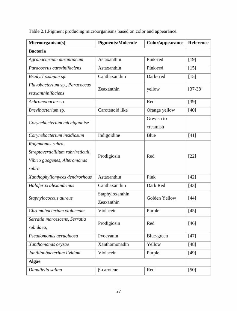
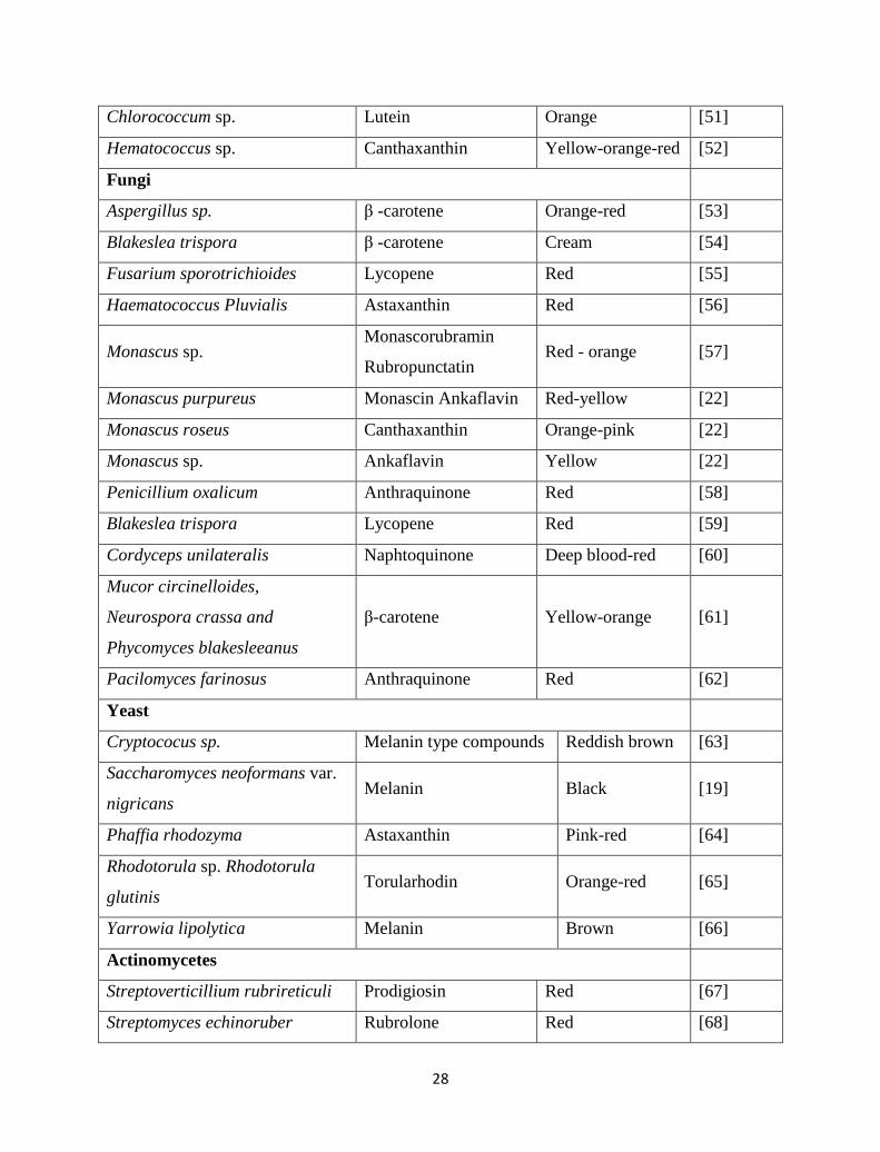
Pigment compounds form microorganisms based on color and appearance can be
classified as shown in Table 2.1.
27
Table 2.1.Pigment producing microorganisms based on color and appearance.
Microorganism(s) Pigments/Molecule Color/appearance Reference
Bacteria
Agrobacterium aurantiacum Astaxanthin Pink-red [19]
Paracoccus carotinifaciens Astaxanthin Pink-red [15]
Bradyrhizobium sp. Canthaxanthin Dark- red [15]
Flavobacterium sp., Paracoccus
zeaxanthinifaciens Zeaxanthin yellow [37-38]
Achromobacter sp. Red [39]
Brevibacterium sp. Carotenoid like Orange yellow [40]
Corynebacterium michigannise Greyish to
creamish
Corynebacterium insidiosum Indigoidine Blue [41]
Rugamonas rubra,
Streptoverticillium rubrireticuli,
Vibrio gaogenes, Alteromonas
rubra
Prodigiosin Red [22]
Xanthophyllomyces dendrorhous Astaxanthin Pink [42]
Haloferax alexandrinus Canthaxanthin Dark Red [43]
Staphylococcus aureus Staphyloxanthin
Zeaxanthin Golden Yellow [44]
Chromobacterium violaceum Violacein Purple [45]
Serratia marcescens, Serratia
rubidaea, Prodigiosin Red [46]
Pseudomonas aeruginosa Pyocyanin Blue-green [47]
Xanthomonas oryzae Xanthomonadin Yellow [48]
Janthinobacterium lividum Violacein Purple [49]
Algae
Dunaliella salina β-carotene Red [50]
28
Chlorococcum sp. Lutein Orange [51]
Hematococcus sp. Canthaxanthin Yellow-orange-red [52]
Fungi
Aspergillus sp. β -carotene Orange-red [53]
Blakeslea trispora β -carotene Cream [54]
Fusarium sporotrichioides Lycopene Red [55]
Haematococcus Pluvialis Astaxanthin Red [56]
Monascus sp. Monascorubramin
Rubropunctatin Red - orange [57]
Monascus purpureus Monascin Ankaflavin Red-yellow [22]
Monascus roseus Canthaxanthin Orange-pink [22]
Monascus sp. Ankaflavin Yellow [22]
Penicillium oxalicum Anthraquinone Red [58]
Blakeslea trispora Lycopene Red [59]
Cordyceps unilateralis Naphtoquinone Deep blood-red [60]
Mucor circinelloides,
Neurospora crassa and
Phycomyces blakesleeanus
β-carotene Yellow-orange [61]
Pacilomyces farinosus Anthraquinone Red [62]
Yeast
Cryptococus sp. Melanin type compounds Reddish brown [63]
Saccharomyces neoformans var.
nigricans Melanin Black [19]
Phaffia rhodozyma Astaxanthin Pink-red [64]
Rhodotorula sp. Rhodotorula
glutinis Torularhodin Orange-red [65]
Yarrowia lipolytica Melanin Brown [66]
Actinomycetes
Streptoverticillium rubrireticuli Prodigiosin Red [67]
Streptomyces echinoruber Rubrolone Red [68]
29
2.7 Microbial pigments of commercial importance
The success of any microbial pigment produced by biotechnological means (for example
fermentation) depends upon its acceptability in the market, regulatory approval, and the size
of the capital investment required in bringing the product to the market. A few years ago,
some expressed doubts about the positive commercialization of fermentation derived food
grade pigments because of the high capital investments requirements for fermentation
facilities and the expensive and time-span toxicity studies required by regulatory agencies
[15, 19].
In addition to the above public perception of biotechnology derived products should
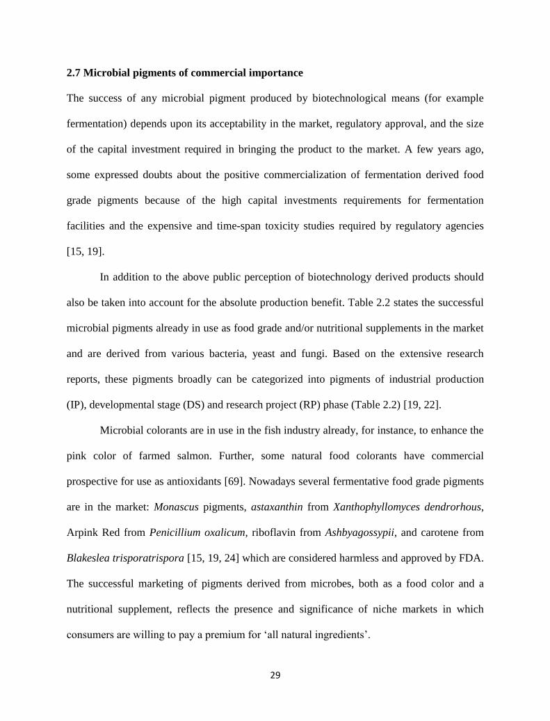
also be taken into account for the absolute production benefit. Table 2.2 states the successful
microbial pigments already in use as food grade and/or nutritional supplements in the market
and are derived from various bacteria, yeast and fungi. Based on the extensive research
reports, these pigments broadly can be categorized into pigments of industrial production
(IP), developmental stage (DS) and research project (RP) phase (Table 2.2) [19, 22].
Microbial colorants are in use in the fish industry already, for instance, to enhance the
pink color of farmed salmon. Further, some natural food colorants have commercial
prospective for use as antioxidants [69]. Nowadays several fermentative food grade pigments
are in the market: Monascus pigments, astaxanthin from Xanthophyllomyces dendrorhous,
Arpink Red from Penicillium oxalicum, riboflavin from Ashbyagossypii, and carotene from
Blakeslea trisporatrispora [15, 19, 24] which are considered harmless and approved by FDA.
The successful marketing of pigments derived from microbes, both as a food color and a
nutritional supplement, reflects the presence and significance of niche markets in which
consumers are willing to pay a premium for ‘all natural ingredients’.
30
Table 2.2. Pigments from various microorganisms which are already in use as natural food
colorants [19, 22].
Pigment Color Microorganism Status
Ankaflavin Yellow Monascus sp. IP
Anthroquinone Red Pencilllium candidum IP
Monascorubramine Red Monascus sp. IP
Riboflavin Yellow Ashbya gossypi IP
Rubropanctatin Orange Monascus sp. IP
β Carotene Yellow-orange Blakeslea trisporia IP
Astaxanthin Pink-red Agrobacterium aurantiacum RP
Astaxanthin Pink-red Paracoccus carotinifaciens RP
Cathaxanthin Dark red Bradirhizobium sp. RP
Lycopene Red Fusarium sporotrichioides RP
Melanin Black Saccharomyces neoformis RP
Napthoquinone Deep blood red Cardyceps unilateralis RP
Zeaxanthin Yellow Paracoccus zeaxanthinifaciens RP
β Carotene Yellow-orange Fusarium sporotrichioides RP
β Carotene Yellow-orange Neurospora crassa RP
β Carotene Yellow-orange Phycomyces blaksleeanus RP
Unknown Red Paecilomyces sinclairii RP
Astaxanthin Pink-red
Xanthophyllomyces
dendrohous
DS
31
Lycopene Red Blakeslea trisporia DS
Rubrolone Red Streptomyces echinoruber DS
Torularhodin Orange-red Rhodotorula sp. DS
Zeaxanthin Yellow Flavobacterium sp. DS
β Carotene Yellow-orange Mucor circinelloides DS
Unknown Red Penicillium purpurogenum DS
The number of approved colorants for food industry is restricted. Some approved food
colorants are recognized by their chemical name (for eg. canthaxanthin) while others are
known by source (eg. fruit juice or vegetable juice). The biocolorants identified by their
chemical name can be synthesized easily by cheaper biotechnological sources particularly by
various microorganisms. And technological limitations are the major hold-up for the
commercial exploitation of the source materials [15, 61]. The success of any pigment
produced by fermentation rely up on its acceptability in the market, regulatory approval, and
the size of the capital investment required in bringing the product to market [61]. Some food
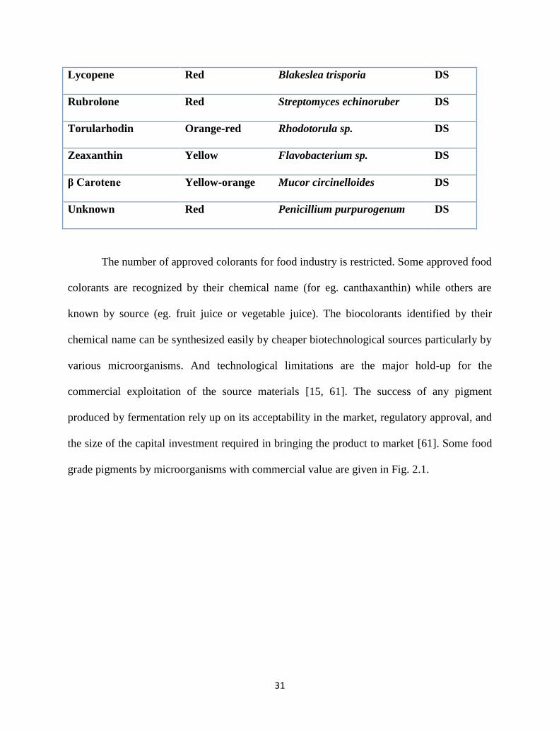
grade pigments by microorganisms with commercial value are given in Fig. 2.1.
32
Fig. 2.1. Some food grade pigments and their structures from microorganisms [15].
33
2.8 Benefits and Applications of Microbial pigments
Pigments produced by microorganisms are of traditional use in oriental countries and have
been a subject of intense research in the present decades because of its potential for
applications. Use of microorganisms and biotechnology would offer solutions to the problems
of various industries especially food colorant industry. Fermentative production of colorants
has a number of benefits that include: cheaper production, readily available raw materials,
high yields and no seasonal variations [70].
In contrast to higher plants, single cell algae and fungi are more appropriate for
biotechnological production because they can be grown using existing culture techniques.
Many fungi produce pigments which have application in both textile and food industries [71].
Fungal pigments are routinely utilized as colorants for both foodstuffs and materials. Some of
the more importantly utilized fungal pigments come from the water soluble orange/red
pigments produced by Monascus sp., frequently used in rice wines in eastern countries [72].
Pigments produced from Monascus purpureus Piedallu are used in wool dying [73], and an
anthraquinone pigment obtained from Penicillium oxalicum Currie & Thom is currently
being developed for use as a ‘natural’ food additive that may have some anticancer effects
[14, 16]. Mycelial extracts of some promising mushrooms are Chroogomplus vinicolor
(which gives red tints), Bankera violascens (which gives greens) and Collybia iocephala
(which gives blues), they have a remarkable potential for dyeing wool and silk fabrics [74].
Carotenoids such as β-carotene and lycopene have been known to be produced by fungal cell
factories. For example, β-carotene by Blakeslea trispora, Mucor circinelloides, and
Phycomyces blakesleeanus and lycopene from Fusarium sporotrichioides, Blakeslea trispora
are already in use as for food colorants [33]. Vitamin, riboflavin (vitamin B2) is a yellow
34
food colorant that is fermentatively produced by the fungi Eremothecium ashbyii and Ashbya
gossypi and has huge commercial potential [75].
Microalgae such as Haematococcus (astaxanthin), Spirulina (phycocyanin), and
Dunaliella (ß-carotene and other carotenoids) are already explored as sources of natural food
colorants with some limitations of low productivity and contamination in the open culture
system, where they are grown [76]. Newly, a blue-green polyphenolic antioxidant pigment
termed marennine from the Diatom Haslea ostrearia has been reported [77]. This may aid as
a promising food coloring additive.
Among yeasts, the marketable success has been with the case of astaxanthin
production by Xanthophyllomyces dendrorhous (formerly Phaffia rhodozyma) and is widely
in use as food additive. Additional example of production of carotenoids is by the yeast
Rhodotorula spp., R. glutinis, R. gracilis, R. rubra, and R. graminis. The chief carotenoids
produced by these yeasts are torulene and torularhodin, with minute quantity of β-carotene
[78-81].
The pigments obtained from bacteria offer the following benefits and advantages [15,
24]; they are progressively attractive to science because of broad ranging activities, easy
propagation and wide-ranging strain selection, high versatile and productive compared to
other sources, fermentation is integrally faster and more productive production when
compared to any other chemical process, simple and fast culturing methods allowing
continuous bioreactor operation, quite easy to manipulate genes, structural complexity suits
for industrial requirements, Cheap substrates used for bulk production and their pigments
extracted using simple liquid-liquid extraction technique minimizing operation cost. Some
important functions of bacterial pigments of commercial potential are 1) Riboflavin
35
synthesized by Bacillus subtilis and used in foods, vitamin enriched milk products and energy
drinks, 2) β-carotene usually produced by Flavobacterium, Agrobacterium aurantiacum and
widely used as food supplement for humans and as food additives for animals and fish, 3)
Prodigiosin by Serratia marcescens, Pseudomonas, Pseudoalteromonas, Alteromonas
denitrificans, Hahella, Vibrio species used as antibacterial, antifungal, antimalarial,
antibiotic, immunosuppressive, anticancer etc., 4) Phycocyanin by Cyanobacteria used as
dietary supplement rich in proteins, and 5) Violacein by Chromobacterium
violaceum,Alteromonas luteoviolacea was used as Antiviral, antibacterial, antiulcerogenic,
antileishmanial, and anticancer properties, potent cytotoxic effects against U937 and HL60
Leukemia cell lines [15, 24].
2.9 Novel practices of microbial pigment production
2.9.1 Strain improvement
Traditionally, strain development was achieved mainly by multiple rounds of random
mutagenesis and selection, which are still very helpful nowadays. In the latest decade, the
development of gene deletion techniques enabled efficient genome DNA inactivation and
greatly improved metabolic engineering of bacteria [15, 82-83]. A more systematic and
integrated approach for biotechnological process for strain improvement became prevalent.
The motivation for industrial strain development is economic, since pigment amounts
produced by wild strains are too low for cost-effective process. It is very essential to isolate
strains which produce pigments with shorter fermentation times. Improvement of microbial
strains for the over production of industrial products has been the characteristic of all
commercial fermentation processes. The strain improvement by common mutagens such as
36
ultraviolet (UV), ethyl methane sulfonate (EMS) and 1-methyl-3-nitro-1-nitrosoguanidine
(NTG) is convenient and can improve pigments yield as proved in several cases [15, 84].
New methods like X-ray and fast neutron of irradiations are also being tried to achieve
microbial strain improvements. For instance, M.purpureus was subjected to the above
irradiations to bring about change in pigmentation and morphology [25].
2.9.2 Fermentation
Fermentation is a metabolic process that converts sugar to acids, gases and/or alcohol and
usually occurs in yeast and bacteria. The production of microbial pigments by fermentation is
an interesting area and a lot of attention is now paid to this biotechnological approach. This
technique can be of solid or submerged fermentation mainly and each approach is having its
own merits and demerits in pigment production.
Submerged fermentation
Submerged fermentation utilizes free flowing liquid substrates, such as molasses and broths.
The bioactive molecules are secreted into the fermentation broth. The substrates are utilized
rapidly; hence need to be constantly replaced or supplemented with nutrients. This
fermentation technique is best suited for microorganisms such as bacteria that require high
moisture content. An added advantage of this technique is that purification of products is
easier [85]. For example Monascus has been successfully cultured submerged condition for
pigment production and versatile substrates like breads, rice and other amylaceous (starch,
dextrins, glucose, maltose and fructose) materials for high productivity of red pigments which
occurs due to glucose and maltose utilization [85]. Carotenoid production by Aspergillus
sp.[86] melanin type pigment by Aspergillus niger [87], water soluble red pigment by
Penicillium purpurogenum [88]are some reported studies from fungi. Furthermore many
37
bacterial pigments too successfully produced using this technique [15]. Various commercial
scale microbial pigments such as prodigiosin, monascorubramie, astaxanthin, canthaxanthin,
beta-carotene and so on are produced using this method. Under submerged conditions, factors
like nitrogen, pH temperature majorly affect the pigment production [15, 25, 33].
Solid- state fermentation
Solid state fermentation (SSF) is defined as any fermentation process performed on a non-
soluble substance that acts both as physical support and source of nutrients in absence of free
flowing liquid [89]. SSF utilizes solid numerous versatile substrates, like bran, bagasse, and
paper pulp etc. The main advantage of using these substrates is that nutrient-rich waste
materials can be easily recycled as substrates [89-90]. In this fermentation technique, the
substrates are utilized very slowly and steadily, so the same substrate can be used for long
fermentation periods. Hence, this technique supports controlled release of nutrients. SSF is
best suited for fermentation techniques involving fungi and microorganisms that require less
moisture content. However, it cannot be used in fermentation processes involving organisms
that require high aw (water activity), such as bacteria [91]. Many pigments were reported
using this technique [92-93] and among all Monascus pigments lead with many reports [94-
96]. For example red rice, a traditional Asian food, has for thousands of years been prepared
by fermentation of steamed rice by Monascus purpureus in SSF. The fungus produces six
different polyketide pigments colored from bright yellow to deep red, which have found
applications both as food additives and pharmaceuticals [25, 97]. For significant amounts of
pigments by molds, substrates like rice, cassava or corn, wheat, oat and barley were used as
substrates [93]. Aspergillus sp. has been grown successfully on starch medium using different
carbon sources wherein dextrin and maltose produced the highest color intensity [25]. Some
38
significant studies include carotenoid by Penicillium sp. [98], riboflavin, a yellow pigment
from Eremothecium ashbyii and Ashbya gossypi, beta-carotene by Dunaliella and D.
bardawil, astaxanthin by Haematococcus pluvialis, phycocianin by Spirulina sp. [92, 99] etc.
2.9.3 Low-cost substrates
Agro-industrial by-products and surpluses that frequently create serious environmental
problems may be possibly used as inexpensive carbohydrate sources for microbial
fermentations, thus decreasing their initial high biological oxygen demand (BOD) while
obtaining biochemical compounds like pigments suitable for pharmaceutical, chemical and
food industries [100]. Low cost by products and residues of agro-industrial origin have shown
their potential in production of different pigments by diverse group microorganisms. Along
this line, variety of substrates and microorganisms has been tested. beta-carotene synthesis by
citrus products [101], carotenoids production using whey ultrafiltrate [102], sauerkraut brine
[81] and peat extract [103], riboflavin in concentrated rectified grape must [104], astaxanthin
on grape juice [105] are some promising studies.
These by-products from industrial processes and other agro or domestic sources are pollutants
to the environmental and their treatment involves high costs. The conversion of these wastes
to value added materials like pigments by microorganisms would provide economic benefits
and reduce waste materials impact on environment as pollutants. Many investigations have
been performed and are under investigation to diminish the costs and optimize the pigments
production [78, 86, 106-111]. Factors such as carbon and nitrogen source are very important
to consider on the selection of wastes as substrates. And pigment production also depends on
minerals and other components in some cases. Pigments produced on several wastes were
shown in Table 2.3.
39
Table 2.3. Different microorganisms and various inexpensive substrates used for pigments
production.
Substrate Microorganism Pigment type References
Whey R. glutinis β -carotene [112]
Potato medium R. mucilaginosa β -carotene [112]
Crude glycerol R. glutinis carotenoids [113]
Chicken feathers R. glutinis carotenoids [114]
Fermented radish brine R. glutinis β -carotene [115]
Mung bean waste flour and sweet
potato extract
R. glutinis carotenoids [79]
Mustard waste X. dendrorhous Astaxanthin [100]
Plant extracts X. dendrorhous Astaxanthin [116]
Coconut milk X. dendrorhous Astaxanthin [117]
Enzymatic hydrolysates of
prehydrolysed wood
X. dendrorhous Astaxanthin [118]
Sugarcane waste marine Streptomyces sp. melanin [119]
2.10 Objectives of the work
The overall objectives of this study are to explore the possibility of pigment production by
different microbial isolates from numerous sources on various substrates. Furthermore, the
enhanced pigment production capability of carotenoid producing microorganisms was also
investigated using fruit waste material as sole substrates. The influential parameters of
increased pigment production by several optimization techniques were also investigated.
40
The specific objectives of this study are:
Isolation, identification and characterization of the microorganisms from different
sources responsible for various pigment production
Investigate and determine effects of substrate (cost-effective), pH, temperature,
moisture, agitation, etc., on the pigment production process by respective
microorganism
Optimize the key parameters for improved pigment production and validation of the
established procedure
Exploring the potential of the obtained pigments as antioxidants, metal chelating
agents, photoprotective agents etc.
Investigating known microbial strains towards enhanced pigment production on
various agro-industrial substrates
References
[1] D. Cristea and G. Vilarem, "Improving light fastness of natural dyes on cotton yarn,"
Dyes and Pigments, vol. 70, pp. 238-245, 2006.
[2] F. Delgado-Vargas, et al., "Natural pigments: carotenoids, anthocyanins, and
betalains—characteristics, biosynthesis, processing, and stability," Critical reviews in
food science and nutrition, vol. 40, pp. 173-289, 2000.
[3] T. Bechtold and R. Mussak, Handbook of natural colorants: John Wiley & Sons,
2009.
[4] F. Delgado-Vargas and O. Paredes-López, Natural colorants for food and
nutraceutical uses: CRC Press, 2002.
41
[5] Z. E. Sikorski, Chemical and functional properties of food components: CRC Press,
2006.
[6] D. M. Freedman, "Reasonable Certainty of No Harm: Reviving the Safety Standard
for Food Additives, Color Additives, and Animal Drugs," Ecology LQ, vol. 7, p. 245,
1978.
[7] J. Neitz, et al., "Color perception is mediated by a plastic neural mechanism that is
adjustable in adults," Neuron, vol. 35, pp. 783-792, 2002.
[8] M. Satake and Y. Mido, Chemistry of colour: Discovery Publishing House, 1995.
[9] M. H. Stafsnes, et al., "Isolation and characterization of marine pigmented bacteria
from Norwegian coastal waters and screening for carotenoids with UVA-blue light
absorbing properties," The Journal of Microbiology, vol. 48, pp. 16-23, 2010.
[10] J. Eyal and M. G. Spencer, "Morchella rotunda sp. useful for producing natural blue
pigment," ed: Google Patents, 1992.
[11] S. Babitha, "Microbial pigments," in Biotechnology for agro-industrial residues
utilisation, ed: Springer, 2009, pp. 147-162.
[12] S. Babitha, et al., "Jackfruit Seed--A novel substrate for the production of Monascus
pigments through solid-state fermentation," Food Technology & Biotechnology, vol.
44, 2006.
[13] H. Sheehan, et al., "Color and scent: how single genes influence pollinator attraction,"
in Cold Spring Harbor symposia on quantitative biology, 2012, pp. 117-133.
[14] S. A. Mapari, et al., "Exploring fungal biodiversity for the production of water-
soluble pigments as potential natural food colorants," Current Opinion in
Biotechnology, vol. 16, pp. 231-238, 2005.
42
[15] C. K. Venil, et al., "Bacterial pigments and their applications," Process Biochemistry,
vol. 48, pp. 1065-1079, 2013.
[16] L. Dufossé, et al., "Microorganisms and microalgae as sources of pigments for food
use: a scientific oddity or an industrial reality?," Trends in Food Science &
Technology, vol. 16, pp. 389-406, 2005.
[17] D. K. Hobson and D. S. Wales, "‘Green’dyes," Journal of the Society of Dyers and
Colourists, vol. 114, pp. 42-44, 1998.
[18] A. B. Soliev, et al., "Bioactive pigments from marine bacteria: applications and
physiological roles," Evidence-Based Complementary and Alternative Medicine, vol.
2011, 2011.
[19] L. Dufossé, "Microbial Production of Food Grade Pigments," Food Technology &
Biotechnology, vol. 44, 2006.
[20] H. J. Nelis and A. De Leenheer, "Microbial sources of carotenoid pigments used in
foods and feeds," Journal of Applied Microbiology, vol. 70, pp. 181-191, 1991.
[21] L. C. Mata-Gómez, et al., "Biotechnological production of carotenoids by yeasts: an
overview," Microbial Cell Factories, vol. 13, p. 12, 2014.
[22] C. K. Venil and P. Lakshmanaperumalsamy, "An insightful overview on microbial
pigment, prodigiosin," Electronic Journal of Biology, vol. 5, pp. 49-61, 2009.
[23] A. Aberoumand, "A review article on edible pigments properties and sources as
natural biocolorants in foodstuff and food industry," World J Dairy Food Sci, vol. 6,
pp. 71-78, 2011.
[24] W. A. Ahmad, et al., Application of Bacterial Pigments as Colorant: Springer, 2012.
43
[25] V. Joshi, et al., "Microbial pigments," Indian journal of biotechnology, vol. 2, pp.
362-369, 2003.
[26] N. Nagpal, et al., "Microbial pigments with health benefits-A mini review," Trends in
Biosciences, vol. 4, pp. 157-160, 2011.
[27] A. Shirata, et al., "Isolation of bacteria producing bluish-purple pigment and use for
dyeing," Japan Agricultural Research Quarterly, vol. 34, pp. 131-140, 2000.
[28] K.-P. Stahmann, et al., "Three biotechnical processes using Ashbya gossypii, Candida
famata, or Bacillus subtilis compete with chemical riboflavin production," Applied
Microbiology and Biotechnology, vol. 53, pp. 509-516, 2000.
[29] D. Asker and Y. Ohta, "Production of canthaxanthin by extremely halophilic
bacteria," Journal of bioscience and bioengineering, vol. 88, pp. 617-621, 1999.
[30] A. A. Woodall, et al., "Carotenoids and protection of phospholipids in solution or in
liposomes against oxidation by peroxyl radicals: relationship between carotenoid
structure and protective ability," Biochimica et Biophysica Acta (BBA)-General
Subjects, vol. 1336, pp. 575-586, 1997.
[31] D. F. Browning, et al., "Light‐induced carotenogenesis in Myxococcus xanthus:
functional characterization of the ECF sigma factor CarQ and antisigma factor CarR,"
Molecular microbiology, vol. 48, pp. 237-251, 2003.
[32] H. Takano, et al., "Light-induced carotenogenesis in Streptomyces coelicolor A3 (2):
identification of an extracytoplasmic function sigma factor that directs
photodependent transcription of the carotenoid biosynthesis gene cluster," Journal of
Bacteriology, vol. 187, pp. 1825-1832, 2005.
44
[33] K. Malik, et al., "Microbial pigments: A review," Int. J. Microbial. Resour. Technol,
vol. 1, pp. 361-365, 2012.
[34] O. Ciferri, "Spirulina, the edible microorganism," Microbiological reviews, vol. 47, p.
551, 1983.
[35] K. H. McClean, et al., "Quorum sensing and Chromobacterium violaceum:
exploitation of violacein production and inhibition for the detection of N-
acylhomoserine lactones," Microbiology, vol. 143, pp. 3703-3711, 1997.
[36] M. Rodríguez-Sáiz, et al., "Xanthophyllomyces dendrorhous for the industrial
production of astaxanthin," Applied Microbiology and Biotechnology, vol. 88, pp.
645-658, 2010.
[37] A. Berry, et al., "Paracoccus zeaxanthinifaciens sp. nov., a zeaxanthin-producing
bacterium," International journal of systematic and evolutionary microbiology, vol.
53, pp. 231-238, 2003.
[38] B. Holmes, et al., "Flavobacterium odoratum: a species resistant to a wide range of
antimicrobial agents," Journal of clinical pathology, vol. 32, pp. 73-77, 1979.
[39] J. A. Duerre and P. J. Buckley, "Pigment production from tryptophan by an
Achromobacter species," Journal of bacteriology, vol. 90, pp. 1686-1691, 1965.
[40] D. Jones, et al., "Taxonomically significant colour changes in Brevibacterium linens
probably associated with a carotenoid-like pigment," Journal of general
microbiology, vol. 77, pp. 145-150, 1973.
[41] M. P. Starr, "The blue pigment of Corynebacterium insidiosum," Archiv für
Mikrobiologie, vol. 30, pp. 325-334, 1958.
45
[42] M. Vázquez, "Effect of the light on carotenoid profiles of Xanthophyllomyces
dendrorhous strains (formerly Phaffia rhodozyma)," Food technology and
biotechnology, vol. 39, pp. 123-128, 2001.
[43] D. Asker and Y. Ohta, "Haloferax alexandrinus sp. nov., an extremely halophilic
canthaxanthin-producing archaeon from a solar saltern in Alexandria (Egypt),"
International journal of systematic and evolutionary microbiology, vol. 52, pp. 729-
738, 2002.
[44] G. Y. Liu, et al., "Staphylococcus aureus golden pigment impairs neutrophil killing
and promotes virulence through its antioxidant activity," The Journal of experimental
medicine, vol. 202, pp. 209-215, 2005.
[45] R. S. Blosser and K. M. Gray, "Extraction of violacein from Chromobacterium
violaceum provides a new quantitative bioassay for N-acyl homoserine lactone
autoinducers," Journal of microbiological methods, vol. 40, pp. 47-55, 2000.
[46] A. A. Deorukhkar, et al., "Identification of a red-pigmented bacterium producing a
potent anti-tumor N-alkylated prodigiosin as Serratia marcescens," Research in
microbiology, vol. 158, pp. 399-404, 2007.
[47] B. A. Kenner and H. P. Clark, "Detection and enumeration of Salmonella and
Pseudomonas aeruginosa," Journal (Water Pollution Control Federation), pp. 2163-
2171, 1974.
[48] A. K. Goel, et al., "Genetic locus encoding functions involved in biosynthesis and
outer membrane localization of xanthomonadin in Xanthomonas oryzae pv. oryzae,"
Journal of Bacteriology, vol. 184, pp. 3539-3548, 2002.
46
[49] F. Pantanella, et al., "Violacein and biofilm production in Janthinobacterium
lividum," Journal of Applied Microbiology, vol. 102, pp. 992-999, 2007.
[50] A. Ben-Amotz, et al., "Mode of action of the massively accumulated β-carotene of
Dunaliella bardawil in protecting the alga against damage by excess irradiation,"
Plant Physiology, vol. 91, pp. 1040-1043, 1989.
[51] R. J. McLean, "New taxonomic criteria in the classification of Chlorococcum species.
I. Pigmentation," Journal of Phycology, vol. 4, pp. 328-332, 1968.
[52] E. Christaki, et al., "Functional properties of carotenoids originating from algae,"
Journal of the Science of Food and Agriculture, vol. 93, pp. 5-11, 2013.
[53] V. Álvarez, et al., "The crtS gene of Xanthophyllomyces dendrorhous encodes a novel
cytochrome-P450 hydroxylase involved in the conversion of β-carotene into
astaxanthin and other xanthophylls," Fungal Genetics and Biology, vol. 43, pp. 261-
272, 2006.
[54] M. Rodríguez-Sáiz, et al., "Blakeslea trispora genes for carotene biosynthesis,"
Applied and environmental microbiology, vol. 70, pp. 5589-5594, 2004.
[55] L. Dufossé, "Mikrobna proizvodnja pigmenata hrane," Food technology and
biotechnology, vol. 44, pp. 313-321, 2006.
[56] T. Lotan and J. Hirschberg, "Cloning and expression in Escherichia coli of the gene
encoding β-C-4-oxygenase, that converts β-carotene to the ketocarotenoid
canthaxanthin in Haematococcus pluvialis," FEBS letters, vol. 364, pp. 125-128,
1995.
[57] D. Wild, et al., "New Monascus metabolite isolated from red yeast rice (angkak, red
koji)," Journal of agricultural and food chemistry, vol. 50, pp. 3999-4002, 2002.
47
[58] S. Zhang, et al., "Biosorption of reactive dyes by the mycelium pellets of a new
isolate of Penicillium oxalicum," Biotechnology letters, vol. 25, pp. 1479-1482, 2003.
[59] W. Hsu, et al., "Carotenoid biosynthesis in Blakeslea trispora," Phytochemistry, vol.
11, pp. 2985-2990, 1972.
[60] P. Unagul, et al., "Production of red pigments by the insect pathogenic fungus
Cordyceps unilateralis BCC 1869," Journal of Industrial Microbiology and
Biotechnology, vol. 32, pp. 135-140, 2005.
[61] C. Socaciu, Food colorants: chemical and functional properties: CRC Press, 2007.
[62] P. Velmurugan, et al., "Water‐soluble red pigments from Isaria farinosa and structural
characterization of the main colored component," Journal of basic microbiology, vol.
50, pp. 581-590, 2010.
[63] S. Chaskes and R. L. Tyndall, "Pigment production by Cryptococcus neoformans
from para-and ortho-Diphenols: effect of the nitrogen source," Journal of clinical
microbiology, vol. 1, pp. 509-514, 1975.
[64] B. Bjerkeng, et al., "Digestibility and muscle retention of astaxanthin in Atlantic
salmon, Salmo salar, fed diets with the red yeast Phaffia rhodozyma in comparison
with synthetic formulated astaxanthin," Aquaculture, vol. 269, pp. 476-489, 2007.
[65] P. Buzzini, "Batch and fed‐batch carotenoid production by Rhodotorula glutinis–
Debaryomyces castellii co‐cultures in corn syrup," Journal of Applied Microbiology,
vol. 90, pp. 843-847, 2001.
[66] A. Carreira, et al., "Brown pigments produced by Yarrowia lipolytica result from
extracellular accumulation of homogentisic acid," Applied and environmental
microbiology, vol. 67, pp. 3463-3468, 2001.
48
[67] N. N. Gerber and D. P. Stahly, "Prodiginine (prodigiosin-like) pigments from
Streptoverticillium rubrireticuli, an organism that causes pink staining of polyvinyl
chloride," Applied microbiology, vol. 30, p. 807, 1975.
[68] N. Palleroni, et al., "Production of a novel red pigment, rubrolone, by Streptomyces
echinoruber sp. nov. I. Taxonomy, fermentation and partial purification," The Journal
of antibiotics, vol. 31, pp. 1218-1225, 1978.
[69] W. A. Schroeder and E. Johnson, "Antioxidant role of carotenoids in Phaffia
rhodozyma," Journal of general microbiology, vol. 139, pp. 907-912, 1993.
[70] A. Mortensen, "Carotenoids and other pigments as natural colorants," Pure and
Applied Chemistry, vol. 78, pp. 1477-1491, 2006.
[71] N. Durán, et al., "Ecological-friendly pigments from fungi," Critical reviews in food
science and nutrition, vol. 42, pp. 53-66, 2002.
[72] J. C. d. Carvalho, et al., "Biopigments from Monascus: strains selection, citrinin
production and color stability," Brazilian Archives of Biology and technology, vol. 48,
pp. 885-894, 2005.
[73] S. C. Robinson, et al., "Utilizing pigment-producing fungi to add commercial value to
American beech (Fagus grandifolia)," Applied Microbiology and Biotechnology, vol.
93, pp. 1041-1048, 2012.
[74] A. Bessette, Mushrooms of North America in color: a field guide companion to
seldom-illustrated fungi: Syracuse University Press, 1995.
[75] Z. Lin, et al., "Polyketides from Penicillium sp. JP-1, an endophytic fungus associated
with the mangrove plant Aegiceras corniculatum," Phytochemistry, vol. 69, pp. 1273-
1278, 2008.
49
[76] W.-L. Chu, "Potential applications of antioxidant compounds derived from algae,"
Current Topics in Nutraceutical Research, vol. 9, 2011.
[77] J.-B. Pouvreau, et al., "Antioxidant and free radical scavenging properties of
marennine, a blue-green polyphenolic pigment from the diatom Haslea ostrearia
(Gaillon/Bory) Simonsen responsible for the natural greening of cultured oysters,"
Journal of agricultural and food chemistry, vol. 56, pp. 6278-6286, 2008.
[78] M. Vazquez and A. M. Martin, "Optimization of Phaffia rhodozyma continuous
culture through response surface methodology," Biotechnology and bioengineering,
vol. 57, pp. 314-320, 1998.
[79] J. Tinoi, et al., "Simplex optimization of carotenoid production by Rhodotorula
glutinis using hydrolyzed mung bean waste flour as substrate," Process Biochemistry,
vol. 40, pp. 2551-2557, 2005.
[80] E. D. Simova, et al., "Effect of aeration on the production of carotenoid pigments by
Rhodotorula rubra-lactobacillus casei subsp. casei co-cultures in whey ultrafiltrate,"
Zeitschrift fur Naturforschung C-Journal of Biosciences, vol. 58, pp. 225-229, 2003.
[81] C. Shih and Y. Hang, "Production of Carotenoids by Rhodotorula rubra from
Sauerkraut Brine," LWT-Food Science and Technology, vol. 29, pp. 570-572, 1996.
[82] V. A. Portnoy, et al., "Adaptive laboratory evolution—harnessing the power of
biology for metabolic engineering," Current Opinion in Biotechnology, vol. 22, pp.
590-594, 2011.
[83] M. Sonderegger and U. Sauer, "Evolutionary engineering of Saccharomyces
cerevisiae for anaerobic growth on xylose," Applied and environmental microbiology,
vol. 69, pp. 1990-1998, 2003.
50
[84] Y. Chen, et al., "Screening and characterization of astaxanthin-hyperproducing
mutants of Haematococcus pluvialis," Biotechnology letters, vol. 25, pp. 527-529,
2003.
[85] M. A. Dhale, "Physiology of Monascus purpureus in relation to metabolite production
and application as functional food," India: Doctor thesis of University of Mysore,
2007.
[86] K. Sanjay, et al., "Optimization of carotenoid production by Aspergillus carbonarius
in submerged fermentation using a response surface methodology," International
Journal of Food Engineering, vol. 3, 2007.
[87] T. R. Jørgensen, et al., "Submerged conidiation and product formation by Aspergillus
niger at low specific growth rates are affected in aerial developmental mutants,"
Applied and environmental microbiology, vol. 77, pp. 5270-5277, 2011.
[88] A. Méndez, et al., "Red pigment production by Penicillium purpurogenum GH2 is
influenced by pH and temperature," Journal of Zhejiang University SCIENCE B, vol.
12, pp. 961-968, 2011.
[89] A. Pandey, et al., "Types of fermentation and factors affecting it," Biotechnology:
Food Fermentation, Educational Publishers, New Delhi, pp. 383-426, 1999.
[90] S. R. Couto and M. A. Sanromán, "Application of solid-state fermentation to food
industry—a review," Journal of Food Engineering, vol. 76, pp. 291-302, 2006.
[91] K. Babu and T. Satyanarayana, "α-Amylase production by thermophilic Bacillus
coagulans in solid state fermentation," Process Biochemistry, vol. 30, pp. 305-309,
1995.
51
[92] L. Thomas, et al., "Current developments in solid-state fermentation," Biochemical
Engineering Journal, vol. 81, pp. 146-161, 2013.
[93] U. Hölker, et al., "Biotechnological advantages of laboratory-scale solid-state
fermentation with fungi," Applied Microbiology and Biotechnology, vol. 64, pp. 175-
186, 2004.
[94] A. Rosenblitt, et al., "Solid substrate fermentation of Monascus purpureus: growth,
carbon balance, and consistency analysis," Biotechnology progress, vol. 16, pp. 152-
162, 2000.
[95] S. Babitha, et al., "Solid-state fermentation for the production of Monascus> pigments
from jackfruit seed," Bioresource Technology, vol. 98, pp. 1554-1560, 2007.
[96] O. Han and R. E. Mudgett, "Effects of Oxygen and Carbon Dioxide Partial Pressures
on Monascus Growth and Pigment Production in Solid‐State Fermentations,"
Biotechnology progress, vol. 8, pp. 5-10, 1992.
[97] M. R. Johns and D. M. Stuart, "Production of pigments by Monascus purpureus in
solid culture," Journal of industrial microbiology, vol. 8, pp. 23-28, 1991.
[98] J.-R. Han, "Sclerotia growth and carotenoid production of Penicillium sp PT95 during
solid state fermentation of corn meal," Biotechnology letters, vol. 20, pp. 1063-1065,
1998.
[99] A. Pandey, "Recent process developments in solid-state fermentation," Process
Biochemistry, vol. 27, pp. 109-117, 1992.
[100] J. Tinoi, et al., "Utilization of mustard waste isolates for improved production of
astaxanthin by Xanthophyllomyces dendrorhous," Journal of Industrial Microbiology
and Biotechnology, vol. 33, pp. 309-314, 2006.
52
[101] A. Ciegler, et al., "Enhancement of β-carotene synthesis by citrus products," Applied
microbiology, vol. 11, pp. 128-131, 1963.
[102] G. Frengova, et al., "Formation of carotenoids by Rhodotorula glutinis in whey
ultrafiltrate," Biotechnology and bioengineering, vol. 44, pp. 888-894, 1994.
[103] A. M. Martin, et al., "Growth parameters for the yeast Rhodotorula rubra grown in
peat extracts," Journal of fermentation and bioengineering, vol. 76, pp. 321-325,
1993.
[104] P. Buzzini and J. Rossi, "Semi-continuous and continuous riboflavin production by
calcium-alginate-immobilized Candida saitoana 4253 in concentrated rectified grape
must.[Erratum: Feb 1999, v. 15 (1), p. 153-159.]," World journal of microbiology &
biotechnology, 1998.
[105] P. Meyer and J. Du Preez, "Astaxanthin production by a Phaffia rhodozyma mutant
on grape juice," World journal of microbiology and biotechnology, vol. 10, pp. 178-
183, 1994.
[106] S. N. Surwase, et al., "Optimization of melanin production by Brevundimonas sp. SGJ
using response surface methodology," 3 Biotech, vol. 3, pp. 187-194, 2013.
[107] P. Park, et al., "Optimization of carotenoid production by Rhodotorula glutinis using
statistical experimental design," World journal of microbiology and biotechnology,
vol. 21, pp. 429-434, 2005.
[108] F. Mantzouridou, et al., "Optimization of β-carotene production from synthetic
medium by Blakeslea trispora in a stirred tank reactor and relationship between
morphological changes and pigment formation," Food Biotechnology, vol. 16, pp.
167-187, 2002.
53
[109] C. Malisorn and W. Suntornsuk, "Optimization of β-carotene production by
Rhodotorula glutinis DM28 in fermented radish brine," Bioresource Technology, vol.
99, pp. 2281-2287, 2008.
[110] B. D. H. Dung, et al., "Optimization carotenoids production by Rhodotorula glutinis
GBDO Uusing design of Plackett-Burman matrix and central composite design-
response surface methodology," in Conference proceedings of Biotechnology for
Green Solutions and Sustainable Environment, 2010, p. 25.
[111] P. Bhosale and R. Gadre, "Optimization of carotenoid production from
hyper‐producing Rhodotorula glutinis mutant 32 by a factorial approach," Letters in
applied microbiology, vol. 33, pp. 12-16, 2001.
[112] I. Marova, et al., "Use of several waste substrates for carotenoid-rich yeast biomass
production," Journal of Environmental Management, vol. 95, pp. S338-S342, 2012.
[113] C. Saenge, et al., "Potential use of oleaginous red yeast Rhodotorula glutinis for the
bioconversion of crude glycerol from biodiesel plant to lipids and carotenoids,"
Process Biochemistry, vol. 46, pp. 210-218, 2011.
[114] M. Taskin, et al., "Use of waste chicken feathers as peptone for production of
carotenoids in submerged culture of Rhodotorula glutinis MT-5," European Food
Research and Technology, vol. 233, pp. 657-665, 2011.
[115] C. Malisorn and W. Suntornsuk, "Improved β-carotene production of Rhodotorula
glutinis in fermented radish brine by continuous cultivation," Biochemical
Engineering Journal, vol. 43, pp. 27-32, 2009.
[116] S. Kim, et al., "Increased carotenoid production in Xanthophyllomyces dendrorhous
G276 using plant extracts," Journal of Mmicrobiology-Seoul-, vol. 45, p. 128, 2007.
54
[117] A. Domínguez-Bocanegra and J. Torres-Muñoz, "Astaxanthin hyperproduction by
Phaffia rhodozyma (now Xanthophyllomyces dendrorhous) with raw coconut milk as
sole source of energy," Applied Microbiology and Biotechnology, vol. 66, pp. 249-
252, 2004.
[118] J. Parajó, et al., "Production of carotenoids by Xanthophyllomyces dendrorhous>
growing on enzymatic hydrolysates of prehydrolysed wood," Food Chemistry, vol.
60, pp. 347-355, 1997.
[119] V. Vasanthabharathi, et al., "Melanin production from marine Streptomyces," African
Journal of Biotechnology, vol. 10, pp. 11224-11234, 2013.
55
Chapter 3
Screening of inexpensive substrates for pigments
production by isolated and known microbial strains -
Bridge to Chapter 3 to 8
3.1 Melanins and Carotenoids - pigments of marketable importance
Owing to a growing worldwide market for pigment compounds by microbial production,
carotenoids and melanin compounds has great potential in terms of applications and strategies
of production. Among both, carotenoids occur as natural colorants with a range of yellow to
red colors, so they have great influence on the acceptability of many foods [1-2]. Moreover,
some carotenoids are precursors of vitamin A; in terms of human health, they are amongst the
bioactive phytochemicals credited that reduce risks for degenerative diseases such as cancer,
cardiovascular diseases, macular degeneration and cataract [3-4]. Carotenoids are naturally
occurring lipid-soluble pigments, the majority being C40 terpenoids, which act as membrane
protective antioxidants scavenging O2 and peroxyl radicals; their antioxidant ability is
56
apparently credited to their structure. Carotenoids pigments occur commonly in
photosynthetic systems of higher plants, algae and phototrophic bacteria. On the other hand,
in non-photosynthetic organisms, carotenoids are significant in protecting against photo-
oxidative damage. Thus, many non-phototrophic bacteria and fungi rely on carotenoids for
protection when growing on conditions where light and air are copious [3-7]. The carotenoids
production by the organisms follow two distinct pathways, the well-known mevalonate
(MVA) pathway and the relatively new 2-C-methyl-D-erythritol 4-phosphate (MEP) pathway.
This pathway produce isopentenyl diphosphate (IPP) and its isomer of dimethylallyl
diphosphate (DMAPP), the universal building blocks for the synthesis of all carotenoids.
Eukaryotes generally use the MVA pathway to convert acetyl–CoA to IPP, which is
subsequently isomerized to DMAPP. Prokaryotes, with some exceptions, use the MEP
pathway to produce IPP and DMAPP via an initial condensation reaction between pyruvate
and glyceraldehyde-3-phosphate (G3P). Plants and Streptomycetes follow both pathways for
the synthesis of these commercially significant pigments [8-9].
Further coming to melanin compounds, these are interesting materials that are
currently being used in diverse fields such as medicine, pharmacology, cosmetics etc. In
addition, many different technological applications including photo-voltaics, sensors,
electronic devices and may prove to be a novel class of semiconducting polymers, bio-
compatibles and with a readily available natural source as natural materials extracted from
pigmented [10-11]. However, despite momentous scientific effort over the past 30 years, the
basic functions of melanins are still a matter of controversy and speculation. This uncertainty
imparts from the few and poorly defined structural and physical–chemical properties that are
known in these molecules that continue to mystify researchers even though their adaptive
57
importance has already been proven [10, 12]. These materials are predominantly intractable
from an analytical perspective because they are chemically and photo-chemically very stable,
and they are virtually insoluble in most common solvents. In microorganisms, melanins may
be found in both extracellular or intracellular environments, and they pretence very
interesting targets for molecular structure determination and organic synthesis [13-16].
Melanins constitute a general class of complex, polyphenolic heteropolymers and are usually
divided into brown–black melanins (called as eumelanins) and brown, red or yellow melanins
(called as pheomelanins). A number of bacterial species have been testified to produce
melanins [11, 17]. These compounds can be produced in the Actinomiceto or Streptomicetos
groups, Bacillus and Azotobacter genuses [11, 18]; however, very few of these pigments have
been molecularly defined. The well-known melanisation pathway is the classic Mason–Raper
pathway. In this pathway, tyrosinases yield the melanin intermediate dihydroxyphenyl-
alanine (DOPA), dihydroxyindole (DHI) and dihydroxyindole carboxylic acid (DHICA) [10-
11, 19]. They are some other melanin types and other synthesis pathways are also reported
that produce melanins; for example, pyomelanin is derived from the catabolism of tyrosine
via p -hydroxyphenylpyruvate and homogentisic acid (HGA) [20].
First and foremost, production of melanin is one of the most universal adaptations of
living organisms to the variable conditions of the earth and these pigments. As these
pigments are widespread among microorganisms, extensive research focused on exploration
around this ubiquitous pigment which of commercial interest and have great application
potentials in the agriculture, cosmetics and pharmaceutical industries. Additionally, the
widespread synthesis calibre of catotenoids by heterotrophic and non-phototropic bacteria,
fungi and yeast suggests that these pigments are crucial for the viability of these organisms in
58
their natural environment. Carotenoids are the largest and most diverse class of natural
pigments known to mankind and they have enormous commercial significance with
promising applications from food to textiles. The first part of our investigation engaged in
isolation of individual melanin and carotenoid producing microorganisms which are majorly
at RP and DS of research. Here microbial cultures were collected from various natural
resources such as marine and soil samples The second part of our work involves the
employment of the selective pigment producing microbial isolates on various low-cost by-
products and solids as substrates for effective pigment (caroteniod or melanin) production.
This chapter ends by extensive screening of the collected inexpensive wastes as sole
substrates for pigment production by the purchased microbial strains.
3.2 Sampling
Water
Marine water samples were collected from different sites along the coast of Vishakhapatnam
beach, Andhra Pradesh, India. Sampling locations were shown in Table 3.1.
Soil
Soil sample was collected from the garden of NIT campus Rourkela. The obtained soil was
thoroughly washed with sterile distilled water to separate the floating dust and debris.
The soil suspension and the collected water samples were then aseptically diluted
serially (10-7
and 10-10
times where ever necessary) and 1ml of it was inoculated on various
mediums and observed for various diffusible pigments and/or pigmented colonies. All the
isolates were cultivated at ambient conditions i.e. at pH 7 and temperature 30 0C. For liquid
media an agitation of 150 rpm was maintained throughout the incubation process.
59
3.3 Screening of pigment producing microorganisms
Isolates from different sources were cultured on their respective growth media and at
specified growth conditions; these were awaited for 2-10 days for visible colored colonies.
Primarily, after incubation for two days, marine isolate from deep sea water gave creamy and
purple colonies; while isolates from shore (near rocks) gave yellow and black colonies. The
observed colonies were picked and cultured separately for individual pure cultures. Coming
to soil isolate; nutrient agar was used as culture medium. Here various colonies were seen
after 2 days, whereas orange pigmented colonies were observed after 10 days of incubation.
These colored colonies were picked and sub-cultured for further experimentation.
3.4 Different substrates and Cultivation of isolates/purchased strains
In this work, cabbage (Brassica oleracea) waste was prepared from the outer leaves of
cabbage that are peeled off before cabbages are distributed in the market. And potato peels
and jack fruit seeds were collected from domestic disposed wastes. The obtained raw wastes
were dried in oven at 60 0C for 12 hrs, powdered and used as substrates for screening
microbial isolates for pigment production. A 5 % solution was prepared from the respective
powders and used as liquid medium for the cultivation of isolates.
Free water or liquid medium does not appear to be natural habitat for the majority of
the microorganisms. Solid substrates in this case enable favorable conditions (close to the
natural environment to which microorganisms are adapted) of growth especially for wild type
microorganisms. Therefore solid substrates such as rice, corn and wheat flour powders
(obtained from local market) were explored by the isolated microorganisms for pigment
60
production. Substrate medium was prepared by autoclaving (at 15 lb pressure for 10 min) 3 g
of individual powders in a petridish (10 cm) with 8 mL distilled water.
Another waste used in the work was from fruit wastes. Fruit waste was obtained from
a fruit juice shop of a local market. The material used is from single batch i.e. used in all the
experiments to minimise the disturbances in the results due to variations in composition. The
waste contains major portion of pine apple and orange waste and minor portions of
pomegranate waste. Fruit waste includes extracted carpels of oranges, core of pineapples, and
crushed seeds along with arils of pomegranate. The soluble sugars are extracted from 1
kilogram of fruit waste by adding 2 litres of distilled water and boiled at 100 0C
for 30
minutes. The resultant straw colour fruit waste extract (FWE) was filtered and stored at 4 0C
for further experimentation.
The prepared solid and liquid media are used for pigment production studies by
marine and soil microbial isolates. These were incubated for 6-7 days at 25 °C and REMI
shaking incubator (operated at 150 rpm) was used for liquid medium substrates. Carotenooid
producing strains Rhodotorula rubra. (MTCC no: 1446) Xanthophyllomyces dendrorhous
(MTCC No: 7536) were obtained from imtech Chandigarh and were cultivated on above
specified wastes for pigment production.
3.5 Strain/substrate selection and further experimentation
Selection of the appropriate substrates - for microbial isolates
The pigment compounds presences in various wastes along with selective media by microbial
isolates are shown in Table 3.1. Upon exploitation, marine isolate taken from near shore area
61
resulted in black color which is moderate in vegetable waste medium as substrate when
compared to significant color change in marine agar.
Furthermore two microbial strains isolated from garden soil; independently resulted in black
color when cultivated on fruit waste extract medium, and an orange color in solid rice powder
medium when used as solitary substrates. Rice powder medium was significantly colored in
orange than the defined nutrient agar medium by garden soil isolate.
The mediums devoid of color change were discarded and omitted from the study. Vegetable
waste from cabbage, fruit waste extract and rice powder are therefore selected for additional
investigations.
Selection of the appropriate substrates - for carotenoid producing microorganisms
Like microbial isolates, various growth mediums were tested by the purchased
microorganisms (yeast) which are potent carotenoid producers on defined and undefined
mediums. Color change of the media was observed for the selection of the best substrate for
pigment production by the obtained microbes (Table 3.2).
Among all the mediums, fruit waste extract medium significantly changed its color when
cultivated by respective yeast. R. rubra produced vibrant pink-orange color, while X.
dendrorhous gave intense orange-red to the medium comparable to the standard nutrient
medium. This FWE medium was subjected to further investigation using these yeasts.
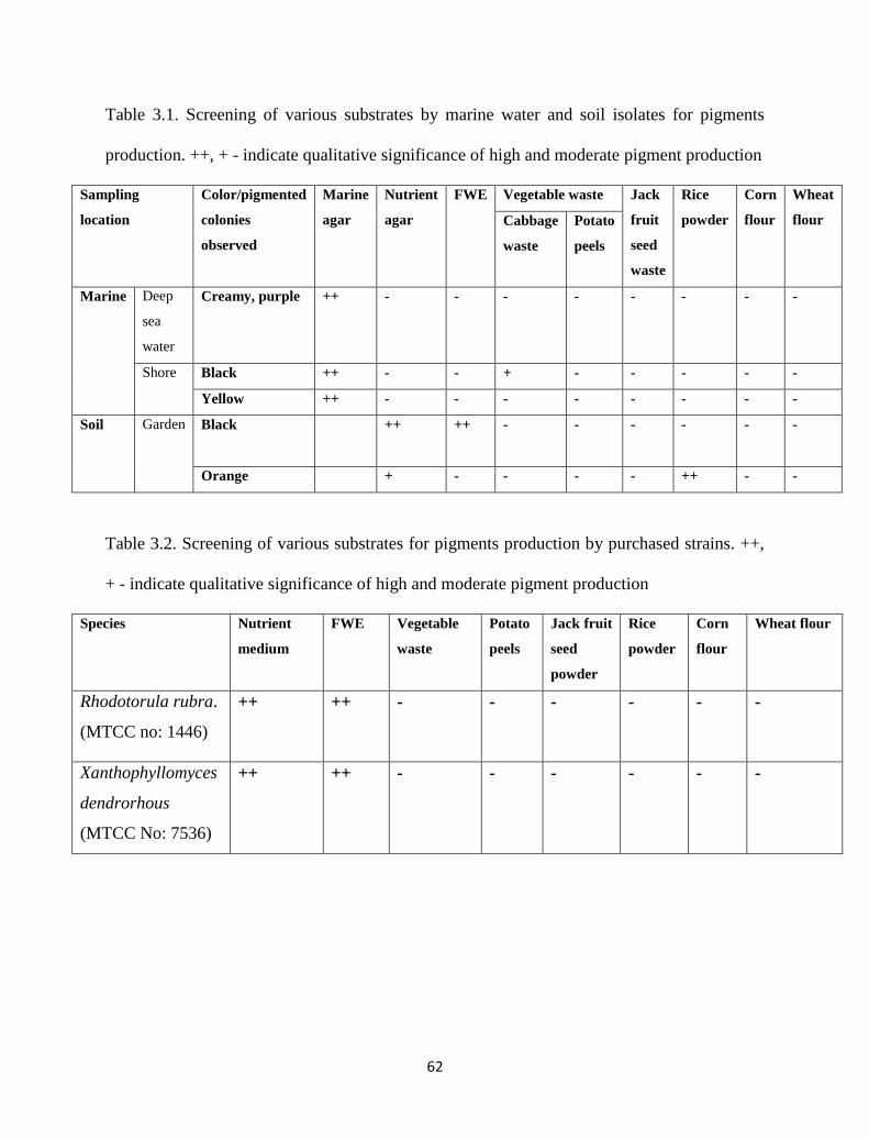
62
Table 3.1. Screening of various substrates by marine water and soil isolates for pigments
production. ++, + - indicate qualitative significance of high and moderate pigment production
Sampling
location
Color/pigmented
colonies
observed
Marine
agar
Nutrient
agar
FWE Vegetable waste Jack
fruit
seed
waste
Rice
powder
Corn
flour
Wheat
flour Cabbage
waste
Potato
peels
Marine Deep
sea
water
Creamy, purple ++ - - - - - - - -
Shore Black ++ - - + - - - - -
Yellow ++ - - - - - - - -
Soil Garden Black
++ ++ - - - - - -
Orange + - - - - ++ - -
Table 3.2. Screening of various substrates for pigments production by purchased strains. ++,
+ - indicate qualitative significance of high and moderate pigment production
Species Nutrient
medium
FWE Vegetable
waste
Potato
peels
Jack fruit
seed
powder
Rice
powder
Corn
flour
Wheat flour
Rhodotorula rubra.
(MTCC no: 1446)
++ ++ - - - - - -
Xanthophyllomyces
dendrorhous
(MTCC No: 7536)
++ ++ - - - - - -
63
3.6 Summary
Use of microbial pigments in processed foods, cosmetics and in other applications is
promising with large economic potential. However, microbial pigments present several
challenges due to high cost, lower stability and disparity in shades due to changes in pH.
At present, none of the microbial pigment can replace synthetic pigments. But recent
advances and efforts in synthetic biology, metabolic engineering etc. will greatly expand the
pigments that could be produced economically in sufficient amounts for industrial
application.
Extensive efforts on fermentation process, utilization of low-cost by-products and
residues of agro-industrial origin as alternative substrates have shown the possibility of
attaining non-toxic microbial pigments in significant amounts. Research in this area is
escalating as the outputs achieved here are fast, have economic potential with less efforts and
these technologies can be implemented immediately than the time taking genetic engineering
of microorganisms. Utilization of wastes, screening of novel microorganisms for non-toxic
pigments, optimization of production conditions are our major objectives of focus in the
coming chapters of the work.
References
[1] L. Ruan, et al., "Melanin Pigment Formation and Increased UV Resistance in Bacillus
thuringiensis Following High Temperature induction," Systematic and applied
microbiology, vol. 27, pp. 286-289, 2004.
[2] S. Kim, et al., "Increased carotenoid production in Xanthophyllomyces dendrorhous
G276 using plant extracts," Journal of Microbiology-Seoul-, vol. 45, p. 128, 2007.
64
[3] M. B. Sporn, The retinoids vol. 1: Academic Press, 1984.
[4] C. N'Soukpoe-Kossi, et al., "Retinol and retinoic acid bind human serum albumin:
stability and structural features," International journal of biological macromolecules,
vol. 40, pp. 484-490, 2007.
[5] T. Lóránd, et al., "FT-IR study of some seco-and apocarotenoids," Journal of
biochemical and biophysical methods, vol. 53, pp. 251-258, 2002.
[6] K. Jacob, et al., "Stability of carotenoids, phenolic compounds, ascorbic acid and
antioxidant capacity of tomatoes during thermal processing," Archivos
Latinoamericanos de Nutricion (ALAN), vol. 60, p. 192, 2010.
[7] G. T. Hayman, et al., "Production of carotenoids by Phaffia rhodozyma grown on
media composed of corn wet-milling co-products," Journal of industrial
microbiology, vol. 14, pp. 389-395, 1995.
[8] A. Das, et al., "An update on microbial carotenoid production: application of recent
metabolic engineering tools," Applied Microbiology and Biotechnology, vol. 77, pp.
505-512, 2007.
[9] L. C. Mata-Gómez, et al., "Biotechnological production of carotenoids by yeasts: an
overview," Microbial Cell Factories, vol. 13, p. 12, 2014.
[10] S. Sajjan, et al., "Purification and physiochemical characterization of melanin
pigment from Klebsiella sp. GSK," Journal of microbiology and biotechnology, vol.
20, pp. 1513-1520, 2010.
[11] A. M. Gómez-Marín and C. I. Sánchez, "Thermal and mass spectroscopic
characterization of a sulphur-containing bacterial melanin from Bacillus subtilis,"
Journal of Non-Crystalline Solids, vol. 356, pp. 1576-1580, 2010.
65
[12] V. Coyne and L. al-Harthi, "Induction of melanin biosynthesis in Vibrio cholerae,"
Applied and environmental microbiology, vol. 58, pp. 2861-2865, 1992.
[13] J. D. Laskin, et al., "Control of melanin synthesis and secretion by B16/C3 melanoma
cells," Journal of cellular physiology, vol. 113, pp. 481-486, 1982.
[14] S. Youngchim, et al., "Production of melanin by Aspergillus fumigatus," Journal of
medical microbiology, vol. 53, pp. 175-181, 2004.
[15] W. W. van de Sande, et al., "Melanin biosynthesis in Madurella mycetomatis and its
effect on susceptibility to itraconazole and ketoconazole," Microbes and Infection,
vol. 9, pp. 1114-1123, 2007.
[16] E. Harki, et al., "Purification, characterisation and analysis of melanin extracted from
Tuber melanosporum Vitt," Food Chemistry, vol. 58, pp. 69-73, 1997.
[17] J. Ravishankar, et al., "Isolation and characterization of melanin from a marine
fungus," Botanica Marina, vol. 38, pp. 413-416, 1995.
[18] J. M. Wang, et al., "The Research Progress of Melanin," Advanced Materials
Research, vol. 204, pp. 2057-2060, 2011.
[19] F. G. Cánovas, et al., "The role of pH in the melanin biosynthesis pathway," Journal
of Biological Chemistry, vol. 257, pp. 8738-8744, 1982.
[20] S. I. Kotob, et al., "Homogentisic acid is the primary precursor of melanin synthesis
in Vibrio cholerae, a Hyphomonas strain, and Shewanella colwelliana," Applied and
environmental microbiology, vol. 61, pp. 1620-1622, 1995.
66
Chapter 4
Approach and investigations
Part I - Isolation of Pigment Producing Bacteria and the
Production of Pigments on Cheaper Substrates
4.1 Melanin from Isolated Marine Pseudomonas sp. using
Vegetable waste
67
4.1.1 Introduction
Melanins are diverse group of macromolecules, synthesized ubiquitously in living organisms
by oxidative polymerization of various phenolic substances in the process of adaption [1]. In
nature, melanins act as photoprotectants (against UV and visible light), charge transport
mediators, free-radical scavengers, antioxidants, metal ion balancers and etc. [2]. Melanins
also have applications in agriculture, medicine, cosmetic and pharmaceutical industries. In
general, melanins are negatively charged, hydrophobic, high molecular weight compounds
with amorphous nature. These are insoluble in common organic solvents, aqueous acids and
water [1],[3].
Based on color and structural classes primarily there are three types of melanins i.e.
eumelanins, pheomelanins and allomelanins. Eumelanins are black to brown color pigments
produced by melanisation by classic Mason-Raper pathway, which produce tyrosine
intermediates or metabolites by the action of tyrosinases. Pheomelanins are brown, red or
yellow color pigments which are produced in course of oxidation of tyrosine and/or
phenylalanine to dihydroxyphenylalanine (DOPA) and dopaquinone. Pheomelanin results
from cysteinylation of DOPA and these are sulphur containing compounds. Allomelanins
include nitrogen free heterogeneous group of polymers formed from catechol precursors
[3],[4]. The eumelanins and pheomelanins commonly occur in animal species, while
allomelanins can be seen in microorganisms and plants [5]. Some of the funguses known to
produce melanins are Cryptococcus neoformans, Sporothrix schenckii, Sepia officinalis,
Aspergillus niger, Penicillium marneffei, Paracoccidioides brasiliensis,
Histoplasmacapsulatum, C. neoforman [6]. some bacterial species include Aeromonas
salmonicida, Azotobacter, Mycobacterium, Micrococcus, Bacillus, Legionella, Streptomyces,
68
Rhizobium, Vibrio, Proteus, Azospirillum, Pseudomonas aeruginosa, Hypomonas sp,
Burkholderia cepacia, E. coli, Bordetella pertusis, Campylobacter jejuni, Yersinia pestis etc.
[2],[5].
Apart from terrestrial microorganisms, explorations of melanin production by marine
microorganisms appear to be inadequate due to limited literature. For instance, Kotob et al [7]
synthesized marine melanin from Vibrio cholerae, a Hyphomonas strain, and Shewanella
colwelliana. They reported that the formed melanin was pyomelanin that resulted due to
catabolism of tyrosine via Tyrosine degradation pathway. Another study by marine bacterium
genus Alteromonas produced melanin in-vivo with the aid of tyrosine precursors [8].
Similarly, other marine bacteria capable of producing melanin are Marinomonas
mediterranea MMB-1T which belong to the phylum Proteobacteria
[9] and thermo-
alkaliphilic Streptomyces (from limestone quarries of the Deccan traps) [10]. Apart from
bacteria, a study by obligate marine fungus Cirrenalia pygmea showed melanin production
ability in its mycelium and conidia [11].
Overall, melanin pigment from various microbial species especially from marine
species is an attractive option of research still in its infancy. In this study, we describe a
procedure for the isolation of melanin producing marine microorganism (Pseudomonas sp.)
and characterized biochemically. Melanin producing ability of the isolate was further tested
on vegetable waste (pure and blended with marine broth) at ambient temperature and pH. The
structure of the bacterium and pigment nature was identified by scanning electron microscopy
and the synthesized melanin was analyzed spectrophotometrically. The extracted and purified
pigment was characterized using energy dispersive spectroscopy and Fourier infrared
spectroscopy.
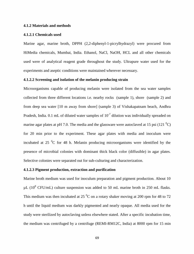
69
4.1.2 Materials and methods
4.1.2.1 Chemicals used
Marine agar, marine broth, DPPH (2,2-diphenyl-1-picrylhydrazyl) were procured from
HiMedia chemicals, Mumbai, India. Ethanol, NaCl, NaOH, HCL and all other chemicals
used were of analytical reagent grade throughout the study. Ultrapure water used for the
experiments and aseptic conditions were maintained wherever necessary.
4.1.2.2 Screening and isolation of the melanin producing strain
Microorganisms capable of producing melanin were isolated from the sea water samples
collected from three different locations i.e. nearby rocks (sample 1), shore (sample 2) and
from deep sea water [10 m away from shore] (sample 3) of Vishakapatnam beach, Andhra
Pradesh, India. 0.1 mL of diluted water samples of 10-7
dilution was individually spreaded on
marine agar plates at pH 7.0. The media and the glassware were autoclaved at 15 psi (121 0C)
for 20 min prior to the experiment. These agar plates with media and inoculum were
incubated at 25 0C for 48 h. Melanin producing microorganisms were identified by the
presence of microbial colonies with dominant thick black color (diffusible) in agar plates.
Selective colonies were separated out for sub-culturing and characterization.
4.1.2.3 Pigment production, extraction and purification
Marine broth medium was used for inoculum preparation and pigment production. About 10
µL (108 CFU/mL) culture suspension was added to 50 mL marine broth in 250 mL flasks.
This medium was then incubated at 25 0C on a rotary shaker moving at 200 rpm for 48 to 72
h until the liquid medium was darkly pigmented and nearly opaque. All media used for the
study were sterilized by autoclaving unless elsewhere stated. After a specific incubation time,
the medium was centrifuged by a centrifuge (REMI-RM12C, India) at 8000 rpm for 15 min
70
to separate the broth (supernatant) and the cells. The solid pellet of cells was separated and
suspended in distilled water. These cells were again centrifuged to collect the supernatant.
Melanin was extracted from the overall supernatant by acidification with 3 N HCl at pH-2
and allowed to stand for 48 h initially at room temperature. This process was repeated for 3
more days until no further precipitation occurred. Then the suspension thus obtained was
boiled for 5 min to prevent the formation of melanoidins [1]. As a final point, crude pigment
pellet was collected after centrifugation at 4000 rpm for 15 min. In addition to marine broth
medium, the pigment production ability of the isolate was tested by culturing them upon
vegetable waste (from cabbage), blend of marine broth and vegetable waste (10:90, 20:80,
30:70) as a nutrient source. Culture conditions and the rest of the protocol maintained same as
described above.
4.1.2.4 DPPH/SPF/metal chelating assay
DPPH assay
DPPH (1, 1diphenyl2picrylhydrazyl) accepts an electron to form a stable diamagnetic
molecule. The methanolic solution of DPPH of violet color has got a strong UV absorbance
at 517nm. The presence of a reducing agent in this methanolic solution pairs the odd
electrons of DPPH radical and further the solution fade color stiochometrically and also the
absorbance of the solution decreases at 517nm. The schematic representation of the process
was shown below
71
The radical scavenging activity by melanin pigment was investigated by modified method of
Ju et al. [12]. Primarily 1 mL of (15‐100 μg/mL) microbial melanin or standard (Ascorbic
acid) was added to 2 mL of DPPH in ethanol. After 30 minutes incubation at 37 oC the
absorbance was measured at 516 nm using UV‐spectrophotometer. Corresponding blanks
were also taken respectively. The experiment was performed in duplicate. The absorbance of
DPPH as control was measured at 516 nm. Lower absorbance of the reaction mixture
indicated higher radical scavenging activity. The scavenging effect (%) was measured using
the following formula:
DPPH inhibition (%) =[(Control absorbance -test absorbance)/Control absorbance] X 100
In vitro sun protection factor
Sunburn, premature skin aging, skin cancers and suppression of the immune system are all
linked to exposure of skin to UV light. The ultraviolet (UV) spectrum of between 200 nm and
400 nm is commonly divided into three regions: UV-A: 320-400 nm; UV-B: 280-320 nm;
UV-C: 200-280 nm.
72
The highest energy region, UV-C, is absorbed wholly by ozone in the stratosphere.
Among the total solar UV radiation reaching the earth’s surface, 6% is in the UV-B region
and 94% in the UV-A. The potential of UV radiation to cause skin damage increases
exponentially with decreasing wavelength. UV light at 280 nm is 1000 times more damaging
than light at 340 nm, thus, a sunscreen’s ability to block UV-B is more important to prevent
the negative effects of sun exposure.
The severe effects of UV-A and UV-B exposure are both short-lived and reversible.
These effects include mostly sunburn (or erythema) and tanning (or pigment darkening).
Largely sunscreen products contain ingredients that provide adequate protection only against
UV-B rays. Even those labelled as broad-spectrum sunscreens may offer only partial
protection against UV-A radiation. Sunscreens are products applied to the skin to protect
against the harmful effects of the sun's UV rays. Sunscreens are usually grouped into two key
categories, namely chemical absorbers and physical blockers. Chemical absorbers absorb
high-intensity UV rays while physical blockers reflect or scatter them. Chemical absorber
compounds include avobenzone, padimate O, octyl methoxycinnamate, octisalate, and
octocrylene. Physical blocker compounds include titanium dioxide and zinc oxide. The
international standard for quantifying the damaging effects of UV radiation on skin is the
erythemal action spectrum and the Calculation of Sun Protection Factor (SPF) was performed
according to Mansur equation [13] stated below.
As per the adapted method, spectra of melanin samples were collected over the spectral range
400-280 nm with 1 nm data point resolution on a UV-visible UV-3200 double beam
spectrophotometer (LABINDIA analytical Instruments Pvt Ltd, India). The SPF values of
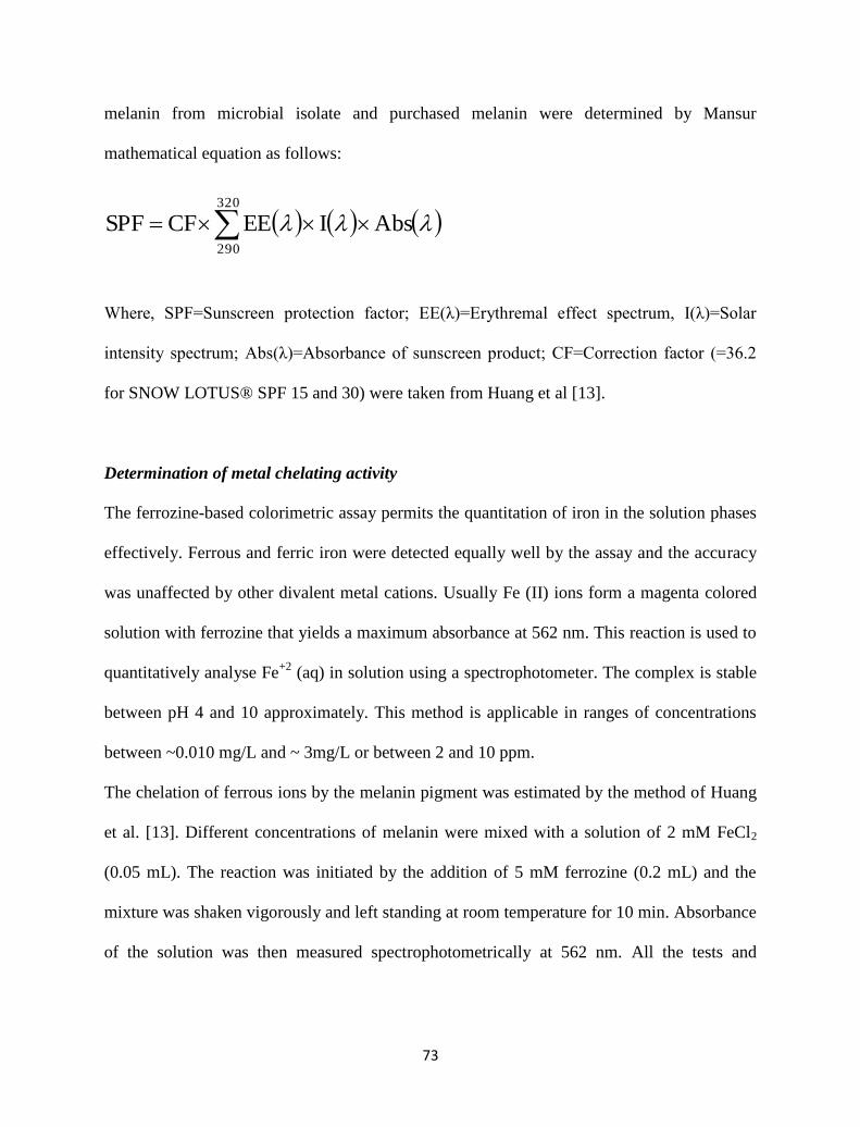
73
melanin from microbial isolate and purchased melanin were determined by Mansur
mathematical equation as follows:
Where, SPF=Sunscreen protection factor; EE(λ)=Erythremal effect spectrum, I(λ)=Solar
intensity spectrum; Abs(λ)=Absorbance of sunscreen product; CF=Correction factor (=36.2
for SNOW LOTUS® SPF 15 and 30) were taken from Huang et al [13].
Determination of metal chelating activity
The ferrozine-based colorimetric assay permits the quantitation of iron in the solution phases
effectively. Ferrous and ferric iron were detected equally well by the assay and the accuracy
was unaffected by other divalent metal cations. Usually Fe (II) ions form a magenta colored
solution with ferrozine that yields a maximum absorbance at 562 nm. This reaction is used to
quantitatively analyse Fe+2
(aq) in solution using a spectrophotometer. The complex is stable
between pH 4 and 10 approximately. This method is applicable in ranges of concentrations
between ~0.010 mg/L and ~ 3mg/L or between 2 and 10 ppm.
The chelation of ferrous ions by the melanin pigment was estimated by the method of Huang
et al. [13]. Different concentrations of melanin were mixed with a solution of 2 mM FeCl2
(0.05 mL). The reaction was initiated by the addition of 5 mM ferrozine (0.2 mL) and the
mixture was shaken vigorously and left standing at room temperature for 10 min. Absorbance
of the solution was then measured spectrophotometrically at 562 nm. All the tests and
AbsIEECFSPF320
290
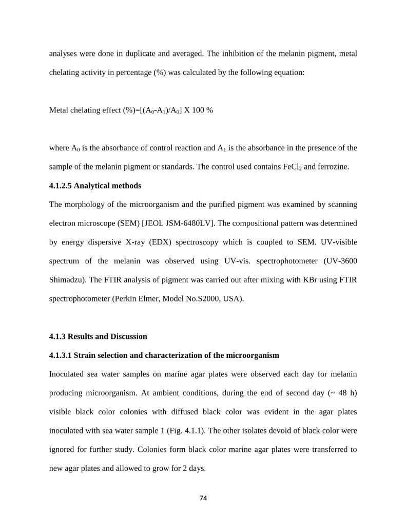
74
analyses were done in duplicate and averaged. The inhibition of the melanin pigment, metal
chelating activity in percentage (%) was calculated by the following equation:
Metal chelating effect (%)=[(A0-A1)/A0] X 100 %
where A0 is the absorbance of control reaction and A1 is the absorbance in the presence of the
sample of the melanin pigment or standards. The control used contains FeCl2 and ferrozine.
4.1.2.5 Analytical methods
The morphology of the microorganism and the purified pigment was examined by scanning
electron microscope (SEM) [JEOL JSM-6480LV]. The compositional pattern was determined
by energy dispersive X-ray (EDX) spectroscopy which is coupled to SEM. UV-visible
spectrum of the melanin was observed using UV-vis. spectrophotometer (UV-3600
Shimadzu). The FTIR analysis of pigment was carried out after mixing with KBr using FTIR
spectrophotometer (Perkin Elmer, Model No.S2000, USA).
4.1.3 Results and Discussion
4.1.3.1 Strain selection and characterization of the microorganism
Inoculated sea water samples on marine agar plates were observed each day for melanin
producing microorganism. At ambient conditions, during the end of second day (~ 48 h)
visible black color colonies with diffused black color was evident in the agar plates
inoculated with sea water sample 1 (Fig. 4.1.1). The other isolates devoid of black color were
ignored for further study. Colonies form black color marine agar plates were transferred to
new agar plates and allowed to grow for 2 days.
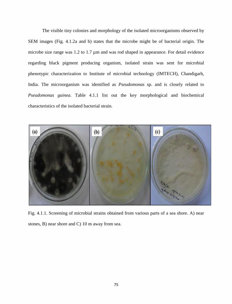
75
The visible tiny colonies and morphology of the isolated microorganisms observed by
SEM images (Fig. 4.1.2a and b) states that the microbe might be of bacterial origin. The
microbe size range was 1.2 to 1.7 µm and was rod shaped in appearance. For detail evidence
regarding black pigment producing organism, isolated strain was sent for microbial
phenotypic characterization to Institute of microbial technology (IMTECH), Chandigarh,
India. The microorganism was identified as Pseudomonas sp. and is closely related to
Pseudomonas guinea. Table 4.1.1 list out the key morphological and biochemical
characteristics of the isolated bacterial strain.
Fig. 4.1.1. Screening of microbial strains obtained from various parts of a sea shore. A) near
stones, B) near shore and C) 10 m away from sea.
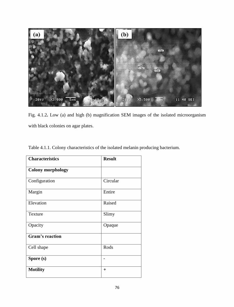
76
Fig. 4.1.2. Low (a) and high (b) magnification SEM images of the isolated microorganism
with black colonies on agar plates.
Table 4.1.1. Colony characteristics of the isolated melanin producing bacterium.
Characteristics Result
Colony morphology
Configuration Circular
Margin Entire
Elevation Raised
Texture Slimy
Opacity Opaque
Gram’s reaction
Cell shape Rods
Spore (s) -
Motility +
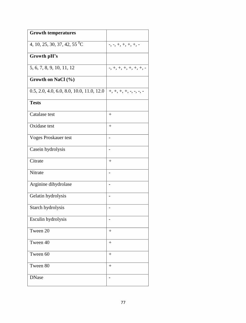
77
Growth temperatures
4, 10, 25, 30, 37, 42, 55 0C -, -, +, +, +, +, -
Growth pH’s
5, 6, 7, 8, 9, 10, 11, 12 -, +, +, +, +, +, +, -
Growth on NaCl (%)
0.5, 2.0, 4.0, 6.0, 8.0, 10.0, 11.0, 12.0 +, +, +, +, -, -, -, -
Tests
Catalase test +
Oxidase test +
Voges Proskauer test -
Casein hydrolysis -
Citrate +
Nitrate -
Arginine dihydrolase -
Gelatin hydrolysis -
Starch hydrolysis -
Esculin hydrolysis -
Tween 20 +
Tween 40 +
Tween 60 +
Tween 80 +
DNase -
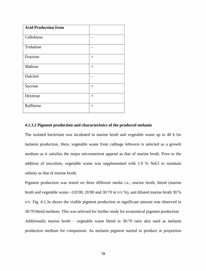
78
Acid Production from
Cellobiose -
Trehalose -
Fructose +
Maltose +
Dulcitol -
Sucrose +
Dextrose +
Raffinose +
4.1.3.2 Pigment production and characteristics of the produced melanin
The isolated bacterium was incubated in marine broth and vegetable waste up to 48 h for
melanin production. Here, vegetable waste from cabbage leftovers is selected as a growth
medium as it satisfies the major micronutrient append as that of marine broth. Prior to the
addition of inoculum, vegetable waste was supplemented with 1.9 % NaCl to maintain
salinity as that of marine broth.
Pigment production was tested on three different media i.e., marine broth, blend (marine
broth and vegetable waste - (10:90; 20:80 and 30:70 in v/v %), and diluted marine broth 30 %
v/v. Fig. 4.1.3a shows the visible pigment production at significant amount was observed in
30:70 blend medium. This was selected for further study for economical pigment production
Additionally marine broth – vegetable waste blend in 30:70 ratio also used as melanin
production medium for comparison. As melanin pigment started to produce at proportion
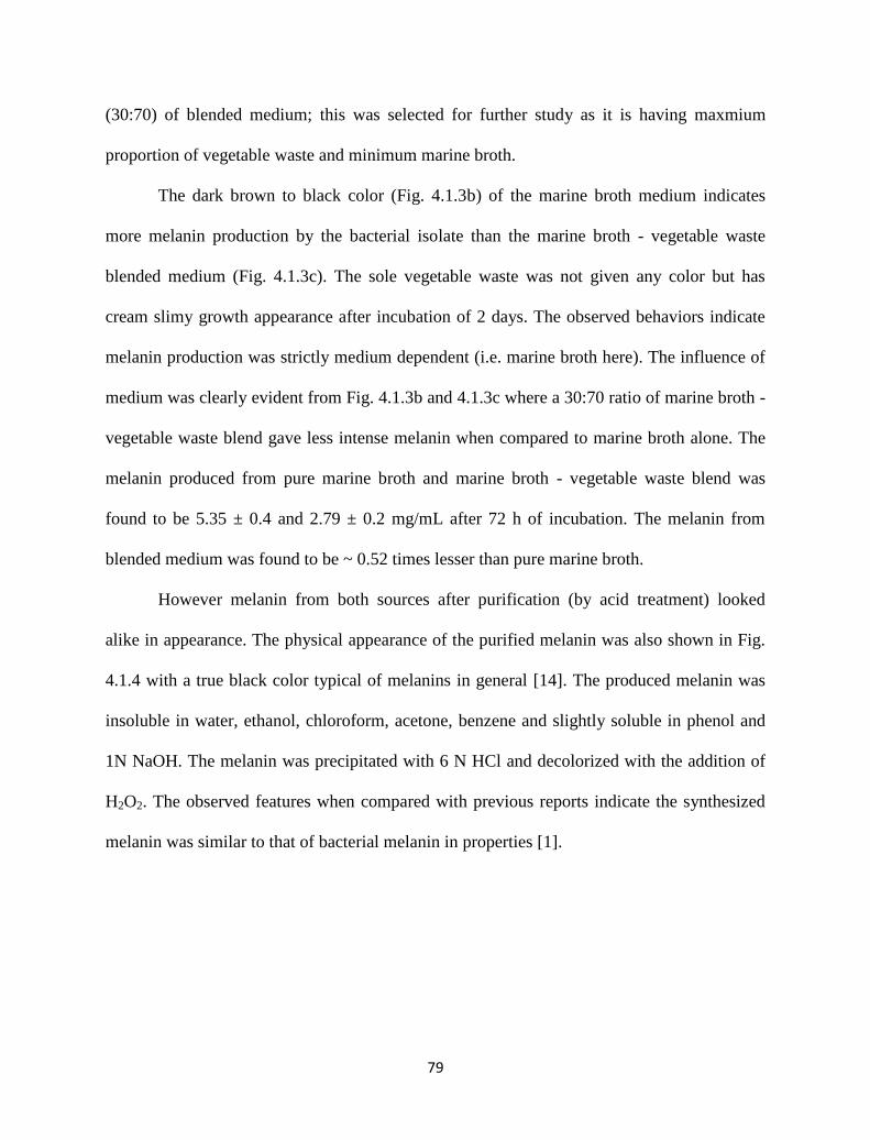
79
(30:70) of blended medium; this was selected for further study as it is having maxmium
proportion of vegetable waste and minimum marine broth.
The dark brown to black color (Fig. 4.1.3b) of the marine broth medium indicates
more melanin production by the bacterial isolate than the marine broth - vegetable waste
blended medium (Fig. 4.1.3c). The sole vegetable waste was not given any color but has
cream slimy growth appearance after incubation of 2 days. The observed behaviors indicate
melanin production was strictly medium dependent (i.e. marine broth here). The influence of
medium was clearly evident from Fig. 4.1.3b and 4.1.3c where a 30:70 ratio of marine broth -
vegetable waste blend gave less intense melanin when compared to marine broth alone. The
melanin produced from pure marine broth and marine broth - vegetable waste blend was
found to be 5.35 ± 0.4 and 2.79 ± 0.2 mg/mL after 72 h of incubation. The melanin from
blended medium was found to be ~ 0.52 times lesser than pure marine broth.
However melanin from both sources after purification (by acid treatment) looked
alike in appearance. The physical appearance of the purified melanin was also shown in Fig.
4.1.4 with a true black color typical of melanins in general [14]. The produced melanin was
insoluble in water, ethanol, chloroform, acetone, benzene and slightly soluble in phenol and
1N NaOH. The melanin was precipitated with 6 N HCl and decolorized with the addition of
H2O2. The observed features when compared with previous reports indicate the synthesized
melanin was similar to that of bacterial melanin in properties [1].
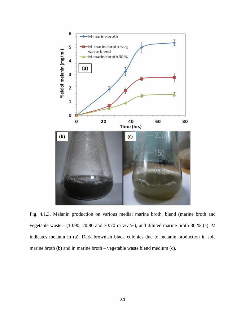
80
Fig. 4.1.3. Melanin production on various media: marine broth, blend (marine broth and
vegetable waste - (10:90; 20:80 and 30:70 in v/v %), and diluted marine broth 30 % (a). M
indicates melanin in (a). Dark brownish black colonies due to melanin production in sole
marine broth (b) and in marine broth – vegetable waste blend medium (c).
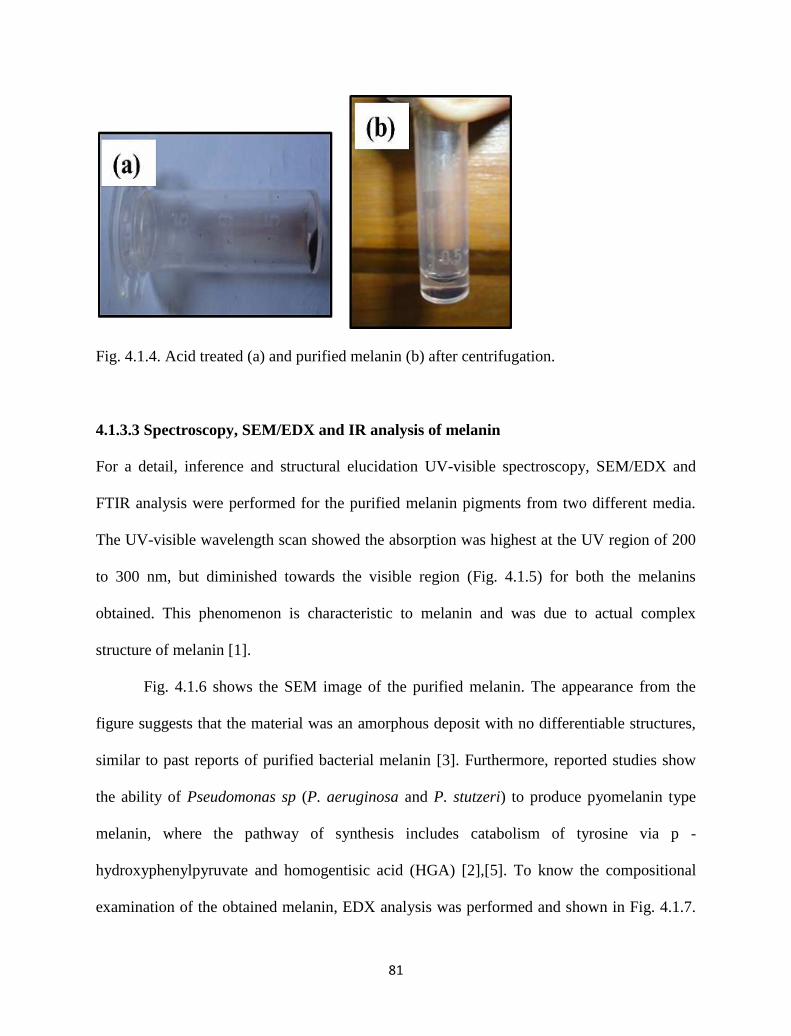
81
Fig. 4.1.4. Acid treated (a) and purified melanin (b) after centrifugation.
4.1.3.3 Spectroscopy, SEM/EDX and IR analysis of melanin
For a detail, inference and structural elucidation UV-visible spectroscopy, SEM/EDX and
FTIR analysis were performed for the purified melanin pigments from two different media.
The UV-visible wavelength scan showed the absorption was highest at the UV region of 200
to 300 nm, but diminished towards the visible region (Fig. 4.1.5) for both the melanins
obtained. This phenomenon is characteristic to melanin and was due to actual complex
structure of melanin [1].
Fig. 4.1.6 shows the SEM image of the purified melanin. The appearance from the
figure suggests that the material was an amorphous deposit with no differentiable structures,
similar to past reports of purified bacterial melanin [3]. Furthermore, reported studies show
the ability of Pseudomonas sp (P. aeruginosa and P. stutzeri) to produce pyomelanin type
melanin, where the pathway of synthesis includes catabolism of tyrosine via p -
hydroxyphenylpyruvate and homogentisic acid (HGA) [2],[5]. To know the compositional
examination of the obtained melanin, EDX analysis was performed and shown in Fig. 4.1.7.
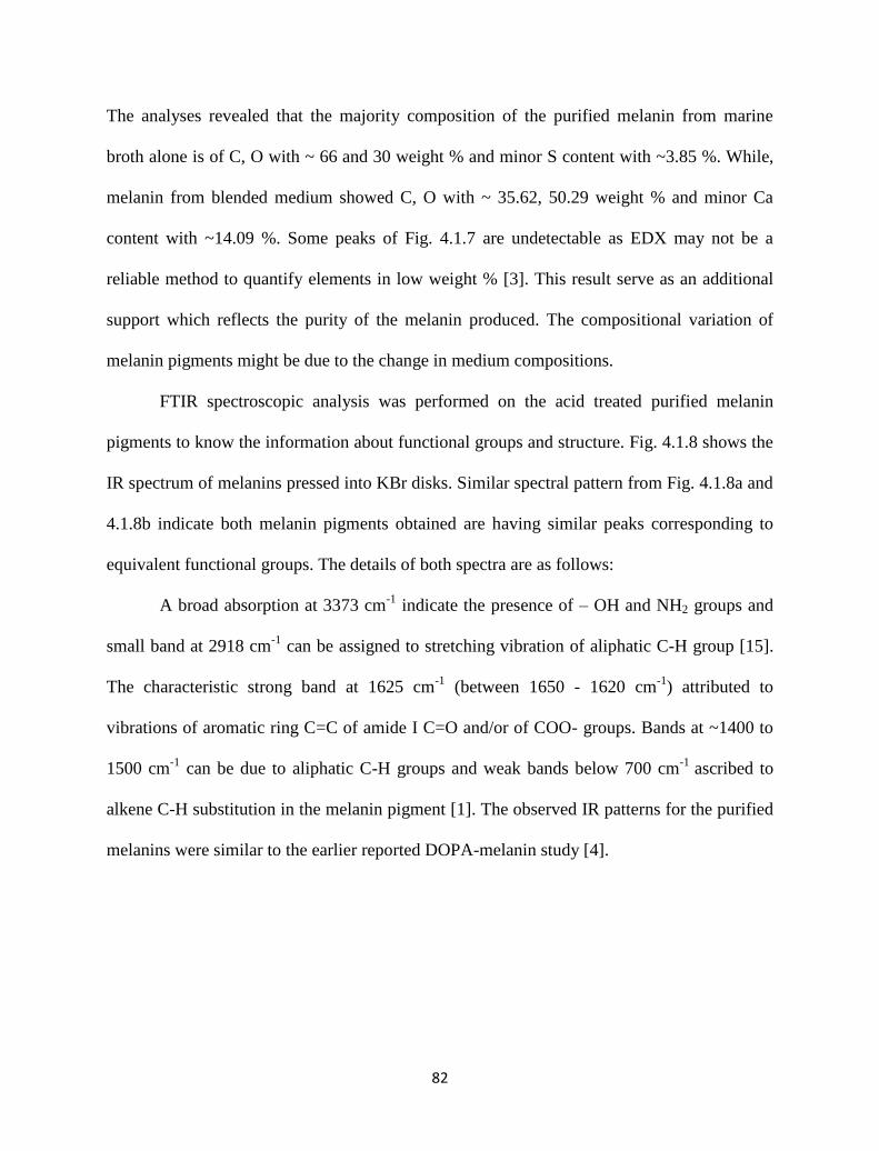
82
The analyses revealed that the majority composition of the purified melanin from marine
broth alone is of C, O with ~ 66 and 30 weight % and minor S content with ~3.85 %. While,
melanin from blended medium showed C, O with ~ 35.62, 50.29 weight % and minor Ca
content with ~14.09 %. Some peaks of Fig. 4.1.7 are undetectable as EDX may not be a
reliable method to quantify elements in low weight % [3]. This result serve as an additional
support which reflects the purity of the melanin produced. The compositional variation of
melanin pigments might be due to the change in medium compositions.
FTIR spectroscopic analysis was performed on the acid treated purified melanin
pigments to know the information about functional groups and structure. Fig. 4.1.8 shows the
IR spectrum of melanins pressed into KBr disks. Similar spectral pattern from Fig. 4.1.8a and
4.1.8b indicate both melanin pigments obtained are having similar peaks corresponding to
equivalent functional groups. The details of both spectra are as follows:
A broad absorption at 3373 cm-1
indicate the presence of – OH and NH2 groups and
small band at 2918 cm-1
can be assigned to stretching vibration of aliphatic C-H group [15].
The characteristic strong band at 1625 cm-1
(between 1650 - 1620 cm-1
) attributed to
vibrations of aromatic ring C=C of amide I C=O and/or of COO- groups. Bands at ~1400 to
1500 cm-1
can be due to aliphatic C-H groups and weak bands below 700 cm-1
ascribed to
alkene C-H substitution in the melanin pigment [1]. The observed IR patterns for the purified
melanins were similar to the earlier reported DOPA-melanin study [4].
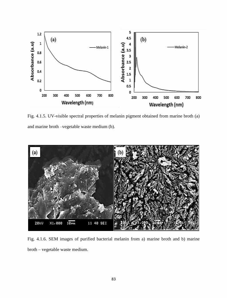
83
Fig. 4.1.5. UV-visible spectral properties of melanin pigment obtained from marine broth (a)
and marine broth –vegetable waste medium (b).
Fig. 4.1.6. SEM images of purified bacterial melanin from a) marine broth and b) marine
broth – vegetable waste medium.
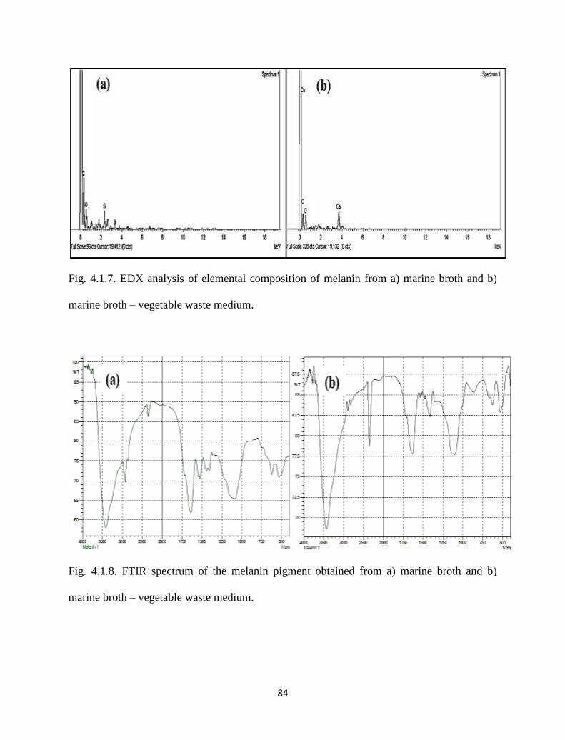
84
Fig. 4.1.7. EDX analysis of elemental composition of melanin from a) marine broth and b)
marine broth – vegetable waste medium.
Fig. 4.1.8. FTIR spectrum of the melanin pigment obtained from a) marine broth and b)
marine broth – vegetable waste medium.
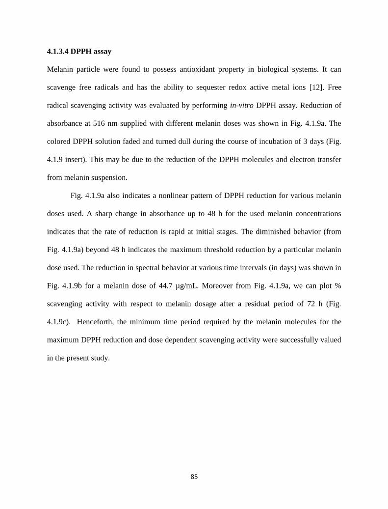
85
4.1.3.4 DPPH assay
Melanin particle were found to possess antioxidant property in biological systems. It can
scavenge free radicals and has the ability to sequester redox active metal ions [12]. Free
radical scavenging activity was evaluated by performing in-vitro DPPH assay. Reduction of
absorbance at 516 nm supplied with different melanin doses was shown in Fig. 4.1.9a. The
colored DPPH solution faded and turned dull during the course of incubation of 3 days (Fig.
4.1.9 insert). This may be due to the reduction of the DPPH molecules and electron transfer
from melanin suspension.
Fig. 4.1.9a also indicates a nonlinear pattern of DPPH reduction for various melanin
doses used. A sharp change in absorbance up to 48 h for the used melanin concentrations
indicates that the rate of reduction is rapid at initial stages. The diminished behavior (from
Fig. 4.1.9a) beyond 48 h indicates the maximum threshold reduction by a particular melanin
dose used. The reduction in spectral behavior at various time intervals (in days) was shown in
Fig. 4.1.9b for a melanin dose of 44.7 µg/mL. Moreover from Fig. 4.1.9a, we can plot %
scavenging activity with respect to melanin dosage after a residual period of 72 h (Fig.
4.1.9c). Henceforth, the minimum time period required by the melanin molecules for the
maximum DPPH reduction and dose dependent scavenging activity were successfully valued
in the present study.
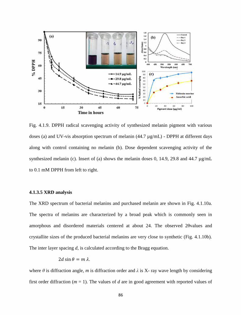
86
Fig. 4.1.9. DPPH radical scavenging activity of synthesized melanin pigment with various
doses (a) and UV-vis absorption spectrum of melanin (44.7 µg/mL) - DPPH at different days
along with control containing no melanin (b). Dose dependent scavenging activity of the
synthesized melanin (c). Insert of (a) shows the melanin doses 0, 14.9, 29.8 and 44.7 µg/mL
to 0.1 mM DPPH from left to right.
4.1.3.5 XRD analysis
The XRD spectrum of bacterial melanins and purchased melanin are shown in Fig. 4.1.10a.
The spectra of melanins are characterized by a broad peak which is commonly seen in
amorphous and disordered materials centered at about 24. The observed 2θvalues and
crystallite sizes of the produced bacterial melanins are very close to synthetic (Fig. 4.1.10b).
The inter layer spacing d, is calculated according to the Bragg equation.
.
where θ is diffraction angle, m is diffraction order and λ is X- ray wave length by considering
first order diffraction (m = 1). The values of d are in good agreement with reported values of
87
the inter layer spacing in the stacked sheets model of the melanin [16]. An estimate of
average grain size of melanins was calculated from the Dedye - schrerrer equation [16].
where FWHM is full width at half maximum of diffraction peak. The closeness of the grain
size values indicates the quality of the purified bacterial melanins. Furthermore %
crystallinity was also calculated for the stated melanins by considering glass substrate as
background. The calculation is as follows:
% crystallinity values from Fig. 4.1.10c indicated that the bacterial melanin from blend and
marine broth were less crystalline than the synthetic melanin studied. Lack of crystallinity is
significant sign of consistent physical property of melanin [17]. And the above results state
that the obtained bacterial melanins are of high pure and amorphous.
Fig. 4.1.10. (a) X-ray diffractograms of the produced melanin (M-marine broth, M-blend) and
purchased melanin (M-synthetic). (b) Interlayer spacing (d-value) and crystallite sizes and (c)
% crystallinity of the different melanins.
100area) (total
area) profile background-area total(itycrystallin %
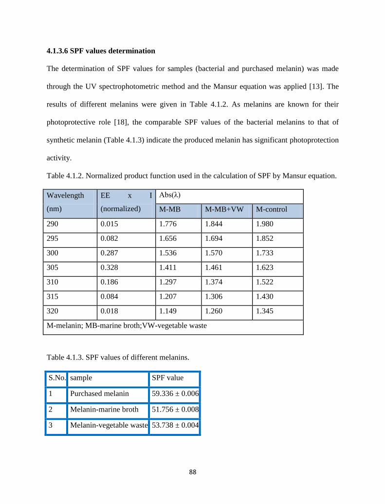
88
4.1.3.6 SPF values determination
The determination of SPF values for samples (bacterial and purchased melanin) was made
through the UV spectrophotometric method and the Mansur equation was applied [13]. The
results of different melanins were given in Table 4.1.2. As melanins are known for their
photoprotective role [18], the comparable SPF values of the bacterial melanins to that of
synthetic melanin (Table 4.1.3) indicate the produced melanin has significant photoprotection
activity.
Table 4.1.2. Normalized product function used in the calculation of SPF by Mansur equation.
Wavelength
(nm)
EE x I
(normalized)
Abs(λ)
M-MB M-MB+VW M-control
290 0.015 1.776 1.844 1.980
295 0.082 1.656 1.694 1.852
300 0.287 1.536 1.570 1.733
305 0.328 1.411 1.461 1.623
310 0.186 1.297 1.374 1.522
315 0.084 1.207 1.306 1.430
320 0.018 1.149 1.260 1.345
M-melanin; MB-marine broth;VW-vegetable waste
Table 4.1.3. SPF values of different melanins.
S.No. sample SPF value
1 Purchased melanin 59.336 ± 0.006
2 Melanin-marine broth 51.756 ± 0.008
3 Melanin-vegetable waste 53.738 ± 0.004
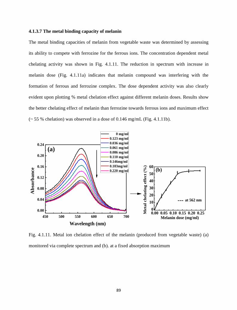
89
4.1.3.7 The metal binding capacity of melanin
The metal binding capacities of melanin from vegetable waste was determined by assessing
its ability to compete with ferrozine for the ferrous ions. The concentration dependent metal
chelating activity was shown in Fig. 4.1.11. The reduction in spectrum with increase in
melanin dose (Fig. 4.1.11a) indicates that melanin compound was interfering with the
formation of ferrous and ferrozine complex. The dose dependent activity was also clearly
evident upon plotting % metal chelation effect against different melanin doses. Results show
the better chelating effect of melanin than ferrozine towards ferrous ions and maximum effect
(~ 55 % chelation) was observed in a dose of 0.146 mg/mL (Fig. 4.1.11b).
Fig. 4.1.11. Metal ion chelation effect of the melanin (produced from vegetable waste) (a)
monitored via complete spectrum and (b). at a fixed absorption maximum
450 500 550 600 650 700
0.00
0.04
0.08
0.12
0.16
0.20
0.24
(a)
Ab
sorb
an
ce
Wavelength (nm)
0 mg/ml
0.123 mg/ml
0.036 mg/ml
0.061 mg/ml
0.086 mg/ml
0.110 mg/ml
0.146mg/ml
0.183mg/ml
0.220 mg/ml
0.00 0.05 0.10 0.15 0.20 0.250
10
20
30
40
50
60(b)
at 562 nm
Meta
l ch
ela
tin
g e
ffect
(%)
Melanin dose (mg/ml)
90
4.1.4 Conclusions
Though different marine isolates were used, it was found that the sample 1 contains the
melanin producing bacterium and was found to be Pseudomonas guinea. Upon investigation
for the melanin production in different media, 5.35 ± 0.4 and 2.79 ± 0.2 mg/mL pigment was
produced when cultured in marine broth alone and blended medium (vegetable waste and
marine broth), respectively. Pigments produced from the different nutrient sources have
shown different elemental compositions with varying weight %. Moreover, the FTIR analysis
revealed that functional groups were conserved in both the melanins and were appeared to be
same. The compositional variation in the pigments might be due to the change in the media
composition although P.guinea, only was used in both the cases. Furthermore, the produced
melanin noticed to have efficient free radical scavenging activity (of a model DPPH radical)
and also exhibited photoprotective and metal ion chelation activity. This study confirms that
the antioxidant melanin pigment can be produced from the cheaper substrates without any
functional variation.
References
[1] S. Sajjan, et al., "Purification and physiochemical characterization of melanin
pigment from Klebsiella sp. GSK," Journal of microbiology and biotechnology, vol.
20, pp. 1513-1520, 2010.
[2] J. Geng, et al., "Bacterial melanin interacts with double-stranded DNA with high
affinity and may inhibit cell metabolism in vivo," Archives of microbiology, vol. 192,
pp. 321-329, 2010.
91
[3] A. M. Gómez-Marín and C. I. Sánchez, "Thermal and mass spectroscopic
characterization of a sulphur-containing bacterial melanin from Bacillus subtilis,"
Journal of Non-Crystalline Solids, vol. 356, pp. 1576-1580, 2010.
[4] E. Harki, et al., "Purification, characterisation and analysis of melanin extracted from
Tuber melanosporum Vitt," Food Chemistry, vol. 58, pp. 69-73, 1997.
[5] V. Coyne and L. al-Harthi, "Induction of melanin biosynthesis in Vibrio cholerae,"
Applied and environmental microbiology, vol. 58, pp. 2861-2865, 1992.
[6] S. Youngchim, et al., "Production of melanin by Aspergillus fumigatus," Journal of
medical microbiology, vol. 53, pp. 175-181, 2004.
[7] S. I. Kotob, et al., "Homogentisic acid is the primary precursor of melanin synthesis
in Vibrio cholerae, a Hyphomonas strain, and Shewanella colwelliana," Applied and
environmental microbiology, vol. 61, pp. 1620-1622, 1995.
[8] F. Solano, et al., "Isolation and characterization of strain MMB-1 (CECT 4803), a
novel melanogenic marine bacterium," Applied and environmental microbiology, vol.
63, pp. 3499-3506, 1997.
[9] P. Lucas-Elío, et al., "Complete genome sequence of the melanogenic marine
bacterium Marinomonas mediterranea type strain (MMB-1T)," Standards in genomic
sciences, vol. 6, p. 63, 2012.
[10] S. R. Quadri and D. Agsar, "Detection of melanin producing thermo-alkaliphilic
Streptomyces from limestone quarries of the Deccan traps," World Journal of Science
and Technology, vol. 2, 2012.
[11] J. Ravishankar, et al., "Isolation and characterization of melanin from a marine
fungus," Botanica Marina, vol. 38, pp. 413-416, 1995.
92
[12] K.-Y. Ju, et al., "Bioinspired polymerization of dopamine to generate melanin-like
nanoparticles having an excellent free-radical-scavenging property,"
Biomacromolecules, vol. 12, pp. 625-632, 2011.
[13] S. Huang, et al., "Antioxidant activities and UV-protective properties of melanin from
the berry of Cinnamomum burmannii and Osmanthus fragrans," Medicinal Chemistry
Research, vol. 20, pp. 475-481, 2011.
[14] W. W. van de Sande, et al., "Melanin biosynthesis in Madurella mycetomatis> and its
effect on susceptibility to itraconazole and ketoconazole," Microbes and Infection,
vol. 9, pp. 1114-1123, 2007.
[15] J. Coates, "Interpretation of infrared spectra, a practical approach," Encyclopedia of
analytical chemistry, 2000.
[16] V. Capozzi, et al., "Optical and photoelectronic properties of melanin," Thin solid
films, vol. 511, pp. 362-366, 2006.
[17] Y. Thathachari and M. Blois, "Physical studies on melanins: II. X-ray diffraction,"
Biophysical journal, vol. 9, pp. 77-89, 1969.
[18] M. Brenner and V. J. Hearing, "The protective role of melanin against UV Damage in
human Skin†," Photochemistry and photobiology, vol. 84, pp. 539-549, 2008.
93
4.2 Melanin by Soil Microbial Isolate on Fruit Waste
Extract: Optimization of Key Production Parameters
4.2.1 Introduction
Melanins are the natural pigments which have their presence in animals, plants and in most of
the microorganisms [1]. They are the dark colored negatively charged high molecular weight
pigments which are formed due to polymerized phenolic and/or indolic compounds. These
complex polymers are amorphous in nature and shows solubility in neither aqueous nor
organic solvents. They showed resistance to concentrated acids and are susceptible to
bleaching by oxidizing agents [2]. They play a vital role in defense and protection
mechanisms that improve the survival and competitiveness of the organisms [3]. Melanin is
known for its absorption capacity of radiation of all wavelengths with an optimum
absorbance at UV range [4] which prevents photo induced damage. Hence it is used in the
preparation of photo absorbing optical lenses and in bioplastics. Besides photo protection it
has versatile biological activities such as radical scavenging, antioxidant, antitumor, anti-
inflammatory [5] and as immune stimulating agent [6].
94
Melanin obtained from microbes has great advantages over melanin from animals and
plants. Microorganisms don’t cause the problems of seasonal variations and are selected
arsenals as they modify them according to the medium and conditions provided to them [7].
Targeting melanogenisis in microbes may help to discover antimicrobial drugs. For example,
melanins produced by Cryptococcus neoformans and Burkholderia cepacia offer virulence
and contribute to the growing resistance of these pathogenic bacteria towards antibiotics [2,
8]. The melanin synthesized by microbes shows metal chelating ability too (sorb the
radioactive wastes uranium) [9]. There are reports that showed the anti HIV properties of
melanin and their role in photo voltage generation and fluorescence studies [10-11].
Therefore all these properties of melanin make them unique and are widely used in cosmetic,
sunscreen protection creams, eye glasses, pharmaceuticals, and food industries
Eying on the potential uses and increasing demand for the melanin pigment there is a
need to conduct studies on the production of melanin from microbes. There are reports on
melanin production from various microorganisms including Bacillus species which are well
known for their pigment production ability in various stress environments [4, 12]. Selection
of substrate for melanin production has economic importance. For instance till date expensive
substrates like NCM media [4], LB (Luria-Bertani) media [12], minimal media supplemented
with L-tyrosine [13], amino acids enriched tryptone broth agar [14] and so on [15-16] were
used for high yield of melanin. Owing to the economy and practicability of the melanin
production process; the need to use economically feasible substrates along with optimization
of key parameters is needed. Taguchi method [17] is a systematic technique of design and
analysis of experiments that has been employed successfully in recent years to design and
95
improve product quality economically [18], CCD design approach has been used to fit a
polynomial model.
In this study, a bacterium capable of producing melanin was isolated from garden soil
and subsequently characterized. The strain was cultivated in the fruit waste extract (FWE) [as
sole source of energy] to produce significant amounts of melanin. The key parameters in
melanin production were identified and optimized using simple two step Taguchi and CCD
(central composite design) approach. Upon purification and characterization, the obtained
melanin was tested for In vitro sun protection effect, free radical scavenging and metal
chelating activities.
4.2.2 Materials and methods
4.2.2.1 Chemicals and microorganism
DPPH (2,2-diphenyl-1-picrylhydrazyl), purchased from HiMedia chemicals, Mumbai, India.
Ascorbic acid was purchased from Merck, India. Ferrozine and melanin (synthetic) was
purchased from Sigma-Aldrich, India. Ethanol, NaCl, NaOH, HCL are from Merck, India and
all other chemicals used were of analytical reagent grade throughout the study. Ultrapure
water was used for the experiments and aseptic conditions were maintained wherever
necessary.
The microorganism used in this study was isolated from garden soil in front of
Department of Chemical Engineering, National Institute of Technology, Rourkela, India
using serial dilution technique on a nutrient agar (NA) [19]. Using 107
dilution soil samples
on NA plates. Melanin producing organism was identified. It was separated by observing a
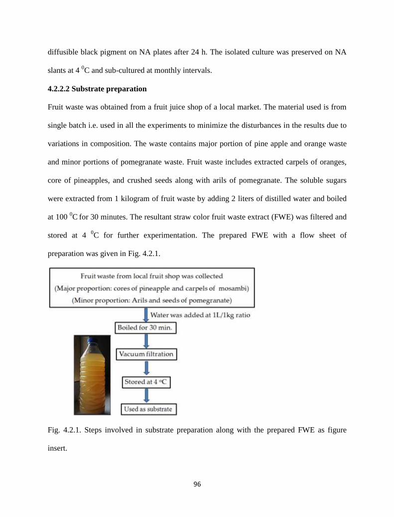
96
diffusible black pigment on NA plates after 24 h. The isolated culture was preserved on NA
slants at 4 0C and sub-cultured at monthly intervals.
4.2.2.2 Substrate preparation
Fruit waste was obtained from a fruit juice shop of a local market. The material used is from
single batch i.e. used in all the experiments to minimize the disturbances in the results due to
variations in composition. The waste contains major portion of pine apple and orange waste
and minor portions of pomegranate waste. Fruit waste includes extracted carpels of oranges,
core of pineapples, and crushed seeds along with arils of pomegranate. The soluble sugars
were extracted from 1 kilogram of fruit waste by adding 2 liters of distilled water and boiled
at 100 0C
for 30 minutes. The resultant straw color fruit waste extract (FWE) was filtered and
stored at 4 0C for further experimentation. The prepared FWE with a flow sheet of
preparation was given in Fig. 4.2.1.
Fig. 4.2.1. Steps involved in substrate preparation along with the prepared FWE as figure
insert.
97
4.2.2.3 Production and purification of melanin
Nutrient broth (peptone-5g/L, beef extract-3g/L, NaCl-5g/L) was used for inoculum
preparation and FWE was used as production medium for melanin. About 10 µL (108
CFU/mL) culture suspension was added to FWE medium in 250 mL flasks with a working
volume 50 ml. The medium was then incubated at 30 0C on a rotary shaker moving at 200
rpm for 24 h. A dark pigmented and nearly opaque FWE medium was observed (Fig. 4.2.2a).
After the incubation time, the medium was centrifuged using REMI-RM12C, India centrifuge
at 8000 rpm for 15 min to separate the broth (supernatant) and the cells. The solid pellet of
cells were separated and suspended in distilled water. The cells were further centrifuged to
collect the supernatant. Melanin was extracted from the overall supernatant by acidification
with 3 N HCl to pH-2 and allowed to stand for 48 h initially at room temperature. This
process was repeated for 7 more days until no precipitate was obtained. The obtained
suspension was boiled for 5 min to prevent the formation of melanoidins. As a final point,
crude pigment pellet was collected after centrifugation at 4000 rpm for 15 min.
4.2.2.4 Parameters optimization using Taguchi and CCD design
Preliminary idea on growth conditions, suggests Taguchi method to be employed for the
optimization of culture conditions for high yield melanin pigment production. Optimization
of three vital factors like pH, temperature and agitation in 6-3-3 levels respectively was done
as a starting point of the study (Table 4.2.1). Then Taguchi method was performed by 18
different experiments by using L18 orthogonal array as shown in Table 4.2.2. Shown values
of the obtained melanin (mg/mL) are the average of results of two replicates. Based on the
obtained results, the optimum conditions of the used parameters were identified and an
analysis of variance (ANOVA) for the obtained results was investigated.
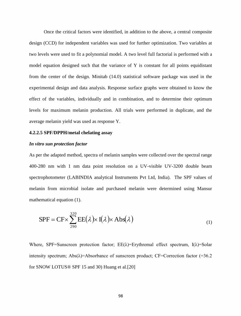
98
Once the critical factors were identified, in addition to the above, a central composite
design (CCD) for independent variables was used for further optimization. Two variables at
two levels were used to fit a polynomial model. A two level full factorial is performed with a
model equation designed such that the variance of Y is constant for all points equidistant
from the center of the design. Minitab (14.0) statistical software package was used in the
experimental design and data analysis. Response surface graphs were obtained to know the
effect of the variables, individually and in combination, and to determine their optimum
levels for maximum melanin production. All trials were performed in duplicate, and the
average melanin yield was used as response Y.
4.2.2.5 SPF/DPPH/metal chelating assay
In vitro sun protection factor
As per the adapted method, spectra of melanin samples were collected over the spectral range
400-280 nm with 1 nm data point resolution on a UV-visible UV-3200 double beam
spectrophotometer (LABINDIA analytical Instruments Pvt Ltd, India). The SPF values of
melanin from microbial isolate and purchased melanin were determined using Mansur
mathematical equation (1).
(1)
Where, SPF=Sunscreen protection factor; EE(λ)=Erythremal effect spectrum, I(λ)=Solar
intensity spectrum; Abs(λ)=Absorbance of sunscreen product; CF=Correction factor (=36.2
for SNOW LOTUS® SPF 15 and 30) Huang et al.[20]
AbsIEECFSPF320
290
99
DPPH assay
The radical scavenging activity by melanin pigment was investigated by modified method of
Ju et al. [21]. Primarily 1 mL of (15‐100 μg/mL) microbial melanin/standard (Ascorbic acid)
was added to 2 mL of DPPH in ethanol. After keeping for 30 minutes at 37oC the absorbance
at 516 nm was measured using UV‐spectrophotometer with reference blank samples. The
experiment was performed in duplicate. The absorbance of DPPH as control was measured at
516 nm. Lower absorbance of the reaction mixture indicated higher radical scavenging
activity. The scavenging effect was measured using equation (2).
DPPH inhibition (%) = [(Control absorbance -test absorbance)/Control absorbance] X 100
(2)
Determination of metal chelating activity
The chelation of ferrous ions by the melanin pigment was estimated by the method of Huang
et al.[20]. Different concentrations of melanin were mixed with a solution of 2 mM FeCl2
(0.05 mL). The reaction was initiated by the addition of 5 mM ferrozine (0.2 mL) and the
mixture was shaken vigorously and left standing at room temperature for 10 min. Absorbance
of the solution was then measured spectrophotometrically at 562 nm. All the tests and
analysis were performed in duplicate and averaged. The inhibition of the melanin pigment
metal chelating activity in percentage (%) was calculated using equation (3).
Metal chelating effect (%) = [(A0-A1)/A0] X 100 % (3)
where A0 is the absorbance of control reaction and A1 is the absorbance in the presence of the
sample of the melanin pigment or standards. The control contains FeCl2 and ferrozine.
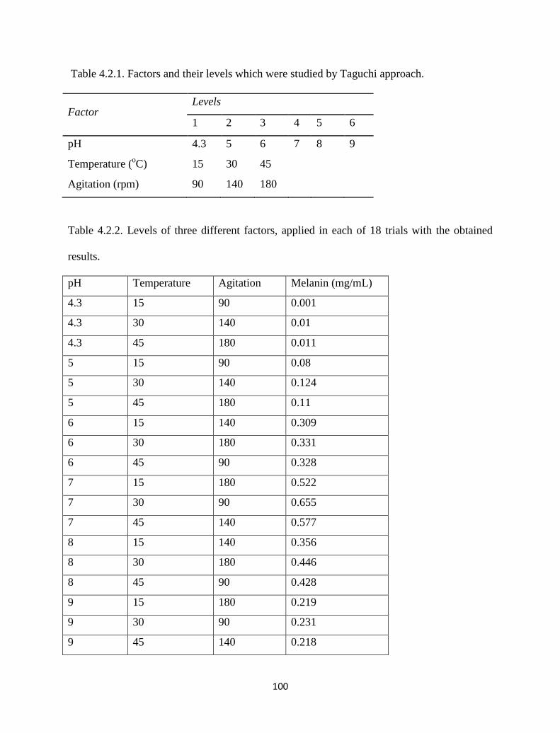
100
Table 4.2.1. Factors and their levels which were studied by Taguchi approach.
Factor Levels
1 2 3 4 5 6
pH 4.3 5 6 7 8 9
Temperature (oC) 15 30 45
Agitation (rpm) 90 140 180
Table 4.2.2. Levels of three different factors, applied in each of 18 trials with the obtained
results.
pH Temperature Agitation Melanin (mg/mL)
4.3 15 90 0.001
4.3 30 140 0.01
4.3 45 180 0.011
5 15 90 0.08
5 30 140 0.124
5 45 180 0.11
6 15 140 0.309
6 30 180 0.331
6 45 90 0.328
7 15 180 0.522
7 30 90 0.655
7 45 140 0.577
8 15 140 0.356
8 30 180 0.446
8 45 90 0.428
9 15 180 0.219
9 30 90 0.231
9 45 140 0.218
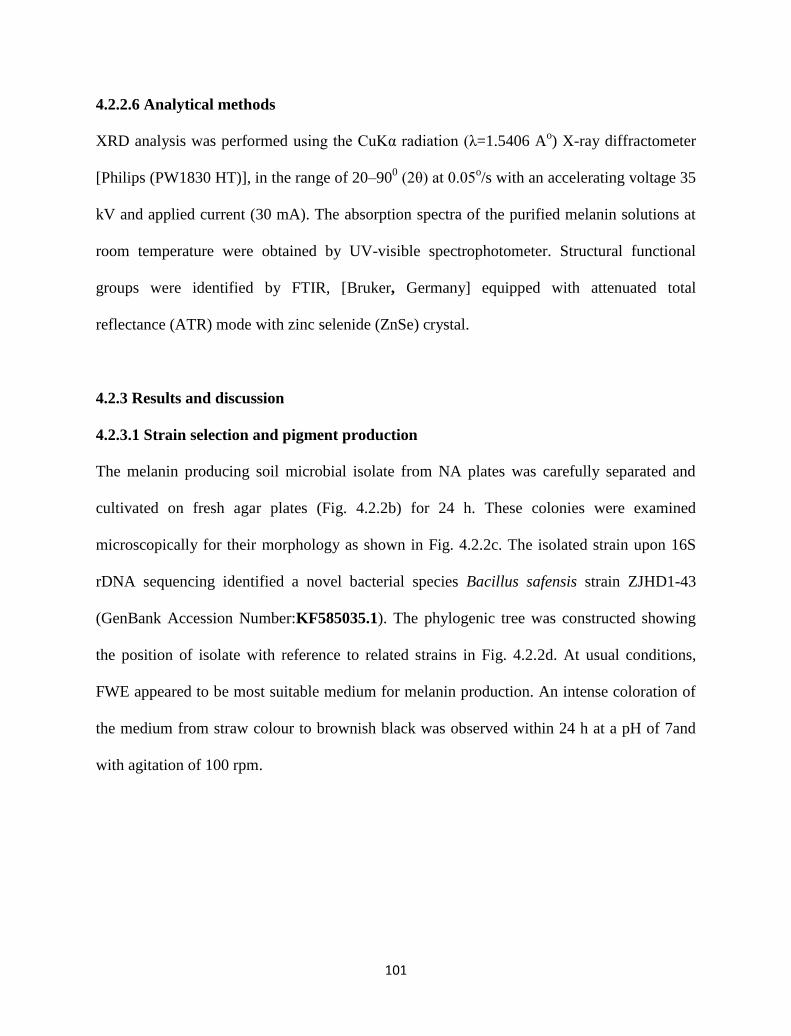
101
4.2.2.6 Analytical methods
XRD analysis was performed using the CuKα radiation (λ=1.5406 Ao) X-ray diffractometer
[Philips (PW1830 HT)], in the range of 20–900 (2θ) at 0.05
o/s with an accelerating voltage 35
kV and applied current (30 mA). The absorption spectra of the purified melanin solutions at
room temperature were obtained by UV-visible spectrophotometer. Structural functional
groups were identified by FTIR, [Bruker, Germany] equipped with attenuated total
reflectance (ATR) mode with zinc selenide (ZnSe) crystal.
4.2.3 Results and discussion
4.2.3.1 Strain selection and pigment production
The melanin producing soil microbial isolate from NA plates was carefully separated and
cultivated on fresh agar plates (Fig. 4.2.2b) for 24 h. These colonies were examined
microscopically for their morphology as shown in Fig. 4.2.2c. The isolated strain upon 16S
rDNA sequencing identified a novel bacterial species Bacillus safensis strain ZJHD1-43
(GenBank Accession Number:KF585035.1). The phylogenic tree was constructed showing
the position of isolate with reference to related strains in Fig. 4.2.2d. At usual conditions,
FWE appeared to be most suitable medium for melanin production. An intense coloration of
the medium from straw colour to brownish black was observed within 24 h at a pH of 7and
with agitation of 100 rpm.
102
Fig. 4.2.2. (a) FWE before (left) and after melanin production (right) by the garden soil
microbial isolate, (b) colonies with diffusible melanin on NA plates, and (c) SEM image of
the microorganism. (d) Phylogenetic tree showing the position of the isolate ZJHD1-43 with
reference to related strains.
4.2.3.2 Taguchi design for screening critical factors
Effect of pH, temperature and agitation were studied employing Taguchi method, which is a
fractional factorial experimental design tool. Experiments performed at experimental
conditions (pH 7; temperature 30 ºC; agitation 90 rpm) produced maximum melanin of 0.655
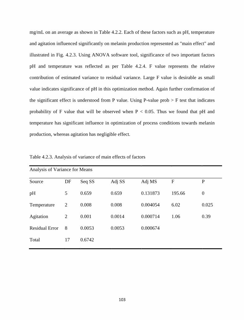
103
mg/mL on an average as shown in Table 4.2.2. Each of these factors such as pH, temperature
and agitation influenced significantly on melanin production represented as "main effect" and
illustrated in Fig. 4.2.3. Using ANOVA software tool, significance of two important factors
pH and temperature was reflected as per Table 4.2.4. F value represents the relative
contribution of estimated variance to residual variance. Large F value is desirable as small
value indicates significance of pH in this optimization method. Again further confirmation of
the significant effect is understood from P value. Using P-value prob > F test that indicates
probability of F value that will be observed when P < 0.05. Thus we found that pH and
temperature has significant influence in optimization of process conditions towards melanin
production, whereas agitation has negligible effect.
Table 4.2.3. Analysis of variance of main effects of factors
Analysis of Variance for Means
Source DF Seq SS Adj SS Adj MS F P
pH 5 0.659 0.659 0.131873 195.66 0
Temperature 2 0.008 0.008 0.004054 6.02 0.025
Agitation 2 0.001 0.0014 0.000714 1.06 0.39
Residual Error 8 0.0053 0.0053 0.000674
Total 17 0.6742
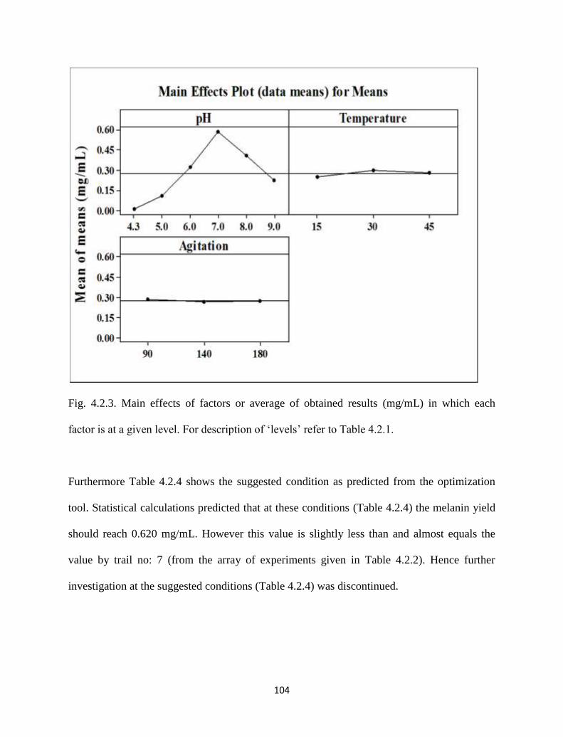
104
Fig. 4.2.3. Main effects of factors or average of obtained results (mg/mL) in which each
factor is at a given level. For description of ‘levels’ refer to Table 4.2.1.
Furthermore Table 4.2.4 shows the suggested condition as predicted from the optimization
tool. Statistical calculations predicted that at these conditions (Table 4.2.4) the melanin yield
should reach 0.620 mg/mL. However this value is slightly less than and almost equals the
value by trail no: 7 (from the array of experiments given in Table 4.2.2). Hence further
investigation at the suggested conditions (Table 4.2.4) was discontinued.
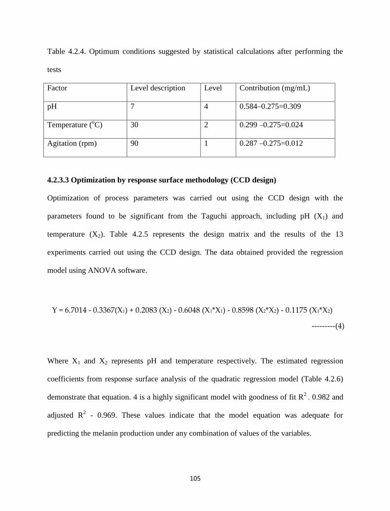
105
Table 4.2.4. Optimum conditions suggested by statistical calculations after performing the
tests
Factor Level description Level Contribution (mg/mL)
pH 7 4 0.584–0.275=0.309
Temperature (oC) 30 2 0.299 –0.275=0.024
Agitation (rpm) 90 1 0.287 –0.275=0.012
4.2.3.3 Optimization by response surface methodology (CCD design)
Optimization of process parameters was carried out using the CCD design with the
parameters found to be significant from the Taguchi approach, including pH (X1) and
temperature (X2). Table 4.2.5 represents the design matrix and the results of the 13
experiments carried out using the CCD design. The data obtained provided the regression
model using ANOVA software.
Where X1 and X2 represents pH and temperature respectively. The estimated regression
coefficients from response surface analysis of the quadratic regression model (Table 4.2.6)
demonstrate that equation. 4 is a highly significant model with goodness of fit R2
- 0.982 and
adjusted R2 - 0.969. These values indicate that the model equation was adequate for
predicting the melanin production under any combination of values of the variables.
Y = 6.7014 - 0.3367(X1) + 0.2083 (X2) - 0.6048 (X1*X1) - 0.8598 (X2*X2) - 0.1175 (X1*X2)
---------(4)
106
Table 4.2.5. Experimental design matrix for the central composite design.
pH Temperature pH values Temperature values Melanin (mg/ml)
0 1 7 35 6.12
0 0 7 30 6.63
0 -1 7 25 5.54
0 0 7 30 6.78
0 0 7 30 6.83
1 0 7.5 30 5.62
-1 1 6.5 35 5.8
-1 -1 6.5 25 5.23
0 0 7 30 6.68
0 0 7 30 6.61
-1 0 6.5 30 6.55
1 1 7.5 35 5.02
1 -1 7.5 25 4.92
4.2.3.4 Interaction effects of variables and validation of the model
The graphical representation provides a method to visualize the relationship between the
response and experimental levels of each variable and the type of interactions between the
test variables in order to identify the optimum conditions. The interaction effects and optimal
levels of the variables were determined by plotting the three dimensional (3D) response
surface curves. The response surface curve in Fig. 4.2.4a,b represents the interaction between
107
pH and temperature, which showed that the maximum melanin yield was obtained toward
neutral pH while melanin yield was significantly affected with alkaline pH.
Validation was carried out under conditions predicted by the model. The optimum
conditions predicted by the model are pH - 6.84, Temp - 30.7 ºC with yield of ~ 6.8 mg/mL
and the actual yield obtained was 6.96 ± 0.6 mg/mL. The close correlation between the
experimental and predicted values signifies the reliability of the response methodology (CCD
design) over traditional optimization approach. In addition, the increased melanin production
was observed with the optimized conditions than the initially used conditions. Additional to
the above, yield of melanin under optimum conditions was observed at different time
intervals and are shown in Fig. 4.2.4c.
Table 4.2.6. Estimated regression coefficients from the model equation.
Response Surface Regression: melanin (mg/ml) versus pH, Temperature
Term Coef SE Coef T P
Constant 6.7014 0.05127 130.714 0
pH -0.3367 0.05041 -6.679 0
Temperature 0.2083 0.05041 4.133 0.004
pH*pH -0.6048 0.07429 -8.141 0
Temperature*Temperature -0.8598 0.07429 -11.573 0
pH*Temperature -0.1175 0.06173 -1.903 0.099
S = 0.1235; R-Sq = 98.20%; R-Sq(adj) = 96.90%
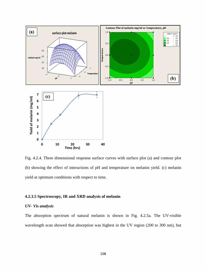
108
Fig. 4.2.4. Three dimensional response surface curves with surface plot (a) and contour plot
(b) showing the effect of interactions of pH and temperature on melanin yield. (c) melanin
yield at optimum conditions with respect to time.
4.2.3.5 Spectroscopy, IR and XRD analysis of melanin
UV- Vis analysis
The absorption spectrum of natural melanin is shown in Fig. 4.2.5a. The UV-visible
wavelength scan showed that absorption was highest in the UV region (200 to 300 nm), but
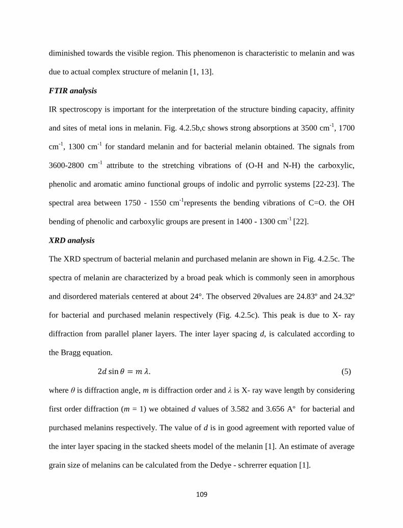
109
diminished towards the visible region. This phenomenon is characteristic to melanin and was
due to actual complex structure of melanin [1, 13].
FTIR analysis
IR spectroscopy is important for the interpretation of the structure binding capacity, affinity
and sites of metal ions in melanin. Fig. 4.2.5b,c shows strong absorptions at 3500 cm-1
, 1700
cm-1
, 1300 cm-1
for standard melanin and for bacterial melanin obtained. The signals from
3600-2800 cm-1
attribute to the stretching vibrations of (O-H and N-H) the carboxylic,
phenolic and aromatic amino functional groups of indolic and pyrrolic systems [22-23]. The
spectral area between 1750 - 1550 cm-1
represents the bending vibrations of C=O. the OH
bending of phenolic and carboxylic groups are present in 1400 - 1300 cm-1
[22].
XRD analysis
The XRD spectrum of bacterial melanin and purchased melanin are shown in Fig. 4.2.5c. The
spectra of melanin are characterized by a broad peak which is commonly seen in amorphous
and disordered materials centered at about 24°. The observed 2θvalues are 24.83º and 24.32º
for bacterial and purchased melanin respectively (Fig. 4.2.5c). This peak is due to X- ray
diffraction from parallel planer layers. The inter layer spacing d, is calculated according to
the Bragg equation.
. (5)
where θ is diffraction angle, m is diffraction order and λ is X- ray wave length by considering
first order diffraction (m = 1) we obtained d values of 3.582 and 3.656 Aº for bacterial and
purchased melanins respectively. The value of d is in good agreement with reported value of
the inter layer spacing in the stacked sheets model of the melanin [1]. An estimate of average
grain size of melanins can be calculated from the Dedye - schrerrer equation [1].
110
(6)
where FWHM is full width at half maximum of diffraction peak. The obtained D values are
0.668 and 0.568 nm for the bacterial and purchased melanins. The closeness of the grain size
values indicates the quality of the purified bacterial melanin. Furthermore % crystallinity was
also calculated for the stated melanins by considering glass substrate as background. The
calculation is as follows:
(7)
Although both melanin samples exhibited the lack of structure in the diffraction pattern
corresponding to any significant crystallinity, the % crystallinity values (Fig. 4.2.5c, picture
indicated with arrow) further indicate bacterial melanin from FWE was far less crystalline
when compared to the purchased melanin. Lack of crystallinity is significant sign of
consistent physical property of melanin [24].
100area) (total
area) profile background-area total(itycrystallin %
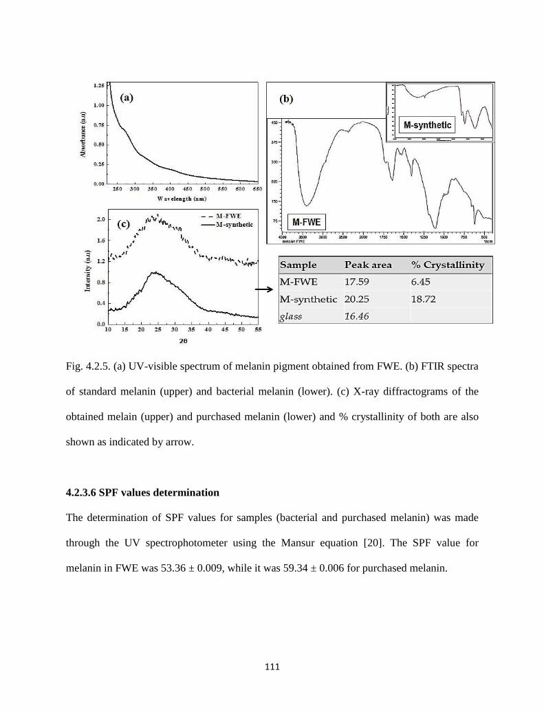
111
Fig. 4.2.5. (a) UV-visible spectrum of melanin pigment obtained from FWE. (b) FTIR spectra
of standard melanin (upper) and bacterial melanin (lower). (c) X-ray diffractograms of the
obtained melain (upper) and purchased melanin (lower) and % crystallinity of both are also
shown as indicated by arrow.
4.2.3.6 SPF values determination
The determination of SPF values for samples (bacterial and purchased melanin) was made
through the UV spectrophotometer using the Mansur equation [20]. The SPF value for
melanin in FWE was 53.36 ± 0.009, while it was 59.34 ± 0.006 for purchased melanin.
112
As melanins are known for their photoprotective role [25], the obtained SPF values state that
melanin from FWE might have profound protection effect against dermal damage related to
photoaging as that of purchased melanin.
4.2.3.7 DPPH assay
DPPH accepts an electron to become a stable diamagnetic molecule. The ethanolic solution
of DPPH (violet color) has got a strong UV absorbance at 516 nm. The presence of a
reducing agent in this ethanolic solution pairs the odd electrons of DPPH radical and further
the solution losses color stiochometrically and also the absorbance of the solution decreases
at 516 nm. Reduction of absorbance at 516 nm and color of DPPH associated with different
melanin doses was verified. The % increase in radical scavenging activity from Fig. 4.2.6a
indicates the diminished behavior of the radical. The data obtained from Fig. 4.2.6a states that
scavenging activity of the melanin was higher than the control ascorbic acid at each and
every dose studied. This behavior shows 30 % enhanced reductive capability of the obtained
bacterial melanin than ascorbic acid for a constant dose of melanin dose of ~100 µg/mL.
4.2.3.8 The metal binding capacity of melanin
The metal binding capacities of melanin from FWE was determined by assessing its ability to
compete with ferrozine for the ferrous ions. The concentration dependent metal chelating
activity was shown in Fig. 4.2.6b and its insert. The reduction in spectrum with increase in
melanin dose indicates that melanin compound was interfering with the formation of ferrous
and ferrozine complex. This suggests the better chelating effect of melanin and good ability
to capture ferrous ions than ferrozine. Maximum effect (~ 64 % chelation) was observed for a
dose of 0.2 mg/mL (Fig. 4.2.6c). The results suggest that the action of melanins as oxidation
protection factors may be predominantly due to their iron binding capacity.
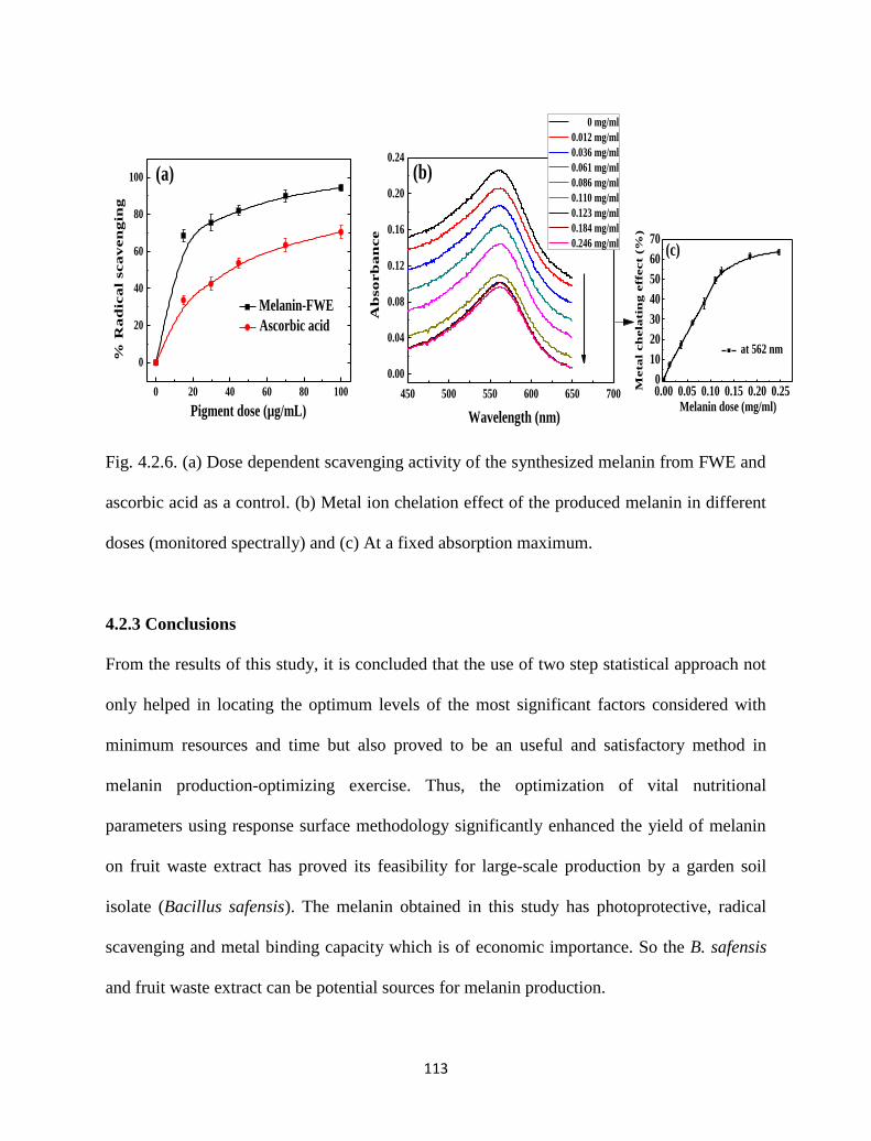
113
Fig. 4.2.6. (a) Dose dependent scavenging activity of the synthesized melanin from FWE and
ascorbic acid as a control. (b) Metal ion chelation effect of the produced melanin in different
doses (monitored spectrally) and (c) At a fixed absorption maximum.
4.2.3 Conclusions
From the results of this study, it is concluded that the use of two step statistical approach not
only helped in locating the optimum levels of the most significant factors considered with
minimum resources and time but also proved to be an useful and satisfactory method in
melanin production-optimizing exercise. Thus, the optimization of vital nutritional
parameters using response surface methodology significantly enhanced the yield of melanin
on fruit waste extract has proved its feasibility for large-scale production by a garden soil
isolate (Bacillus safensis). The melanin obtained in this study has photoprotective, radical
scavenging and metal binding capacity which is of economic importance. So the B. safensis
and fruit waste extract can be potential sources for melanin production.
450 500 550 600 650 700
0.00
0.04
0.08
0.12
0.16
0.20
0.24
(b)
Ab
so
rb
an
ce
Wavelength (nm)
0 mg/ml
0.012 mg/ml
0.036 mg/ml
0.061 mg/ml
0.086 mg/ml
0.110 mg/ml
0.123 mg/ml
0.184 mg/ml
0.246 mg/ml
0.00 0.05 0.10 0.15 0.20 0.250
10
20
30
40
50
60
70(c)
at 562 nm
Meta
l ch
ela
tin
g e
ffect (
%)
Melanin dose (mg/ml)0 20 40 60 80 100
0
20
40
60
80
100 (a)
% R
ad
ica
l sca
ven
gin
g
Pigment dose (µg/mL)
Melanin-FWE
Ascorbic acid
114
References
[1] V. Capozzi, et al., "Optical and photoelectronic properties of melanin," Thin solid
films, vol. 511, pp. 362-366, 2006.
[2] J. D. Nosanchuk and A. Casadevall, "Impact of melanin on microbial virulence and
clinical resistance to antimicrobial compounds," Antimicrobial agents and
chemotherapy, vol. 50, pp. 3519-3528, 2006.
[3] J. Zhang, et al., "Characterization of melanin produced by a wild-type strain of
Bacillus cereus," Frontiers of Biology in China, vol. 2, pp. 26-29, 2007.
[4] L. Ruan, et al., "Melanin pigment formation and increased UV resistance in Bacillus
thuringiensis following high temperature induction," Systematic and applied
microbiology, vol. 27, pp. 286-289, 2004.
[5] A. El-Obeid, et al., "Herbal melanin modulates tumor necrosis factor alpha (TNF- α),
interleukin 6 (IL-6) and vascular endothelial growth factor (VEGF) production,"
Phytomedicine, vol. 13, pp. 324-333, 2006.
[6] V. M. Sava, et al., "A novel melanin-like pigment derived from black tea leaves with
immuno-stimulating activity," Food Research International, vol. 34, pp. 337-343,
2001.
[7] Q. Gao and F. Garcia-Pichel, "Microbial ultraviolet sunscreens," Nature Reviews
Microbiology, vol. 9, pp. 791-802, 2011.
[8] J. D. Nosanchuk and A. Casadevall, "The contribution of melanin to microbial
pathogenesis," Cellular microbiology, vol. 5, pp. 203-223, 2003.
[9] J. McLean, et al., "Role for lichen melanins in uranium remediation," Nature, vol.
391, pp. 649-650, 1998.
115
[10] S. N. Surwase, et al., "Optimization of melanin production by Brevundimonas sp. SGJ
using response surface methodology," 3 Biotech, vol. 3, pp. 187-194, 2013.
[11] I. Hanyz and D. Wróbel, "Photovoltage generation and fluorescence of charged
tetraphenylporphyrins with dopa melanin," Crystal Research and Technology, vol. 38,
pp. 325-330, 2003.
[12] A. M. Gómez-Marín and C. I. Sánchez, "Thermal and mass spectroscopic
characterization of a sulphur-containing bacterial melanin from Bacillus subtilis,"
Journal of Non-Crystalline Solids, vol. 356, pp. 1576-1580, 2010.
[13] S. Sajjan, et al., "Purification and physiochemical characterization of melanin
pigment from Klebsiella sp. GSK," Journal of microbiology and biotechnology, vol.
20, pp. 1513-1520, 2010.
[14] V. Coyne and L. al-Harthi, "Induction of melanin biosynthesis in Vibrio cholerae,"
Applied and environmental microbiology, vol. 58, pp. 2861-2865, 1992.
[15] M. T. Cubo, et al., "Melanin production by Rhizobium strains," Applied and
environmental microbiology, vol. 54, pp. 1812-1817, 1988.
[16] V. Lagunas‐Muñoz, et al., "Optimum melanin production using recombinant
Escherichia coli," Journal of Applied Microbiology, vol. 101, pp. 1002-1008, 2006.
[17] G. Taguchi, Introduction to quality engineering: designing quality into products and
processes, 1986.
[18] B. Harlizius, et al., "Genomics for food safety and sustainable animal production,"
Journal of Biotechnology, vol. 113, pp. 33-42, 2004.
[19] K. Aneja, Experiments in microbiology, plant pathology and biotechnology: New Age
International, 2003.
116
[20] S. Huang, et al., "Antioxidant activities and UV-protective properties of melanin from
the berry of Cinnamomum burmannii and Osmanthus fragrans," Medicinal Chemistry
Research, vol. 20, pp. 475-481, 2011.
[21] K.-Y. Ju, et al., "Bioinspired polymerization of dopamine to generate melanin-like
nanoparticles having an excellent free-radical-scavenging property,"
Biomacromolecules, vol. 12, pp. 625-632, 2011.
[22] M. Magarelli, et al., "Purification, characterization and analysis of sepia melanin from
commercial sepia ink (Sepia officinalis)," Revista CES Medicina Veterinaria y
Zootecnia, vol. 5, pp. 18-28, 2010.
[23] J. Coates, "Interpretation of infrared spectra, a practical approach," Encyclopedia of
analytical chemistry, 2000.
[24] Y. Thathachari and M. Blois, "Physical studies on melanins: II. X-Ray Diffraction,"
Biophysical journal, vol. 9, pp. 77-89, 1969.
[25] M. Brenner and V. J. Hearing, "The protective role of melanin against UV damage in
human skin†," Photochemistry and photobiology, vol. 84, pp. 539-549, 2008.
117
4.3 Carotenoid by Bacillus clausi Using Rice Powder as
the Sole Substrate: Pigment Analyses and Optimization of
Key Production Parameters
4.3.1 Introduction
Pigments are particulate solids that disperse into a medium without significant interaction in
solutions [1]. Their wide applications in fields of food, cosmetics, paints, pharmaceuticals,
textiles, glass etc. has contributed to its increasing demand [2]. Currently there are two
methods employed so far to produce β-carotene; they are based on Wittig reaction developed
by Badische Anilin and Soda Fabrik (BASF) and the second is based on Grinard reaction
elaborated by Hoffman-La Roche [3-4]. However the microbial method has gained maximum
interest as they are naturally produced and are environmentally friendly. Carotenoids are the
secondary metabolites which are usually accumulate in the organisms during biotic and
abiotic stress. Among all natural pigments, carotenoids represent the largest and most diverse
class compounds known to mankind [5].
118
Carotenoids of microbial origin have found maximum application in the food and
cosmetic industries which has exhibited increase in demand in recent years. Considerable
research has been focused on these pigments as nutritional supplements with some $ 935
million to billion dollars market in 2005 [6-7]. Besides, carotenoids play a significant role in
human health as precursors of vitamin A, scavengers of active oxygen, enhancers of in vitro
antibody production, anticancer agents and so on [6, 8].
In view of high cost of current technology there is a need to develop low cost
processes for the production of pigments which could replace the synthetic pigments in
industrial scale. Production of pigments essentially depends on the type of substrate on which
the microbe grows [9]. A variety of substrates are tested for carotenoids production using
different microbes with special emphasis on cheaper substrates like maple sap [10], coconut
milk [11], sugar manufacture processing waste [12], fermented radish brain [13], corn meal,
pea nut meal, soybean meal, coconut residue [14], whey waste [7], corn syrup [15], grape
must, beet molasses, soybean flour extract, maize flour extract [16], jack fruit seed [2], plant
extract [17], prehydrolysed wood [18] etc. Efforts have been made in order to reduce the
production cost of microbial pigments as compared to synthetic, plant and animal derived
pigments [16, 19].
This paper outlines the following objectives. 1) Isolation of the pigment producing
bacteria from garden soil, 2) Examine the potential of cheap natural substrate like rice
powder for the production of pigment, 3) Extraction, purification and analyzing the chemical
nature of the produced pigment, 4) Investigating the stability of the obtained pigment, and 5)
Optimization of key production parameters, using Taguchi experimental design tool.
119
4.3.2 Materials and methods
4.3.2.1 Sampling, microscopy and strain characterization
Carotenoid producing microbial cultures was isolated from soil sample collected from the
premises of NIT campus Rourkela. Screening procedure adopted was plating of 1 mL (10-10
)
diluted soil samples on nutrient agar (Himedia Pvt Ltd, India) with inoculation at 25 °C. The
orange colored colonies developed on the plates were observed. The isolate showing
maximum diameter was propagated on rice plates at 25 °C for 24 h. Characterization and
identification of the isolate with pigment production ability was carried out morphologically
with optical microscope (Hund-H600). In order to study the morphological characters such as
size and shape of the microorganism, various images were captured at different
magnifications using SEM (Scanning Electron Microscope) analysis [JEOL JSM-6480LV].
16s-rDNA characterization was performed on this isolated strain at Xcelris labs Ltd,
Ahmedabad, India.
4.3.2.2 Pigment production
Finely ground rice powder was used as a substrate for pigment production. Experiments were
conducted in petriplates of 10 cm diameter containing 3 g of rice powder. The substrate was
moistened with double distilled water (8 mL) at neutral pH - 7 and autoclaved to maintain
sterile conditions throughout the study. On cooling to room temperature; moisture content of
the substrate was analyzed using moisture analyzer (Sartorious MA-150) and was estimated
as 52.04 %. The substrate in plates was inoculated with 1 mL inoculum culture and incubated
at ambient room temperature ~ 30 °C for 10 days. After observing intense coloration in rice
plates, batch experiments at pH 6 and 8 were also studied for pigment production.
120
4.3.2.3 Purification and stability of the pigment
Pigment was extracted using a modified method adapted from Babitha et al. [20]. To evaluate
the solubility of the pigment a definite quantity of substrate (rice powder) 1g was extracted
with fixed volume 5 mL of different solvents such as acetone, methanol, ethanol, chloroform,
water and 10 % NaOH [21]. The pigment-solvent mixture was allowed to stand after shaking
for 15 minutes. Based on high extraction of pigment and fair dispersion with 10 % NaOH,
ethanol was selected as extracting solvent. About 1:5 rice powder (grams) to solvent (mL)
ratio was used for pigment extraction by thorough dispersion as per the reported method [2].
The mixture was centrifuged at 9000 rpm and the supernatant was collected. The above
process was repeated until the substrate was colorless. Phase separation of the extracted
pigment in the supernatant was achieved on adding equal volumes of diethyl ether and NaCl
(0.1 M) to obtain pure pigment devoid of cellular disturbances [11]. The obtained pigment
solution was further purified using thin layer chromatography using stationary phase - TLC
silica gel 60 F 254 and hexane: methanol (7:3) as stationary phase and solvent phase
respectively. To obtain orange colored powdered pigment the TLC extract was subjected to
vacuum evaporation.
The extracted pigment was assessed on 0.50 g of dry pigment and adjusted to 50 ml
constant volume of ethanol before measuring its absorbance at (λmax) using a UV-3600
Shimadzu UV-Visible spectrophotometer. The structural analysis of the extracted pigment
was examined by FTIR (Bruker ATR-FTIR, USA) at room temperature. Stability of the
purified pigment was also tested after thermal treatment at 45 0C for 8 h at ambient conditions
in an oven. It was subjected to UV-visible, fluorescence and Attenuated total reflectance
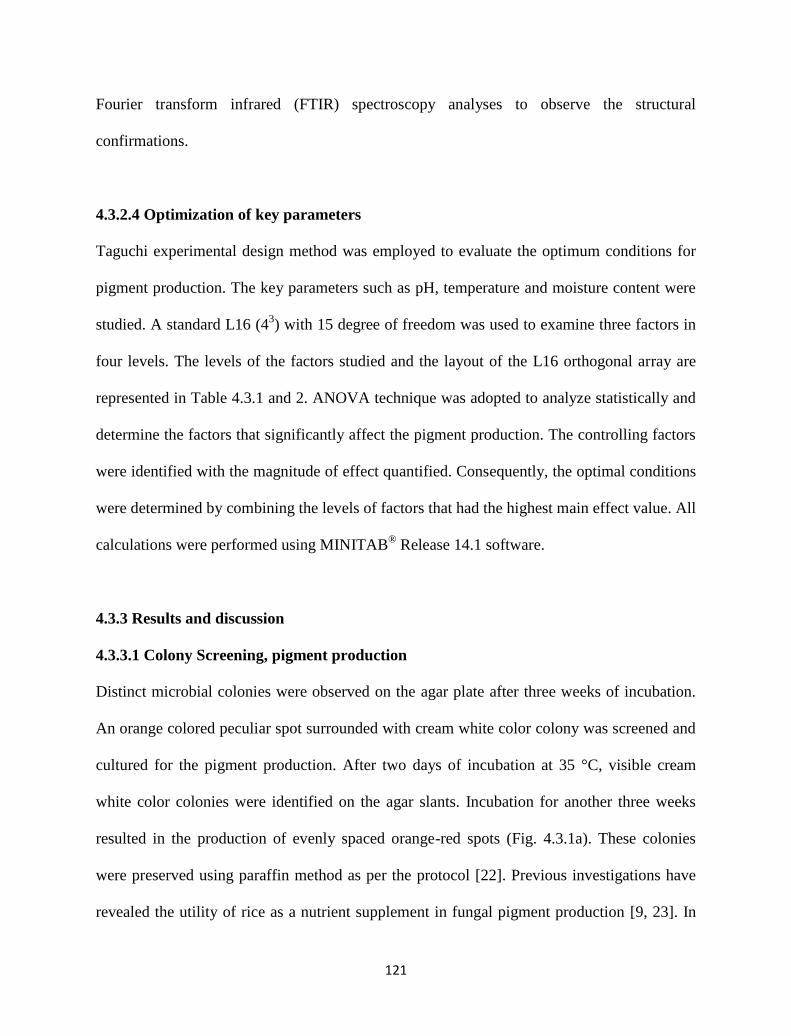
121
Fourier transform infrared (FTIR) spectroscopy analyses to observe the structural
confirmations.
4.3.2.4 Optimization of key parameters
Taguchi experimental design method was employed to evaluate the optimum conditions for
pigment production. The key parameters such as pH, temperature and moisture content were
studied. A standard L16 (43) with 15 degree of freedom was used to examine three factors in
four levels. The levels of the factors studied and the layout of the L16 orthogonal array are
represented in Table 4.3.1 and 2. ANOVA technique was adopted to analyze statistically and
determine the factors that significantly affect the pigment production. The controlling factors
were identified with the magnitude of effect quantified. Consequently, the optimal conditions
were determined by combining the levels of factors that had the highest main effect value. All
calculations were performed using MINITAB® Release 14.1 software.
4.3.3 Results and discussion
4.3.3.1 Colony Screening, pigment production
Distinct microbial colonies were observed on the agar plate after three weeks of incubation.
An orange colored peculiar spot surrounded with cream white color colony was screened and
cultured for the pigment production. After two days of incubation at 35 °C, visible cream
white color colonies were identified on the agar slants. Incubation for another three weeks
resulted in the production of evenly spaced orange-red spots (Fig. 4.3.1a). These colonies
were preserved using paraffin method as per the protocol [22]. Previous investigations have
revealed the utility of rice as a nutrient supplement in fungal pigment production [9, 23]. In
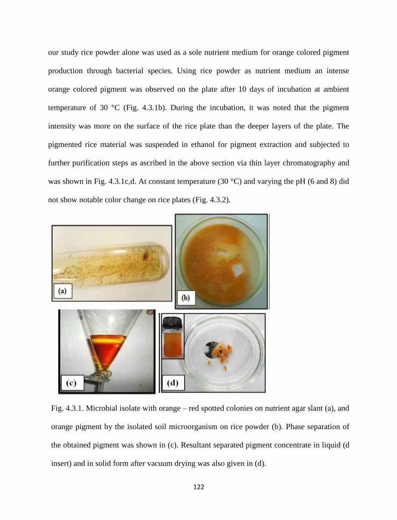
122
our study rice powder alone was used as a sole nutrient medium for orange colored pigment
production through bacterial species. Using rice powder as nutrient medium an intense
orange colored pigment was observed on the plate after 10 days of incubation at ambient
temperature of 30 °C (Fig. 4.3.1b). During the incubation, it was noted that the pigment
intensity was more on the surface of the rice plate than the deeper layers of the plate. The
pigmented rice material was suspended in ethanol for pigment extraction and subjected to
further purification steps as ascribed in the above section via thin layer chromatography and
was shown in Fig. 4.3.1c,d. At constant temperature (30 °C) and varying the pH (6 and 8) did
not show notable color change on rice plates (Fig. 4.3.2).
Fig. 4.3.1. Microbial isolate with orange – red spotted colonies on nutrient agar slant (a), and
orange pigment by the isolated soil microorganism on rice powder (b). Phase separation of
the obtained pigment was shown in (c). Resultant separated pigment concentrate in liquid (d
insert) and in solid form after vacuum drying was also given in (d).
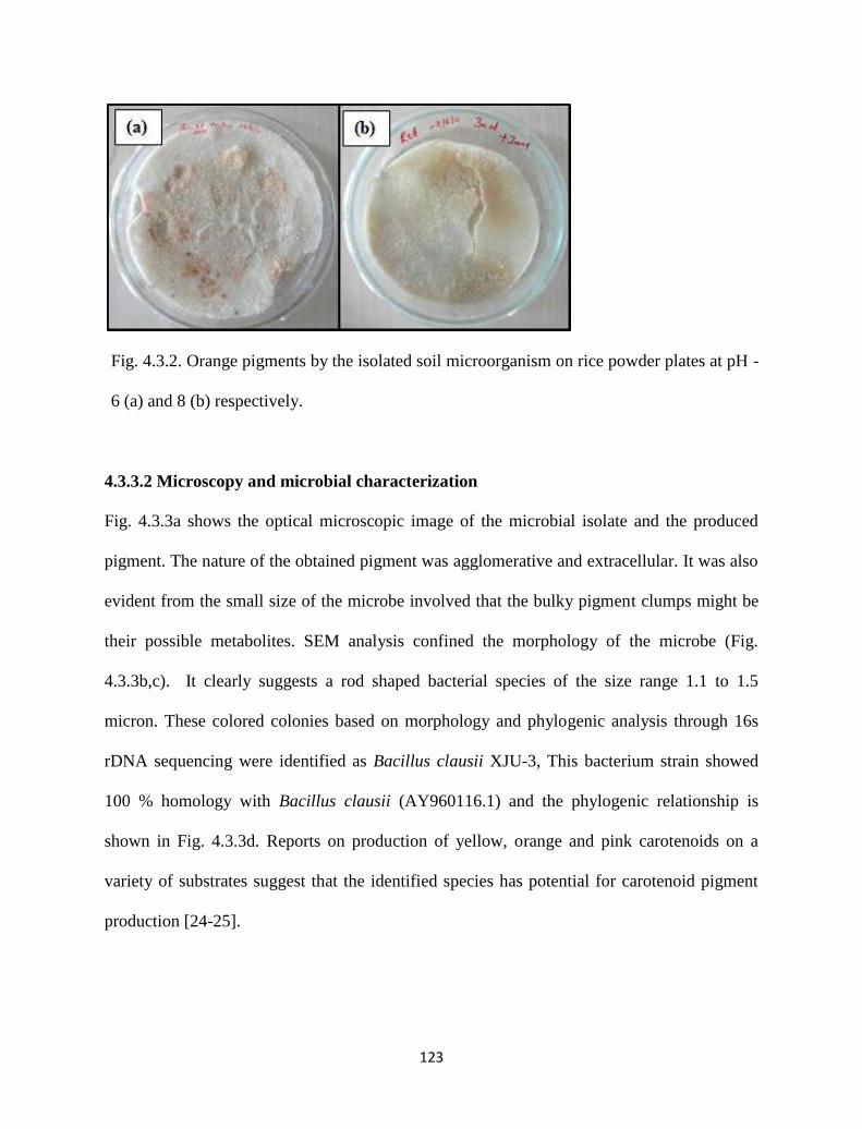
123
Fig. 4.3.2. Orange pigments by the isolated soil microorganism on rice powder plates at pH -
6 (a) and 8 (b) respectively.
4.3.3.2 Microscopy and microbial characterization
Fig. 4.3.3a shows the optical microscopic image of the microbial isolate and the produced
pigment. The nature of the obtained pigment was agglomerative and extracellular. It was also
evident from the small size of the microbe involved that the bulky pigment clumps might be
their possible metabolites. SEM analysis confined the morphology of the microbe (Fig.
4.3.3b,c). It clearly suggests a rod shaped bacterial species of the size range 1.1 to 1.5
micron. These colored colonies based on morphology and phylogenic analysis through 16s
rDNA sequencing were identified as Bacillus clausii XJU-3, This bacterium strain showed
100 % homology with Bacillus clausii (AY960116.1) and the phylogenic relationship is
shown in Fig. 4.3.3d. Reports on production of yellow, orange and pink carotenoids on a
variety of substrates suggest that the identified species has potential for carotenoid pigment
production [24-25].
124
Fig 4.3.3. Pigment producing bacterial species with an orange pigment (insert) magnified by
an optical microscope at 400 X magnification (a). SEM images of the identified pigment
producing bacteria with (b) 5000 X, (c) 9000 X magnifications and (d) Phylogenetic tree
showing the position of the isolate XJU-3 with reference to related strains
(d)
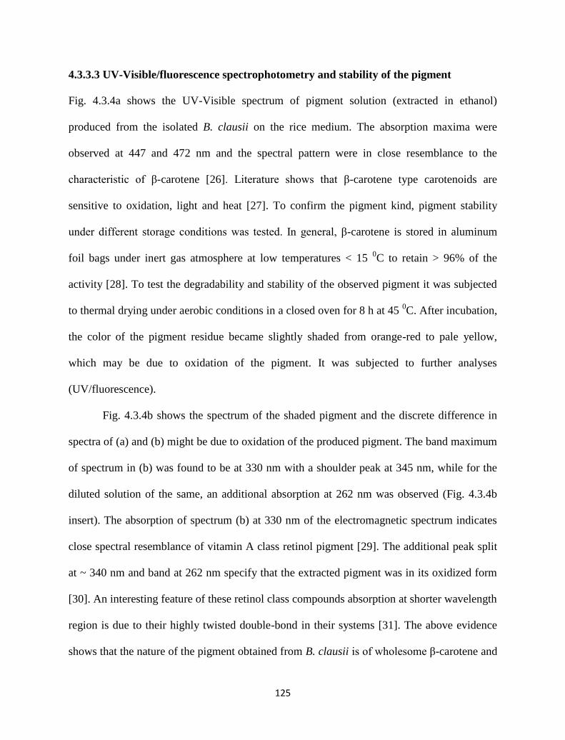
125
4.3.3.3 UV-Visible/fluorescence spectrophotometry and stability of the pigment
Fig. 4.3.4a shows the UV-Visible spectrum of pigment solution (extracted in ethanol)
produced from the isolated B. clausii on the rice medium. The absorption maxima were
observed at 447 and 472 nm and the spectral pattern were in close resemblance to the
characteristic of β-carotene [26]. Literature shows that β-carotene type carotenoids are
sensitive to oxidation, light and heat [27]. To confirm the pigment kind, pigment stability
under different storage conditions was tested. In general, β-carotene is stored in aluminum
foil bags under inert gas atmosphere at low temperatures < 15 0C to retain > 96% of the
activity [28]. To test the degradability and stability of the observed pigment it was subjected
to thermal drying under aerobic conditions in a closed oven for 8 h at 45 0C. After incubation,
the color of the pigment residue became slightly shaded from orange-red to pale yellow,
which may be due to oxidation of the pigment. It was subjected to further analyses
(UV/fluorescence).
Fig. 4.3.4b shows the spectrum of the shaded pigment and the discrete difference in
spectra of (a) and (b) might be due to oxidation of the produced pigment. The band maximum
of spectrum in (b) was found to be at 330 nm with a shoulder peak at 345 nm, while for the
diluted solution of the same, an additional absorption at 262 nm was observed (Fig. 4.3.4b
insert). The absorption of spectrum (b) at 330 nm of the electromagnetic spectrum indicates
close spectral resemblance of vitamin A class retinol pigment [29]. The additional peak split
at ~ 340 nm and band at 262 nm specify that the extracted pigment was in its oxidized form
[30]. An interesting feature of these retinol class compounds absorption at shorter wavelength
region is due to their highly twisted double-bond in their systems [31]. The above evidence
shows that the nature of the pigment obtained from B. clausii is of wholesome β-carotene and
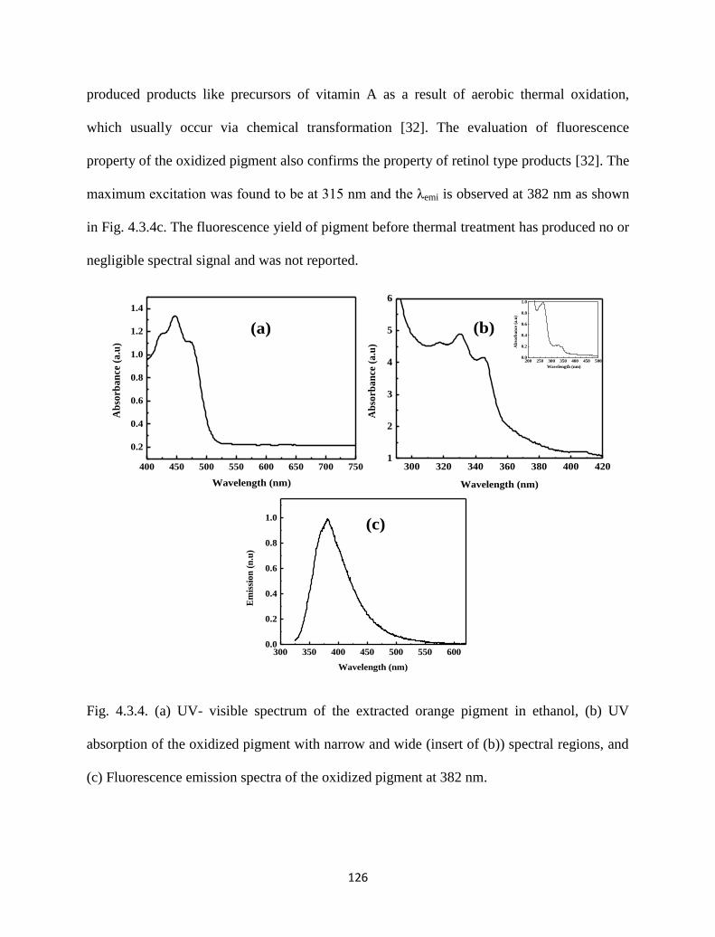
126
produced products like precursors of vitamin A as a result of aerobic thermal oxidation,
which usually occur via chemical transformation [32]. The evaluation of fluorescence
property of the oxidized pigment also confirms the property of retinol type products [32]. The
maximum excitation was found to be at 315 nm and the λemi is observed at 382 nm as shown
in Fig. 4.3.4c. The fluorescence yield of pigment before thermal treatment has produced no or
negligible spectral signal and was not reported.
Fig. 4.3.4. (a) UV- visible spectrum of the extracted orange pigment in ethanol, (b) UV
absorption of the oxidized pigment with narrow and wide (insert of (b)) spectral regions, and
(c) Fluorescence emission spectra of the oxidized pigment at 382 nm.
200 250 300 350 400 450 5000.0
0.2
0.4
0.6
0.8
1.0
Ab
sorb
an
ce (
a.u
)
Wavelength (nm)
300 320 340 360 380 400 4201
2
3
4
5
6
Ab
sorb
an
ce (
a.u
)
Wavelength (nm)
(c)
300 350 400 450 500 550 6000.0
0.2
0.4
0.6
0.8
1.0
Em
issi
on
(n
.u)
Wavelength (nm)
400 450 500 550 600 650 700 750
0.2
0.4
0.6
0.8
1.0
1.2
1.4
Ab
sorb
an
ce (
a.u
)
Wavelength (nm)
(b)(a)
127
4.3.3.4 FTIR analysis
In order to understand the pigment structure FTIR analysis was performed. The purified
pigment was subjected to FTIR spectroscopy before and after oxidation steps (Fig. 4.3.5a,b).
Fig. 4.3.5a shows the IR spectrum of the pigment with major peaks at 3320, 2943, 2832,
1652, 1448, 1405, 1100 and 1020 cm-1
. The majority of the peaks from the obtained pigment
show a close resemblance to a β-carotene structure [33-34]. Peaks at 2943 and 2832 cm-1
are
due to asymmetric and symmetric stretching vibrations of CH2 and CH3 groups [34]. And
peaks at 1020 cm-1
is for wagging vibration of (RH).C=C.(RH) groups of the synthesized
pigment. Peaks at 1405 and 1448 cm-1 are due to symmetric deformation of δ CH3 and
deformation vibration of δ CH2 groups. Moreover the peaks at 3320, 1652 and 1020 cm-1
are
due to vibrational modes of water interference in the analyzed pigment [33, 35].
Fig. 4.3.5b shows FTIR peaks after the oxidation of the pigment. The major peaks
observed here are at 3433, 3229, 3000, 2883, 1672, 1388, 1343, 1089, 1082, 884, 708 and
631 cm-1
. The spectral pattern highly resembles retinol compound of carotenoids family [36].
The absorptions in the region of 3200 - 2700 cm-1
are normally characteristic of carbon and
hydrogen containing species, and can be assigned to various forms of C-H stretching. The
absorbance at and above 3000 cm-1
suggest that the compound is likely to have unsaturated
C=C. The absorption at 3433 cm-1
which is in the region of 3650-3250 cm-1
mainly occurs for
hydroxy or amino groups.
The additional intense bands in 1600-1300, 1200-1000 and 800-600 cm-1
in Fig.
4.3.5b confirms that the oxidized compound consists a simple hydroxy compound with O-H
absorption and is likely to be an alcohol. The absorption at the low end and below 1700 cm-1
was due to carboxylate (carboxylic acid salt). In our case the peak at 1672 cm-1
may be due to
128
conjugation at C=O. (may be our oxidized compound is a mixture of retinol and retinoic
acid). Peaks at 3000 cm-1
and 2986 cm-1
which are just above and below 3000 cm-1
may be
due to unsaturated and saturated C-H absorptions in the compound. Bands at ~ 1000 cm-1
and
880 cm-1
indicate C-H out of plane bending which is usually seen for all trans retinol
compounds. Further the olefinic double bond type unsaturation can be confirmed by a single
peak around ~ 890 cm-1
along with the absorptions at ~1650 and ~3000 cm-1
[35]. The
structural confirmations by IR analysis in concurrence with UV-Visible analysis infer that the
produced pigment is exclusively close to β-carotene in all aspects. And the retinol derivatives
of the obtained pigment on thermal treatment further confirm the instability of these β-
carotene type pigments.
Fig. 4.3.5. ATR-FTIR analysis of the extracted and purified orange pigment before (a) and
after (b) oxidation process.
4000 3600 3200 2800 2400 2000 1600 1200 800
Wavenumber (cm-1)
1299
1343
1388
631
708
884
10821089
15
51
15
84
1672
2883
3000
3229
3433(b)
3500 3000 2500 2000 1500 1000
% T
ra
nsm
itta
nce (
n.u
)
Wavenumber (cm-1)
1020
1100
14051448
1652
28322943
3320
(a)
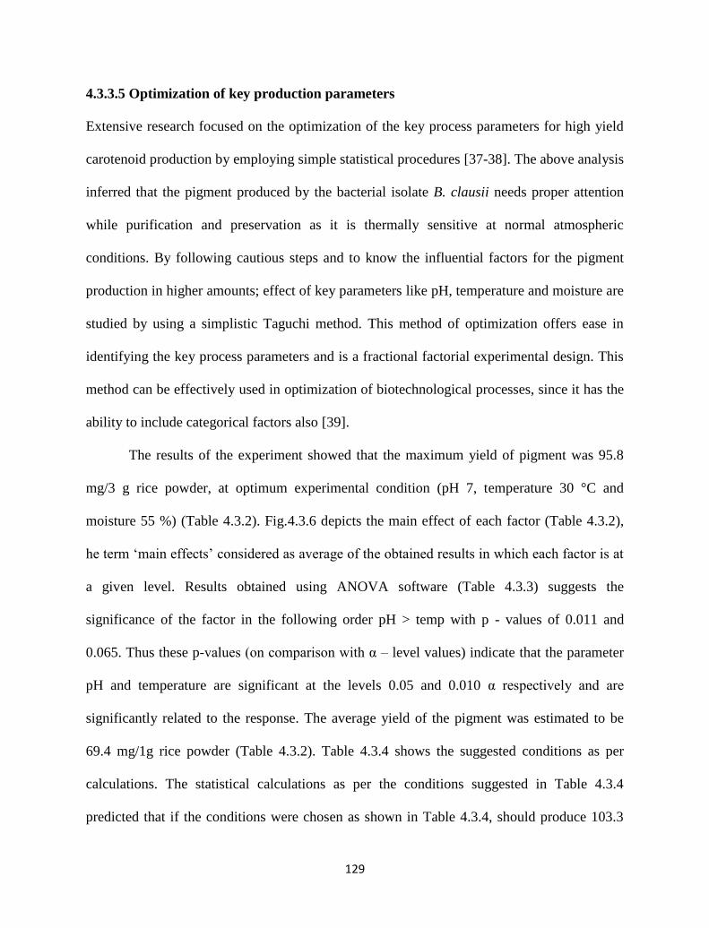
129
4.3.3.5 Optimization of key production parameters
Extensive research focused on the optimization of the key process parameters for high yield
carotenoid production by employing simple statistical procedures [37-38]. The above analysis
inferred that the pigment produced by the bacterial isolate B. clausii needs proper attention
while purification and preservation as it is thermally sensitive at normal atmospheric
conditions. By following cautious steps and to know the influential factors for the pigment
production in higher amounts; effect of key parameters like pH, temperature and moisture are
studied by using a simplistic Taguchi method. This method of optimization offers ease in
identifying the key process parameters and is a fractional factorial experimental design. This
method can be effectively used in optimization of biotechnological processes, since it has the
ability to include categorical factors also [39].
The results of the experiment showed that the maximum yield of pigment was 95.8
mg/3 g rice powder, at optimum experimental condition (pH 7, temperature 30 °C and
moisture 55 %) (Table 4.3.2). Fig.4.3.6 depicts the main effect of each factor (Table 4.3.2),
he term ‘main effects’ considered as average of the obtained results in which each factor is at
a given level. Results obtained using ANOVA software (Table 4.3.3) suggests the
significance of the factor in the following order pH > temp with p - values of 0.011 and
0.065. Thus these p-values (on comparison with α – level values) indicate that the parameter
pH and temperature are significant at the levels 0.05 and 0.010 α respectively and are
significantly related to the response. The average yield of the pigment was estimated to be
69.4 mg/1g rice powder (Table 4.3.2). Table 4.3.4 shows the suggested conditions as per
calculations. The statistical calculations as per the conditions suggested in Table 4.3.4
predicted that if the conditions were chosen as shown in Table 4.3.4, should produce 103.3

130
mg pigment per 1 g rice powder which is 48.9 % more yield. However, the experimental
result showed 107 ± 1.2 mg pigment production using 1 g rice powder since the deviation in
actual and predicted value was only about 3.58 %, which is acceptable. Therefore on
comparing with 95.8 mg pigment produced before, a further increase of about 11.6 % was
achieved.
1 2 3 4
55
60
65
70
75
80
85
90
Pig
men
t y
ield
(m
g)
Levels of factors
pH
Temperature
Moisture
Fig. 4.3.6. Main effects of factors or average of obtained results (pigment per 3 gm rice
powder) in which each factor is at a given level. For detail about ‘levels’ refer to Table 4.3.1.
Table 4.3.1. Factors and their levels studied by Taguchi method.
Factor Level 1 Level 2 Level 3 Level 4
pH 6 6.5 7 7.5
Temperature (oC) 25 30 35 40
Moisture % (w/w) 40 45 50 55
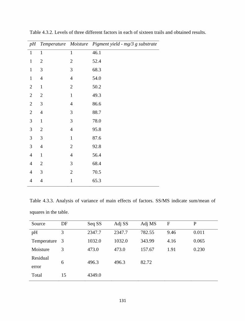
131
Table 4.3.2. Levels of three different factors in each of sixteen trails and obtained results.
pH Temperature Moisture Pigment yield - mg/3 g substrate
1 1 1 46.1
1 2 2 52.4
1 3 3 68.3
1 4 4 54.0
2 1 2 50.2
2 2 1 49.3
2 3 4 86.6
2 4 3 88.7
3 1 3 78.0
3 2 4 95.8
3 3 1 87.6
3 4 2 92.8
4 1 4 56.4
4 2 3 68.4
4 3 2 70.5
4 4 1 65.3
Table 4.3.3. Analysis of variance of main effects of factors. SS/MS indicate sum/mean of
squares in the table.
Source DF Seq SS Adj SS Adj MS F P
pH 3 2347.7 2347.7 782.55 9.46 0.011
Temperature 3 1032.0 1032.0 343.99 4.16 0.065
Moisture 3 473.0 473.0 157.67 1.91 0.230
Residual
error 6 496.3 496.3 82.72
Total 15 4349.0
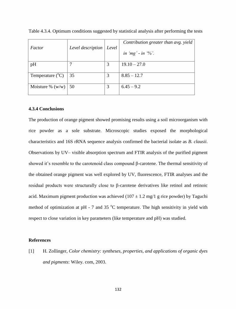
132
Table 4.3.4. Optimum conditions suggested by statistical analysis after performing the tests
Factor Level description Level
Contribution greater than avg. yield
in ‘mg’ - in ‘%’.
pH 7 3 19.10 – 27.0
Temperature (oC) 35 3 8.85 – 12.7
Moisture % (w/w) 50 3 6.45 – 9.2
4.3.4 Conclusions
The production of orange pigment showed promising results using a soil microorganism with
rice powder as a sole substrate. Microscopic studies exposed the morphological
characteristics and 16S rRNA sequence analysis confirmed the bacterial isolate as B. clausii.
Observations by UV– visible absorption spectrum and FTIR analysis of the purified pigment
showed it’s resemble to the carotenoid class compound β-carotene. The thermal sensitivity of
the obtained orange pigment was well explored by UV, fluorescence, FTIR analyses and the
residual products were structurally close to β-carotene derivatives like retinol and retinoic
acid. Maximum pigment production was achieved (107 ± 1.2 mg/1 g rice powder) by Taguchi
method of optimization at pH - 7 and 35 oC temperature. The high sensitivity in yield with
respect to close variation in key parameters (like temperature and pH) was studied.
References
[1] H. Zollinger, Color chemistry: syntheses, properties, and applications of organic dyes
and pigments: Wiley. com, 2003.
133
[2] S. Babitha, et al., "Jackfruit Seed--A novel substrate for the production of Monascus
pigments through solid-state fermentation," Food Technology & Biotechnology, vol.
44, 2006.
[3] G. Wittig and H. Pommer, "Ger. Patent 954,247, 1959," in Chem. Abstr, 1959, p.
2279.
[4] T. Storebakken, et al., "Carotenoids in diets for salmonids: IV. Pigmentation of
Atlantic salmon with astaxanthin, astaxanthin dipalmitate and canthaxanthin,"
Aquaculture, vol. 65, pp. 279-292, 1987.
[5] G. Britton, et al., Carotenoids: handbook: Springer, 2004.
[6] P. Park, et al., "Optimization of carotenoid production by Rhodotorula glutinis using
statistical experimental design," World journal of microbiology and biotechnology,
vol. 21, pp. 429-434, 2005.
[7] I. Marova, et al., "Use of several waste substrates for carotenoid-rich yeast biomass
production," Journal of Environmental Management, vol. 95, Supplement, pp. S338-
S342, 2012.
[8] M. M. S. Cabral, et al., "Carotenoids production from a newly isolated Sporidiobolus
pararoseus strain by submerged fermentation," European Food Research and
Technology, vol. 233, pp. 159-166, 2011.
[9] L. Kagliwal, et al., "A novel medium for the production of cephamycin C by
Nocardia lactamdurans using solid-state fermentation," Bioresource Technology, vol.
100, pp. 2600-2606, 2009.
[10] A. E. Wasserman, "Absorption and fluorescence of water-soluble pigments produced
by four species of Pseudomonas," Applied microbiology, vol. 13, pp. 175-180, 1965.
134
[11] A. Domínguez-Bocanegra and J. Torres-Muñoz, "Astaxanthin hyperproduction by
Phaffia rhodozyma (now Xanthophyllomyces dendrorhous) with raw coconut milk as
sole source of energy," Applied Microbiology and Biotechnology, vol. 66, pp. 249-
252, 2004.
[12] G. T. Hayman, et al., "Production of carotenoids by Phaffia rhodozyma grown on
media composed of corn wet-milling co-products," Journal of industrial
microbiology, vol. 14, pp. 389-395, 1995.
[13] C. Malisorn and W. Suntornsuk, "Optimization of β-carotene production by
Rhodotorula glutinis DM28 in fermented radish brine," Bioresource Technology, vol.
99, pp. 2281-2287, 2008.
[14] S. I. Mussatto, et al., "Use of Agro-industrial wastes in solid-state fermentation
processes," Industrial waste. Croatia: InTech, pp. 121-40, 2012.
[15] P. Buzzini, "Batch and fed‐batch carotenoid production by Rhodotorula glutinis–
Debaryomyces castellii co‐cultures in corn syrup," Journal of Applied Microbiology,
vol. 90, pp. 843-847, 2001.
[16] P. Buzzini, "An optimization study of carotenoid production by Rhodotorula glutinis
DBVPG 3853 from substrates containing concentrated rectified grape must as the sole
carbohydrate source," Journal of Industrial Microbiology and Biotechnology, vol. 24,
pp. 41-45, 2000.
[17] S. Kim, et al., "Increased carotenoid production in Xanthophyllomyces dendrorhous
G276 using plant extracts," Journal of Microbiology-Seoul-, vol. 45, p. 128, 2007.
135
[18] J. Parajó, et al., "Production of carotenoids by Xanthophyllomyces dendrorhous
growing on enzymatic hydrolysates of prehydrolysed wood," Food Chemistry, vol.
60, pp. 347-355, 1997.
[19] L. Dufossé, et al., "Microorganisms and microalgae as sources of pigments for food
use: a scientific oddity or an industrial reality?," Trends in Food Science &
Technology, vol. 16, pp. 389-406, 2005.
[20] S. Babitha, et al., "Solid-state fermentation for the production of Monascus pigments
from jackfruit seed," Bioresource Technology, vol. 98, pp. 1554-1560, 2007.
[21] W. Decleir and A. Richard, "A study of the orange-red pigment from the accessory
nidamental glands of the cephalopod Sepia officinalis L," Biol. Jb. Dodonaea, vol. 40,
pp. 188-197, 1972.
[22] K. Aneja, Experiments in microbiology, plant pathology and biotechnology: New Age
International, 2003.
[23] X. Lian, et al., "Identification of new red pigments produced by Monascus ruber,"
Dyes and Pigments, vol. 73, pp. 121-125, 2007.
[24] L. H. Duc, et al., "Carotenoids present in halotolerant Bacillus spore formers," FEMS
microbiology letters, vol. 255, pp. 215-224, 2006.
[25] R. Khaneja, et al., "Carotenoids found in Bacillus," Journal of Applied Microbiology,
vol. 108, pp. 1889-1902, 2010.
[26] P. Park, et al., "Chemical disruption of yeast cells for the isolation of carotenoid
pigments," Separation and purification technology, vol. 53, pp. 148-152, 2007.
136
[27] K. Jacob, et al., "Stability of carotenoids, phenolic compounds, ascorbic acid and
antioxidant capacity of tomatoes during thermal processing," Archivos
Latinoamericanos de Nutricion (ALAN), vol. 60, p. 192, 2010.
[28] E. F. S. Authority, "Scientific Opinion on the safety and efficacy of beta-carotene as a
feed additive for all animal species and categories.," 2012.
[29] C. N'Soukpoe-Kossi, et al., "Retinol and retinoic acid bind human serum albumin:
stability and structural features," International journal of biological macromolecules,
vol. 40, pp. 484-490, 2007.
[30] S. Martras, et al., "Kinetics of human alcohol dehydrogenase with ring-oxidized
retinoids: effect of Tween 80," Archives of biochemistry and biophysics, vol. 430, pp.
210-217, 2004.
[31] M. B. Sporn, The retinoids vol. 1: Academic Press, 1984.
[32] S. Adhikari, et al., "Pulse radiolytic oxidation of β-carotene with halogenated
alkylperoxyl radicals in a quaternary microemulsion: formation of retinol,"
Biophysical chemistry, vol. 88, pp. 111-117, 2000.
[33] M. Baranska, et al., "Determination of lycopene and β-carotene content in tomato
fruits and related products: comparison of FT-Raman, ATR-IR, and NIR
spectroscopy," Analytical Chemistry, vol. 78, pp. 8456-8461, 2006.
[34] W. Ammawath and Y. Bin Che Man, "a rapid method for determination commercial
β-carotene in rbd palm olein by fourier transform infrared spectroscopy," Asian
Journal of Food and Agro-Industry, 3 (4), 443-452., 2010.
[35] J. Coates, "Interpretation of infrared spectra, a practical approach," Encyclopedia of
analytical chemistry, 2000.
137
[36] T. Lóránd, et al., "FT-IR study of some seco-and apocarotenoids," Journal of
biochemical and biophysical methods, vol. 53, pp. 251-258, 2002.
[37] P. Bhosale and R. Gadre, "Optimization of carotenoid production from
hyper‐producing Rhodotorula glutinis mutant 32 by a factorial approach," Letters in
applied microbiology, vol. 33, pp. 12-16, 2001.
[38] R. Sarada, et al., "A response surface approach for the production of natural pigment
astaxanthin from green alga, Haematococcus pluvialis: effect of sodium acetate,
culture age, and sodium chloride," Food Biotechnology, vol. 16, pp. 107-120, 2002.
[39] M. Azin, et al., "Production of xylanase by Trichoderma longibrachiatum on a
mixture of wheat bran and wheat straw: Optimization of culture condition by Taguchi
method," Enzyme and microbial technology, vol. 40, pp. 801-805, 2007.
138
Chapter 4 Approach and investigations
Part II - Facile Synthesis of Commercial Pigments by
Purchased Strains on Cheaper Substrates
4.4 Carotenoid by Rhodotorula sp. on Fruit Waste
Extract as a Sole Carbon Source and Optimization of key
production parameters
4.4.1 Introduction
Pigments are of great commercial interest and have received considerable attention because
of their potential beneficial effects on human health besides pigmenting properties [1]. In
recent years there is an increasing demand for microbial pigments as promising alternatives
139
for synthetic pigments that are widely used in food industries [2]. The pigments from
microorganisms are nature selected and have advantages over plant and animal derived
pigments with no seasonal variations, no geographic inconsistency in the production, high
productivity and are easily manipulated in the processing schemes [3-4].
Among microbial derived pigments, carotenoids are fat soluble diverse class of
yellow, orange, red and purple natural pigments which are unanimously produced by a wide
range of microorganisms and plants. Due to the recent discovery of anticancer and
antioxidant properties of carotenoid pigments, their use in pharmaceuticals and
neutraceuticals is expected along with their applications in the fields of food, cosmetics,
chemicals and so on [5-7]. Globally carotenoids are estimated to supersede USD S 280
million in 2015 [8]. In order to improve the yield of carotenoid pigments and to decrease the
cost of production, various studies have been performed using several microorganisms using
numerous different substrates [9-10]. Amongst different microorganisms Rhodotorula sp.
stood as a potential and scientifically favorable candidate for high yield carotenoids
production [11].The yeast Rhodotorula sp. is one of very few types of yeast which is able to
produce a large number of carotenoid pigments which are mainly β - carotene, torulene,
torularhodin [12]. Moreover, many investigations have been done using this genus to make
the process economical [3, 6, 13-14]. Key process parameters like type of substrate, nutrients
availability, pH, temperature and so on are optimized in various processes [15]. However,
there are still extensive investigations focused on this versatile genus Rhodotorula to produce
carotenoids in more economical ways.
The cost of the substrate has an important contribution to the overall pigment
production cost, and it can be minimized by using cheaper organic waste. Substrate
140
composition, typically carbon source has a significant effect on carotenoid production and its
cost. Many substrates have been considered as potential substrates for carotenoid production
[16]. Using a low cost or cheap substrate for the production of high value products may help
to make the process economical. Till now, several cheaper substrates have been used (e.g.
sugar cane juice, peat extract, whey, grape must, beet molasses, hydrolyzed mug bean waste
flour, soyabean and corn flour extract and sugar cane molasses etc.) for the production of
carotenoids [17]. In this study, we have used fruit waste extract (FWE) as the substrate for the
production of carotenoids. In general, fruit processing units dispose the wastes which are rich
in soluble sugars and micronutrients that support the microbial growth. Nowadays, fruit waste
disposal is one of the problems that the fruit processing industries are facing. Eyeing on this,
we have chosen fruit waste extract as potential substrate for pigment production which can
reduce the production cost and make the product economical. Using FWE as a suitable sole
substrate; we examined the ability of Rhodotorula rubra for carotenoids production.
In recent years statistical design has been successfully employed to identify the
optimum level of various parameters involved in the process. Statistical design is a powerful
tool that accounts for the main as well as interactive influences of the parameters on the
process performance. The disadvantages of other classical methods are that they are time
consuming, laborious and expensive. In contrast, the use of statistical tools such as RSM
methodology provides a great amount of information based on only a small number of
experiments [18]. In this study a combination of traditional non statistical and statistical
method based experimental design has been employed to optimize the biomass and
carotenoid production. Primarily, one factor at a time, a classical approach was practiced that
involves various levels of one factor when the other factors are constant. Using this method,
141
the key parameters which influenced the pigment production significantly, were identified.
Box–Behnken experimental design method is useful for rapidly optimizing the process with
limited number of experiments. Hence the objective of this study is to explore the
effectiveness of the sole substrate (mixed fruit extract) on pigment production using the Box-
Behnken statistical tool. Optimization of process parameters such as pH, temperature, and
agitation for high yield of biomass and pigment production was experimented.
4.4.2 Experimental section
4.4.2.1 Reagents and chemicals
Methanol, acetone, HCL, NaCl, and diethyl ether were procured from Merck, Mumbai,
India., malt yeast extract medium, malt yeast extract agar were purchased from HiMedia,
India. All other chemicals used were of analytical reagent grade throughout the study. Double
distilled water was used through the study and aseptic conditions were maintained wherever
necessary.
4.4.2.2 Microorganism and its maintenance
The microorganism Rhodotorula rubra (MTCC no: 1446) used in this study was obtained
from Microbial Type Culture Collection and Gene Bank (MTCC), Chandigarh, India. The
microorganism was grown and maintained on malt yeast extract medium and stock cultures
were preserved on malt yeast extract agar slants at 4 0C and sub-cultured at monthly intervals.
4.4.2.3 Substrate preparation
Fruit waste typically containing pineapple, pomegranate and orange is obtained from a fruit
juice shop of a local market. It includes extracted carpels of oranges, core of pineapples, and
crushed seeds along with arils of pomegranate. The soluble sugars are extracted from 1
142
kilogram of fruit waste by adding 2 liters of distilled water at 100 0C
for 30 minutes. The
resultant straw colored fruit waste extract (FWE) is filtered and stored at 4 0C for further
experimentation.
4.4.2.4 Pigment production and extraction
The biomass (5 mg) was activated in malt yeast extract broth (100 ml) at 30 0C for 24 hrs.
1ml of the activated culture was inoculated in 250 ml conical flasks containing 50 mL
autoclaved FWE medium maintained at different pH’s (4.3, 5, 6, 7, 8). Until significant color
appearance the incubation at 30 OC was continued. The pigmented biomass collected after
centrifugation (9000 rpm, 10 min) was washed repeatedly (thrice) with distilled water and
subsequently treated with 1 N HCl (60 0C, 10 min).
The treated cells were further washed in sterile distilled water and subjected to
repeated solvent washes to extract the intracellular pigment. Methanol: acetone (1:1) was
used as solvent for the microbial pellet wash and washing step continued till colorless pellet
was achieved. The washed colored solvent was collected separately and extracted with equal
parts of diethyl ether and NaCl (10 %) as a part of further purification step of the carotenoid
pigment. The obtained pigment here was subjected to thin layer chromatographic purification
step as stated in Chapter 4.3. The total carotenoid concentration (measured as β - carotene) in
diethyl ether extract was determined using standard method [13]. It was estimated according
to the absorbance at 448 nm using spectrophotometer (UV-3600 Shimadzu). The pigment
quantity is estimated using absorption coefficient E1%
1cm = 2659 via spectral analysis. To
obtain purified pigment in solid form, the collected ether layer subjected to vacuum drying.
143
4.4.2.5 DPPH assay
The effect of carotenoid form R. rubra on DPPH free radical was studied following the
method of [19] with some modifications. 0.1 mM DPPH solution in ethanol (99.5 %) was
prepared. Pigment in ethanol (1.65 mg/ml) in different volumes (10, 20, 30, 50 and 70 µl)
was made up to 100 µl with ethanol and was added to 1 ml DPPH solution. The mixture was
thoroughly mixed and allowed to stand for 40 min at room temperature in the dark. The
decrease in the absorbance at 517 nm was then measured. Ascorbic acid is used as positive
control and the pigment solution without DPPH as negative control. The radical scavenging
activity was measured as a decrease in the absorbance of DPPH and was calculated using the
following formula:
100)(A
)A-A(acticity Scavenging %
c
sc
Where Ac,As are absorbance’s from control and sample.
4.4.2.6 Analysis
Cell dry weight was measured by harvesting the cells after centrifugation of the growth
medium at 5000 rpm for 10 minutes and subsequent washing thrice with distilled water. The
cells were dried at 105 0C till constant weight was attained. Reducing sugars were estimated
during experimentation by DNS assay method [20]. Elemental analyses were determined on a
Vario EL Cube CHNS Analyzer for FWE, before and after carotenoid production at pH -7.
4.4.2.7 Experimental design
One factor at a time and Box-Behnken design were used for optimization of carotenoid
production. The significant effect of pH towards carotenoid and biomass yield was
determined by one factor at a time method. Knowing the safe operating zones of other
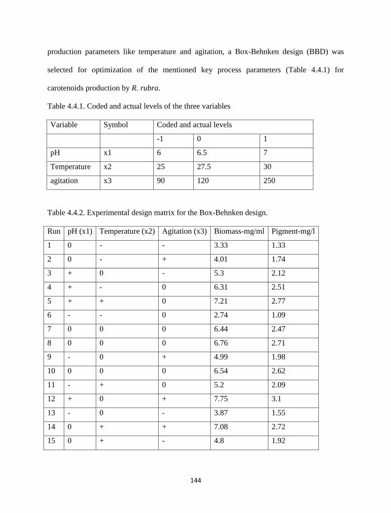
144
production parameters like temperature and agitation, a Box-Behnken design (BBD) was
selected for optimization of the mentioned key process parameters (Table 4.4.1) for
carotenoids production by R. rubra.
Table 4.4.1. Coded and actual levels of the three variables
Variable Symbol Coded and actual levels
-1 0 1
pH x1 6 6.5 7
Temperature x2 25 27.5 30
agitation x3 90 120 250
Table 4.4.2. Experimental design matrix for the Box-Behnken design.
Run pH (x1) Temperature (x2) Agitation (x3) Biomass-mg/ml Pigment-mg/l
1 0 - - 3.33 1.33
2 0 - + 4.01 1.74
3 + 0 - 5.3 2.12
4 + - 0 6.31 2.51
5 + + 0 7.21 2.77
6 - - 0 2.74 1.09
7 0 0 0 6.44 2.47
8 0 0 0 6.76 2.71
9 - 0 + 4.99 1.98
10 0 0 0 6.54 2.62
11 - + 0 5.2 2.09
12 + 0 + 7.75 3.1
13 - 0 - 3.87 1.55
14 0 + + 7.08 2.72
15 0 + - 4.8 1.92
145
The experimental design and statistical analysis of the data were carried out using the Minitab
(14.0) statistical software package. BBD with three factors and 15 runs was chosen; a model
was developed and also validated. Table 4.4.1 shows the variables and experimental design
levels for response surface and Table 4.4.2 shows the design matrix of BBD.
4.4.3 Results and discussion
4.4.3.1 Effect of pH on biomass and carotenoid yield
Using first approach the significant parameters that affected carotenoid production were
identified as pH, temperature and agitation. Effect of pH on biomass growth and carotenoid
production is illustrated in Fig. 4.4.1(a). The figure shows native FWE’s pH (4.31) including
different values studied. For evaluating the influence of pH on biomass and pigment
production, R. rubra was cultivated in FWE medium at 30 0C for 7 days. The microbial
culture is inoculated in the medium with adjusted pH's 5, 6, 7, 8, and 9 along with initial pH
4.31. Among various pHs, biomass growth was identified at values of 5 to 9 and pigment
production was observed only at 6 and 7. So, the results suggest that pigment production was
highly dependent on pH irrespective of biomass growth with high yield caused at pH 7. The
prepared FWE and fermented FWE with the yeast R. rubra are shown in Fig. 4.4.1(b).
The reducing sugar content in FWE was identified to be 19.90 mg/ml before
experimentation. CHNS analysis was performed for the prepared FWE so as to estimate the
presence of major and minor nutrients. CHNS analysis was done before and after carotenoid
production (Fig. 4.4.1(c)). The values stated suggests that significant utilization of macro and
micro nutrients by the microorganism followed the following order of H > N > C > S. In
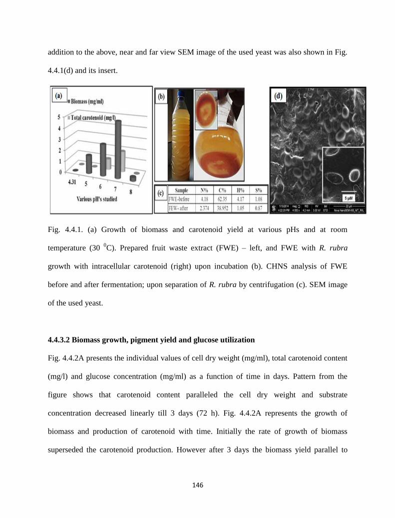
146
addition to the above, near and far view SEM image of the used yeast was also shown in Fig.
4.4.1(d) and its insert.
Fig. 4.4.1. (a) Growth of biomass and carotenoid yield at various pHs and at room
temperature (30 0C). Prepared fruit waste extract (FWE) – left, and FWE with R. rubra
growth with intracellular carotenoid (right) upon incubation (b). CHNS analysis of FWE
before and after fermentation; upon separation of R. rubra by centrifugation (c). SEM image
of the used yeast.
4.4.3.2 Biomass growth, pigment yield and glucose utilization
Fig. 4.4.2A presents the individual values of cell dry weight (mg/ml), total carotenoid content
(mg/l) and glucose concentration (mg/ml) as a function of time in days. Pattern from the
figure shows that carotenoid content paralleled the cell dry weight and substrate
concentration decreased linearly till 3 days (72 h). Fig. 4.4.2A represents the growth of
biomass and production of carotenoid with time. Initially the rate of growth of biomass
superseded the carotenoid production. However after 3 days the biomass yield parallel to
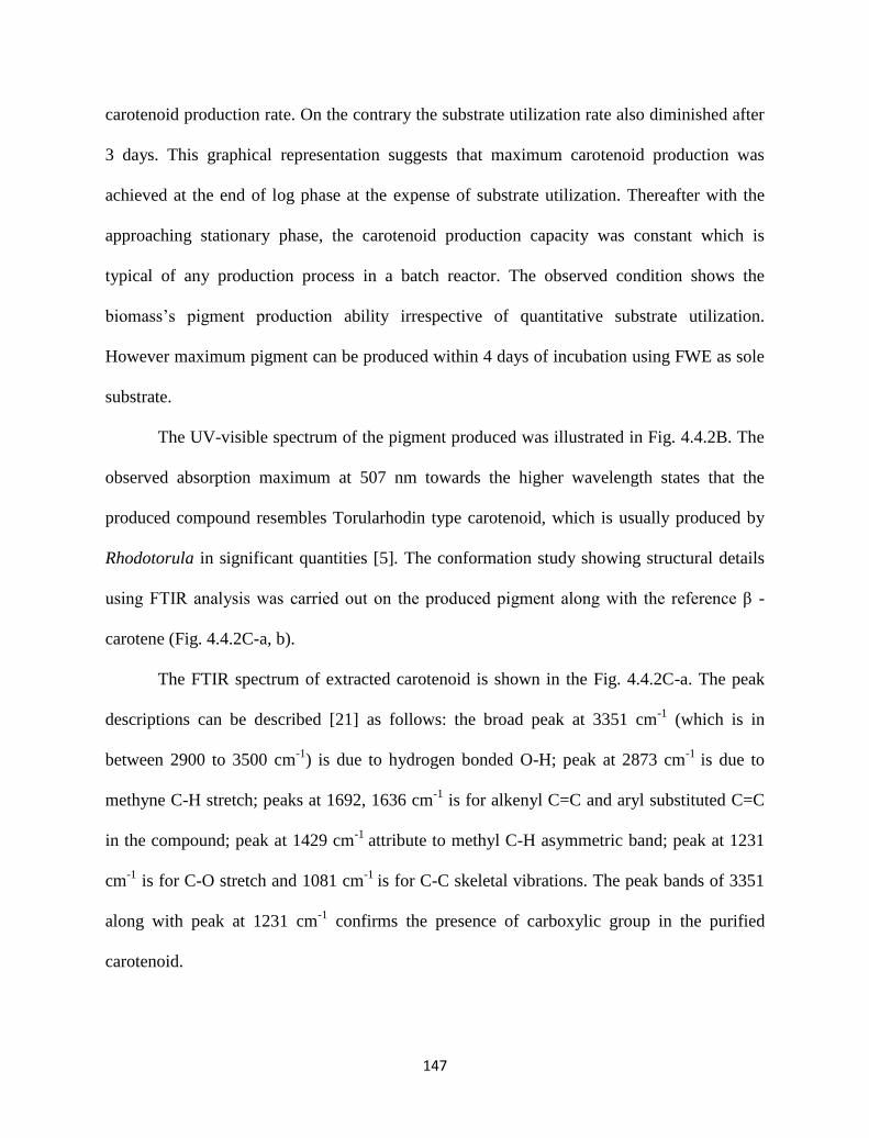
147
carotenoid production rate. On the contrary the substrate utilization rate also diminished after
3 days. This graphical representation suggests that maximum carotenoid production was
achieved at the end of log phase at the expense of substrate utilization. Thereafter with the
approaching stationary phase, the carotenoid production capacity was constant which is
typical of any production process in a batch reactor. The observed condition shows the
biomass’s pigment production ability irrespective of quantitative substrate utilization.
However maximum pigment can be produced within 4 days of incubation using FWE as sole
substrate.
The UV-visible spectrum of the pigment produced was illustrated in Fig. 4.4.2B. The
observed absorption maximum at 507 nm towards the higher wavelength states that the
produced compound resembles Torularhodin type carotenoid, which is usually produced by
Rhodotorula in significant quantities [5]. The conformation study showing structural details
using FTIR analysis was carried out on the produced pigment along with the reference β -
carotene (Fig. 4.4.2C-a, b).
The FTIR spectrum of extracted carotenoid is shown in the Fig. 4.4.2C-a. The peak
descriptions can be described [21] as follows: the broad peak at 3351 cm-1
(which is in
between 2900 to 3500 cm-1
) is due to hydrogen bonded O-H; peak at 2873 cm-1
is due to
methyne C-H stretch; peaks at 1692, 1636 cm-1
is for alkenyl C=C and aryl substituted C=C
in the compound; peak at 1429 cm-1
attribute to methyl C-H asymmetric band; peak at 1231
cm-1
is for C-O stretch and 1081 cm-1
is for C-C skeletal vibrations. The peak bands of 3351
along with peak at 1231 cm-1
confirms the presence of carboxylic group in the purified
carotenoid.
148
Fig. 4.4.2. Carotenoid production (mg/l) and biomass yield (mg/ml) along with glucose
utilization (mg/ml) given in [A]. The picture insert shows the colored biomass from 1 to 6
days. UV-visible spectrum of the purified carotenoid in methanol medium [B]. FTIR spectra
of the synthesized carotenoid (a) and the purchased β-carotene (b) pigment.
Moreover on comparison with the purchased β - carotene’s spectrum (Fig. 4.4.2C-b), the
obtained carotenoid spectrum has some similarities and some variations. The spectrum of β-
carotene displayed major peaks at 2912 and 2860 cm-1
(for asymmetric and symmetric
vibrations of the CH2 and CH3); peak at 1446 cm-1
(due to CH2 group); peak at 1365 cm-1
(for
splitting due to dimethyl; group); peaks at 1021 cm-1
(is for in plane –CH-) and 965 cm-1
(is
149
for trans conjugated alkene -CH=CH- out-of-plane deformation). Further peaks at 700 to 900
cm-1
are for skeletal vibration of C-C stretch. The IR analysis along with UV spectrum of the
obtained carotenoid closely resembles the structures of torularhodin [14] foremost, and has
some similarities with β-carotene (as shown in Fig. 4.4.2C-b). Also this spectral pattern is
obvious as the mentioned pigments are predominantly obtained by Rhodotoula sp [5].
4.4.3.3 Box–Behnken design
The influence of pH on pigment productivity was identified as described in the above section,
this indicates a slight deviation of pH values from 6 to 7 resulted in negative effects with
low/negligible pigment yield. The above synthesis conditions were at static temperature 30
0C and agitation (100 rpm). The Box–Behnken design was employed to study the interactions
among the significant factors and also determine their optimal levels. This methodology is
essentially an assortment of statistical and regression techniques. The first step involves
framing a statistically significant empirical model capable of describing the effect of multiple
factors on a response. A commonly used empirical model of the response surface analysis is a
quadratic polynomial of the type:
where y is the predicted response, xixj are input variables which influence the response
variable y; b0 is the offset term; bi is the ith
linear coefficient; bii the ith
quadratic coefficient
and bij is the ijth
interaction coefficient.
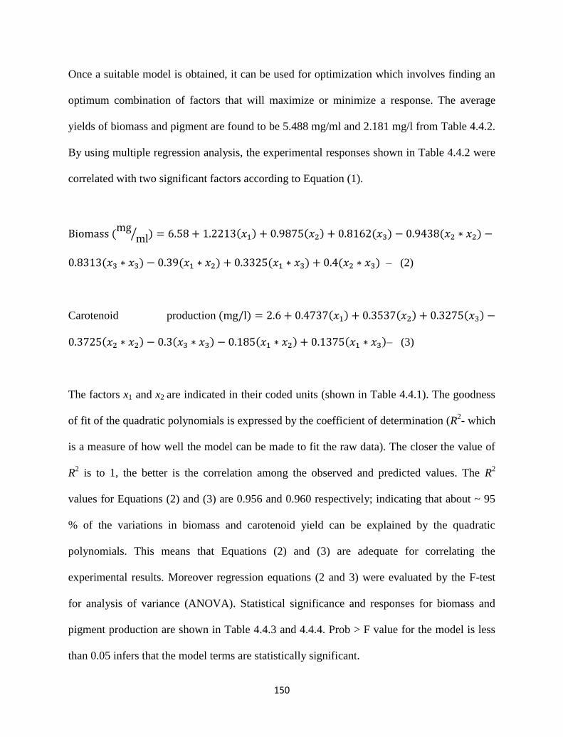
150
Once a suitable model is obtained, it can be used for optimization which involves finding an
optimum combination of factors that will maximize or minimize a response. The average
yields of biomass and pigment are found to be 5.488 mg/ml and 2.181 mg/l from Table 4.4.2.
By using multiple regression analysis, the experimental responses shown in Table 4.4.2 were
correlated with two significant factors according to Equation (1).
– (2)
Carotenoid production
– (3)
The factors x1 and x2 are indicated in their coded units (shown in Table 4.4.1). The goodness
of fit of the quadratic polynomials is expressed by the coefficient of determination (R2- which
is a measure of how well the model can be made to fit the raw data). The closer the value of
R2 is to 1, the better is the correlation among the observed and predicted values. The R
2
values for Equations (2) and (3) are 0.956 and 0.960 respectively; indicating that about ~ 95
% of the variations in biomass and carotenoid yield can be explained by the quadratic
polynomials. This means that Equations (2) and (3) are adequate for correlating the
experimental results. Moreover regression equations (2 and 3) were evaluated by the F-test
for analysis of variance (ANOVA). Statistical significance and responses for biomass and
pigment production are shown in Table 4.4.3 and 4.4.4. Prob > F value for the model is less
than 0.05 infers that the model terms are statistically significant.
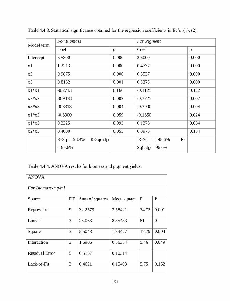
151
Table 4.4.3. Statistical significance obtained for the regression coefficients in Eq’s .(1), (2).
Model term For Biomass For Pigment
Coef p Coef p
Intercept 6.5800 0.000 2.6000 0.000
x1 1.2213 0.000 0.4737 0.000
x2 0.9875 0.000 0.3537 0.000
x3 0.8162 0.001 0.3275 0.000
x1*x1 -0.2713 0.166 -0.1125 0.122
x2*x2 -0.9438 0.002 -0.3725 0.002
x3*x3 -0.8313 0.004 -0.3000 0.004
x1*x2 -0.3900 0.059 -0.1850 0.024
x1*x3 0.3325 0.093 0.1375 0.064
x2*x3 0.4000 0.055 0.0975 0.154
R-Sq = 98.4% R-Sq(adj)
= 95.6%
R-Sq = 98.6% R-
Sq(adj) = 96.0%
Table 4.4.4. ANOVA results for biomass and pigment yields.
ANOVA
For Biomass-mg/ml
Source DF Sum of squares Mean square F P
Regression 9 32.2579 3.58421 34.75 0.001
Linear 3 25.063 8.35433 81 0
Square 3 5.5043 1.83477 17.79 0.004
Interaction 3 1.6906 0.56354 5.46 0.049
Residual Error 5 0.5157 0.10314
Lack-of-Fit 3 0.4621 0.15403 5.75 0.152
152
Pure Error 2 0.0536 0.0268
Total 14 32.7736
For Pigment-mg/l
Source DF Sum of squares Mean square F P
Regression 9 4.70655 0.52295 38.78 0
Linear 3 3.65468 1.21823 90.34 0
Square 3 0.80132 0.26711 19.81 0.003
Interaction 3 0.25055 0.08352 6.19 0.039
Residual Error 5 0.06742 0.01348
Lack-of-Fit 3 0.03802 0.01267 0.86 0.576
Pure Error 2 0.0294 0.0147
Total 14 4.77397
The actual and predicted values of responses for biomass and carotenoid
concentrations versus the corresponding values calculated by regression models are shown in
Fig. 4.4.3a,b respectively. Actual values are the measured values for a particular experiment,
whereas predicted values are generated by using the approximating functions. The line of
perfect fit is also shown in these figures and visualization of the two regression models
provides an accurate description of the experimental data. In addition the values of R2 and
adjusted R2 (Table 4.4.3) have advocated a high correlation between actual and predicted
values. The response surface and contour plots were constructed using the regression models
and represented in Fig. 4.4.4a-d. Fig. 4.4.4a depicts the interactive effect of the agitation and
153
temperature on the biomass concentration. At low to moderate agitation values, increases in
temperature led to increased production of biomass initially up to center point and then
decreased. At low to moderate temperature values, increases in agitation values showed
increase in biomass yield. Similar trends were not observed for pigment yield as a function of
pH and temperature (Fig. 4.4.4c). At high pH values, increases in temperature showed
significant increase in pigment yield up to the center point and decreased thereafter. While
with increase in temperature values, increase in pH led to increased pigment production.
The contour plots in Fig. 4.4.4b,c indicate that a local optimum exists in the area
experimentally studied; a set of values on the two factors that leads to maximum biomass or
pigment production. The location of these optimal points can be obtained by differentiating
Equations (2) and (3). Equation (3) was used to derive the most efficient combination of x1 to
x3 to produce carotenoids from Rhodotura sp. According to Equation (3), a maximum
carotenoid recovery of 3.17 mg/l could be attained with pH (7.0), temperature (28.2 0C), and
agitation (150 rpm). Experimental validation of the optimum x1-x3 combination gave a
carotenoid yield of 3.298 ± 0.28 mg/l (with 7.83 mg/ml biomass concentration). Equation (2)
depicted that at pH (7.0), temperature (30 0C), and agitation (150 rpm) maximum yield of
7.97 mg/ml biomass could be achieved. The experimental validation resulted in biomass yield
of 8.27 ± 0.33 mg/ml with 2.43 mg/l carotenoid production.
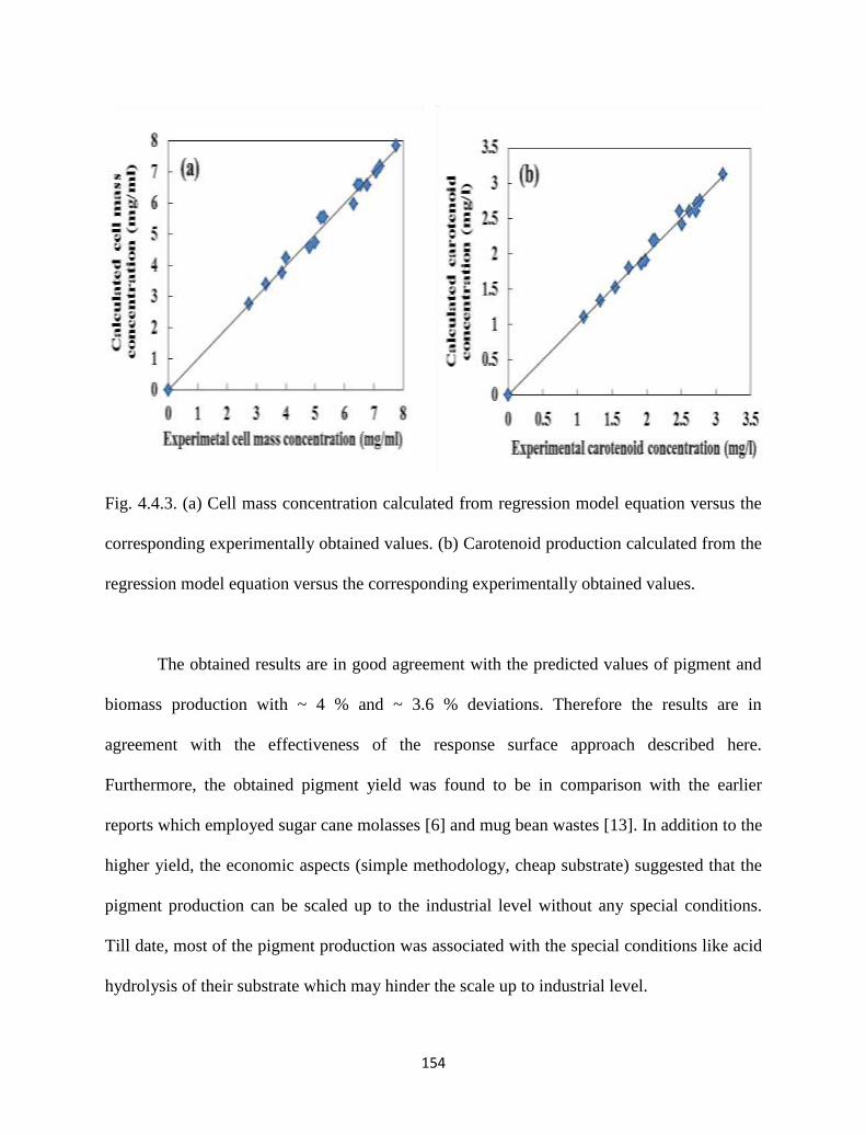
154
Fig. 4.4.3. (a) Cell mass concentration calculated from regression model equation versus the
corresponding experimentally obtained values. (b) Carotenoid production calculated from the
regression model equation versus the corresponding experimentally obtained values.
The obtained results are in good agreement with the predicted values of pigment and
biomass production with ~ 4 % and ~ 3.6 % deviations. Therefore the results are in
agreement with the effectiveness of the response surface approach described here.
Furthermore, the obtained pigment yield was found to be in comparison with the earlier
reports which employed sugar cane molasses [6] and mug bean wastes [13]. In addition to the
higher yield, the economic aspects (simple methodology, cheap substrate) suggested that the
pigment production can be scaled up to the industrial level without any special conditions.
Till date, most of the pigment production was associated with the special conditions like acid
hydrolysis of their substrate which may hinder the scale up to industrial level.
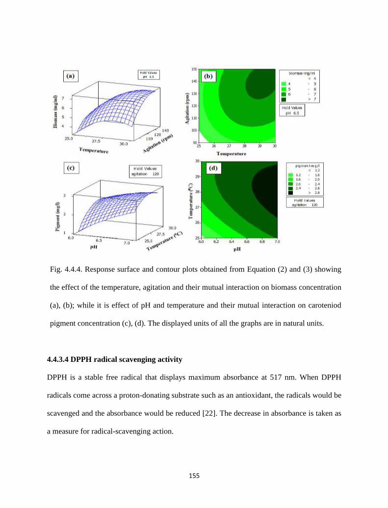
155
Fig. 4.4.4. Response surface and contour plots obtained from Equation (2) and (3) showing
the effect of the temperature, agitation and their mutual interaction on biomass concentration
(a), (b); while it is effect of pH and temperature and their mutual interaction on caroteniod
pigment concentration (c), (d). The displayed units of all the graphs are in natural units.
4.4.3.4 DPPH radical scavenging activity
DPPH is a stable free radical that displays maximum absorbance at 517 nm. When DPPH
radicals come across a proton-donating substrate such as an antioxidant, the radicals would be
scavenged and the absorbance would be reduced [22]. The decrease in absorbance is taken as
a measure for radical-scavenging action.
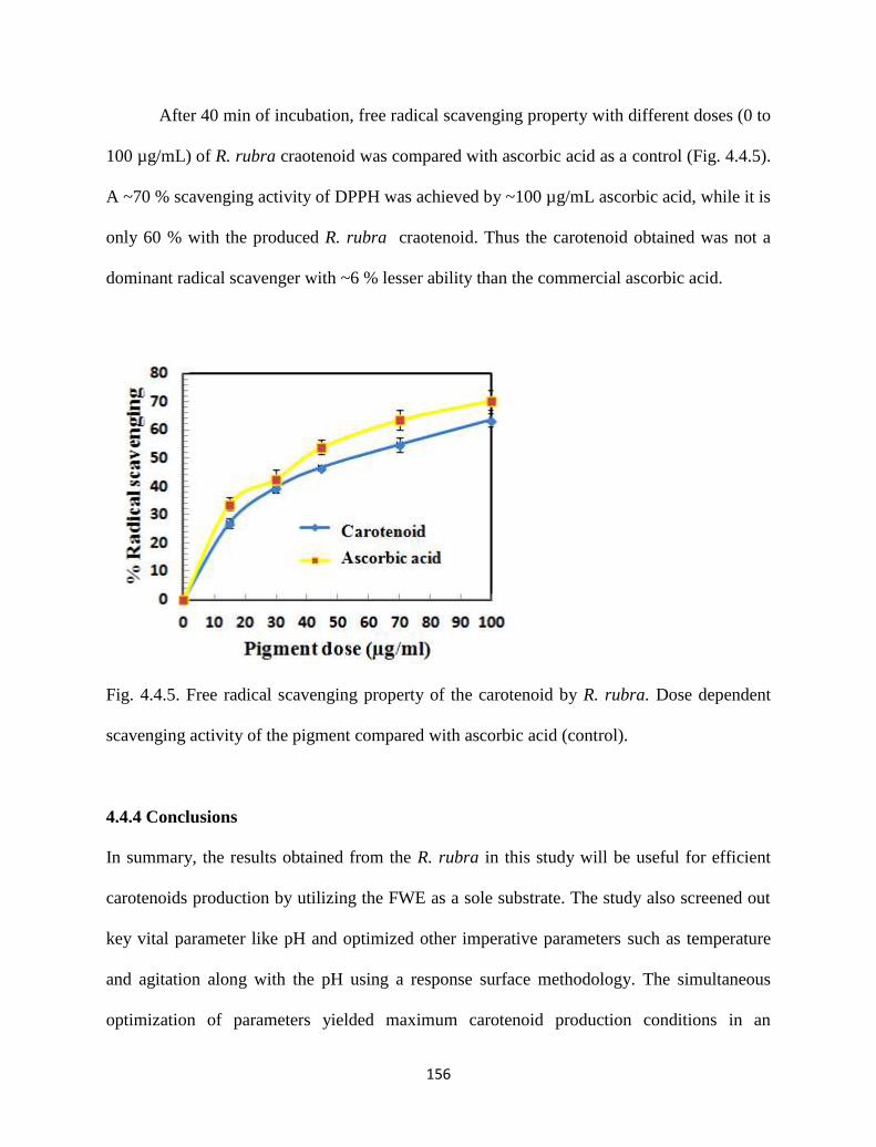
156
After 40 min of incubation, free radical scavenging property with different doses (0 to
100 µg/mL) of R. rubra craotenoid was compared with ascorbic acid as a control (Fig. 4.4.5).
A ~70 % scavenging activity of DPPH was achieved by ~100 µg/mL ascorbic acid, while it is
only 60 % with the produced R. rubra craotenoid. Thus the carotenoid obtained was not a
dominant radical scavenger with ~6 % lesser ability than the commercial ascorbic acid.
Fig. 4.4.5. Free radical scavenging property of the carotenoid by R. rubra. Dose dependent
scavenging activity of the pigment compared with ascorbic acid (control).
4.4.4 Conclusions
In summary, the results obtained from the R. rubra in this study will be useful for efficient
carotenoids production by utilizing the FWE as a sole substrate. The study also screened out
key vital parameter like pH and optimized other imperative parameters such as temperature
and agitation along with the pH using a response surface methodology. The simultaneous
optimization of parameters yielded maximum carotenoid production conditions in an
157
economical way, for its large scale production using a cheaper substrate (FWE). Our results
demonstrated that FWE could be profitably used as a suitable substrate without any
additional growth supplement for noteworthy carotenoid production. The response
optimization of parameters pH (7), temperature (28.2 0C) and agitation (150 rpm) by Box–
Behnken design resulted in 51.2 % more enhancement of the mean carotenoid production.
The produced carotenoid pigment is having potential antioxidant applications in food
industries. This can be valued for industrial scale utilization of FWE to generate high value
carotenoids using R. rubra and opens up scope for exploring other high value microbial
pigments.
References
[1] A. Das, et al., "An update on microbial carotenoid production: application of recent
metabolic engineering tools," Applied Microbiology and Biotechnology, vol. 77, pp.
505-512, 2007.
[2] L. Dufossé, et al., "Microorganisms and microalgae as sources of pigments for food
use: a scientific oddity or an industrial reality?," Trends in Food Science &
Technology, vol. 16, pp. 389-406, 2005.
[3] E. D. Simova, et al., "Effect of aeration on the production of carotenoid pigments by
Rhodotorula rubra-lactobacillus casei subsp. casei co-cultures in whey ultrafiltrate,"
Zeitschrift fur Naturforschung C-Journal of Biosciences, vol. 58, pp. 225-229, 2003.
[4] I. Marova, et al., "Use of several waste substrates for carotenoid-rich yeast biomass
production," Journal of Environmental Management, vol. 95, Supplement, pp. S338-
S342, 2012.
158
[5] P. Park, et al., "Chemical disruption of yeast cells for the isolation of carotenoid
pigments," Separation and purification technology, vol. 53, pp. 148-152, 2007.
[6] P. Park, et al., "Optimization of carotenoid production by Rhodotorula glutinis using
statistical experimental design," World journal of microbiology and biotechnology,
vol. 21, pp. 429-434, 2005.
[7] M. M. S. Cabral, et al., "Carotenoids production from a newly isolated Sporidiobolus
pararoseus strain by submerged fermentation," European Food Research and
Technology, vol. 233, pp. 159-166, 2011.
[8] B. Ribeiro, et al., "Technological Aspects of β-Carotene Production," Food and
Bioprocess Technology, vol. 4, pp. 693-701, 2011/07/01 2011.
[9] H. J. Nelis and A. De Leenheer, "Microbial sources of carotenoid pigments used in
foods and feeds," Journal of Applied Microbiology, vol. 70, pp. 181-191, 1991.
[10] S. Liaaen-Jensen and A. Andrewes, "Microbial carotenoids," Annual Reviews in
Microbiology, vol. 26, pp. 225-248, 1972.
[11] G. I. Frengova and D. M. Beshkova, "Carotenoids from Rhodotorula and Phaffia:
yeasts of biotechnological importance," Journal of industrial microbiology &
biotechnology, vol. 36, pp. 163-180, 2009.
[12] B. D. H. Dung, et al., "Optimization carotenoids production by Rhodotorula glutinis
GBDO Uusing design of Plackett-Burman matrix and central composite design-
response surface methodology," in Conference proceedings of Biotechnology for
Green Solutions and Sustainable Environment, 2010, p. 25.
159
[13] J. Tinoi, et al., "Simplex optimization of carotenoid production by Rhodotorula
glutinis using hydrolyzed mung bean waste flour as substrate," Process Biochemistry,
vol. 40, pp. 2551-2557, 2005.
[14] B. Latha and K. Jeevaratnam, "Purification and characterization of the pigments from
Rhodotorula glutinis DFR-PDY isolated from natural source," Global J Biotech
Biochem, vol. 5, pp. 166-174, 2010.
[15] P. Bhosale and R. Gadre, "Optimization of carotenoid production from
hyper‐producing Rhodotorula glutinis mutant 32 by a factorial approach," Letters in
applied microbiology, vol. 33, pp. 12-16, 2001.
[16] C. Malisorn and W. Suntornsuk, "Optimization of β-carotene production by
Rhodotorula glutinis DM28 in fermented radish brine," Bioresource Technology, vol.
99, pp. 2281-2287, 2008.
[17] P. Buzzini, "An optimization study of carotenoid production by Rhodotorula glutinis
DBVPG 3853 from substrates containing concentrated rectified grape must as the sole
carbohydrate source," Journal of Industrial Microbiology and Biotechnology, vol. 24,
pp. 41-45, 2000.
[18] S. N. Surwase, et al., "Optimization of melanin production by Brevundimonas sp. SGJ
using response surface methodology," 3 Biotech, pp. 1-8, 2012.
[19] A. Gramza-Michałowska and B. Stachowiak, "The antioxidant potential of carotenoid
extract from Phaffia rhodozyma," Acta Scientiarum Polonorum. Technologia
Alimentaria, vol. 9, 2010.
[20] S. K. Thimmaiah, Standard methods of biochemical analysis: Kalyani publishers,
2006.
160
[21] J. Coates, "Interpretation of infrared spectra, a practical approach," Encyclopedia of
analytical chemistry, 2000.
[22] R. Lo Scalzo, "Organic acids influence on DPPH scavenging by ascorbic acid," Food
Chemistry, vol. 107, pp. 40-43, 2008.
161
4.5 Astaxanthin by Xanthophyllomyces dendrorhous
Using Fruit Waste Extract as Sole Source of Energy:
Optimization of Culture Conditions by Taguchi method
for Improved Pigment Production
4.5.1 Introduction
Carotenoid class astaxanthin belongs to the group of lipophilic tetraterpenes, which are
combination of eight isoprene units (C5) that has important application in the neutraceutical,
cosmetics, food and feed industries due to its high antioxidant activity [1-3]. Astaxanthin
synthesized chemically contains only 8% astaxanthin and costs $25,000-30,000 Kg-1
[4].
Synthetic astaxanthin is the major source of carotenoid currently being used in fish feeds [5].
However, food and feed additives from biotechnological processes are preferred by
consumers to those produced by chemical technologies [6]. Among the microorganisms
Brevibacterium [7], Mycobacterium lacticola [8], Agrobacterium auratium [9],
Haematococcus pluvalis [10] and X. dendrorhous [4, 11] can be used for commercial
162
production of astaxanthin as 80-90% of the total carotenoids produced by these yeasts
accounts for astaxanthin.
Among all red pigmented heterobasidiomycetous yeast, X. dendrrorhous has the
potential to produce carotenoids on cheaper substrates like different agro-industrial raw
materials with high yields [12]. This yeast strain has ability for the assimilation and
metabolizing mono, di and polysaccharides, organic acid and alcohols. Moreover till date
research is focused on this versatile strain on various cheaper substrates for significant
astaxanthin production. Aiming towards cost cutting strategies, substrates such as waste
streams like those from sugar manufacturing processes or the corn wet milling industry, white
grape juice, enzymatic eucalyptus wood hydrolyzates [13], hemicellulosic hydrolyzates of
eucalyptus globulus [14], yucca medium (based on date juice) [15], peat hydrolysate [16],
corn steep liquor [17] etc. were successfully employed for the production of significant
carotenoid class astaxanthin.
Very limited research has been carried out so far on fruit wastes and their extracts as
sole source of energy for astaxanthin production using X. dendrrorhous. India contributes to
10 % of the world’s fruit production [18]. Over last few years research focus is towards
reprocessing and reuse of various fruit wastes for the conversion of nutritive and valuable
products [19-20]. Previous studies on fruit wastes signify that it is a rich source of
carbohydrates and other minor nutrients to support the microbial growth [19-20]. The surplus
fruit wastes those are frequently disposed in the environment after exhaustive extraction of
their juice may be possibly used as low cost substrates for production of microbial bioactive
compounds like pigments. The availability of readymade simple sugars along with macro and
micro nutrients make fruit waste as a suitable compound for the production of numerous
163
pigments by a variety of microorganisms. The above reported studies used special conditions
and/or enriched the substrate media for high yield pigment production. In contrast, eying on
cheaper, economical and quick synthesis process devoid of providing special conditions, we
explored the capacity of readily prepared fruit waste extract (FWE) as sole substrate for
astaxanthin production by acquired X. dendrrorhous. Thus this paper outlined the optimum
production conditions for carotenoid pigment by X. dendrrorhous on FWE using Taguchi
method.
4.5.2 Materials and methods
4.5.2.1 Chemicals and microorganism
Acetone, ascorbic acid, methanol, NaCl, HCL and diethyl ether were procured from Merck,
India. The strain Xanthophyllomyces dendrorhous (7536) was obtained from MTCC
Chandigarh. The yeast was maintained on Yeast extract/ malt extract (YM) media agar plates
at 4 0C and sub cultured monthly in YM media broth. YM media and agar were purchased
from HiMedia chemicals, India.
4.5.2.2 Substrate preparation
Fruit waste is obtained from local juice shop from local market, Rourkela, Odisha, India,
which includes pineapple waste, orange waste and minor portion of pomegranate waste. Fruit
waste extract (FWE) media is prepared from mixed waste by boiling and extracting the
soluble sugars for 30 min using distilled water.
4.5.2.3 Culture conditions
The inoculum was prepared in YM broth at 22 0C for 48h. The yeast cells were harvested by
centrifugation and washed twice with distilled water. 1 mL of microbial solution (1 % w/v)
164
was inoculated in a 250 ml Erlenmeyer flask containing 50 ml FWE medium. The inoculated
cultures were kept at 22 0C in incubator shaker at a shaking speed of 150 rpm for pigment
production. Yeast extract/malt extract (YM) medium is used as the reference medium for
comparison with FWE medium. Each experiment was performed thrice to minimize the error.
4.5.2.4 Pigment production and extraction/ Sugar determination
Yeast cells obtained after centrifugation was washed twice with distilled water. Pigmented
cells were treated with methanol: acetone (1:1) solvents and centrifuged. This process was
repeated until colorless biomass was obtained. The extracted pigment was subjected to phase
separation using equal volumes of petroleum ether and 10 % NaCl. The carotenoid was
collected from the diethyl ether phase and analyzed at 474 nm by UV-visible
spectrophotometer using an extension coefficient of = 2,100 [21]. Obtained carotenoids
were further purified using thin layer chromatography using stationary phase - TLC silica gel
60 F 254 and hexane: methanol (7:3) as stationary phase and solvent phase respectively. The
purified pigment was further analyzed in FTIR to determine the functional groups.
The 3,5- dinitrosalicyclic acid (DNS) method of miller [22] was used to determine the
reducing sugars in FWE medium. About 1ml sample was centrifuged at 3,500 X g for 5 min,
and 1 mL DNS reagent was added to the supernatant and boiled for 5 min. The sample was
placed in ice –bath for rapid cooling. Distilled water was added to the samples and vortexed
for 5 min. The optical density was calculated at 575 nm. In addition, CHNS analysis was
performed before and after the pigment production
4.5.2.5 Taguchi design and statistical analysis
Simple Taguchi orthogonal array design was selected where there are three factors and three
levels. From the reported studies, pH (4-6), temperature (15-25 0C) and agitation (100-300
165
rpm) were found to be significant in obtaining higher amounts of astaxanthin production by
X. dendrorhous [23-25]. In this study Taguchi method was used for optimization of pigment
production conditions. Optimization of stated factors in three different levels is illustrated in
Table 4.5.1. To perform the Taguchi method, 9 different experiments using L9 orthogonal
array was run as shown in Table 4.5.2. Based on the primary results a verification test was
employed to check the optimum condition. An analysis of variance (ANOVA) for the
obtained results was examined. Design of experiments, ANOVA and optimization of process
were accomplished using MINITAB- 14 software.
4.5.2.6 Determination of free radical scavenging activity
The effect of astaxanthin from X. dendrorhous on DPPH free radical was studied as per the
method of Gramza-Michałowska and Stachowiak[26] with some modifications. A 2 mg/ml
pigment suspension was prepared in varying volumes of ethanol (10 to 60 µl). To the
suspension 1 ml DPPH solution containing 0.1 mM DPPH and 99.5 % ethanol (99.5 %) was
added. The mixture was thoroughly mixed and allowed to stand for 40 min at room
temperature in the dark. Ascorbic acid is used as positive control and the pigment solution
without DPPH as negative control. The radical scavenging activity was measured as a
decrease in the absorbance of DPPH at 517 nm wavelength and was calculated using the
following formula:
100)(A
)A-A(activity Scavenging %
c
sc
Where Ac, As are absorbance’s from control and sample.
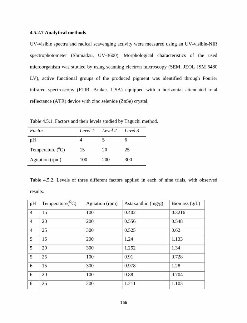
166
4.5.2.7 Analytical methods
UV-visible spectra and radical scavenging activity were measured using an UV-visible-NIR
spectrophotometer (Shimadzu, UV-3600). Morphological characteristics of the used
microorganism was studied by using scanning electron microscopy (SEM, JEOL JSM 6480
LV), active functional groups of the produced pigment was identified through Fourier
infrared spectroscopy (FTIR, Bruker, USA) equipped with a horizontal attenuated total
reflectance (ATR) device with zinc selenide (ZnSe) crystal.
Table 4.5.1. Factors and their levels studied by Taguchi method.
Factor Level 1 Level 2 Level 3
pH 4 5 6
Temperature (oC) 15 20 25
Agitation (rpm) 100 200 300
Table 4.5.2. Levels of three different factors applied in each of nine trials, with observed
results.
pH Temperature(OC) Agitation (rpm) Astaxanthin (mg/g) Biomass (g/L)
4 15 100 0.402 0.3216
4 20 200 0.556 0.548
4 25 300 0.525 0.62
5 15 200 1.24 1.133
5 20 300 1.252 1.34
5 25 100 0.91 0.728
6 15 300 0.978 1.28
6 20 100 0.88 0.704
6 25 200 1.211 1.103
167
4.5.3 Results and discussion
Among various yeasts, X. dendrorhous has been exploited as major source of astaxanthin
with potential commercial applications in the fields of cosmetics, pharmaceuticals and
agriculture [5, 7]. However, astaxanthin production from cheaper substrates has limited
reports. So optimization of its process parameters for astaxanthin production has been
extensively studied to emphasize its production in commercial scale. Substrate
utilization/growth and pigment production by yeast was significantly influenced by key
factors like pH, temperature and agitation; hence these factors play a vital role in the cost
effectiveness of pigment production. The pigmentation of the FWE and the SEM image of
the studied yeast were given in Fig. 4.5.1.
Fig. 4.5.1. Pigmentation of FWE (a) by before X. dendrorhous yeast with lower (b) and
higher (c) magnification SEM images.
168
4.5.3.1 Experimental design by Taguchi method
Effects of pH, temperature and agitation were studied using Taguchi method, which is a
fractional factorial experimental design. The results of experiments performed in this section
(Table 4.5.2) showed that the maximum average yield of pigment 1.252 mg/g, at
experimental conditions pH 5, temperature 20 oC and agitation 300 rpm. Fig. 4.5.2 depicts the
main effect of each of these factors that implies the average results obtained for each factor in
order to screen out significant factors the statistical technique of analysis of variance
(ANOVA) with F test was employed to the simulation reflecting the data shown in Table
4.5.3. The ANOVA results implicates from F value of 23.71 that pH has significant effect on
pigment production. Similarly temperature showed negligible effect. Thus the optimum
experimental condition was selected from the obtained result as pH 5, temp 20 oC and
agitation 300 rpm. When the results were analyzed, an optimum condition was proposed by
calculations.
4.5.3.2 Validation of the model
Table 4.5.4 shows the suggested conditions for maximum pigment production. Statistical
calculations predicted that if the conditions were chosen as shown in Table 4.5.4, the pigment
production should reach 1.264 mg/g biomass. However after performing the experiment at
said condition, the produced astaxanthin was 1.40 ± 0.14 mg/g. The difference between
predicted and actual result was about 10 %, and is regarded as acceptable and promising.
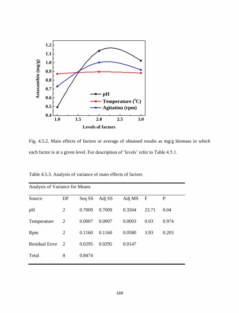
169
1.0 1.5 2.0 2.5 3.00.4
0.5
0.6
0.7
0.8
0.9
1.0
1.1
1.2
pH
Temperature (oC)
Agitation (rpm)
A
sta
xa
nth
in (
mg
/g)
Levels of factors
Fig. 4.5.2. Main effects of factors or average of obtained results as mg/g biomass in which
each factor is at a given level. For description of ‘levels’ refer to Table 4.5.1.
Table 4.5.3. Analysis of variance of main effects of factors
Analysis of Variance for Means
Source DF Seq SS Adj SS Adj MS F P
pH 2 0.7009 0.7009 0.3504 23.71 0.04
Temperature 2 0.0007 0.0007 0.0003 0.03 0.974
Rpm 2 0.1160 0.1160 0.0580 3.93 0.203
Residual Error 2 0.0295 0.0295 0.0147
Total 8 0.8474
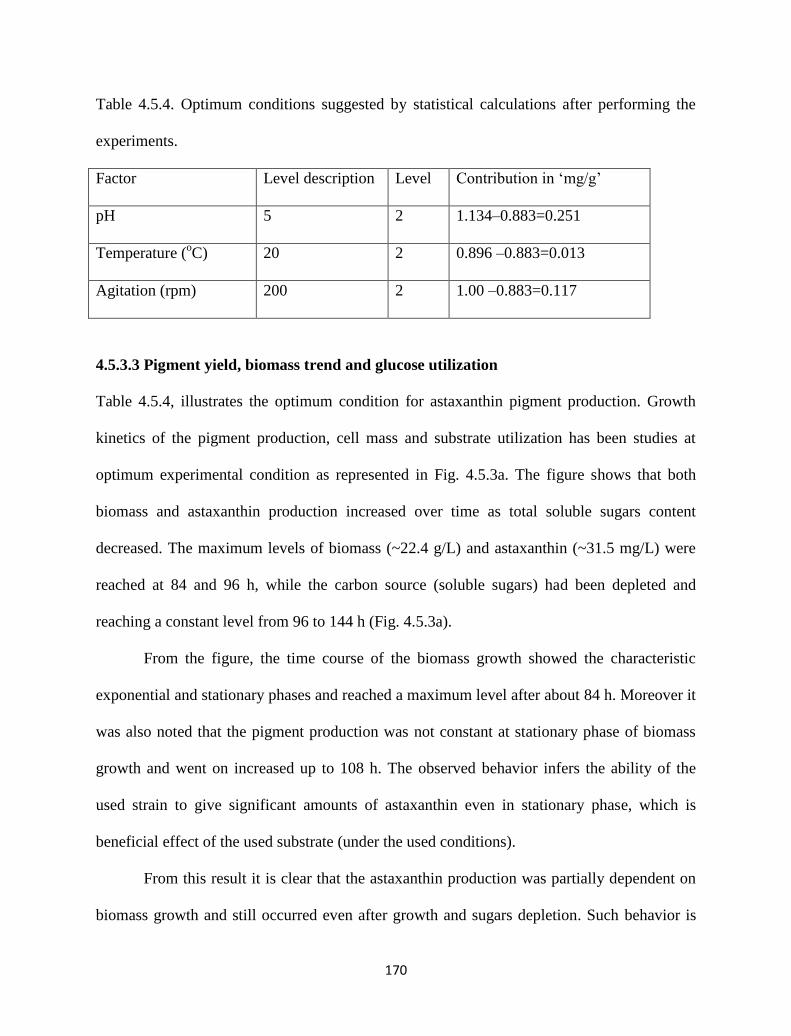
170
Table 4.5.4. Optimum conditions suggested by statistical calculations after performing the
experiments.
Factor Level description Level Contribution in ‘mg/g’
pH 5 2 1.134–0.883=0.251
Temperature (oC) 20 2 0.896 –0.883=0.013
Agitation (rpm) 200 2 1.00 –0.883=0.117
4.5.3.3 Pigment yield, biomass trend and glucose utilization
Table 4.5.4, illustrates the optimum condition for astaxanthin pigment production. Growth
kinetics of the pigment production, cell mass and substrate utilization has been studies at
optimum experimental condition as represented in Fig. 4.5.3a. The figure shows that both
biomass and astaxanthin production increased over time as total soluble sugars content
decreased. The maximum levels of biomass (~22.4 g/L) and astaxanthin (~31.5 mg/L) were
reached at 84 and 96 h, while the carbon source (soluble sugars) had been depleted and
reaching a constant level from 96 to 144 h (Fig. 4.5.3a).
From the figure, the time course of the biomass growth showed the characteristic
exponential and stationary phases and reached a maximum level after about 84 h. Moreover it
was also noted that the pigment production was not constant at stationary phase of biomass
growth and went on increased up to 108 h. The observed behavior infers the ability of the
used strain to give significant amounts of astaxanthin even in stationary phase, which is
beneficial effect of the used substrate (under the used conditions).
From this result it is clear that the astaxanthin production was partially dependent on
biomass growth and still occurred even after growth and sugars depletion. Such behavior is
171
certainly related to the fact that X. dendrorhous excretes and stocks some extracellular carbon
intermediates, which stimulate late carotenogenesis [7].
Fig. 4.5.3. (a) Time course of the growth and production of astaxanathin by X. dendrorhous
in FWE. Experiment was carried out at optimum conditions i.e. pH (5), temperature (20 oC)
and agitation (300 rpm). (b) UV-Visible and FTIR spectrum (c) of the produced pigment in
methanol. (d) Antioxidant activity (DPPH radical scavenging) of astaxanthin and ascorbic
acid.
0 20 40 60 80 100 120
0
20
40
60
80
100
% R
ad
ica
l sc
av
en
gin
g
Pigment dose (µg/mL)
Astaxanthin
Ascorbic acid
(c) (d)
(b)
3600 3150 2700 2250 1800 1350 9001.88
1.90
1.92
1.94
1.96
1.98
2.00
2.02
1380
9501163
17362970
Inte
nsi
ty (
a.u
)
Wavenumber (cm-1
)
3362
300 375 450 525 600 6750.1
0.2
0.3
0.4
0.5
0.6
0.7
0.8
0.9
Ab
sorb
an
ce (
a.u
)
Wavelength (nm)
0 20 40 60 80 100 120 1400
5
10
15
20
25
30
35
Glucose depletion (g/L)
Astaxanthin (mg/L)
Biomass (g/L)
Time (hours)
Glu
co
se d
ep
leti
on
(g
/L)
0
5
10
15
20
25
30
35
Asta
xa
nth
in (m
g/L
); Bio
ma
ss (g/L
)
(a)
172
4.5.3.4 UV analysis
The spectral property of the produced astaxanthin in FWE was shown in Fig. 4.5.3b. The
absorption maximum was identified to be at 474 nm in methanol and was in good agreement
with the reported value [27].
4.5.3.5 FTIR analysis
The purified pigment from the TLC plate was dissolved in ethanol and analyzed for FTIR
spectral analysis and the results are shown in (Fig. 4.5.3c). According to Coates et al. [28] the
peak descriptions of the FTIR spectra can be described as follows: The peak at 3362 shows
the presence of O-H hydrogen bond. The peak at 2970 is due to methyne C-H stretch; a peak
at 1736 shows the presence of C=O bond in the molecule. The peak at 1380 is due to methyl
C-H asymmetric band. The peak at 1163 is for C-O stretch and the peak at 950 represents the
skeletal vibrations. The spectral behavior of the pigment closely resembles the structure of
astaxanthin which is predominantly obtained by X. dendrorhous [29].
4.5.3.6 DPPH radical scavenging activity
DPPH is a stable free radical that displays maximum absorbance at 517 nm. When DPPH
radicals come across a proton-donating substrate such as an antioxidant, the radicals would be
scavenged and the absorbance would be reduced [30]. The decrease in absorbance is taken as
a measure for radical-scavenging action. The DPPH radical-scavenging activity was
investigated at different concentrations (0 to 109 µg/mL) of the produced astaxanthin. The
results are shown in Fig. 4.5.3d.
173
After 40 min of incubation, it could be observed that the radical scavenging activity
increased promptly with an increase in astaxanthin dose when compared to ascorbic acid
(control) and reached up to ~85 % at the dose of 109 µg/ml. However, it is also noted that
ascorbic acid helped in achieving only ~70 % under the same conditions. Although
astaxanhin and ascorbic acid both showing an increase in % radical scavenging, it is
noteworthy that the former was higher than the latter at any moment within the studied
pigment dose (Fig. 4.5.3d).
In summary, the key factors that independently influence biomass growth and
pigment production were identified by Taguchi method. At identified key parameters, FWE
appears to be an excellent substrate since a remarkable astaxanthin production (1.4 ± 0.14
mg/g biomass) was obtained when compared to YM medium i.e. 1.66 ± 0.21 mg/g biomass
at same culture conditions. YM is an expensive source for astaxanthin production with C to N
proportion of 1.0 and 0.3 %, while FWE is an inexpensive alternative with C to N proportion
found to be 1.0 and 0.066 % (observed by CHNS analysis). This work shows that FWE
medium plays a key role in astaxanthin production by X. dendrorhous and it is worth noting
that, in our case we achieved enhanced astaxanthin production with free radical scavenging
activity without any substrate supplements and special conditions.
4.5.4 Conclusions
This study shows the suitability of the FWE medium for the production of astaxanthin by X.
dendrorhous yeast strain. Critical parameters screened by simple Taguchi approach and
optimum conditions for the enhanced production of pigment were established. Increase yield
was observed at pH - 5, Temp - 20 °C and Agitation-200 rpm with optimum production of
174
1.4 ± 0.14 mg/g biomass. UV-visible and IR analysis of the purified pigment illustrated the
structural confirmations of the pigment. In addition, free radical scavenging activity of the
obtained pigment gave 15 % better activity than ascorbic acid and encourages its applicability
as a constitutional ingredient in cosmetics.
References
[1] H. T. H. AN, et al., "Determination of astaxanthin and other carotenoids in
vietnamese crustaceans by HPLC." TAp Chí Khoa Hoc Và Công Nghe, vol. 46, pp.
47-58, 2008.
[2] R. N. Rao, et al., "Preparative isolation and characterization of some minor impurities
of astaxanthin by high-performance liquid chromatography," Journal of
chromatography A, vol. 1076, pp. 189-192, 2005.
[3] I. Schmidt, et al., "Biotechnological production of astaxanthin with Phaffia
rhodozyma/Xanthophyllomyces dendrorhous," Applied Microbiology and
Biotechnology, vol. 89, pp. 555-571, 2011.
[4] J. Tinoi, et al., "Utilization of mustard waste isolates for improved production of
astaxanthin by Xanthophyllomyces dendrorhous," Journal of Industrial Microbiology
and Biotechnology, vol. 33, pp. 309-314, 2006.
[5] I. Higuera-Ciapara, et al., "Astaxanthin: a review of its chemistry and applications,"
Critical reviews in food science and nutrition, vol. 46, pp. 185-196, 2006.
[6] D. Pleissner and C. S. K. Lin, "Valorisation of food waste in biotechnological
processes," Sustainable Chemical Processes, vol. 1, p. 21, 2013.
175
[7] E. A. Johnson and G.-H. An, "Astaxanthin from microbial sources," Critical Reviews
in Biotechnology, vol. 11, pp. 297-326, 1991.
[8] H. J. Nelis and A. De Leenheer, "Microbial sources of carotenoid pigments used in
foods and feeds," Journal of Applied Microbiology, vol. 70, pp. 181-191, 1991.
[9] J. A. Bon, et al., "Isolation of astaxanthin-overproducing mutants of Phaffia
rhodozyma," Biotechnology letters, vol. 19, pp. 109-112, 1997.
[10] L. Waldenstedt, et al., "Effects of astaxanthin-rich algal meal (Haematococcus
pluvalis) on growth performance, Caecal Campylobacter and Clostridial counts and
tissue astaxanthin concentration of broiler chickens," Animal feed science and
technology, vol. 108, pp. 119-132, 2003.
[11] L. M. Ducrey Sanpietro and M. R. Kula, "Studies of astaxanthin biosynthesis in
Xanthophyllomyces dendrorhous (Phaffia rhodozyma). Effect of inhibitors and low
temperature," Yeast, vol. 14, pp. 1007-1016, 1998.
[12] G. I. Frengova and D. M. Beshkova, "Carotenoids from Rhodotorula and Phaffia:
yeasts of biotechnological importance," Journal of industrial microbiology &
biotechnology, vol. 36, pp. 163-180, 2009.
[13] J. Cruz and J. Parajo, "Improved astaxanthin production by Xanthophyllomyces
dendrorhous growing on enzymatic wood hydrolysates containing glucose and
cellobiose," Food Chemistry, vol. 63, pp. 479-484, 1998.
[14] J. C. Parajo, et al., "Production of carotenoids by Phaffia rhodozyma growing on
media made from hemicellulosic hydrolysates of Eucalyptus globulus wood,"
Biotechnology and bioengineering, vol. 59, pp. 501-506, 1998.
176
[15] . Ram rez, et al., "Optimization of astaxanthin production by Phaffia rhodozyma
through factorial design and response surface methodology," Journal of
Biotechnology, vol. 88, pp. 259-268, 2001.
[16] M. Vazquez and A. M. Martin, "Optimization of Phaffia rhodozyma continuous
culture through response surface methodology," Biotechnology and bioengineering,
vol. 57, pp. 314-320, 1998.
[17] S. S. Kesava, et al., "An industrial medium for improved production of carotenoids
from a mutant strain of Phaffia rhodozyma," Bioprocess Engineering, vol. 19, pp.
165-170, 1998.
[18] FAOSTAT, "These are food and agriculture classification groups. For definition with
list of botanical species covered under each classification, consult FAOSTAT of the
United Nations," 2011.
[19] P. Viswanath, et al., "Anaerobic digestion of fruit and vegetable processing wastes for
biogas production," Bioresource Technology, vol. 40, pp. 43-48, 1992.
[20] M. Mahadevaswamy and L. Venkataraman, "Integrated utilization of fruit-processing
wastes for biogas and fish production," Biological wastes, vol. 32, pp. 243-251, 1990.
[21] W. A. Schroeder and E. Johnson, "Antioxidant role of carotenoids in Phaffia
rhodozyma," Journal of general microbiology, vol. 139, pp. 907-912, 1993.
[22] G. L. Miller, "Use of dinitrosalicylic acid reagent for determination of reducing
sugar," Analytical Chemistry, vol. 31, pp. 426-428, 1959.
[23] S. Kim, et al., "Increased carotenoid production in Xanthophyllomyces dendrorhous
G276 using plant extracts," Journal of Microbiology-Seoul-, vol. 45, p. 128, 2007.
177
[24] Y.-G. Zheng, et al., "Large-Scale Production of Astaxanthin by Xanthophyllomyces
Dendrorhous," Food and bioproducts processing, vol. 84, pp. 164-166, 2006.
[25] A. Domínguez-Bocanegra and J. Torres-Muñoz, "Astaxanthin hyperproduction by
Phaffia rhodozyma (now Xanthophyllomyces dendrorhous) with raw coconut milk as
sole source of energy," Applied Microbiology and Biotechnology, vol. 66, pp. 249-
252, 2004.
[26] A. Gramza-Michałowska and B. Stachowiak, "The antioxidant potential of carotenoid
extract from Phaffia rhodozyma," Acta Scientiarum Polonorum. Technologia
Alimentaria, vol. 9, 2010.
[27] M. Buchwald and W. P. Jencks, "Optical properties of astaxanthin solutions and
aggregates," Biochemistry, vol. 7, pp. 834-843, 1968.
[28] J. Coates, "Interpretation of infrared spectra, a practical approach," Encyclopedia of
analytical chemistry, 2000.
[29] X. Chen, et al., "The preparation and stability of the inclusion complex of astaxanthin
with β-cyclodextrin," Food Chemistry, vol. 101, pp. 1580-1584, 2007.
[30] R. Lo Scalzo, "Organic acids influence on DPPH scavenging by ascorbic acid," Food
Chemistry, vol. 107, pp. 40-43, 2008.
178
Chapter 5 Conclusions and future perspectives
Conclusions
The main outcome of this work is that we have screened some novel potent pigment
producing microorganisms, bacteria in particular, which can produce melanins and
carotenoids on cheaper and renewable substrates in high amounts. This work also explores
the pigment producing ability of the obtained microbial strains (yeasts) on various cheaper
substrates and accomplished noticeable high yields on a fruit waste extract medium. The
beauty of the work lies in the identification of the key parameters of pigment production (for
isolated as well as obtained strains) and optimization of the influential parameters for
improved pigments production.
The present exertion starts with the production of melanin by a marine isolate in part I-
4.1 of Chapter 4. Here marine bacterium capable of melanin production on marine
broth/agar was isolated and identified as Pseudomonas sp. (closely related to guinea) on
phenotypic characterization. Melanin production activity of the isolate was studied in
liquid media such as pure marine broth and vegetable cabbage waste. In pure marine
179
broth, melanin yield was ~5.35 mg/mL and pigment production was negligible in pure
vegetable waste medium. However in the presence of marine broth (as starter culture)
melanin yield increased to ~2.79 mg/mL. This indicates melanin production may be
initiated austerely by marine broth. The characterized melanin was very near to synthetic
dihydroxyphenylalanine (DOPA)-melanin in all aspects. Furthermore, the purified
melanin upon analyzing for sun protection activity showed promising result. SPF value
of the melanin here was found to be 53.74 where, it was 59.34 for synthetic melanin.
Additionally radical scavenging activity of the obtained melanin was higher than the
ascorbic acid (control) at all doses which showed 40% of enhanced reductive capability.
~55% ferrous ion chelation activity was noticed for the dose of 0.146 mg/mL which was
greater than synthetic melanin.
The pigment outcome from a soil microbial isolate addressed in part I–4.2 of Chapter 4
was a melanin and abundantly produced on a fruit waste extract medium. The garden
soil isolate here was identified to be Bacillus safensis. The optimization of process
parameters influencing melanin production was attempted using a simple two step
methodology. Taguchi approach was adopted for screening of critical parameters for
pigment production, and optimization study was performed using the response surface
methodology (RSM) with a central composite design. At optimum conditions of pH -
6.84 and Temp - 30.7 ºC, a significant yield of 6.96 ± 0.6 mg/mL was observed.
Statistical analysis revealed that the experimental results fitted well to this statistical
model with model R2 value 0.982. The optimization of process parameters using RSM
reported a 15 % increase in the yield of melanin than average yield by the trails of the
studied model. The melanin pigment obtained here too exhibited significant
180
photoprotective capability with sunscreen protection factor 53.36, while it was 59.34 for
synthetic melanin. It also showed ~ 30% greater radical scavenging activity than
ascorbic acid for a constant melanin dose of 100 µg/mL. and metal chelating activity
studies showed that obtained melanin has maximum chelation ~ 64 % for a dose of 0.2
mg/mL.
Microbial method of carotenoid pigment (β-carotene type) production was attained by
Bacillus clausii (a garden soil isolate) using rice powder as the sole substrate in part I-4.3
of Chapter 4. Influence of substrate (rice powder) on β-carotenoid production has been
extensively studied at optimum process conditions such as pH, temperature etc. The
maximum yield of β-carotenoid 48.9 % using B.clausii was achieved at pH 7 and 35 oC
utilizing rice powder as a sole substrate. A statistical design technique, Taguchi method
was applied to evaluate the optimal process conditions such as pH, temperature etc. for
maximum pigment production.
The investigations of part II-4.4 and 4.5 of Chapter 4 shows the effectiveness of
abundantly available cheaper fruit waste extract as a sole substrate in obtaining
carotenoids in significant amounts by R. rubra and X. dendrorhous yeast strains. In 4.4,
a two-step simple sequential strategy was employed for the optimization of carotenoid
production by R. rubra. In the first step, one factor at a time was employed to evaluate
the impact of pH on carotenoid production. The outcome revealed that pH has a
noteworthy influence on pigment production at ambient conditions with constant
temperature and agitation. A Box–Behnken design was then applied in the second step to
optimize the pH, temperature and agitation to obtain high pigment yield. The statistical
experimental design predicted the high yield conditions of different responses. The
181
interaction between pH and temperature stood vital for improved carotenoid production
(2.98 ± 0.28 mg/l) with biomass yield of 7.83 mg/ml by the optimization of significant
parameters. The optimum conditions followed for high yield carotenoids are pH (7.0),
temperature (28.2 0C), and agitation (150 rpm). The purified pigment upon testing for
free radical scavenging activity displayed moderately comparable and ~6 % lesser ability
than the tested ascorbic acid as a control.
While addressing the later study in 4.5, Taguchi method was employed to find the
optimum pigment production conditions of the yeast in FWE medium. Effect of key
parameters (pH, temperature and agitation) was investigated using L9 orthogonal array
by three factor-three level approach and significant parameters influencing pigment yield
was checked by analysis of variance. Key parameter’s contribution was suggested by
statistical calculations and assessed by a validation test. Conducting the experiments at
optimum conditions (pH-5, Temp-20 0C and Agitation-200 rpm), an increase in 10 %
astaxanthin was achieved. This study shows a simple and cost effective synthesis of
astaxanthin by X. dendrorhous using FWE. Additionally free radical scavenging activity
of the purified pigment gave 15 % better activity than the ascorbic acid with a constant
dose of 100 µg/mL.
The above evidences suggest that, FWE has huge potential to be an economical substrate for
various microorganisms for novel pigments production with no medium enrichments and
special conditions. The established optimized conditions in FWE have huge scope to be
scaled-up for the large scale production of melanins and carotenoids by selective
microorganisms which are having significant applications in cosmetics, agriculture and
medicine sectors.
182
Future Perspectives
A systematic screening of other microbial habitants for pigment producing microbes and
their exploitation of a wide range of different cheaper substrates is highly recommended.
Evaluation of potent pigment producing organisms like fungi and bacteria has to be
exploited on different inexpensive materials as a sole source of substrates will warrant
pigments production economical.
Robust Taguchi and response surface approaches should be implemented during the
cultivation procedures to identify the key process parameters and to develop a model
based production strategy. Statistical methods not only save time, but also eliminate the
wastage of media and materials.
Toxicity issues to be thoroughly studied for the novel identified pigments by
developmental stage for marketability and consumer acceptance.
183
Publications from the work
Tarangini Korumilli and Susmita Mishra, Production of Melanin by Soil Microbial
Isolate on Fruit Waste Extract: Two Step Optimization of Key Parameters.
Biotechnology Reports, 4, 39-146 (2014).
Tarangini Korumilli and Susmita Mishra, Carotenoid Production by Bacillus clausii
Using Rice Powder as the Sole Substrate: Pigment Analyses and Optimization of
Key Production Parameters, Journal of Biochemical Technology, 5, 788-794 (2014).
Tarangini Korumilli and Susmita Mishra, Carotenoid production by Rhodotorula sp.
on fruit waste extract as a sole source and optimization of key parameters. Iranian
journal of Chemistry and Chemical Engineering, 33, 97-106 (2014).
Tarangini Korumilli and Susmita Mishra, Production, Characterization and
Analysis of Melanin from Isolated Marine Pseudomonas sp. using Vegetable waste,
Research Journal of Engineering Science, 2, 40-46 (2013).
Tarangini Korumilli and Susmita Mishra, Astaxanthin by Xanthophyllomyces
dendrorhous Using Fruit Waste Extract as Sole Source of Energy: Optimization of
Culture Conditions by Taguchi method for Improved Pigment Production.
Biotechnology Reports - Submitted (In review) 2014.
184
CURRICULUM VITAE
TARANGINI KORUMILLI C/O Annapurna Pharmacy, DoNo:12-21-16
Aryapuram. Rajahmundry-533014,
East Godavari District, Andhra Pradesh, India
Contact: 09439783304
E-mail: [email protected]
Career Goal
To work in a challenging academic position that gives me an opportunity to prove myself
and to be an outstanding asset to the organization in which I work.
Current status
Doctorial Research scholar engaged in “Microbial pigments” research, especially on
“Exploring simple and economical methods of production of microbial pigments on cheaper
substrates and investigating their biological applications”.
Academics
Course College/University Year of Passing Class / % of
Marks
M.Tech (Res)
(Chemical Engg)
National Institute of Technology,
Rourkela, Orissa, India. 2009
First Class/
(86.80 %)
B.Tech.
(Bio-Technology)
Godavari Institute of Eng &
Technology, JNTU,
(Rajahmundry, A.P, India)
2006 First Class/
(76.40 %)
Intermediate
(Bi.P.C)
Aditya Junior College, (Board Of
Intermediate Education, A.P.) 2001
First Class/
(80.50 %)
Areas of Interest
Biochemical engineering, Food science and technology, Biosorption, Molecular Biology,
Genetics, Bioprocess Engineering, Immunology, and associated areas of Biotechnology.
Skill Profile
Expertise with instruments like TGA, AAS, UV-visible spectroscopy, Sonicator,
Spray drier, HPLC etc. with all basic analytical techniques.
Other skills: System languages C; Operating System-Windows XP, Windows 7
185
Assets
Good communication, practical & positive thinking, interpersonal and leadership
skills. Thrive in both independent and collaborative work environments.
Quick learner with an ability to rapidly achieve organizational integration, assimilate
job requirements and employ new ideas, concepts, methods and technologies
Efficient in handling projects and a team.
Major Achievements
International Publications
Tarangini Korumilli and Susmita Mishra, Production of Melanin by Soil Microbial
Isolate on Fruit Waste Extract: Two Step Optimization of Key Parameters.
Biotechnology Reports, 4, 39-146 (2014).
Tarangini Korumilli and Susmita Mishra, Carotenoid Production by Bacillus clausii
Using Rice Powder as the Sole Substrate: Pigment Analyses and Optimization of
Key Production Parameters, Journal of Biochemical Technology, 5, 788-794 (2014).
Tarangini Korumilli and Susmita Mishra, Carotenoid production by Rhodotorula
sp. on fruit waste extract as a sole source and optimization of key parameters.
Iranian journal of Chemistry and Chemical Engineering, 33, 97-106 (2014).
Tarangini Korumilli and Susmita Mishra, Production, Characterization and
Analysis of Melanin from Isolated Marine Pseudomonas sp. using Vegetable waste,
Research Journal of Engineering Science, 2, 40-46 (2013).
Tarangini Korumillii, Arvind Kumar, G. R. Satpathy, Vikas Kumar, SangalStatistical
optimization of process parameters for Cr (VI) biosorption onto mixed cultures of
Pseudomonas aeruginosa and Bacillus subtilis, CLEAN - Soil, Air, Water, Wiley Inter
Science. 37, 319-327 (2009).
Tarangini Korumilli and G. R. Satpathy, Optimization of heavy metal biosorption using
attenuated cells of Bacillus subtilis and Pseudomonas aeruginosa. Journal of Environmental
Research and Development, 3, 677-684 (2009).
Tarangini Korumilli and Susmita Mishra, Astaxanthin by Xanthophyllomyces dendrorhous
Using Fruit Waste Extract as Sole Source of Energy: Optimization of Culture Conditions by
Taguchi method for Improved Pigment Production. Biotechnology Reports - Submitted
(In review) 2014.
186
National and International Conferences
Presented a paper in International Congress on Environmental Research and
Development (ICER-08) on Optimization of heavy metal biosorption using attenuated cells
of Bacillus subtilis and Pseudomonas aeruginosa. 2008.
Secured second place in National Level Paper Presentation on Solar Photo catalyzed
Oxidation Reactions using TiO2 for killing bacteria at S.N.E.T – 2003.
Secured second place in State Level Paper Presentation on Bio Hydrogen Production
from Rice Bra Oil Mill waste at Srujana – 2005.
Participated in National Level Symposium, held in Vellore Institute of Technology on
Bio Hydrogen Production from Paper Mill waste – 2005.
Participated in National Level Symposium, held in Ananthapur J.N.T.U on A Non –
Conventional approach for Bio Hydrogen Production -2005.
Presented a Paper at National Seminar on Advances in the frontiers of environment
research at Andhra University – Nov 2005.
Working Model on Microbial analysis of soil in and around Rajahmundry at Srujana –
2004.
Working Model on Mushroom cultivation at Srujana – 2005.
Academic Projects
Title “Optimization of Heavy metal Biosorption using attenuated cultures of
Bacillus subtilis and Pseudomonas aeruginosa.”
Project theme Eco-friendly and cost effective removal of heavy metals from the aqueous
solutions using Biosorption technique
Application
of work
Today’s world is facing serious problem of environmental pollution where
water pollution occupies a major place. So, to reduce this water pollution
we are removing dissolved hazardous and carcinogenic metals by
Biosorption technique by using individual and mixed cultures of Bacillus
and Pseudomonas species.
Personnel Details
Date of Birth : 25th Nov 1983
Alternate address : D/o Chittibabu, D.No.2-7-2, Ramaraopet, Peddapuram-533437
East Godavari District, Andhra Pradesh, Phone: 08852 – 244355.
References : Available on request