Embed Size (px)
Citation preview
Journal of Steroid Biochemistry. 1971, Vol. 2. pp. 203-213. Pergamon Press. PrinlcdinGreat Britain
STUDY OF A5-3p-HYDROXYSTEROID DEHYDROGENASE AND A5-3-KETOSTEROID
ISOMERASE IN IMMATURE MOUSE OVARIAN TISSUE
S. SULlMOVlCl and B. LUNENFELD
Institute of Endocrinology, Tel Hashomer Government Hospital and the Department of Life Sciences, Bar-Ban University, Israel
(Received 30 November 1970)
SUMMARY
The transformation of As-3B-hydroxysteroids to A*-3-ketosteroids, a two-stage reaction which requires the participation of a A5-3P-hydroxysteroid dehydrogenase (EC 1.1.1.5 1) and A5-3- ketosteroid isomerase (EC 5.3.3.1). has been studied in ovaries of immature mice. The sub- strates employed in this investigation were: 3&hydroxy-5-pregnen-20-one (pregnenolone) and 3/3-hydroxy-5-androsten-17-one (dehydroepiandrosterone).
The products of the enzymic reaction, progesterone and 4-androstenedione respectively, were identified by thin layer chromatography, gas-liquid chromatography and mass spectro- metry. The radiochemical purity was established by recrystallization to constant specific activity.
The evidence accumulated is sufficient to establish that the ovaries of immature mice ‘devoid of corpora lutea’ are capable of converting exogenous pregnenoione to progesterone, and dehydroepiandrosterone to 4-androstenedione.
INTRODUCTION
THE oxidation of the A5-3@hydroxysteroids to A4-3-ketosteroids is catalysed in steroidogenic tissues by a NAD+ dependent A5-3P-hydroxysteroid dehydrogenase, and a A5-3-ketosteroid isomerase[ I]. The presence of these enzymes in steroid producing glands has been demonstrated both histochemically[2-51 and bio- chemically [6-91.
It is assumed that changes in concentration, relative activity, and inter and intracellular localization of this enzyme system may control the rate of steroid hormone biosynthesis. Therefore, various morphological changes in a steroid- producing gland may influence one of the above mentioned factors, and this, in turn, might affect steroidogenesis.
The ovary is an interesting organ for a study of the relationship between the morphological state of the tissue and its capacity to produce steroid hormones. Rubin et al.[ lo] investigated changes in A5-3/3-hydroxysteroid dehydrogenase activity in rat ovarian tissue. These authors found an increase in enzymic activity upon the appearance of the first corpus luteum. Luteinization of the prepuberal ovary by administration of pregnant mare’s serum gonadotropin (PMSG) and human chorionic gonadotropin (HCG) was also accompanied by an increase in A5- 3/3-hydroxysteroid dehydrogenase activity [ 1 I]. Studies by Ryan and Petro [ 121 showed that human ovarian follicles are capable of converting pregnenolone to progesterone. This conversion was found to be more active with granulosa cells than with theta cells. Granulosa cells of follicles undergoing atresia were found to be very active in As-3P-hydroxysteroid dehydrogenase [ 131. Thus a relation-
203
704 S. SULIMOVICI and B. LUNENFELD
ship clearly exists between the morphological state of the ovary and its capacity to produce AA-3-ketosteroids.
Direct biochemical evidence for the presence of AZ-3/3-hydroxysteroid de- hydrogenase in immature rat ovarian tissue has been reported [ 10. 14. 151. In these studies the dehydrogenase activity was measured by the increase in optical density at 240 nm caused by the formation of Ai-“-3-ketosteroids. For the final confirmation of the presence of the enzymes in immature ovaries. it is felt that the identification and characterization of the products of the enzymic reaction is necessary.
The present article is concerned with the capacity of the immature mouse ovarian tissue (devoid of corpora lutea) to convert pregnenolone to progesterone and dehydroepiandrosterone to 4-androstenedione. Criteria for the identification of the products of the enzymic reaction are also presented.
EXPERIMENTAL
Animals Immature female Swiss albino mice were used for all experiments. The animals
were housed in an air-conditioned room (20°C) and fed on Purina lab. chow and given water ad libitum up to the time of sacrifice. The animals varied in age from 21-25 days and in weight trom lo- 13 g at the commencement of the experiment. Histological examination of ovaries of animals of this age showed no signs of ovulation. The components of such ovaries were follicles and interstitial cells only.
Radioactive substrates [7a-3H]Pregnenolone (14.70 Cilmmol) and [7@H]dehydroepiandrosterone
(1590 Cilmmol) in benzene solution were purchased from the Radiochemical Centre (Amersham, England). The radiochemi~al purities were verified by thin- layer chromatography on silica gel-G, using benzene-ethyl acetate (4: 1, v/v) together with standard reference samples. Each batch of [7a-“H]pregnenolone and [7a-:‘H]dehydroepiandrosterone respectively was tested in this way prior to use.
Chemicals Nicotinamide adenine dinucleotide (NAD’) was obtained from Sigma
(Engfand). Silica gel-G was purchased from Merck (Germany). Standard steroids: pregnenolone. progesterone. dehydroepiandrosterone and 4-androstenedione were acquired from Ikapharm (Israel). Scintillators for liquid scintillation counting were obtained from Packard Instrument Company (U.S.A.).
All other chemicals and organic solvents were of reagent grade. The acetone. methanol and carbon disulphide were distilled before use. The rest of the organic solvents were employed without further purification.
Prepuration of ovfiriun tissue
The animals were all killed by dislocation of the cervical spine. and the ovaries were removed by a ventral approach. The uteri were inspected and only ovaries from animals with an infantile uterus were used. Each ovary was rapidly cleaned. relieved of superficial fat and connective tissue, and kept on ice until the next stage of preparation. Within 60 min after the killing of the animals. the
Ovarian ti-3@hydroxysteroid dehydrogenase 205
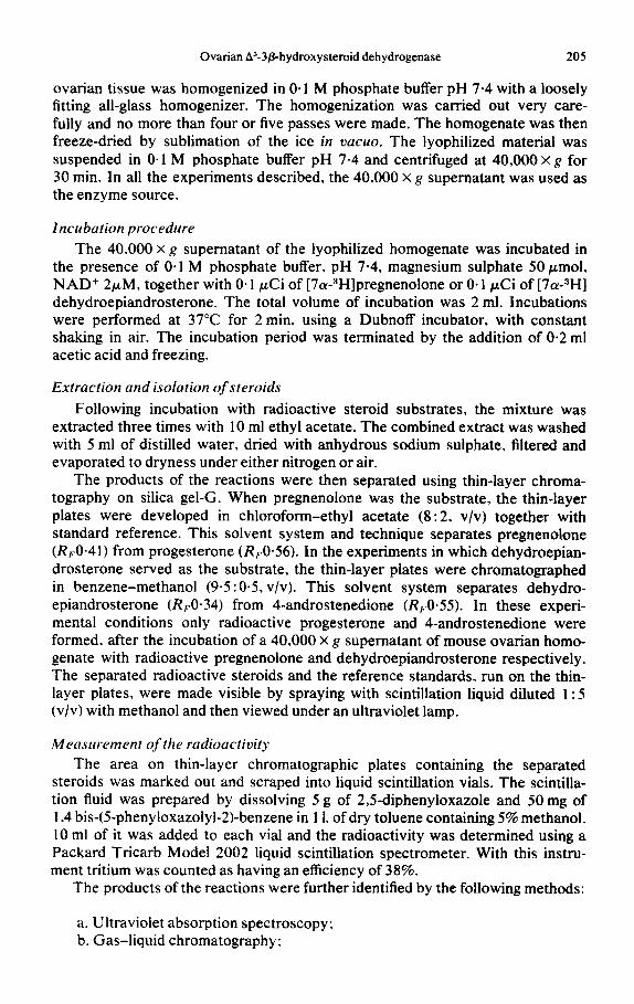
ovarian tissue was homogenized in 0.1 M phosphate buffer pH 7.4 with a loosely fitting all-glass homogenizer. The homogenization was carried out very care- fully and no more than four or five passes were made. The homogenate was then freeze-dried by sublimation of the ice in vucuo. The lyophilized material was suspended in O-1 M phosphate buffer pH 7.4 and centrifuged at 40,000 x a for 30 min. In all the experiments described, the 40,000 X g supernatant was used as the enzyme source.
Incubation procedure
The 40.000 x g supematant of the lyophilized homogenate was incubated in the presence of 0.1 M phosphate buffer, pH 7.4, magnesium sulphate 50 pmol, NAD+ 2/*M, together with 0.1 &i of [7a-“Hlpregnenolone or 0.1 @i of [7&H] dehydroepiandrosterone. The total volume of incubation was 2 ml. Incubations were performed at 37°C for 2 min. using a Dubnoff incubator, with constant shaking in air. The incubation period was terminated by the addition of 0.2 ml acetic acid and freezing.
Extraction and isolation of steroids
Following incubation with radioactive steroid substrates, the mixture was extracted three times with 10 ml ethyl acetate. The combined extract was washed with 5 ml of distilled water, dried with anhydrous sodium sulphate, filtered and evaporated to dryness under either nitrogen or air.
The products of the reactions were then separated using thin-layer chroma- tography on silica gel-G. When pregnenolone was the substrate, the thin-layer plates were developed in chloroform-ethyl acetate (8: 2, v/v) together with standard reference. This solvent system and technique separates pregnenolone (R,O-41) from progesterone (Rr0*56). In the experiments in which dehydroepian- drosterone served as the substrate, the thin-layer plates were chromatographed in benzene-methanol (9.5 : 0.5, v/v). This solvent system separates dehydro- epiandrosterone (R,O*34) from 4-androstenedione (Rp0*55). In these experi- mental conditions only radioactive progesterone and 4-androstenedione were formed, after the incubation of a 40,000 X g supematant of mouse ovarian homo- genate with radioactive pregnenolone and dehydroepiandrosterone respectively. The separated radioactive steroids and the reference standards, run on the thin- layer plates, were made visible by spraying with scintillation liquid diluted 1 : 5 (v/v) with methanol and then viewed under an ultraviolet lamp.
Measurement of the radioactivity
The area on thin-layer chromatographic plates containing the separated steroids was marked out and scraped into liquid scintillation vials. The scintilla- tion fluid was prepared by dissolving 5 g of 2,5-diphenyloxazole and 50 mg of 1.4 bis-(5-phenyloxazolyl-2)-benzene in 11. of dry toluene containing 5% methanol. 10 ml of it was added to each vial and the radioactivity was determined using a Packard Tricarb Model 2002 liquid scintillation spectrometer. With this instru- ment tritium was counted as having an efficiency of 38%.
The products of the reactions were further identified by the following methods:
a. Ultraviolet absorption spectroscopy; b. Gas-liquid chromatography:
206 S. SULIMOVICI and B. LUNENFELD
c. Mass spectrometry; d. Reduction of 4-androstenedione to testosterone: e. Recrystallization to constant specific activity.
~~tra~iulet absorption spectroscopy
The 40,000X g supernatant of mouse ovarian homogenate was separately incubated with cold pregnenolone or dehydroepiandrosterone (IO. 25. 50 and 100 kg) and NAD+ (0.1 mM). The steroids were extracted and chromatographed on silica gel plates as described above. The area corresponding to progesterone after incubation with pregnenolone and the area of 4-androstenedione following incubation with dehydroepiandrosterone was eluted from the thin-layer plates with acetone and evaporated to dryness. 2 ml of methanol was added to the dry residue and the ultraviolet absorbing material was measured in a P.M.Q. II Zeiss spectrophotometer.
Aliquots eluted from the thin-layer plates were examined by gas chromato- graphy using a Packard Model 87 1. This instrument was equipped with a flame ionization detector and had a column 1.96 m long and a 4 mm i.d. The stationary phase was S.E. 30 on IOO- 120 mesh Gas Chrom Q. The column temperature was kept at 235°C with the detector at 245°C. Samples were introduced into the column with a IO ~1 Hamilton syringe.
Mass spectrometry A Hitachi Perkin-Elmer R.M.U. 6E. mass spectrometer was employed for
mass spectrometric analysis. The sample eluted from the thin-layer plate was dissolved in acetone and introduced into the sample tube of the direct inlet system by means of a hypodermic syringe. The sample temperature was I30- 170°C and the ion source temperature was kept at 250°C. Spectra were observed at 70 eV.
Reduction of 4-androste~edia~e to testosterone
4-androstenedione was reduced to testosterone by incubation at - 15°C for 60 min in 1 ml methanol containing 100 mg anhydrous NaBH,. Distilled water (5 ml) was added to the incubation mixture and the product was extracted with ether and applied to thin-layer chromatography. The chromatography was carried out in benzene-ethyl acetate (6: 4, v/v). The testosterone was then eluted from the plates and acetylated with pyridine and acetic anhydride.
Enzymic assay
The enzymic assay was the same as previously described [ 161 and consisted of the conversion of [7a-:‘H]pregnenolone to [7a-“HIprogesterone and the con- version of [7&H]dehydroepiandrosterone to [7cu-JH]4-androstenedione. The enzymic activity. expressed as a percentage of the progesterone or 4-androstene- dione formed per mg proteinlmin. was calculated from the data obtained from the conversion of the added radioactive As-3fi-hydroxysteroid to radioactive AA-3- ketosteroid and the protein content of the enzyme solution. This assay was based on the assumption that the 40.000 X g supematant of immature mouse ovaries contained very little endogenous pregnenofone or dehydroepiandrosterone. The
Ovarian A$-3phydroxysteroid dehydrogenase 207
amounts of endogenous pregnenolone and dehydroepiandrosterone were dis- regarded, being negligible. Protein concentration was estimated by the method of Lowry ez a/.[ 171 with bovine serum albumin used as standard.
RESULTS
A. Identification of progesterone
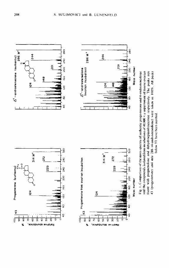
Samples of the product isolated following incubation of the 40,000 X g super- natant of ovarian homogenate with pregnenolone were eluted from the thin layer plates as described above. The ultraviolet spectrum was taken from 220-280 nm in methanol. A peak at 240 nm was found, indicating the formation of A4-3- ketosteroid, presumably progesterone. When an aliquot of this material, after elution from the thin-layer plate, was subjected to gas chromatography, the re- tention time (tR) of the substance injected, in relation to pregnenolone acetate (internal standard tR = 24 min) was identical to the relative retention time of standard progesterone (0.67). The mass spectrum of this compound was found to be similar to the mass spectrum of authentic progesterone (Fig. 1).
B. Identi@ation of I-androstenedione
The 40.000 x g supematant fluid of the homogenate of mouse ovarian tissue was incubated with cold dehydroepiandrosterone. The steroids were extracted and separated on thin-layer chromatography. The area corresponding to 4- androstenedione was eluted from the thin-layer plates and the ultraviolet spectrum taken in methanol. A peak at 240 nm due to A4-3-ketosteroid, presumably 4- androstenedione, was observed. After elution from the thin-layer plate 4-andro- stenedione was further reduced to testosterone and the latter steroid acetylated as described above. The 4-androstenedione and the acetylated testosterone were injected into the gas-liquid chromatograph, with column and condition of opera- tion the same as previously described. The retention time of the substances in- jected in relation to pregnenolone acetate (internal standard tR = 24 min) were identical to the relative retention time of standard 4-androstenedione (O-58) and testosterone acetate (O-83). The mass spectrum of the 4-androstenedione isolated from the incubation was shown to be similar to that of the authentic compound (Fig. 1.).
C. Recrystallization to constant specific activity Proof that the substances reported in the present study as [7a-3H]progesterone
and [ 7a-3H14-androstenedione were radiochemically pure is presented in Table 1. After additions of appropriate steroids, the compounds were recrystallized from four different solvents. It can be seen (Table 1) that the specific activities of all samples (crystals and mother liquors) remained constant through four crystal- lizations and that no significant fall in specific activity occurred as the result of the first crystallization.
D. Time course of the enzymic reaction
There is a linear relationship between the amount of progesterone formed and the concentration of enzyme (Fig. 2a). Analogous results were obtained when dehydroepiandrosterone was employed as substrate (Fig. 2b); however, dehydro- epiandrosterone was metabolized faster than pregnenolone. The rate of formation
100
43
so
* 80
* 8 70
c g 60
c 50
2 P
40
0 ;r
” 30
%
20
o?
IO
0
Pro
gest
eron
e (a
uthe
ntic
) $H3
20
160
200
240
280
320
4c
13
Pro
gest
eron
e fr
om
ovar
ian
incu
batio
n
1 124
I 31
4 M
+
40
80
120
160
200
240
260
320
24
0 31
4 tv
l+
1272
r A
4 -and
rost
snad
iona
(a
uthe
ntic
) 26
6 M
244
120
160
200
240
2i
A4-
andr
oste
nedi
one
(ova
rian
incu
batio
n)
286
M+
40
80
120
160
200
240
280
Mas
s nu
mbe
r M
ass
num
ber
Fig
. 1.
Com
paris
on
of t
he m
ass
spec
tra
of a
uthe
ntic
pr
oges
tero
ne
and
4-an
dros
tene
dion
e
and
the
com
poun
ds
isol
ated
fr
om
incu
batio
ns
of 4
0.00
0 x
g su
pern
atan
t of
mou
se o
varia
n
tissu
e w
ith
pre
gn
eno
lon
e an
d
deh
ydro
epia
nd
rost
ero
ne
resp
ecti
vely
. T
he
pea
ks
m/e
43
(p
rog
este
ron
e)
and
m
/e
286
t4-a
nd
rost
ened
ion
e)
wer
e ta
ken
as
IO
W.
All
pea
ks
bel
ow
5%
hav
e b
een
om
itte
d.

Ovarian ~-3~-hyd~xyste~id dehydrogenase 209
Table 1. Recrystallization of [7&H] progesterone and [7&H]4-androstenedione to constant specific activity”
[7&H] progesterone 17nJHI 4-~drosten~ione
Specific activity Specific activity (d.p.m./mg) @.p.m./mg)
Recrystal- Crystals Mother Recrystal- C~StalS Mother hzation Solvent hquor lization Solvent liquor
After After addition addition
of carrier - 3700 - of carrier 2870 - 1st Acetone1 3600 3040 1st Acetone/ 2940 2500
hexane ligroin 2nd Aqueous 3520 2920 2nd Ethyl 2820 2440
ethanol acetate 3rd Ethyl 3590 2690 3rd Aqueous 2880 2460
acetate/ ethanol hexane
4th Methanol 3660 3000 4th Benzene/ 2850 2450 hexane
*Specific activity. d.p.m.fmg, after isolation from thin-layer c~mat~phy and addition of carrier steroids fapprox. 20 mg).
I I 4
Fig. 2. The formation of progesterone from pregnenolone (af and of 4-androstenedione from dehydroepiandrosterone (b) by a 40,000 xg supematant of immature mouse ovarian tissue as a function of enzyme concentrations. The incubation mixture contained i&easing amounts of enzyme protein in 0.1 M phosphate buffer pH 7-4, 50 rmol magnesium sulphate. 2 PM NAD+, O-1 &i[7&HI pregnenolone or 0.1 yCi[7&H]- dehydroepiandrostercme. to a total volume of 2 ml. incubations were carried out in air
at 37°C for 2 min.
of 4-androstenedione was 0.657 X IO+ mol/mg proteinlmin and that of proges- terone was 0.453 X IO-” moltmg proteinlmin.
The conversion of [7~-3H]pregnenol~ne to [7a-3H]progesterone and of [7~-sH]de~yd~oepiandrosterone to r7~-3H]4-androstene~one at different con-
210 S. SULIMOVICI and B. LUNENFELD
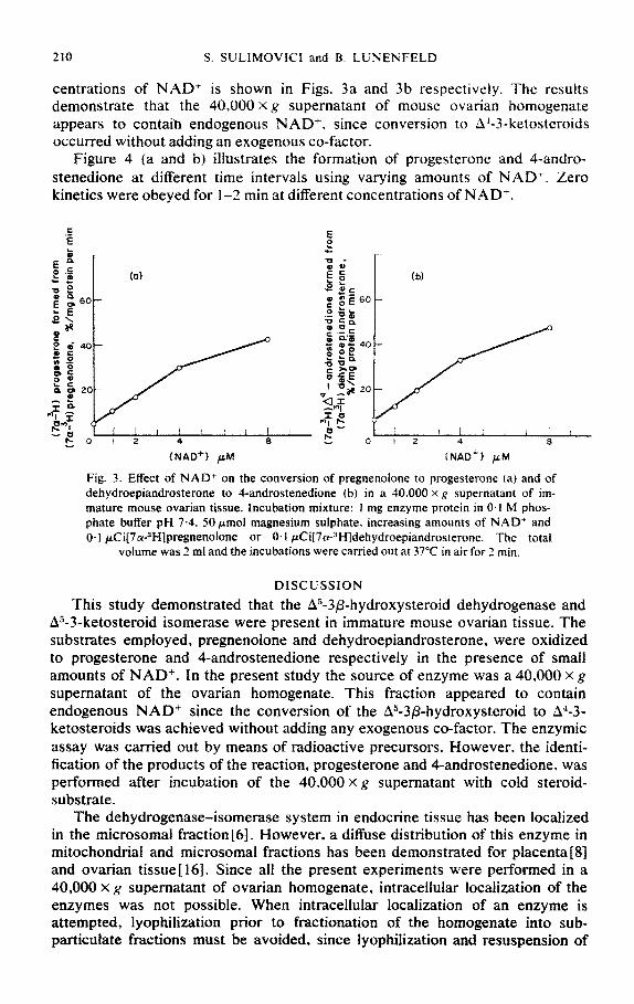
centrations of NAD+ is shown in Figs. 3a and 3b respectively. The results demonstrate that the 40,000 X g supernatant of mouse ovarian homogenate appears to contaih endogenous NAD+. since conversion to A.&-3-ketosteroids occurred without adding an exogenous co-factor.
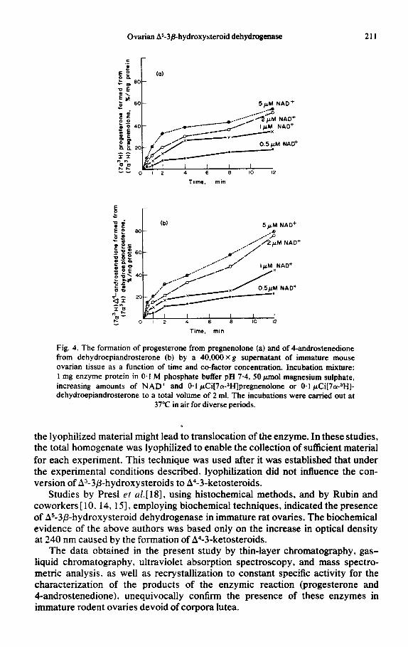
Figure 4 (a and b) illustrates the formation of progesterone and 4-andro- stenedione at different time intervals using varying amounts of NAD+. Zero kinetics were obeyed for I-2 min at different concentrations of NAD+.
(a)
Yi I! i 8 I II f I 0 I 2 4 8
(~~0’1 p4
E e
r
Fig. 3. Effect of NAD+ on the conversion of pregnenolone to progesterone la) and of dehydroepiandrosterone to 4-androstenedione (b) in a 40,000 x g supernatant of im- mature mouse ovarian tissue. Incubation mixture: 1 mg enzyme protein in 0.1 M phos- phate buffer pH 7.4. 50pmol magnesium sulphate. increasing amounts of NAD’ and O- 1 ~Ci[7~-3H]pregnenolone or 0, I ~C~~7~-3H]dehydroepjandrosterone. The total
volume was 2 ml and the incubations were carried out at 37°C in air for 2 min.
DISCUSSION
This study demonstrated that the As-3p-hydroxysteroid dehydrogenase and A5-3-ketosteroid isomerase were present in immature mouse ovarian tissue. The substrates employed, pregnenoione and dehydroepiandrosterone, were oxidized to progesterone and 4-androstenedione respectively in the presence of small amounts of NAD’. In the present study the source of enzyme was a 40.000 X g
supematant of the ovarian homogenate. This fraction appeared to contain endogenous NAD’ since the conversion of the A5-3&hydroxysteroid to AJ-3- ketosteroids was achieved without adding any exogenous co-factor. The enzymic assay was carried out by means of radioactive precursors. However. the identi- fication of the products of the reaction, progesterone and 4-androstenedione, was performed after incubation of the 40,000 x g supematant with cold steroid- substrate.
The dehydrogenase-isomerase system in endocrine tissue has been localized in the microsomal fraction [6]. However, a diffuse distribution of this enzyme in mitochond~al and microsomal fractions has been demonstrated for placenta [S] and ovarian tissuei 161. Since all the present experiments were performed in a 40,000 x g supematant of ovarian homogenate, intracellular localization of the enzymes was not possible. When intracellular localization of an enzyme is attempted, lyophilization prior to fractionation of the homogenate into sub- particulate fractions must be avoided, since lyophilization and resuspension of
Ovarian AS-3&hydroxysteroid dehydrogenase 211
I I I I I 0 I2 4 6 6 IO 12
Tame. min
5pM NAD+
60-
0 I 2 4 6 6 10 12
Time. min
Fig. 4. The formation of progesterone from pregnenolone (a) and of 4-androstenedione from dehydroepiandrosterone (b) by a 40,OOOXg supematant of immature mouse ovarian tissue as a function of time and co-factor concentration. Incubation mixture: 1 mg enzyme protein in 0.1 M phosphate buffer pH 7.4, 50 pmol magnesium sulphate, increasing amounts of NAD+ and 0.1 &i[7a-3H]pregnenolone or 0.1 &i[7c?H]- dehydroepiandrosterone to a total volume of 2 ml. The incubations were carried out at
37°C in air for diverse periods.
the lyophilized material might lead to translocation of the enzyme. In these studies, the total homogenate was lyophilized to enable the collection of sufficient material for each experiment. This technique was used after it was established that under the experimental conditions described. lyophilization did not influence the con- version of As-3P-hydroxysteroids to A4-3-ketosteroids.
Studies by Presl et a/.[ 181, using histochemical methods, and by Rubin and coworkers [ 10. 14, 151, employing biochemical techniques, indicated the presence of As-3@-hydroxysteroid dehydrogenase in immature rat ovaries. The biochemical evidence of the above authors was based only on the increase in optical density at 240 nm caused by the formation of A4-3-ketosteroids.
The data obtained in the present study by thin-layer chromatography, gas- liquid chromatography, ultraviolet absorption spectroscopy, and mass spectro- metric analysis. as well as recrystallization to constant specific activity for the characterization of the products of the enzymic reaction (progesterone and 4-androstenedione), unequivocally confirm the presence of these enzymes in immature rodent ovaries devoid of corpora lutea.
‘12 S. SULIMOVICI and 3. LUNENFELD
Since no attempt was made to separate the granulosa. thecal and interstitial cells of the ovary. prior to homogenization, the intercellular localization of these enzymes was not studied. Although this tissue contained no corpora lutea. it could be claimed that the ~-3~-hydroxysteroid dehydrogenase activity might be derived from atretic follicles which may be present in smaller or greater num- bers in 21-day-old mice. Histochemical evidence in mouse ovarian tissue[5] supports the view that the main source of A”-3/3-hydroxysteroid dehydrogenase in the immature mouse is atretic follicles or follicles doomed to undergo atresia. Electron microscopic studies demonstrated that organetles of interstitial cells of immature rodents are indicative of steroid secretory function [ 191. ~istochemica1 studies in immature mice showed the appearance of formazan granules in inter- stitial cells after incubation with pregnenolone or dehydroepiandrosterone [20]. Therefore, the interstitial cells of immature rodents seem also to contain A”-3p- hydroxysteroid dehydrogenase activity.
Although no direct evidence on secretion of biologically active steroids is available, indirect evidence seems to indicate such processes in infancy. At the age of 14 days. uterine weights of animals spayed at the age of 3 days were dis- tinctly lower than normal [2 11. Indication that ovarian steroids may play a role in gonadotropin feed-back mechanism in infancy can be derived from studies with animals placed in parabiosis, where it was shown that the infantile hypo- physis following removal of the ovaries increased secretion of gonadotropins
WI.
ACKNOWLEDGEMENTS
Our thanks are due to Dr. B. Sklarz of the Chemistry Department, Bar-flan University. for his help in performing the mass spectrometric analysis. The technical assistance of Mrs. V. Rotary is gratefully acknowledged. This research study was supported by Ford Foundation Grant No. 67-470.
REFERENCES
I. L. T. Samuels: In Metubolic Parhwc~ys (Edited by D. M. Greenberg). Academic Press. New York and London. Vol. I ( 1960) p. 43 1.
2. L. W. Wattenberg: J. ~~sro~lz~tn. C~rochrm. 6 f 1958122.5. 3. H. Lewy. H. W. Deane and B. L. Rubin: Endocrinology 65 ( 1959) 932. 4. B. Goldberg, G. E. Jones and D. A. Turner: Am. J. Ohsret. C.ww. 86 ( 19631349. 5. M. M. Ferguson: J. enhcr. 32 (19651365. 6. K. F. Beyer and L. T. Samuels: J. hiol. C’lwm. 219 ( 1956) 69. 7. J. Kowal. E. Forchielli and R. I. Dorfman: Strrc&& 3 ( 1964153 1. 8. S. S. Koide and M. T. Torres: ~i~)~/zjrn. biup~?ys. Acfcz 105 t 1965) 1 15. 9. S. G. Cheatum and J. C. Warren: Biocizim. biophys. Acfa 122 (1966) 1.
IO. B. L. Rubin. H. W. Deane, J. A. Hamilton and E. C. Driks: Enducrinolo,q.v 72 ( 19631924. I I. B. L. Rubin and H. W. Deane: Endocrinoloev 76 ( 1965) 382. IL K. J. Ryan and Z. Petro: J. Clin. Endocr. Met. 26 ( 1966) 46. 13. 6. L. Lobel. R. M. Rosenbaum and H. W. Deane: Endoocrinology 68 ( 196 I) 132.
14. B. L. Rubin. J. A. Hamilton. T. J. Karlson and R. I. Tufaro: Endorrinthp.v 77 ( 1965) 909. IS. B. L. Rubin: ~n~~~~~j~ff~~~,~_v 83 f 1968) 626. I6 S. Sulimovici and G. S. Boyd: cur. J. B&hem. 7 ( 1969) 549. 17. 0. H. Lowry. N. J. Rosebrough. A. L. Farr and R. J. Randall: J. bid. Clam. 193 ( I95 I) 165. IS. J. Presl. J. Jirdsek. J. Horsky and M. Henzl: J. Endocr. 31( 1965) 293. 19. H E. Stegner: Workslrnp Mrctin,q on Deur/opmru/ o! IIIC 0w1ry in fnjirnc~~. Birmingham 1969
(in press). 20. E. Rachamim: M.Sc.-Thesis. Bar-llan University. lswel t 1970).
Ovarian k5-3/I-hydroxysteroid dehydrogenase
21. D.Price:Anar.Rec.97(1947)519. 22. T. Martins and A. Rocha: Endocrinology 15 ( 193 1) 42 1. 23. S. Sulimovici and B. Lunenfeld: Hormorte und Merubolic Research (197 1) (in press).
213


![Evidence for Endogenous Neurosteroid Production in the ... · tetrahydroDOC in the brain by the enzymes 5α reductase and 3α hydroxysteroid dehydrogenase (HSD) [5,12]. Cholesterol](https://img.pdfslide.net/doc/110x75/5fd9b663408dab2eba43865a/evidence-for-endogenous-neurosteroid-production-in-the-tetrahydrodoc-in-the.jpg)