Embed Size (px)
Citation preview
Study of a Special System of Transportation in the Epicarps of Urticdceae"
By EGIL RAMSTAD
The epidermis of the mature fruits of most Urticaceae is filled with mucilage. The mucilage develops from the bottom of the cells of the epicarpic layer and thus isolates tke layer from the rest of the pericar . Yet the individual cells of this layer still are able to maintain communication witg the subjacent layers of the pericarp through a special system of communication. This system consists of cells, named intercalary cells, distributed at even interkals throughout the pericarp. The intercalary cell is devoid of mucilage, and every mucilage cell touches at one point or other, one and only one, intercalary cell. The exchange of metabolites and the transport of con- structive material from the mesocarp for the formation of the mucilage take place
through the intercalary cell.
N A PREVIOUS paper (I) the author has shown I the existence of regularly distributed, non- mucilaginiferous cells in the mucilaginous epicarps of many species of the Nettle family. These cells have been overlooked by previous investigators who have studied the structure of the pericarps of the Nettle family. Since, in a sense, these structures are unique in the plant world, additional and detailed information about them is reported in this paper.
DESCRIPTION
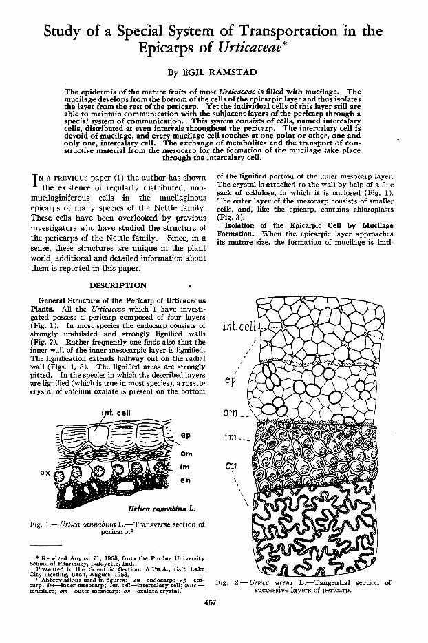
General Structure of the Pericarp of Urticaceous Plants.-All the Urticaceae which I have investi- gated possess a pericarp composed of four layers (Fig. 1). In most species the endocarp consists of strongly undulated and strongly lignified walls (Fig. 2). Rather frequently one finds also that the inner wall of the inner mesocarpic layer is lignified. The lignification extends halfway out on the radial wall (Figs. 1, 3). The lignified areas are strongly pitted. In the species in which the described layers are lignified (which is true in most species), a rosette crystal of calcium oxalate is present on the bottom
ox
int. cell
eP
OWl
im
en
rtica cannabina 1.
Fig. 1.- Urtica cannabina L.-Transverse section of pericarp.'
* Received August 21, 1953, from the Purdue University School of Pharmacy, Lafayette, Ind.
Presented to the Scientific Section, A.Pm.A., Salt Lake City meeting, Utah, August, 1953.
1 Abbreviations used in figures: en-endocarp; cp-epi- carp; im-inner mesocarp; in:. cell-intercalary cell; muc.- mucilage; om-vuter mesocarp; ox-oxalate crystal.
of the lignified portion of the inner mesocarp layer. The crystal is attached t o the wall by help of a fine sack of cellulose, in which it is enclosed (Fig. 1). The outer layer of the mesocarp consists of smaller cells, and, like the epicarp, contains chloroplasts (Fig. 3).
Isolation of the Epicarpic Cell by Mucilage Formation.-When the epicarpic layer approaches its mature size, the formation of mucilage is initi-
int. c
/ I
I
eP
om -
im.
en \ \ \
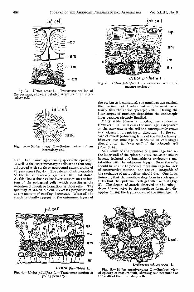
Fig. 2.- Urtica w e n s L.-Tangential section of successive layers of pericarp.
457
158 JOURNAL OF THE AMERICAN PHARMACEUTICAL ASSOCIATION Vol. XLIII, NO. 8
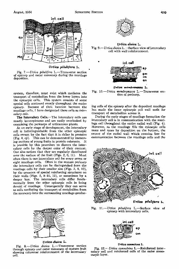
int. cell
---ep
Fig. 3a.- Urtica urens L.-Transverse section of the pericarp, showing detailed structure of an inter- calary cell.
,Ii,. int.cel1
Fig. 3b.-Urtica wens L.-Surface view of an intercalary cell.
ated. In the mucilage-forming species the epicarpic as well as the outer mesocarpic cells are a t that stage all gorged with single or compound starch grains of varying sizes (Fig. 4). The calcium oxalate crystals of the inner mesocarp layer are then laid down. At this time a fine hyaline layer appears on the bot- tom of the epidermal cells, which constitutes the initiation of mucilage formation by those cells. The quantity of starch present decreases proportionally as the amount of mucilage increases. When all the starch originally present in the outermost layers of
in t. ce 11
Urticu pilulifera L. Fig. 4.- Urtica pilulifera L.7Transverse section of
‘young pericarp.
int. cell
Fig. 5.- Urtica pilulifera L.-Transverse section of mature pericarp.
the pericarps is consumed, the mucilage has reached the maximum of development and, in most cases, nearly fills the entire epicarpic cells. During the later stages of mucilage deposition the endocarpic layer becomes strongly lignified.
Many seeds possess a mucilaginous epidermis However, in all such cases the mucilage is deposited on the outer wall of the cell and consequently grows in thickness in a centripetal direction. In the epi- c ~ p of mucilage-forming fruits of the Nettle family, however, the mucilage is deposited in centrifugal direction on the inner wall of the epicarpic cell (Figs. 3, 4).
As a result of the presence of a mucilage bed on the inner wall of the epicarpic cells, the latter should become isolated and incapable of exchanging me- tabolites with the subjacent layers. Soon the cells should be unable to produce more mucilage for lack of constructive material, and the cell, incapable of the exchange of metabolites, should die. One finds, however, that the mucilage does form in such quan- tities that the epidermal cells get filled with it (Fig. 3 ) . The depots of starch observed in the subepi- dermal layer prior to the mucilage formation dis- appear during the laying-down of the mucilage. A
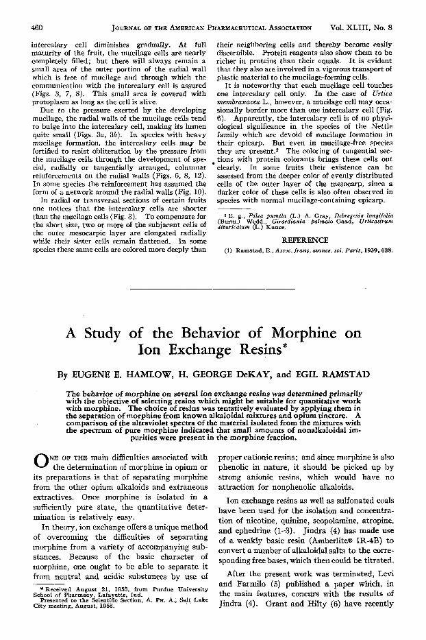
Fig. 6.- Urtica membranacea L.-Surface view of epicarp of mature fruit, showing reinforcement of the walls of the intercalary cells.
August, 1954 SCIENTIFIC EDITION 469
vrtica dioica t. Fig. 9.-Urtica dioica L.-Surface view of intercalary
cell with wall reinforcement.
int. cell
Fig. 7.-Urtica pilulifera L.-Transverse section of epicarp and outer mesocarp during the mucilage deposition.
eP Urtica pilulifera L. . 7: en
system, therefore, must exist which mediates the transport of metabolites from the lower layers into the epicarpic cells. This system consists of some special cells scattered evenly throughout the entire epicarp. Because of their location between the mucilage cells, I have designated these cells as inter- calary cells.
The Intercalary Cells.-The intercalary cells are mostly ipconspicuous and are easily overlooked by examining the pericarps of urticaceous plants.
At an early stage of development, the intercalary cell is indistinguishable from the other epicarpic cells except by the fact that it is richer in proteins (Fig. 4, ep). This can be demonstrated by immers- ing sections of young fruits in protein colorants. It is possible by this procedure to discern the inter- calary cells by the deeper color of their content. One also notices that they are regularly distributed over the surface of the fruit (Figs. 2, 6, 11). Most often there is one intercalary cell for every seven or eight mucilage cells. Often in the mature pericarp the intercalary cells can be distinguished from the mucilage cells by their smaller size (Figs. 3, 8, lo}, by the presence of special reinforcing structures on their walls (Figs. 5, 8-10, 12), or sometimes by a deeper hue. The intercalary cells differ funda- mentally from the other epicarpic cells in being devoid of mucilage. Consequently they can serve as cells mediating the transport of metabolites from the mesocarp into the surrounding mucilage-produc-
Urtica dioica L. Fig. 8.- Urtica dioica L.-Transverse section
through epicarp and outer mesocarp of mature fruit showinP: columnar reinforcement of the intercalary
Urtfccr mem6rrmeeea L. Fig. 10.- Urtica membranacea L.-Transverse sec-
tion of pericarp.
ing cells of the epicarp after the deposited mucilage has made the inner epicarpic cell wall unfit for transport of metabolites across it.
During the early stages of mucilage formation the intercalary cell is in communication with the muci- lage cell throughout the entire radial wall (Fig. 4). However, as the mucilage fills the epicarpic cells more and more by deposition on the bottom, the extent of the radial wall which remains free for communication between the mucilage cells and the
,int. cell
Fig. ll.-Urtica pilulifera L.-Surface view of epicarp with intercalary cells.
. int. cell
Udica cannabina L. Fig. 12.- Urtica cannabina L.-Reinforced inter-
calary cell and reinforced cells of the outer meso- - cell. carp& layer.
460 JOURNAL OF THE AMERICAN PHARMACEUTICAL ASSOCIATION VOl. XLIII. NO. 8
intercalary cell diminishes gradually. At full maturity of the fruit, the mucilage cells are nearly completely filled; but there will always remain a small area of the outer portion of the radial wall which is free of mucilage and through which the communication with the intercalary cell is assured (Figs. 3, 7, 8). This small area is covered with protoplasm as long as the cell is alive.
Due to the pressure exerted by the developing mucilage, the radial walls of the mucilage cells tend to bulge into the intercalary cell, making its lumen quite small (Figs. 3a, 3b) . In species with heavy mucilage formation, the intercalary cells may be fortified to resist obliteration by the pressure from the mucilage cells through the development of spe- cial, radially or tangentially arranged, columnar reinforcements on the radial walls (Figs. 6, 8, 12). In some species the reinforcement has assumed the form of a network around the radial walls (Fig. 10).
In radial or transversal sections of certain fruits one notices that the intercalary cells are shorter than the mucilage cells (Fig. 3 ) . To compensate for the short size, two or more of the subjacent cells of the outer mesocarpic layer are elongated radially while their sister cells remain flattened. In some species these same cells are colored more deeply than
their neighboring cells and thereby become easily discernible. Protein reagents also show them to be richer in proteins than their equals. It is evident that they also are involved in a vigorous transport of plastic material to the mucilage-forming cells.
It is noteworthy that each mucilage cell touches one intercalary cell only. In the case of Urtica rnembranacea L., however, a mucilage cell may occa- sionally border more than one intercalary cell (Fig. 6). Apparently, the intercalary cell is of no physi- ological significance in the species of the Nettle family which are devoid of mucilage formation in their epicarp. But even in mucilage-free species they are present.2 The coloring of tangential sec- tions with protein colorants brings these cells out
In some fruits their existence can be assessed from the deeper color of evenly distributed cells of the outer layer of the mesocarp, since a darker color of these cells is also often observed in species with normal mucilage-containing epicarp.
clearly.
2 E. g., Pilea pumila (L.) A. Gray, Debregesia longifolia (Burm.) Wedd., Girardiania galmato Gaad, Urticastrum diuaricatum (L.) Kunze.
REFERENCE (1) Ramstad, E., Assoc. franp. auancc. sci. Paris, 1939,638.
A Study of the Behavior of Morphine Ion Exchange Resins*
on
By EUGENE E. HAMLOW, H. GEORGE DeKAY, and EGIL RAMSTAD
The behavior of morphine on several ion exchange resins was determined primarily with the objective of selecting resins which might be suitable for quantitative work with morphine. The choice of resins was tentatively evaluated by applying them in the separation of morphine from known alkaloidal mixtures and opium tincture. A comparison of the ultraviolet spectra of the material isolated from the mixtures with the spectrum of pure morphine indicated that small amounts of nonalkaloidal im-
purities were present in the morphine fraction.
NE OF THE main difficulties associated with 0 the determination of morphine in opium or its preparations is that of separating morphine from the other opium alkaloids and extraneous extractives. Once morphine is isolated in a sufficiently pure state, the quantitative deter- mination is relatively easy.
In theory, ion exchange offers a unique method of overcoming the difficulties of separating morphine from a variety of accompanying sub- stances. Because of the basic character of morphine, one ought to be able to separate it from neutral and acidic substances by use of
* Received August 21, 1953, from Purdue University
Presented to the Scientific Section, A. PH. A., Salt Lake School of Pbarmaey, Lafayette. Ind.
City meeting, August, 1953.
proper cationic resins; and since morphine is also phenolic in nature, it should be picked up by strong anionic resins, which would have no attraction for nonphenolic alkaloids.
Ion exchange resins as well as sulfonated coals have been used for the isolation and concentra- tion of nicotine, quinine, scopolamine, atropine, and ephedrine (1-3). Jindra (4) has made use of a weakly basic resin (AmberliteB IR-4B) to convert a number of alkaloidal salts to the corre- sponding free bases, which then could be titrated.
After the present work was terminated, Levi and Farmilo (5) published a paper which, in the main features, concurs with the results of Jindra (4). Grant and Hilty (6) have recently


![A2-01 Polyploidy in Pilea brevicornuta Hayata (Urticaceae ...Proceedings of 3rd International Conference on Environmental Aspects of Bangladesh [ICEAB 2012]; October 13~14, 2012 45](https://img.pdfslide.net/doc/110x75/61123ba8bd6b9e16975146f1/a2-01-polyploidy-in-pilea-brevicornuta-hayata-urticaceae-proceedings-of-3rd.jpg)