Embed Size (px)
Citation preview
Study of Aging in Oil Paintings by 1D and 2D NMRSpectroscopy
Apostolos Spyros†,* and Demetrios Anglos‡
NMR Laboratory, Department of Chemistry, University of Crete, Knossos Avenue, 71409 Heraklion, Crete, Greece, andInstitute of Electronic Structure and Laser, Foundation for Research and TechnologysHellas (IESL-FORTH),P.O. Box 1527, 71110 Heraklion, Crete, Greece
Nuclear magnetic resonance spectroscopy is proposed asan efficient analytical tool in the study of painted artworks.The binding medium from two original oil paintings, datedfrom the early 20th and the late 17th century, was studiedvia high-resolution 1D and 2D NMR, establishing theadvanced state of hydrolysis and oxidation of the oil paint.Studies of the solvent-extractable component from modelsamples of various drying oils, raw oil paints, and agedoil paints allowed the definition of several markers basedon the integral ratios of various chemical species presentin the 1H and 13C NMR spectra. These markers aresensitive to hydrolytic and oxidative processes that reflectthe extent of aging in oil paintings. The rapidity, simplicity,and nondestructive nature of the proposed analytical NMRmethodology represents a great advantage, since theusually minute sample quantities available from originalartwork can be subsequently analyzed further by otheranalytical techniques, if necessary.
The study and characterization of materials in painted worksof art represents a challenge for the analyst, who is called toidentity the chemical constituents of heterogeneous matrixes,composed of both inorganic and organic substances, to gainknowledge on the paint materials and techniques employed orassess the state of preservation of a painting.1 In particular, organicmaterials used in paintings, such as for example terpenoid resinsor drying oils, employed as varnishes and binders, respectively,are natural products (plant or tree extracts) and thus multicom-ponent mixtures whose composition is chemically rich andmoreover subject to continuous changes over time because ofongoing degradation processes including oxidation, polymeriza-tion, and hydrolysis.2,3 Obviously, powerful analytical techniquesand methodologies are required to characterize such complexmaterials, which are typically available in very low quantities dueto the strict limitations imposed on sampling works of art. In this
respect, gas chromatography/mass spectrometry (GC/MS) incombination with various off-line4-6 or on-line7 derivatization pro-cedures, direct temperature resolved mass spectrometry,8 andFT-IR spectrometry9 has been used extensively to study thecomposition and degradation of organic paint binders.1 Staticsecondary ion mass spectrometry10 and FT-IR microscopic imag-ing11 have been recently used for the examination of paint crosssections, providing an insight to the stratigraphy of the paint byidentifying the different pigments and the binding medium.
Despite its established and prominent role in lipids research,12-14
NMR spectroscopy has been restricted in terms of applicationsto studies of the chemical drying of oils15,16 and alkyd resins,17-19
while the direct analysis of painted artwork has not received anyattention. Recently, the introduction of gradient 2D NMR tech-niques has enabled the thorough characterization of complexorganic mixtures, while in parallel, detection limits have beenreduced significantly. Thus, we were prompted to examine theanalytical capabilities of NMR spectroscopy in the study of originalpainted works of art and in this paper present results from theanalysis of binding media in oil paints.
A paint consists of pigments, in the form of a fine powder,dispersed in a suitable matrix, which normally is called the bindingmedium, and can be of proteinaceous, oil, or synthetic polymer
* Corresponding author: (e-mail) [email protected]; (tel) +30 2810393685; (fax) +30 2810 393601.
† University of Crete.‡ IESL-FORTH.
(1) Zadrozna, I.; Polec-Pawlak, K.; Gluch, I.; Ackacha, M. A.; Mojski, M.;Witowska-Jarosz, J.; Jarosz, M. J. Sep. Sci. 2003, 26, 996-1004.
(2) Mills, J. S.; White, R. The Organic Chemistry of Museum Objects, 2nd ed.;Butterworh Heinemann, Oxford, 1994.
(3) Frankel, E. N. Lipid Oxidation; The Oily Press: Dundee, 1998.
(4) Colombini, M. P.; Modugno, F.; Menicagli, E.; Fuoco, R.; Giacomelli, A.Microchem. J. 2000, 67, 291-300.
(5) Colombini, M. P.; Modugno, F.; Fuoco, R.; Tognazzi, A. Microchem. J. 2002,73, 175-185.
(6) van den Berg, J. D. J.; van den Berg, K. J.; Boon, J. J. Prog. Org. Coat.2001, 41, 143-155.
(7) van den Berg, J. D. J.; van den Berg, K. J.; Boon, J. J. J. Chromatogr., A2002, 950, 195-211 and references therein.
(8) Boon, J. J.; van den Berg, K. J.; Pureveen, J.; van der Doelen, G. A.; Groen,K. M.; van Och, J.; van Grevenstein, A. Proceedings of the 43rd ASMSConference on Mass Spectrometry and Allied Topics, Atlanta, 1995; p 745.
(9) Carbo, M. T. D.; Reig, F. B.; Adelantado, J. V. G.; Martinez V. P. Anal.Chim. Acta 1996, 330, 207-215.
(10) Keune, K.; Boon, J. J. Anal. Chem. 2004, 76, 1374-1385.(11) van der Weerd, J.; Brammer, H.; Boon, J. J.; Heeren, R. M. A. Appl. Spectrosc.
2002, 56, 275-283(12) Marcel, S. F.; Jie, L. K.; Mustafa, J. Lipids 1997, 32, 1019-1034.(13) Silwood, C. J. L.; Grootveld, M. Lipids 1999, 34, 741-756.(14) Guillen, M. D.; Ruiz, A. Trends Food Sci. Technol. 2001, 12, 328-338.(15) Marshall, G. L. Eur. Polym. J. 1986, 22, 231-241.(16) Marshall, M. J. Oil Color. Chem. Assoc. 1983, 66, 285-293.(17) Muizebelt, W. J.; Hubert, J. C.; Venderbosch, R. A. M. Prog. Org. Coat.
1994, 24, 263-279.(18) Muizebelt, W. J.; Donkerbroek, J. J.; Nielen, M. W. F.; Hussem, J. B.;
Biemond, M. E. F.; Klaasen, R. P.; Zabel, K. H. J. Coat. Technol. 1998, 70,83-93.
(19) Spyros, A. J. Appl. Polym. Sci. 2003, 88, 1881-1888.
Anal. Chem. 2004, 76, 4929-4936
10.1021/ac049350k CCC: $27.50 © 2004 American Chemical Society Analytical Chemistry, Vol. 76, No. 17, September 1, 2004 4929Published on Web 07/29/2004
origin.1,2 Linseed, poppyseed, and walnut oils have traditionallyfound most use in western European painting practice as thebinding media of oil paints. Cross-polymerization of the triglycerideunsaturated fatty acids during drying solidifies the paint, therebytrapping and stabilizing the pigments. A varnish coating is appliedon the paint, functioning as a transparent, protective layer thatenhances color and gloss.2 Storage and cleaning history, pastrestoration attempts, and the original materials used by the artistare all important factors that need to be understood for a properanalysis of an oil painting. The “mobile phase” of an oil paintsample is easily separated by solvent extraction from the insoluble,highly cross-linked “stationary phase”and the solid pigmentgrains.6 It consists of tri-, di-, and monoglycerides of fatty acids,free fatty acids, and lipid oxidation products such as diacids andhydroxy acids.6,7,20-22 The composition of the solvent-extractablefraction can provide information regarding the levels of hydrolysis6
and oxidation that have occurred during aging of an oil paintingand can be of great interest to art conservators, who often usesolvents during cleaning and restoration of painted artworks.20,23
In this report, we present the study of the “mobile phase” fromtwo oil paintings by high-resolution NMR spectroscopy, using ananalytical protocol that contains no chemical workup and utilizessimply sonication in a deuterated NMR solvent for the directextraction of organic material from the painting sample. Thecomposition of the solvent-extractable fraction is directly deter-mined by NMR spectroscopy and will be used to provide insightinto the extent of hydrolysis and oxidation processes during agingof the painted works of art. A systematic study of drying oils, theaging of drying oil model films, artists’ oil paints, and aged oilpaints using the same analytical protocol was carried out beforethe analysis of the two original paintings, to afford a betterunderstanding of the aging procedure of oil paintings. Gradient2D NMR spectroscopy was used to assign the peaks observed inthe 1H and 13C 1D NMR spectra of the paint extracts. The mainadvantage of NMR spectroscopy over GC/MS analytical methodsinvolving derivatization procedures that have been used to studysolvent extracts of paints4-6 is its nondestructive character.Complications that may arise from incomplete derivatizationreactions or side products during chemical workup are avoided,and the whole of the paint sample can be reclaimed and madeavailable for further analyses upon completion of the NMRexperiments, if required. The ability to apply a series of differentanalytical methodologies is critical, since the amount of chemicalinformation obtainable from limited quantities of original materialfrom works of art is thus maximized.
EXPERIMENTAL SECTIONDeuterated acetone-d6 and methanol-d4 were obtained from
Aldrich Chemical Co., Inc. Films of ∼0.1-mm thickness wereprepared by coating glass slides with linseed oil (LO), boiledlinseed oil (b-LO), and poppy seed oil (PO). These model filmswere kept under room temperature conditions and monitored byNMR spectroscopy over a period of five years. Model oil paint
samples in the form of thin coatings were investigated includingtitanium white (TW), lamp black (LB), cadmium red (CR), andlead white (LW). TW and LB were commercially available paints(Rowney Georgian) while CR and LW were made in the laboratoryby mixing the corresponding pigment in powder form with LO.These samples were naturally aged in the laboratory over a periodof five years.
Two paintings from a private collection were investigated(Figure S-6 in Supporting Information): the first one, Portrait ofYoung Man (oil on canvas) by S. Vandoros, a modern Greekpainter, was dated to the early 20th century;24 the second, TheDuke (oil on canvas), by an unknown artist, was dated to the late17th century.24 Samples from the two original paintings werecollected by carefully removing a small quantity (∼10 mg) of paintfrom the side of the painted canvas to avoid intervention with thetop surface of the painting and minimize sampling of varnishmaterial. For all samples studied in this report, the bindingmaterial was dissolved in 0.6 mL of acetone-d6 and extracted for30 min in an ultrasonic bath. The solvent was then filtered throughglass wool directly into a 5-mm NMR tube.
1H and 13C NMR 1D spectra were obtained on either a BrukerMSL-300 or a Bruker AMX-500 spectrometer using standardinstrument software and pulse sequences,25 at a probe temperatureof 26 °C. Quantitative 13C NMR spectra were acquired using low(30°) flip angle and a long relaxation delay (10 s). For the 13CNMR spectra, a line broadening of 1 Hz and drift correction wereapplied prior to Fourier transformation. Polynomial fourth-orderbaseline correction was performed before manual integration ofall NMR spectra. Chemical shifts in acetone-d6 are reported relativeto internal TMS. 1H-1H homonuclear gradient COSY 2D NMRspectra19,25 were obtained using 256 increments of 1K data points,16 scans, and 4 dummy scans with a recycle delay of 1 s. 1H-13Cheteronuclear gradient multiple quantum correlation (gHMQC)and multiple bond correlation (gHMBC) 2D NMR spectra wereobtained using 128 increments of 1K data points, 16 scans, and 4dummy scans with a recycle delay of 1 s. The gHMQC experimentwas optimized for one-bond 1H-13C couplings of 140 Hz by settingthe evolution delay to 3 ms. The gHMBC experiment used anevolution delay of 60 ms optimized for long-range 1H-13CJ-couplings of ∼8 Hz.25 Before Fourier transformation, all 2D datasets were zero-filled to a 1K × 1K matrix, and a square-sinusoidalwindow function was used for processing. Details of the 31P NMRprotocol used for the quantification of diglycerides (DG), monoglyc-erides (MG) and free fatty acids (FA) in LO and b-LO can befound elsewhere.26,27
RESULTS AND DISCUSSIONThe NMR spectra of filtered acetone extracts from an oil paint,
in the absence of a varnish, are expected to contain mainly signalsfrom the oleaginous binding medium. Figure 1 presents the 1Hand 13C NMR spectra of the acetone-d6 extract of fresh lamp blackoil color and a five-year-old paint film of the same material.Although the paint contains the pigment component made of soot
(20) White, R.; Roy, A. Stud. Conserv. 1998, 43, 159-176.(21) Tumosa, C. S.; Millard, J.; Erhardt, D.; Mecklenburg, M. F. In Preprints of
the 12th Triennial ICOM CC Meeting; Bridgland J., Ed.; Lyon, 1999; p 347.(22) Sutherland, K.; Shibayama, N. In Preprints of the 12th Triennial ICOM CC
Meeting; Bridgland J., Ed.; Lyon, 1999; p 341.(23) Sutherland, K. Stud. Conserv. 2000, 45, 54-62.
(24) Private communication, Dr. M. Doulgeridis, National Gallery, Athens, June2004.
(25) Braun, S.; Kalinowski, H.-O. 150 & More Basic NMR Experiments: A PracticalCourse, 2nd exp. ed.; Wiley-WCH, Weinheim, 1998.
(26) Spyros, A.; Dais, P. J. Agric. Food. Chem. 2000, 48, 802-805.(27) Vigli, G.; Fillipidis, A.; Spyros, A.; Dais, P. J. Agric. Food. Chem. 2003, 51,
5715-5722.
4930 Analytical Chemistry, Vol. 76, No. 17, September 1, 2004
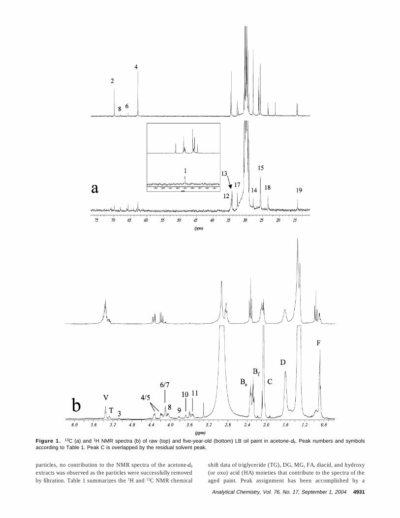
particles, no contribution to the NMR spectra of the acetone-d6
extracts was observed as the particles were successfully removedby filtration. Table 1 summarizes the 1H and 13C NMR chemical
shift data of triglyceride (TG), DG, MG, FA, diacid, and hydroxy(or oxo) acid (HA) moieties that contribute to the spectra of theaged paint. Peak assignment has been accomplished by a
Figure 1. 13C (a) and 1H NMR spectra (b) of raw (top) and five-year-old (bottom) LB oil paint in acetone-d6. Peak numbers and symbolsaccording to Table 1. Peak C is overlapped by the residual solvent peak.
Analytical Chemistry, Vol. 76, No. 17, September 1, 2004 4931
combination of 13C-DEPT, gradient 2D NMR spectroscopy (FigureS-1, gCOSY; Figure S-2, gHMQC), and literature data whereavailable.12-14 The chemical shift data for TG and DG are in goodagreement with literature data in a less polar solvent (CDCl3).28
The chemical drying of the paint by cross-linking is evident inthe reduction of intensity of all peaks originating from linolenicand linoleic acid moeities.28 The iodine value (IV), a measure ofvinyl bond abundance in a given lipid mixture, can be easilycalculated from the proton signal integrals of peaks V and F.27
The intensity of peak F in the 1H NMR spectra does not dependon the extent of TG hydrolysis or diacid formation, (the numberof fatty acid end-methyl protons remains unchanged during theseprocesses)6 and thus can be used as a normalization factor forother peak integrals. Signals originating from various (oligomeric)glycerides, TG, 1,2-DG, 1,3-DG, and 1-MG, are well resolved inthe region δ 61-73 (13C, Figure 1a) and 3.5-5.25 (1H, Figure 1b)of the paint film, while the fresh paint contains mainly TG andvery few 1,3-DG, as expected.27 Through NMR signal integration,the molar ratio of glycerides (TG + DG + MG ) 1) can becalculated from both 1H and 13C NMR spectra. Thus, quantitativeinformation on the extent of hydrolytic processes that have takenplace in the oil paint during aging is obtained. The chemical shiftof the methylene CH2-COOH group of fatty moieties produces asignal (peaks Bf, 13 in Figure 1) separate from that of esterifiedCH2-COOR groups (peaks Be, 12 in Figure 1) in both 1H and 13CNMR spectra, as verified by gHMBC 2D NMR spectroscopy(Figure S-3 in Supporting Information). This provides an excellentmeans to quantify the ratio of free-to-total carboxyl groups in oilpaint samples as Bf/B by 1H and 13C NMR spectral integration. Itis worth noting that the direct use of quaternary carbon signalssuch as those of the carbonyl groups for quantitative integrationis not experimentally realistic.25 The ratio Bf/B does not dependsolely on glyceride hydrolysis but is also affected by the extentof fatty acid chain scission that leads to the formation of C8-C9
diacids in oil paints,29,30 since diacids contribute with two freecarboxylic groups per molecule to Bf. Other signals in the 1H NMRspectrum of the aged paint also depend on diacid formation andcan be used to extract information on the extent of this procedure.The integral ratio Di/FA ) (3B - 1)/8F represents the ratio ofdiacids/fatty acids in the paint film extracts (see Appendix) andhas a zero value for raw paints in which oxidative cross-linkinghas not been initiated. This ratio is expected to increase duringaging because of diacid formation; however, diacids are them-selves susceptible to further degradation to lower molecularweight products and volatiles. After subtracting the 1,3-DGmethine proton contribution, signal 8 in the 1H NMR spectrumcan be used to evaluate the quantities of hydroxy acids presentin the acetone extracts of the film samples, by normalizing itsintegral to that of the fatty acid methyl groups F, defining HA/FA ) 3(H - 8)/F. Finally, the amount of triglyceride moieties inthe extracts can be estimated by normalizing the integral of signalT (1H NMR in Figure 1b) to F as TG/FA ) 9T/F.
Drying Oils. To evaluate the applicability of the aboveanalytical NMR approach to the study of solvent extracts of oilpaintings, we first examined the cross-linking and film formationof some pure drying oils, which are the simplest model analoguesof paints. Stand oils are processed drying oils produced byintroducing a prepolymerizing step of heating in the absence ofoxygen, leading to improved drying performance.2 To examinethe effect of oil processing on drying properties, two different setsof experiments were performed. In the first set, the compositionof pure LO and b-LO was compared using 31P NMR spectroscopyin conjuction with a well-known derivatization procedure26 for thecalculation of the absolute (µmol/g) DG, MG, and FA concentra-tions of the raw oils, while 1H NMR was used to calculate thefatty acid composition and the IV of the oils.27 It was found thatthe linolenic and linoleic acid content of b-LO was lower than thatof LO, leading to a lower IV (169 vs 176), a result attributed to
(28) Sacchi, R.; Addeo, F.; Paolillo, P. Magn. Reson. Chem. 1997, 35, S133-S145.
(29) Wexler, W. Chem. Rev. 1964, 64, 591-611.(30) Rasti, F.; Scott, G.; Stud. Conserv. 1980, 25, 145-156.
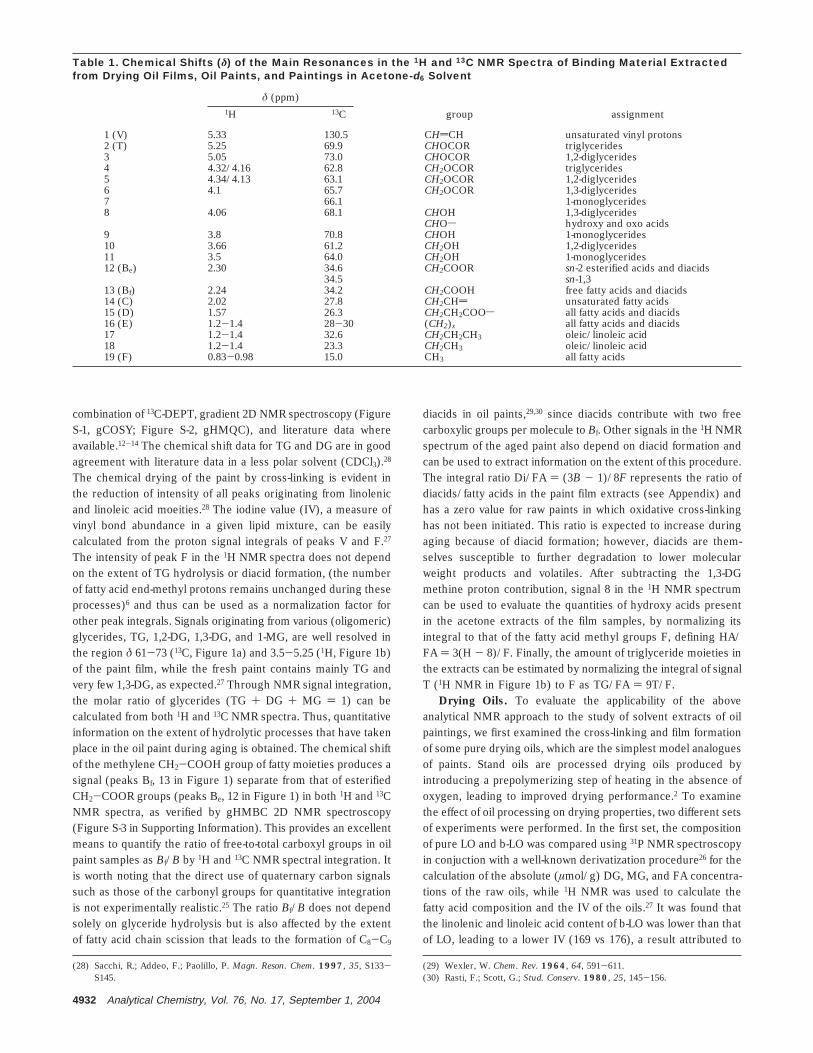
Table 1. Chemical Shifts (δ) of the Main Resonances in the 1H and 13C NMR Spectra of Binding Material Extractedfrom Drying Oil Films, Oil Paints, and Paintings in Acetone-d6 Solvent
δ (ppm)1H 13C group assignment
1 (V) 5.33 130.5 CHdCH unsaturated vinyl protons2 (T) 5.25 69.9 CHOCOR triglycerides3 5.05 73.0 CHOCOR 1,2-diglycerides4 4.32/4.16 62.8 CH2OCOR triglycerides5 4.34/4.13 63.1 CH2OCOR 1,2-diglycerides6 4.1 65.7 CH2OCOR 1,3-diglycerides7 66.1 1-monoglycerides8 4.06 68.1 CHOH 1,3-diglycerides
CHOs hydroxy and oxo acids9 3.8 70.8 CHOH 1-monoglycerides10 3.66 61.2 CH2OH 1,2-diglycerides11 3.5 64.0 CH2OH 1-monoglycerides12 (Be) 2.30 34.6 CH2COOR sn-2 esterified acids and diacids
34.5 sn-1,313 (Bf) 2.24 34.2 CH2COOH free fatty acids and diacids14 (C) 2.02 27.8 CH2CHd unsaturated fatty acids15 (D) 1.57 26.3 CH2CH2COOs all fatty acids and diacids16 (E) 1.2-1.4 28-30 (CH2)x all fatty acids and diacids17 1.2-1.4 32.6 CH2CH2CH3 oleic/linoleic acid18 1.2-1.4 23.3 CH2CH3 oleic/linoleic acid19 (F) 0.83-0.98 15.0 CH3 all fatty acids
4932 Analytical Chemistry, Vol. 76, No. 17, September 1, 2004
partial glyceride oligomerization and oxidation during process-ing.2,31 Furthermore, the 31P NMR measurements showed that inb-LO the DG and FA concentrations are much higher ([DG] )124.8 µmol/g of oil, 77.3 µmol/g of oil in LO, and [FA] ) 59.1µmol/g of oil, 12.4 in LO). The equimolar increase in concentrationfor DG and FA indicates that they are produced by hydrolysis oftriglycerides triggered by extensive heating of linseed oil duringprocessing. The effect of heat-induced hydrolysis has recentlybeen observed during linseed oil processing under variousexperimental conditions31 and is further supported by the iden-tification of small amounts (6.0 µmol/g of oil) of 1-monoglycerides,which are products of diglyceride hydrolysis, in our present b-LOsample, not observed in LO (data not shown).
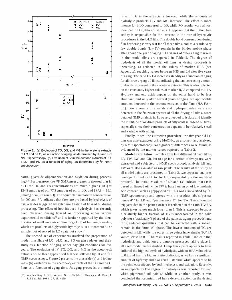
The second set of experiments involved the preparation ofmodel thin films of LO, b-LO, and PO on glass plates and theirstudy as a function of aging under daylight conditions for fiveyears. The evolution of IV, TG, DG, and MG in the acetone-d6
extracts of the three types of oil film was followed by 1H and 13CNMR spectroscopy. Figure 2 presents the glyceride (a) and iodinevalue (b) evolution in the acetone-d6 extracts of the LO and b-LOfilms as a function of aging time. As aging proceeds, the molar
ratio of TG in the extracts is lowered, while the amounts ofhydrolytic products DG and MG increase. The effect is moreintense for b-LO compared to LO, while PO results were almostidentical to LO (data not shown). It appears that the higher freeacidity is responsible for the increase in the rate of hydrolyticprocedures in the b-LO film. The double bond consumption duringfilm hardening is very fast for all three films, and as a result, veryfew double bonds (low IV) remain in the binder mobile phaseafter about one year of aging. The values of other aging markersin the model films are reported in Table 2. The degree ofhydrolysis of all the model oil films as drying proceeds isincreasing, as reflected in the values of marker HFA (seeAppendix), reaching values between 0.35 and 0.4 after five yearsof aging. The ratio Di/FA increases steadily as a function of agingfor all three drying oil films, indicating that an increasing amountof diacids is present in their acetone extracts. This is also reflectedon the constantly higher values of marker Bf/B compared to HFA.Hydroxy and oxo acids appear on the other hand to be lessabundant, and only after several years of aging are appreciableamounts detected in the acetone extracts of the films (HA/FA <0.1). Low amounts of alkanals and hydroperoxides were alsodetected in the 1H NMR spectra of all the drying oil films. Moredetailed NMR analysis is, however, needed to isolate and identifythe multitude of oxidized products of fatty acids in linseed oil films,especially since their concentration appears to be relatively smalland variable with aging.
Finally, to test the extraction procedure, the five-year-old LOfilm was also extracted using MeOH-d4 as a solvent and analyzedby NMR spectroscopy. No significant differences were found, asevidenced by the marker values reported in Table 2.
Model Paint Films. Samples from four different oil paint films,LB, TW, LW, and CR, left to age for a period of five years, wereextracted and subjected to NMR spectroscopic analysis. LB andTW were also available as raw paints. The results of the study ofall model paints are presented in Table 2, two separate analysesbeing performed for LB to check the repeatability of the analyticalprotocol. The initial IV values of 173 and 139 indicate that LB isbased on linseed oil, while TW is based on an oil of low linolenicacid content, such as poppyseed oil. This was also verified by 13CNMR spectroscopy and agrees with the product labels,“perma-nence 4*” for LB and “permanence 3*” for TW. The amount oftriglycerides in the paint extracts is reflected in the ratio TG/FA,which takes values much lower than 1. This is expected becausea relatively higher fraction of TG is incorporated in the solidpolymer (“stationary”) phase of the paint as aging proceeds, andthus, reduced quantities that can be extracted with a solventremain in the “mobile” phase. The lowest amounts of TG aredetected in LB, while the other three paints have similar TG/FAvalues, close to 0.5. The results reported in Table 2 indicate thathydrolysis and oxidation are ongoing processes taking place inall aged model paints studied. Lamp black paint appears to havesuffered the highest levels of hydrolysis, with an HFA value closeto 0.3, and has the highest ratio of diacids, as well as a significantamount of hydroxy and oxo acids. Titanium white appears to bethe paint least affected by both hydrolysis and oxidation. Recently,an unexpectedly low degree of hydrolysis was reported for leadwhite pigmented oil paints,6 while in another study, it wasconcluded that cadmium red has a delaying action on the drying
(31) van den Berg, J. D. J.; Vermist, N. D.; Carlyle, L.; Holcapek, M.; Boon, J.J. J. Sep. Sci. 2004, 27, 181-199.
Figure 2. (a) Evolution of TG, DG, and MG in the acetone extractsof LO and b-LO) as a function of aging, as determined by 1H and 13CNMR spectroscopy. (b) Evolution of IV in the acetone extracts of LO,b-LO, and PO as a function of aging, as determined by 1H NMRspectroscopy.
Analytical Chemistry, Vol. 76, No. 17, September 1, 2004 4933
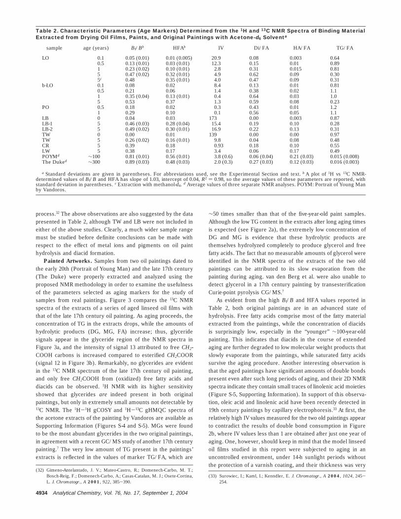
process.32 The above observations are also suggested by the datapresented in Table 2, although TW and LB were not included ineither of the above studies. Clearly, a much wider sample rangemust be studied before definite conclusions can be made withrespect to the effect of metal ions and pigments on oil painthydrolysis and diacid formation.
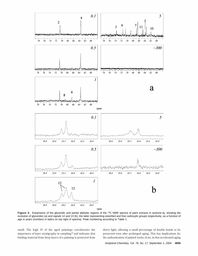
Painted Artworks. Samples from two oil paintings dated tothe early 20th (Portrait of Young Man) and the late 17th century(The Duke) were properly extracted and analyzed using theproposed NMR methodology in order to examine the usefulnessof the parameters selected as aging markers for the study ofsamples from real paintings. Figure 3 compares the 13C NMRspectra of the extracts of a series of aged linseed oil films withthat of the late 17th century oil painting. As aging proceeds, theconcentration of TG in the extracts drops, while the amounts ofhydrolytic products (DG, MG, FA) increase; thus, glyceridesignals appear in the glyceride region of the NMR spectra inFigure 3a, and the intensity of signal 13 attributed to free CH2-COOH carbons is increased compared to esterified CH2COOR(signal 12 in Figure 3b). Remarkably, no glycerides are evidentin the 13C NMR spectrum of the late 17th century oil painting,and only free CH2COOH from (oxidized) free fatty acids anddiacids can be observed. 1H NMR with its higher sensitivityshowed that glycerides are indeed present in both originalpaintings, but only in extremely small amounts not detectable by13C NMR. The 1H-1H gCOSY and 1H-13C gHMQC spectra ofthe acetone extracts of the painting by Vandoros are available asSupporting Information (Figures S-4 and S-5). MGs were foundto be the most abundant glycerides in the two original paintings,in agreement with a recent GC/MS study of another 17th centurypainting.7 The very low amount of TG present in the paintings’extracts is reflected in the values of marker TG/FA, which are
∼50 times smaller than that of the five-year-old paint samples.Although the low TG content in the extracts after long aging timesis expected (see Figure 2a), the extremely low concentration ofDG and MG is evidence that these hydrolytic products arethemselves hydrolyzed completely to produce glycerol and freefatty acids. The fact that no measurable amounts of glycerol wereidentified in the NMR spectra of the extracts of the two oldpaintings can be attributed to its slow evaporation from thepainting during aging. van den Berg et al. were also unable todetect glycerol in a 17th century painting by transesterificationCurie-point pyrolysis CG/MS.7
As evident from the high Bf/B and HFA values reported inTable 2, both original paintings are in an advanced state ofhydrolysis. Free fatty acids comprise most of the fatty materialextracted from the paintings, while the concentration of diacidsis surprisingly low, especially in the “younger” ∼100-year-oldpainting. This indicates that diacids in the course of extendedaging are further degraded to low molecular weight products thatslowly evaporate from the paintings, while saturated fatty acidssurvive the aging procedure. Another interesting observation isthat the aged paintings have significant amounts of double bondspresent even after such long periods of aging, and their 2D NMRspectra indicate they contain small traces of linolenic acid moieties(Figure S-5, Supporting Information). In support of this observa-tion, oleic acid and linolenic acid have been recently detected in19th century paintings by capillary electrophoresis.33 At first, therelatively high IV values measured for the two old paintings appearto contradict the results of double bond consumption in Figure2b, where IV values less than 1 are obtained after just one year ofaging. One, however, should keep in mind that the model linseedoil films studied in this report were subjected to aging in anuncontrolled environment, under 14-h sunlight periods withoutthe protection of a varnish coating, and their thickness was very
(32) Gimeno-Antelantado, J. V.; Mateo-Castro, R.; Domenech-Carbo, M. T.;Bosch-Reig, F.; Domenech-Carbo, A.; Casas-Catalan, M. J.; Osete-Cortina,L. J. Chromatogr., A 2001, 922, 385-390.
(33) Surowiec, I.; Kaml, I.; Kenndler, E. J. Chromatogr., A 2004, 1024, 245-254.
Table 2. Characteristic Parameters (Age Markers) Determined from the 1H and 13C NMR Spectra of Binding MaterialExtracted from Drying Oil Films, Paints, and Original Paintings with Acetone-d6 Solventa
sample age (years) Bf/Bb HFAb IV Di/FA HA/FA TG/FA
LO 0.1 0.05 (0.01) 0.01 (0.005) 20.9 0.08 0.003 0.640.5 0.13 (0.01) 0.03 (0.01) 12.3 0.15 0.01 0.891 0.23 (0.02) 0.10 (0.01) 2.8 0.31 0.015 0.815 0.47 (0.02) 0.32 (0.01) 4.9 0.62 0.09 0.305c 0.48 0.35 (0.01) 4.0 0.47 0.09 0.31
b-LO 0.1 0.08 0.02 8.4 0.13 0.01 0.810.5 0.21 0.06 1.4 0.38 0.02 1.11 0.35 (0.04) 0.13 (0.01) 0.4 0.64 0.03 1.05 0.53 0.37 1.3 0.59 0.08 0.23
PO 0.5 0.18 0.02 0.3 0.43 0.01 1.21 0.29 0.10 0.1 0.56 0.05 1.1
LB 0 0.04 0.03 173 0.00 0.003 0.87LB-1 5 0.46 (0.03) 0.28 (0.04) 15.4 0.19 0.10 0.28LB-2 5 0.49 (0.02) 0.30 (0.01) 16.9 0.22 0.13 0.31TW 0 0.00 0.01 139 0.00 0.00 0.97TW 5 0.26 (0.02) 0.16 (0.01) 9.8 0.04 0.08 0.48CR 5 0.39 0.18 0.93 0.18 0.10 0.55LW 5 0.38 0.17 3.4 0.06 0.17 0.49POYMd ∼100 0.81 (0.01) 0.56 (0.01) 3.8 (0.6) 0.06 (0.04) 0.21 (0.03) 0.015 (0.008)The Duked ∼300 0.89 (0.03) 0.48 (0.03) 2.0 (0.3) 0.27 (0.03) 0.12 (0.03) 0.016 (0.003)
a Standard deviations are given in parentheses. For abbreviations used, see the Experimental Section and text. b A plot of 1H vs 13C NMR-determined values of Bf/B and HFA has slope of 1.03, intercept of 0.04, R2 ) 0.98, so the average values of these parameters are reported, withstandard deviation in parentheses. c Extraction with methanol-d4. d Average values of three separate NMR analyses. POYM: Portrait of Young Manby Vandoros.
4934 Analytical Chemistry, Vol. 76, No. 17, September 1, 2004
small. The high IV of the aged paintings corroborates theimportance of layer stratigraphy in sampling10 and indicates thatbinding material from deep layers of a painting is protected from
direct light, allowing a small percentage of double bonds to bepreserved even after prolonged aging. This has implications forthe authentication of painted works of art, in that accelerated aging
Figure 3. Expansions of the glyceridic and partial aliphatic regions of the 13C NMR spectra of paint extracts in acetone-d6, showing theevolution of glycerides (a) and signals 12 and 13 (b), the latter representing esterified and free carboxylic groups respectively, as a function ofage in years (numbers in italics on top right of spectra). Peak numbering according to Table 1.
Analytical Chemistry, Vol. 76, No. 17, September 1, 2004 4935
might not be able to reproduce the chemical state of an originalwork aged under unknown conditions.34
Hydroxy derivatives of fatty acids are a class of compoundsthat have been identified by GC/MS on relatively young painter’sfilms and in smaller concentrations in old paintings.6,7 9-10-Dihydroxyoctanoic acid is often detected when old oil paint layersare analyzed.7 By comparing the integral of the methine CH(OH)proton (signal 8 in Figure 1b) of hydroxy acids to integral F offatty acids, the molar ratio of hydroxyacids to total fatty acids inthe extracts of the two original paintings was calculated as 0.21( 0.03 for the painting by Vandoros and 0.12 ( 0.03 for the olderpainting The Duke. These values are much higher than those ofaged linseed oil films, but of the same order with the five-year-old model paints in Table 2. Overall, the parameter marker thatbest differentiates the two old original paintings from youngerpaints appears to be the ratio TG/FA, which assumes muchsmaller values in the former (by a factor of ∼50). Such low TG/FA values, combined with high values for Bf/B and HFA appearto be indicative of oil paintings of significant age.
An important issue with respect to the NMR analysis of worksof art relates to the quantity of material required. In this study, itwas demonstrated that using conventional NMR probes a fewmilligrams of paint are enough to provide a good characterizationof the paint’s “mobile phase”, which represents 1-5% of the totalpaint sample weight. However, nowadays, cryogenically cooledNMR probes are available that offer on average a 4-fold increasein S/N ratio over that of conventional probes,35 while capillaryprobes with 2.5-µL NMR-active sample volume have made possiblethe acquisition of 1H NMR spectra of 1 µg of analyte (5 nmol) in∼1 min.36 It is estimated that, taking advantage of advanced probetechnology, the paint sample size can be reduced to 0.1-0.2 mg,bringing NMR on par with current analytical techniques used inthe study of works of art.
CONCLUSIONSThe results presented demonstrate the capabilities of NMR
spectroscopy with respect to its use as an analytical tool to studythe organic components in painted works of art. More specifically,by employing 1D and 2D NMR spectroscopy it was possible tocharacterize the chemical composition of the “mobile phase”(solvent-extractable component) of the binding medium from oilpaintings and reveal important information regarding the ongoingprocesses of hydrolysis and oxidation. During aging, the triglyc-eride content of a paint extract appears to decrease. On the otherhand, the concentrations of the di- and monoglyceride componentsreach a maximum in the extract, followed by a slow decrease withfurther aging, since these compounds suffer further hydrolysisto glycerol and free fatty acids. The amount of free fatty acidsincreases monotonically with aging and reflects the degree ofhydrolysis of the oleaginous binding medium. Our studies with aseries of model paint samples indicate that a low ratio of TG/FAin combination with high values of Bf/B and HFA represents anaccurate set of markers to characterize the extent of aging of anoil paint.
The use of sophisticated cryoprobes and hyphenated LCNMR35 techniques are expected to minimize the amount ofmaterial needed to perform NMR analysis of oil paintings andmake NMR a more favorable technique for the analyst, in view ofits rapidity and experimental simplicity. Thus, modern NMRspectroscopy has the potential to become an important analyticaltool for the detailed study of painted artworks. Further work,involving the study of acrylic and tempera paintings, varnishes,and the utilization of hyphenated LC NMR techniques is inprogress.
ACKNOWLEDGMENTThe authors are grateful to Prof. P. Dais, University of Crete,
for his support and also thank Dr. M. Doulgeridis, National Galleryof Athens, for providing information on the oil paintings and Ms.K. Melessanaki (IESL-FORTH) for assisting with sampling.
SUPPORTING INFORMATION AVAILABLEA listing of five additional figures. Figure S-1: 1H-1H homo-
nuclear gradient COSY 2D NMR spectrum of the acetone-d6
extract of aged LB paint; Figure S-2: 1H-13C heteronucleargradient HMQC 2D NMR spectrum of acetone-d6 extracts of agedLB paint. Figure S-3: Carbonyl region of the 1H-13C heteronucleargradient HMBC 2D NMR spectrum of the methanol-d4 extractsof a five-year-old linseed oil film, showing the assignment via thelong-range 1H-13C coupling of methylene protons with free andesterified carbonyl groups. Figure S-4: 1H-1H homonucleargradient COSY 2D NMR spectrum of the acetone-d6 extracts ofan early 20th century oil painting by Vandoros. Figure S-5: 1H-13C heteronuclear gradient HMQC 2D NMR spectrum of of theacetone-d6 extracts of an early 20th century oil painting byVandoros. Figure S-6: (a) Portrait of Young Man, by S. Vandoros,oil on canvas, early 20th century, private collection; (b) The Duke,unknown artist, oil on canvas, late 17th century, private collection.This material is available free of charge via the Internet at http://pubs.acs.org.
APPENDIXAssuming that the acetone-d6 extracts of a paint film contain a
mol of esterified fatty acids, b mol of free fatty acids, and c mol ofdiacids, integral values in the 1H NMR spectrum are expressedas
where TG, DG, and MG are the molar ratios of each type ofglyceride.
Received for review May 3, 2004. Accepted June 29, 2004.
AC049350K
(34) Erhardt, D.; Tumosa, C. S.; Mecklenburg, M. F. Polym. Prepr. 2000, 41,1790-1791.
(35) Spraul, M.; Freund, A. S.; Nast, R. E.; Withers, R. S.; Maas, W. E.; CorcoranO. Anal. Chem. 2003, 75, 1536-1541.
(36) Schlotterbeck, G.; Ross, A.; Hochstrasser, R.; Senn, H.; Kuhn, T.; Marek,D.; Schett O. Anal. Chem. 2002, 74, 4464-4471.
Be ) 2a (1)
Bf ) 2b + 4c (2)
B ) D ) 2a + 2b + 4c (3)
F ) 3a + 3b (4)
b ) (F/3) - (Be/2) (5)
c ) (B/4) - (F/6) (6)
Di/FA ) (6B - 1)/8F (7)
HFA ) (2DG + 4MG)/6(TG + DG + MG) (8)
4936 Analytical Chemistry, Vol. 76, No. 17, September 1, 2004