Embed Size (px)
Citation preview

Study of an RNA helicase implicatessmall RNA–noncoding RNA interactionsin programmed DNA eliminationin TetrahymenaLucia Aronica,1,6 Janna Bednenko,2,6 Tomoko Noto,1,6 Leroi V. DeSouza,3 K.W. Michael Siu,3
Josef Loidl,4 Ronald E. Pearlman,5 Martin A. Gorovsky,2 and Kazufumi Mochizuki1,7
1Institute of Molecular Biotechnology of the Austrian Academy of Sciences (IMBA), A-1030 Vienna, Austria; 2Departmentof Biology, University of Rochester, Rochester, New York 14627, USA; 3Department of Chemistry and Center for Researchin Mass Spectrometry, York University, Toronto, Ontario M3J 1P3, Canada; 4Department of Chromosome Biology, Max F.Perutz Laboratories, University of Vienna, A-1030 Vienna, Austria; 5Department of Biology and Center for Research in MassSpectronomy, York University, Toronto, Ontario M3J 1P3, Canada
Tetrahymena eliminates micronuclear-limited sequences from the developing macronucleus during sexualreproduction. Homology between the sequences to be eliminated and ∼28-nucleotide small RNAs (scnRNAs)associated with an Argonaute family protein Twi1p likely underlies this elimination process. However, themechanism by which Twi1p–scnRNA complexes identify micronuclear-limited sequences is not wellunderstood. We show that a Twi1p-associated putative RNA helicase Ema1p is required for the interactionbetween Twi1p and chromatin. This requirement explains the phenotypes of EMA1 KO strains, including lossof selective down-regulation of scnRNAs homologous to macronuclear-destined sequences, loss of H3K9 andK27 methylation in the developing new macronucleus, and failure to eliminate DNA. We further demonstratethat Twi1p interacts with noncoding transcripts derived from parental and developing macronuclei and thisinteraction is greatly reduced in the absence of Ema1p. We propose that Ema1p functions in DNA eliminationby stimulating base-pairing interactions between scnRNAs and noncoding transcripts in both parental anddeveloping new macronuclei.
[Keywords: RNA; heterochromatin; small RNA; noncoding RNA; Tetrahymena]
Supplemental material is available at http://www.genesdev.org.
Received April 7, 2008; revised version accepted June 25, 2008.
Heterochromatin functions in various chromosomal pro-cesses, including regulation of gene expression, chromo-some segregation, and nuclear organization (for review,see Grewal and Jia 2007). In diverse eukaryotes, RNAi-related mechanisms involving small RNAs complexedwith Argonaute family proteins mediate heterochroma-tin formation (for review, see Martienssen and Moazed2006; Grewal and Jia 2007). However, the mechanism bywhich small RNAs target heterochromatin formation isnot completely understood. In ciliated protozoans, het-erochromatin formation is also induced by an RNAi-re-lated mechanism, followed by programmed DNA elimi-nation of germline-specific sequences from the develop-ing somatic nucleus (for review, see Meyer and Chalker2006). Thus, programmed DNA elimination in ciliates
serves as a model to study small RNA-mediated hetero-chromatin formation.
Like most ciliated protozoans, Tetrahymena thermo-phila exhibits nuclear dimorphism. Each cell contains agermline micronucleus (Mic) and a somatic macro-nucleus (Mac). It is likely that only the Mac contributesto gene expression. In vegetative growth, the Mic andMac replicate/divide, and sister nuclei are segregated todaughter cells. In the sexual process of conjugation (Fig.1A; see also Supplemental Fig. S1), the Mic undergoesmeiosis to form two haploid pronuclei, one of which isreciprocally exchanged between the two conjugatingcells. The migratory and stationary pronuclei then fuseto create a zygotic nucleus that divides mitotically twiceto produce the next generation of new Macs and Mics.Then, paired cells separate, one of the two new Mics andthe parental Mac are destroyed and, if fed, they resumevegetative growth.
After Pair Separation, ∼6000 internal eliminated se-quences (IESs) are deleted from the new Mac, and the
6These authors contributed equally to this work.7Corresponding author.E-MAIL [email protected]; FAX 43-1-79044-110.Article is online at http://www.genesdev.org/cgi/doi/10.1101/gad.481908.
2228 GENES & DEVELOPMENT 22:2228–2241 © 2008 by Cold Spring Harbor Laboratory Press ISSN 0890-9369/08; www.genesdev.org
Cold Spring Harbor Laboratory Press on November 15, 2021 - Published by genesdev.cshlp.orgDownloaded from

flanking sequences are ligated (for review, see Yao et al.2002). The Mic sequences that remain in the future Macare called Mac-destined sequences (MDSs). IESs in Tet-rahymena vary in size (∼0.5–20 kb) and account for ∼15%of the Mic genome. Most IESs are moderately repeated inthe Mic genome and many are related to transposableelements. IESs have not been found in coding sequencesof Tetrahymena, although some are located in introns.Excision of IESs can occur reproducibly at a specific siteor with a limited number of alternative boundaries.
IES elimination is epigenetically regulated by a mecha-nism that prevents sequences present in the old Macfrom being eliminated in the new Mac (Chalker and Yao1996). Small (∼28 nucleotide [nt]) scan RNAs (scnRNAs)expressed during conjugation (Mochizuki et al. 2002; Leeand Collins 2006) are involved in epigenetic regulation ofIES elimination (Mochizuki et al. 2002; Yao et al. 2003).A Dicer-like protein, Dcl1p, is required for IES elimi-nation and for processing Mic noncoding RNA (ncRNA,
also called nongenic RNA) transcripts derived from bothstrands of the Mic sequences (Chalker and Yao 2001) toscnRNAs (Malone et al. 2005; Mochizuki and Gorovsky2005). An Argonaute protein Twi1p is complexed withscnRNAs (Mochizuki and Gorovsky 2004b) and is alsorequired for IES elimination (Mochizuki et al. 2002).
Heterochromatin is involved in IES elimination.Methylation of histone H3 on Lys 9 (H3K9me) and/orLys 27 (H3K27me), as well as chromodomain proteinsthat can bind to these methylated histones, are hall-marks of heterochromatin in diverse eukaryotes. In Tet-rahymena, both H3K9me and H3K27me occur in thedeveloping Mac and are specifically associated witheliminated IES sequences (Taverna et al. 2002; Liu et al.2007). Both H3K9me and K27me in the developing newMac are essential for IES elimination and depend onEzl1p, an E(z)-related histone methyltransferase (Liu etal. 2007). The chromodomain protein Pdd1p is also essen-tial for IES elimination (Coyne et al. 1999), can bind toboth H3K9me and K27me, and could act as a “reader” ofthese modifications (Taverna et al. 2002; Liu et al. 2007).IES elimination is also sensitive to treatment with a his-Figure 1. Identification and characterization of EMA1/Ema1p.
(A) Conjugation. In the sexual process of conjugation, two cellsmate (1); the Mic undergoes meiosis to form two haploid pro-nuclei, one of which is reciprocally exchanged between the twoconjugating cells (2); the migratory and stationary pronucleithen fuse to create a zygotic nucleus (3); the zygotic nucleusdivides mitotically twice to produce the next generation of newMacs and Mics (4); paired cells separate and one of the two newMics and the parental Mac are destroyed (5); and cells resumevegetative growth with Mic mitosis, followed by cytokinesis (6).(B) Copurification of Ema1p with Flag-HA-Twi1p. Two wild-type strains (No-tag) or two Flag-HA-TWI1 strains were matedand were harvested at 9 h post-mixing. Flag-HA-Twi1p-contain-ing complexes were enriched by gel filtration and immuno-af-finity purified first with anti-Flag antibody and then with anti-HA antibody. The purified proteins were separated by SDS-PAGE and analyzed by silver staining. The arrow and theasterisk indicate the positions of Flag-HA-Twi1p and theTwi1p-associated proteins identified by mass spectrometryanalysis, respectively. (C) Twi1p coimmunoprecipitates withEma1p. An EMA1-HA strain and a wild-type strain were matedand lysed at 6 h post-mixing. (Right) The Ema1p-HA-containingcomplex was pulled down with an anti-HA antibody. (Left) As acontrol, two wild-type strains were crossed and processed simi-larly. Coimmunoprecipitated proteins (anti-HA IP) or total pro-teins (Input) used for immunoprecipitation were analyzed onWestern blot using anti-Twi1p antiserum. (D) Expression ofEMA1 mRNA. Total RNAs from exponentially growing (E),starved (S), and conjugating (2, 4, 6, 8, 10, 12, and 14 h post-mixing) wild-type cells were analyzed by Northern hybridiza-tion. RPL21 was used as a loading control. (E–L) Localization ofEma1p. Mating pairs of wild-type cells in the early (E) and late(F) premeiosis, pronuclear exchange (G), Mac Anlagen (H,I),Nuclear Alignment (J), Pair Separation (K), and Mic Elimination(L) stages were processed for immunostaining. See Supplemen-tal Figure S1 for the conjugation stages. Ema1p was localizedusing anti-Ema1p antiserum (left) and DNA was stained byDAPI (right). Arrows, arrowheads, and arrowheads marked with“An” indicate Macs, Mics, and developing new Macs, respec-tively. The staining detected at the junction of cells (doublearrowheads) was also observed in �EMA1 strains (see Fig. 7O,P)and thus represents cross-reaction of the antiserum with otherproteins.
RNA–RNA interaction in DNA elimination
GENES & DEVELOPMENT 2229
Cold Spring Harbor Laboratory Press on November 15, 2021 - Published by genesdev.cshlp.orgDownloaded from

tone deacetylase inhibitor (Duharcourt and Yao 2002).Together, these results indicate involvement of hetero-chromatin in DNA elimination. Dcl1p and Twi1p, re-quired for production and accumulation of scnRNAs, re-spectively, are required for accumulation and/or target-ing of H3K9/K27me and for DNA elimination (Liu et al.2004, 2007; Malone et al. 2005; Mochizuki and Gorovsky2005). Thus, heterochromatin formation occurs down-stream from the RNAi-related mechanism in the IESelimination pathway.
scnRNAs homologous to IES sequences became en-riched as conjugation proceeded (Mochizuki and Gor-ovsky 2004b) and most of the scnRNAs cloned from thelate stage of conjugation are complementary to IES se-quences (Lee and Collins 2006). Thus, scnRNAs are se-lected for IES specificity after they are diced. Based onthese and other observations, we proposed that thescnRNA model (Mochizuki et al. 2002; Mochizuki andGorovsky 2004a), which postulates that althoughscnRNAs are derived from both MDSs and IESs of theMic genome, scnRNAs having homology with any MacDNA sequence (=MDSs) are degraded in the parentalMac (“scanning” for selection of scnRNA). As a result,scnRNAs become enriched in IES sequences. We furtherproposed that these IES-specific scnRNAs, complexed toTwi1p, move to the developing Mac, where they identifyhomologous sequences as IESs and target them for het-erochromatin formation via accumulation of H3K9/K27me and Pdd1p, followed by DNA elimination. In themodel, a given scnRNA should interact with genomic se-quences either in the parental Mac to induce its own deg-radation or in the developing Mac to induce H3K9/K27me.
Here we report that a Twi1p-associated RNA helicase,Ema1p, plays an important role in the interactions be-tween Twi1p and Mac chromatins and between Twi1pand Mac ncRNAs. We further show that Ema1p is re-quired for IES elimination of some loci, for selectivedown-regulation of scnRNAs and for accumulation ofH3K9me and H3K27me in the developing new Mac.These data suggest that Ema1p facilitates the interactionbetween nascent ncRNAs from Mac chromatins and theTwi1p–scnRNA complex, and this interaction plays im-portant roles in DNA elimination.
Results
Ema1p interacts with Twi1p
Twi1p, a Tetrahymena Argonaute protein is required forscnRNA accumulation and for IES elimination (Mochi-zuki et al. 2002) and is physically associated withscnRNAs (Mochizuki and Gorovsky 2004b). To furtherunderstand the Twi1p–scnRNA complex, we sought toidentify Twi1p-associated proteins. A complex contain-ing N-terminally tagged Flag-HA-Twi1p was purifiedfrom conjugating cells at 9 h post-mixing when Mac de-velopment is occurring and analyzed by SDS-PAGE andsilver staining (Fig. 1B). Two closely migrating proteinscopurified with Flag-HA-Twi1p (asterisk in Fig. 1B) wereidentified by mass spectrometry analysis (SupplementalTable S1).
One of the copurifying proteins was encoded byTTHERM_00088150 (http://www.ciliate.org), a gene wenamed EMA1. Ema1p, the protein predicted from theEMA1 cDNA sequence (AB292216 in DDBJ/EMBL/Gen-Bank), is a DExH box RNA helicase. Although manyRNA helicases are involved in RNAi-related pathways ina variety of eukaryotes, none of them are close relativesof Ema1p. The proteins closely related to Ema1p areXP_001458153 and XP_001423262 in Paramecium,DHX57 and DHX36 in human, AT2G35920 in Arabi-dopsis, and GH07148p in the fruit fly. DHX36 was re-cently identified as an AGO1- and AGO2-binding pro-tein (Höck et al. 2007). Thus, these proteins may have aconserved role in RNAi-related mechanisms by modu-lating the functions of Argonaute proteins.
We confirmed the Twi1p–Ema1p interaction by coim-munoprecipitation using a strain expressing EMA1-HAand an anti-HA antibody. Twi1p was highly enriched inthe immunoprecipitate from mating EMA1-HA cells(Fig. 1C). Thus, Ema1p reciprocally associates withTwi1p.
Ema1p localizes in old and new Macsduring conjugation
EMA1 mRNA was detected from early (2 h; meiotic pro-phase) to late (14 h; when DNA elimination occurs) con-jugation stages, but not in growing cells or in starvedcells (Fig. 1D). EMA1 mRNA is therefore expressed ex-clusively during conjugation.
Ema1p was localized by immunostaining using anti-Ema1p antiserum. Ema1p was not detected in vegetativecells (data not shown). During conjugation, Ema1p wasfirst observed in the parental Mac when Mics were inmeiotic prophase (Fig. 1A [stage 1],E). Ema1p localized inthe parental Mac until the new Macs developed (Fig. 1A[until stage 4], F,G), after which, staining in the parental(old) Mac disappeared and became localized in the newMacs (Fig. 1H,I). Ema1p remained localized in the newMacs until the Pair Separation (Fig. 1A [stage 5], J,K).Then, it disappeared before DNA elimination occurred(∼14 h post-mixing) (Fig. 1L). Similar transitions of local-ization from the old Mac to the new Mac were also ob-served for Twi1p, Ezl1p, Pdd1p, and Pdd2p, all of whichare required for DNA elimination (Coyne et al. 1999;Nikiforov et al. 1999; Mochizuki et al. 2002; Liu et al.2007).
Parental EMA1 is required for producing viableprogeny
To determine the function(s) of EMA1, EMA1 knockout(KO, �EMA1) strains were constructed. Part of the cod-ing sequence, including the conserved helicase domain,was replaced by a drug resistance marker in all EMA1loci in the polyploid Mac (Fig. 2A). By Southern hybrid-ization, a strong band with the expected size of the KOlocus, and a faint band with the expected size of theendogenous EMA1 locus, were observed (Fig. 2B). The
Aronica et al.
2230 GENES & DEVELOPMENT
Cold Spring Harbor Laboratory Press on November 15, 2021 - Published by genesdev.cshlp.orgDownloaded from

faint band was ∼30 times less intense than the KO band,reflecting the relative amounts of DNA in Mic and Mac(Woodard et al. 1972), suggesting that complete replace-ment of the Mac EMA1 gene had occurred. Consistentwith this, Ema1p was not detected in �EMA1 cells dur-ing the early to mid stages of conjugation (Fig. 2C). Smallamounts of Ema1p were detected in �EMA1 cells in latestages of conjugation (Fig. 2C, at 12 h post-mixing), in-dicating that in �EMA1 cells the intact EMA1 loci in thenew Macs could be expressed (zygotic expression). In�EMA1 cells, Ema1p was never detected in the parentalMac, but first appeared in the new Mac at Nuclear Align-ment stage at 12 h post-mixing (Fig. 7O,P, below; Supple-mental Fig. S2), although it was detected in the new Macas early as 6 h post-mixing in the wild-type cells (Fig.1H). These results argue that, in �EMA1 strains, Ema1pin the parental Mac is completely eliminated and its ac-cumulation in the developing new Mac is severely de-layed.
The �EMA1 strains showed no obvious defects duringvegetative growth (data not shown). They mate normallybut meiosis (between stages 1 and 2 in Fig. 1A; stage E2in Supplemental Fig. S1) and Pair Separation (stages 4and 5 in Fig. 1A; stages L1–L2 in Supplemental Fig. S1)were delayed (Fig. 2D). The significance of the meioticdelay is not clear. Delayed Pair Separation in �EMA1strains is possibly related to defective heterochromatinformation (see below). Nonetheless, exconjugants (prog-eny) containing new Macs and Mics were produced in
the �EMA1 cells (L2 in Fig. 2D). Thus, at the cytologicallevel, nuclear differentiation appeared to occur normallywithout parental EMA1.
The exconjugants from wild-type cells eliminate oneof the two Mics (stage 5 in Fig. 1A; stage L3 in Supple-mental Fig. S1) and, if the cells are fed, the remainingMic divides, followed by resumption of vegetativegrowth. However, the majority of the exconjugants from�EMA1 cells retained two Mics (arrested in stage L2 inSupplemental Fig. S1), even at 36 h post-mixing (Fig. 2E).Therefore, the progeny of �EMA1 cells were expectednot to resume vegetative growth. Indeed, all single pairsof �EMA1 cells placed into nutrient medium did notgrow and eventually died (Fig. 2F). Thus, parental EMA1is essential for the formation of viable conjugation prog-eny.
Figure 2. Characterization of �EMA1 cells. (A) EMA1 locusand KO construct. A part of the EMA1 coding sequence, includ-ing the conserved helicase domains (shown in gray) was re-placed by the drug-resistance marker neo3. Upon transforma-tion, the KO construct was introduced into the EMA1 locus byhomologous recombination. (B) Southern hybridization of�EMA1 strains. Total DNA isolated from wild-type (WT) or�EMA1 strains was digested with NdeI (N in A), and the blotwas hybridized with the probe shown in A. Positions of thebands for wild-type and KO loci are indicated with arrowheads.(C) Ema1p expression in �EMA1 strains. (Top panel) Ema1pexpression in the wild-type (W) and �EMA1 (�) strains instarved (S) and mating (4, 8, and 12 h post-mixing) cells wasanalyzed by Western blot using anti-Ema1p antiserum. (Bottompanel) For a loading control, the amount of �-tubulin was ana-lyzed. (D) Developmental profiles of conjugation in wild-typeand �EMA1 strains. Conjugation stage wild-type(CU427 × CU428) and �EMA1 [(7)-3-1 X (8)-1-1] cells were ob-served by DAPI staining. The stages categorized were singleunmated cells (S), premeiosis (E1), meiosis (E2), prezygotic (M1),post-zygotic (M2), Mac development (L1), Pair Separation (2Mics) (L2), and Mic elimination (L3). See Supplemental FigureS1 for the developmental stages. (E) �EMA1 causes arrest at PairSeparation stage. At 36 h post-mixing, the progeny of wild-type(CU427 and CU428) or �EMA1 cells were fixed and nuclei wereobserved by DAPI staining. The stages categorized were thesame as in D except stages E1∼L1 were combined. (F) �EMA1cells fail to produce viable progeny. At 6∼8 h post-mixing, singlemating pairs were placed into drops of medium and incubatedfor ∼60 h at 30°C. Completion of conjugation was confirmed bytesting for expression of the marker specific for newly developedMacs.
RNA–RNA interaction in DNA elimination
GENES & DEVELOPMENT 2231
Cold Spring Harbor Laboratory Press on November 15, 2021 - Published by genesdev.cshlp.orgDownloaded from

Elimination of a subset of IESs is inhibited in �EMA1
Lack of viable conjugation progeny and retention of thetwo Mics observed in the �EMA1 strains are phenotypesreported in mutant strains in which genes affecting IESelimination (DCL1, EZL1, PDD1, PDD2, and TWI1)were disrupted (Coyne et al. 1999; Nikiforov et al. 1999;Mochizuki et al. 2002; Malone et al. 2005; Mochizukiand Gorovsky 2005; Liu et al. 2007). To determinewhether EMA1 is required for IES elimination, we ana-lyzed the elimination of four different IESs: M, R, Cal,and Tlr1 elements (Fig. 3). Pairs of mating �EMA1strains were isolated at ∼8 h post-mixing and allowed tocomplete conjugation. At 36 h post-mixing, one of thetwo separated exconjugants was analyzed by nested PCRto assess DNA elimination. Because IES elimination oc-curs at around 14 h post-mixing in wild-type cells, wereasoned that 36 h post-mixing should allow observationof DNA elimination in mutant cells, even if it was de-layed. As negative controls for DNA elimination, pairs of�TWI1 and �EZL1 (encoding H3K9/K27 methyltransfer-ase) strains (Supplemental Fig. S3) were also analyzed.
As expected, PCR products from the rearranged Macforms of DNA were detected for all of the IES sites testedin the control wild-type strains (Fig. 3, control lanes) andonly unrearranged, Mic forms of DNA were observed inthe �TWI1 and �EZL1 strains (Fig. 3), arguing that theRNAi machinery and heterochromatin were required forDNA elimination of all of the IESs tested here. Surpris-ingly, disruption of parental EMA1 had different effectson different IESs: elimination of the M and Tlr1 ele-ments was inhibited in the progeny of �EMA1 strains,while elimination of the R and Cal elements was not(Fig. 3). Thus, parental EMA1 is required for the elimi-nation of some loci, but not of others. As far as we know,�EMA1 is the first Tetrahymena mutant reported to bedefective in the DNA elimination of a subset of IESs .
The elimination of the M element was also inhibitedin an EMA1 point mutant K188A (Walker-type ATP-binding motif substitution) (Supplemental Fig. S4), argu-ing that Ema1p was likely to have ATP-dependent RNAhelicase activity that was involved in DNA elimination.
Ema1p is not required for scnRNA loadingand passenger strand removal
RNA helicase A is required for the loading of siRNA toArgonaute proteins in human cells (Robb and Rana2007). To determine whether Ema1p is required for theloading of scnRNAs to Twi1p, a Twi1p-containing com-plex was immunoprecipitated from wild-type and�EMA1 strains at 4 h post-mixing using an anti-Twi1pantibody, and the presence of coimmunoprecipitatedscnRNAs was analyzed. Denaturing gel analysis (Supple-mental Fig. S5A) revealed that the amount of scnRNAscomplexed to Twi1p was similar in wild-type and in�EMA1 strains, suggesting that Ema1p was not requiredfor scnRNA loading onto Twi1p.
scnRNAs are made from double-stranded Mic ncRNAsby the Dicer-like protein Dcl1p (Malone et al. 2005;Mochizuki and Gorovsky 2005). There should thereforebe a mechanism to remove one of two scnRNA strands(the passenger strand); otherwise, the scnRNA would notbe able to recognize its complementary sequences. Na-tive gel analysis of the scnRNAs prepared above indi-cated that the amount of double-stranded scnRNAs as-sociated with Twi1p in the �EMA1 strains was similarto that in the wild-type strains (Supplemental Fig. S5B).Thus, Ema1p is not required for the passenger strandremoval of scnRNAs.
Ema1p is required for Twi1p–chromatin interaction
Next, we compared the localization of Twi1p in the pres-ence and absence of Ema1p using an anti-Twi1p anti-body. Both in wild-type and �EMA1 cells, Twi1p waslocalized in the parental Mac in early to mid-stages(Supplemental Fig. S6A,B,D,E) and in the developing newMac in late stages (Supplemental Fig. S6C,F) of conjuga-tion. Thus, Twi1p localized in Macs normally in the ab-sence of Ema1p.
We previously hypothesized that the Twi1p–scnRNAcomplex interacts with chromatin to induce scnRNAdegradation in the parental Mac, followed by H3K9meand DNA elimination in the new Mac (Mochizuki and
Figure 3. DNA elimination in the progeny of�EMA1 cells. (Left) Schematic drawings ofthe IES elimination assays. Solid horizontallines indicate MDSs and the open boxes indi-cate IESs. The M and R elements are ∼2.5 kbapart on the same Mic chromosome. Fourprimers (arrows) that flank each IES were usedfor nested PCR. For the assays of Cal andTlr-1 elements, primers complementary toIESs were also used for the same PCR. (Right)Results of the IES elimination assays. Thesizes of the unprocessed (Mic form) and theprocessed (Mac form) products are marked byarrowheads with “i” and “a”, respectively.(m) Molecular weight marker.
Aronica et al.
2232 GENES & DEVELOPMENT
Cold Spring Harbor Laboratory Press on November 15, 2021 - Published by genesdev.cshlp.orgDownloaded from
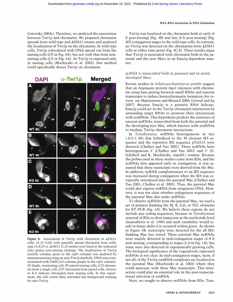
Gorovsky 2004c). Therefore, we analyzed the associationbetween Twi1p and chromatin. We prepared chromatinspreads from wild-type and �EMA1 strains and analyzedthe localization of Twi1p on the chromatin. In wild-typecells, Twi1p colocalized with DNA spread out from themating cells (CP in Fig. 4A), but not with that from non-mating cells (CS in Fig. 4A). As Twi1p is expressed onlyin mating cells (Mochizuki et al. 2002), this methodcould specifically detect Twi1p on chromatin.
Twi1p was localized on the chromatin both at early (4h post-mixing) (Fig. 4B) and late (8 h post-mixing) (Fig.4D) conjugation stages in the wild-type cells. In contrast,no Twi1p was detected on the chromatins from �EMA1cells at either time point (Fig. 4C,E). These results arguethat Twi1p is associated with chromatin both in the pa-rental and the new Macs in an Ema1p-dependent man-ner.
ncRNA is transcribed both in parental and in newlydeveloped Macs
Recent studies in Schizosaccharomyces pombe suggestthat an Argonaute protein Ago1 interacts with chroma-tin using base pairing between small RNAs and nascenttranscripts to induce heterochromatin formation (for re-view, see Martienssen and Moazed 2006; Grewal and Jia2007). Because Ema1p is a putative RNA helicase,Ema1p could act in the Twi1p–chromatin interaction byunwinding target RNAs to promote their interactionswith scnRNAs. This hypothesis predicts the existence ofnascent ncRNAs, transcribed from both the parental andthe developing new Mac, which interact with scnRNAsto mediate Twi1p–chromatin interactions.
In Tetrahymena, ncRNAs heterogeneous in size(∼0.2–1 kb) that hybridized to the M element IES se-quence and the repetitive IES sequence pTt2512 weredetected (Chalker and Yao 2001). These ncRNAs haveheterogeneous 3� (Chalker and Yao 2001) and 5� (U.Schöberl and K. Mochizuki, unpubl.) termini. Becausethe probes used in these studies came from IESs, and thencRNAs first appeared early in conjugation, it was as-sumed that these transcripts were derived from the Mic.In addition, ncRNA complementary to an IES sequencewas increased during conjugation when the IES was ec-topically introduced into the parental Mac (Chalker andYao 2001; Chalker et al. 2005). Thus, the parental Maccould also express ncRNA from exogenous DNA. How-ever, it was not clear whether endogenous sequences ofthe parental Mac also make ncRNAs.
To observe ncRNAs from the parental Mac, we used aset of primers flanking the M, R, Cal, or Tlr1 elementsfor RT–PCR (Fig. 5A). We believe these regions do notinclude any coding sequences, because in Tetrahymenaremoval of IESs is often imprecise at the nucleotide level(Austerberry et al. 1989) and such variability would re-sult in frame shifts if it occurred within genes. As shownin Figure 5B, transcripts were detected for the all IES-flanking Mac loci tested. These parental Mac ncRNAswere mainly detected in mid-conjugation stages (4–8 hpost-mixing, corresponding to stages 2–4 in Fig. 1A), butsome were also detected in exponentially growing cells.The biological significance of the vegetatively expressedncRNAs is not clear. In mid-conjugation stages, most, ifnot all, of the Twi1p–scnRNA complexes are localized inthe parental Mac (Mochizuki et al. 2002) where theycould associate with these Mac transcripts. This inter-action could play an essential role in the post-transcrip-tional selection of scnRNA.
Next, we sought to observe ncRNAs from IESs. Tran-
Figure 4. Association of Twi1p with chromatin in �EMA1cells. (A–E) Cells with partially spread chromatin from wild-type (A,B,D) or �EMA1 (C,E) strains were fixed at the indicatedtime points post-mixing (mating). The localization of Twi1p(middle column; green in the right column) was analyzed byimmunostaining using an anti-Twi1p antibody. DNA was coun-terstained with DAPI (left column; purple in the right column).(S) Single, nonmating cell; (P) paired mating cells; (CS) chroma-tin from a single cell; (CP) chromatin from paired cells. Arrowsin B–E indicate chromatin from mating cells. In this experi-ment, the cell cortex (blue asterisks) has background stainingfor anti-Twi1p.
RNA–RNA interaction in DNA elimination
GENES & DEVELOPMENT 2233
Cold Spring Harbor Laboratory Press on November 15, 2021 - Published by genesdev.cshlp.orgDownloaded from

scription from IESs could occur from the Mic and fromthe developing new Mac that has not yet undergoneDNA elimination. The same cDNAs described abovewere used to detect ncRNAs from IES sequences usingprimer sets specific for M, R, Cal, and Tlr1 element IESs(Fig. 5C,D). As expected of Mic transcripts, ncRNAsfrom all of these IESs were detected in early conjugation
stages (during Mic meiosis, 2 and 4 h post-mixing) (be-fore stage 2 in Fig. 1A; stages E1 and E2 in SupplementalFig. S1) and fell to low or undetectable levels at mid-conjugation (6 h post-mixing) (right before stage 4 in Fig.1A; stage M2 in Supplemental Fig. S1). Importantly, thelevel of these transcripts increased again at 8–10 h post-mixing (stage 4 in Fig. 1A; stage L1 in Supplemental Fig.S1), when new Mac development started. This biphasicpattern suggests that ncRNA transcription from IES se-quences occurs twice, first at meiotic prophase, the onlystage when RNA synthesis and the transcription appara-tus are detected in the Mic (Sugai and Hiwatashi 1974;Stargell and Gorovsky 1994; Mochizuki and Gorovsky2004c), and then when new Macs develop.
Combining the data above, we conclude that threetypes of ncRNA transcription occur during conjugation(Fig. 5E). The first is transcription in the Mic at meioticprophase (Fig. 5E, blue line). These Mic ncRNAs areprobably processed to scnRNAs (Malone et al. 2005;Mochizuki and Gorovsky 2005). The second is transcrip-tion from the parental Mac at mid-conjugation stages(Fig. 5E, red line). The last is the transcription from IESsin the developing Mac at late stages of conjugation (Fig.5E, green line). Since ncRNAs from the intergenic se-quence between HHF2 and HHT2 were also detected
Figure 5. Three types ncRNA. (A–D) Exponentially growing(E), starved (S), and conjugating (2, 4, 6, 8, 10, and 12 h post-mixing) wild-type cells were analyzed by RT–PCR. (A) Sche-matic drawing of the analysis of ncRNA transcription from theparental Mac. Sets of primers (arrows) that flank four differentIESs were used to amplify the cDNAs. (B) Results of RT–PCRfor Mac loci flanking the indicated elements. The red horizontalbar indicates stages in which the ncRNAs are up-regulated.Constitutively expressed RPL21 mRNA was amplified as thepositive control for the RT reaction. (C) Schematic drawing ofncRNA analysis from IESs. Sets of primers (arrows) in four dif-ferent IESs were used to amplify the cDNAs. (D) The results ofRT–PCR for the indicated IES elements. Blue and green hori-zontal bars indicate stages in which the ncRNAs are up-regu-lated. (E) Schematic summary of ncRNA transcription analyses.Relative expression levels (vertical axis) and expression timing(horizontal axis) of the ncRNAs from the Mic (blue), the paren-tal Mac (red), and the developing Mac (green) are represented. (F)Coimmunoprecipitation of ncRNAs with Twi1p. (Lanes labeledas A) Conjugating wild-type strains at 4.5 or 9 h post-mixingwere lysed and the Twi1p complex was immunoprecipitatedwith anti-Twi1p antibody. Coimmunoprecipitated RNA wasanalyzed by RT–PCR (RT+) to detect M-MDS (from parentalMac) or M-IES (from developing new Mac) transcripts as above.As a control, a similar experiment was performed with preim-mune serum (lanes labeled as P). In parallel, a similar experi-ment was performed without the reverse transcription reaction(RT−). (G) Coimmunoprecipitation assay of ncRNAs withTwi1p in the wild-type (W) or �EMA1 strains (�). Conjugatingcells were lysed at 4.5 or 9 h post-mixing and the Twi1p com-plex was immunoprecipitated with anti-Twi1p antibody (IP).Coimmunoprecipitated RNA was analyzed by RT–PCR (RT+)to detect M-MDS or M-IES transcripts. RNA was also extractedfrom part of the lysate and analyzed by RT–PCR (Input). As anegative control, a similar experiment was performed withoutthe reverse transcription reaction (RT−).
Aronica et al.
2234 GENES & DEVELOPMENT
Cold Spring Harbor Laboratory Press on November 15, 2021 - Published by genesdev.cshlp.orgDownloaded from

from early to late conjugation stages (Supplemental Fig.S7), ncRNA transcriptions are not limited from IES-as-sociated sequences, but probably occur genome-wide.
Similar RT–PCR studies using �EMA1 strains showedthat the expression of all three classes of ncRNAs oc-curred normally (Supplemental Fig. S8), suggesting thatthe defects observed in �EMA1 strains are not caused byabnormal ncRNA transcription.
Ema1p is required for efficient Twi1p–ncRNAinteraction
Twi1p associated with chromatin in an Ema1p-depen-dent manner in both the parental and developing newMacs (Fig. 4), and ncRNA was detected in both types ofMac (Fig. 5). These results led us to test the possibilitythat the interaction between Twi1p and chromatin ismediated by nascent ncRNA.
Cell lysates were prepared from mating wild-type cellsat 4.5 h (around stage 2 in Fig. 1A) and 9 h post-mixing(stage 4 in Fig. 1A; stage L1 in Supplemental Fig. S1),when Twi1p is present in (Mochizuki et al. 2002) and thencRNAs are expressed from (Fig. 5) the parental and de-veloping new Macs, respectively. Twi1p was immuno-precipitated with an anti-Twi1p antibody and the coim-munoprecipitated RNA was used for cDNA production.Immunoprecipitation with preimmune serum was usedas a control. ncRNAs from the parental Mac and thedeveloping new Mac were observed by RT–PCR usingprimers flanking the M elements described above andwith primers in the M element IES, respectively. Asshown in Figure 5F, the parental Mac ncRNAs were spe-cifically coimmunoprecipted with Twi1p at 4.5 h post-mixing (Fig. 5F). Similarly, the ncRNAs from the newMac were detected specifically in the RNA coimmuno-precipted with Twi1p at 9 h post-mixing (Fig. 5F). Thus,Twi1p interacts with the ncRNA transcribed in both theparental and the developing new Mac.
Next, to determine whether these Twi1p–ncRNA in-teractions were dependent on Ema1p, a similar experi-ment was performed using �EMA1 strains. The ncRNAscoimmunoprecipitated with Twi1p were greatly reducedin the absence of EMA1 (Fig. 5G), indicating that Ema1pfacilitates interaction between Twi1p and the ncRNAs.Because Ema1p is also required for the interaction be-tween Twi1p and Mac chromatin (Fig. 4), these findingssuggest that Ema1p mediates Twi1p association with thechromatin-associated nascent ncRNAs.
EMA1 is required for selective down-regulationof scnRNAs homologous to a repeated MDS
Because Twi1p associates with scnRNAs and is requiredfor their accumulation (Mochizuki and Gorovsky2004b), it was important to analyze the expression ofscnRNA in �EMA1 strains. Because specific scnRNAsthemselves have not been well characterized, we firststudied the expression patterns of scnRNAs in wild-typecells.
We demonstrated previously that the total amount ofscnRNAs remained relatively constant during conjuga-tion (Fig. 6A; Mochizuki et al. 2002). In addition, we usedendogenous scnRNAs as probes for hybridization toSouthern blots containing Mac and Mic DNA to demon-strate that scnRNAs homologous to IES sequences be-came enriched as conjugation proceeds (Mochizuki andGorovsky 2004b). These observations suggested thatscnRNAs derived from IESs should persist in the laterstages of conjugation, while those derived from MDSsshould disappear. However, Chalker et al. (2005) re-ported that scnRNAs homologous to the M element IESdetected by Northern hybridization were present in largeamounts in very early conjugation stages, but were re-duced to low levels in mid to late conjugation.
In an attempt to reconcile these observations, we ex-amined the expression of scnRNAs homologous to the Melement IES. Six 50-b DNA probes homologous to differ-ent regions of the M element IES (see Supplemental Fig.S9) were used to detect scnRNAs on Northern blots ofRNA from different stages of conjugation. Five of the sixprobes tested failed to detect any scnRNAs, while one ofthem, the Mi-9 probe, detected scnRNAs (Fig. 6C) ex-pressed in conjugating cells 2–4 h post-mixing (beforestage 2 in Fig. 1A), but not thereafter. Similar resultswere obtained using the probe Mi-9A, which wascomplementary to Mi-9 (Fig. 6D), demonstrating thespecificity of the hybridization.
To understand why different probes for the M element
Figure 6. Expression of scnRNAs in �EMA1 cells. Total RNAwas extracted from starved (S) or conjugating (2, 4, 6, 8, 10, and12 h post-mixing) wild-type (W) or �EMA1 (�) strains and sepa-rated in sequencing gels. (A,B) Bulk scnRNAs (∼28 nt) werevisualized by staining the gels with GelRed. (C–K) Blots werehybridized with the probes indicated (right). Quantitation of thesignals are shown as ratios of the signals (�EMA1/wild-type)obtained from gel staining and Northern blots.
RNA–RNA interaction in DNA elimination
GENES & DEVELOPMENT 2235
Cold Spring Harbor Laboratory Press on November 15, 2021 - Published by genesdev.cshlp.orgDownloaded from

IES gave such different results, we examined the Tetra-hymena genome database and confirmed that a ∼190-bpsequence (referred to as the MI repeat), including thesequence in the Mi-9 probe, was highly repeated in thesequences found in the Tetrahymena “Mac” genome da-tabase, as also recently noted by Kowalczyk et al. (2006).MI repeats were found both in long (�10 kb) and in short(<10 kb) genomic scaffolds (Supplemental Fig. S9). Shortscaffolds are thought to represent contaminants of theMic genome in the Mac preparations used for genomiclibrary construction, while long scaffolds are thought torepresent true assemblies of Mac chromosomes (Eisen etal. 2006). We conclude, therefore, that the scnRNAs de-tected by the Mi-9 probe and by the M-IES probes studiedby Chalker et al. (2005) were derived not only from the Melement IES, but also from other MI repeats located inmany different MDS and IES loci. Since the other (non-Mi-9) M element probes are probably complementaryonly to the single-copy sequences of the M element IES,we believe the expression levels of the scnRNAs ho-mologous to them were too low to be detected in ourexperiments. Because it is actually an MDS sequence,the large amount of scnRNAs homologous to MI repeatsin early conjugation and their dramatic reduction in themidstages of conjugation is consistent with our previousobservation that scnRNAs homologous to sequences inthe parental Mac (=MDSs) are reduced in the midstagesof conjugation (Mochizuki and Gorovsky 2004b).
Next, we examined MI repeat scnRNAs in the �EMA1strains. In �EMA1 cells, bulk scnRNA levels were com-parable with those in the wild-type cells (Fig. 6A,B).Thus, EMA1 is not required for the accumulation of bulkscnRNAs. Interestingly, in the absence of EMA1 expres-sion, scnRNAs homologous to Mi-9 probes were con-tinuously detected, even in late stages of conjugationwhere they were not detected in wild-type cells (Fig.6C,E). This difference was not due to developmental de-fects in �EMA1 cells because although they proceedmore slowly through the meiotic stage, most �EMA1strains exhibit normal initiation of the appearance ofscnRNAs between 0 and 2 h (Fig. 6) and cytologicallynormal new Macs by 8 h post-mixing (Fig. 2D). Thus, weconclude that Ema1p is involved in the selective reduc-tion of scnRNAs homologous to widely dispersed MI re-peats and possibly of scnRNAs homologous to otherMDSs.
scnRNAs homologous only to IESs are not affectedin the absence of EMA1
Next, we analyzed the expression of scnRNA homolo-gous to IES-specific sequences using 50 b DNA probeshomologous to moderately repeated sequences foundonly in IESs. The probe Tlr1-1 is complementary to theTlr1-element IES (Wells et al. 1994). The probe TP-1 iscomplementary to the antisense strand of a predictedORF (TTHERM_01785770). Both are related to transpos-able elements (Wells et al. 1994; Eisen et al. 2006). Sev-eral sequences identical or similar to Tlr1-1 or TP-1 werefound only in very short (<4 kb) scaffolds in the Tetra-
hymena Mac genome database (data not shown) and,therefore, were likely to be in IESs as described above.
In dramatic contrast to the scnRNA homologous tothe MI repeat (Fig. 6C), in wild-type cells, the scnRNAshomologous to the Tlr1-1 and TP-1 were detectedthroughout conjugation (Fig. 6F,I). Similar results wereobtained using the probes Tlr1-1A and TP-1A, whichwere complementary to Tlr1-1 and TP-1, respectively(Fig. 6G,J). These results are consistent with our previousobservation that scnRNAs homologous to IES sequencespersist to late stages of conjugation (Mochizuki and Gor-ovsky 2004b).
Next, we examined IES-specific scnRNAs present inthe �EMA1 strains by Northern hybridization using theTlr1-1 and TP-1 probes and found that there were noobvious differences in their expression between the wild-type and the �EMA1 strains (Fig. 6, cf. F and H or I andK). Together with above results, we conclude that Ema1pis not required for the production of scnRNAs, but isinvolved in the selective down-regulation of scnRNAshomologous to MDSs.
H3K9 and K27me in the developing new Macare affected in �EMA1 Cells
H3K9me2 and H3K27me3 accumulate specifically inIES-associated chromatin during DNA elimination inthe developing new Mac (Taverna et al. 2002; Liu et al.2007). H3K9me3 also specifically accumulates in the de-veloping new Mac (Liu et al. 2007). Because Twi1p isassociated with Ema1p and is essential for the accumu-lation of H3K9me2 and H3K27me3 (Liu et al. 2004, 2007)in the developing new Mac, we compared the accumu-lation of these modifications in wild-type and �EMA1strains by immunostaining.
As reported, all of these modifications accumulated inthe developing new Macs from the Nuclear Alignmentstage (9 h post-mixing) (Figs. 1A, [slightly after stage 4],7A,E,I; stage L1 in Supplemental Fig. S1) to the Pair Sepa-ration stage before DNA elimination started (12 h post-mixing) (Figs. 1A [stage 5], 7B,F,J; stage L2 in Supplemen-tal Fig. S1) in wild-type cells. In �EMA1 cells, thesemodifications were differentially affected. H3K27me3occurred in the developing new Mac at 9 h post-mixing,but it showed punctate localization at the periphery ofthe nuclei (Fig. 7C) instead of the even distribution ob-served in the wild-type cells at the same stage (Fig. 7A).In the later stages, H3K27me3 was evenly distributed inthe new Mac in the �EMA1 strains (Fig. 7D), possiblydue to the activity of zygotically expressed Ema1p (Fig.7P).
In �EMA1 strains, both H3K9me2 and H3K9me3 werenot detected in the developing Macs in early develop-mental stages (9 h post-mixing) (Fig. 7G,K). In most ofthe �EMA1 cells in the late stages of conjugation (12 hpost-mixing), H3K9me2 accumulation reached a levelsimilar to that of wild-type cells (Fig. 7H). This delayedappearance was well correlated with the accumulation ofzygotic Ema1p in �EMA1 strains (Fig. 7P). H3K9me3 wasnot detected even in the very late stages of conjugation
Aronica et al.
2236 GENES & DEVELOPMENT
Cold Spring Harbor Laboratory Press on November 15, 2021 - Published by genesdev.cshlp.orgDownloaded from

(Fig. 7L). Thus, Ema1p is essential for the accumulationof H3K9me3. We conclude that Ema1p is required forproper induction of H3K9me2 and H3K9me3, and onlyH3K9me2 can be partially restored by zygotic Ema1p.
Liu et al. (2007) reported that H3K27me was upstreamof H3K9me. Thus, H3K27me3, H3K9me2, andH3K9me3 should occur sequentially, and the differenteffect of the loss of parental EMA1 on these modifica-tions correlates well with this sequential event. Thesesuggest that Ema1p is likely essential for accumulationof all of these modifications, and the zygotic Ema1p canrestore only the earlier steps of the sequential events.
Discussion
We identified a putative RNA helicase, Ema1p, as part ofa complex containing the Argonaute protein Twi1p.Ema1p is required for interaction between Twi1p andchromatin in both parental and developing new Macs.We also demonstrated the presence of ncRNAs in bothparental and developing new Macs and showed thatEma1p is essential for the efficient interaction betweenTwi1p and these ncRNAs. In the parental Mac, Ema1p isrequired for selective down-regulation of scnRNAs ho-mologous to MI repeat MDS sequences. In the new Mac,it is required for the induction of H3K9/K27me, and forthe elimination of a subset of IESs. We propose thatEma1p functions similarly in both parental and new
Macs to facilitate interaction between the Twi1p–scnRNA complex and nascent ncRNAs to induce theseevents.
Possible functions of Ema1p in DNA elimination
We propose that Ema1p mediates scnRNA–chromatininteraction by facilitating the interaction betweenscnRNA and nascent ncRNA because (1) it is required forTwi1p–chromatin interaction in both parental and newMacs (Fig. 4), (2) is required for efficient interaction be-tween Twi1p and the ncRNA transcribed from bothparental and new Macs (Fig. 5) and, (3) Twi1p interactwith scnRNA (Mochizuki and Gorovsky 2004b). ThisscnRNA–nascent RNA interaction most likely serves toidentify the scnRNAs homologous to MDS sequencesthat will be down-regulated in the parental Mac (Fig. 6)and those homologous to the IESs that will induceH3K9/K27me (Fig. 7) and IES elimination (Fig. 3) in thedeveloping new Mac. While there is no direct evidencesupporting the idea that ncRNAs bridge the Twi1p–chromatin interaction, the observations that the loss ofEma1p disrupts both the binding of Twi1p to chromatinand its association with ncRNA support this hypothesis.Future studies will be designed to understand the biogen-esis of ncRNA and to perturb it genetically to clarify theprecise role (or roles) of ncRNA in the DNA elimination.
In the fission yeast S. pombe, association of the Argo-
Figure 7. H3K9/K27me in �EMA1 cells. Wild-type (WT, top) and �EMA1 (bottom) conjugating cells at 9 or 12 h post-mixing wereprocessed for immunofluorescent staining (green) using anti-H3K27me3 (A–D), anti-H3K9me2 (E–H), anti-H3K9me3 (I–L), or anti-Ema1p (M–P) Abs. DNA was stained with DAPI (purple). Arrowheads indicate developing new Macs.
RNA–RNA interaction in DNA elimination
GENES & DEVELOPMENT 2237
Cold Spring Harbor Laboratory Press on November 15, 2021 - Published by genesdev.cshlp.orgDownloaded from

naute protein Ago1 with chromatin and the initiationand spreading of heterochromatin are proposed to in-volve base pairing between siRNAs and nascent RNApolymerase II transcripts (Motamedi et al. 2004; Djupe-dal et al. 2005; Kato et al. 2005; Bühler et al. 2006). Aputative RNA helicase Hrr1 in S. pombe associates withAgo1 and is required for its localization to the centro-meric repeats whose transcription initiates heterochro-matin formation (Motamedi et al. 2004). Although Hrr1and Ema1p belong to different classes of RNA helicasesand their exact roles are unclear, they could still haveanalogous functions, as they both interact with Argo-naute proteins and are essential for making heterochro-matin.
scnRNA selection is likely to be achievedby the selective degradation of scnRNAcomplementary to MDSs
We demonstrated previously that scnRNA complemen-tary to IES sequences were selectively enriched duringthe course of conjugation (Mochizuki and Gorovsky2004b). In this study, we show that scnRNAs comple-mentary to the MI repeat, which was found in both IESsand MDSs, were selectively down-regulated in the midto late stages of conjugation, while scnRNAs comple-mentary to the IES sequences were stably expressedthroughout the conjugation. Thus, as suggested (Mochi-zuki et al. 2002), scnRNA selection is likely to beachieved, at least in part, by the selective down-regula-tion of scnRNAs complementary to MDSs.
Since the Dicer-like protein Dcl1p, which is respon-sible for the production of scnRNAs, is detected exclu-sively in the Mic in early conjugation, and the Mic-de-rived ncRNA found in early conjugation increases whenDCL1 is knocked out (Malone et al. 2005; Mochizukiand Gorovsky 2005), scnRNAs are likely to be derivedfrom Mic ncRNAs in early conjugation stages. The factthat ectopic expression of ncRNAs from parental Macdid not cause up-regulation of scnRNAs from these tran-scripts suggested that the parental Mac ncRNA does notcontribute to produce scnRNAs (Chalker et al. 2005).These results suggest that the down-regulation ofscnRNAs complementary to MDSs is likely to occur bytheir selective degradation.
Why is elimination of only a subset of IESs dependenton parental Ema1p?
Parental EMA1 is essential for the elimination of only asubset of IESs (Fig. 3). Because two RNAi machineries,Twi1p and Dcl1p, are required for elimination of the Rand Cal elements (Fig. 8; Malone et al. 2005), whoseelimination is not dependent on parental EMA1 expres-sion, there may be an IES elimination pathway that isdependent on scnRNA, but not on scnRNA selection andparental Ema1p. Mic ncRNA, which produces scnRNAs,might be biased for IESs at some loci and the derivedscnRNAs could possess sufficient specificity to causeIES elimination without the scnRNA selection process.
Alternatively, ncRNA in the new Mac might be pro-duced more efficiently from IESs than from their flank-ing MDSs at some loci and be targeted correctly even byunselected scnRNAs. It is interesting to note in this re-gard that the DNA elimination of a foreign sequenceinserted into the Mic is known to be subject to the po-sition effect (Liu et al. 2005) and thus, ncRNA transcrip-tion in the Mic and/or new Mac may occur nonuni-formly.
It is known that the centromeric histone H3 variantCna1p plays an essential role in IES elimination (Cui andGorovsky 2006). Thus, the physical location of IESs inMic may contribute to the position effect. Differences insensitivity to homology-dependent maternal effects havealso been reported (Fillingham et al. 2001). However, thepatterns of dependence on Ema1p do not correlate witheither of these processes. The characterization of morethan the currently known small number of IESs in Tet-rahymena will enable us to better define what causessensitivity to this protein.
Figure 8. A refined scnRNA model. The sequentially occur-ring events are drawn from top to bottom. The approximatestages when the events occur are shown on the right with ar-rows. See the text for details.
Aronica et al.
2238 GENES & DEVELOPMENT
Cold Spring Harbor Laboratory Press on November 15, 2021 - Published by genesdev.cshlp.orgDownloaded from

Refining the scnRNA model
The present study enables the inclusion of Ema1p andncRNAs in a refined version of the scnRNA model (Fig.8). In the early stages of conjugation (soon after mating,in the first 4 h post-mixing, before stage 2 in Fig. 1A),bidirectional ncRNA transcription occurs in the Mic inmeiotic prophase. The resulting double-stranded MicncRNAs (shown as blue wavy lines in Fig. 8) are pro-cessed to ∼28-nt scnRNAs by Dcl1p. The scnRNAs arethen transferred to the cytoplasm, where they form acomplex with accumulating Twi1p. Then, the scnRNA–Twi1p complex localizes in the parental Mac in the mid-stages of conjugation (4 to ∼7 h post-mixing, before stage4 in Fig. 1A). In parallel, ncRNAs are made from parentalMac chromosomes (red wavy lines in Fig. 8). We proposethat scnRNAs homologous to the parental Mac are se-lectively degraded in a homology-dependent process thatis achieved by interactions between scnRNAs and theparental Mac ncRNAs. Ema1p (drawn in purple) likelyfunctions in this selective elimination of scnRNAs ei-ther by unwinding the parental Mac ncRNAs to enhancethe scnRNA–ncRNAs interaction and/or to enhance itsturnover. scnRNAs complexed with ncRNAs could bedigested by a specific ribonuclease or the interactionmight displace Twi1p from the scnRNA, exposing thescnRNA to nonspecific ribonucleases. Next, we hypoth-esize that the remaining IES-specific scnRNAs, accom-panied by Twi1p and Ema1p, localize to the developingnew Mac in the late stages of conjugation (>7 h, afterstage 4 in Fig. 1A) and target the methylation of H3K9/K27 to the chromatin transcribing ncRNAs (green wavylines in Fig. 8) that are complementary with them. Wesuggest that the interaction between scnRNA–ncRNAsrecruits a complex containing a histone methyltransfer-ase, Ezl1p, to induce H3K9/K27me. Again, we proposethat Ema1p is involved in the homology-dependentmethylation of H3K9/27 by enhancing the interactionbetween scnRNA and their complementary nascentncRNAs. Then, H3K9/27me attracts Pdd1p, a chromo-domain protein, to establish a heterochromatin-likestructure. Finally, we propose that this heterochromatinserves as a platform to attract an unidentified endonucle-ase, Excisase, which cuts out the IES and rejoins theflanking sequences.
A pleasing aspect of this refined model is that it arguesthat both of the essential processes of IES elimination inTetrahymena, scanning and IES targeting, are likely tobe mediated by small RNA-nascent RNA interactions.Thus, in both heterochromatin formation in S. pombeand IES elimination in Tetrahymena, epigenetic modifi-cations of chromatin function are likely to have at theircore mechanisms that are similar to those in conven-tional RNAi.
Materials and methods
Strains and culture conditions
Wild-type B2086, CU427, and CU428 strains of T. thermophilawere provided by Dr. P.J. Bruns (Cornell University, Ithaca, NY).
Flag-HA-TWI1, EMA1 knockout (����1), EMA1-HA, �EZL1,P1-EMA1 (wild type), and P1-EMA1 (K188A) strains are de-scribed in the Supplemental Material. �RPB3 homozygous het-eterokaryon strains and �TWI1(somatic) strains were describedpreviously (Mochizuki et al. 2002; Mochizuki and Gorovsky2004b). Cells were grown in SPP medium (Gorovsky et al. 1975)containing 1% or 2% proteose peptone at 30°C. For conjugation,growing cells (∼5 × 10⁵ cells per milliliter) of two different mat-ing types were washed, prestarved (∼12–24 h) and mixed in 10mM Tris (pH 7.5) at 30°C. In the experiments shown in Figure6, the culture was refed at 4 h post-mixing by adding 1/3 vol of4× SPP medium to limit the initiation of mating to that period.
Immunopurifications
Tandem affinity purification and identification of Twi1p-asso-ciated proteins, immunoprecipitation of Ema1p-HA, and analy-sis of RNA associated with Twi1p are described in the Supple-mental Material.
Immunostainings
Cells were fixed and processed as described (Mochizuki et al.2002; Loidl and Scherthan 2004) with a 1:200 dilution of anti-Ema1p serum (see Supplemental Material), 1:500 dilution ofanti-Twi1p serum (see Supplemental Material), 1:50 dilution ofanti-dimethyl histone H3 Lys 9 (H3K9me2) antibody or 1:250anti-H3K9me3 antibody or 1:250 anti-H3K27me3 antibody fol-lowed by incubation in 1:500 to 1:2000 diluted Alexa488-con-jugated anti-rabbit IgG (Invitrogen). Anti-methyl histone anti-bodies were a gift from T. Jenuwein (IMP, Vienna, Austria).
Progeny viability test
Viability of progeny was analyzed as described (Mochizuki et al.2002), except the mating pairs were isolated at 6–8 h post-mix-ing and the growth of cells was examined at ∼60 h after cloning.
DNA elimination assays
Pairs of the RPB3 KO homozygous heterokaryon (control),�EMA1, �EZL1 or �TWI1 strains were isolated into drops ofSPP medium at ∼8 h post-mixing and allowed to complete con-jugation. One of the two separated exconjugants was analyzed at∼36 h post-mixing by PCR as described previously (Mochizukiet al. 2002). Details of the DNA elimination assays are in theSupplemental Material.
Chromatin spreading
Chromatin spreading was performed (Loidl et al. 1998) with thefollowing modifications. Mating cells (20 µL) concentrated to∼5 × 10⁶ cells per milliliter in 10 mM Tris (pH7.5) were droppedonto slides and mixed with 100 µL of ice-cold 5% Lipsol deter-gent (Barloworld Scientific). After ∼5–10 sec incubation, 200 µLof ice-cold fixation solution (4% paraformaldehyde and 3.4%sucrose) were added. The mixture was spread and dried. Theslide was washed with PBS twice and used for immunofluores-cent staining as described above.
RT–PCR
Total RNA was extracted using TRIzol reagent (Invitrogen)from wild-type (B2086 and CU428) or �EMA1 [�EMA1 (7)-3-1and �EMA1 (8)-1-1] strains and analyzed by RT–PCR using the
RNA–RNA interaction in DNA elimination
GENES & DEVELOPMENT 2239
Cold Spring Harbor Laboratory Press on November 15, 2021 - Published by genesdev.cshlp.orgDownloaded from

primers listed in Supplemental Table S2. Detailed descriptionsfor RT–PCR can be found in the Supplemental Material.
Northern hybridization
Total RNA extracted using TRIzol reagent (Invitrogen) from6.7 × 105 starved or mating cells was separated on 15% dena-turing polyacrylamide gels, transferred to Hybond N+ mem-branes (GE Healthcare) by semidry electroblotting (2 mA/cm2,75 min), fixed by UV (1200 J/cm2), and baked for 30 min at 80°C.Membranes were probed with 10 pmol 5�-32P-labeled DNA oli-gos (Supplemental Table S2) in Ultra-Hyb Oligo solution (Am-bion) for ∼24 h at 37°C. Membranes were washed twice in 2×SSC containing 0.5% SDS for 30 min at 37°C and subjected toautoradiography. The intensity of the signals was quantified byImageJ version 1.37.
Acknowledgments
We thank Thomas Jenuwein (IMP, Vienna) for the anti H3K9meand H3K27me antibodies, and Jürgen Knoblich and HenrietteKurth (IMBA) for critical reading of the manuscript. Tetrahy-mena genome sequence data were obtained from the J. CraigVenter Institute (http://www.tigr.org). This work was supportedby National Institutes of Health grants GM21793 and GM72752to M.A.G. and a Canadian Institutes for Health Research grant(MOP13347) to R.E.P. L.A. is supported by the IMBA Ph.D. pro-gram (European Research Council). K.W.M.S. obtained infra-structure support from the Ontario Research and DevelopmentChallenge Fund and MDS SCIEX. K.M. is a Junior Group Leaderat IMBA, funded by the Austrian Academy of Sciences.
Note added in proof
Recent work by Lepere et al. (2008) suggests that the noncodingRNA from the parental Mac plays an essential role in DNAelimination in another ciliate, Paramecium tetraurelia.
References
Austerberry, C.F., Snyder, R.O., and Yao, M.C. 1989. Sequencemicroheterogeneity is generated at junctions of programmedDNA deletions in Tetrahymena thermophila. Nucleic AcidsRes. 17: 7263–7272.
Bühler, M., Verdel, A., and Moazed, D. 2006. Tethering RITS toa nascent transcript initiates RNAi- and heterochromatin-dependent gene silencing. Cell 125: 873–886.
Chalker, D.L. and Yao, M.C. 2001. Nongenic, bidirectional tran-scription precedes and may promote developmental DNAdeletion in Tetrahymena thermophila. Genes & Dev. 15:1287–1298.
Chalker, D.L. and Yao, M.C. 1996. Non-Mendelian, heritableblocks to DNA rearrangement are induced by loading thesomatic nucleus of Tetrahymena thermophila with germline-limited DNA. Mol. Cell Biol. 16: 3658–3667.
Chalker, D.L., Fuller, P., and Yao, M.C. 2005. Communicationbetween parental and developing genomes during Tetrahy-mena nuclear differentiation is likely mediated by homolo-gous RNAs. Genetics 169: 149–160.
Coyne, R.S., Nikiforov, M.A., Smothers, J.F., Allis, C.D., andYao, M.C. 1999. Parental expression of the chromodomainprotein Pdd1p is required for completion of programmedDNA elimination and nuclear differentiation. Mol. Cell 4:865–872.
Cui, B. and Gorovsky, M.A. 2006. Centromeric histone H3 isessential for vegetative cell division and for DNA elimina-tion during conjugation in Tetrahymena thermophila. Mol.Cell. Biol. 26: 4499–4510.
Djupedal, I., Portoso, M., Spahr, H., Bonilla, C., Gustafsson,C.M., Allshire, R.C., and Ekwall, K. 2005. RNA Pol II sub-unit Rpb7 promotes centromeric transcription and RNAi-directed chromatin silencing. Genes & Dev. 19: 2301–2306.
Duharcourt, S. and Yao, M.C. 2002. Role of histone deacetyla-tion in developmentally programmed DNA rearrangementsin Tetrahymena thermophila. Eukaryot. Cell 1: 293–303.
Eisen, J.A., Coyne, R.S., Wu, M., Wu, D., Thiagarajan, M., Wort-man, J.R., Badger, J.H., Ren, Q., Amedeo, P., Jones, K.M., etal. 2006. Macronuclear genome sequence of the ciliate Tet-rahymena thermophila, a model eukaryote. PLoS Biol. 4:e286. doi: 10.1371/journal.pbio.0040286.
Fillingham, J.S., Bruno, D., and Pearlman, R.E. 2001. Cis-actingrequirements in flanking DNA for the programmed elimina-tion of mse2.9: A common mechanism for deletion of inter-nal eliminated sequences from the developing macronucleusof Tetrahymena thermophila. Nucleic Acids Res. 29: 488–498.
Grewal, S.I. and Jia, S. 2007. Heterochromatin revisited. Nat.Rev. Genet. 8: 35–46.
Gorovsky, M.A., Yao, M.C., Keevert, J.B., and Pleger, G.L. 1975.Isolation of micro- and macronuclei of Tetrahymena pyrifor-mis. Methods Cell Biol. 9: 311–327.
Höck, J., Weinmann, L., Ender, C., Rudel, S., Kremmer, E., Ra-abe, M., Urlaub, H., and Meister, G. 2007. Proteomic andfunctional analysis of Argonaute-containing mRNA-proteincomplexes in human cells. EMBO Rep. 8: 1052–1060.
Kato, H., Goto, D.B., Martienssen, R.A., Urano, T., Furukawa,K., and Murakami, Y. 2005. RNA polymerase II is requiredfor RNAi-dependent heterochromatin assembly. Science309: 467–469.
Kowalczyk, C.A., Anderson, A.M., Arce-Larreta, M., andChalker, D.L. 2006. The germ line limited M element ofTetrahymena is targeted for elimination from the somaticgenome by a homology-dependent mechanism. Nucleic Ac-ids Res. 34: 5778–5789.
Lee, S.R. and Collins, K. 2006. Two classes of endogenous smallRNAs in Tetrahymena thermophila. Genes & Dev. 20: 28–33.
Lepere, G., Betermier, M., Meyer, E., and Duharcourt, S. 2008.Maternal noncoding transcripts antagonize the targeting ofDNA elimination by scanRNAs in Paramecium tetraurelia.Genes & Dev. 22: 1501–1512.
Liu, Y., Mochizuki, K., and Gorovsky, M.A. 2004. Histone Hlysine 9 methylation is required for DNA elimination indeveloping macronuclei in Tetrahymena. Proc. Natl. Acad.Sci. 101: 1679–1684.
Liu, Y., Song, X., Gorovsky, M.A., and Karrer, K.M. 2005. Elimi-nation of foreign DNA during somatic differentiation in Tet-rahymena thermophila shows position effect and is dosagedependent. Eukaryot. Cell 4: 421–431.
Liu, Y., Taverna, S.D., Muratore, T.L., Shabanowitz, J., Hunt,D.F., and Allis, C.D. 2007. RNAi-dependent H3K27 methyl-ation is required for heterochromatin formation and DNAelimination in Tetrahymena. Genes & Dev. 21: 1530–1545.
Loidl, J. and Scherthan, H. 2004. Organization and pairing ofmeiotic chromosomes in the ciliate Tetrahymena thermo-phila. J. Cell Sci. 117: 5791–5801.
Loidl, J., Klein, F., and Engebrecht, J. 1998. Genetic and mor-phological approaches for the analysis of meiotic chromo-somes in yeast. Methods Cell Biol. 53: 257–285.
Malone, C.D., Anderson, A.M., Motl, J.A., Rexer, C.H., and
Aronica et al.
2240 GENES & DEVELOPMENT
Cold Spring Harbor Laboratory Press on November 15, 2021 - Published by genesdev.cshlp.orgDownloaded from

Chalker, D.L. 2005. Germ line transcripts are processed by aDicer-like protein that is essential for developmentally pro-grammed genome rearrangements of Tetrahymena thermo-phila. Mol. Cell. Biol. 25: 9151–9164.
Martienssen, R. and Moazed, D. 2006. RNAi and heterochro-matin assembly In Epigenitics. (eds. C.D. Allis et al.), pp.151–166. Cold Spring Habor Laboratory Press, Cold SpringHarbor, New York.
Meyer, E. and Chalker, D.L. 2006. Epigenetics in Ciliates. InEpigenetics (eds. C.D. Allis et al.), pp. 127–150. Cold SpringHabor Laboratory Press, Cold Spring Harbor, New York.
Mochizuki, K. and Gorovsky, M.A. 2004a. Small RNAs in ge-nome rearrangement in Tetrahymena. Curr. Opin. Genet.Dev. 14: 181–187.
Mochizuki, K. and Gorovsky, M.A. 2004b. Conjugation-specificsmall RNAs in Tetrahymena have predicted properties ofscan (scn) RNAs involved in genome rearrangement. Genes& Dev. 18: 2068–2073.
Mochizuki, K. and Gorovsky, M.A. 2004c. RNA polymerase IIlocalizes in Tetrahymena thermophila meiotic micronucleiwhen micronuclear transcription associated with genomerearrangement occurs. Eukaryot. Cell 3: 1233–1240.
Mochizuki, K. and Gorovsky, M.A. 2005. A Dicer-like proteinin Tetrahymena has distinct functions in genome rearrange-ment, chromosome segregation, and meiotic prophase.Genes & Dev. 19: 77–89.
Mochizuki, K., Fine, N.A., Fujisawa, T., and Gorovsky, M.A.2002. Analysis of a piwi-related gene implicates small RNAsin genome rearrangement in Tetrahymena. Cell 110: 689–699.
Motamedi, M.R., Verdel, A., Colmenares, S.U., Gerber, S.A.,Gygi, S.P., and Moazed, D. 2004. Two RNAi complexes,RITS and RDRC, physically interact and localize to noncod-ing centromeric RNAs. Cell 119: 789–802.
Nikiforov, M.A., Smothers, J.F., Gorovsky, M.A., and Allis,C.D. 1999. Excision of micronuclear-specific DNA requiresparental expression of pdd2p and occurs independently fromDNA replication in Tetrahymena thermophila. Genes &Dev. 13: 2852–2862.
Robb, G.B. and Rana, T.M. 2007. RNA helicase A interacts withRISC in human cells and functions in RISC loading. Mol.Cell 26: 523–537.
Stargell, L.A. and Gorovsky, M.A. 1994. TATA-binding proteinand nuclear differentiation in Tetrahymena thermophila.Mol. Cell. Biol. 14: 723–734.
Sugai, T. and Hiwatashi, K. 1974. Cytologic and autoradio-graphic studies of the micronucleus at meiotic prophase inTetrahymena pyriformis. J. Protozool. 21: 542–548.
Taverna, S.D., Coyne, R.S., and Allis, C.D. 2002. Methylation ofhistone h3 at lysine 9 targets programmed DNA eliminationin Tetrahymena. Cell 110: 701–711.
Wells, J.M., Ellingson, J.L., Catt, D.M., Berger, P.J., and Karrer,K.M. 1994. A small family of elements with long invertedrepeats is located near sites of developmentally regulatedDNA rearrangement in Tetrahymena thermophila. Mol.Cell. Biol. 14: 5939–5949.
Woodard, J., Kaneshiro, E., and Gorovsky, M.A. 1972. Cyto-chemical studies on the problem of macronuclear subnucleiin Tetrahymena. Genetics 70: 251–260.
Yao, M.C., Duharcourt, S., and Chalker, D.L. 2002. Genome-wide rearrangements of DNA in ciliates. In Mobile DNA II(eds. N. Craig et al.), pp. 730–758. Academic Press, NewYork.
Yao, M.C., Fuller, P., and Xi, X. 2003. Programmed DNA dele-tion as an RNA-guided system of genome defense. Science300: 1581–1584.
RNA–RNA interaction in DNA elimination
GENES & DEVELOPMENT 2241
Cold Spring Harbor Laboratory Press on November 15, 2021 - Published by genesdev.cshlp.orgDownloaded from
10.1101/gad.481908Access the most recent version at doi: 22:2008, Genes Dev.
Lucia Aronica, Janna Bednenko, Tomoko Noto, et al.
Tetrahymenainteractions in programmed DNA elimination in noncoding RNA−Study of an RNA helicase implicates small RNA
Material
Supplemental
http://genesdev.cshlp.org/content/suppl/2008/07/29/22.16.2228.DC1
References
http://genesdev.cshlp.org/content/22/16/2228.full.html#ref-list-1
This article cites 38 articles, 22 of which can be accessed free at:
License
ServiceEmail Alerting
click here.right corner of the article or
Receive free email alerts when new articles cite this article - sign up in the box at the top
Copyright © 2008, Cold Spring Harbor Laboratory Press
Cold Spring Harbor Laboratory Press on November 15, 2021 - Published by genesdev.cshlp.orgDownloaded from