Embed Size (px)
Citation preview

University of Nebraska - LincolnDigitalCommons@University of Nebraska - Lincoln
Papers in Natural Resources Natural Resources, School of
2012
Study of archaeological nits/eggs of Pediculushumanus capitis by scanning electron microscopyBernardo ArriazaUniversidad de Tarapacá
Vivien StandenUniversidad de Tarapacá
Hipólito NúñezUniversidad de Tarapacá
Karl ReinhardUniversity of Nebraska-Lincoln, [email protected]
Follow this and additional works at: http://digitalcommons.unl.edu/natrespapers
This Article is brought to you for free and open access by the Natural Resources, School of at DigitalCommons@University of Nebraska - Lincoln. Ithas been accepted for inclusion in Papers in Natural Resources by an authorized administrator of DigitalCommons@University of Nebraska - Lincoln.
Arriaza, Bernardo; Standen, Vivien; Núñez, Hipólito; and Reinhard, Karl, "Study of archaeological nits/eggs of Pediculus humanuscapitis by scanning electron microscopy" (2012). Papers in Natural Resources. Paper 481.http://digitalcommons.unl.edu/natrespapers/481
1. Introduction
Pediculus humanus capitis is the subspecies of the human head louse. The females of P. humanus capitis are larger than males, showing clear sexual dimorphism (Campos et al., 2007, p. 29). About a day after mating, females begin laying eggs. Each female lays about 3–10 per day. Over 4–5 weeks, a female lays about 60–100 eggs. The eggs are whitish and about 800 μm long (Atias, 1999, p. 465). The females have terminal portion called gonopod and a uterine gland that secretes cement-like glue, which serves to fix the eggs to the hair during egg laying. These eggs are strongly attached to the hair by this glue, pre-venting them from easy removal but leaving open the opercu-lum with the aeropyles necessary for respiration of the embryo (Zuñiga and Caro, 2010, p. 57). The glue is composed of pro-teins similar to hair keratin, but it is more rigid, probably due to tyrosine and phenylalanine residues which hardens the nit/egg sheath (Burkhart and Burkhart, 2005, pp. 130–131).
Season and general climate affects the position of eggs. In cold weather the position of the eggs is closer to the scalp and only one egg per hair is laid. By contrast, in warmer climates more than one egg per hair are laid by a single female (Zuñiga and Caro, 2010, p. 57). Laboratory experiments show that the proximity of the egg to the scalp provides the ideal conditions of temperature (27–31 °C) and relative humidity (45–75%) for
its development (Mougabure et al., 2006, p. 260). With these op-timal parameters, the eggs embryonates within 5–10 days and then a very labile larva hatches. The larva must feed quickly to survive (Zuñiga and Caro, 2010) (p. 58). An adult state is reached after three nymphal stages over 6–9 days (Botero, 1998, pp. 387–388), (Heukelbach, 2010, pp. 29–30). The cycle from egg to adult takes about 2–4 weeks and all of this happens in the head of the host (Botero, 1998, p. 388).
The firm adherence of the nits/eggs to the hair and kera-tinous composition of the shell of the nits/eggs allows them to survive for a long time, even in the archaeological record. This throws some light on the extent of the infestation and the lifestyles of ancient populations. Thus, Araújo et al. (2000, p. 269) have reported the finding of a louse egg on a human hair at an archaeological site in northeastern Brazil dating back to 10,000-years. Also Rivera et al. (2008, p. 31) reported a high de-gree of infestation in 6 of 7 tested mummies, associated with the late stage of the Chinchorro tradition (ca. 2000 B.C.) of north-ern Chile. And in the south of Perú in the Chiribaya culture a range of 18% to 71% infestation of P. humanus capitis was found between high elevation and coastal sites (Reinhard and Buiks-tra, 2003, p. 178). Another case in a Maitas Chiribaya mummy from Arica was reported, in northern Chile, dating back to A.D. 760 that was severely infested with P. humanus capitis nits/eggs (Arriaza et al., 2012).
Published in Micron 45 (February 2013), pp. 145–149; doi: 10.1016/j.micron.2012.10.018Copyright © 2012 Elsevier Ltd. Used by permission.Submitted July 26, 2012; revised October 26, 2012; accepted October 29, 2012.
S h o r t c o m m u n i c a t i o n
Study of archaeological nits/eggs of Pediculus humanus capitis by scanning electron microscopy
Bernardo Arriaza,1 Vivien Standen,2 Hipólito Núñez,1 and Karl Reinhard3
1. Instituto de Alta Investigación, Universidad de Tarapacá, Arica, Chile2. Departamento de Antropología, Universidad de Tarapacá, Arica, Chile
3. University of Nebraska–Lincoln, Lincoln, Nebraska, USA
AbstractThis paper presents and discusses archaeological samples of Pediculus humanus capitis nits/eggs in Arica, northern Chile, dating between 2000 B.C. and A.D. 500. Eight samples of nits/eggs taken directly from seven mummified bod-ies of both the valley and the coast of Arica, were collected and studied. Samples were analyzed with scanning elec-tron microscopy (SEM), uncoated, using low and variable pressure modes. The aim was to study the morphology of the nits/eggs, the different degrees of preservation and their research potential. All samples were in good external condi-tion and due to manipulation before SEM analysis, the oldest ones were fractured allowing the observation in situ of the hatching ad portas of an embryo. This inside view of the egg allowed observation and identification of microstructures of the embryo such as abdominal and thoracic spiracles and claws. In the most recent and best preserved samples, ex-ternal structures characteristic of the egg such as aeropyles and operculum were observed. SEM can contribute signif-icantly to the study of ectoparasites that affected ancient American populations and in this particular case to illustrate the stages and morphology of Andean archaeological specimens of P. humanus capitis.
Keywords: Pediculus humanus capitis, Chile, Mummies, Variable pressure, Operculum, Spiracles
145
digitalcommons.unl.edu
146 a r r i a z a , S t a n d e n , n ú ñ e z , & r e i n h a r d i n M i c r o n 45 (2013)
This study reviews and reports different mummified archae-ological nits/eggs samples from various sites in Northern Chile (ca. 2000 B.C. to A.D. 500) providing new data that contributes to the evolutionary study of head lice. Using SEM, in variable pressure mode, the morphological details of the mummified nits/eggs and embryos are identified.
2. Materials and methods
We analyzed eight nits/eggs of P. humanus capitis extracted directly from bodies that were naturally mummified. The macro analysis was undertaken at the laboratory of physical anthropology of the Archaeological Museum of San Miguel of Azapa of the Universidad de Tarapacá (MASMA). These nits/eggs are from five archaeological sites in the city of Arica, northern Chile: Quiani (n = 1), Morro 1 (n = 2), Camarones 15 (n = 2), Azapa 14 (n = 2), TR40A (n = 1). The first two sites cor-respond to the Early Period (Chinchorro Fishermen and Gath-erer, ca. 1500–1300 B.C.) and the third to the fifth correspond to the Fomative Groups (ca. 1500 B.C–A.D. 500), (Agüero, 1995; Agüero and Cases, 2004; Arriaza and Standen, 2008; Dauels-berg, 1974; Rivera et al., 2008). Following the methodology pro-posed by Reinhard and Buikstra (2003, p. 173) an area on the head of 2 cm × 2 cm in the temporal and occipital of the mum-mified individuals was selected to quantify the presence of nits/eggs. From this area, eight specimens were extracted and are reported in this study. It should be mentioned that the spec-imens of the nits/eggs analyzed were selected because of being attached to the hair of pre-Columbian individuals and their de-gree of preservation and integrity.
The collected samples were analyzed at the Instituto de Alta Investigación, Universidad de Tarapacá, Arica, Chile. The spec-imens were mounted on aluminum stubs on a double contact carbon film and positioned on the stubs using a stereoscopic microscope model SZX-7. Samples were analyzed uncoated using a SEM, model EVO-LS 10 in low vacuum and variable pressure (VP) mode. The chamber pressure was 150 Pa, and column 2 × 10⁻⁵ Pa high vacuum was, the working distance was 4–8 mm with a tilt of 0°. The images were recorded at 3024 × 2304 pixels and a scanning speed of 12 min 54 s.
3. Results
The nits/eggs revealed excellent preservation, but in a few cases placing them in the stubs created microfractures, which
allowed the observation of anatomical structures in situ. These features are described below including an intact egg to a hatch-ing egg. Samples were not rehydrated because this SEM model permits to work with variable pressure mode, without altering the morphology of the samples. In the future, fractures might be prevented by rehydrating the remains in 0.5% trisodium phosphate which might make the lice more pliable.
In Figure 1, specimen TR40A T10 of the Formative Period shows a louse egg in excellent condition. The image allows us to identify aeropyles (A), cementing substance (C), hair (H), and operculum (OP). These structures are in very good condi-tion similar to a modern sample. We cannot determine if it is embryonated. Such determination depends on imaging the in-ternal structures of the egg which we can only do by fracturing the egg. Not all of the eggs are embryonated on the mummies.
In Figure 2, specimen Az14T65A, a louse egg from the For-mative Period is dirty with indeterminate elements at this mag-nification. The following structures are visible: A, C, H, and OP. In addition, there is a fracture of the egg, a result of the han-dling and fragility of the specimen. The morphology of the egg corresponds to P. humanus capitis, but we cannot be certain that is embryonated.
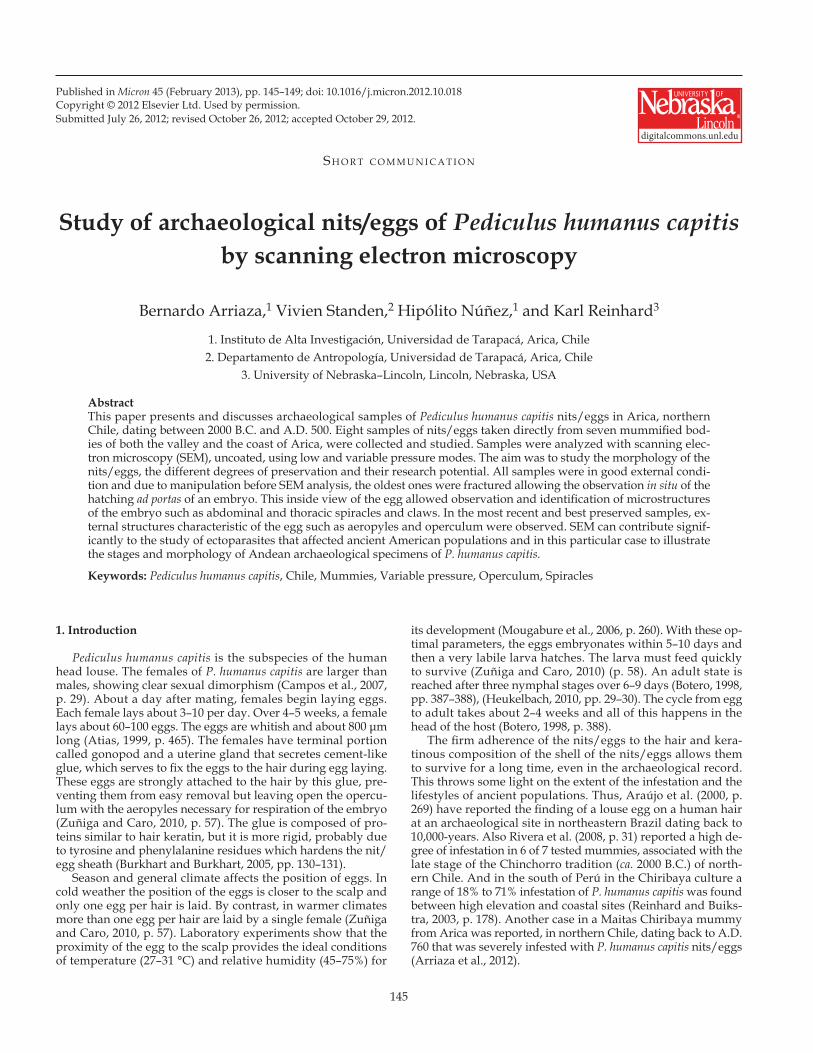
In Figure 3, specimen Cam15A sector 4, 97, a louse egg from the Formative Period, the following structures are visible: A, C, H, and OP. In addition, there is a fracture of the egg resulting from manipulation and positioning the nit/egg onto the stub. The split (microfracture) of the egg is not wide enough to al-low the observation of anatomical structures of an embryo of P. humanus capitis.
In Figure 4, specimen Az14T16, a louse egg from the Forma-tive Period, the following structures are visible: A, C, and OP. There is a post-mortem fracture allowing visualization of the in-side of the egg, showing part of a claw (CL). The egg morphol-ogy, size and shape are consistent with an embryonated P. hum-anus capitis egg.
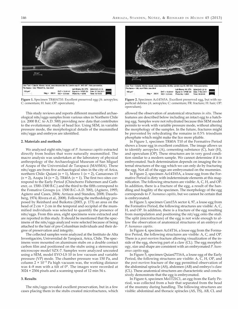
In Figure 5, specimen Quiani7T16A, a louse egg of the Early Period, the following structures are visible: A, C, H, OP, and the post-mortem fracture of the egg permitted observation of the abdominal spiracle (AS), abdomen (AB) and embryo’s claw (CL). These anatomical structures are characteristic and conclu-sively demonstrate that the egg is embryonated.
In Figure 6, specimen Mo1T21C1, an egg from the Early Pe-riod, was collected from a hair that separated from the head of the mummy during handling. The following structures are visible: abdominal and thoracic spiracles (AS, TS), AB, CL and
Figure 1. Specimen TR40AT10. Excellent preserved egg (A: aeropyles; C: cementum; H: hair; OP: operculum).
Figure 2. Specimen Az14T65A. Excellent preserved egg, but with su-perficial debries (A: aeropyles; C: cementum; FR: fracture; H: hair; OP: operculum).
Sem S t u d y o f a r c h a e o l o g i c a l P e d i c u l u s h u M a n u s c a P i t i s n i t S / e g g S 147
Figure 3. Specimen Cam 15A sector 4 Pieza 97. Post-mortem fractured egg (A: aeropyles; C: cementum; FR: fracture; H: hair; OP: operculum).
Figure 4. Specimen Az14T16. Post-mortem fractured egg (A: aeropyles; C: cementum; FR: fracture; CL: claw; OP: operculum).
Figure 5. Specimen Quiani7T16A. Severe post-mortem fractured egg (A: aeropyles; AB: abdomen; AS: abdominal spiracle; CL: claw; FR: frac-ture; H: hair; OP: operculum).
Figure 6. Specimen Mo1T21C1. Severe post-mortem fractured egg and embryo (AB: abdomen; AS: abdominal spiracle; CL: claw; T: thorax; TS: thoracic spiracle).
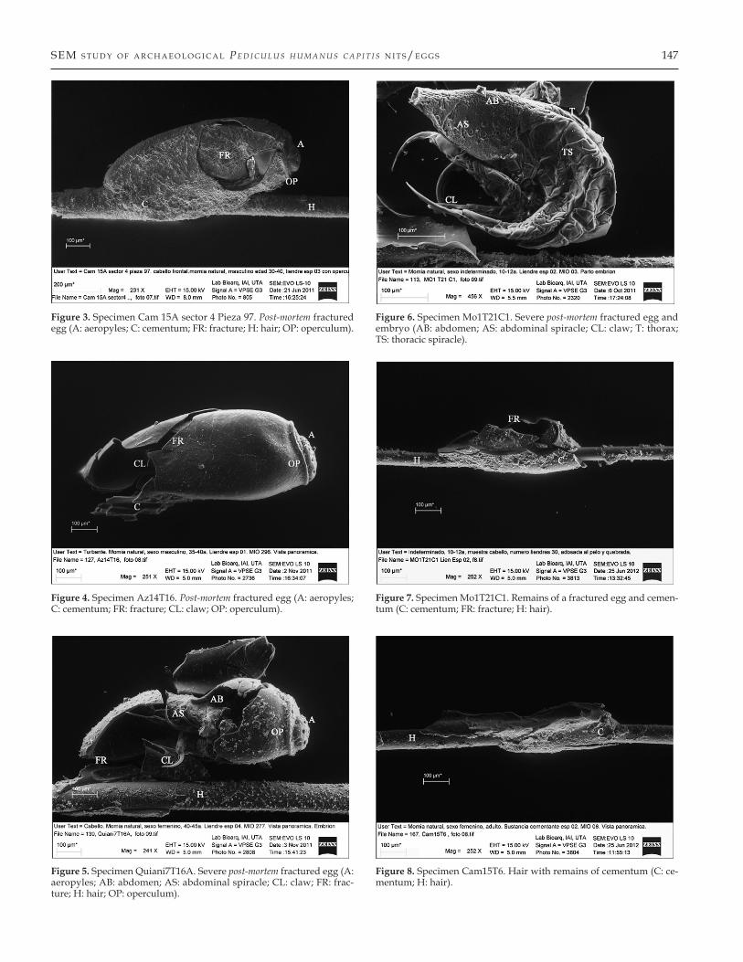
Figure 7. Specimen Mo1T21C1. Remains of a fractured egg and cemen-tum (C: cementum; FR: fracture; H: hair).
Figure 8. Specimen Cam15T6. Hair with remains of cementum (C: ce-mentum; H: hair).
148 a r r i a z a , S t a n d e n , n ú ñ e z , & r e i n h a r d i n M i c r o n 45 (2013)
thorax (T). The post-mortem fracture allowed the observation of anatomical structures that demonstrate the presence of an em-bryo of P. humanus capitis. This ectoparasite is more than 3000 years old, and shows a nymph on the verge of hatching from its egg in situ.
In Figure 7, specimen Mo1T21C1 a fractured louse egg from the Early Period. The fracture was likely caused by the combi-nation of the fragility of the specimen, rough manipulation and storage conditions. Even though the louse egg is broken the ce-mentum substance appears intact. The following structures are visible: C, H, and a fracture (FR).
In Figure 8, specimen Cam15T6, hair and cement substance from the Formative Period, shows that minimal remains of the egg/nit are present, represented by glue residue.
4. Discussion
The archaeological samples of nits/eggs have varying de-grees of preservation and the images of SEM (Figures 1 & 2) reveal anatomical structures of louse eggs in perfect condition despite the passage of time and their handling during museum curation and laboratory research. In these images we can con-firm that the cementing substance is a very strong structure, in spite of time it still continues to hold the nits/eggs to the hair. In Figures 1–3, typical anatomical nits/eggs structures can be seen, but SEM analysis cannot indicate if these eggs are embry-onated because these eggs are intact. In contrast, Figures 4–6 show that the eggs are embryonated since the eggs are frac-tured. Interestingly, the eggs for which the embryo is observed, resulting from the breakdown of the egg, are the oldest archae-ological samples. This would indicate that their frailty would be related to the age of the specimen of about 3000 years. In addition, accidental fracture during handling shows an em-bryo-nymph in very good condition that was on the verge of hatching (Figure 6). In Figures 4–6 we can observe the micro-structures characteristics of P. humanus capitis, also the degree of internal preservation is much better than the egg itself. In life, the eggshell protects the embryo from different environ-mental pollutants and manipulation. After thousands of years, the egg gradually becomes weaker and loses its elasticity easily fracturing when subjected to a minimum pressure on a mount-ing stub. Interestingly, through SEM analysis of uncoated sam-ples, we can not only observe micromorphology, but also pre-serve the specimen for subsequent analysis such as DNA. The specimens can be mechanically or chemically removed from the stubs because they are not gold or carbon coated. Extraction of ancient DNA is difficult, and contamination is always a prob-lem, thus we also left some unmounted samples for future mo-lecular study, independent of the SEM work. Also, unhatched eggs are protected by their shells.
For quantifying infestation, the residue of the cementum may be most important. In Figure 1 and Figure 3 the cemen-tum substance can be clearly observed. In addition, in Figure 7 and Figure 8, although the egg shell is broken, the cementum still is attached to the hair, showing its resilience and durabil-ity. Ancient nit-picking, grooming, and washing may remove the eggs, but traces of the cement glue remain. Therefore, mum-mies that are apparently nit/egg free should be examined for traces of cementum to document infestation that was controlled by the time of death.
In summary, microscopic studies of archaeological spec-imens of the Americas are useful to verify the types of ecto-parasites that affected the daily lives of ancient populations and in this particular case, they contribute to the study of the stages and micromorphology of P. humanus capitis. The samples
shown here demonstrate that nits/eggs can become very brittle with time, but the cementum endures through time. The study of microsamples and SEM analyses can be a very useful tool to shed light onto ancient ectoparasites and human cultural be-havior. Our study of louse eggs in ancient mummies reveals heavy infestations by sex, age and cultural periods. Reinhard and Buikstra (2003) showed that louse infestation prevalence was very high in prehistoric Andean villages, sometimes reach-ing 70% at some villages, but most individuals were lightly in-fected. Most of the lice were found on a very few individuals. Indeed, 84% of the lice were recovered from 10% of the most heavily infested mummies. Arriaza et al. (2012) demonstrated that in this background in infestation prevalence, individuals could become overwhelmed with lice such that every centime-ter of the scalp was covered with lice. Atacama Desert temper-atures decrease significantly at night and in antiquity housing raw materials were minimal, thus people lived in small huts and in crowding conditions. Cultural innovation could also be at play. Early fishing populations did not manufactured hair combs making it more difficult to delouse and control head lice infestations. Lastly, most Andeans wore long hair and during agropastoral times farmers had elaborate hairstyles and delous-ing combs. All these cultural variables influenced the likelihood and degree of head louse infestations. The combined research of these workers shows that cultural patterns such as groom-ing, headwear, and hair style created unusual epidemiological patterns. Prehistoric Andean prevalence patterns were the op-posite of louse infestation in the modern world: children were least infected and men were most infected. Using SEM methods as presented here, we will be able to expand our studies to look for traces of infestation represented by cementum that may re-veal prehistoric success at controlling infestations. With archae-ological evidence of individual status, we will be able to assess the risk level posed by poverty in prehistoric times. In essence, SEM analysis opens a more detailed method to assess the nu-ances of infestation related to behavior.
Acknowledgments — Project Fondecyt No 1100059, Convenio de Desempeño UTA–MINEDUC and FIC Regional EQU-19. Special thanks to Natalia Aravena and Octavio Lagos for their technical assistant.
References
Agüero, C., 1995. Indicadores textiles de grupos formativos: Proposición de una tipología de turbantes. In: Actas del XIII Congreso Nacional de Arqueología. Hombre y Desierto 9, Tomo II, pp. 97–110.
Agüero, C., Cases, B., 2004. Quillahua y los textiles formativos del norte grande de Chile. Chungara 36 (suplemento espe-cial 2), 599–617.
Araújo, A., Ferreira, L.F., Guidon, N., Maues Da Serra Freire, N., Reinhard, K.J., Dittmar, K., 2000. Ten thousand years of head lice infection. Parasitology Today 16 (7), 269.
Arriaza, B., Orellana, N., Barbosa, H., Menna-Barreto, R., Araújo, A., Standen, V., 2012. Severe head lice infestation in an Andean mummy of Arica, Chile. Journal of Parasitol-ogy 98 (2), 433–436.
Arriaza, B., Standen, V., 2008. Bioarqueología. Editorial Univer-sitaria, Santiago.
Atias, A., 1999. Piojos y Pulgas. In: Parasitología Médica. Editorial
Sem S t u d y o f a r c h a e o l o g i c a l P e d i c u l u s h u M a n u s c a P i t i s n i t S / e g g S 149
Mediterráneo, Santiago, Capítulo 52, 465–470. Botero, D., 1998. Parasitosis Humanas: Conceptos Generales Sobre
Parasitología, Cuarta Edición. Corporación para Investigacio-nes Biológicas, CIB, Medellín, CO, pp. 387–389.
Burkhart, C.N., Burkhart, C.G., 2005. Head lice: Scientific as-sessment of the nit sheath with clinical ramifications and therapeutic options. Journal American Academy Dermatology 53 (1), 129–133.
Campos, B., Jofré, L., Neira, P., Noemi, I., Saavedra, T., San Martin, A., 2007. Guía clínica Sarna y Pediculosis. Subsecre-taria de Salud Pública. Ministerio de Salud, Gobierno de Chile, pp. 27–46.
Dauelsberg, P., 1974. Excavaciones arqueológicas en Quiani. Chungara 4, 7–38.
Heukelbach, J., 2010. Management and Control of Head Lice Infes-tations. Editorial UNI-MED Verlag AG, Bremen.
Mougabure, G., Zerba, E., Picollo, M., 2006. Embryonic devel-opment of human lice: Rearing conditions and suscepti-bility to spinosad. Memorias Instituto Oswaldo Cruz 101 (3), 257–261.
Reinhard, K., Buikstra, J., 2003. Louse infestation of the Chiri-baya culture southern Perú: Variation in prevalence by age and sex. Memórias do Instituto Oswaldo Cruz 98, 173–179.
Rivera, M., Mumcuoglu, K., Matheny, R., Matheny, D., 2008. Head lice eggs, Anthropophthirus capitis, from mummies of the Chinchorro tradition, Camarones 15D, northern Chile. Chungara. Revista de Antropología Chilena 40, 31–39.
Zũniga, I., Caro, J., 2010. Pediculosis: Una ectoparasitosis emer-gente en México. Revista de Enfermedades Infecciosas en Pedi-atría XXIV (94), 56–63.
[Appendix A, with 5 Supplemental Figures, follows.]
Glossary
A: aeropylesAB: abdomenAS: abdominal spiracleC: cementumCL: clawFR: fractureH: hairOP: operculumT: thoraxTS: thoracic spiracle
Suppl. a r r i a z a , S t a n d e n , n ú ñ e z , & r e i n h a r d i n M i c r o n 45 (2013)
Appendix A. Supplementary data
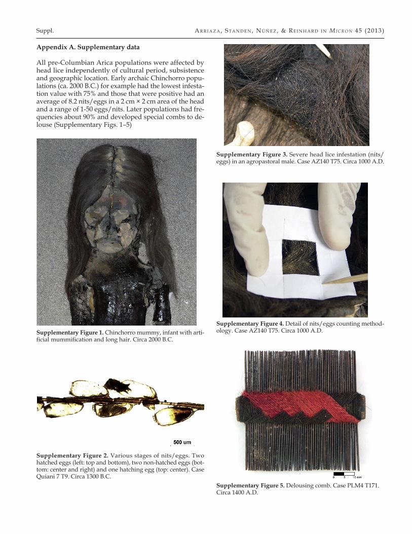
All pre-Columbian Arica populations were affected by head lice independently of cultural period, subsistence and geographic location. Early archaic Chinchorro popu-lations (ca. 2000 B.C.) for example had the lowest infesta-tion value with 75% and those that were positive had an average of 8.2 nits/eggs in a 2 cm × 2 cm area of the head and a range of 1-50 eggs/nits. Later populations had fre-quencies about 90% and developed special combs to de-louse (Supplementary Figs. 1–5)
Supplementary Figure 1. Chinchorro mummy, infant with arti-ficial mummification and long hair. Circa 2000 B.C.
Supplementary Figure 2. Various stages of nits/eggs. Two hatched eggs (left: top and bottom), two non-hatched eggs (bot-tom: center and right) and one hatching egg (top: center). Case Quiani 7 T9. Circa 1300 B.C.
Supplementary Figure 3. Severe head lice infestation (nits/eggs) in an agropastoral male. Case AZ140 T75. Circa 1000 A.D.
Supplementary Figure 4. Detail of nits/eggs counting method-ology. Case AZ140 T75. Circa 1000 A.D.
Supplementary Figure 5. Delousing comb. Case PLM4 T171. Circa 1400 A.D.


![Clinical and videodermoscopic look of pediculosis corporis....humanus corporis in detail on all sites infected with lice [3]. Figure 2: Dermoscopy of a louse of Pediculus humanus corporis](https://img.pdfslide.net/doc/110x75/6088caa7aa1079274b4aef57/clinical-and-videodermoscopic-look-of-pediculosis-humanus-corporis-in-detail.jpg)