Embed Size (px)
Citation preview
67Arq Bras Oftalmol. 2012;75(1):67-70
Relato de Caso | Case RepoRt
INTRODUCTIONLoa loa infestation fits in the category of helminthic diseases
called filariases. It is popularly known as “African eye worm”, due to a peculiar ocular manifestation of the disease(1). The adult worms live freely in the subcutaneous space of humans and occasionally may migrate into the subconjunctival space(2) where they invariably produce exuberant symptoms such as ocular pain, pruritus, tearing and foreign body sensation(3). The adult worm has been described also in the anterior chamber of the eye and in the subcutaneous of the eyelids(4). Other common manifestations are the so-called Calabar swellings: localized angioedemas found predominantly in the limbs, near the joints(5).
The nematode is transmitted by the bite of an infected Chrysops fly. Both the agent and vector are originally found only in equatorial Africa(1). The disease affects millions of people in that region but is rarely found in other continents, generally in African immigrants or travelers (the disease may remain asymptomatic for several years)(1). In fact, there are only case reports in sporadic countries: USA, Ger-many, Spain, Italy, Norway, Korea, Australia, to name a few(2,4-11) (Table 1). This is the first case described in Brazil.
CASE REPORTA 33 year-old woman presented to our emergency service com-
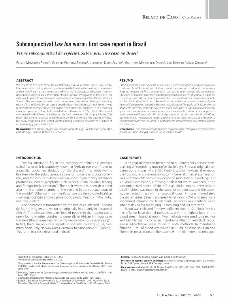
plaining of “something moving” in the left eye. She was original from Cameroon and was living in São Paulo, Brazil, for five years. She denied previous ocular or systemic symptoms. General physical examination was unremarkable with no evidence of subcutaneous swellings. On slit lamp examination, a moving opalescent worm was seen in the subconjunctival space of the left eye. Under topical anesthesia, a small incision was made in the superior conjunctiva and the worm was extracted intact with a forceps (Figure 1). It was immediately placed in saline (later transferred to ethanol 70%) and sent for a specialized Parasitology Department. The worm was identified as an adult male Loa loa measuring 33 mm long and 0.4 mm wide.
Blood was collected from two different sites at 12 o’clock (Loa loa microfilariae have diurnal periodicity, with the highest load in the blood stream found at noon). Two methods were used to search for and identify the microfilariae: membrane filtration and thick blood smear. Microfilariae were found in both methods. In membrane filtration, 1 mL of blood was diluted in 10 mL of saline solution and filtered in polycarbonate filters with 25 mm diameter and micropo-
Subconjunctival Loa loa worm: first case report in BrazilVerme subconjuntival da espécie Loa loa: primeiro caso no Brasil
Renato Magalhães Passos1, CaRolina PelegRini BaRBosa1, Juliana de souza alMeida2, guilheRMe MaeRsChneR ogawa3, luis MaRCelo aRanha CaMaRgo4
Submitted for publication: February, 11, 2011 Accepted for publication: September 14, 2011
Study carried out at the Departamento de Oftalmologia da Universidade Federal de São Paulo - UNIFESP - and at the Instituto de Ciências Biológicas da Universidade de São Paulo - USP - Porto Velho (RO).
1 Physician, Department of Ophthalmology, Universidade Federal de São Paulo - UNIFESP - São Paulo (SP), Brazil.
2 Biomedical, Department of Medicine, Faculdade São Lucas, Porto Velho (RO), Brazil.3 Biologist, Biomedical Science Institute 5, Universidade de São Paulo - USP - Rondônia, Brazil.4 Physician, Biomedical Science Institute 5, Universidade de São Paulo - USP - Rondônia, Brazil.
Funding: No specific financial support was available for this study.
Disclosure of potential conflicts of interest: R.M.Passos, None; C.P.Barbosa, None; J.S.Almeida, None; G.M.Ogawa, None; L.M.A.Camargo, None.
Correspondence address: Renato M. Passos. Rua Botucatu, 821 - São Paulo (SP) - 04024-062 - Brazil - E-mail: [email protected]
ABSTRACTWe report the first case of ocular infestation by Loa loa in Brazil. Loiasis is caused by infestation with Loa loa, a filarial parasite originally found in the rainforests of western and central Africa. It is transmitted by the bite of the fly Chrysops and has been recently described in other places other than Africa, in African immigrants or travellers. Our case is a 33 year-old woman from Cameroon who was living in São Paulo, Brazil, for 5 years. She was asymptomatic until one morning she started feeling “something moving” in the left eye. Under topical anesthesia, on the slit lamp, a moving worm was removed from the subconjunctival space, which later was confirmed to be a male Loa loa adult specimen. Blood tests revealed microfilaraemia of 129 mf/mL. The patient was treated with 400 mg oral albendazole for 3 weeks and 60 mg prednisone. This report illustrates an unusual ocular disease, which is extremely rare outside of Africa, but easily diagnosed and treated. Ophthalmologists should be aware of it, in face of an increasingly globalized world.
Keywords: Loa; Loiasis; Conjunctival diseases/parasitology; Eye infections, parasitic/parasitology; Filariosis; Brazil; Case reports
RESUMOEste é o primeiro relato na literatura nacional e internacional de infestação ocular por Loa loa no Brasil. A loíase é uma filariose causada pelo parasita Loa loa, encontrado nas florestas tropicais da África equatorial. A transmissão se dá pela picada do mosquito Chrysops e casos têm sido descritos em países não africanos, em imigrantes e viajantes. O presente caso trata-se de uma paciente de 33 anos natural de Camarões e residente em São Paulo, Brasil, há 5 anos. Até então assintomática, uma manhã sentiu algo “se mexendo” em seu olho esquerdo. Sob anestesia tópica, na lâmpada de fenda, um verme altamente móvel foi removido do espaço subconjuntival e enviado para identificação, que confirmou tratar-se de um espécime macho adulto de Loa loa. Testes sanguíneos revelaram microfilaremia de 129 mf/mL. A paciente foi tratada com albendazol 400 mg e prednisona 60 mg esquema regressivo por 3 semanas. Este relato ilustra uma doença excepcionalmente rara no Brasil, e praticamente desconhecida dos oftalmologistas em nosso país.
Descritores: Loa; Loíase; Doenças da túnica conjuntiva/parasitologia; Infecções oculares parasitárias/parasitologia; Filariose; Brasil; Relatos de casos
Subconjunctival Loa Loa worm: firSt caSe report in brazil
68 Arq Bras Oftalmol. 2012;75(1):67-70
Figure 1. Highly mobile worm in the subconjunctival space and its removal through small incision under topical anesthesia.
Table 1. A few case reports of ocular Loiasis outside Africa
PatientOcular
manifestationsExtra-ocular
manifestationsLast exposure in endemic region Treatment Reference
Brazil, african 33yo immigrant
Subconjunctival worm
None 5 years Worm removal + albendazole +
prednisone
Passos et al, 2011
England, african 21yo medical student
Subconjunctival worm
Calabar swelling in the elbow
6 years Worm removal + diethylcarbamazine +
prednisolone
Bowler et al, 2011(8)
Italy, swiss 33yo woman
Subconjunctival worm
None 7 years Worm removal Aiello et al, 2010(7)
Norway, norwegian 38yo woman
Subconjunctival worm
Sporadic episodes of worm movement in
subcutaneous tissues
Worked regularly in african countries
Worm removal + diethylcarbamazine
Varhaug, 2009(6)
Australia, nigerian 42yo immigrant
Subconjunctival worm
None 2 years Worm removal + albendazole +
prednisone
Jain et al, 2008(2)
South Korea, african 29yo immigrant
Subconjunctival worm
Calabar swelling on the forearm
5 years Worm removal + ivermectin
Cho et al, 2008(5)
Spain, african 24yo immigrant
Subconjunctival worm
None Visited Cameroon once a year
Worm removal López-Rodríguez et al, 2007(9)
USA, african 29yo immigrant
Subconjunctival worm
None Not described Worm removal Nam et al, 2008(10)
Germany, african 23yo student
Eyelid subcutaneous worm
Calabar swelling on the forearms
5 years Worm removal + diethylcarbamazine
Sbeity et al, 2006(11)
India, indian 48yo man
Anterior chamber worm
None Never went to Africa Worm removal + diethylcarbamazine +
prednisolone
Barua et al, 2005(4)
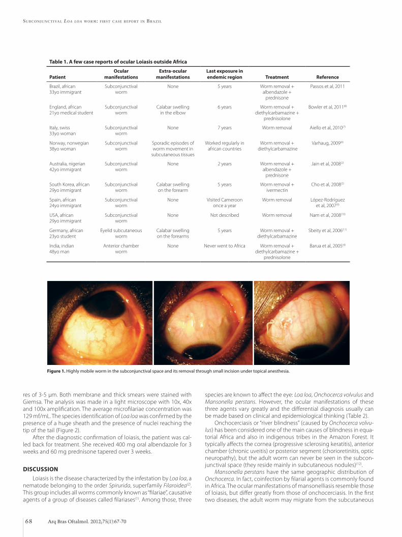
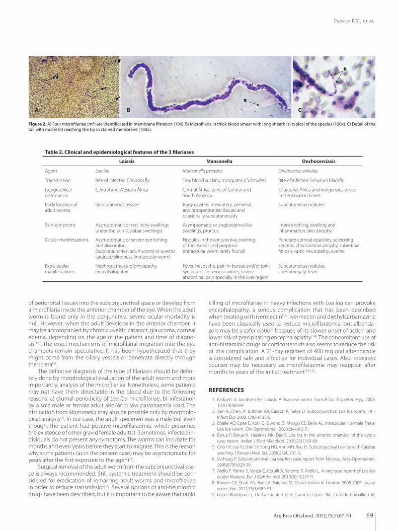
res of 3-5 µm. Both membrane and thick smears were stained with Giemsa. The analysis was made in a light microscope with 10x, 40x and 100x amplification. The average microfilariae concentration was 129 mf/mL. The species identification of Loa loa was confirmed by the presence of a huge sheath and the presence of nuclei reaching the tip of the tail (Figure 2).
After the diagnostic confirmation of loiasis, the patient was cal-led back for treatment. She received 400 mg oral albendazole for 3 weeks and 60 mg prednisone tapered over 3 weeks.
DISCUSSIONLoiasis is the disease characterized by the infestation by Loa loa, a
nematode belonging to the order Spirurida, superfamily Filaroidea(2). This group includes all worms commonly known as “filariae”, causati ve agents of a group of diseases called filariases(1). Among those, three
species are known to affect the eye: Loa loa, Onchocerca volvulus and Mansonella perstans. However, the ocular manifestations of these three agents vary greatly and the differential diagnosis usually can be made based on clinical and epidemiological thinking (Table 2).
Onchocerciasis or “river blindness” (caused by Onchocerca volvu-lus) has been considered one of the main causes of blindness in equa -torial Africa and also in indigenous tribes in the Amazon Forest. It typically affects the cornea (progressive sclerosing keratitis), anterior chamber (chronic uveitis) or posterior segment (chorioretinitis, optic neuropathy), but the adult worm can never be seen in the subcon-junctival space (they reside mainly in subcutaneous nodules)(12).
Mansonella perstans have the same geographic distribution of Onchocerca. In fact, coinfection by filarial agents is commonly found in Africa. The ocular manifestations of mansonelliasis resemble those of loiasis, but differ greatly from those of onchocerciasis. In the first two diseases, the adult worm may migrate from the subcutaneous
Passos RM, et al.
69Arq Bras Oftalmol. 2012;75(1):67-70
Figure 2. A) Four microfilariae (mf) are identificated in membrane filtration (10x). B) Microfilaria in thick blood smear with long sheath (s) typical of the species (100x). C) Detail of the tail with nuclei (n) reaching the tip in stained membrane (100x).
A B C
Table 2. Clinical and epidemiological features of the 3 filariases
Loiasis Mansonella Onchocerciasis
Agent Loa loa Mansonella perstans Onchocerca volvulus
Transmission Bite of infected Chrysops fly Tiny blood sucking mosquitos (Culicoides) Bite of infected Simulium blackfly
Geographical distribution
Central and Western África Central África, parts of Central and South America
Equatorial África and indigenous tribes in the Amazon Forest
Body location of adult worms
Subcutaneous tissues Body cavities, mesentery, perirenal, and retroperitoneal tissues and ocasionally subcutaneously
Subcutaneous nodules
Skin symptoms Asymptomatic or red, itchy swellings under the skin (Calabar swellings)
Asymptomatic or angioedema-like swellings, pruritus
Intense itching, swelling and inflammation, skin atrophy
Ocular manifestations Asymptomatic or severe eye itching and discomfort (subconjunctival adult worm) or uveitis/cataract/blindness (intraocular worm)
Nodules in the conjunctiva, swelling of the eyelids and proptosis (intraocular worm rarely found)
Punctate corneal opacities, sclerosing keratitis, chorioretinal atrophy, subretinal fibrosis, optic neuropathy, uveitis
Extra-ocular manifestations
Nephropathy, cardiomyopathy, encephalopathy
Fever, headache, pain in bursae and/or joint synovia, or, in serous cavities, severe abdominal pain specially in the liver region
Subcutaneous nodules, adenomegaly, fever
of periorbital tissues into the subconjunctival space or develop from a microfilaria inside the anterior chamber of the eye. When the adult worm is found only in the conjunctiva, severe ocular morbidity is null. However, when the adult develops in the anterior chamber, it may be accompanied by chronic uveitis, cataract, glaucoma, corneal edema, depending on the age of the patient and time of diagno-sis(3,4). The exact mechanisms of microfilarial migration into the eye chambers remain speculative. It has been hypothesized that they might come from the ciliary vessels or penetrate directly through the sclera(5).
The definitive diagnosis of the type of filariasis should be defini-tely done by morphological evaluation of the adult worm and more importantly, analysis of the microfilariae. Nonetheless, some patients may not have them detectable in the blood due to the following reasons: a) diurnal periodicity of Loa loa microfilariae, b) infestation by a sole male or female adult and/or c) low parasitaemia load. The distinction from Mansonella may also be possible only by morpholo-gical analysis(1). In our case, the adult specimen was a male but even though, the patient had positive microfilaraemia, which presumes the existence of other gravid female adult(s). Sometimes, infected in-dividuals do not present any symptoms. The worms can incubate for months and even years before they start to migrate. This is the reason why some patients (as in the present case) may be asymptomatic for years after the first exposure to the agent(1).
Surgical removal of the adult worm from the subconjunctival spa-ce is always recommended. Still, systemic treatment should be con-sidered for eradication of remaining adult worms and microfilariae in order to reduce transmission(1). Several options of anti-helminthic drugs have been described, but it is important to be aware that rapid
killing of microfilariae in heavy infections with Loa loa can provoke en cephalopathy, a serious complication that has been described when treating with ivermectin(13). Ivermectin and diethylcarbamazine have been classically used to reduce microfilaraemia, but albenda-zole may be a safer option because of its slower onset of action and lower risk of precipitating encephalopathy(14). The concomitant use of anti-histaminic drugs or corticosteroids also seems to reduce the risk of this complication. A 21-day regimen of 400 mg oral albendazole is considered safe and effective for individual cases. Also, repeated courses may be necessary, as microfilaraemia may reappear after months to years of the initial treatment(13,14).
REFERENCES 1. Padgett JJ, Jacobsen KH. Loiasis: African eye worm. Trans R Soc Trop Med Hyg. 2008;
102(10):983-9. 2. Jain R, Chen JY, Butcher AR, Casson R, Selva D. Subconjunctival Loa loa worm. Int J
Infect Dis. 2008;12(6):e133-5. 3. Eballe AO, Epée E, Koki G, Owono D, Mvogo CE, Bella AL. Intraocular live male filarial
Loa loa worm. Clin Ophthalmol. 2008;2(4):965-7. 4. Barua P, Barua N, Hazarika NK, Das S. Loa loa in the anterior chamber of the eye: a
case report. Indian J Med Microbiol. 2005;23(1):59-60. 5. Cho HY, Lee YJ, Shin SY, Song HO, Ahn MH, Ryu JS. Subconjuctival Loa loa with Calabar
swelling. J Korean Med Sci. 2008;23(4):731-3. 6. Varhaug P. Subconjunctival Loa loa: first case report from Norway. Acta Ophthalmol.
2009;87(8):929-30. 7. Aiello F, Palma S, Varesi C, Cerulli A, Valente R, Aiello L. A rare case report of Loa loa
ocular filariasis. Eur J Ophthalmol. 2010;20(1):237-9. 8. Bowler GS, Shah AN, Bye LA, Saldana M. Ocular loiasis in London 2008-2009: a case
series. Eye. 2011;25(3):389-91. 9. López-Rodríguez I, De-La-Fuente-Cid R, Carnero-López JM, Cordido-Carballido M,
Subconjunctival Loa Loa worm: firSt caSe report in brazil
70 Arq Bras Oftalmol. 2012;75(1):67-70
Zúñiga-Rodríguez C. Loiasis. [Approach to a form of ocular parasitosis]. Arch Soc Esp Oftalmol. 2007;82(1):55-8. Spanish
10. Nam JN, Reddy S, Charles NC. Surgical management of conjunctival loiasis. Ophthal Plast Reconstr Surg. 2008;24(4):316-7.
11. Sbeity ZH, Jaksche A, Martin S, Loeffl er KU. Loa loa macrofi lariasis in the eyelid: case report of the fi rst periocular subcutaneous manifestation in Germany. Graephes Arch Clin Exp Ophthalmol. 2006;244(7):883-4.
12. Newland HS, White AT, Greene BM, Murphy RP, Taylor HR. Ocular manifestations
of onchocerciasis in a rain forest area of west Africa. Br J Ophthalmol. 1991;75(3): 163-9.
13. Kombila M, Duong TH, Ferrer A, Perret JL, Marion MC, Nguiri C, et al. Short- and long-term action of multiple doses of ivermectin on loiasis microfi laremia. Am J Trop Med Hyg. 1998;58(4):458-60.
14. Klion AD, Massougbodji A, Horton J, Ekoué S, Lanmasso T, Ahouissou NL, Nutman TB. Albendazole in human loiasis: results of a double-blind, placebo-controlled trial. J Infect Dis. 1993;168(1):202-6.