Embed Size (px)
Citation preview
Romanian Neurosurgery (2011) XVIII 3: 331 – 339 331
Subdural hematoma and arachnoid cyst - case report
L. Nuteanu, A.Tascu, A.V. Ciurea
“Bagdasar-Arseni”Clinical Emergency Hospital, Bucharest, Romania
Abstract We present a case of a previously
asymptomatic 15-year-old boy who was examined in the emergency services unit for syndrome of intracranial hypertension. He was addmited for headache, vertigo, vomiting and somnolence, GCS 14, and on local examination no traumatic signs. On admission, a CT-scan of his head revealed left fronto-temporo-parietal expansive process with important mass effect to the right. The patient underwent a left-sided craniotomy for evacuation of the subdural hematoma as well as the intracystic hematoma and cyst fenestration into the basal cisterns. The patient tolerated the procedure well and recovered completely. Intracranial arachnoid cysts are considered to be congenital malformations with a predilection for the temporal fossa. A review of clinical symptoms, etiology and mechanisms, diagnosis and treatment is made, followed by the necessary discussions. Taking into account the possible mechanisms of subdural hematomas in arachnoid cysts, in our opinion is logically to perform craniotomy with membranectomy, taking out the membrane from the vessels, in order to prevent another hemorrhage. Other authors propose subdural hematoma drainage without any specific treatment (shunt or fenestration) of the arachnoid cyst, for this category of patient. The diagnosis of a subdural hematoma superposed on a
arachnoid cyst can be tricky for a doctor from other specialty or for a young neurosurgeon. It is important to identify and report such rare complications with intracranial arachnoid cyst, so that asymptomatic patients with an intracranial arachnoid cyst can be counseled about such possibilities following head trauma.
Keywords: subdural hematomas, arachnoid cysts, fenestration, head trauma.
Introduction The Plymouth Neurosurgical Unit has
treated twenty patients with arachnoid cysts of the middle fossa (ACMF) between 1976-1985, seven of these being complicated by subdural haematoma (SDH). There was an age range of 11-56 years in those with SDH. Six of the sev-en patients with ACMF and SDH gave no significant trauma history. Four of these were males aged 11 to 20 years. In the SDH alone group 100% suffered major skull trauma and 80% had demonstrable skull fractures. In addition the patients with ACMF were compared with patients presenting with other supratentorial arachnoid cysts (AC) in Plymouth. Only ACMF were associated with the development of SDH in their study. Three patients demonstrated total masking of the ACMF by isodense intracystic haematoma on computed tomography. In two of these patients the presence of an ACMF was suspected due to plain radiographic and CT enlargement of
332 L. Nuteanu et al Subdural hematoma and arachnoid cyst
the middle fossa (12). Intracranial ACs are considered to be
congenital malformations with a predilection for the temporal fossa (18). ACs most frequently occur in the middle fossa, followed by the posterior fossa, convexity, and suprasellar regions (11, 14).
For the temporal cysts, the haematoma frequency is 6.5% (18).
Headache, convulsion, deformity of the cranium with bulging and thinning of the adjacent bone, focal neurological deficit, and mental retardation are known symptoms of intracranial AC (11).
The reported symptoms of AC and SDH are headache, followed by vomiting and he-miparesis in the patients with arachnoid cyst. Seizures are also reported (8). The most fre-quent symptoms are gait disturbance followed by hemiparesis in the SDH patients without AC (11).
Case report A previously asymptomatic 15-year-old
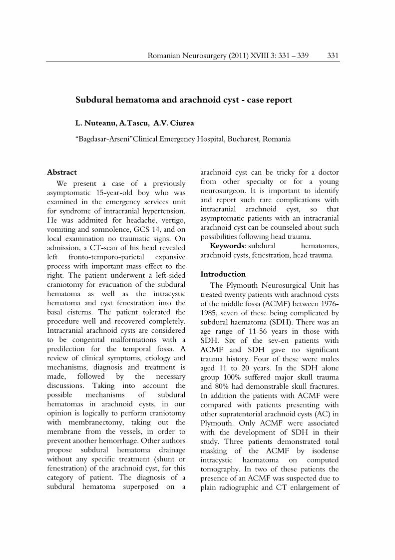
boy was examined in the emergency services unit for syndrome of intracranial hypertension. He reported no loss of consciousness and no external injuries. On examination, the patient was fully conscious and demonstrated no focal neurological deficit. The admission motivation was: headache, vertigo, vomiting and somno-lence; GCS was 14. On local examination: no traumatic signs.
On admission, a CT-scan of his head revealed left fronto-temporo-parietal expansive process of 92x103x70 mm, infiltrative, dense, important mass effect to the right.
Admission diagnostic: expansive process extradural, left fronto-temporo-parietal.
Romanian Neurosurgery (2011) XVIII 3: 331 – 339 333
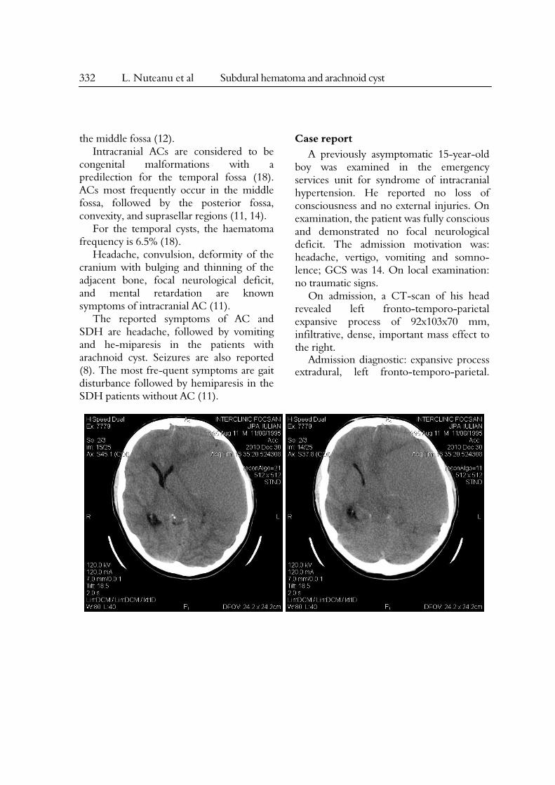
Figure 1 A The native CT-scan could not differentiate between tumor and liquid accumulation.
Figure 1 B The CT-scan with contrast can differentiate between tumor and liquid accumulation.
334 L. Nuteanu et al Subdural hematoma and arachnoid cyst
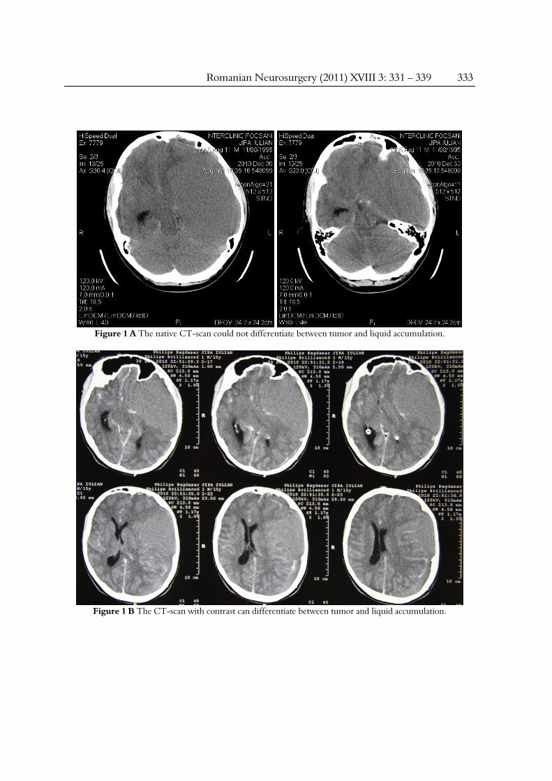
A. frontal images
B. sagital images
C. axial images
Figure 2 MRI with contrast: Chronic subdural hematoma and arachnoid cyst.
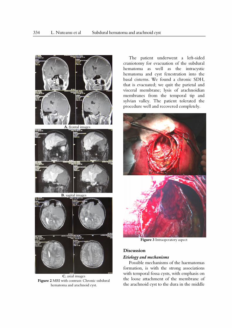
The patient underwent a left-sided craniotomy for evacuation of the subdural hematoma as well as the intracystic hematoma and cyst fenestration into the basal cisterns. We found a chronic SDH, that is evacuated; we quit the parietal and visceral membrane; lysis of arachnoidian membranes from the temporal tip and sylvian valley. The patient tolerated the procedure well and recovered completely.
Figure 3 Intraoperatory aspect
Discussion Etiology and mechanisms
Possible mechanisms of the haematomas formation, is with the strong associations with temporal fossa cysts, with emphasis on the loose attachment of the membrane of the arachnoid cyst to the dura in the middle
Romanian Neurosurgery (2011) XVIII 3: 331 – 339 335
fossa, and its possible role as an "extra wall" covering easy-bleeding vascular structures in the dura (18). Although these lesions are considered congenital, the exact etiology is still not clear. They are often asymptomatic but can sometimes be symptomatic due to enlargement or hemorrhage. There are multiple case reports of ACs becoming symptomatic with hemorrhagic complications following head trauma. In such cases, the bleeding often appears to the side ipsilateral to the AC. Occurrence of contralateral subdural hematomas in patients with temporal fossa AC has rarely been observed and is reported less frequently. It is likely that lack of adequate intracranial cushioning in the presence of an intracranial AC may result in injury not
only to ipsilateral but also to contralateral bridging veins, following head trauma.
Hemorrhage into an AC and the associated SDH following head trauma are well do-cumented, although the mechanism and true incidence are not clearly understood. The an-nual risk for hemorrhage in patients with a ACMF probably remains below 0.1%.
In a recent study by Wester et al. (18), the incidence of chronic subdural or intracystic haematomas was reported as 4.6% of all patients with intracranial AC referred for treatment. Some authors (14) propose two mechanisms leading to formation of subdural hemorrhage. First, the cyst membrane is loosely attached to the convexity dura.
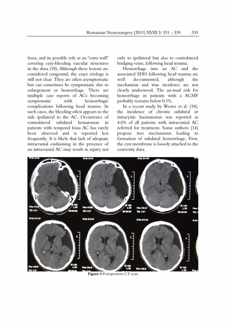
Figure 4 Postoperatory CT-scan
336 L. Nuteanu et al Subdural hematoma and arachnoid cyst
The mechanical forces that are sustained during a moderate head trauma can cause the cyst membrane to be detached from the dura and thus cause a bleeding episode. Second, the parietal cyst membrane also covers the area where the bridging Sylvian veins, or the veins that traverse the membrane unsupported by brain tissue, enter into the dural venous sinuses behind the sphenoid ridge. Even a moderate manipulation of the parietal membrane can disrupt these veins, leading to bleeding into subdural space (18).
Parsch and colleagues (13) suggest an approximately 5-fold greater prevalence (2.43% versus 0.46%) of ACMF in patients with chronic subdural hematomas (CSDH) than in the general population who undergo magnetic resonance imaging.
ACMF is now recognized as one of the causes of CSDH after head injury, especially in young people, as the cysts appear to be more susceptible to hemorrhagic complications, including subdural and intracystic hematomas. The membrane is vascular, and bridging veins are often observed traversing the cyst wall. This could in part explain the liability of intracystic subdural bleeding in these patients (13).
Occurrence of contralateral subdural hematomas with arachnoid cysts was reported by Mori et al. (two cases) (11) and Parsch et al. (one case) (13), Pillai et al. (two cases) (14). This finding reinforces the notion that an arachnoid cyst, being a large fluid-filled lesion, is less compliant than normal brain parenchyma, making both ipsilateral and contralateral bridging veins prone to injury. Even though it is rare, there have been reports of ruptured AC, presenting with a subdural CSF collection without evidence of hemorrhage (8). The
sudden collapse of the cyst can cause a sudden shift of the brain, which, along with the force of the trauma, can lead to stretching, and tearing of bridging veins on the opposite side. This could explain the occurrence of contralateral SDH. Thus, we should inform patients with AC and their families of the possibility of such complications and advise care to avoid head injury in daily life, regardless of the size and symptoms of the cyst (14).
The etiology of CSDH is not completely understood. Traumatic subdural effusion is widely accepted as a precursor stage in the formation of CSDH. Arachnoid tear is also ac-cepted as a causative factor in the development of traumatic subdural effusion, supported by the observation that CSDH tends to occur in elderly people because brain atrophy causes en-largement of the subarachnoid space and stretching of the bridging veins, and these preexist-ing conditions facilitate tearing of the arachnoid membrane and leakage of bloody CSF into the subdural space after mild head injury. CSF with or without blood leaks into the subdural space then facilitates the formation of the “outer membrane” under the dura mater, which forms internal capillaries or sinusoids. These blood vessels in the outer membrane are fene-strated and allow plasma fluid leakage and resultant enlargement of the subdural effusion. Bleeding then occurs repeatedly from the capillaries with degenerating endothelium accompanied by local hyperfibrinolysis, which is one of the causes of the development of effusions into CSDH (11). The rupture of arachnoid cyst can even occur spontaneously. Tearing of the outer wall of the arachnoid cyst is associated with subdural and/or intracystic hemorrhage caused by rupture of bridging veins,
Romanian Neurosurgery (2011) XVIII 3: 331 – 339 337
unsupported blood vessels around the cyst wall, and leptomeningeal vessels in the base of the cyst.
The association of CSDH and AC in the posterior fossa might be a coincidence. The studies are not conclusive and the subject needs further investigation (11).
CSDH tends to occur in elderly patients with a history of mild head injury at a few months prior to the onset of symptoms. CSDH in patients with previous arachnoid cysts ap-pear in younger ages. The clinical symptoms are also significantly different. The most fre-quent symptom is headache followed by vomiting in the patients with arachnoid cyst, while gait disturbance and hemiparesis predominated in patients without arachnoid cyst (11).
We can conclude that even a small AC can be a risk factor for CSDH after mild head injury in young patients and symptoms of increased intracranial pressure are common. The results of some authors (11) also suggest that CSDH formation may be preceded by subdural hygroma caused by the rupture of arachnoid cyst.
Accumulation of CSF outside the outer wall of the AC was found to induce the rupture of well-developed, fragile leptomeningeal vessels at the base of the middle cranial fossa. This phenomenon is noteworthy as a pitfall in surgery for intracranial arachnoid cyst and suggests the bleeding source of SDH occasionally associated with this lesion (1).
SDHs occur in association with previously asymptomatic ACMF. The aetiology of haematoma formation has not been conclusively described, but may partly result from a high pressure intracranial system with decreased compliance (15).
A retrospective review of data for 50 children (11) who underwent keyhole
craniotomy for fenestration of temporal arachnoid cysts between 1994 and 2001 was performed after institutional review board approval. During that period, the first-line treatment for all symptomatic middle fossa arachnoid cysts was microcraniotomy for fenestration. Microsurgical dissection to create communications between the cyst cavity and basal cisterns was the goal. Complications included spontaneously resolving pseudomeningocele (10%), transient Cranial Nerve III palsy (6%), cerebrospinal fluid leak (6%), subdural hematoma (4%), and wound infection (2%).
It was also described a case with subdural hematoma in AC after a birth delivery (3).
The rupture of an aneurysm into an arachnoid cyst and subdural space is unusual. A 25-year-old man was admitted 2 weeks after having undergone a burr hole drainage for a chronic subdural haematoma elsewhere. An angiogram revealed a small aneurysm at the bi-furcation of the middle cerebral artery. The aneurysm was clipped and the cyst communicated with the basal cisterns (9). Diagnosis
CT and/or MR imaging are performed in all patients to confirm the diagnosis. The history is of mild head injury, operation for arachnoid cyst, cystoperitoneal shunting and cyst opening (11).
When the hematoma was chronic and of equal hypodensity with the cyst, a clear-cut differentiation was not possible from the CT-scan. The presence of a SDH could only be sug-gested by thickened arachnoid structures crossing the hypodense area, indicating the wall between cyst and hematoma. The cyst could often be diagnosed by bulging of the skull bone and a temporal lobe defect. Differences in
338 L. Nuteanu et al Subdural hematoma and arachnoid cyst
density between cyst and hematoma, such as in subacute SDH, delineated both entities (2). Treatment
Hematoma evacuation through burr holes improves the symptoms in all patients with arachnoid cyst (11).
A microsurgical keyhole approach to arachnoid cyst fenestration is a safe effective method for treating middle fossa cysts. This procedure can be performed with minimal mor-bidity via a minicraniotomy. Compared with an endoscopic approach, better control of hemostasis can be obtained, because of the ability to use bipolar forceps and other standard instruments. The operative time and length of hospital stay are not excessively increased (10). Hematomas are usually evacuated and irrigated with normal saline through two burr holes and closed system subdural drainage was continued for one or two days after the operation (5, 11). Cystoperitoneal shunting surgery after the drainage surgery can be used if the arachnoid cyst cause mass effect to the surrounding brain (11).
All patients with CSDH and arachnoid cyst become symptom-free after the burr hole irrigation. Follow-up CT or MR imaging showed spontaneous disappearance of arachnoid cyst or decreased size of the arachnoid cyst (11).
Therapeutic recommendations can include fenestration or extirpation of the cyst wall, in addition to evacuation of the space-occupying lesion. No additional symptoms from the arachnoid cysts occurred in a follow-up period of up to 14 years after therapy (13).
Cysto-peritoneal shunt can be recommended as the surgical treatment after evacuation of the haematoma (15).
Old patients can also be treated conservatively (8).
Usually, before this event the cyst is
asymptomatic and unknown. Magnetic resonance imaging is the most contributive radiologic exam. Surgical procedure is usually limited to subdural hematoma evacuation. The internal wall of subdural hematoma can be opened (7).
The clinical outcome is good. The CT or MRI scan follow-up 3 months later reveal nearly total disappearance of subdural hematoma. Even the volume of arachnoid cyst can decrease (7), or even disappear, as in the case of a 7-year-old boy whose arachnoid cyst ruptured into the subdural space following a mild head injury and disappeared after draining the subdural haematoma by burr-holes (17).
No bleeding could be attributed to ruptured bridging veins. In two cases the source of bleeding was identified at the interface between the dura mater and the outer membrane at the temporal skull base. Some authors suggest that, even if wide outer membranectomy is probably not indicated, careful coagulation of the membrane at the skull base is necessary to avoid bleeding within the cyst (16).
Taking into account the possible mechanisms of CSDH in AC, in our oppinion is logically to perform craniotomy with membranectomy, taking out the membrane from the vessels, in order to prevent another hemorrhage.
Some authors propose subdural hematoma drainage without any specific treatment (shunt or fenestration) of the arachnoid cyst, for this category of patient (7).
The overwhelming majority of cases of acute SDH in the forensic setting occur as a result of head trauma. It is also a report of a case of sudden unexpected death in a middle-aged woman with a history of arachnoid cyst who had sudden spontaneous onset of severe headache that was rapidly followed by collapse and death.
Romanian Neurosurgery (2011) XVIII 3: 331 – 339 339
A postmortem multiple-slice computed tomographic scan showed a large acute subdural hematoma associated with hemorrhage into an arachnoid cyst. Subdural hemorrhage is an uncommon but well-described complication of an AC (4).
Conclusions The diagnosis of a SDH superposed on
a AC can be tricky for a doctor from other specialty or for a young neurosurgeon.
It is important to identify and report such rare complications with intracranial AC, so that asymptomatic patients with an intracranial AC can be counseled about such possibilities following head trauma (8, 14).
Rarely, posttraumatic or spontaneous rupture of ACs can result in intracystic hemorr-hage, SDH or subdural hygroma. The patients usually harbor ACMF and suffered mild closed head injuries (6).
Correspondent author: Dr. Al. Tascu “Bagdasar-Arseni” Clinical Emergency Hospital, Bucharest, Romania [email protected]
References 1. Aoki N, Sakai T: Intraoperative subdural hematoma in a patient with arachnoid cyst in the middle cranial fossa. Childs Nerv Syst, 6(1):44-6, 1990. 2. Auer LM, Gallhofer B, Ladurner G, Sager WD, Heppner F, Lechner H: Diagnosis and treatment of middle fossa arachnoid cysts and subdural hematomas. J Neurosurg, 54(3):366-9, 1981. 3. Bilginer B, Onal MB, Oguz KK, Akalan N: Arachnoid cyst associated with subdural hema-toma: report of three cases and review of the literature. Childs Nerv Syst, 25(1):119-24, 2009. 4. Burke MP, O'Donnell C, Opeskin K: Spontaneous acute subdural hematoma complicating arachnoid cyst. Am J Forensic Med Pathol. 31(4):382-4, 2010. 5. Domenicucci M, Russo N, Giugni E, Pierallini A: Relationship between supratentorial arachnoid cyst and chronic subdural hematoma: neuroradiological evidence and surgical treatment. J Neurosurg, 110(6):1250-5, 2009. 6. Donaldson JW, Edwards-Brown M, Luerssen TG:
Arachnoid cyst rupture with concurrent subdural hygroma. Pediatr Neurosurg. 32(3):137-9, 2000. 7. Fuentes S, Palombi O, Pouit B, Bernard C, Desgeorges M: (Arachnoid cysts of the middle fossa and associated subdural hematoma. Three case reports and review of the literature). (Article in French) Neurochirurgie, 46(4):376-82, 2000. 8. Gelabert-González M, Castro-Bouzas D, Arcos-Algaba A, Santín-Amo JM, Díaz-Cabanas L, Serramito-García R, Arán-Echabe E, Prieto-González A, García-Allut A: (Chronic subdural hematoma associated with arachnoid cyst. Report of 12 cases). Neurocirugia (Astur). 21(3):222-7, 2010. (Article in Spanish) 9. Kocaeli H, Korfali E: Rupture of a small middle cerebral artery aneurysm into middle fossa arachnoid cyst presenting as a chronic subdural haematoma. Acta Neurochir (Wien), 150(4):407-8, 2008. 10. Levy ML, Wang M, Aryan HE, Yoo K, Meltzer H: Microsurgical keyhole approach for middle fossa arachnoid cyst fenestration. Neurosurgery, 53(5):1138-44; 2003. 11. Mori K, Yamamoto T, Horinaka N, Maeda M: Arachnoid Cyst Is a Risk Factor for Chronic Subdural Hematoma in Juveniles: Twelve Cases of Chronic Subdural Hematoma Associated with Arachnoid Cyst. Journal of Neurotrauma, 19, 9, 2002. 12. Page AC, Mohan D, Paxton RM: Arachnoid cysts of the middle fossa predispose to sub-dural haematoma formation fact or fiction? Acta Neurochir Suppl (Wien), 42:210-5, 1988. 13. Parsch CS, Krauss J, Hofmann E, Meixensberger J, Roosen K: Arachnoid cysts associated with subdural hematomas and hygromas: analysis of 16 cases, long-term follow-up, and review of the literature. Neurosurgery, 40(3):483-90, 1997. 14. Pillai P, Menon SK, Manjooran RP, Kariyattil R, Pillai AB, Panikar D: Temporal fossa arachnoid cyst presenting with bilateral subdural hematoma following trauma: two case reports. Journal of Medical Case Reports, 3:53, 2009. 15. Rogers MA, Klug GL, Siu KH: Middle fossa arachnoid cysts in association with subdural haematomas. A review and recommendations for management. Br J Neurosurg, 4(6):497-502, 1990. 16. Servadei F, Vergoni G, Frattarelli M, Pasini A, Arista A, Fagioli L: Arachnoid cyst of middle cranial fossa and ipsilateral subdural haematoma: diagnostic and therapeutic implications in three cases. Br J Neurosurg, 7(3):249-53, 1993. 17. Yilmaz C, Cetinalp E, Caner H, Altinors N: Dissapearance of arachnoid cyst after rupturing into subdural space. Acta Neurochir (Wien), 149(7):731-3, 2007. 18. Wester K, Helland CA: How often do chronic extra-cerebral haematomas occur in pa-tients with intracranial arachnoid cysts? J Neurol Neurosurg Psychiatry. 79(1):72-5, 2008.