Embed Size (px)
Citation preview
Substitution of 68
Ga-DOTA-peptide
PET/CT scanning in lieu of Octreotide for patients undergoing
somatostatin receptor diagnostic
imaging under MBS item 61369
February 2017
Mini assessment report
VERSION CONTROL
DOCUMENT HISTORY
Version Number
Date Changed Author Reason for Change
1.0 10-Feb-2017 Adelaide Health Technology Assessment
Draft of mini assessment
2.0 23-Feb-2017 Adelaide Health Technology Assessment
Final of mini-assessment
DOCUMENT APPROVAL
Version Number
Date Changed Author Reason for Change
1.0
© Commonwealth of Australia 2017
ISSN (Online) 1443-7139 Internet site http://www.msac.gov.au/ This work is copyright. You may download, display, print and reproduce this material in unaltered form only (retaining this notice) for your personal, non-commercial use or use within your organisation. Apart from any use as permitted under the Copyright Act 1968, all other rights are reserved. Requests and inquiries concerning reproduction and rights should be addressed to Commonwealth Copyright Administration, Attorney-General's Department, Robert Garran Offices, National Circuit, Barton ACT 2600 or posted at http://www.ag.gov.au/. Electronic copies of the report can be obtained from the Medical Service Advisory Committee’s Internet site at http://www.msac.gov.au/ Enquiries about the content of the report should be emailed to [email protected]. The technical information in this document is used by the Medical Services Advisory Committee (MSAC) to inform its deliberations. MSAC is an independent committee which has been established to provide advice to the Minister for Health on the strength of evidence available on new and existing medical technologies and procedures in terms of their safety, effectiveness and cost effectiveness. This advice will help to inform government decisions about which medical services should attract funding under Medicare. MSAC’s advice does not necessarily reflect the views of all individuals who participated in the MSAC evaluation. This min-assessment was prepared by Dr Judy Morona and Dr Ruchi Mittal from Adelaide Health Technology Assessment (AHTA). Clinical advice was provided by A/Prof Paul Roach, Dr David Wyld and Prof Nick Pavlakis. The mini-assessment was commissioned by the Australian Government Department of Health. It was edited by Dr Debra Gum and Ms Skye Newton. The suggested citation for this document is: Morona JK, Mittal R. (2017). Substitution of
68Ga-DOTA-peptide PET/CT scanning in lieu of Octreotide for
patients undergoing somatostatin receptor diagnostic imaging under MBS item 61369. Mini-assessment Report. Commonwealth of Australia, Canberra, ACT.

MSAC mini CA for MBS item no. 61369 v
CONTENTS
Version Control ............................................................................................................................ ii
Document History .................................................................................................................... ii
Document Approval ................................................................................................................. ii
Contents ............................................................................................................................ v
Tables .................................................................................................................................... viii
Figures ..................................................................................................................................... x
Executive Summary .................................................................................................................... 11
Substitution of 68Ga-DOTA-peptide PET/CT scanning in lieu of 111In-octreotide
SPECT/CT for patients undergoing somatostatin receptor diagnostic imaging under
MBS item 61369 .................................................................................................................... 11
Alignment with agreed PICO Confirmation ................................................................. 11
Proposed Medical Service ........................................................................................... 11
Proposal for Public Funding ........................................................................................ 12
Population ................................................................................................................... 12
Comparator Details ..................................................................................................... 13
Clinical management algorithm .................................................................................. 13
Key Differences in the Delivery of the Proposed Medical Service and the Main
Comparator ................................................................................................................. 14
Clinical Claim ............................................................................................................... 14
Approach Taken to the Evidence Assessment ............................................................ 14
Characteristics of the Evidence Base .......................................................................... 14
Results ......................................................................................................................... 15
Safety ........................................................................................................................... 15
Direct effectiveness ..................................................................................................... 16
Effectiveness from linked evidence............................................................................. 16
Estimated Extent of Use and Financial Implications ................................................... 18
Consumer impact summary ........................................................................................ 19
Other Relevant Considerations ................................................................................... 19
Acronyms and Abbreviations ...................................................................................................... 20
Section A Context .............................................................................................................. 22
A1 Items in the agreed PICO Confirmation ...................................................................... 22
A2 Proposed Medical Service ........................................................................................... 22
The radiopharmaceutical 68Ga-DOTA-peptide ............................................................ 23
The use of 68Ga for PET/CT scanning for diagnosis of GEP NETs in Australia .............. 23
MSAC mini CA for MBS item no. 61369 vi
A3 Proposal for Public Funding ........................................................................................ 24
A4 Proposed Population ................................................................................................... 25
Identification of patients eligible for SRS - Prior tests required to diagnose GEP NET26
The incidence and prevalence of GEP NETs ................................................................ 26
A5 Comparator Details ..................................................................................................... 27
A6 Clinical Management Algorithm(s) .............................................................................. 28
A7 Key Differences in the Proposed Medical Service and the Main Comparator ............ 29
A8 Clinical Claim ............................................................................................................... 30
A9 Summary of the PICO .................................................................................................. 31
A10 Consumer impact statement .................................................................................... 31
Section B Clinical Evaluation .............................................................................................. 33
B1 Direct Evidence .................................................................................................. 33
B2 Linked evidence approach .................................................................................. 34
B2.1 Basis for linked evidence ........................................................................................... 34
B2.2 Steps for linked analysis ............................................................................................ 34
B3 Diagnostic performance ..................................................................................... 35
B3.1 Reference standard ................................................................................................... 35
B3.2 Evidence Base............................................................................................................ 35
Diagnostic accuracy of 68Ga-DOTA-peptide PET/CT compared with the composite
reference standard ...................................................................................................... 35
Diagnostic accuracy of 68Ga-DOTA-peptide PET/CT compared with 111In-octreotide
SPECT±CT ..................................................................................................................... 36
B3.3 Risk of Bias Assessment ............................................................................................ 36
B3.4 Characteristics of the Evidence Base ........................................................................ 37
B3.5 Outcome Measures and Analysis .............................................................................. 37
B3.6 Results of the Systematic Literature review ............................................................. 40
Is it accurate? .............................................................................................................. 40
B3.6.1 Diagnostic accuracy of 68Ga-DOTA-peptide PET/CT compared with the
composite reference standard .................................................................................... 40
B3.6.2 Diagnostic accuracy of 111In-octreotide SPECT±CT compared with the
composite reference standard .................................................................................... 42
B3.7 Extended assessment of reliability evidence ............................................................ 42
B3.8 Concordance analysis ................................................................................................ 43
B3.9 Interpretation of evidence on diagnostic performance ........................................... 43
B4 Clinical Validity................................................................................................... 44
MSAC mini CA for MBS item no. 61369 vii
B4.1 Measures of clinical validity ...................................................................................... 44
B4.1.1 to B4.1.4 ..................................................................................................................... 44
B4.1.5 Outcome Measures and Analysis .......................................................................... 44
B4.1.6 Results of the Systematic Literature review ......................................................... 45
Is it accurate in the target population? ....................................................................... 45
B4.2 Prognosis or predisposition ...................................................................................... 46
B5 Clinical utility ..................................................................................................... 47
B5.1 Impact on clinical management (Therapeutic efficacy) ........................................ 47
B5.1.1 Evidence Base ........................................................................................................ 47
B5.1.2 Risk of Bias Assessment ........................................................................................ 48
B5.1.3 Characteristics of the Evidence Base .................................................................... 48
B5.1.4 Outcome Measures and Analysis .......................................................................... 48
B5.1.5 Results of the Systematic Literature review ......................................................... 49
Does it impact on clinical management? .................................................................... 49
Summary of the types of changes in management resulting from 68Ga-DOTA-peptide
PET/CT ......................................................................................................................... 54
B5.2 Therapeutic effectiveness (including impact of effect modification) ..................... 56
B5.2.1 Evidence Base ........................................................................................................ 56
B5.2.2 Risk of Bias Assessment ........................................................................................ 56
B5.2.3 Characteristics of the Evidence Base .................................................................... 57
B5.2.4 Outcome Measures and Analysis .......................................................................... 57
B5.2.5 Results of the Systematic Literature review ......................................................... 57
Does the change in management improve health outcomes? ................................... 57
B5.2.5.1 Effect of SSA therapy on survival in patients with GEP NETs ..................... 59
B5.2.5.2 Effect of PRRT on survival in patients with GEP NETs ................................ 61
B5.2.5.3 Effect of Surgery on survival in patients with GEP NETs ............................ 63
B6 Impact of repeat testing/monitoring ................................................................... 65
B7 Extended assessment of comparative harms ....................................................... 66
B7.1 Short-term safety ...................................................................................................... 66
B7.2 Long-term safety ....................................................................................................... 67
B8 Interpretation of the clinical evidence ................................................................. 68
Section C Translation Issues ............................................................................................... 72
Section D Economic Evaluation .......................................................................................... 72
MSAC mini CA for MBS item no. 61369 viii
Section E Financial Implications ......................................................................................... 73
E.1. Justification of the Selection of Data Sources ............................................................. 73
E.2. Use and Costs of 68Ga-DOTA-peptide PET/CT ............................................................. 74
Expected use ............................................................................................................... 74
Expected costs ............................................................................................................. 77
E.3. Changes in Use and Cost of Other Medical Services ................................................... 79
Estimated Services Offset ............................................................................................ 79
E.4. Financial Implications for the MBS .............................................................................. 80
E.5. Financial Implications for Government Health Budgets ............................................. 80
E.6. Identification, Estimation and Reduction of Uncertainty ........................................... 81
Section F Other relevant considerations ............................................................................ 82
Appendix A Clinical Experts and Assessment Group ............................................................... 84
Appendix B Studies included in the Systematic Review .......................................................... 85
Appendix C Extracted Data from Included Studies ................................................................. 92
Appendix D Additional information for financial analysis ....................................................... 97
References .......................................................................................................................... 98
TABLES
Table 1 Proposed replacement for MBS item number 61369 ...................................................... 12
Table 2 Key features of the included linked evidence .................................................................. 15
Table 3 Pooled summary estimates for 68Ga-DOTATATE PET/CT and 68Ga-DOTA-peptide
PET/CT compared to 111In-octreotide SPECT/CT, against the composite reference
standard............................................................................................................................ 16
Table 4 Total costs to the MBS associated with 68Ga-DOTA-peptide PET/CT service,
2017–18 to 2021–22 ........................................................................................................ 19
Table 5 Proposed replacement for MBS item number 61369 ...................................................... 24
Table 6 MBS item descriptor for the comparator ......................................................................... 27
Table 7 MBS utilisation for item 61369 2010/11 through to 2015/16 ......................................... 28
Table 8 Criteria for identifying and selecting studies to determine the safety and direct
effectiveness of 68Ga-DOTA-peptide PET/CT scanning in patients with GEP NETs .......... 31
Table 9 Quality appraisal and risk of bias for the SRs ................................................................... 37
Table 10 Outcomes reported by the SRs ........................................................................................ 38
MSAC mini CA for MBS item no. 61369 ix
Table 11 Diagnostic accuracy data extraction ................................................................................ 38
Table 12 Overview of the SRs .......................................................................................................... 41
Table 13 Pooled sensitivity and specificity of 111In-octreotide SPECT±CT compared with
the composite reference standard ................................................................................... 42
Table 14 PPV and NPV of 68Ga-DOTATATE PET/CT, 68Ga-DOTA-peptide PET/CT and 111In-
octreotide SPECT/CT ......................................................................................................... 45
Table 15 Quality appraisal and risk of bias for the SRs ................................................................... 48
Table 16 Meta-analysis of the impact of 68Ga-DOTATATE PET/CT compared to 111In-
octreotide on patient management ................................................................................. 50
Table 17 Change in management due to 68Ga-DOTA-peptide PET/CT results compared to 111In-octreotide SPECT±CT ................................................................................................ 51
Table 18 Identification of primary tumour site in patients with NETS of unknown origin
using 68Ga-DOTA-peptide PET/CT ..................................................................................... 53
Table 19 Clinical results of PRRT with either 90Y-octreotide or 177Lu-octreotate in GEP
NETs .................................................................................................................................. 61
Table 20 Overall survival and progression-free survival for patients having PRRT using 90Y-
octreotide compared with 177Lu-octreotate ..................................................................... 62
Table 21 Long-term toxicity of PRRT with either 90Y-octreotide or 177Lu-octreotate ..................... 62
Table 22 Parameters and data sources used in the financial analysis ............................................ 73
Table 23 Estimated number of patients who would be eligible for 68Ga-DOTA-peptide
PET/CT to provide diagnostic clarification to confirm/refute a diagnosis of GEP
NET (subgroup 1), 2017–18 to 2021–22 ........................................................................... 75
Table 24 Estimated number of patients who would be eligible for 68Ga-DOTA-peptide PET
in surgically amenable patients to exclude additional disease sites (subgroup 2),
2017–18 to 2021–22 ........................................................................................................ 76
Table 25 Estimated number of services expected to be MBS funded, 2017–18 to 2021–22 ......... 77
Table 26 MBS data and cost of 111In-octreotide SPECT and CT scan, 2015–16 ............................... 77
Table 27 Estimated MBS and patient contribution costs of the proposed 68Ga-DOTA-
peptide PET/CT service, 2017–18 to 2021–22 ................................................................. 78
Table 28 Estimation of the number of comparator services offset ................................................ 79
Table 29 Total costs offset by replacement of 111In-octreotide SPECT±CT services ....................... 79
Table 30 Net costs to the MBS associated with 68Ga-DOTA-peptide PET/CTs ................................ 80
Table 31 Cost implications for other healthcare budgetsa.............................................................. 81
MSAC mini CA for MBS item no. 61369 x
Table 32 Net costs to patients associated with listing of 68Ga-DOTA-peptide PET/CT ................... 81
Table 33 Sensitivity analysis of financial implications of listing 68Ga-DOTA-peptide PET/CT ......... 81
Table 34 Characteristics of the evidence base included in the SRs for diagnostic accuracy .......... 85
Table 35 Characteristics of the evidence base included in the SRs for change in
management .................................................................................................................... 86
Table 36 Study profiles of the studies reporting a change in management ................................... 86
Table 37 Study profiles for the non-comparative studies reporting on the effectiveness of
a change in management leading to either surgery or SSA therapy ................................ 90
Table 38 Sensitivity and specificity of 68Ga-DOTA-peptide PET/CT compared with the
reference standard ........................................................................................................... 92
Table 39 Studies included in the meta-analysis of 111In-octreotide SPECT±CT compared
with the reference standard............................................................................................. 94
Table 40 The MBS utilisation for item 61369 since 2002–03.......................................................... 97
FIGURES
Figure 1 The 5-year survival rate of patients with NETs undergoing various tretaments ............. 18
Figure 2 Clinical management algorithm for the diagnosis of GEP NETs ....................................... 29
Figure 3 Meta-analysis of the proportion of patients who had a change in management
after 68Ga-DOTATATE PET/CT compared to 111In-octreotide SPECT±CT .......................... 51
Figure 4 The proportion of management decisions that resulted in either an intra-
modality or an inter-modality change .............................................................................. 51
Figure 5 Algorithm for the treatment of GEP NET from the ESMO Clinical Practice
Guidelines. ........................................................................................................................ 55
Figure 6 The 5-year survival rate of patients with NETs undergoing various tretaments ............. 60
Figure 7 Forest plot showing the sensitivity of 111In-octreotide SPECT and SPECT/CT
compared with the composite reference standard ......................................................... 95
Figure 8 Forest plot showing the specificity of 111In-octreotide SPECT and SPECT/CT
compared with the composite reference standard ......................................................... 96
Figure 9 Projected number of 111In-octreotide SPECT services that would be MBS funded
(2017–18 to 2021–22) if not replaced by 68Ga-DOTA-Peptide PET .................................. 97
MSAC mini CA for MBS item no. 61369 11
EXECUTIVE SUMMARY
Substitution of 68Ga-DOTA-peptide PET/CT scanning in lieu of 111In-octreotide
SPECT/CT for patients undergoing somatostatin receptor diagnostic imaging
under MBS item 61369
This contracted mini assessment examines the evidence to the support listing of 68Gallium-1,4,7,10-
tetra-azacyclododecane-1,4,7,10-tetraacetic acid-peptide (68Ga-DOTA-peptide) positron emission
tomography (PET) / computed tomography (CT) scanning for the diagnosis of gastroenteropancreatic
neuroendocrine tumours (GEP NETs) on the Medicare Benefits Schedule (MBS). The target
population are people with clinically suspected GEP NETs. The applicant has claimed that the
successful listing of the technology in the target population and setting will lead to a reduction in the
number of repeated tests and superior safety in terms of faster acquisition time and lower radiation
exposure.
A systematic literature review was not undertaken for this mini-assessment; and therefore, the
evidence base is incomplete. No comparative studies were identified to inform on therapeutic
efficacy or effectiveness
ALIGNMENT WITH AGREED PICO CONFIRMATION
This contracted mini assessment of 68Ga-DOTA-peptide PET/CT scanning for the diagnosis of GEP
NETs addresses all of the PICO1 elements that were pre-specified in the draft PICO Confirmation
submitted to the PICO Confirmation Advisory Sub-Committee of the MSAC.
PROPOSED MEDICAL SERVICE
The proposed medical service is a combined PET/CT scan for functional (PET) and anatomical (CT)
imaging of GEP NETs using a 68Ga-DOTA-labelled somatostatin analogue. Similar to 111Indium (111In)-
octreotide, these analogues are also derived from octreotide, a somatostatin octapeptide that binds
to the somatostatin receptor. Three different DOTA-peptides—DOTA–D-Phe1-Tyr3-Thr8-octreotate
(DOTATATE), DOTA–D-Phe1-Tyr3-octreotide (DOTATOC), and DOTA-Phe1-NaI3-octreotide
(DOTANOC)—are currently used in conjunction with 68Ga for PET/CT imaging of GEP NETs.
In Australia, the DOTATATE peptide is coupled to 68Ga. This peptide is supplied by Auspep, which is
licensed by the TGA to manufacture active pharmaceutical ingredients (licence MI-07122005-LI-
001046-11). The 68Ga-DOTA-peptide is not listed on the Australian Register of Therapeutic Goods
1 Population, Intervention, Comparator, Outcomes
MSAC mini CA for MBS item no. 61369 12
(ARTG), as it is reconstituted from its components. While several 68Ga generators are available
commercially, none are currently registered in Australia. TGA registration is still under review.
68Ga-DOTATATE PET scanning has been performed in lieu of 111In-octreotide SPECT for several years
in a number of Australian public hospitals (under the public hospital exemption), so there is local
experience and expertise with its use at several hospitals. The Australasian Association of Nuclear
Medicine Specialists is requesting that public funding should be provided for Good Manufacturing
Practice compliant 68Ga generators.
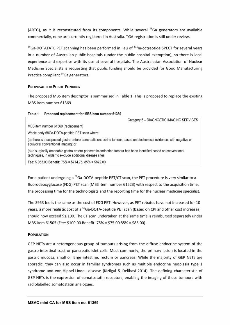
PROPOSAL FOR PUBLIC FUNDING
The proposed MBS item descriptor is summarised in Table 1. This is proposed to replace the existing
MBS item number 61369.
Table 1 Proposed replacement for MBS item number 61369
Category 5 – DIAGNOSTIC IMAGING SERVICES
MBS item number 61369 (replacement)
Whole body 68Ga-DOTA-peptide PET scan where:
(a) there is a suspected gastro-entero-pancreatic endocrine tumour, based on biochemical evidence, with negative or equivocal conventional imaging; or
(b) a surgically amenable gastro-entero-pancreatic endocrine tumour has been identified based on conventional techniques, in order to exclude additional disease sites
Fee: $ 953.00 Benefit: 75% = $714.75, 85% = $872.80
For a patient undergoing a 68Ga-DOTA-peptide PET/CT scan, the PET procedure is very similar to a
fluorodeoxyglucose (FDG) PET scan (MBS item number 61523) with respect to the acquisition time,
the processing time for the technologists and the reporting time for the nuclear medicine specialist.
The $953 fee is the same as the cost of FDG PET. However, as PET rebates have not increased for 10
years, a more realistic cost of a 68Ga-DOTA-peptide PET scan (based on CPI and other cost increases)
should now exceed $1,100. The CT scan undertaken at the same time is reimbursed separately under
MBS item 61505 (Fee: $100.00 Benefit: 75% = $75.00 85% = $85.00).
POPULATION
GEP NETs are a heterogeneous group of tumours arising from the diffuse endocrine system of the
gastro-intestinal tract or pancreatic islet cells. Most commonly, the primary lesion is located in the
gastric mucosa, small or large intestine, rectum or pancreas. While the majority of GEP NETs are
sporadic, they can also occur in familiar syndromes such as multiple endocrine neoplasia type 1
syndrome and von-Hippel-Lindau disease (Kizilgul & Delibasi 2014). The defining characteristic of
GEP NETs is the expression of somatostatin receptors, enabling the imaging of these tumours with
radiolabelled somatostatin analogues.
MSAC mini CA for MBS item no. 61369 13
Approximately two-thirds of GEP NETS are carcinoid tumours, originating in the enterochromaffin
cells of the gut. Many do not cause symptoms, but the metastases from some carcinoid GEP NETs
(mostly mid-gut originating in the small intestine, appendix or proximal large bowel) may secrete
serotonin and other vasoactive substances, causing carcinoid syndrome. Approximately one-third of
GEP NETS are pancreatic tumours, originating from the islet cells. The majority of pancreatic cancers
are adenocarcinomas, which arise from the exocrine pancreas. Up to 60% of pancreatic NETs are
non-functional. The functional tumours are often classified by the hormone most strongly secreted.
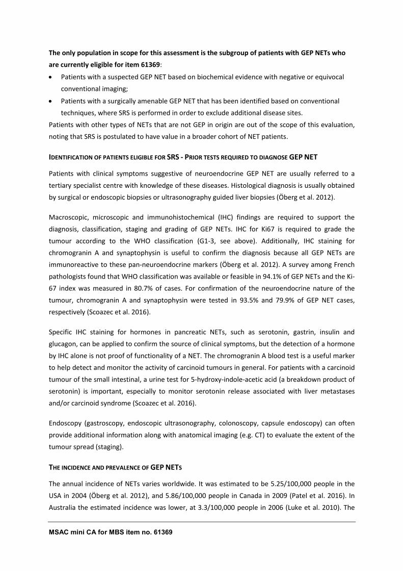
The only population in scope for this assessment is the subgroup of patients with GEP NETs who
are currently eligible for item 61369:
Patients with a suspected GEP NET based on biochemical evidence with negative or equivocal
conventional imaging;
Patients with a surgically amenable GEP NET that has been identified based on conventional
techniques, where somatostatin receptor scintigraphy (SRS) is performed in order to exclude
additional disease sites.
Patients with other types of NETs that are not GEP in origin are out of the scope of this evaluation,
noting that SRS is postulated to have value in a broader cohort of NET patients.
COMPARATOR DETAILS
In Australia, the only approved diagnostic radiopharmaceutical for SRS is OctreoScan® (111In-
octreotide), which was listed on the ARTG in 1996 (number 55928). It is covered by MBS item 61369,
with a schedule fee of $2015.75 (Table 6 in section A6). This item was included in the MBS in the
early 2000s following a recommendation by MSAC in 1999 (Application 1003). Item 61369 is usually
performed using SPECT with a gamma camera. If a concomitant CT is performed, it is reimbursed
under MBS item 61505 (Fee: $100.00 Benefit: 75% = $75.00 85% = $85.00).
The gold standard for the diagnosis of GEP NETs is histopathology. Most guidelines, such as those
from The Clinical Oncological Society of Australia (COSA 2008), European Society for Medical
Oncology (Öberg et al. 2012) and the Canadian evidence-based consensus recommendations (Singh
et al. 2016), stipulate that histology of surgical or biopsy tissue is mandatory in all cases for the
diagnosis of GEP NETs. However, the systematic reviews (SRs) that provide the evidence base for
diagnostic accuracy all used a composite reference standard, which included the results from
histopathology and/or conventional imaging and/or clinical follow-up of at least 1 year. Thus, this
composite reference standard has been used for this mini-assessment.
CLINICAL MANAGEMENT ALGORITHM
Currently, functional SRS assessment of the suspected GEP NET using 111In-octreotide SPECT±CT
imaging is funded on the MBS. In the proposed pathway 111In-octreotide SPECT±CT imaging is
replaced by 68Ga-DOTA-peptide PET/CT imaging for functional assessment. The current and
proposed diagnostic pathways are shown in Figure 2 in section A6.
MSAC mini CA for MBS item no. 61369 14
KEY DIFFERENCES IN THE DELIVERY OF THE PROPOSED MEDICAL SERVICE AND THE MAIN COMPARATOR
The key differences between the two tests are:
The time taken to complete the test is 90 minutes to 2 hours for 68Ga-DOTATATE PET/CT
compared with 2 days for 111In-octreotide SPECT/CT.
The radiation dose from 68Ga-DOTATATE PET (2–3 mSv, 28–41 MBq) is less than that received
with 111In-octreotide SPECT (8–16 mSv, 111–222 MBq).
The cost of 111In-octreotide SPECT/CT is much higher than 68Ga-DOTATATE PET/CT.
CLINICAL CLAIM
A claim has been made that introducing 68Ga-DOTA-peptide PET/CT scanning in lieu of 111In-
octreotide SPECT/CT will reduce the amount of repeat testing that supposedly occurs with 111In-
octreotide SPECT. 68Ga-DOTA-peptide PET/CT scanning is also claimed to have superior safety over
the comparator in terms of faster acquisition time and lower radiation exposure.
APPROACH TAKEN TO THE EVIDENCE ASSESSMENT
This mini assessment of the effectiveness of 68Ga-DOTA-peptide PET/CT scanning in lieu of 111In-
octreotide SPECT/CT scanning is constrained by a lack of information. A systematic search of the
literature is required to properly assess these technologies. To complement the limited evidence
provided by the applicant, a quick literature search of the PubMed database to identify recent
publications to help evaluate these technologies was undertaken. Therefore, the conclusions drawn
from this mini-assessment are based on an incomplete evidence base.
CHARACTERISTICS OF THE EVIDENCE BASE
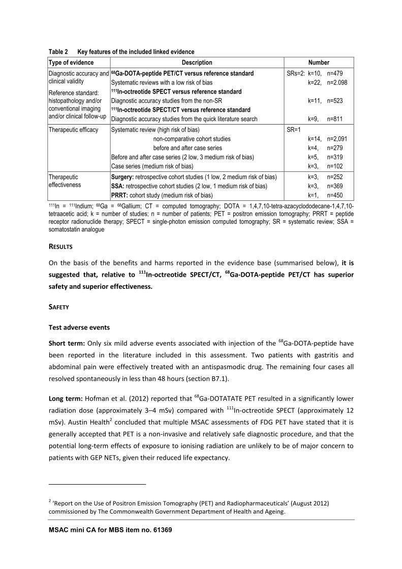
The characteristics of the evidence informing each step of the linked analysis are shown in Table 2.
There was no direct evidence. Whilst some studies were identified to provide evidence for each step
of the linked evidence, data that directly compared the outcomes for 68Ga-DOTA-peptide PET/CT
with those for 111In-octreotide SPECT/CT were not available for either therapeutic efficacy or
therapeutic effectiveness.
MSAC mini CA for MBS item no. 61369 15
Table 2 Key features of the included linked evidence
Type of evidence Description Number
Diagnostic accuracy and clinical validity
Reference standard: histopathology and/or conventional imaging and/or clinical follow-up
68Ga-DOTA-peptide PET/CT versus reference standard
Systematic reviews with a low risk of bias 111In-octreotide SPECT versus reference standard
Diagnostic accuracy studies from the non-SR 111In-octreotide SPECT/CT versus reference standard
Diagnostic accuracy studies from the quick literature search
SRs=2: k=10, n=479
k=22, n=2.098
k=11, n=523
k=9, n=811
Therapeutic efficacy Systematic review (high risk of bias)
non-comparative cohort studies
before and after case series
Before and after case series (2 low, 3 medium risk of bias)
Case series (medium risk of bias)
SR=1
k=14, n=2,091
k=4, n=279
k=5, n=319
k=3, n=102
Therapeutic effectiveness
Surgery: retrospective cohort studies (1 low, 2 medium risk of bias)
SSA: retrospective cohort studies (2 low, 1 medium risk of bias)
PRRT: cohort study (medium risk of bias)
k=3, n=252
k=3, n=369
k=1, n=450
111In = 111Indium; 68Ga = 68Gallium; CT = computed tomography; DOTA = 1,4,7,10-tetra-azacyclododecane-1,4,7,10-tetraacetic acid; k = number of studies; n = number of patients; PET = positron emission tomography; PRRT = peptide receptor radionuclide therapy; SPECT = single-photon emission computed tomography; SR = systematic review; SSA = somatostatin analogue
RESULTS
On the basis of the benefits and harms reported in the evidence base (summarised below), it is
suggested that, relative to 111In-octreotide SPECT/CT, 68Ga-DOTA-peptide PET/CT has superior
safety and superior effectiveness.
SAFETY
Test adverse events
Short term: Only six mild adverse events associated with injection of the 68Ga-DOTA-peptide have
been reported in the literature included in this assessment. Two patients with gastritis and
abdominal pain were effectively treated with an antispasmodic drug. The remaining four cases all
resolved spontaneously in less than 48 hours (section B7.1).
Long term: Hofman et al. (2012) reported that 68Ga-DOTATATE PET resulted in a significantly lower
radiation dose (approximately 3–4 mSv) compared with 111In-octreotide SPECT (approximately 12
mSv). Austin Health2 concluded that multiple MSAC assessments of FDG PET have stated that it is
generally accepted that PET is a non-invasive and relatively safe diagnostic procedure, and that the
potential long-term effects of exposure to ionising radiation are unlikely to be of major concern to
patients with GEP NETs, given their reduced life expectancy.
2 ‘Report on the Use of Positron Emission Tomography (PET) and Radiopharmaceuticals’ (August 2012)
commissioned by The Commonwealth Government Department of Health and Ageing.
MSAC mini CA for MBS item no. 61369 16
DIRECT EFFECTIVENESS
There was no direct evidence in inform of the effectiveness of 68Ga-DOTA-peptide PET/CT compared
with 111In-octreotide SPECT/CT.
EFFECTIVENESS FROM LINKED EVIDENCE
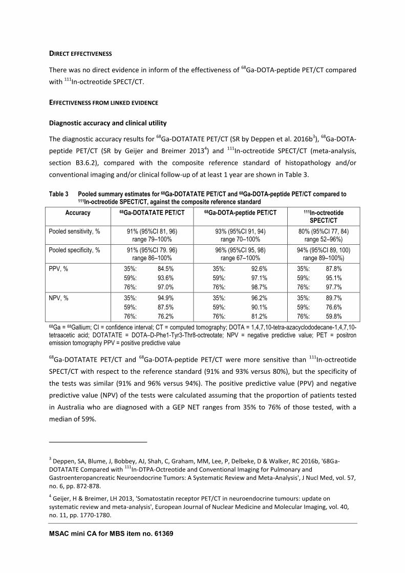
Diagnostic accuracy and clinical utility
The diagnostic accuracy results for 68Ga-DOTATATE PET/CT (SR by Deppen et al. 2016b3), 68Ga-DOTA-
peptide PET/CT (SR by Geijer and Breimer 20134) and 111In-octreotide SPECT/CT (meta-analysis,
section B3.6.2), compared with the composite reference standard of histopathology and/or
conventional imaging and/or clinical follow-up of at least 1 year are shown in Table 3.
Table 3 Pooled summary estimates for 68Ga-DOTATATE PET/CT and 68Ga-DOTA-peptide PET/CT compared to 111In-octreotide SPECT/CT, against the composite reference standard
Accuracy 68Ga-DOTATATE PET/CT 68Ga-DOTA-peptide PET/CT 111In-octreotide SPECT/CT
Pooled sensitivity, % 91% (95%CI 81, 96) range 79–100%
93% (95%CI 91, 94) range 70–100%
80% (95%CI 77, 84) range 52–96%)
Pooled specificity, % 91% (95%CI 79. 96) range 86–100%
96% (95%CI 95, 98) range 67–100%
94% (95%CI 89, 100) range 89–100%)
PPV, % 35%: 84.5%
59%: 93.6%
76%: 97.0%
35%: 92.6%
59%: 97.1%
76%: 98.7%
35%: 87.8%
59%: 95.1%
76%: 97.7%
NPV, % 35%: 94.9%
59%: 87.5%
76%: 76.2%
35%: 96.2%
59%: 90.1%
76%: 81.2%
35%: 89.7%
59%: 76.6%
76%: 59.8%
68Ga = 68Gallium; CI = confidence interval; CT = computed tomography; DOTA = 1,4,7,10-tetra-azacyclododecane-1,4,7,10-tetraacetic acid; DOTATATE = DOTA–D-Phe1-Tyr3-Thr8-octreotate; NPV = negative predictive value; PET = positron emission tomography PPV = positive predictive value
68Ga-DOTATATE PET/CT and 68Ga-DOTA-peptide PET/CT were more sensitive than 111In-octreotide
SPECT/CT with respect to the reference standard (91% and 93% versus 80%), but the specificity of
the tests was similar (91% and 96% versus 94%). The positive predictive value (PPV) and negative
predictive value (NPV) of the tests were calculated assuming that the proportion of patients tested
in Australia who are diagnosed with a GEP NET ranges from 35% to 76% of those tested, with a
median of 59%.
3 Deppen, SA, Blume, J, Bobbey, AJ, Shah, C, Graham, MM, Lee, P, Delbeke, D & Walker, RC 2016b, '68Ga-
DOTATATE Compared with 111
In-DTPA-Octreotide and Conventional Imaging for Pulmonary and Gastroenteropancreatic Neuroendocrine Tumors: A Systematic Review and Meta-Analysis', J Nucl Med, vol. 57, no. 6, pp. 872-878.
4 Geijer, H & Breimer, LH 2013, 'Somatostatin receptor PET/CT in neuroendocrine tumours: update on
systematic review and meta-analysis', European Journal of Nuclear Medicine and Molecular Imaging, vol. 40, no. 11, pp. 1770-1780.
MSAC mini CA for MBS item no. 61369 17
With a prevalence of 59%, the PPV for 111In-octreotide SPECT/CT was very similar to that for 68Ga-
DOTATATE PET/CT and 68Ga-DOTA-peptide PET/CT (95.1% versus 93.6% and 97.1%), the NPV values
varied by more than 10% (76.6% versus 87.5% and 90.1%). Thus, 23% of people who had a negative 111In-octreotide SPECT/CT result would actually have a GEP NET compared with 10–12% of those
scoring negative after 68Ga-DOTA-peptide PET/CT. Thus, approximately twice as many people with a
negative result would actually have disease after 111In-octreotide SPECT/CT scanning compared with 68Ga-DOTA-peptide PET/CT scanning. This is likely to be of clinical significance.
Therapeutic efficacy (change in management)
Barrio et al. (2017) concluded that management changes as a result of 68Ga-DOTATATE PET/CT
occurred in 44% of all patients. An updated meta-analysis found 38% of patients who had had a prior 111In-octreotide SPECT/CT had a change in management after having a 68Ga-DOTATATE PET/CT. Even
though these data were non-comparative and no data were available to determine whether or not
the initial management decisions would have differed in the absence of the 111In-octreotide
SPECT±CT, the results of the 111In-octreotide SPECT±CT appeared to be of little value when
determining a patient’s management plan.
Only two patients (2/322; 0.6%) for whom 68Ga-DOTATATE PET/CT may have resulted in a
suboptimal treatment plan were identified from the six studies investigating a change in
management after 68Ga-DOTATATE PET/CT in patients who had a prior 111In-octreotide SPECT±CT
(Hofman et al. 2012; Srirajaskanthan et al. 2010).
Taken together, the non-comparative studies forming the evidence base for the therapeutic efficacy
of 68Ga-DOTA-peptide PET/CT found that a change in management was usually a direct consequence
of the improved spatial resolution and clarity of the 68Ga-DOTA-peptide PET/CT image compared
with the 111In-octreotide SPECT/CT image. There were four main scenarios that led to a potentially
major impact on patient management from 68Ga-DOTA-peptide PET/CT imaging:
1. Approximately half of histopathology-positive GEP NET patients who were falsely negative with 111In-octreotide SPECT/CT could become eligible for somatostatin analogue (SSA) therapy or
peptide receptor radionuclide therapy (PRRT) after 68Ga-DOTA-peptide PET/CT imaging due to
its better NPV compared to 111In-octreotide SPECT/CT (section B4.6);
2. Histopathology-positive GEP NET patients who are SRS-negative with 68Ga-DOTA-peptide
PET/CT would most likely be directed away from PRRT and SSA therapy due to the lack of
somatostatin receptors on the tumour cell surface;
3. Identification of the primary tumour site with 68Ga-DOTA-peptide PET/CT imaging in patients in
whom it is otherwise not detected could lead to surgical resection; and
4. Identification of more metastases with 68Ga-DOTA-peptide PET/CT imaging may lead to patients
receiving PRRT instead of, or in addition to, any planned surgical procedures.
MSAC mini CA for MBS item no. 61369 18
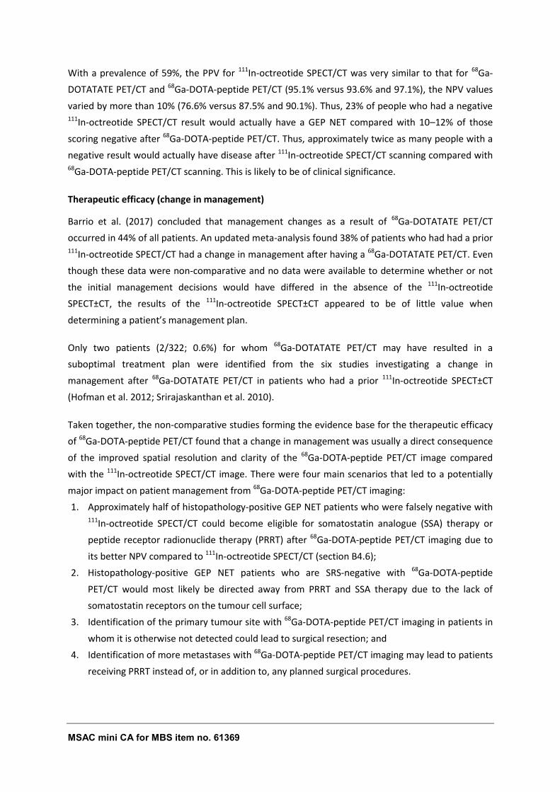
Therapeutic effectiveness (health benefit from change in management)
The most common management decisions resulting from 68Ga-DOTA-peptide scanning were referral
for surgery, PRRT or SSA therapy.
As a whole, the non-comparative evidence base for the treatment effectiveness of surgery and PRRT
supported the findings in the review by Bodei et al. (2014). This review included an indirect
comparison of the 5-year survival rates for patients who had various treatments and found that
surgery had better survival outcomes compared to other therapies (Figure 1). However, it should be
noted that this category would include most patients with early stage disease who would generally
be expected to live longer than those with advanced late-stage disease. As PRRT is used as an
alternative to chemotherapy, the patients groups are likely to have similar disease characteristics.
Bodei et al. (2014) reported that PRRT was an efficient and relatively safe treatment with patients
surviving longer compared to chemotherapy (Figure 1). The non-comparative evidence base for the
effectiveness of long-acting release (LAR) SSA therapy supported the findings by Saglam et al. (2015)
who reported an estimated 5-year survival rate of 58% for therapy with LAR SSAs. This was included
in Figure 1. The literature suggests that LAR-SSAs are used at all stages of disease and, as seen in
Figure 1, the estimated 5-year survival rate for LAR SSA therapy was second only to surgical
resection in improved survival outcomes (Figure 1).
Figure 1 The 5-year survival rate of patients with NETs undergoing various tretaments
Source: adapted from Bodei et al. (2014); LAR SSA therapy from Saglam et al. (2015)
PRRT = peptide receptor radionuclide therapy; R0 = resection for cure or complete remission; R1 = microscopic residual tumour; R2 = macroscopic residual tumour; SSA = somatostatin analogue
Thus, treatment of GEP NETs with surgery, SSA therapy or PRRT appears to be more effective than
no treatment or chemotherapy and relatively safe. However, randomised controlled trials are
needed to determine if these conclusions are accurate.
ESTIMATED EXTENT OF USE AND FINANCIAL IMPLICATIONS
An epidemiological approach (combined with additional data from the literature review and clinical
expert advice) has been used to estimate the number of services and financial implications
associated with the substitution of 68Ga-DOTA-peptide PET for 111In-octreotide SPECT in MBS item
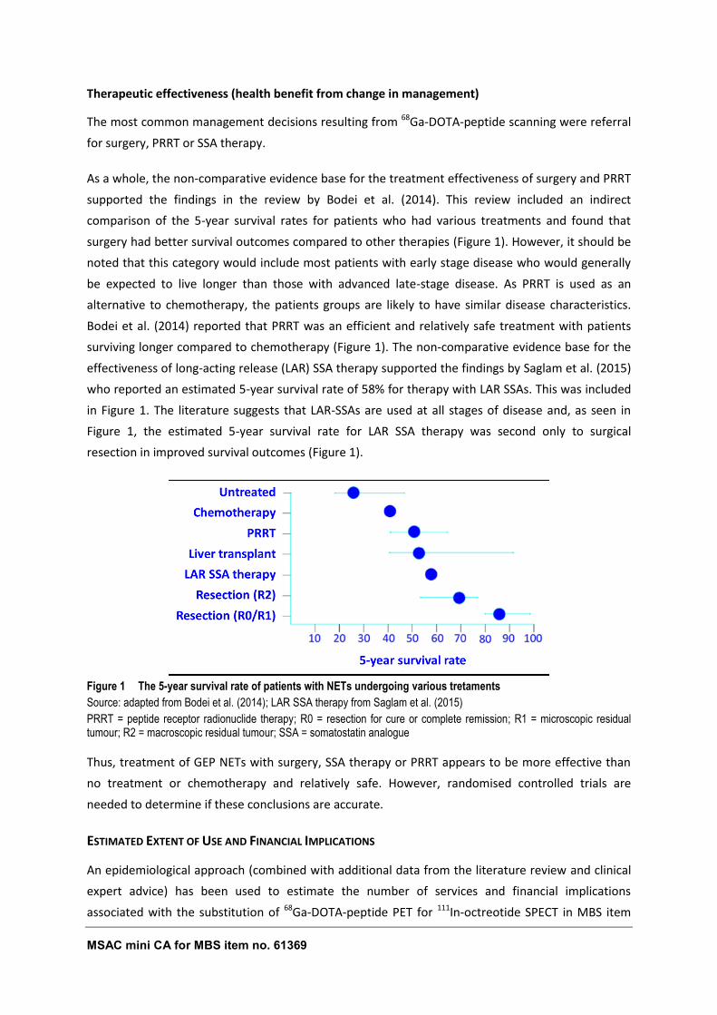
MSAC mini CA for MBS item no. 61369 19
61369. Table 4 summarises the financial implications to the MBS resulting from the proposed listing
of 68Ga-DOTA-peptide PET.
Table 4 Total costs to the MBS associated with 68Ga-DOTA-peptide PET/CT service, 2017–18 to 2021–22
2017–18 2018–19 2019–20 2020–21 2021–22
Number of services estimated to be MBS funded 445 566 691 819 951
Costs to MBS $438,794 $557,793 $680,469 $806,816 $936,765
CT = computed tomography; DOTA = 1,4,7,10-tetra-azacyclododecane-1,4,7,10-tetraacetic acid; 68Ga = 68GalliumPET = positron emission tomography; MBS = Medical Benefits Schedule;
CONSUMER IMPACT SUMMARY
Not applicable.
OTHER RELEVANT CONSIDERATIONS
None identified.
MSAC mini CA for MBS item no. 61369 20
ACRONYMS AND ABBREVIATIONS
111In 111Indium
177Lu 177Lutetium
68Ga 68Gallium
90Y 90Yttrium
AANMS Australasian Association of Nuclear Medicine Specialists
ABS Australian Bureau of Statistics
ARTG Australian Register of Therapeutic Goods
CI confidence interval
CT computed tomography
CUP carcinoma with unknown primary
DOTA 1,4,7,10-tetra-azacyclododecane-1,4,7,10-tetraacetic acid
DOTANOC DOTA-Phe1-NaI3-octreotide
DOTATATE DOTA–D-Phe1-Tyr3-Thr8-octreotate
DOTATOC DOTA–D-Phe1-Tyr3-octreotide
DTPA diethylene-triamino-penta-acetic acid
ESMO European Society for Medical Oncology
FDG fluorodeoxyglucose
GEP gastroenteropancreatic
GMP Good Manufacturing Practice
HIAA hydroxyindoleacetic acid
HTA health technology assessment
IHC immunohistochemical
LAR long-acting release
MBS Medicare Benefits Schedule
MR magnetic resonance
MSAC mini CA for MBS item no. 61369 21
MSAC Medical Services Advisory Committee
NET neuroendocrine tumour
NPV negative predictive value
PET positron emission tomography
PFS progression-free survival
PICO population, intervention, comparator, outcomes
PPV positive predictive value
PRRT peptide receptor radionuclide therapy
SPECT single-photon emission computed tomography
SR systematic review
SROC summary receiver operator characteristic
SRS somatostatin receptor scintigraphy
SSA somatostatin analogue
TGA Therapeutic Goods Administration
TTP time to progression
WHO World Health Organization
MSAC mini CA for MBS item no. 61369 22
SECTION A CONTEXT
This contracted mini assessment of 68Gallium-1,4,7,10-tetra-azacyclododecane-1,4,7,10-tetraacetic
acid-peptide (68Ga-DOTA-peptide) positron emission tomography (PET) / computed tomography (CT)
scanning for the diagnosis of gastroenteropancreatic neuroendocrine tumours (GEP NETs) is
intended for the Medical Services Advisory Committee (MSAC). MSAC evaluates new and existing
health technologies and procedures for which funding is sought under the Medicare Benefits
Schedule (MBS) in terms of their safety, effectiveness and cost-effectiveness, while taking into
account other issues such as access and equity. MSAC adopts an evidence-based approach to its
assessments, based on reviews of the scientific literature and other information sources, including
clinical expertise.
The proposal originated from the Australasian Association of Nuclear Medicine Specialists (AANMS)
and was referred to MSAC for consideration by the MBS Review Taskforce for targeted assessment.
Adelaide Health Technology Assessment has been commissioned by the Australian Government
Department of Health to conduct a mini assessment of the management/health outcomes of the
incremental diagnostic information obtained from 68Ga-DOTA-peptide PET/CT scanning compared to 111Indium (111In)-labelled octreotide study (111In-octreotide SPECT/CT) in patients with GEP NETs. This
assessment has been undertaken in order to inform MSAC’s decision-making regarding whether the
proposed medical service should be publicly funded.
Appendix A provides a list of the people involved in the development of this assessment report,
including clinical expertise.
The proposed use of 68Ga-DOTA-peptide PET/CT scanning in Australian clinical practice was outlined
in a draft PICO Confirmation that was prepared by the department in response to a request from
MSAC Executive Teleconference on 24 November 2016.
A1 ITEMS IN THE AGREED PICO CONFIRMATION
This contracted mini assessment of MBS item number 61369 has primarily drawn upon material
provided to the Department of Health by the applicant, as well as the European Society of Medical
Oncology (ESMO) Clinical Practice Guidelines published by Oberg et al. (Öberg et al. 2012).
This mini assessment addresses all of the PICO elements that were pre-specified in the draft PICO
Confirmation submitted to the PICO Confirmation Advisory Sub-Committee of the MSAC. PROPOSED
MEDICAL SERVICE
The proposed medical service is a combined PET/CT scan for functional (PET) and anatomical (CT)
imaging of GEP NETs using a 68Ga-DOTA-labelled somatostatin analogue. The current ESMO clinical
MSAC mini CA for MBS item no. 61369 23
guidelines recommend that preoperative staging of GEP NETs should include somatostatin receptor
scintigraphy (SRS) (Öberg et al. 2012). However, the guidelines also say that conventional SRS with 111In-octreotide single-photon emission computed tomography (SPECT) with or without CT and/or a
gamma camera can be replaced by SRS using 68Ga-DOTA-peptide PET/CT for higher spatial resolution
and quantification, resulting in higher sensitivity and specificity. 68Ga-DOTA-peptide PET also has a
faster acquisition time than conventional SRS (2 hours compared to 2 days with SPECT) and the
patients are exposed to less radiation (see section B7.2). However, not all GEP NETs express a
significant number of somatostatin receptors; in the later stages of disease, the tumour
characteristics change from being well differentiated to being poorly differentiated with greatly
increased metabolic activity and reduced levels of somatostatin receptor expression. Therefore,
anatomical imaging (e.g. CT) should always be done in conjunction with 68Ga-DOTA-peptide PET
functional imaging (Öberg et al. 2012).
THE RADIOPHARMACEUTICAL 68GA-DOTA-PEPTIDE
Three different DOTA-peptides—DOTA–D-Phe1-Tyr3-Thr8-octreotate (DOTATATE), DOTA–D-Phe1-
Tyr3-octreotide (DOTATOC), and DOTA-Phe1-NaI3-octreotide (DOTANOC)—are currently used in
conjunction with 68Ga for PET/CT imaging of GEP NETs. Similar to 111In-octreotide, these peptides are
also derived from octreotide, a somatostatin octapeptide that bind to the somatostatin receptor.
In Australia, the DOTATATE peptide is coupled to 68Ga. This peptide is supplied by Auspep, which is
licensed by the TGA to manufacture active pharmaceutical ingredients (licence MI-07122005-LI-
001046-11).
The 68Ga-DOTA-peptide is not listed on the Australian Register of Therapeutic Goods (ARTG), as it is
reconstituted from its components. The radioactive isotope is eluted from a Good Manufacturing
Practice (GMP)-compliant 68Ga generator. 68Ga is then coupled with the non-radioactive (or cold)
DOTA-peptide. This ‘radiolabelling’ process is routine, and is done on a daily basis in most Australian
nuclear medicine departments for the preparation of commonly used radiopharmaceuticals.
Examples include: Tc-99m MDP (bone scans), Tc-99m MAA (perfusion lung scans), Tc-99m DTPA
(renal scans) and Tc-99m sestamibi (cardiac perfusion scans and parathyroid scans).
THE USE OF 68GA FOR PET/CT SCANNING FOR DIAGNOSIS OF GEP NETS IN AUSTRALIA
While several 68Ga generators are available commercially, none are currently registered in Australia.
The TGA has yet to decide whether radiopharmaceutical generators will be fully exempt from
regulation in Australia. The issue of TGA registration is out of scope for the purpose of this
assessment and is an issue that will be progressed in parallel with this assessment. When MSAC
considers this assessment the Department will separately provide the committee with an update
on the TGA status of the Ga-68 generators.
MSAC mini CA for MBS item no. 61369 24
AANMS accepts that public funding should only be provided when the 68Ga generator used is GMP
compliant but does not consider registration by a respected overseas regulator mandatory given
that these generators have been used for many years both locally and internationally and, as a
result, the safety profile is well established.
In Australia, 68Ga-DOTATATE PET scanning has been performed in lieu of 111In-octreotide SPECT for
several years in a number of public hospitals (under the public hospital exemption), so there is local
experience and expertise with its use at several hospitals. AANMS is requesting that public funding
should be provided for GMP compliant 68Ga generators.
A3 PROPOSAL FOR PUBLIC FUNDING
The proposed MBS item descriptor is summarised in Table 5. This will replace the existing MBS item
number 61369.
Table 5 Proposed replacement for MBS item number 61369
Category 5 – DIAGNOSTIC IMAGING SERVICES
MBS item number 61369 (replacement)
Whole body 68Ga-DOTA-peptide PET scan where:
(a) there is a suspected gastro-entero-pancreatic endocrine tumour, based on biochemical evidence, with negative or equivocal conventional imaging; or
(b) a surgically amenable gastro-entero-pancreatic endocrine tumour has been identified based on conventional techniques, in order to exclude additional disease sites
Fee: $ 953.00 Benefit: 75% = $714.75, 85% = $872.80
For a patient undergoing a 68Ga-DOTA-peptide PET/CT scan, the PET procedure is very similar to a
fluorodeoxyglucose (FDG) PET scan (MBS item number 61523). Following injection of the
radiopharmaceutical, there is an uptake period of 45-60 minutes, after which the patient undergoes
a scan on a PET/CT scanner. The acquisition time of the scan as well as the processing time for the
technologists is comparable to a FDG PET scan, as is the reporting time for the reporting nuclear
medicine specialist.
The $953 fee is the same as the cost of FDG PET. However, as PET rebates have not increased for 10
years, a more realistic cost of a 68Ga-DOTA-peptide PET scan (based on CPI and other cost increases)
should now exceed $1,100. The CT scan undertaken at the same time is reimbursed separately under
MBS item 61505 (Fee: $100.00 Benefit: 75% = $75.00 85% = $85.00).
Due to the short half-life of 68Ga (68 minutes), the commercial sale of individual patient doses of the
radiopharmaceutical 68Ga-DOTA-peptide will not be feasible in nearly all circumstances. As a result,
nuclear medicine facilities offering this service will need to have a 68Ga generator on site, a synthesis
MSAC mini CA for MBS item no. 61369 25
module to perform the labelling and quality control, as well as consumables (including chemicals,
cartridges and the DOTA-peptide).
A4 PROPOSED POPULATION
GEP NETs are a heterogeneous group of tumours arising from the diffuse endocrine system of the
gastro-intestinal tract or pancreatic islet cells. Most commonly, the primary lesion is located in the
gastric mucosa, small or large intestine, rectum or pancreas. While the majority of GEP NETs are
sporadic, they can also occur in familiar syndromes such as multiple endocrine neoplasia type 1
syndrome, von-Hippel-Lindau disease, tuberosclerosis and neurofibromatosis type 1 (Kizilgul &
Delibasi 2014). The defining characteristic of GEP NETs is the expression of somatostatin receptors,
enabling the imaging of these tumours with radiolabelled somatostatin analogues.
The 2010 World Health Organization (WHO) classification splits GEP NETs into 3 categories with
different malignant potential and histology: well-differentiated neoplasms or tumours that are
usually low grade (G1, Ki67 <2%) as well as intermediate grade (G2, Ki67 3–20%), and poorly
differentiated neoplasms or carcinomas that represent late stages of disease (G3, Ki-67 >20%)
(Berardi et al. 2016a). The Ki67 protein is a cellular marker for proliferation. A higher percentage
suggests a faster-growing, more aggressive tumour.
GEP NETs are characterized by their ability to synthesize, store, and secrete a variety of neuro-
amines and peptides. They can be functioning (hormone secreting and symptomatic), or non-
functioning. They are usually slow-growing malignancies that can be difficult to diagnose because of
vague and diffuse clinical presentations. Hence, approximately 65% of patients with GEP NETs
present with metastatic disease (Modlin et al. 2010).
Approximately two-thirds of GEP NETS are carcinoid tumours, originating in the enterochromaffin
cells of the gut. Many do not cause symptoms even when they have metastasized. However, the
metastases from some carcinoid GEP NETs (mostly mid-gut originating in the small intestine,
appendix or proximal large bowel) may secrete serotonin and other vasoactive substances causing
carcinoid syndrome. The symptoms include flushing, wheezing, diarrhoea, abdominal cramping,
peripheral oedema, heart palpitations and eventual congestive heart disease. Congestive heart
failure is due to chronic exposure to high levels of serotonin, which causes thickening of the heart
valves (Oladejo 2009).
Approximately one-third of GEP NETS are pancreatic tumours, originating from the islet cells. The
majority of pancreatic cancers are adenocarcinomas, which arise from the exocrine pancreas. Up to
60% of pancreatic NETs are non-functional. The functional tumours are often classified by the
hormone most strongly secreted, such as: insulinomas, glucagonomas, gastrinomas and
somatostatinomas (Kizilgul & Delibasi 2014).
MSAC mini CA for MBS item no. 61369 26
The only population in scope for this assessment is the subgroup of patients with GEP NETs who
are currently eligible for item 61369:
Patients with a suspected GEP NET based on biochemical evidence with negative or equivocal
conventional imaging;
Patients with a surgically amenable GEP NET that has been identified based on conventional
techniques, where SRS is performed in order to exclude additional disease sites.
Patients with other types of NETs that are not GEP in origin are out of the scope of this evaluation,
noting that SRS is postulated to have value in a broader cohort of NET patients.
IDENTIFICATION OF PATIENTS ELIGIBLE FOR SRS - PRIOR TESTS REQUIRED TO DIAGNOSE GEP NET
Patients with clinical symptoms suggestive of neuroendocrine GEP NET are usually referred to a
tertiary specialist centre with knowledge of these diseases. Histological diagnosis is usually obtained
by surgical or endoscopic biopsies or ultrasonography guided liver biopsies (Öberg et al. 2012).
Macroscopic, microscopic and immunohistochemical (IHC) findings are required to support the
diagnosis, classification, staging and grading of GEP NETs. IHC for Ki67 is required to grade the
tumour according to the WHO classification (G1-3, see above). Additionally, IHC staining for
chromogranin A and synaptophysin is useful to confirm the diagnosis because all GEP NETs are
immunoreactive to these pan-neuroendocrine markers (Öberg et al. 2012). A survey among French
pathologists found that WHO classification was available or feasible in 94.1% of GEP NETs and the Ki-
67 index was measured in 80.7% of cases. For confirmation of the neuroendocrine nature of the
tumour, chromogranin A and synaptophysin were tested in 93.5% and 79.9% of GEP NET cases,
respectively (Scoazec et al. 2016).
Specific IHC staining for hormones in pancreatic NETs, such as serotonin, gastrin, insulin and
glucagon, can be applied to confirm the source of clinical symptoms, but the detection of a hormone
by IHC alone is not proof of functionality of a NET. The chromogranin A blood test is a useful marker
to help detect and monitor the activity of carcinoid tumours in general. For patients with a carcinoid
tumour of the small intestinal, a urine test for 5-hydroxy-indole-acetic acid (a breakdown product of
serotonin) is important, especially to monitor serotonin release associated with liver metastases
and/or carcinoid syndrome (Scoazec et al. 2016).
Endoscopy (gastroscopy, endoscopic ultrasonography, colonoscopy, capsule endoscopy) can often
provide additional information along with anatomical imaging (e.g. CT) to evaluate the extent of the
tumour spread (staging).
THE INCIDENCE AND PREVALENCE OF GEP NETS
The annual incidence of NETs varies worldwide. It was estimated to be 5.25/100,000 people in the
USA in 2004 (Öberg et al. 2012), and 5.86/100,000 people in Canada in 2009 (Patel et al. 2016). In
Australia the estimated incidence was lower, at 3.3/100,000 people in 2006 (Luke et al. 2010). The
MSAC mini CA for MBS item no. 61369 27
estimated prevalence in the USA was 35/100,000 people in 2004 (Öberg et al. 2012). The most
common primary site for NET was GEP (60% of all NETs), with patients generally diagnosed in their
late 50s or early 60s, but those with familial NET syndromes may have a clinical onset of disease 15–
20 years earlier than patients with sporadic disease (Yao et al. 2008).
NETs comprised 0.6% of all invasive cancers recorded on the South Australian Cancer Registry from
2000–2006 (Luke et al. 2010). The annual age-standardised incidence per 100,000 people increased
by 86.8% from 1.74 between 1980 and 1989 to 3.25 between 2000 and 2006. The NETs originated in
the lung in 25.9% of cases, 54.1% were GEP NETs, and 20% had an unknown or other origin (Luke et
al. 2010). The most common primary sites for GEP NETs were the small intestine (38.1%), large
bowel (21.2%), appendix (17.6%), pancreas (12.0%), and stomach (6.8%).
The 5-year survival rate for patients diagnosed with GEP NETs in South Australia between 1980 and
2006 was higher for those whose tumour originated in the appendix (93.8%), rectum (85.9%) or
small intestine (74.6%) compared with the pancreas (42.4%), colon excluding appendix (64.6%) or
stomach (66.4%). An increase in survival was seen in later calendar years, with the 5-year survival
rate for patients diagnosed with NETs in South Australia between 2000 and 2006 being 73.4% ± 3.0%
for all NETs, 84.8% ± 0.1% for stomach, 80.9% ± 8.8% for colon and 100% for appendix NETs (Luke et
al. 2010).
The 5-year survival rate for patients with pancreatic NETs is estimated to be 60–100% for localized
disease, 40% for regional, 25% for metastatic and 80% for all stages.
A5 COMPARATOR DETAILS
In Australia, the only approved diagnostic radiopharmaceutical for SRS is OctreoScan® (111In-
octreotide), which was listed on the ARTG in 1996 (number 55928). It is covered by MBS item 61369,
with a schedule fee of $2015.75 (Table 6). This item was included in the MBS in the early 2000s
following a recommendation by MSAC in 1999 (Application 1003). Item 61369 is usually performed
using SPECT with a gamma camera. If a concomitant CT is performed, it is reimbursed under MBS
item 61505 (Fee: $100.00 Benefit: 75% = $75.00 85% = $85.00).
Table 6 MBS item descriptor for the comparator
Category 5 – DIAGNOSTIC IMAGING SERVICES
MBS item number 61369
INDIUM-LABELLED OCTREOTIDE STUDY - including single photon emission tomography when undertaken, where:
(a) there is a suspected gastro-entero-pancreatic endocrine tumour, based on biochemical evidence, with negative or equivocal conventional imaging; or
(b) a surgically amenable gastro-entero-pancreatic endocrine tumour has been identified based on conventional techniques, in order to exclude additional disease sites. (R)
Fee: $2,015.75 Benefit: 75% = $1,511.85 85% = $1,935.55
MSAC mini CA for MBS item no. 61369 28
Octreotide is a long-acting somatostatin analogue and has been an important agent in the initial
evaluation and management of NETs for nearly 30 years. Octreotide is conjugated with diethylene-
triamine-pentaacetic acid (DTPA) and labelled with 111In to form 111In-DTPA-D-Phe1-octreotide, also
known as 111In-pentetreotide (111In-octreotide). The radiotracer is injected intravenously, followed by
imaging at several time-points over the next 1-2 days, usually using SPECT to obtain both two-
dimensional ‘planar’ imaging and three-dimensional cross-sectional images (Rufini, Calcagni & Baum
2006). The results from SRS are easiest to interpret when hybrid SPECT/CT scanners are used to
provide both functional (SPECT) and anatomical (CT) information. SRS provides information on the
primary tumour location and the extent of disease, as well as predicting the response to therapy
with unlabelled or labelled somatostatin analogues (Rufini, Calcagni & Baum 2006).
The use of item 61369 in Australia has decreased in recent years (Table 7) as treating clinicians and
nuclear medicine specialists increasingly use 68Ga-DOTA-peptide PET/CT for SRS. Additionally, the
increasing costs of performing SRS with 111In-octreotide SPECT compared with the MBS fee makes
the test less economical to perform.
Table 7 MBS utilisation for item 61369 2010/11 through to 2015/16
Financial year 2010/11 2011/12 2012/13 2013/14 2014/15 2015/16
Total number of services for item 61369 693 484 419 236 146 106
A6 CLINICAL MANAGEMENT ALGORITHM(S)
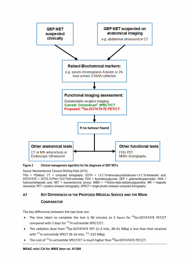
The current and proposed diagnostic pathways are shown in Figure 2. Currently, functional SRS
assessment of the suspected GEP NET using 111In-octreotide SPECT/CT imaging is funded on the MBS
(shown in green). In the proposed pathway 111In-octreotide SPECT/CT imaging is replaced by 68Ga-
DOTA-peptide PET/CT imaging (shown in red) for functional assessment.
MSAC mini CA for MBS item no. 61369 29
Figure 2 Clinical management algorithm for the diagnosis of GEP NETs
Source: Neuroendocrine Tumours Working Party (2013). 68Ga = 68Gallium; CT = computed tomography; DOTA = 1,4,7,10-tetra-azacyclododecane-1,4,7,10-tetraacetic acid; DOTATATE = DOTA–D-Phe1-Tyr3-Thr8-octreotate; FDG = fluorodeoxyglucose; GEP = gastroenteropancreatic; HIAA = hydroxyindoleacetic acid; NET = neuroendocrine tumour; MIBG = 123Iodine-meta-iodobenzylguanidine; MR = magnetic resonance; PET = positron emission tomography; SPECT = single-photon emission computed tomography
A7 KEY DIFFERENCES IN THE PROPOSED MEDICAL SERVICE AND THE MAIN
COMPARATOR
The key differences between the two tests are:
The time taken to complete the test is 90 minutes to 2 hours for 68Ga-DOTATATE PET/CT
compared with 2 days for 111In-octreotide SPECT/CT.
The radiation dose from 68Ga-DOTATATE PET (2–3 mSv, 28–41 MBq) is less than that received
with 111In-octreotide SPECT (8–16 mSv, 111–222 MBq).
The cost of 111In-octreotide SPECT/CT is much higher than 68Ga-DOTATATE PET/CT.
MSAC mini CA for MBS item no. 61369 30
The US Library of Medicine website5 provides information on the use and safety of both (111In-
octreotide and 68Ga-DOTATATE (marketed as NETSPOT).
The indication for 111In-octreotide is as an agent for the scintigraphic localization of primary and
metastatic NETs bearing somatostatin receptors. 68Ga-DOTATATE is indicated by the US Food and
Drug Administration for use with PET for localization of somatostatin receptor positive NETs in adult
and paediatric patients. There are no contra-indications for either radiopharmaceutical.
The safety of 68Ga-DOTATATE was evaluated in three single centre studies (Deppen et al. 2016a;
Haug et al. 2014; Haug et al. 2012) and in a survey of the scientific literature. No serious adverse
reactions were identified. As both 68Ga-DOTATATE and 111In-octreotide are derivatives of octreotide,
they are likely to have similar adverse reactions and precautions.
The adverse reactions and precautions listed in the TGA product information6 for 111In-octreotide
are:
Octreotide therapy can produce severe hypoglycaemia in patients with insulinomas and in
diabetic patients receiving high doses of insulin. An intravenous solution containing glucose
should be administered just before and during administration of 111In-octreotide.
Since 111In-octreotide is eliminated primarily by renal excretion, use in patients with impaired
renal function should be carefully considered.
As with any other radioactive material, appropriate shielding should be used to avoid
unnecessary radiation exposure to the patient, occupational workers, and other persons.
Evidence of mutagenicity was not found when 111In-octreotide was evaluated in an in vivo
mouse micronucleus assay.
Safety and effectiveness in pregnant women, lactating mothers and paediatric patients have not
been established.
A8 CLINICAL CLAIM
A claim has been made that introducing 68Ga-DOTA-peptide PET/CT scanning in lieu of 111In-
octreotide SPECT/CT will reduce the amount of repeat testing that supposedly occurs with 111In-
octreotide SPECT. 68Ga-DOTA-peptide PET/CT scanning is also claimed to have superior safety over
the comparator in terms of faster acquisition time and lower radiation exposure.
5 Available from URL: https://dailymed.nlm.nih.gov/dailymed/index.cfm [accessed 20 December 2016].
6 Available from URL:
https://www.ebs.tga.gov.au/ebs/picmi/picmirepository.nsf/PICMI?OpenForm&t=&k=O&r=https://www.ebs.tga.gov.au/ [accessed 20 December 2016].
MSAC mini CA for MBS item no. 61369 31
A9 SUMMARY OF THE PICO
The guiding framework of a PICO Confirmation is recommended by MSAC for each assessment. The
PICO Confirmation describes current clinical practice and reflects the likely future practice with the
proposed medical service. The PICO that were pre-specified in the PICO confirmation are presented
in Table 8.
Table 8 Criteria for identifying and selecting studies to determine the safety and direct effectiveness of 68Ga-DOTA-peptide PET/CT scanning in patients with GEP NETs
Selection criteria Description
Population Patients with GEP NETs, specifically those patients with this tumour currently eligible to receive item 61369.
Prior tests Conventional imaging, histopathology and various sophisticated biomarkers in a specialised tertiary setting
Intervention 68Ga-DOTA-peptide PET±CT scanning (direct substitution to comparator)
Comparator Indium labelled octreotide study (111In-octreotide SPECT±CT) currently covered by MBS items 61369 (SPECT) and 61505 (CT)
Outcomes • Relative Safety
• Relative Diagnostic accuracy (sensitivity/specificity)
• Impact on clinical management including net change on clinical management arising from differential accuracy
• Impact on clinical utility through a linked evidence approach (in the absence of direct evidence) as per Investigative Guidelines
• Health resource impacts and cost/consequence analysis
Questions for direct evidence
What is the safety and effectiveness of 68Ga-DOTA-peptide PET/CT scanning compared with 111In-octreotide in patients with GEP NETs?
Questions for linked evidence
What is the diagnostic accuracy of 68Ga-DOTA-peptide PET/CT scanning compared with 111In-octreotide SPECT±CT in patients with GEP NETs?
What is the clinical validity of 68Ga-DOTA-peptide PET/CT scanning compared with 111In-octreotide SPECT±CT in patients with GEP NETs?
Is there a change in management from 68Ga-DOTA-peptide PET/CT scanning in patients with GEP NETs compared with 111In-octreotide SPECT±CT?
Does the change in management due to 68Ga-DOTA-peptide PET/CT scanning improve patient outcomes?
111In = 111Indium; 68Ga = 68Gallium; CT = computed tomography; DOTA = 1,4,7,10-tetra-azacyclododecane-1,4,7,10-tetraacetic acid; GEP = gastroenteropancreatic; NET = neuroendocrine tumour; PET = positron emission tomography; SPECT = single-photon emission computed tomography
A10 CONSUMER IMPACT STATEMENT
Not applicable.

MSAC mini CA for MBS item no. 61369 33
SECTION B CLINICAL EVALUATION
This mini assessment evaluates the management/health outcomes of incremental diagnostic
information obtained from 68Ga-DOTA-peptide PET/CT scanning compared to 111In-octreotide
SPECT/CT in patients with GEP NETs.
Determination of the clinical effectiveness of an investigative medical service requires either:
evidence of the effectiveness of 68Ga-DOTA-peptide PET/CT scanning from high-quality
comparative studies evaluating the use of 68Ga-DOTA-peptide PET/CT scanning and
subsequent treatment compared to 111In-octreotide SPECT/CT and treatment (direct
evidence). Randomised controlled trials provide the highest quality evidence for this
comparison. Or, if this is not available:
evidence of the treatment effectiveness from high-quality comparative studies evaluating
the treatment for GEP NET, linked with applicable and high-quality evidence of the accuracy
of 68Ga-DOTA-peptide PET/CT scanning compared to 111In-octreotide SPECT/CT for the
diagnosis of GEP NET. This is called ‘linked evidence’.
The Department of Health stipulated that a systematic literature review was not required for this
mini assessment. The evidence base consisted of material published by the ESMO, primarily Oberg
et al. (2012), The ‘Report on the Use of Positron Emission Tomography (PET) and
Radiopharmaceuticals’ (August 2012) by Austin Health7, as well as material provided to the
Department of Health by AANMS.
To supplement the limited evidence base provided, a quick search of the literature in the PubMed
database was undertaken to identify recent systematic reviews (SRs) and studies reporting on the
use of 68Ga-DOTA-peptide PET/CT in the diagnosis and management of patients with or suspected of
having GEP NETs.
B1 DIRECT EVIDENCE
No studies were provided or identified in the quick literature search that reported on the safety or
effectiveness of 68Ga-DOTA-peptide PET/CT scanning directly compared with 111In-octreotide
SPECT/CT in patients with GEP NETs.
7 The Commonwealth Government Department of Health and Ageing contracted Austin Health to undertake a
review of the Broader Use of Positron Emission Tomography (PET) and Radiopharmaceuticals in 2012.
MSAC mini CA for MBS item no. 61369 34
B2 LINKED EVIDENCE APPROACH
B2.1 BASIS FOR LINKED EVIDENCE
As there was no direct evidence identified, a linked evidence approach was undertaken.
B2.2 STEPS FOR LINKED ANALYSIS
To construct a linked evidence analysis, different evidence requirements are required.
Consideration of the diagnostic performance and clinical validity (where relevant) of the
investigative medical service (sections B3 and B4);
Consideration of the clinical utility of the investigative medical service in terms of impact of
positive versus negative test results on patient management, the contribution and clinical
importance of false negatives versus false positives and direct impact of each therapeutic
model service option on health outcomes (section B5);
Considerations of the impact of repeat testing (if appropriate) (section B6); and
Consideration of the relative safety of performing the investigative service, both immediate
safety issues of directly performing the test and ‘flow on’ safety issues that arise as a result
of conducting the investigative service (section B7).
Conclusions linking these steps are made in section B8.
MSAC mini CA for MBS item no. 61369 35
B3 DIAGNOSTIC PERFORMANCE
B3.1 REFERENCE STANDARD
Most guidelines, such as those from The Clinical Oncological Society of Australia (COSA 2008), ESMO
(Öberg et al. 2012) and the Canadian evidence-based consensus recommendations (Singh et al.
2016), stipulate that histology of surgical or biopsy tissue is mandatory in all cases for the diagnosis
of GEP NETs. The histopathology tests include:
• Routine haematoxylin and eosin-staining to identify the typical morphology of GEP NETs ;
• IHC for neuroendocrine markers, including synaptophysin and chromogranin A.
Although GEP NETs are a heterogeneous in nature, they are all immune-reactive to
neuroendocrine markers;
• IHC for the cell-cycle-dependent marker, Ki67 antigen, to determine the aggressiveness of the
tumour. The Ki67 index serves as the basis for WHO grading of tumours (G1-3);
• Additional biochemical pathology tests verifying increased hormone levels may also be
performed, especially in symptomatic patients.
Thus, the gold standard for the diagnosis of GEP NETs is histopathology.
However, the SRs that provide the evidence base for diagnostic accuracy all used a composite
reference standard, which included the results from histopathology and/or conventional imaging
and/or clinical follow-up of at least 1 year. Thus, this composite reference standard will be used for
this mini-assessment.
B3.2 EVIDENCE BASE
The evidence base for this review was provided by the Department of Health and the applicant. A
quick search of the PubMed database was conducted for recent publications to supplement the
provided evidence when needed.
DIAGNOSTIC ACCURACY OF 68GA-DOTA-PEPTIDE PET/CT COMPARED WITH THE COMPOSITE REFERENCE
STANDARD
The ‘Report on the Use of Positron Emission Tomography (PET) and Radiopharmaceuticals’ (August
2012) by Austin Health8 conducted a systematic search of the literature for original research papers,
including SRs, using PubMed and Cochrane databases, and a search of websites of international
health technology assessment (HTA) agencies for existing HTA reports. This report included a SR that
8 The Commonwealth Government Department of Health and Ageing contracted Austin Health to undertake a
review of the Broader Use of Positron Emission Tomography (PET) and Radiopharmaceuticals in 2012.
MSAC mini CA for MBS item no. 61369 36
evaluated the diagnostic accuracy of 68Ga-DOTA-peptide PET/CT compared with histology and/or
morphological imaging (Treglia et al. 2012). The applicant provided an additional two SRs that
compared the diagnostic accuracy of 68Ga-DOTA-peptide PET/CT with the composite reference
standard (Geijer & Breimer 2013; Mojtahedi et al. 2014).
A quick search of the PubMed database identified another 2 SRs that looked at diagnostic accuracy;
both Deppen et al. (2016b) and Yang et al. (2014) compared 68Ga-DOTA-peptide PET/CT with the
composite reference standard.
DIAGNOSTIC ACCURACY OF 68GA-DOTA-PEPTIDE PET/CT COMPARED WITH
111IN-OCTREOTIDE SPECT±CT
The Report by Austin Health9 and three of the SRs mentioned above provided a narrative review of
the diagnostic accuracy of 68Ga-DOTA-peptide PET/CT compared with 111In-octreotide SPECT/CT
using the composite reference standard (Deppen et al. 2016b; Geijer & Breimer 2013; Mojtahedi et
al. 2014).
A quick search of the PubMed database identified a non-systematic review that compared the
diagnostic accuracy of 111In-octreotide SPECT±CT with the composite reference standard (Koopmans
et al. 2009). An additional 9 studies were also identified that compared 111In-octreotide SPECT/CT
with the composite reference standard.
B3.3 RISK OF BIAS ASSESSMENT
The five SRs that reported on the diagnostic accuracy of 68Ga-DOTA-peptide PET/CT compared with
the composite reference standard and the non-SR comparing the diagnostic accuracy of 111In-
octreotide SPECT±CT with composite reference standard were evaluated using the AMSTAR checklist
(Shea et al. 2007). A summary of the risk of bias for each SR is shown in Table 9.
9 The Commonwealth Government Department of Health and Ageing contracted Austin Health to undertake a
review of the Broader Use of Positron Emission Tomography (PET) and Radiopharmaceuticals in 2012.
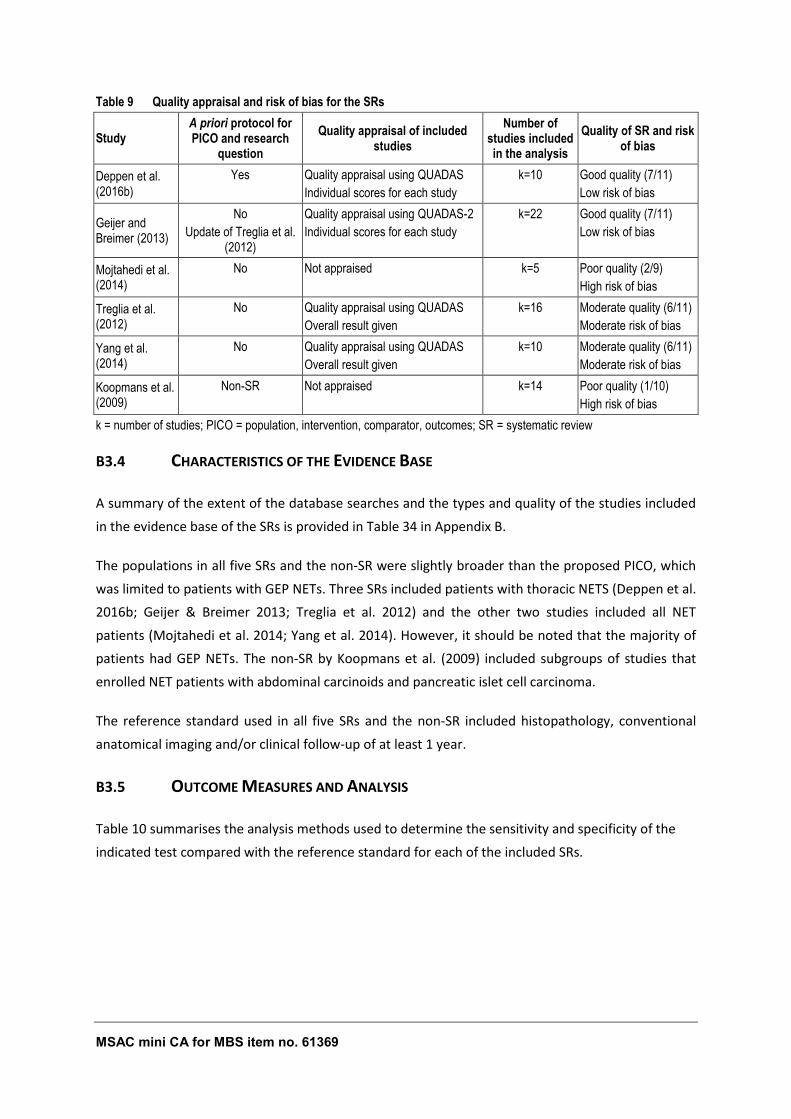
MSAC mini CA for MBS item no. 61369 37
Table 9 Quality appraisal and risk of bias for the SRs
Study A priori protocol for PICO and research
question
Quality appraisal of included studies
Number of studies included in the analysis
Quality of SR and risk of bias
Deppen et al. (2016b)
Yes Quality appraisal using QUADAS
Individual scores for each study
k=10 Good quality (7/11)
Low risk of bias
Geijer and Breimer (2013)
No
Update of Treglia et al. (2012)
Quality appraisal using QUADAS-2
Individual scores for each study
k=22 Good quality (7/11)
Low risk of bias
Mojtahedi et al. (2014)
No Not appraised k=5 Poor quality (2/9)
High risk of bias
Treglia et al. (2012)
No Quality appraisal using QUADAS
Overall result given
k=16 Moderate quality (6/11)
Moderate risk of bias
Yang et al. (2014)
No Quality appraisal using QUADAS
Overall result given
k=10 Moderate quality (6/11)
Moderate risk of bias
Koopmans et al. (2009)
Non-SR Not appraised k=14 Poor quality (1/10)
High risk of bias
k = number of studies; PICO = population, intervention, comparator, outcomes; SR = systematic review
B3.4 CHARACTERISTICS OF THE EVIDENCE BASE
A summary of the extent of the database searches and the types and quality of the studies included
in the evidence base of the SRs is provided in Table 34 in Appendix B.
The populations in all five SRs and the non-SR were slightly broader than the proposed PICO, which
was limited to patients with GEP NETs. Three SRs included patients with thoracic NETS (Deppen et al.
2016b; Geijer & Breimer 2013; Treglia et al. 2012) and the other two studies included all NET
patients (Mojtahedi et al. 2014; Yang et al. 2014). However, it should be noted that the majority of
patients had GEP NETs. The non-SR by Koopmans et al. (2009) included subgroups of studies that
enrolled NET patients with abdominal carcinoids and pancreatic islet cell carcinoma.
The reference standard used in all five SRs and the non-SR included histopathology, conventional
anatomical imaging and/or clinical follow-up of at least 1 year.
B3.5 OUTCOME MEASURES AND ANALYSIS
Table 10 summarises the analysis methods used to determine the sensitivity and specificity of the
indicated test compared with the reference standard for each of the included SRs.
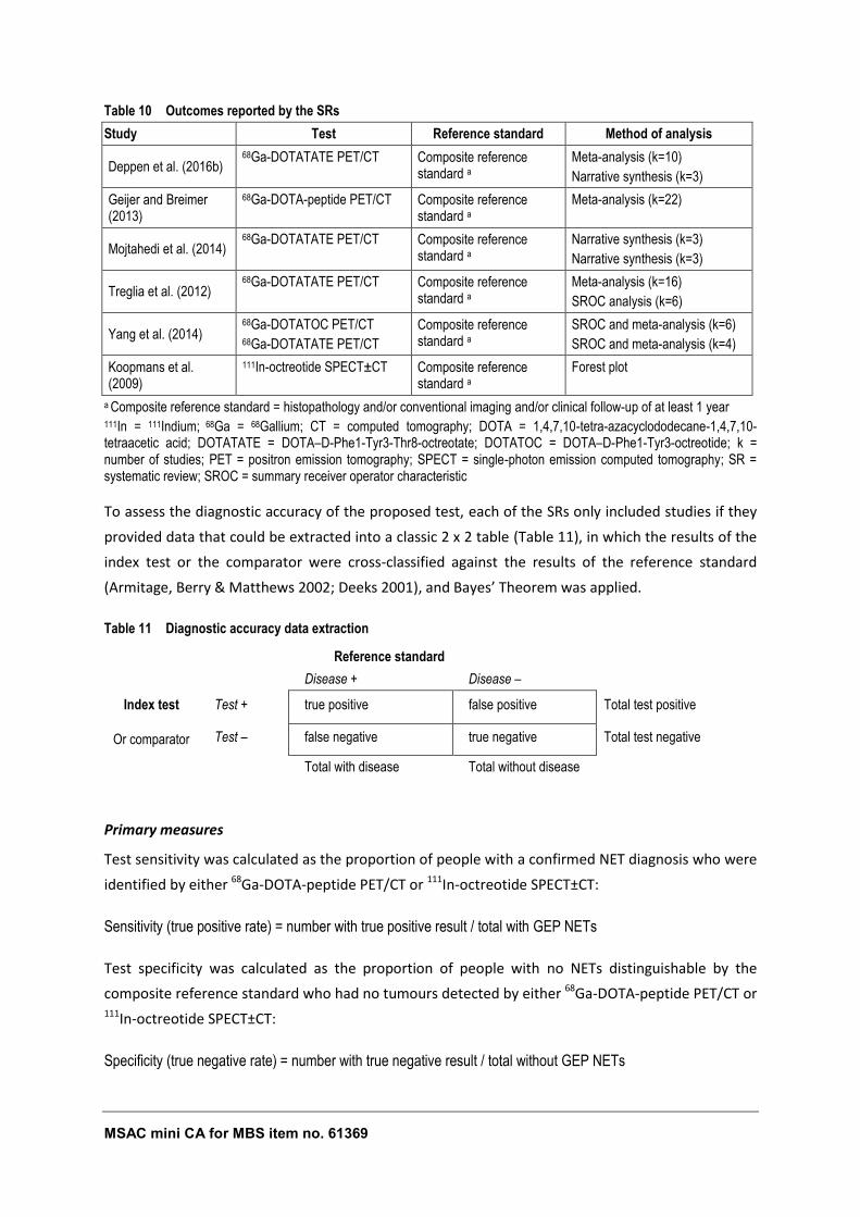
MSAC mini CA for MBS item no. 61369 38
Table 10 Outcomes reported by the SRs
Study Test Reference standard Method of analysis
Deppen et al. (2016b) 68Ga-DOTATATE PET/CT Composite reference
standard a Meta-analysis (k=10)
Narrative synthesis (k=3)
Geijer and Breimer (2013)
68Ga-DOTA-peptide PET/CT Composite reference standard a
Meta-analysis (k=22)
Mojtahedi et al. (2014) 68Ga-DOTATATE PET/CT Composite reference
standard a Narrative synthesis (k=3)
Narrative synthesis (k=3)
Treglia et al. (2012) 68Ga-DOTATATE PET/CT Composite reference
standard a Meta-analysis (k=16)
SROC analysis (k=6)
Yang et al. (2014) 68Ga-DOTATOC PET/CT 68Ga-DOTATATE PET/CT
Composite reference standard a
SROC and meta-analysis (k=6)
SROC and meta-analysis (k=4)
Koopmans et al. (2009)
111In-octreotide SPECT±CT Composite reference standard a
Forest plot
a Composite reference standard = histopathology and/or conventional imaging and/or clinical follow-up of at least 1 year 111In = 111Indium; 68Ga = 68Gallium; CT = computed tomography; DOTA = 1,4,7,10-tetra-azacyclododecane-1,4,7,10-tetraacetic acid; DOTATATE = DOTA–D-Phe1-Tyr3-Thr8-octreotate; DOTATOC = DOTA–D-Phe1-Tyr3-octreotide; k = number of studies; PET = positron emission tomography; SPECT = single-photon emission computed tomography; SR = systematic review; SROC = summary receiver operator characteristic
To assess the diagnostic accuracy of the proposed test, each of the SRs only included studies if they
provided data that could be extracted into a classic 2 x 2 table (Table 11), in which the results of the
index test or the comparator were cross-classified against the results of the reference standard
(Armitage, Berry & Matthews 2002; Deeks 2001), and Bayes’ Theorem was applied.
Table 11 Diagnostic accuracy data extraction
- - Reference standard -
- - Disease + Disease – -
Index test Test + true positive false positive Total test positive
Or comparator Test – false negative true negative Total test negative
- - Total with disease Total without disease -
Primary measures
Test sensitivity was calculated as the proportion of people with a confirmed NET diagnosis who were
identified by either 68Ga-DOTA-peptide PET/CT or 111In-octreotide SPECT±CT:
Sensitivity (true positive rate) = number with true positive result / total with GEP NETs
Test specificity was calculated as the proportion of people with no NETs distinguishable by the
composite reference standard who had no tumours detected by either 68Ga-DOTA-peptide PET/CT or 111In-octreotide SPECT±CT:
Specificity (true negative rate) = number with true negative result / total without GEP NETs
MSAC mini CA for MBS item no. 61369 39
The 95%CI was calculated by the exact binomial method.
Summary measures
Diagnostic test accuracy meta-analysis was undertaken to assess the accuracy of 111In-octreotide
SPECT±CT compared with the composite reference standard in the diagnosis of GEP NETs using Stata
version 14.1 (StataCorp 2015). Only studies that provided raw (2 × 2) data were included. Forest
plots were generated using the ‘midas’ command in Stata, which requires a minimum of 4 studies
for analysis and calculates summary operating sensitivity and specificity (with confidence and
prediction contours in summary receiver operator characteristic (SROC) space). Heterogeneity was
calculated using the formula I2 = 100% x (Q – df)/Q, where Q is Cochran's heterogeneity statistic and
df is the degrees of freedom (Higgins et al. 2003). Summary estimates for sensitivity and specificity
were also calculated.
MSAC mini CA for MBS item no. 61369 40
B3.6 RESULTS OF THE SYSTEMATIC LITERATURE REVIEW
IS IT ACCURATE?
Summary – What is the diagnostic accuracy of 68Ga-DOTA-peptide PET/CT scanning compared with 111In-
octreotide SPECT±CT in patients with GEP NETs?
Diagnostic accuracy of 68Ga-DOTA-peptide PET/CT compared with the composite reference standard
Two good quality SRs conducted meta-analyses to determine the accuracy of 68Ga-DOTA-peptide PET/CT
compared with the composite reference standard. The SR by Geijer and Breimer (2013) included 22 studies with
a pooled sensitivity and specificity of 93% (95%CI 91, 94; range 70–100%) and 96% (95%CI 95, 98; range 67–
100%), respectively. The SR by Deppen et al. (2016b) included 10 studies comparing 68Ga-DOTATATE PET/CT
with the composite reference standard. The pooled sensitivity and specificity were 91% (95%CI 81, 96; range
79–100%) and 91% (95%CI 79. 96, range 86–100%), respectively. The results for 68Ga-DOTA-peptide PET/CT
and 68Ga-DOTATATE PET/CT compared with the composite reference standard were similar.
Diagnostic accuracy of 111In-octreotide SPECT±CT compared with the composite reference standard
Meta-analysis of 11 studies that compared 111In-octreotide SPECT with the composite reference standard
estimated the pooled sensitivity and specificity to be 84% (95%CI 80, 87; range 54–96%) and 75% (95%CI 59,
91; range 60–84%), respectively. Meta-analysis of 9 studies that compared 111In-octreotide SPECT/CT with the
composite reference standard estimated the pooled sensitivity and specificity to be 80% (95%CI 77, 84; range
52–96%) and 94% (95%CI 89, 100; range 89–100%), respectively. The addition of CT to 111In-octreotide SPECT
had little effect on the pooled estimate for sensitivity (84% versus 80%) but markedly improved the specificity
(75% versus 94%).
68Ga-DOTATATE PET/CT compared with 111In-octreotide SPECT/CT using the composite reference
standard
When the pooled estimates for 68Ga-DOTATATE PET/CT (relevant to Australian clinical practice) and 111In-
octreotide SPECT/CT were compared, 68Ga-DOTATATE PET/CT was more sensitive than 111In-octreotide
SPECT/CT (91% versus 80%) with respect to the reference standard but the specificity of the tests were similar
(91% versus 94%).
B3.6.1 DIAGNOSTIC ACCURACY OF 68GA-DOTA-PEPTIDE PET/CT COMPARED WITH THE COMPOSITE
REFERENCE STANDARD
Five SRs were identified that investigated the diagnostic accuracy of 68Ga-DOTA-peptide PET/CT
compared with the composite reference standard (Table 12). The pooled sensitivities calculated in
the SRs ranged from 91% to 96% and the pooled specificities ranged from 85% to 100%. However,
after evaluation of the quality and the applicability of the reported outcomes in the five SRs, only the
results from the two good quality SRs with a low risk of bias have been discussed further.
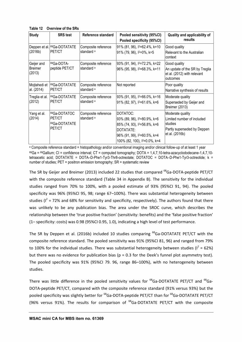
MSAC mini CA for MBS item no. 61369 41
Table 12 Overview of the SRs
Study SRS test Reference standard Pooled sensitivity (95%CI)
Pooled specificity (95%CI)
Quality and applicability of results
Deppen et al. (2016b)
68Ga-DOTATATE PET/CT
Composite reference standard a
91% (81, 96), I2=62.4%, k=10
91% (79, 96), I2=0%, k=5
Good quality
Relevant to the Australian context
Geijer and Breimer (2013)
68Ga-DOTA-peptide PET/CT
Composite reference standard a
93% (91, 94), I2=72.2%, k=22
96% (95, 98), I2=68.3%, k=11
Good quality
An update of the SR by Treglia et al. (2012) with relevant outcomes
Mojtahedi et al. (2014)
68Ga-DOTATATE PET/CT
Composite reference standard a
Not reported Poor quality
Narrative synthesis of results
Treglia et al. (2012)
68Ga-DOTATATE PET/CT
Composite reference standard a
93% (91, 95), I2=66.0%, k=16
91% (82, 97), I2=61.6%, k=6
Moderate quality
Superseded by Geijer and Breimer (2013)
Yang et al. (2014)
68Ga-DOTATOC PET/CT 68Ga-DOTATATE PET/CT
Composite reference standard a
DOTATOC:
93% (89, 96), I2=80.9%, k=6
85% (74, 93), I2=56.8%, k=6
DOTATATE:
96% (91, 99), I2=60.5%, k=4
100% (82, 100), I2=0.0%, k=4
Moderate quality
Limited number of included studies
Partly superseded by Deppen et al. (2016b)
a Composite reference standard = histopathology and/or conventional imaging and/or clinical follow-up of at least 1 year 68Ga = 68Gallium; CI = confidence interval; CT = computed tomography; DOTA = 1,4,7,10-tetra-azacyclododecane-1,4,7,10-tetraacetic acid; DOTATATE = DOTA–D-Phe1-Tyr3-Thr8-octreotate; DOTATOC = DOTA–D-Phe1-Tyr3-octreotide; k = number of studies; PET = positron emission tomography; SR = systematic review
The SR by Geijer and Breimer (2013) included 22 studies that compared 68Ga-DOTA-peptide PET/CT
with the composite reference standard (Table 34 in Appendix B). The sensitivity for the individual
studies ranged from 70% to 100%, with a pooled estimate of 93% (95%CI 91, 94). The pooled
specificity was 96% (95%CI 95, 98; range 67–100%). There was substantial heterogeneity between
studies (I2 = 72% and 68% for sensitivity and specificity, respectively). The authors found that there
was unlikely to be any publication bias. The area under the SROC curve, which describes the
relationship between the ‘true positive fraction’ (sensitivity: benefits) and the ‘false positive fraction’
(1– specificity: costs) was 0.98 (95%CI 0.95, 1.0), indicating a high level of test performance.
The SR by Deppen et al. (2016b) included 10 studies comparing 68Ga-DOTATATE PET/CT with the
composite reference standard. The pooled sensitivity was 91% (95%CI 81, 96) and ranged from 79%
to 100% for the individual studies. There was substantial heterogeneity between studies (I2 = 62%)
but there was no evidence for publication bias (p = 0.3 for the Deek’s funnel plot asymmetry test).
The pooled specificity was 91% (95%CI 79. 96, range 86–100%), with no heterogeneity between
studies.
There was little difference in the pooled sensitivity values for 68Ga-DOTATATE PET/CT and 68Ga-
DOTA-peptide PET/CT, compared with the composite reference standard (91% versus 93%) but the
pooled specificity was slightly better for 68Ga-DOTA-peptide PET/CT than for 68Ga-DOTATATE PET/CT
(96% versus 91%). The results for comparison of 68Ga-DOTATATE PET/CT with the composite
MSAC mini CA for MBS item no. 61369 42
reference standard is more relevant in the Australian clinical context as only DOTATATE is currently
available for use in Australia.
It should be noted that the patients included in both of these SRs were not limited to those with
proven or suspected GEP NETs. Although the majority of the patients had GEP NETs, a significant
proportion had thoracic NETs, NETs of unknown origin and other NETs (Table 34 in Appendix B).
Although the population is broader than that specified in the PICO, it is likely that this had little
effect on the diagnostic accuracy of 68Ga-DOTA-peptide PET/CT compared with the composite
reference standard.
B3.6.2 DIAGNOSTIC ACCURACY OF 111IN-OCTREOTIDE SPECT±CT COMPARED WITH THE COMPOSITE
REFERENCE STANDARD
111In-octreotide SPECT has been successfully used for over 2 decades, and it has been reported that
the sensitivity of the test is increased when the functional SPECT is combined with an anatomical CT
(Deroose et al. 2016). However, no meta-analyses comparing the diagnostic accuracy of this test
with the composite reference standard, or any other test, were identified during a quick literature
search of PubMed. One non-systematic review by Koopmans et al. (2009) did present a forest plot
showing the sensitivity of the test compared with the composite reference standard for subgroups
of patients with different NETs, but did not provide a pooled estimate. Eleven studies that compared 111In-octreotide SPECT with the composite reference standard in patients with GEP NETs, sourced
form Koopmans et al. (2009), and 9 studies that compared 111In-octreotide SPECT/CT with the
composite reference standard that were identified in a quick search of PubMed, were meta-analysed
(Figure 7 and Figure 8, Appendix C). The pooled estimates are summarised in Table 13.
Table 13 Pooled sensitivity and specificity of 111In-octreotide SPECT±CT compared with the composite reference standard
SRS test Pooled sensitivity (95%CI) Pooled specificity (95%CI)
111In-octreotide SPECT 84% (80, 87), range 54–96%, I2=72.3%, k=11 75% (59, 91), range 60–84%, I2=0%, k=3
111In-octreotide SPECT/CT
80% (77, 84), range 52–96%, I2=85.4%, k=9 94% (89, 100), range 89–100%, I2=0%, k=6
111In = 111Indium; CI = confidence interval; CT = computed tomography; k = number of studies; SPECT = single-photon emission computed tomography
Although the pooled sensitivity and specificity values were not derived from a systematic literature
search, they are more robust than the estimated sensitivity range published in reviews of between
80% and 100%. The meta-analysis showed that the addition of CT to 111In-octreotide SPECT had little
effect on the sensitivity of the test but did markedly improve the specificity (94% versus 75%).
B3.7 EXTENDED ASSESSMENT OF RELIABILITY EVIDENCE
Deppen at al. (2016a) reported that the bias-corrected Fleiss kappa was 0.82 (95%CI 0.74, 0.89)
between the 3 blinded reviewers in their interpretation of 97 68Ga-DOTATATE PET/CT scans,
MSAC mini CA for MBS item no. 61369 43
demonstrating a high level of inter-observer reproducibility. No other study reported on intra- or
inter-observer reproducibility.
B3.8 CONCORDANCE ANALYSIS
Not required.
B3.9 INTERPRETATION OF EVIDENCE ON DIAGNOSTIC PERFORMANCE
The pooled sensitivity and specificity estimates for 68Ga-DOTATATE PET/CT were compared to 111In-
octreotide SPECT/CT. 68Ga-DOTATATE PET/CT combines both functional and anatomical imaging,
therefore its accuracy against the composite reference standard will be compared with 111In-
octreotide SPECT/CT, which also combines functional and anatomical imaging.
When the pooled estimates for 68Ga-DOTATATE PET/CT and 111In-octreotide SPECT/CT were
compared, 68Ga-DOTATATE PET/CT (91%) was more sensitive than 111In-octreotide SPECT/CT (80%)
with respect to the composite reference standard but the specificity of the tests was similar (91%
versus 94%).
The increased sensitivity is largely due to the increased spatial resolution of 68GA-DOTA-peptide PET
compared with 111In-octreotide SPECT10. Etchebehere et al. (2014) reported that the spatial
resolution for PET was 3–6 mm versus 10–15 mm for SPECT. Krausz et al. (2011) also found that 68GA-DOTATOC PET images were clearer than 111In-octreotide SPECT images. PET was especially
useful in detecting small lesions (Etchebehere et al. 2014), particularly in the bones (Frilling et al.
2010; Putzer et al. 2009), as well as identifying the primary tumour site in patients where it was
unknown (Prasad et al. 2010).
A preliminary clinical study by Hofman et al. (2001) in a small patient cohort of 8 patients with
histologically proven metastatic NET (6 with GEP NET) demonstrated that 68GA-DOTATOC PET
achieved higher tumour to non-tumour binding ratios than 111In-octreotide SPECT. 68Ga-DOTATOC
PET identified all previously known lesions, whereas 111In-octreotide SPECT identified only 85%. In
addition, 68Ga-DOTATOC PET detected previously unknown small lesions, including brain metastases.
10 Austin Health. ‘Report on the Use of Positron Emission Tomography (PET) and Radiopharmaceuticals’
(August 2012)
MSAC mini CA for MBS item no. 61369 44
B4 CLINICAL VALIDITY
B4.1 MEASURES OF CLINICAL VALIDITY
The positive predictive value (PPV) and negative predictive value (NPV) are clinically relevant
statistical measures that indicate how likely individuals who screen positive (or negative) have (or do
not have) the disease. PPV and NPV depends on both the test performance and on the prevalence of
the condition in the population tested. The PPV and NPV of the 68Ga-DOTA-peptide PET/CT and 111In-
octreotide SPECT/CT tests were calculated using the proportion of patients who were suspected of
having GEP NETs that were actually diagnosed with the disease in the included studies.
B4.1.1 TO B4.1.4
As for B3.1 to B3.4
B4.1.5 OUTCOME MEASURES AND ANALYSIS
The pooled sensitivity and specificity estimates and the prevalence of test-positive patients in the
included studies were used to assess the positive predictive value (PPV) and negative predictive
value (NPV) of 68Ga-DOTA-peptide PET/CT and 111In-octreotide SPECT/CT.
PPV was calculated as the proportion of people with histopathologically-confirmed NETs who were
identified by 68Ga-DOTA-peptide PET/CT out of all patients positive by 68Ga-DOTA-peptide PET/CT:
PPV = number true positives / total test positive
= sensitivity × prevalence sensitivity × prevalence + (1 – specificity) × (1 – prevalence)
NPV was calculated as the proportion of people with no NETs distinguishable by histopathology who
had no tumours detected by 68Ga-DOTA-peptide PET/CT out of all patients negative by 68Ga-DOTA-
peptide PET/CT:
NPV = number true negatives / total test negative
= specificity × (1 – prevalence) (1 – sensitivity) × prevalence + specificity × (1 – prevalence)
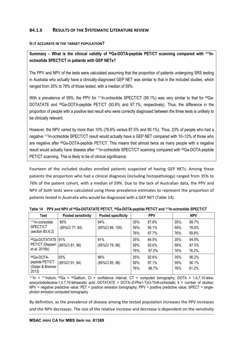
MSAC mini CA for MBS item no. 61369 45
B4.1.6 RESULTS OF THE SYSTEMATIC LITERATURE REVIEW
IS IT ACCURATE IN THE TARGET POPULATION?
Summary – What is the clinical validity of 68Ga-DOTA-peptide PET/CT scanning compared with 111In-
octreotide SPECT/CT in patients with GEP NETs?
The PPV and NPV of the tests were calculated assuming that the proportion of patients undergoing SRS testing
in Australia who actually have a clinically-diagnosed GEP NET was similar to that in the included studies, which
ranged from 35% to 76% of those tested, with a median of 59%.
With a prevalence of 59%, the PPV for 111In-octreotide SPECT/CT (95.1%) was very similar to that for 68Ga-
DOTATATE and 68Ga-DOTA-peptide PET/CT (93.6% and 97.1%, respectively). Thus, the difference in the
proportion of people with a positive test result who were correctly diagnosed between the three tests is unlikely to
be clinically relevant.
However, the NPV varied by more than 10% (76.6% versus 87.5% and 90.1%). Thus, 23% of people who had a
negative 111In-octreotide SPECT/CT result would actually have a GEP NET compared with 10–12% of those who
are negative after 68Ga-DOTA-peptide PET/CT. This means that almost twice as many people with a negative
result would actually have disease after 111In-octreotide SPECT/CT scanning compared with 68Ga-DOTA-peptide
PET/CT scanning. This is likely to be of clinical significance.
Fourteen of the included studies enrolled patients suspected of having GEP NETs. Among these
patients the proportion who had a clinical diagnosis (including histopathology) ranged from 35% to
76% of the patient cohort, with a median of 59%. Due to the lack of Australian data, the PPV and
NPV of both tests were calculated using these prevalence estimates to represent the proportion of
patients tested in Australia who would be diagnosed with a GEP NET (Table 14).
Table 14 PPV and NPV of 68Ga-DOTATATE PET/CT, 68Ga-DOTA-peptide PET/CT and 111In-octreotide SPECT/CT
Test Pooled sensitivity Pooled specificity PPV NPV
111In-octreotide SPECT/CT (section B3.6.2)
80%
(95%CI 77, 84)
94%
(95%CI 89, 100)
35%: 87.8%
59%: 95.1%
76%: 97.7%
35%: 89.7%
59%: 76.6%
76%: 59.8%
68Ga-DOTATATE PET/CT (Deppen et al. 2016b)
91%
(95%CI 81, 96)
91%
(95%CI 79, 96)
35%: 84.5%
59%: 93.6%
76%: 97.0%
35%: 94.9%
59%: 87.5%
76%: 76.2%
68Ga-DOTA-peptide PET/CT (Geijer & Breimer 2013)
93%
(95%CI 91, 94)
96%
(95%CI 95, 98)
35%: 92.6%
59%: 97.1%
76%: 98.7%
35%: 96.2%
59%: 90.1%
76%: 81.2%
111In = 111Indium; 68Ga = 68Gallium; CI = confidence interval; CT = computed tomography; DOTA = 1,4,7,10-tetra-azacyclododecane-1,4,7,10-tetraacetic acid; DOTATATE = DOTA–D-Phe1-Tyr3-Thr8-octreotate; k = number of studies; NPV = negative predictive value; PET = positron emission tomography; PPV = positive predictive value; SPECT = single-photon emission computed tomography
By definition, as the prevalence of disease among the tested population increases the PPV increases
and the NPV decreases. The size of the relative increase and decrease is dependent on the sensitivity
MSAC mini CA for MBS item no. 61369 46
and specificity of the test. When 59% of those tested were assumed to have a GEP NET, the PPV of 111In-octreotide SPECT/CT (95.1%) was very similar to that for 68Ga-DOTATATE PET/CT and 68Ga-
DOTA-peptide PET/CT (93.6% and 97.1%, respectively). The difference between tests does not
increase greatly if the prevalence decrease to 35% (Table 14). Thus, the difference in the proportion
of people with a positive test result who were correctly diagnosed with a GEP NET using the 111In-
octreotide SPECT/CT and 68Ga-DOTA-peptide PET/CT tests is unlikely to be clinically relevant.
However, with a prevalence of 59% the NPV varied by more than 10% (76.6% versus 87.5% and
90.1%). Thus, 23% of people who had a negative 111In-octreotide SPECT/CT result would actually
have a GEP NET compared with 10–12% of those scoring negative after 68Ga-DOTA-peptide PET/CT.
With a higher prevalence of 76% the difference in the proportion of people who are true negatives
increases from 11-13% to 16–21%. The difference in the proportion of patients who would have a
negative test result but actually have disease is likely to be of clinical significance.
It should also be noted that a proportion of those patients who have histologically proven GEP NET
will be true negative with respect to 68Ga-DOTA-peptide PET/CT imaging as some patients with
poorly differentiated (WHO grade G3) disease have tumours that do not express high levels of the
somatostatin receptor, and therefore, would not bind sufficient 68Ga-DOTA-peptide ligand for
detection by PET.
B4.2 PROGNOSIS OR PREDISPOSITION
Not required.
MSAC mini CA for MBS item no. 61369 47
B5 CLINICAL UTILITY
Clinical utility refers to how likely the test is to significantly impact on patient management and
health outcomes.
B5.1 IMPACT ON CLINICAL MANAGEMENT (THERAPEUTIC
EFFICACY)
B5.1.1 EVIDENCE BASE
The Report by Austin Health11 included one before and after study that reported on changes in
clinical management of GEP NET after 68Ga-DOTA-peptide PET/CT compared with 111In-octreotide
SPECT/CT (Hofman et al. 2012) and one case series that looked at the impact of identifying the
unknown primary tumour site on management of GEP NETs (Prasad et al. 2010).
The applicant provided a SR by Mojtahedi et al. (2014) that included three before and after studies
reporting on the change in management after 68Ga-DOTA-peptide PET/CT compared with 111In-
octreotide SPECT±CT (Deppen et al. 2016a; Hofman et al. 2012; Srirajaskanthan et al. 2010). The
applicant also provided an additional study reporting on clinical management outcomes after 68Ga-
DOTA-peptide PET/CT scanning compared to other tests (Skoura et al. 2016). As the comparator did
not include 111In-octreotide SPECT±CT, this study was not included in the evidence base.
During the quick literature search a recent SR was identified that reported on the impact of 68Ga-
DOTA-peptide PET/CT scanning on the management of patients with NETs (Barrio et al. 2017). This
SR included fourteen before and after studies that reported on the change in management after 68Ga-DOTA-peptide PET/CT compared with all prior tests with or without 111In-octreotide SPECT±CT,
four of which included 111In-octreotide SPECT±CT (Deppen et al. 2016a; Krausz et al. 2011; Sadowski
et al. 2016; Srirajaskanthan et al. 2010). Another case series that reported on the impact of
identifying the unknown primary tumour site using 68Ga-DOTA-peptide PET/CT on clinical
management was also identified (Alonso et al. 2014).
11 The Commonwealth Government Department of Health and Ageing contracted Austin Health to undertake a
review of the Broader Use of Positron Emission Tomography (PET) and Radiopharmaceuticals in 2012.
MSAC mini CA for MBS item no. 61369 48
B5.1.2 RISK OF BIAS ASSESSMENT
The SR by Mojtahedi et al. (2014) provided by the applicant was assessed in section B3.3 and was
found to be of poor quality with a high risk of bias. The SR by Barrio et al. (2017) was assessed using
the AMSTAR checklist (Shea et al. 2007). A summary of the risk of bias shown in Table 15.
Table 15 Quality appraisal and risk of bias for the SRs
Study A priori protocol for PICO and research
question
Quality appraisal of included
studies
Number of studies included in the analysis
Quality of SR and risk of bias
Barrio et al. (2017)
No Not assessed k=14 vs conventional imaging
k=4 vs 111In-octreotide SPECT±CT
Poor quality (2/11)
High risk of bias
k = number of studies; PICO = population, intervention, comparator, outcomes; SR = systematic review
Five before and after studies reporting on the change in management after 68Ga-DOTA-peptide
PET/CT compared with 111In-octreotide SPECT±CT were assessed using the Institute of Health
Economics (IHE) Case Series checklist (IHE 2014). Two case series reporting on the impact of
identifying the unknown primary tumour site on management of GEP NETs were also assessed using
the IHE checklist. The risk of bias for all studies was either low or medium (Table 36 in Appendix B).
B5.1.3 CHARACTERISTICS OF THE EVIDENCE BASE
See Table 35 and Table 36 in Appendix B for details on the individual studies included in the evidence
base.
The populations in all included studies were slightly broader than the proposed PICO, which was
limited to patients with GEP NETs. Although the majority of patients in the studies had GEP NETs, a
small proportion of patients had thoracic or other NETS, or NETs with an unknown primary tumour
site. In the two case series reporting on the identification of previously unknown primary tumour
sites, the majority of NET sites found were GEP.
B5.1.4 OUTCOME MEASURES AND ANALYSIS
See Table 35 and Table 36 in Appendix B for details on the outcomes measured in the included
studies.
The studies that were included for assessment of the impact of testing on clinical management all
reported on resulting changes in the treatment pathway. The outcomes were reported as the
proportion of patients in whom management was changed as a result of testing, which was
appropriate.
Meta-analysis of the proportion of patients who had a change in management after 68Ga-DOTATATE
PET/CT compared to 111In-octreotide SPECT±CT was undertaken using Stata version 14.1 (StataCorp
2015). Forest plots were generated using the ‘metan’ command in Stata.
MSAC mini CA for MBS item no. 61369 49
B5.1.5 RESULTS OF THE SYSTEMATIC LITERATURE REVIEW
DOES IT IMPACT ON CLINICAL MANAGEMENT?
Summary – Is there a change in management from 68Ga-DOTA-peptide PET/CT scanning in patients with
GEP NETs compared with 111In-octreotide SPECT±CT?
Barrio et al. (2017) concluded that management changes as a result of 68Ga-DOTATATE PET/CT occurred in
44% of all patients whether or not they had had a prior 111In-octreotide SPECT±CT. Meta-analysis found 38% of
patients who had a prior 111In-octreotide SPECT/CT had a change in management after 68Ga-DOTATATE
PET/CT. None of the included studies reported on whether or not the initial management decisions would have
differed in the absence of 111In-octreotide SPECT±CT. Thus, no direct comparison between management
changes as a result of 68Ga-DOTA-peptide PET/CT and those resulting from 111In-octreotide SPECT±CT can be
made. Nevertheless, the available evidence suggests that 111In-octreotide SPECT±CT may add little value to
patient management.
Hofman et al. (2012), Sadowski et al. (2016) and Srirajaskanthan et al. (2010) noted that the most frequent
consequence of 68Ga-DOTATATE PET/CT scanning was to increase the number of lesions detected. This
resulted in a change in management for many patients, who often received systemic therapy rather than
undergoing surgery. Alonso et al. (2014), Prasad et al. (2010) and Sadowski et al. (2016) found that 68Ga-DOTA-
peptide PET/CT imaging was useful in identifying the primary lesion (mostly GEP) in NET cases where it was
previously unknown. These findings also had implications for surgical management in 17–41% of identified
cases.
Only two studies reported on any patient for whom the 68Ga-DOTATATE PET/CT result may have resulted in a
suboptimal treatment plan. Hofman et al. (2012) reported on one false positive case with moderately increased
uptake in the pancreas which was concordant with earlier 111In-octreotide imaging. The patient underwent
surgery and histology revealed no evidence of a NET. Srirajaskanthan et al. (2010) reported that 68Ga-
DOTATATE PET/CT was false-negative in 1 intermediate-grade non-functional pancreatic tumour. 111In-
octreotide imaging showed faint uptake in the region corresponding to the site of liver metastases. The effect on
clinical management for this patient was not discussed. This suggests that few patients would be likely to receive
inappropriate treatment after 68Ga-DOTA-peptide PET/CT.
Most of the identified changes in management are as a direct consequence of the improved spatial resolution
and clarity of the 68Ga-DOTA-peptide PET/CT image compared with the 111In-octreotide SPECT/CT image. There
are four main scenarios that lead to a potentially major impact on patient management from 68Ga-DOTA-peptide
PET/CT imaging: histopathology-positive GEP NET patients who were falsely negative with 111In-octreotide
SPECT/CT and are positive with 68Ga-DOTA-peptide PET/CT could become eligible for either somatostatin
analogue (SSA) therapy or peptide receptor radionucleotide therapy (PRRT); histopathology-positive GEP NET
patients who are negative with 68Ga-DOTA-peptide PET/CT would be appropriately directed away from PRRT
and SSA therapy; identification of the primary tumour site for patients in whom it is otherwise not detected could
lead to appropriate surgical resection; and identification of more metastases may lead to patients receiving
PRRT instead of or in addition to any planned surgical procedures.
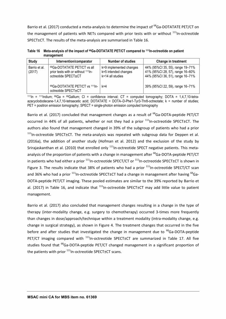
MSAC mini CA for MBS item no. 61369 50
Barrio et al. (2017) conducted a meta-analysis to determine the impact of 68Ga-DOTATATE PET/CT on
the management of patients with NETs compared with prior tests with or without 111In-octreotide
SPECT±CT. The results of the meta-analysis are summarised in Table 16.
Table 16 Meta-analysis of the impact of 68Ga-DOTATATE PET/CT compared to 111In-octreotide on patient management
Study Intervention/comparator Number of studies Change in treatment
Barrio et al. (2017)
68Ga-DOTATATE PET/CT vs all prior tests with or without 111In-octreotide SPECT±CT
68Ga-DOTATATE PET/CT vs 111In-octreotide SPECT±CT
k=9 implemented changes k=5 intended changes k=14 all studies
k=4
44% (95%CI 35, 55), range 19–71% 41% (95%CI 28, 57), range 16–60% 44% (95%CI 36, 51), range 16–71%
39% (95%CI 22, 59), range 16–71%
111In = 111Indium; 68Ga = 68Gallium; CI = confidence interval; CT = computed tomography; DOTA = 1,4,7,10-tetra-azacyclododecane-1,4,7,10-tetraacetic acid; DOTATATE = DOTA–D-Phe1-Tyr3-Thr8-octreotate; k = number of studies; PET = positron emission tomography; SPECT = single-photon emission computed tomography
Barrio et al. (2017) concluded that management changes as a result of 68Ga-DOTA-peptide PET/CT
occurred in 44% of all patients, whether or not they had a prior 111In-octreotide SPECT±CT. The
authors also found that management changed in 39% of the subgroup of patients who had a prior 111In-octreotide SPECT±CT. The meta-analysis was repeated with subgroup data for Deppen et al.
(2016a), the addition of another study (Hofman et al. 2012) and the exclusion of the study by
Srirajaskanthan et al. (2010) that enrolled only 111In-octreotide SPECT negative patients. This meta-
analysis of the proportion of patients with a change in management after 68Ga-DOTA-peptide PET/CT
in patients who had either a prior 111In-octreotide SPECT/CT or 111In-octreotide SPECT±CT is shown in
Figure 3. The results indicate that 38% of patients who had a prior 111In-octreotide SPECT/CT scan
and 36% who had a prior 111In-octreotide SPECT±CT had a change in management after having 68Ga-
DOTA-peptide PET/CT imaging. These pooled estimates are similar to the 39% reported by Barrio et
al. (2017) in Table 16, and indicate that 111In-octreotide SPECT±CT may add little value to patient
management.
Barrio et al. (2017) also concluded that management changes resulting in a change in the type of
therapy (inter-modality change, e.g. surgery to chemotherapy) occurred 3-times more frequently
than changes in dose/approach/technique within a treatment modality (intra-modality change, e.g.
change in surgical strategy), as shown in Figure 4. The treatment changes that occurred in the five
before and after studies that investigated the change in management due to 68Ga-DOTA-peptide
PET/CT imaging compared with 111In-octreotide SPECT±CT are summarized in Table 17. All five
studies found that 68Ga-DOTA-peptide PET/CT changed management in a significant proportion of
the patients with prior 111In-octreotide SPECT±CT scans.
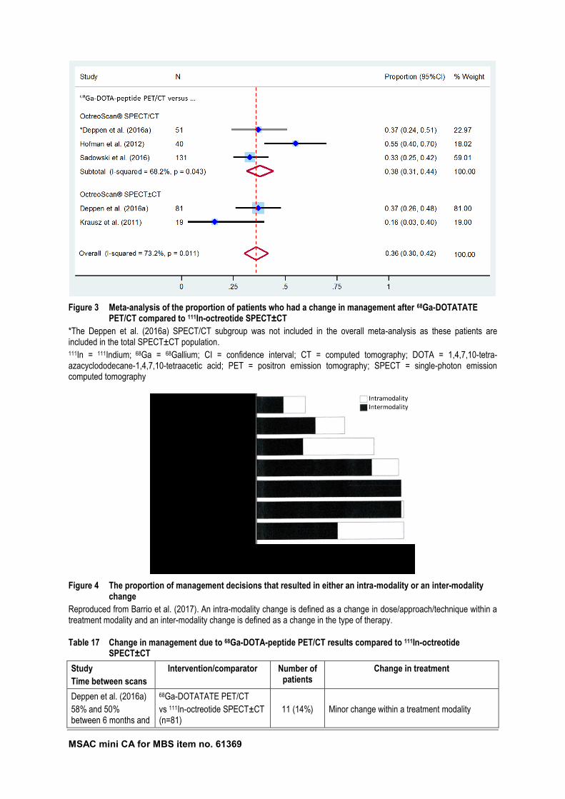
MSAC mini CA for MBS item no. 61369 51
Figure 3 Meta-analysis of the proportion of patients who had a change in management after 68Ga-DOTATATE PET/CT compared to 111In-octreotide SPECT±CT
*The Deppen et al. (2016a) SPECT/CT subgroup was not included in the overall meta-analysis as these patients are included in the total SPECT±CT population. 111In = 111Indium; 68Ga = 68Gallium; CI = confidence interval; CT = computed tomography; DOTA = 1,4,7,10-tetra-azacyclododecane-1,4,7,10-tetraacetic acid; PET = positron emission tomography; SPECT = single-photon emission computed tomography
Figure 4 The proportion of management decisions that resulted in either an intra-modality or an inter-modality change
Reproduced from Barrio et al. (2017). An intra-modality change is defined as a change in dose/approach/technique within a treatment modality and an inter-modality change is defined as a change in the type of therapy.
Table 17 Change in management due to 68Ga-DOTA-peptide PET/CT results compared to 111In-octreotide SPECT±CT
Study
Time between scans
Intervention/comparator Number of patients
Change in treatment
Deppen et al. (2016a)
58% and 50% between 6 months and
68Ga-DOTATATE PET/CT
vs 111In-octreotide SPECT±CT (n=81)
11 (14%)
Minor change within a treatment modality
MSAC mini CA for MBS item no. 61369 52
Study
Time between scans
Intervention/comparator Number of patients
Change in treatment
3 years
Remainder less than 6 months
vs 111In-octreotide SPECT/CT
(n=51)
19 (23%)
8 (16%)
11 (22%)
Major change of treatment modality
(8 surgery cancelled, 12 PRRT)
Minor change within a treatment modality
Major change of treatment modality
Hofman et al. (2012)
40% 6–18 months
60% within 6 months
68Ga-DOTATATE PET/CT
vs 111In-octreotide SPECT/CT
(n=40)
33 (83%)
22 (55%)
Identified additional lesions
High impact changes
(PRRT, SSA therapy, chemotherapy, surgery)
Krausz et al.(2011)
Median 24 days (range 10–65)
68Ga-DOTANOC PET/CT
vs 111In-octreotide SPECT±CT
(n=19 patients who were 111In-octreotide SPECT±CT positive)
4 (21%)
3 (16%)
Implications for disease staging
Implications for patient management
2 referred for PRRT 1 more intensive follow-up
Sadowski et al. (2016)
Within 3 months
68Ga-DOTATATE PET/CT
vs 111In-octreotide SPECT/CT
(n=131)
93 (71%)
44 (34%)
15 (11%)
43 (33%)
19 (15%)
24 (18%)
Detected additional lesions
Detected metastatic lesions
Detected evidence of disease
Change in management
Additional patients had surgery
Additional patients had targeted chemotherapy or PRRT
Srirajaskanthan et al. (2010)
Median 4 months (range 1–8)
68Ga-DOTATATE PET/CT
vs 111In-octreotide SPECT
(n=47 with evidence of disease)
(n=51 patients who were 111In-octreotide SPECT negative or equivocal)
41 (80%)
36 (71%)
20 (39%)
4 (8%)
7 (14%)
3 (6%)
1 (2%)
1 (2%)
Identified lesions
Change in management
Patients had PRRT
Excluded from PRRT due to negative PET/CT
Patients commenced SSA therapy
Patients had surgery
Refused recommended surgery
Confirmation of no disease (No CT or biochemical evidence of disease but some uptake with 111In-octreotide SPECT)
111In = 111Indium; 68Ga = 68Gallium; CT = computed tomography; DOTA = 1,4,7,10-tetra-azacyclododecane-1,4,7,10-tetraacetic acid; DOTANOC = DOTA-Phe1-NaI3-octreotide; DOTATATE = DOTA–D-Phe1-Tyr3-Thr8-octreotate; PET = positron emission tomography PRRT = peptide receptor radionuclide therapy; SPECT = single-photon emission computed tomography; SSA = somatostatin analogue
Even though Deppen et al. (2016a) reported that after reviewing patient records, 111In-octreotide
SPECT±CT did not add value compared with histopathology and anatomical tests in any patient, no
information supporting this statement was provided. None of the other four studies reported on
whether or not the initial management decisions would have differed in the absence of the 111In-
octreotide SPECT±CT. Thus, no direct comparison between management changes as a result of 68Ga-
DOTA-peptide PET/CT and those resulting from 111In-octreotide SPECT±CT can be made.
The study by Deppen et al. (2016a) had the longest time frame between the two SRS tests of up to
three years. For these patients, it is reasonable to expect that the difference between the tests and
any resultant change in management may be at least partially due to disease progression. However,
the authors found that when the time between tests was broken into 3 categories (0–90. 91–180,
and >180 days), the highest proportion of scans having an impact on treatment were in the 0–90 day
category (44% versus 36% for >180 days), though the differences between categories were not
MSAC mini CA for MBS item no. 61369 53
significant. The study by Hofman et al. (2012) with up to 18 months between tests also found that
the greatest proportion of patients with a high management impact had both SRS tests within 3
months (71% versus 50% for >6 months). This suggest that the results have not been confounded by
disease progression between SRS tests in these studies.
Three studies reported that 68Ga-DOTA-peptide PET/CT provided additional information for disease
staging (Hofman et al. 2012; Krausz et al. 2011; Sadowski et al. 2016). Hofman et al. (2012) and
Sadowski et al. (2016) noted that the most frequent consequence of 68Ga-DOTATATE PET/CT
scanning was to increase the number of lesions detected (Table 17), and this resulted in many
patients receiving systemic therapy rather than undergoing surgery. Srirajaskanthan et al. (2010)
found that in many patients with negative or equivocal 111In-octreotide SPECT findings, 68Ga-
DOTATATE PET/CT identified additional lesions and altered management in most cases. Sadowski et
al. (2016) concluded that 68Ga-DOTATATE PET/CT imaging should be implemented in the initial
management and follow-up of patients with GEP NETs as it significantly improves patient care
decisions.
Three studies found that 68Ga-DOTA-peptide PET/CT imaging was useful in identifying the primary
lesion in NET cases where it is unknown (Table 18). It should be noted that the patients had a prior 111In-octreotide SPECT/CT in only one of these studies (Sadowski et al. 2016). This study identified
only 29% of the unknown primary tumour sites, compared with 59% in the other two studies. This
suggests that for a proportion of the patients, both 68Ga-DOTA-peptide PET/CT and 111In-octreotide
SPECT/CT imaging would have identified the primary tumours. Nevertheless, the additional patients
having their primary tumour site identified after 68Ga-DOTA-peptide PET/CT imaging is likely to be of
clinical significance.
These findings had implications for surgical management in 17–41% of identified cases. The authors
of these studies also concluded that 68Ga-DOTATATE PET/CT is a clinically useful imaging technique
for the localization of primary tumours and can play a major role in patient management.
Table 18 Identification of primary tumour site in patients with NETS of unknown origin using 68Ga-DOTA-peptide PET/CT
Study Study population Primary lesion identified Change in treatment
Alonso et al. (2014)
DOTATATE
29 patients with negative conventional imaging studies
(contrast enhanced CT and MRI) for primary tumour identification
17 (59%) – all GEP
9 midgut: 7 ileum, 1 duodenum, 1 colon
8 foregut: 7 pancreas, 1 stomach
7 (41%) patients had surgery
Prasad et al. (2010)
DOTANOC
59 patients with negative physical examination and conventional
imaging (multislice CT, MRI and ultrasonography)
35 (59%)
32 (91%)GEPs: 14 ileum/jejunum, 16 pancreas, 2 rectum/colon,
3 (9%) other: 2 lung and 1 paraganglioma
6 (17%) patients had surgery
29 (83%) had advanced metastases and were not
suitable for surgery
Sadowski et al. (2016)
DOTATATE
14 patients with negative 111In-octreotide SPECT/CT and
conventional (CT or MRI) imaging
4 (29%)
1 small bowel, 3 site not reported
Not reported
68Ga = 68Gallium; CT = computed tomography; DOTA = 1,4,7,10-tetra-azacyclododecane-1,4,7,10-tetraacetic acid;
MSAC mini CA for MBS item no. 61369 54
DOTANOC = DOTA-Phe1-NaI3-octreotide; DOTATATE = DOTA–D-Phe1-Tyr3-Thr8-octreotate; GEP = gastroenteropancreatic; MRI = magnetic resonance imaging; NET = neuroendocrine tumour; PET = positron emission tomography
Only two studies reported on any patient for whom the 68Ga-DOTATATE PET/CT result may have
resulted in a suboptimal treatment plan. Hofman et al. (2012) reported on one false positive 68Ga-
DOTATATE PET/CT result for a case that had been confirmed histologically. In this case, there was
moderately increased uptake of 68Ga-DOTATATE in the pancreas which was concordant with earlier 111In-octreotide imaging. Although it is now known that such uptake can be physiological, the patient
underwent surgery and histology revealed no evidence of a GEP NET. Srirajaskanthan et al. (2010)
reported that 68Ga-DOTATATE PET/CT was false-negative in one intermediate-grade non-functional
pancreatic tumour. In this patient, 111In-octreotide SPECT imaging showed faint uptake in the region
corresponding to the site of liver metastases. The effect on clinical management for this patient was
not discussed. Nevertheless, it seems that few patients would receive inappropriate treatment as a
result of a 68Ga-DOTA-peptide PET/CT scan.
SUMMARY OF THE TYPES OF CHANGES IN MANAGEMENT RESULTING FROM 68GA-DOTA-PEPTIDE PET/CT
Most of the identified changes in management are as a direct consequence of the improved spatial
resolution and clarity of the 68Ga-DOTA-peptide PET/CT image compared with the 111In-octreotide
SPECT/CT image. There are four main scenarios that lead to a potentially major impact on patient
management:
1. Histopathology-positive GEP NET patients who were falsely negative with 111In-octreotide
SPECT/CT are suspected of having low levels of the somatostatin receptor expressed on the
tumour cell surface, and would therefore be limited to surgical and/or chemotherapy treatment
options. Due to the better NPV of 68Ga-DOTA-peptide PET/CT compared to 111In-octreotide
SPECT/CT (section B4.6), approximately half of these patients are likely to be positive after 68Ga-
DOTA-peptide PET/CT and could become eligible for either SSA therapy or PRRT, depending on
their disease status.
2. Histopathology-positive GEP NET patients who remain SRS negative after 68Ga-DOTA-peptide
PET/CT are likely to have poorly differentiated disease that does not express the somatostatin
receptor; and therefore, would not benefit from either SSA therapy or PRRT. These patients
would appropriately be directed towards other surgical or chemotherapy options.
3. 68Ga-DOTA-peptide PET/CT can identify the primary tumour sites for many patients in whom it
is otherwise not detected (Table 18). This could lead to resection of the primary tumour.
4. 68Ga-DOTA-peptide PET/CT can identify smaller lesions than 111In-octreotide SPECT/CT and
hence can identify patients who have more extensive metastatic disease than previously
realised. These patients may receive PRRT instead of, or in addition to, any planned surgical
procedures.
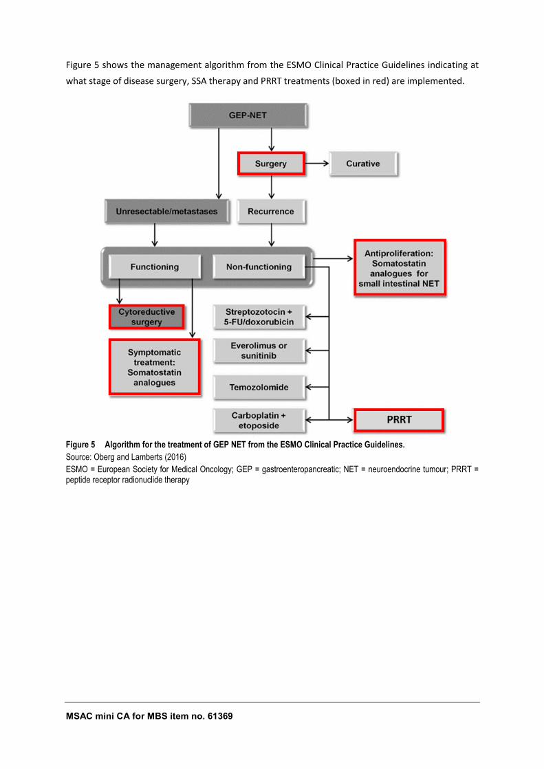
MSAC mini CA for MBS item no. 61369 55
Figure 5 shows the management algorithm from the ESMO Clinical Practice Guidelines indicating at
what stage of disease surgery, SSA therapy and PRRT treatments (boxed in red) are implemented.
Figure 5 Algorithm for the treatment of GEP NET from the ESMO Clinical Practice Guidelines.
Source: Oberg and Lamberts (2016)
ESMO = European Society for Medical Oncology; GEP = gastroenteropancreatic; NET = neuroendocrine tumour; PRRT = peptide receptor radionuclide therapy
MSAC mini CA for MBS item no. 61369 56
B5.2 THERAPEUTIC EFFECTIVENESS (INCLUDING IMPACT
OF EFFECT MODIFICATION)
68Ga-DOTATATE PET/CT is proposed to replace 111In-octreotide SPECT±CT in diagnosing GEP NETs.
Most studies found that 68Ga-DOTATATE PET/CT provided additional information for disease staging
that was of value in determining clinical management. The most common management changes
were referral for surgery, PRRT or SSA therapy.
In order to determine the likely impact of these therapies on patient outcomes, the effectiveness of
these therapies are discussed below.
B5.2.1 EVIDENCE BASE
The ‘Report on the Use of Positron Emission Tomography (PET) and Radiopharmaceuticals – Part 2’
(August 2012) was based on a systematic search of the literature using PubMed and Cochrane
databases for original research papers, including SRs and websites of international HTA agencies for
existing HTA reports. This report included two studies investigating SSA therapy (di Bartolomeo et al.
1996; Townsend et al. 2010) and two reviews investigating PRRT (Bodei et al. 2009; van Essen et al.
2009). The study by di Bartolomeo et al. (1996) was excluded as several updated studies and reviews
were identified in a quick literature search.
A quick literature search of the PubMed database identified five reviews: an updated review on
PRRT by Bodei et al. (2014), a review that looked at the safety of PRRT (Sabet, Biersack & Ezziddin
2016), three reviews that looked at the effectiveness and safety of SSA therapy (Oberg & Lamberts
2016; Sidéris, Dubé & Rinke 2012) and a review on surgical management (Tamburrino et al. 2016).
Six retrospective studies were also identified: an Australian study by Townsend et al. (2010) that
investigated the changing patterns of care over two decades for patients diagnosed with carcinoid
tumours in the North West Adelaide Health Service, a multi-centre study that looked at the
effectiveness and safety of PRRT (Horsch et al. 2016), two retrospective studies investigating the
effectiveness of long-acting release (LAR)-SSAs (Laskaratos et al. 2016; Saglam et al. 2015), and two
studies on surgical treatments for GEP NETs (Keck et al. 2017; Pasqual et al. 2016).
B5.2.2 RISK OF BIAS ASSESSMENT
While the reviews provided an overview of the literature and provided useful information in the lack
of primary studies, they were not systematic and could not be critically appraised.
The six non-comparative studies were assessed for their risk of bias using the IHE Case Series
checklist (IHE 2014). The risk of bias for all studies was either low or medium (Table 37 in Appendix
B).
MSAC mini CA for MBS item no. 61369 57
B5.2.3 CHARACTERISTICS OF THE EVIDENCE BASE
See Table 37 in Appendix B for details on the individual studies included in the evidence base.
B5.2.4 OUTCOME MEASURES AND ANALYSIS
See Table 37 in Appendix B for details on the outcomes measured in the included studies.
Two studies reported survival over a fixed time period as a proportion, which was appropriate. The
remaining four studies reported time to radiological progression, partial response rate, overall
survival and progression-free survival. The analysis used in these studies included Cox univariate and
multi-variate regression analysis and Kaplan–Meier techniques, which were appropriate.
B5.2.5 RESULTS OF THE SYSTEMATIC LITERATURE REVIEW
DOES THE CHANGE IN MANAGEMENT IMPROVE HEALTH OUTCOMES?
Summary – Does the change in management due to 68Ga-DOTA-peptide PET/CT scanning improve
patient outcomes?
The most common management changes resulting from 68Ga-DOTA-peptide PET/CT scanning were referral for
SSA therapy, PRRT or surgery.
Effect of SSA on survival in patients with GEP NETs
68Ga-DOTA-peptide PET/CT scanning has a better negative predictive value than 111In-octreotide SPECT/CT.
Those patients who would have been falsely classified as negative on 111In-octreotide SPECT/CT, but receive
positive results on 68Ga-DOTA-peptide PET/CT scanning, would be eligible to receive SSA therapy or PRRT.
Additionally, patients with multiple small metastatic lesions are more likely to have these detected with 68Ga-
DOTA-peptide PET/CT scanning than 111In-octreotide SPECT/CT. The detection of more extensive disease in
these patients may result in treatments such as PRRT or SSA therapy instead of, or in addition to, any planned
surgical procedures.
Three reviews found that SSA therapy with octreotide controlled clinical symptoms arising from hormone
secretion in somatostatin receptor-expressing NETs and two of them also report that recently published data
have established an anti-proliferative effect for SSAs. Approximately half of the patients with GEP NET on SSA
therapy achieved stabilization of tumour growth with a duration of 8–16 months and 10–20% showed tumour
regression. Stable disease was achieved in 15–67% patients with poorly differentiated, functioning or non-
functioning GEP NETs treated with octreotide and up to 26–88% in those receiving long-acting release (LAR)
octreotide. Two recent retrospective non-comparative studies supported the findings (Laskaratos et al. 2016;
Saglam et al. 2015).
MSAC mini CA for MBS item no. 61369 58
Saglam et al. (2015) reported an estimated 5-year survival of 58% for patients treated with LAR SSA therapy.
When this was compared with the 5-year survival rates for other treatments reported by Bodei et al. (2014), only
surgical resection had a longer 5-year survival rate (70–85%). Patients receiving chemotherapy and PRRT had
5-year survival rates of 40% and 50%, respectively. These patients would be expected to have similar disease
profiles (metastatic disease) to the patients enrolled in the study by Saglam et al. (2015), suggesting that LAR
SSA therapy may be an effective treatment option in these patients.
A retrospective study by Townsend et al. (2010) found that the median overall survival was 112 months for
patients receiving octreotide LAR compared with 53 months for those who received SSA therapy using non-LAR
analogues (p=0.021, hazard ratio: 2.46), and 10 year survival was 40% compared with 22%, respectively. Thus,
LAR SSA therapy appears to provide some survival benefits over non-LAR SSAs to GEP NET patients.
Octreotide has a well-established favourable safety profile with mild to moderately severe gastrointestinal-related
complaints, which mostly resolve after a few weeks of therapy, were the most frequently reported side effects.
Almost half of all patients with advanced GEP NET are at risk of developing gallstones and/or biliary sludge while
receiving SSA therapy, but only 1% of patients develop symptoms sufficiently acute to require a
cholecystectomy.
Effect of PRRT on survival in patients with GEP NETs
Bodei et al. (2014) reviewed PRRT and concluded that it was an efficient and relatively safe treatment of
unresectable or metastatic NETs. They found that the median time to progression ranged from 10–29 months for
90Y-DOTATOC compared with 36 months for 177Lu-DOTATATE, and the median progression-free survival ranged
from 16–29 months and 29–33 months, respectively. Horsch et al. (2016) also found that overall survival and
progression-free survival were significantly inferior in the patients treated with solely 90Y-DOTATOC compared to
177Lu-DOTATATE.
Bodei et al. (2014) also reported on the proportion of patients surviving for longer than 5 years after PRRT.
Approximately 50% of patients undergoing PRRT survived for at least 5 years compared with 40% of those
undergoing chemotherapy. As these patients groups are likely to have similar disease characteristics, PRRT may
be more effective than chemotherapy. However, as the evidence base was non-comparative, the indirect
comparisons between treatments are likely to be confounded, so randomised controlled trials are needed to
determine any true differences between treatments.
Sabet et al. (2016) reported that the high radiation doses associated with 90Y-DOTATOC PRRT can lead to renal
impairment and/or delayed end-stage renal disease and significant bone marrow toxicities. When 177Lu-
DOTATATE is used instead of 90Y-DOTATOC the number of patients with both haematological and renal
toxicities are reduced. Thus, 177Lu-DOTATATE is likely to be safer than 90Y-DOTATOC. The safety of 177Lu-
DOTATATE also compares favourably with reported toxicities for common chemotherapy regimens including 5-
fluorouracil orstreptozocin (20%-30%, grades 3/4) and sunitinib (>30%, grades 3/4) for pancreatic NET.
Effect of surgery on survival in patients with GEP NETs
Patients with unknown primary tumour sites are more likely to have these detected with 68Ga-DOTA-peptide
PET/CT scanning than 111In-octreotide SPECT/CT, enabling these patients to have surgical resections.
MSAC mini CA for MBS item no. 61369 59
A review by Tamburrino et al. (2016) concluded that surgical resection improves survival compared with no
surgery. Another review by Bodei et al. (2014) reported that the proportion of patients whose primary tumours
were surgically resected surviving for longer than 5 years was approximately 70–85% compared with 25% of
untreated patients, 40% of those receiving chemotherapy and 50% of patients on PRRT. However, it should be
noted that there are large differences in the patient characteristics between treatments. Those receiving
“curative” surgery would have earlier stage disease compared to those receiving chemotherapy or PRRT,
confounding the results. Nevertheless, surgery remains the only “curative” treatment and the promising long-term
survival of these patients suggests that surgery is an effective treatment for GEP NET.
B5.2.5.1 EFFECT OF SSA THERAPY ON SURVIVAL IN PATIENTS WITH GEP NETS
Histopathology-positive GEP NET patients who were falsely negative with 111In-octreotide SPECT/CT
would be limited to surgical and/or chemotherapy treatment options as they would be expected to
have low levels of the somatostatin receptor expressed on the tumour cell surface. The better NPV
of 68Ga-DOTA-peptide PET/CT compared to 111In-octreotide SPECT/CT (section B4.6) indicates that
approximately half of these patients are likely to be positive after 68Ga-DOTA-peptide PET/CT and
could become eligible for either SSA therapy or PRRT, depending on their disease status.
Furthermore, as 68Ga-DOTA-peptide PET/CT can identify smaller lesions than 111In-octreotide
SPECT/CT, it can identify patients who have more extensive metastatic disease than previously
realised. These patients may receive PRRT or SSA therapy instead of, or in addition to, any planned
surgical procedures.
Three reviews found that SSA therapy controlled clinical symptoms arising from hormone secretion
in somatostatin receptor-expressing NETs (Narayanan & Kunz 2016; Oberg & Lamberts 2016; Sidéris,
Dubé & Rinke 2012). Two of these reviews also reported that recently published data have
established an anti-proliferative effect for SSAs. Oberg and Lamberts (2016) stated that the clinical
use of octreotide has contributed to improved patient survival since 1987. Sideris et al. (2012)
concluded that stable disease in patients with poorly differentiated, functioning or non-functioning
GEP NETs was achieved in 15–67% of patients treated with octreotide and in 26–88% of those
receiving octreotide LAR.
Two recent retrospective studies agreed with the findings in the reviews. The study by Laskaratos et
al.(2016) looked at the response to SSA therapy using octreotide LAR in 254 treatment naïve patients
with advanced NETs and found that a partial response occurred in 5% of patients and the median
time to radiological progression was 37 (95%CI 32, 52) months. The study by Saglam et al. (2015)
found that patients with locally inoperable or metastatic well-differentiated non-functional NETs
who received octreotide LAR treatment had a progression-free survival of 25.0±3.4 months (95%CI
18.4, 31.5) and an overall survival of 71.3±9.5 months (95%CI 52.7, 89.9). The estimated 5-year
survival rate for patients receiving Lar SSA therapy was 58%. A study by Townsend et al. (2010)
found that patients who received octreotide LAR had improved outcomes; the median overall
survival was 112 months compared with 53 months for those who received SSA therapy using non-
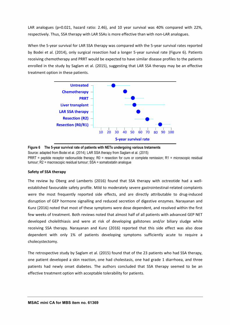
MSAC mini CA for MBS item no. 61369 60
LAR analogues (p=0.021, hazard ratio: 2.46), and 10 year survival was 40% compared with 22%,
respectively. Thus, SSA therapy with LAR SSAs is more effective than with non-LAR analogues.
When the 5-year survival for LAR SSA therapy was compared with the 5-year survival rates reported
by Bodei et al. (2014), only surgical resection had a longer 5-year survival rate (Figure 6). Patients
receiving chemotherapy and PRRT would be expected to have similar disease profiles to the patients
enrolled in the study by Saglam et al. (2015), suggesting that LAR SSA therapy may be an effective
treatment option in these patients.
Figure 6 The 5-year survival rate of patients with NETs undergoing various tretaments
Source: adapted from Bodei et al. (2014); LAR SSA therapy from Saglam et al. (2015)
PRRT = peptide receptor radionuclide therapy; R0 = resection for cure or complete remission; R1 = microscopic residual tumour; R2 = macroscopic residual tumour; SSA = somatostatin analogue
Safety of SSA therapy
The review by Oberg and Lamberts (2016) found that SSA therapy with octreotide had a well-
established favourable safety profile. Mild to moderately severe gastrointestinal-related complaints
were the most frequently reported side effects, and are directly attributable to drug-induced
disruption of GEP hormone signalling and reduced secretion of digestive enzymes. Narayanan and
Kunz (2016) noted that most of these symptoms were dose dependent, and resolved within the first
few weeks of treatment. Both reviews noted that almost half of all patients with advanced GEP NET
developed cholelithiasis and were at risk of developing gallstones and/or biliary sludge while
receiving SSA therapy. Narayanan and Kunz (2016) reported that this side effect was also dose
dependent with only 1% of patients developing symptoms sufficiently acute to require a
cholecystectomy.
The retrospective study by Saglam et al. (2015) found that of the 23 patients who had SSA therapy,
one patient developed a skin reaction, one had cholestasis, one had grade 1 diarrhoea, and three
patients had newly onset diabetes. The authors concluded that SSA therapy seemed to be an
effective treatment option with acceptable tolerability for patients.
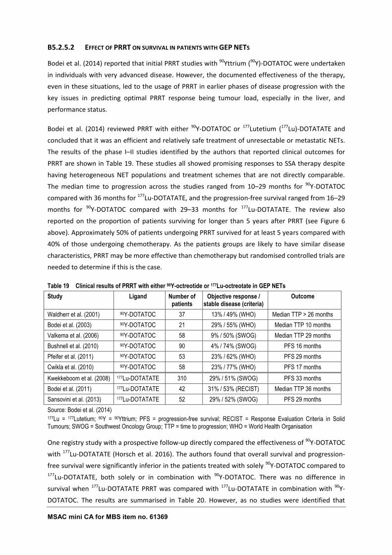
MSAC mini CA for MBS item no. 61369 61
B5.2.5.2 EFFECT OF PRRT ON SURVIVAL IN PATIENTS WITH GEP NETS
Bodei et al. (2014) reported that initial PRRT studies with 90Yttrium (90Y)-DOTATOC were undertaken
in individuals with very advanced disease. However, the documented effectiveness of the therapy,
even in these situations, led to the usage of PRRT in earlier phases of disease progression with the
key issues in predicting optimal PRRT response being tumour load, especially in the liver, and
performance status.
Bodei et al. (2014) reviewed PRRT with either 90Y-DOTATOC or 177Lutetium (177Lu)-DOTATATE and
concluded that it was an efficient and relatively safe treatment of unresectable or metastatic NETs.
The results of the phase I–II studies identified by the authors that reported clinical outcomes for
PRRT are shown in Table 19. These studies all showed promising responses to SSA therapy despite
having heterogeneous NET populations and treatment schemes that are not directly comparable.
The median time to progression across the studies ranged from 10–29 months for 90Y-DOTATOC
compared with 36 months for 177Lu-DOTATATE, and the progression-free survival ranged from 16–29
months for 90Y-DOTATOC compared with 29–33 months for 177Lu-DOTATATE. The review also
reported on the proportion of patients surviving for longer than 5 years after PRRT (see Figure 6
above). Approximately 50% of patients undergoing PRRT survived for at least 5 years compared with
40% of those undergoing chemotherapy. As the patients groups are likely to have similar disease
characteristics, PRRT may be more effective than chemotherapy but randomised controlled trials are
needed to determine if this is the case.
Table 19 Clinical results of PRRT with either 90Y-octreotide or 177Lu-octreotate in GEP NETs
Study Ligand Number of patients
Objective response / stable disease (criteria)
Outcome
Waldherr et al. (2001) 90Y-DOTATOC 37 13% / 49% (WHO) Median TTP > 26 months
Bodei et al. (2003) 90Y-DOTATOC 21 29% / 55% (WHO) Median TTP 10 months
Valkema et al. (2006) 90Y-DOTATOC 58 9% / 50% (SWOG) Median TTP 29 months
Bushnell et al. (2010) 90Y-DOTATOC 90 4% / 74% (SWOG) PFS 16 months
Pfeifer et al. (2011) 90Y-DOTATOC 53 23% / 62% (WHO) PFS 29 months
Cwikla et al. (2010) 90Y-DOTATOC 58 23% / 77% (WHO) PFS 17 months
Kwekkeboom et al. (2008) 177Lu-DOTATATE 310 29% / 51% (SWOG) PFS 33 months
Bodei et al. (2011) 177Lu-DOTATATE 42 31% / 53% (RECIST) Median TTP 36 months
Sansovini et al. (2013) 177Lu-DOTATATE 52 29% / 52% (SWOG) PFS 29 months
Source: Bodei et al. (2014) 177Lu = 177Lutetium; 90Y = 90Yttrium; PFS = progression-free survival; RECIST = Response Evaluation Criteria in Solid Tumours; SWOG = Southwest Oncology Group; TTP = time to progression; WHO = World Health Organisation
One registry study with a prospective follow-up directly compared the effectiveness of 90Y-DOTATOC
with 177Lu-DOTATATE (Horsch et al. 2016). The authors found that overall survival and progression-
free survival were significantly inferior in the patients treated with solely 90Y-DOTATOC compared to 177Lu-DOTATATE, both solely or in combination with 90Y-DOTATOC. There was no difference in
survival when 177Lu-DOTATATE PRRT was compared with 177Lu-DOTATATE in combination with 90Y-
DOTATOC. The results are summarised in Table 20. However, as no studies were identified that
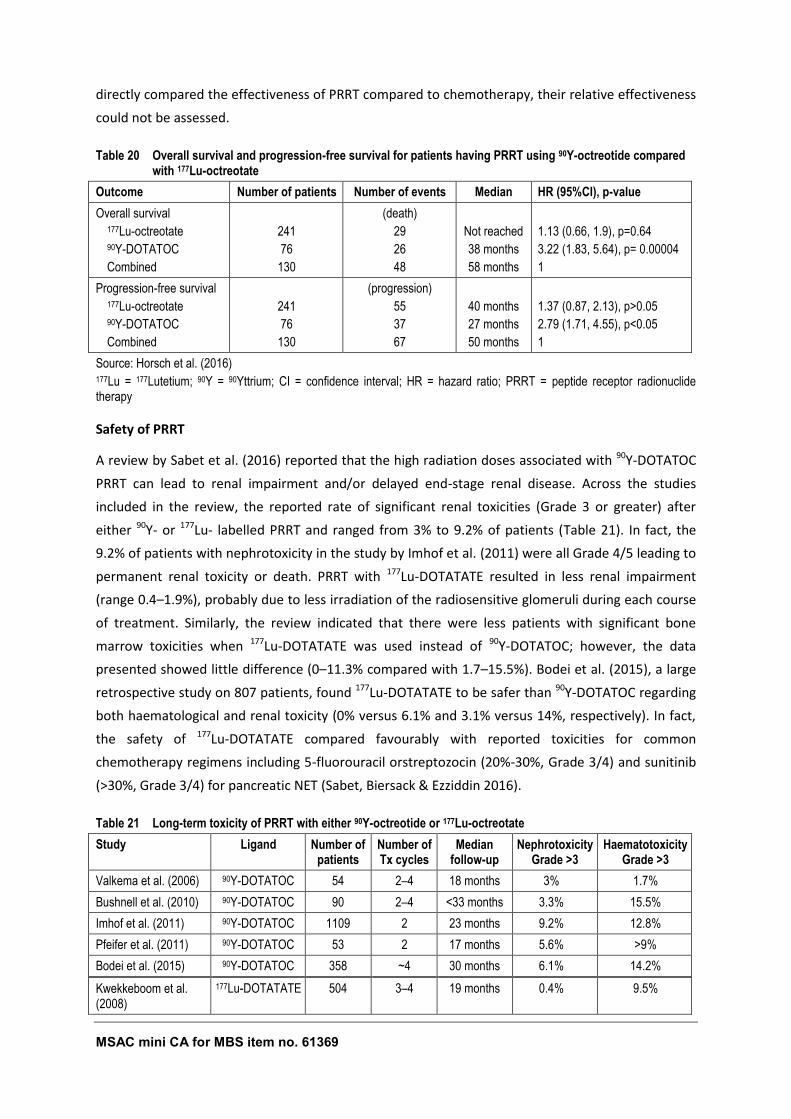
MSAC mini CA for MBS item no. 61369 62
directly compared the effectiveness of PRRT compared to chemotherapy, their relative effectiveness
could not be assessed.
Table 20 Overall survival and progression-free survival for patients having PRRT using 90Y-octreotide compared with 177Lu-octreotate
Outcome Number of patients Number of events Median HR (95%CI), p-value
Overall survival 177Lu-octreotate 90Y-DOTATOC
Combined
241
76
130
(death)
29
26
48
Not reached
38 months
58 months
1.13 (0.66, 1.9), p=0.64
3.22 (1.83, 5.64), p= 0.00004
1
Progression-free survival 177Lu-octreotate 90Y-DOTATOC
Combined
241
76
130
(progression)
55
37
67
40 months
27 months
50 months
1.37 (0.87, 2.13), p>0.05
2.79 (1.71, 4.55), p<0.05
1
Source: Horsch et al. (2016) 177Lu = 177Lutetium; 90Y = 90Yttrium; CI = confidence interval; HR = hazard ratio; PRRT = peptide receptor radionuclide therapy
Safety of PRRT
A review by Sabet et al. (2016) reported that the high radiation doses associated with 90Y-DOTATOC
PRRT can lead to renal impairment and/or delayed end-stage renal disease. Across the studies
included in the review, the reported rate of significant renal toxicities (Grade 3 or greater) after
either 90Y- or 177Lu- labelled PRRT and ranged from 3% to 9.2% of patients (Table 21). In fact, the
9.2% of patients with nephrotoxicity in the study by Imhof et al. (2011) were all Grade 4/5 leading to
permanent renal toxicity or death. PRRT with 177Lu-DOTATATE resulted in less renal impairment
(range 0.4–1.9%), probably due to less irradiation of the radiosensitive glomeruli during each course
of treatment. Similarly, the review indicated that there were less patients with significant bone
marrow toxicities when 177Lu-DOTATATE was used instead of 90Y-DOTATOC; however, the data
presented showed little difference (0–11.3% compared with 1.7–15.5%). Bodei et al. (2015), a large
retrospective study on 807 patients, found 177Lu-DOTATATE to be safer than 90Y-DOTATOC regarding
both haematological and renal toxicity (0% versus 6.1% and 3.1% versus 14%, respectively). In fact,
the safety of 177Lu-DOTATATE compared favourably with reported toxicities for common
chemotherapy regimens including 5-fluorouracil orstreptozocin (20%-30%, Grade 3/4) and sunitinib
(>30%, Grade 3/4) for pancreatic NET (Sabet, Biersack & Ezziddin 2016).
Table 21 Long-term toxicity of PRRT with either 90Y-octreotide or 177Lu-octreotate
Study Ligand Number of patients
Number of Tx cycles
Median follow-up
Nephrotoxicity Grade >3
Haematotoxicity Grade >3
Valkema et al. (2006) 90Y-DOTATOC 54 2–4 18 months 3% 1.7%
Bushnell et al. (2010) 90Y-DOTATOC 90 2–4 <33 months 3.3% 15.5%
Imhof et al. (2011) 90Y-DOTATOC 1109 2 23 months 9.2% 12.8%
Pfeifer et al. (2011) 90Y-DOTATOC 53 2 17 months 5.6% >9%
Bodei et al. (2015) 90Y-DOTATOC 358 ~4 30 months 6.1% 14.2%
Kwekkeboom et al. (2008)
177Lu-DOTATATE 504 3–4 19 months 0.4% 9.5%
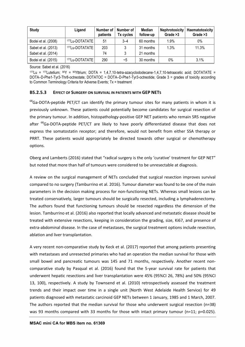
MSAC mini CA for MBS item no. 61369 63
Study Ligand Number of patients
Number of Tx cycles
Median follow-up
Nephrotoxicity Grade >3
Haematotoxicity Grade >3
Bodei et al. (2008) 177Lu-DOTATATE 51 3–4 60 months 1.9% 0%
Sabet et al. (2013)
Sabet et al. (2014)
177Lu-DOTATATE 203
74
3
3
31 months
21 months
1.3% 11.3%
Bodei et al. (2015) 177Lu-DOTATATE 290 ~5 30 months 0% 3.1%
Source: Sabet et al. (2016) 177Lu = 177Lutetium; 90Y = 90Yttrium; DOTA = 1,4,7,10-tetra-azacyclododecane-1,4,7,10-tetraacetic acid; DOTATATE = DOTA–D-Phe1-Tyr3-Thr8-octreotate; DOTATOC = DOTA–D-Phe1-Tyr3-octreotide; Grade 3 = grades of toxicity according to Common Terminology Criteria for Adverse Events; Tx = treatment
B5.2.5.3 EFFECT OF SURGERY ON SURVIVAL IN PATIENTS WITH GEP NETS
68Ga-DOTA-peptide PET/CT can identify the primary tumour sites for many patients in whom it is
previously unknown. These patients could potentially become candidates for surgical resection of
the primary tumour. In addition, histopathology-positive GEP NET patients who remain SRS negative
after 68Ga-DOTA-peptide PET/CT are likely to have poorly differentiated disease that does not
express the somatostatin receptor; and therefore, would not benefit from either SSA therapy or
PRRT. These patients would appropriately be directed towards other surgical or chemotherapy
options.
Oberg and Lamberts (2016) stated that “radical surgery is the only ‘curative’ treatment for GEP NET”
but noted that more than half of tumours were considered to be unresectable at diagnosis.
A review on the surgical management of NETs concluded that surgical resection improves survival
compared to no surgery (Tamburrino et al. 2016). Tumour diameter was found to be one of the main
parameters in the decision making process for non-functioning NETs. Whereas small lesions can be
treated conservatively, larger tumours should be surgically resected, including a lymphadenectomy.
The authors found that functioning tumours should be resected regardless the dimension of the
lesion. Tamburrino et al. (2016) also reported that locally advanced and metastatic disease should be
treated with extensive resections, keeping in consideration the grading, size, Ki67, and presence of
extra-abdominal disease. In the case of metastases, the surgical treatment options include resection,
ablation and liver transplantation.
A very recent non-comparative study by Keck et al. (2017) reported that among patients presenting
with metastases and unresected primaries who had an operation the median survival for those with
small bowel and pancreatic tumours was 145 and 71 months, respectively. Another recent non-
comparative study by Pasqual et al. (2016) found that the 5-year survival rate for patients that
underwent hepatic resections and liver transplantation were 45% (95%CI 26, 78%) and 50% (95%CI
13, 100), respectively. A study by Townsend et al. (2010) retrospectively assessed the treatment
trends and their impact over time in a single unit [North West Adelaide Health Service) for 49
patients diagnosed with metastatic carcinoid GEP NETs between 1 January, 1985 and 1 March, 2007.
The authors reported that the median survival for those who underwent surgical resection (n=38)
was 93 months compared with 33 months for those with intact primary tumour (n=11; p=0.025).
MSAC mini CA for MBS item no. 61369 64
However, the decision for surgery was based on standard clinical practice indicating that those who
did not have surgery most likely had unresectable tumours, making the two groups not directly
comparable. Pasqual et al. (2016) concluded that although the results are encouraging, randomized
clinical trials are necessary to more adequately evaluate the effect of surgery on survival in GEP NET
patients.
The review by Bodei et al. (2014) reported on the proportion of patients surviving for longer than 5
years after surgical resection, liver transplantation, chemotherapy, and PRRT compared to untreated
patients (Figure 6). Approximately 70–85% of patients whose primary tumours are surgically
resected survived for at least 5 years compared with 25% of untreated patients and 40% of those
receiving chemotherapy. The median 5-year survival rate is similar (approximately 50%) for patients
who have a liver transplant and for patients on PRRT. However, it should be noted that there are
large differences in the patient characteristics between treatments. Those receiving “curative”
surgery would have earlier stage disease compared to those receiving chemotherapy or PRRT. These
differences would confound the results making a direct comparison between surgery and other
treatments uncertain. Nevertheless, surgery remains the only “curative” treatment and the
promising long-term survival of these patients suggests that surgery is an effective treatment for
GEP NET.
MSAC mini CA for MBS item no. 61369 65
B6 IMPACT OF REPEAT TESTING/MONITORING
The clinical claim in the PICO Confirmation was that introducing 68Ga-DOTA-peptide PET/CT scanning
in lieu of 111In-octreotide SPECT/CT will reduce the number of tests that would need to be repeated.
No evidence was provided by the Department of Health, or identified in the quick literature search
to inform on repeat testing. The studies included in the meta-analyses to determine the pooled
sensitivity and specificity of 111In-octreotide SPECT±CT did not report any patients in whom the test
was inconclusive. Similarly, no reports of failed 68Ga-DOTA-peptide PET/CT scans were identified in
the evidence base.
However, the accuracy data did indicate that almost twice as many patients with histologically
proven GEP NETs will have a negative 111In-octreotide SPECT/CT compared with a negative 68Ga-
DOTA-peptide PET/CT. Thus, fewer patients with suspected GEP NET would need additional
anatomical or functional tests, as shown in the clinical algorithm (Figure 2 in section A6), to obtain a
final diagnosis.
Some of the studies included in the evidence base tested patients who had recurrent disease, or
required follow-up after surgical or other treatments with 68Ga-DOTA-peptide PET/CT. Thus,
repeated testing may be of clinical importance for long-term management of these patients.
However, the effectiveness of follow-up testing was not evaluated as it was outside the scope of this
assessment.
MSAC mini CA for MBS item no. 61369 66
B7 EXTENDED ASSESSMENT OF COMPARATIVE HARMS
The ‘Report on the Use of Positron Emission Tomography (PET) and Radiopharmaceuticals’ (August
2012) by Austin Health12 reported on the safety of 68Ga-DOTA-peptide PET/CT compared with 111In-
octreotide SPECT/CT and included the safety results reported by Hofman et al. (2012). The applicant
provided a recent study by Deppen et al. (2016a) that looked at the safety and efficacy of 68Ga-
DOTATATE PET/CT.
The US Library of Medicine website13 provides information on the use and safety of both 111In-
octreotide® and 68Ga-DOTATATE (marketed as NETSPOT). This site reported that the safety of 68Ga-
DOTATATE was evaluated in three single centre studies (Deppen et al. 2016a; Haug et al. 2014; Haug
et al. 2012) and that no serious adverse reactions were identified. Of these three studies, only
Deppen et al. (2016a) published any adverse event data.
The SR by Deppen et al. (2016b) also identified three studies that reported on adverse events
(Deppen et al. 2016a; Etchebehere et al. 2014; Kunikowska et al. 2014).
B7.1 SHORT-TERM SAFETY
Austin Health found that there were no documented serious adverse events associated with
injection of the 68Ga, or with performing the PET/CT scan and that the studies included in their
review did not raise any new safety concerns. Austin Health also concluded that multiple MSAC
assessments of FDG PET have stated that it is generally accepted that PET is a non-invasive and
relatively safe diagnostic procedure.
Only six mild adverse events associated with injection of the 68Ga-DOTA-peptide were reported in
the studies included in this assessment (adverse events associated with 111In-octreotide SPECT±CT
were not reported in any study). Deppen et al. (2016a) reported that minor adverse events occurred
in three patients. One had minor itching at the 68Ga-DOTATATE injection site the next day. One
patient had an unexplained drop in post-scan oxygen saturation on room air from 98% before
injection to 90% after scanning. Both of these resolved spontaneously. The third patient had an
asymptomatic post-scan tachycardia of 112 beats/minute (baseline 87 beats/minute), spontaneously
returning to less than 100 beats/minute within an hour. Kunikowska et al. (2014) reported that two
patients with a history of gastritis had abdominal pain associated with 68Ga-DOTATATE
12 The Commonwealth Government Department of Health and Ageing contracted Austin Health to undertake a
review of the Broader Use of Positron Emission Tomography (PET) and Radiopharmaceuticals in 2012.
13 Available from URL: https://dailymed.nlm.nih.gov/dailymed/index.cfm [accessed 20 December 2016].
MSAC mini CA for MBS item no. 61369 67
administration, which was effectively treated with an antispasmodic drug. The SR by Deppen et al.
(2016b) reported that one patient had an adverse event in the study by Etchebehere et al. (2014);
this patient had unilateral whole-body oedema ipsilateral to the injected arm occurring within 24
hours of injection, which resolved spontaneously in less than 48 hours. However, this adverse event
was not reported in the published study and the source for this information was not reported in the
review.
B7.2 LONG-TERM SAFETY
Hartmann et al. (2009) found that 68Ga-DOTA-peptide PET resulted in a significantly lower radiation
dose (approximately 3–4 mSv) compared with 111In-octreotide SPECT (approximately 12 mSv).
Walker et al. (2013) found similar results with patients undergoing 68Ga-DOTA-peptide PET being
exposed to 3.1–4.8 mSv compared with 5.9 mSv for 111In-octreotide SPECT. The use of low dose CT
for anatomical localisation of the PET or SPECT data delivers radiation exposure up to 1 mSv
(Hartmann et al. 2009). McLean et al. (1989) reported that 111In also causes radiation damage via
emission of Auger electrons, which may be underestimated by traditional dosimetry models. Austin
Health concluded that the potential long-term effects of exposure to ionising radiation are unlikely
to be of major concern to these patients, given their reduced life expectancy.
MSAC mini CA for MBS item no. 61369 68
B8 INTERPRETATION OF THE CLINICAL EVIDENCE
This mini assessment of the effectiveness of 68Ga-DOTA-peptide PET/CT scanning in lieu of
Octreotide SPECT/CT scanning is constrained by a lack of information. To comprehensively assess
these technologies a systematic search of the literature is required. To complement the limited
evidence provided by the applicant a quick literature search of the PubMed database to identify
recent publications to help evaluate these technologies was undertaken. Therefore, the conclusions
drawn from this mini-assessment are based on an incomplete evidence base and the implications of
this are unknown.
On the basis of the evidence presented in section B, it is suggested that, relative to 111In-octreotide
SPECT/CT, 68Ga-DOTA-peptide PET/CT has superior safety and superior effectiveness.
Barrio et al. (2017) found that management changes occurred in 44% of patients as a result of 68Ga-
DOTATATE PET/CT. A change in the type of therapy (inter-modality change) occurred 3-times more
frequently than a change in dose/approach/technique within a treatment modality (intra-modality
change). An updated meta-analysis found 38% of patients who had had a prior 111In-octreotide
SPECT/CT had a change in management after having a 68Ga-DOTATATE PET/CT. The most common
management changes were referral for surgery, PRRT or SSA therapy. No data were available to
determine whether or not management decisions would have differed in the absence of the 111In-
octreotide SPECT±CT. Thus, no direct comparisons between management changes as a result of 68Ga-DOTA-peptide PET/CT and those resulting from 111In-octreotide SPECT±CT can be made.
Nevertheless, as the proportion of patients whose management changed after 68Ga-DOTA-peptide
PET/CT with or without a prior 111In-octreotide SPECT±CT are similar, the results of the 111In-
octreotide SPECT/CT appear to be of little value when determining a patient’s management plan.
68Ga-DOTATATE PET/CT was found to be more sensitive, with less false negative results, than 111In-
octreotide SPECT/CT when compared to the composite reference standard (91% versus 80%). At an
assumed diagnostic yield of 59%, the NPV values for the two tests indicated that 23% of people who
had a negative 111In-octreotide SPECT/CT would actually have a GEP NET compared with 13% of
those who were negative after 68Ga-DOTA-peptide PET/CT. Thus, almost twice as many people who
actually have a GEP NET would have a negative result after 111In-octreotide SPECT/CT compared with 68Ga-DOTA-peptide PET/CT. This difference is likely be of clinical significance and would have
implications for patient management.
Without a 68Ga-DOTA-peptide PET/CT scan, the clinicians would have considered that these patients
expressed the somatostatin receptor poorly based on the negative 111In-octreotide SPECT/CT scan.
As a result, these patients would have been incorrectly directed away from targeted therapies such
as PRRT or SSA therapy as they not be expected to benefit from them and would have probably
MSAC mini CA for MBS item no. 61369 69
received chemotherapy. The 13% of patients with a negative 111In-octreotide SPECT/CT scan who
had a positive 68Ga-DOTA-peptide PET/CT scan would become eligible for PRRT or SSA therapy.
Even though the evidence base was subject to confounding, both of these therapies appear to be
effective treatment options in patients with GEP NETs. Saglam et al. (2015) reported that the 5-year
survival rate for patients on LAR SSA therapy was 58%. When the 5-year survival for LAR SSA therapy
was compared with the 5-year survival rates reported by Bodei et al. (2014), only surgical resection
had a longer 5-year survival rate (Figure 6). Patients receiving chemotherapy and PRRT would be
expected to have similar disease profiles to the patients enrolled in the study by Saglam et al. (2015),
suggesting that LAR SSA therapy may be an effective treatment option in these patients.
Bodei et al. (2014) reviewed PRRT and concluded that it was an efficient and relatively safe
treatment for unresectable or metastatic NETs. The authors also reported on the proportion of
patients surviving for longer than 5 years after PRRT (see Figure 6 above). Approximately 50% of
patients undergoing PRRT survived for at least 5 years compared with 40% of those undergoing
chemotherapy. As these patient groups are likely to have similar disease characteristics, PRRT may
be more effective than chemotherapy but randomised controlled trials are needed to determine if
this is the case.
Thus, patients with negative 111In-octreotide SPECT/CT scans and positive 68Ga-DOTA-peptide PET/CT
scan would most likely have a change in management leading to improved health outcomes.
Conversely, histopathology-positive GEP NET patients who were negative with 68Ga-DOTA-peptide
PET/CT are actually likely to have tumours expressing low levels of the somatostatin receptor, and
would be appropriately directed away from PRRT and SSA therapy towards surgery and/or
chemotherapy options.
The main advantage with 68Ga-DOTANOC PET appeared to be the increased clarity of the images in
comparison with 111In-octreotide SPECT making them easier to interpret (Krausz et al. 2011). This
increased clarity resulted in an increase in the number of lesions detected (Hofman et al. 2012;
Sadowski et al. 2016; Srirajaskanthan et al. 2010). The detection of more lesions also had
implications for patient management in a large number of cases. The detection of more advanced
disease than previously realised would result in clinicians treating patients with PRRT or SSA therapy
instead of, or in addition to, any previously planned surgical procedures.
68Ga-DOTA-peptide PET/CT imaging was also useful in identifying the site of the primary lesion in
NET cases where it was previously unknown (Alonso et al. 2014; Prasad et al. 2010; Sadowski et al.
2016). These studies were non-comparative, and it is likely that the primary tumour site for a
proportion of these patients would have been detected by both 68Ga-DOTA-peptide PET/CT and 111In-octreotide SPECT/CT. Nonetheless, the additional patients having their primary tumour site
identified by 68Ga-DOTA-peptide PET/CT but not by 111In-octreotide SPECT/CT is likely to be of clinical
MSAC mini CA for MBS item no. 61369 70
significance. These patients may be eligible for surgical resection instead of or in addition to other
systemic therapies.
The review by Oberg and Lamberts (2016) stated that “radical surgery is the only ‘curative’
treatment for GEP NET”. A review by Bodei et al. (2014) reported that the proportion of patients
whose primary tumours were surgically resected surviving for longer than 5 years was approximately
70–85% compared with 25% of untreated patients, 40% of those receiving chemotherapy and 50%
of patients on PRRT. However, it should be noted that there are large differences in the patient
characteristics between treatments. Those receiving “curative” surgery would have earlier stage
disease compared to those receiving chemotherapy or PRRT, confounding the results. Nevertheless,
surgery remains the only “curative” treatment and the promising long-term survival of these
patients suggests that surgery is an effective treatment for GEP NET.
Only two studies reported on any patient for whom the 68Ga-DOTATATE PET/CT result may have
resulted in a suboptimal treatment plan. Hofman et al. (2012) reported on one false positive case
with moderately increased uptake in the pancreas which was concordant with earlier 111In-
octreotide SPECT/CT. The patient underwent surgery and histology revealed no evidence of a NET.
Srirajaskanthan et al. (2010) reported that 68Ga-DOTATATE PET/CT was false-negative in 1
intermediate-grade non-functional pancreatic tumour. 111In-octreotide SPECT showed faint uptake in
the region corresponding to the site of liver metastases. The effect on clinical management for this
patient was not discussed. Thus, very few patients (2/322; 0.6%) are likely to be managed incorrectly
based on their 68Ga-DOTA-peptide PET/CT results.
There were no studies identified that reported on the safety of surgery, but surgery remains the only
potentially curative therapy available to patients with GEP NETs. Several reviews reported that
octreotide has a well-established favourable safety profile. The most frequently reported adverse
events were mild to moderately severe gastrointestinal-related complaints, which mostly resolve
after a few weeks of therapy. Almost half of all patients with advanced GEP NET developed
cholelithiasis while receiving SSA therapy, but only 1% of patients developed symptoms sufficiently
acute to require a cholecystectomy. Of the two main PRRT radiopeptides, 177Lu-DOTATATE has a
superior safety profile compared to 90Y-DOTATOC. The higher radiation doses associated with 90Y-
DOTATOC PRRT leads to more renal impairment and/or delayed end-stage renal disease and
significant bone marrow toxicities than 177Lu-DOTATATE. The safety of 177Lu-DOTATATE also
compares favourably with reported toxicities for common chemotherapy regimens including 5-
fluorouracil orstreptozocin (20%-30%, grades 3/4) and sunitinib (>30%, grades 3/4) for pancreatic
NET (Sabet, Biersack & Ezziddin 2016). Thus, treatment of GEP NETs with surgery, SSA therapy or
PRRT appears to be more effective than no treatment or chemotherapy and relatively safe.
Only six patients with mild adverse events associated with injection of the 68Ga-DOTA-peptide were
reported in the studies included in this assessment. Two patients with gastritis and abdominal pain
were effectively treated with an antispasmodic drug. The remaining four cases all resolved
MSAC mini CA for MBS item no. 61369 71
spontaneously in less than 48 hours. Austin Health14 concluded that multiple MSAC assessments of
FDG PET have stated that it is generally accepted that PET is non-invasive and relatively safe.
Additionally, 68Ga-DOTA-peptide PET resulted in a significantly lower radiation dose (approximately
3–5 mSv) compared with 111In-octreotide SPECT (approximately 6–12 mSv). An extra 1 mSv exposure
occurs from the use of low dose CT for transmission correction of the PET or SPECT data. Thus, 68Ga-
DOTA-peptide PET/CT was found to be a relatively safe procedure in comparison to 111In-octreotide
SPECT/CT. Furthermore, Austin Health concluded that any potential long-term effects of exposure to
ionising radiation from 68Ga-DOTA-peptide PET/CT are unlikely to be of major concern to these
patients, given their reduced life expectancy.
In conclusion, 68Ga-DOTA-peptide PET/CT is more sensitive, with an improved NPV indicating that
there are fewer false negative patients compared with 111In-octreotide SPECT/CT. Additionally,
increased clarity of the 68Ga-DOTA-peptide PET images in comparison with 111In-octreotide SPECT
enabled a more accurate detection of the extent of disease and localisation of the primary tumour.
The increased accuracy and clarity resulted in a change in management in approximately 40% of all
patients, irrespective of whether or not the patients had had prior 111In-octreotide SPECT/CT
imaging. These changes in management resulted in more patients being directed towards surgery,
SSA therapy or PRRT rather than chemotherapy. Treatment of GEP NETs with surgery, SSA therapy or
PRRT appear to be more effective than no treatment or chemotherapy and are relatively safe. 68Ga-
DOTA-peptide PET/CT is also safer with lower radiation exposure and quicker (2 hours versus 2 days)
than 111In-octreotide SPECT/CT. When taken together, these results suggest that replacement of 111In-octreotide SPECT/CT with 68Ga-DOTA-peptide PET/CT is likely to lead to better patient
outcomes in up to 30% of patients; i.e. those who had an inter-modality change in management
(Barrio et al. 2017).
14 The Commonwealth Government Department of Health and Ageing contracted Austin Health to undertake a
review of the Broader Use of Positron Emission Tomography (PET) and Radiopharmaceuticals in 2012.
MSAC mini CA for MBS item no. 61369 72
SECTION C TRANSLATION ISSUES
Not applicable.
SECTION D ECONOMIC EVALUATION
Not applicable.
MSAC mini CA for MBS item no. 61369 73
SECTION E FINANCIAL IMPLICATIONS
E.1. JUSTIFICATION OF THE SELECTION OF DATA SOURCES
68Ga-DOTA-peptide PET is proposed as a replacement test (replacing existing listing for 111In-
octreotide SPECT, MBS item 61369), for diagnostic clarification in patients with suspected GEP NETs
(subgroup 1) or to exclude additional disease sites in patients identified with surgically amenable
disease (subgroup 2).
To estimate the target patient population, an epidemiological approach (combined with additional
data from the literature review and clinical expert advice) has been used to estimate the number of
services and financial implications associated with the substitution of 68Ga-DOTA-peptide PET for 111In-octreotide SPECT in MBS item 61369. This is difficult to validate with a market-based estimate,
as the use of existing comparator 111In-octreotide SPECT has decreased in recent years despite the
increasing incidence and prevalence of GEP NETs. Table 40 in Appendix D shows the MBS utilisation
for item 61369 since 2002–03. This may be attributed to treating clinicians and Nuclear Medicine
specialists increasingly using 68Ga-DOTA-peptide PET/CT for SRS in lieu of 111In-octreotide SPECT±CT.
Beginning with an estimate of the number of new cases of GEP NETs anticipated each year, using
population projections by Australian Bureau of Statistics and published/advised incidence and
prevalence rates, the estimated number of 68Ga-DOTA-peptide PET services required to obtain the
number of diagnoses is back-calculated using an estimate of diagnostic yield.
The data sources used to calculate the financial impact of the MBS listing of 68Ga-DOTA-peptide PET
are summarised in Table 22.
Table 22 Parameters and data sources used in the financial analysis
Data source Purpose Value
Epidemiological data - -
Expert advice and Fraenkel et al. (2014)
Estimate of incidence of GEP NETs in Australia
3.0–3.6 per 100,000 per yeara
Fraenkel et al. (2014) and Patel et al. (2016)
Estimate of prevalence of GEP NETs in Australia
21 per 100,000 a
Table 14 in Section B.4 Estimated diagnostic yield in the target population to derive the suspected number of cases eligible for diagnostic clarification with the proposed test
Base case: 59%
Sensitivity analysis: 35% and 76%
ABS data catalogue no. 3222, series B (2013)
Projection of Australian population, all ages in 2017–2022
Row A, Table 23
Market data - -
Medicare item reports Number of octreotide services (item 61369) that are currently MBS funded
Table 40, Appendix D
Expert advice Proportion of new cases receiving a follow-up scan in the same year before being considered
Base case: 10%
Sensitivity analysis: 20% and 30%
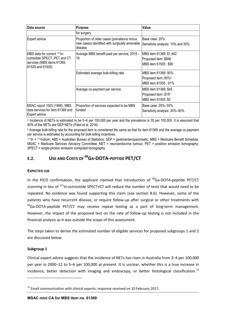
MSAC mini CA for MBS item no. 61369 74
Data source Purpose Value
for surgery
Expert advice Proportion of older cases (prevalence minus new cases) identified with surgically amenable disease
Base case: 20%
Sensitivity analysis: 10% and 30%
MBS data for current 111In-octreotide SPECT, PET and CT services (MBS items 61369, 61529 and 61505)
Average MBS benefit paid per service, 2015–16
MBS item 61369: $1,942
Proposed item: $896
MBS item 61505 : $90
- Estimated average bulk-billing rate MBS item 61369: 90%
Proposed item: 90%b
MBS item 61505 : 91%
- Average co-payment per service MBS item 61369: $45
Proposed item: $15b
MBS item 61505: $3
MSAC report 1003 (1999), MBS data services for item 61369 and Expert advice
Proportion of services expected to be MBS funded
Base case: 20%–30%
Sensitivity analysis: 30%–50%
a Incidence of NETs is estimated to be 5–6 per 100,000 per year and the prevalence is 35 per 100,000. It is assumed that 60% of the NETs are GEP NETs (Patel et al. 2016) b Average bulk-billing rate for the proposed item is considered the same as that for item 61369 and the average co-payment per service is estimated by accounting for bulk-billing incentives. 111In = 111Indium; ABS = Australian Bureau of Statistics; GEP = gastroenteropancreatic; MBS = Medicare Benefit Schedule; MSAC = Medicare Services Advisory Committee; NET = neuroendocrine tumour; PET = positron emission tomography; SPECT = single-photon emission computed tomography
E.2. USE AND COSTS OF 68GA-DOTA-PEPTIDE PET/CT
EXPECTED USE
In the PICO confirmation, the applicant claimed that introduction of 68Ga-DOTA-peptide PET/CT
scanning in lieu of 111In-octreotide SPECT±CT will reduce the number of tests that would need to be
repeated. No evidence was found supporting this claim (see section B.6). However, some of the
patients who have recurrent disease, or require follow-up after surgical or other treatments with 68Ga-DOTA-peptide PET/CT may receive repeat testing as a part of long-term management.
However, the impact of the proposed test on the rate of follow-up testing is not included in the
financial analysis as it was outside the scope of this assessment.
The steps taken to derive the estimated number of eligible services for proposed subgroups 1 and 2
are discussed below.
Subgroup 1
Clinical expert advice suggests that the incidence of NETs has risen in Australia from 3–4 per 100,000
per year in 2000–12 to 5–6 per 100,000 at present. It is unclear, whether this is a true increase in
incidence, better detection with imaging and endoscopy, or better histological classification.15
15 Email communication with clinical experts; response received on 10 February 2017.
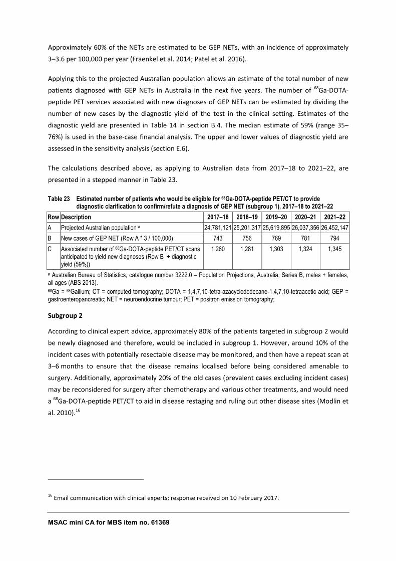
MSAC mini CA for MBS item no. 61369 75
Approximately 60% of the NETs are estimated to be GEP NETs, with an incidence of approximately
3–3.6 per 100,000 per year (Fraenkel et al. 2014; Patel et al. 2016).
Applying this to the projected Australian population allows an estimate of the total number of new
patients diagnosed with GEP NETs in Australia in the next five years. The number of 68Ga-DOTA-
peptide PET services associated with new diagnoses of GEP NETs can be estimated by dividing the
number of new cases by the diagnostic yield of the test in the clinical setting. Estimates of the
diagnostic yield are presented in Table 14 in section B.4. The median estimate of 59% (range 35–
76%) is used in the base-case financial analysis. The upper and lower values of diagnostic yield are
assessed in the sensitivity analysis (section E.6).
The calculations described above, as applying to Australian data from 2017–18 to 2021–22, are
presented in a stepped manner in Table 23.
Table 23 Estimated number of patients who would be eligible for 68Ga-DOTA-peptide PET/CT to provide diagnostic clarification to confirm/refute a diagnosis of GEP NET (subgroup 1), 2017–18 to 2021–22
Row Description 2017–18 2018–19 2019–20 2020–21 2021–22
A Projected Australian population a 24,781,121 25,201,317 25,619,895 26,037,356 26,452,147
B New cases of GEP NET (Row A * 3 / 100,000) 743 756 769 781 794
C Associated number of 68Ga-DOTA-peptide PET/CT scans anticipated to yield new diagnoses (Row B ÷ diagnostic yield (59%))
1,260 1,281 1,303 1,324 1,345
a Australian Bureau of Statistics, catalogue number 3222.0 – Population Projections, Australia, Series B, males + females, all ages (ABS 2013). 68Ga = 68Gallium; CT = computed tomography; DOTA = 1,4,7,10-tetra-azacyclododecane-1,4,7,10-tetraacetic acid; GEP = gastroenteropancreatic; NET = neuroendocrine tumour; PET = positron emission tomography;
Subgroup 2
According to clinical expert advice, approximately 80% of the patients targeted in subgroup 2 would
be newly diagnosed and therefore, would be included in subgroup 1. However, around 10% of the
incident cases with potentially resectable disease may be monitored, and then have a repeat scan at
3–6 months to ensure that the disease remains localised before being considered amenable to
surgery. Additionally, approximately 20% of the old cases (prevalent cases excluding incident cases)
may be reconsidered for surgery after chemotherapy and various other treatments, and would need
a 68Ga-DOTA-peptide PET/CT to aid in disease restaging and ruling out other disease sites (Modlin et
al. 2010).16
16 Email communication with clinical experts; response received on 10 February 2017.
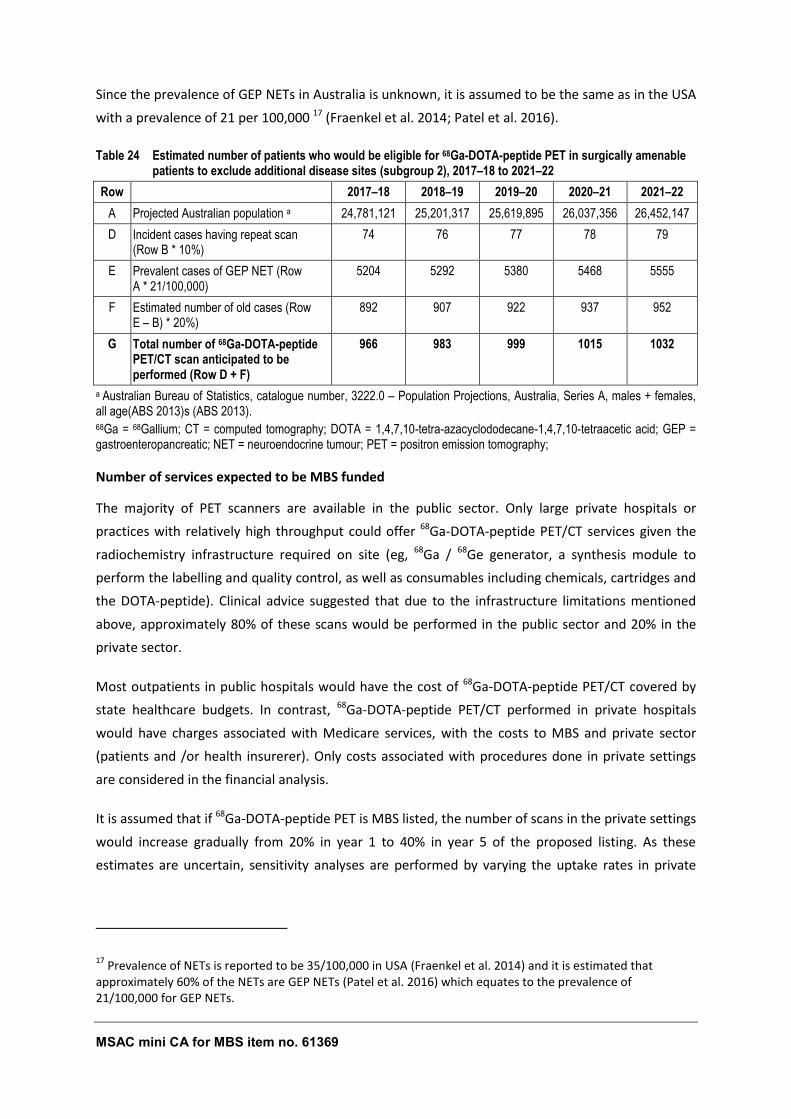
MSAC mini CA for MBS item no. 61369 76
Since the prevalence of GEP NETs in Australia is unknown, it is assumed to be the same as in the USA
with a prevalence of 21 per 100,000 17 (Fraenkel et al. 2014; Patel et al. 2016).
Table 24 Estimated number of patients who would be eligible for 68Ga-DOTA-peptide PET in surgically amenable patients to exclude additional disease sites (subgroup 2), 2017–18 to 2021–22
Row 2017–18 2018–19 2019–20 2020–21 2021–22
A Projected Australian population a 24,781,121 25,201,317 25,619,895 26,037,356 26,452,147
D Incident cases having repeat scan (Row B * 10%)
74 76 77 78 79
E Prevalent cases of GEP NET (Row A * 21/100,000)
5204 5292 5380 5468 5555
F Estimated number of old cases (Row E – B) * 20%)
892 907 922 937 952
G Total number of 68Ga-DOTA-peptide PET/CT scan anticipated to be performed (Row D + F)
966 983 999 1015 1032
a Australian Bureau of Statistics, catalogue number, 3222.0 – Population Projections, Australia, Series A, males + females, all age(ABS 2013)s (ABS 2013). 68Ga = 68Gallium; CT = computed tomography; DOTA = 1,4,7,10-tetra-azacyclododecane-1,4,7,10-tetraacetic acid; GEP = gastroenteropancreatic; NET = neuroendocrine tumour; PET = positron emission tomography;
Number of services expected to be MBS funded
The majority of PET scanners are available in the public sector. Only large private hospitals or
practices with relatively high throughput could offer 68Ga-DOTA-peptide PET/CT services given the
radiochemistry infrastructure required on site (eg, 68Ga / 68Ge generator, a synthesis module to
perform the labelling and quality control, as well as consumables including chemicals, cartridges and
the DOTA-peptide). Clinical advice suggested that due to the infrastructure limitations mentioned
above, approximately 80% of these scans would be performed in the public sector and 20% in the
private sector.
Most outpatients in public hospitals would have the cost of 68Ga-DOTA-peptide PET/CT covered by
state healthcare budgets. In contrast, 68Ga-DOTA-peptide PET/CT performed in private hospitals
would have charges associated with Medicare services, with the costs to MBS and private sector
(patients and /or health insurerer). Only costs associated with procedures done in private settings
are considered in the financial analysis.
It is assumed that if 68Ga-DOTA-peptide PET is MBS listed, the number of scans in the private settings
would increase gradually from 20% in year 1 to 40% in year 5 of the proposed listing. As these
estimates are uncertain, sensitivity analyses are performed by varying the uptake rates in private
17 Prevalence of NETs is reported to be 35/100,000 in USA (Fraenkel et al. 2014) and it is estimated that
approximately 60% of the NETs are GEP NETs (Patel et al. 2016) which equates to the prevalence of 21/100,000 for GEP NETs.
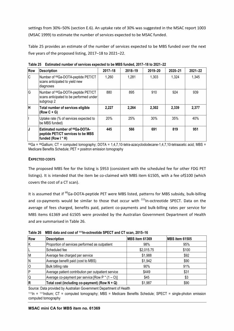
MSAC mini CA for MBS item no. 61369 77
settings from 30%–50% (section E.6). An uptake rate of 30% was suggested in the MSAC report 1003
(MSAC 1999) to estimate the number of services expected to be MSAC funded.
Table 25 provides an estimate of the number of services expected to be MBS funded over the next
five years of the proposed listing, 2017–18 to 2021–22.
Table 25 Estimated number of services expected to be MBS funded, 2017–18 to 2021–22
Row Description 2017–18 2018–19 2019–20 2020–21 2021–22
C Number of 68Ga-DOTA-peptide PET/CT scans anticipated to yield new diagnoses
1,260 1,281 1,303 1,324 1,345
G Number of 68Ga-DOTA-peptide PET/CT scans anticipated to be performed under subgroup 2
880 895 910 924 939
H Total number of services eligible (Row C + G)
2,227 2,264 2,302 2,339 2,377
I Uptake rate (% of services expected to be MBS funded)
20% 25% 30% 35% 40%
J Estimated number of 68Ga-DOTA-peptide PET/CT services to be MBS funded (Row I * H)
445 566 691 819 951
68Ga = 68Gallium; CT = computed tomography; DOTA = 1,4,7,10-tetra-azacyclododecane-1,4,7,10-tetraacetic acid; MBS = Medicare Benefits Schedule; PET = positron emission tomography
EXPECTED COSTS
The proposed MBS fee for the listing is $953 (consistent with the scheduled fee for other FDG PET
listings). It is intended that the item be co-claimed with MBS item 61505, with a fee of$100 (which
covers the cost of a CT scan).
It is assumed that if 68Ga-DOTA-peptide PET were MBS listed, patterns for MBS subsidy, bulk-billing
and co-payments would be similar to those that occur with 111In-octreotide SPECT. Data on the
average of fees charged, benefits paid, patient co-payments and bulk-billing rates per service for
MBS items 61369 and 61505 were provided by the Australian Government Department of Health
and are summarised in Table 26.
Table 26 MBS data and cost of 111In-octreotide SPECT and CT scan, 2015–16
Row Description MBS item 61369 MBS item 61505
K Proportion of services performed as outpatient 98% 95%
L Scheduled fee $2,015.75 $100
M Average fee charged per service $1,988 $92
N Average benefit paid (cost to MBS) $1,942 $90
O Bulk billing rate 90% 91%
P Average patient contribution per outpatient service $449 $31
Q Average co-payment per service [Row P * (1 – O)] $45 $3
R Total cost (including co-payment) (Row N + Q) $1,987 $90
Source: Data provided by Australian Government Department of Health 111In = 111Indium; CT = computed tomography; MBS = Medicare Benefits Schedule; SPECT = single-photon emission computed tomography

MSAC mini CA for MBS item no. 61369 78
Applying a similar pattern to 68Ga-DOTA-peptide PET/CT services, it is assumed that all tests would
be conducted in an out-of-hospital setting (98% of 111In-octreotide SPECT±CT services were in
outpatient settings, Table 26), and 90 per cent services would be bulk-billed (with a 100% rebate due
to the bulk-billing incentive).
The total cost to the MBS per service is calculated as $985.38, derived from the average benefit paid
per outpatient service for 68Ga-DOTA-peptide PET/CT (average benefit paid; $895.82) and the
associated use of CT scan (MBS item 61505, average benefit paid; $89.56)18.
The proportion of patients that are bulk-billed (90%) and the patient contribution ($145.81) for
proposed service are estimated based on the data for current MBS services (item 61369 and items
for FDG PET in 2015–16). Therefore, the estimated patient contribution per 68Ga-DOTA-peptide PET
test is $14.5819 and for the CT scan is $2.75. The total patient contribution associated with each 68Ga-
DOTA-peptide PET/CT service is thus $17.33 ($14.58 + $2.75). Application of these costs to the usage
estimates are shown in Table 27, disaggregated by payer (the MBS and the patient). The average
total cost of 68Ga-DOTA-peptide PET/CT testing is estimated to reach $953,000 in year five of the
proposed listing.
Table 27 Estimated MBS and patient contribution costs of the proposed 68Ga-DOTA-peptide PET/CT service, 2017–18 to 2021–22
Row Description 2017–18 2018–19 2019–20 2020–21 2021–22
J Number of services estimated to be MBS funded 445 566 691 819 951
S Total MBS contribution costs (Row J × $985.38) $438,794 $557,793 $680,469 $806,816 $936,765
T Total patient contribution costs (Row J × $17.33) $7,717 $9,810 $11,968 $14,190 $16,475
U Total costs of listing (Row Row S + T) $446,511 $567,603 $692,436 $821,006 $953,240
68Ga = 68Gallium; CT = computed tomography; DOTA = 1,4,7,10-tetra-azacyclododecane-1,4,7,10-tetraacetic acid; MBS = Medical Benefits Schedule; PET = positron emission tomography
The average benefit paid per outpatient service may differ for the proposed item than used for other
FDG-PET services taken as a referral standard. Therefore, a sensitivity analysis is presented in section
E.6 assuming the cost to the MBS is 85 per cent of the proposed fee, and the co-payments is
15 per cent of the proposed fee.
18 Average benefit paid for
68Ga-DOTA-peptide PET is estimated based on the Medicare statistics data available
for FDG-PET items, and take into account the bulk-billing incentives;
<http://medicarestatistics.humanservices.gov.au/statistics/mbs_item.jsp>; accessed on 8 February 2017.
19 $145.48 × (1 90%)
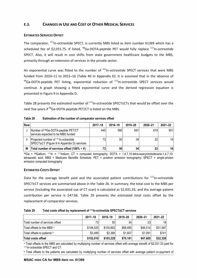
MSAC mini CA for MBS item no. 61369 79
E.3. CHANGES IN USE AND COST OF OTHER MEDICAL SERVICES
ESTIMATED SERVICES OFFSET
The comparator, 111In-octreotide SPECT, is currently MBS listed as item number 61369 which has a
scheduled fee of $2,015.75. If listed, 68Ga-DOTA-peptide PET would fully replace 111In-octreotide
SPECT. Also, it will result in cost shifts from state government healthcare budgets to the MBS,
primarily through an extension of services in the private sector.
An exponential curve was fitted to the number of 111In-octreotide SPECT services that were MBS
funded from 2010–11 to 2015–16 (Table 40 in Appendix D). It is assumed that in the absence of 68Ga-DOTA-peptide PET listing, exponential reduction of 111In-octreotide SPECT services would
continue. A graph showing a fitted exponential curve and the derived regression equation is
presented in Figure 9 in Appendix D.
Table 28 presents the estimated number of 111In-octreotide SPECT±CTs that would be offset over the
next five years if 68Ga-DOTA-peptide PET/CT is listed on the MBS.
Table 28 Estimation of the number of comparator services offset
Row 2017–18 2018–19 2019–20 2020–21 2021–22
J Number of 68Ga-DOTA-peptide PET/CT services expected to be MBS funded
445 566 691 819 951
V Projected number of 111In-octreotide SPECT±CT (Figure 9 in Appendix D) services
73 50 34 23 16
W Total number of services offset (100% × V) 73 50 34 23 16
68Ga = 68Gallium; 111In = 111Indium; CT = computed tomography; DOTA = 1,4,7,10-tetra-azacyclododecane-1,4,7,10-tetraacetic acid; MBS = Medicare Benefits Schedule; PET = positron emission tomography; SPECT = single-photon emission computed tomography
ESTIMATED COSTS OFFSET
Data for the average benefit paid and the associated patient contributions for 111In-octreotide
SPECT±CT services are summarised above in the Table 26. In summary, the total cost to the MBS per
service (including the associated use of CT scan) is calculated as $2,031.33, and the average patient
contribution per service is $47.66. Table 29 presents the estimated total costs offset by the
replacement of comparator services.
Table 29 Total costs offset by replacement of 111In-octreotide SPECT±CT services
2017–18 2018–19 2019–20 2020–21 2021–22
Total number of services offset 73 50 34 23 16
Total offsets to the MBS a $148,525 $100,862 $68,495 $46,514 $31,587
Total offsets to patients b $3,485 $2,366 $1,607 $1,091 $741
Total costs offset $152,010 $103,228 $70,101 $47,605 $32,328
a Total offsets to the MBS are calculated by multiplying number of services offset with average benefit of $2,031.33 paid for 111In-octreotide SPECT and CT. b Total offsets to the patients are calculated by multiplying number of services offset with average patient co-payment of
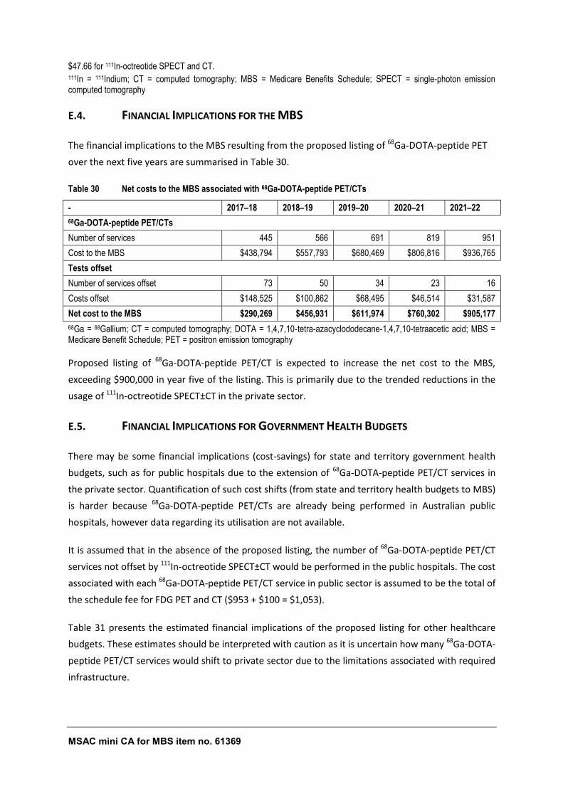
MSAC mini CA for MBS item no. 61369 80
$47.66 for 111In-octreotide SPECT and CT. 111In = 111Indium; CT = computed tomography; MBS = Medicare Benefits Schedule; SPECT = single-photon emission computed tomography
E.4. FINANCIAL IMPLICATIONS FOR THE MBS
The financial implications to the MBS resulting from the proposed listing of 68Ga-DOTA-peptide PET
over the next five years are summarised in Table 30.
Table 30 Net costs to the MBS associated with 68Ga-DOTA-peptide PET/CTs
- 2017–18 2018–19 2019–20 2020–21 2021–22
68Ga-DOTA-peptide PET/CTs
Number of services 445 566 691 819 951
Cost to the MBS $438,794 $557,793 $680,469 $806,816 $936,765
Tests offset
Number of services offset 73 50 34 23 16
Costs offset $148,525 $100,862 $68,495 $46,514 $31,587
Net cost to the MBS $290,269 $456,931 $611,974 $760,302 $905,177
68Ga = 68Gallium; CT = computed tomography; DOTA = 1,4,7,10-tetra-azacyclododecane-1,4,7,10-tetraacetic acid; MBS = Medicare Benefit Schedule; PET = positron emission tomography
Proposed listing of 68Ga-DOTA-peptide PET/CT is expected to increase the net cost to the MBS,
exceeding $900,000 in year five of the listing. This is primarily due to the trended reductions in the
usage of 111In-octreotide SPECT±CT in the private sector.
E.5. FINANCIAL IMPLICATIONS FOR GOVERNMENT HEALTH BUDGETS
There may be some financial implications (cost-savings) for state and territory government health
budgets, such as for public hospitals due to the extension of 68Ga-DOTA-peptide PET/CT services in
the private sector. Quantification of such cost shifts (from state and territory health budgets to MBS)
is harder because 68Ga-DOTA-peptide PET/CTs are already being performed in Australian public
hospitals, however data regarding its utilisation are not available.
It is assumed that in the absence of the proposed listing, the number of 68Ga-DOTA-peptide PET/CT
services not offset by 111In-octreotide SPECT±CT would be performed in the public hospitals. The cost
associated with each 68Ga-DOTA-peptide PET/CT service in public sector is assumed to be the total of
the schedule fee for FDG PET and CT ($953 + $100 = $1,053).
Table 31 presents the estimated financial implications of the proposed listing for other healthcare
budgets. These estimates should be interpreted with caution as it is uncertain how many 68Ga-DOTA-
peptide PET/CT services would shift to private sector due to the limitations associated with required
infrastructure.
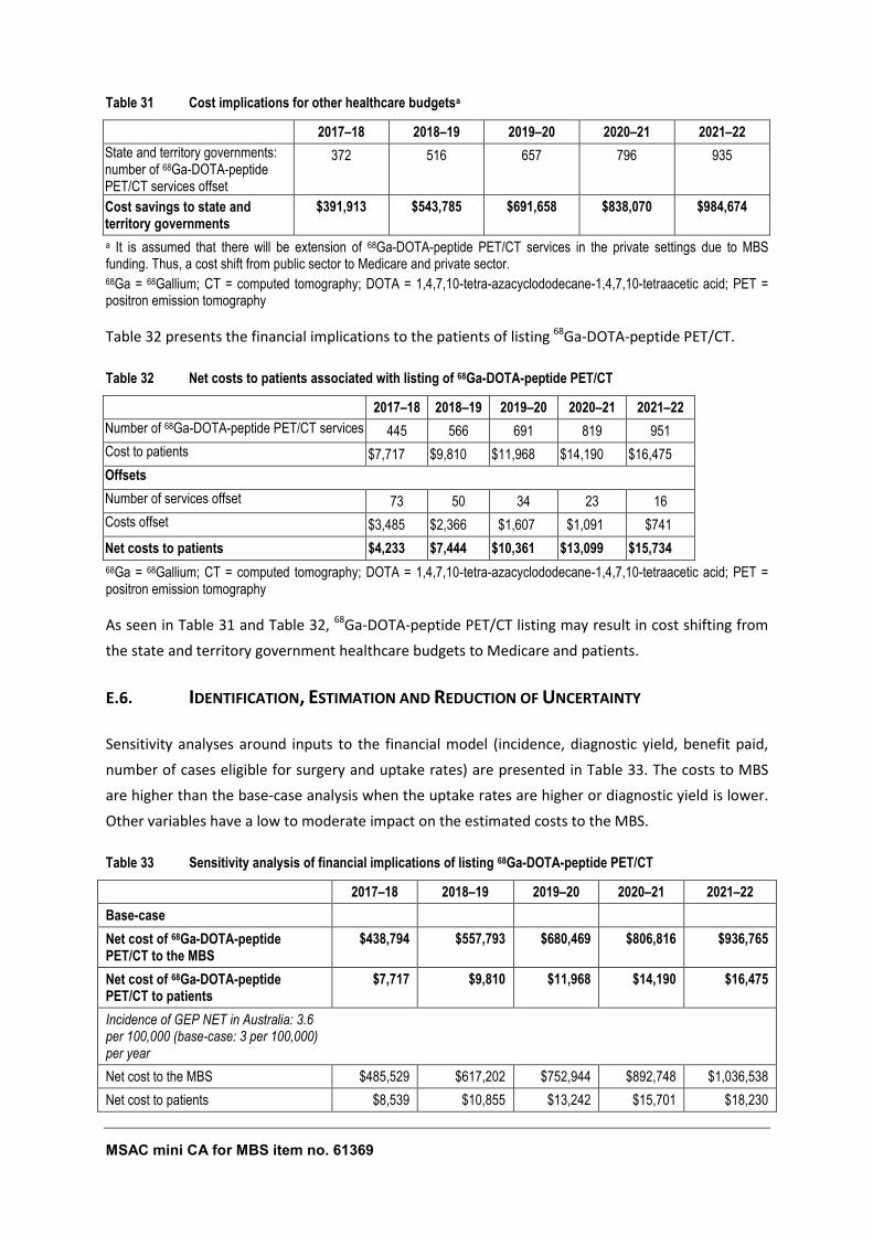
MSAC mini CA for MBS item no. 61369 81
Table 31 Cost implications for other healthcare budgetsa
2017–18 2018–19 2019–20 2020–21 2021–22
State and territory governments: number of 68Ga-DOTA-peptide PET/CT services offset
372 516 657 796 935
Cost savings to state and territory governments
$391,913 $543,785 $691,658 $838,070 $984,674
a It is assumed that there will be extension of 68Ga-DOTA-peptide PET/CT services in the private settings due to MBS funding. Thus, a cost shift from public sector to Medicare and private sector. 68Ga = 68Gallium; CT = computed tomography; DOTA = 1,4,7,10-tetra-azacyclododecane-1,4,7,10-tetraacetic acid; PET = positron emission tomography
Table 32 presents the financial implications to the patients of listing 68Ga-DOTA-peptide PET/CT.
Table 32 Net costs to patients associated with listing of 68Ga-DOTA-peptide PET/CT
2017–18 2018–19 2019–20 2020–21 2021–22
Number of 68Ga-DOTA-peptide PET/CT services 445 566 691 819 951
Cost to patients $7,717 $9,810 $11,968 $14,190 $16,475
Offsets
Number of services offset 73 50 34 23 16
Costs offset $3,485 $2,366 $1,607 $1,091 $741
Net costs to patients $4,233 $7,444 $10,361 $13,099 $15,734
68Ga = 68Gallium; CT = computed tomography; DOTA = 1,4,7,10-tetra-azacyclododecane-1,4,7,10-tetraacetic acid; PET = positron emission tomography
As seen in Table 31 and Table 32, 68Ga-DOTA-peptide PET/CT listing may result in cost shifting from
the state and territory government healthcare budgets to Medicare and patients.
E.6. IDENTIFICATION, ESTIMATION AND REDUCTION OF UNCERTAINTY
Sensitivity analyses around inputs to the financial model (incidence, diagnostic yield, benefit paid,
number of cases eligible for surgery and uptake rates) are presented in Table 33. The costs to MBS
are higher than the base-case analysis when the uptake rates are higher or diagnostic yield is lower.
Other variables have a low to moderate impact on the estimated costs to the MBS.
Table 33 Sensitivity analysis of financial implications of listing 68Ga-DOTA-peptide PET/CT
2017–18 2018–19 2019–20 2020–21 2021–22
Base-case
Net cost of 68Ga-DOTA-peptide PET/CT to the MBS
$438,794 $557,793 $680,469 $806,816 $936,765
Net cost of 68Ga-DOTA-peptide PET/CT to patients
$7,717 $9,810 $11,968 $14,190 $16,475
Incidence of GEP NET in Australia: 3.6 per 100,000 (base-case: 3 per 100,000) per year
Net cost to the MBS $485,529 $617,202 $752,944 $892,748 $1,036,538
Net cost to patients $8,539 $10,855 $13,242 $15,701 $18,230
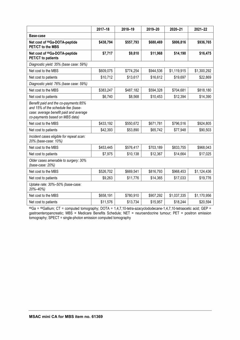
MSAC mini CA for MBS item no. 61369 82
2017–18 2018–19 2019–20 2020–21 2021–22
Base-case
Net cost of 68Ga-DOTA-peptide PET/CT to the MBS
$438,794 $557,793 $680,469 $806,816 $936,765
Net cost of 68Ga-DOTA-peptide PET/CT to patients
$7,717 $9,810 $11,968 $14,190 $16,475
Diagnostic yield: 35% (base case: 59%)
Net cost to the MBS $609,075 $774,254 $944,536 $1,119,915 $1,300,292
Net cost to patients $10,712 $13,617 $16,612 $19,697 $22,869
Diagnostic yield: 76% (base case: 59%)
Net cost to the MBS $383,247 $487,182 $594,328 $704,681 $818,180
Net cost to patients $6,740 $8,568 $10,453 $12,394 $14,390
Benefit paid and the co-payments:85% and 15% of the schedule fee (base-case: average benefit paid and average co-payments based on MBS data)
Net cost to the MBS $433,192 $550,672 $671,781 $796,516 $924,805
Net cost to patients $42,393 $53,890 $65,742 $77,948 $90,503
Incident cases eligible for repeat scan: 20% (base-case: 10%)
Net cost to the MBS $453,445 $576,417 $703,189 $833,755 $968,043
Net cost to patients $7,975 $10,138 $12,367 $14,664 $17,025
Older cases amenable to surgery: 30% (base-case: 20%)
Net cost to the MBS $526,702 $669,541 $816,793 $968,453 $1,124,436
Net cost to patients $9,263 $11,776 $14,365 $17,033 $19,776
Uptake rate: 30%–50% (base-case: 20%–40%)
Net cost to the MBS $658,191 $780,910 $907,292 $1,037,335 $1,170,956
Net cost to patients $11,576 $13,734 $15,957 $18,244 $20,594
68Ga = 68Gallium; CT = computed tomography; DOTA = 1,4,7,10-tetra-azacyclododecane-1,4,7,10-tetraacetic acid; GEP = gastroenteropancreatic; MBS = Medicare Benefits Schedule; NET = neuroendocrine tumour; PET = positron emission tomography; SPECT = single-photon emission computed tomography

MSAC mini CA for MBS item no. 61369 83
SECTION F OTHER RELEVANT CONSIDERATIONS
None identified
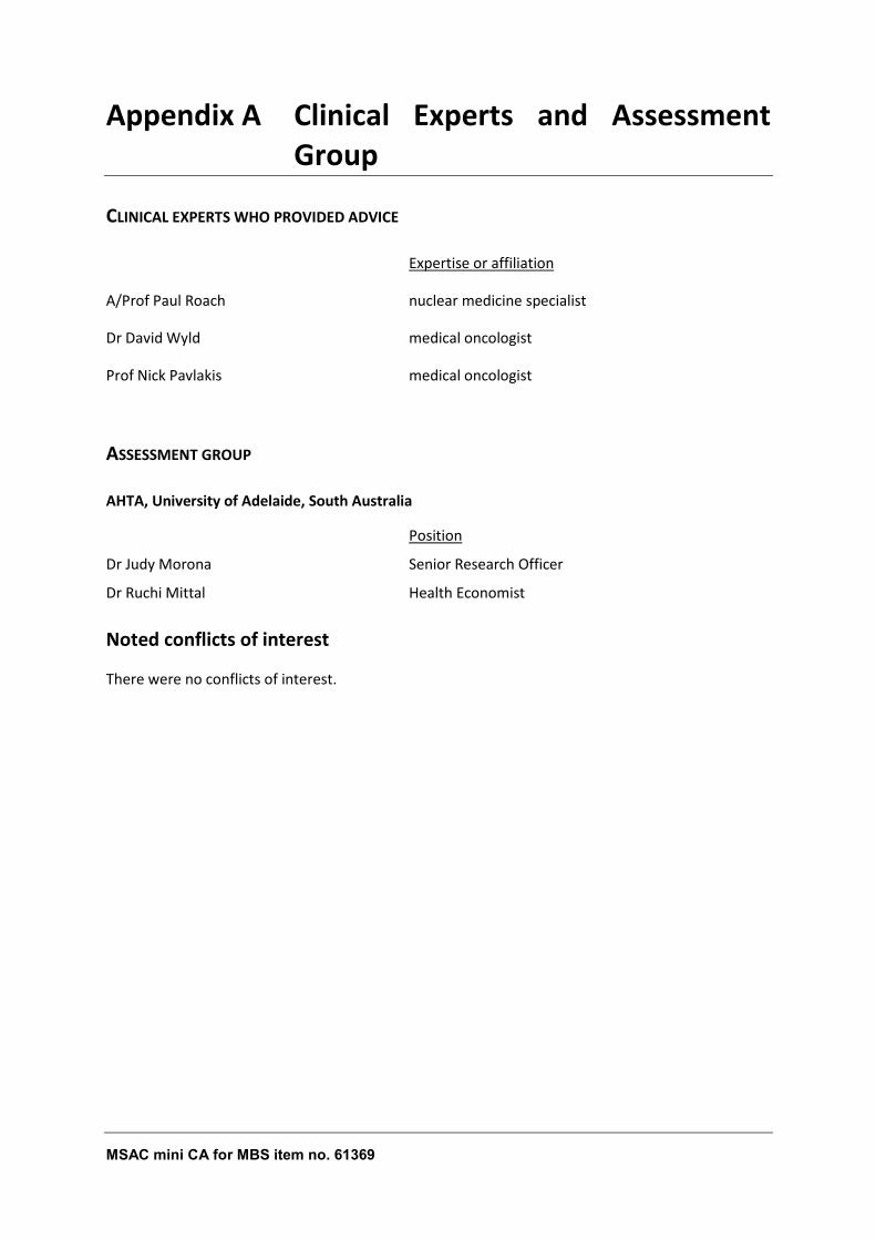
MSAC mini CA for MBS item no. 61369 84
Appendix A Clinical Experts and Assessment Group
CLINICAL EXPERTS WHO PROVIDED ADVICE
Expertise or affiliation
A/Prof Paul Roach nuclear medicine specialist
Dr David Wyld medical oncologist
Prof Nick Pavlakis medical oncologist
ASSESSMENT GROUP
AHTA, University of Adelaide, South Australia
Position
Dr Judy Morona Senior Research Officer
Dr Ruchi Mittal Health Economist
Noted conflicts of interest
There were no conflicts of interest.
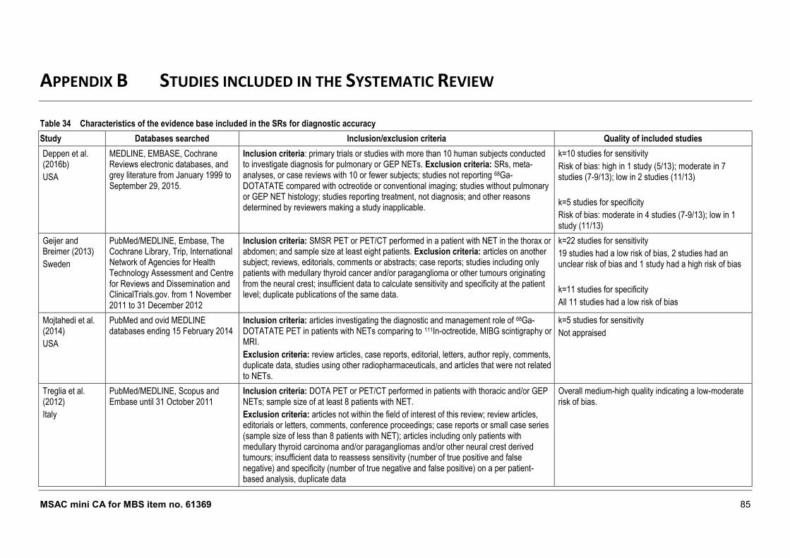
MSAC mini CA for MBS item no. 61369 85
APPENDIX B STUDIES INCLUDED IN THE SYSTEMATIC REVIEW
Table 34 Characteristics of the evidence base included in the SRs for diagnostic accuracy
Study Databases searched Inclusion/exclusion criteria Quality of included studies
Deppen et al. (2016b)
USA
MEDLINE, EMBASE, Cochrane Reviews electronic databases, and grey literature from January 1999 to September 29, 2015.
Inclusion criteria: primary trials or studies with more than 10 human subjects conducted to investigate diagnosis for pulmonary or GEP NETs. Exclusion criteria: SRs, meta-analyses, or case reviews with 10 or fewer subjects; studies not reporting 68Ga-DOTATATE compared with octreotide or conventional imaging; studies without pulmonary or GEP NET histology; studies reporting treatment, not diagnosis; and other reasons determined by reviewers making a study inapplicable.
k=10 studies for sensitivity
Risk of bias: high in 1 study (5/13); moderate in 7 studies (7-9/13); low in 2 studies (11/13)
k=5 studies for specificity
Risk of bias: moderate in 4 studies (7-9/13); low in 1 study (11/13)
Geijer and Breimer (2013)
Sweden
PubMed/MEDLINE, Embase, The Cochrane Library, Trip, International Network of Agencies for Health Technology Assessment and Centre for Reviews and Dissemination and ClinicalTrials.gov. from 1 November 2011 to 31 December 2012
Inclusion criteria: SMSR PET or PET/CT performed in a patient with NET in the thorax or abdomen; and sample size at least eight patients. Exclusion criteria: articles on another subject; reviews, editorials, comments or abstracts; case reports; studies including only patients with medullary thyroid cancer and/or paraganglioma or other tumours originating from the neural crest; insufficient data to calculate sensitivity and specificity at the patient level; duplicate publications of the same data.
k=22 studies for sensitivity
19 studies had a low risk of bias, 2 studies had an unclear risk of bias and 1 study had a high risk of bias
k=11 studies for specificity
All 11 studies had a low risk of bias
Mojtahedi et al. (2014)
USA
PubMed and ovid MEDLINE databases ending 15 February 2014
Inclusion criteria: articles investigating the diagnostic and management role of 68Ga-DOTATATE PET in patients with NETs comparing to 111In-octreotide, MIBG scintigraphy or MRI.
Exclusion criteria: review articles, case reports, editorial, letters, author reply, comments, duplicate data, studies using other radiopharmaceuticals, and articles that were not related to NETs.
k=5 studies for sensitivity
Not appraised
Treglia et al. (2012)
Italy
PubMed/MEDLINE, Scopus and Embase until 31 October 2011
Inclusion criteria: DOTA PET or PET/CT performed in patients with thoracic and/or GEP NETs; sample size of at least 8 patients with NET.
Exclusion criteria: articles not within the field of interest of this review; review articles, editorials or letters, comments, conference proceedings; case reports or small case series (sample size of less than 8 patients with NET); articles including only patients with medullary thyroid carcinoma and/or paragangliomas and/or other neural crest derived tumours; insufficient data to reassess sensitivity (number of true positive and false negative) and specificity (number of true negative and false positive) on a per patient-based analysis, duplicate data
Overall medium-high quality indicating a low-moderate risk of bias.
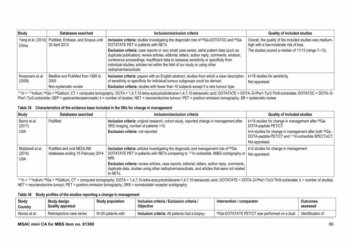
MSAC mini CA for MBS item no. 61369 86
Study Databases searched Inclusion/exclusion criteria Quality of included studies
Yang et al. (2014)
China
PubMed, Embase, and Scopus until 30 April 2013
Inclusion criteria: studies investigating the diagnostic role of 68Ga-DOTATOC and 68Ga-DOTATATE PET in patients with NETs
Exclusion criteria: case reports or very small case series; same patient data (such as duplicate publication); review articles, editorial, letters, author reply, comments, erratum, conference proceedings; insufficient data to reassess sensitivity or specificity from individual studies; articles not within the field of our study or using other radiopharmaceuticals
Overall, the quality of the included studies was medium–high with a low-moderate risk of bias.
The studies scored a median of 11/13 (range 7–13).
Koopmans et al. (2009)
Medline and PubMed from 1995 to 2005
Non-systematic review
Inclusion criteria: papers with an English abstract, studies from which a clear description of sensitivity or specificity for individual tumour subgroups could be derived.
Exclusion criteria: studies with fewer than 10 subjects except if a rare tumour type
k=18 studies for sensitivity
Not appraised
111In = 111Indium; 68Ga = 68Gallium; CT = computed tomography; DOTA = 1,4,7,10-tetra-azacyclododecane-1,4,7,10-tetraacetic acid; DOTATATE = DOTA–D-Phe1-Tyr3-Thr8-octreotate; DOTATOC = DOTA–D-Phe1-Tyr3-octreotide; GEP = gastroenteropancreatic; k = number of studies; NET = neuroendocrine tumour; PET = positron emission tomography; SR = systematic review
Table 35 Characteristics of the evidence base included in the SRs for change in management
Study Databases searched Inclusion/exclusion criteria Quality of included studies
Barrio et al. (2017)
USA
PubMed Inclusion criteria: original research, cohort study, reported change in management after SRS imaging, number of patients >10.
Exclusion criteria: not reported
k=14 studies for change in management after 68Ga-DOTA-peptide PET/CT
k=4 studies for change in management after both 68Ga-DOTA-peptide PET/CT and 111In-octreotide SPECT±CT
Not appraised
Mojtahedi et al. (2014)
USA
PubMed and ovid MEDLINE databases ending 15 February 2014
Inclusion criteria: articles investigating the diagnostic and management role of 68Ga-DOTATATE PET in patients with NETs comparing to 111In-octreotide, MIBG scintigraphy or MRI.
Exclusion criteria: review articles, case reports, editorial, letters, author reply, comments, duplicate data, studies using other radiopharmaceuticals, and articles that were not related to NETs.
k=2 studies for change in management
Not appraised
111In = 111Indium; 68Ga = 68Gallium; CT = computed tomography; DOTA = 1,4,7,10-tetra-azacyclododecane-1,4,7,10-tetraacetic acid; DOTATATE = DOTA–D-Phe1-Tyr3-Thr8-octreotate; k = number of studies; NET = neuroendocrine tumour; PET = positron emission tomography; SRS = somatostatin receptor scintigraphy
Table 36 Study profiles of the studies reporting a change in management
Study
Country
Study design Quality appraisal
Study population Inclusion criteria / Exclusion criteria / Objective
Intervention / comparator Outcomes assessed
Alonso et al. Retrospective case series N=29 patients with Inclusion criteria: All patients had a biopsy- 68Ga-DOTATATE PET/CT was performed on a dual- Identification of
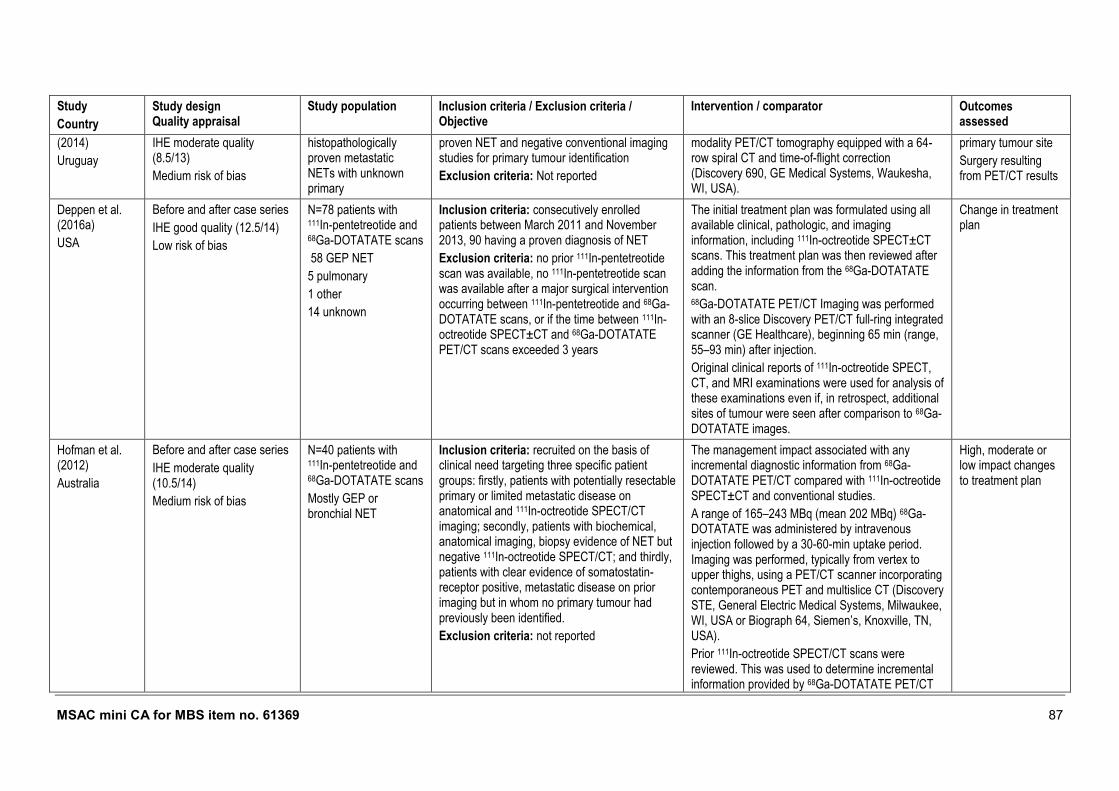
MSAC mini CA for MBS item no. 61369 87
Study
Country
Study design Quality appraisal
Study population Inclusion criteria / Exclusion criteria / Objective
Intervention / comparator Outcomes assessed
(2014)
Uruguay
IHE moderate quality (8.5/13)
Medium risk of bias
histopathologically proven metastatic NETs with unknown primary
proven NET and negative conventional imaging studies for primary tumour identification
Exclusion criteria: Not reported
modality PET/CT tomography equipped with a 64-row spiral CT and time-of-flight correction (Discovery 690, GE Medical Systems, Waukesha, WI, USA).
primary tumour site
Surgery resulting from PET/CT results
Deppen et al. (2016a)
USA
Before and after case series
IHE good quality (12.5/14)
Low risk of bias
N=78 patients with 111In-pentetreotide and 68Ga-DOTATATE scans
58 GEP NET
5 pulmonary
1 other
14 unknown
Inclusion criteria: consecutively enrolled patients between March 2011 and November 2013, 90 having a proven diagnosis of NET
Exclusion criteria: no prior 111In-pentetreotide scan was available, no 111In-pentetreotide scan was available after a major surgical intervention occurring between 111In-pentetreotide and 68Ga-DOTATATE scans, or if the time between 111In-octreotide SPECT±CT and 68Ga-DOTATATE PET/CT scans exceeded 3 years
The initial treatment plan was formulated using all available clinical, pathologic, and imaging information, including 111In-octreotide SPECT±CT scans. This treatment plan was then reviewed after adding the information from the 68Ga-DOTATATE scan. 68Ga-DOTATATE PET/CT Imaging was performed with an 8-slice Discovery PET/CT full-ring integrated scanner (GE Healthcare), beginning 65 min (range, 55–93 min) after injection.
Original clinical reports of 111In-octreotide SPECT, CT, and MRI examinations were used for analysis of these examinations even if, in retrospect, additional sites of tumour were seen after comparison to 68Ga-DOTATATE images.
Change in treatment plan
Hofman et al. (2012)
Australia
Before and after case series
IHE moderate quality (10.5/14)
Medium risk of bias
N=40 patients with 111In-pentetreotide and 68Ga-DOTATATE scans
Mostly GEP or bronchial NET
Inclusion criteria: recruited on the basis of clinical need targeting three specific patient groups: firstly, patients with potentially resectable primary or limited metastatic disease on anatomical and 111In-octreotide SPECT/CT imaging; secondly, patients with biochemical, anatomical imaging, biopsy evidence of NET but negative 111In-octreotide SPECT/CT; and thirdly, patients with clear evidence of somatostatin-receptor positive, metastatic disease on prior imaging but in whom no primary tumour had previously been identified.
Exclusion criteria: not reported
The management impact associated with any incremental diagnostic information from 68Ga-DOTATATE PET/CT compared with 111In-octreotide SPECT±CT and conventional studies.
A range of 165–243 MBq (mean 202 MBq) 68Ga-DOTATATE was administered by intravenous injection followed by a 30-60-min uptake period. Imaging was performed, typically from vertex to upper thighs, using a PET/CT scanner incorporating contemporaneous PET and multislice CT (Discovery STE, General Electric Medical Systems, Milwaukee, WI, USA or Biograph 64, Siemen’s, Knoxville, TN, USA).
Prior 111In-octreotide SPECT/CT scans were reviewed. This was used to determine incremental information provided by 68Ga-DOTATATE PET/CT
High, moderate or low impact changes to treatment plan
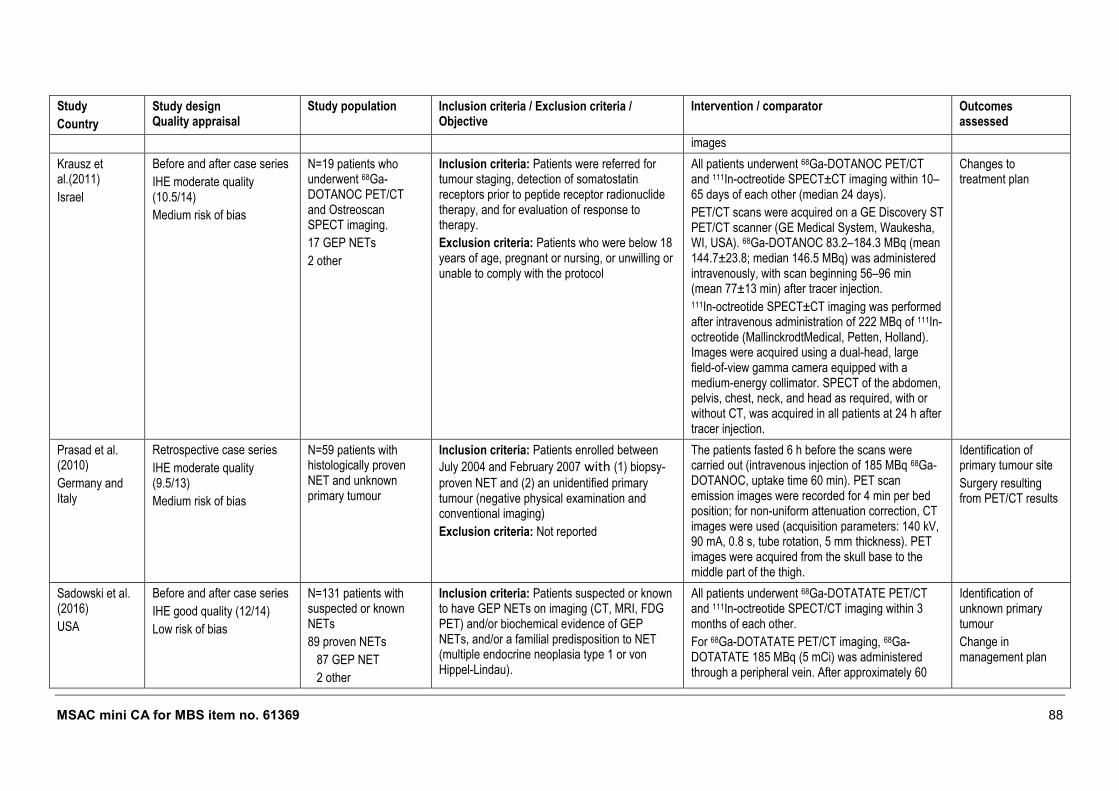
MSAC mini CA for MBS item no. 61369 88
Study
Country
Study design Quality appraisal
Study population Inclusion criteria / Exclusion criteria / Objective
Intervention / comparator Outcomes assessed
images
Krausz et al.(2011)
Israel
Before and after case series
IHE moderate quality (10.5/14)
Medium risk of bias
N=19 patients who underwent 68Ga-DOTANOC PET/CT and Ostreoscan SPECT imaging.
17 GEP NETs
2 other
Inclusion criteria: Patients were referred for tumour staging, detection of somatostatin receptors prior to peptide receptor radionuclide therapy, and for evaluation of response to therapy.
Exclusion criteria: Patients who were below 18 years of age, pregnant or nursing, or unwilling or unable to comply with the protocol
All patients underwent 68Ga-DOTANOC PET/CT and 111In-octreotide SPECT±CT imaging within 10–65 days of each other (median 24 days).
PET/CT scans were acquired on a GE Discovery ST PET/CT scanner (GE Medical System, Waukesha, WI, USA). 68Ga-DOTANOC 83.2–184.3 MBq (mean 144.7±23.8; median 146.5 MBq) was administered intravenously, with scan beginning 56–96 min (mean 77±13 min) after tracer injection. 111In-octreotide SPECT±CT imaging was performed after intravenous administration of 222 MBq of 111In-octreotide (MallinckrodtMedical, Petten, Holland). Images were acquired using a dual-head, large field-of-view gamma camera equipped with a medium-energy collimator. SPECT of the abdomen, pelvis, chest, neck, and head as required, with or without CT, was acquired in all patients at 24 h after tracer injection.
Changes to treatment plan
Prasad et al. (2010)
Germany and Italy
Retrospective case series
IHE moderate quality (9.5/13)
Medium risk of bias
N=59 patients with histologically proven NET and unknown primary tumour
Inclusion criteria: Patients enrolled between
July 2004 and February 2007 with (1) biopsy-
proven NET and (2) an unidentified primary tumour (negative physical examination and conventional imaging)
Exclusion criteria: Not reported
The patients fasted 6 h before the scans were carried out (intravenous injection of 185 MBq 68Ga-DOTANOC, uptake time 60 min). PET scan emission images were recorded for 4 min per bed position; for non-uniform attenuation correction, CT images were used (acquisition parameters: 140 kV, 90 mA, 0.8 s, tube rotation, 5 mm thickness). PET images were acquired from the skull base to the middle part of the thigh.
Identification of primary tumour site
Surgery resulting from PET/CT results
Sadowski et al. (2016)
USA
Before and after case series
IHE good quality (12/14)
Low risk of bias
N=131 patients with suspected or known NETs
89 proven NETs
87 GEP NET
2 other
Inclusion criteria: Patients suspected or known to have GEP NETs on imaging (CT, MRI, FDG PET) and/or biochemical evidence of GEP NETs, and/or a familial predisposition to NET (multiple endocrine neoplasia type 1 or von Hippel-Lindau).
All patients underwent 68Ga-DOTATATE PET/CT and 111In-octreotide SPECT/CT imaging within 3 months of each other.
For 68Ga-DOTATATE PET/CT imaging, 68Ga-DOTATATE 185 MBq (5 mCi) was administered through a peripheral vein. After approximately 60
Identification of unknown primary tumour
Change in management plan
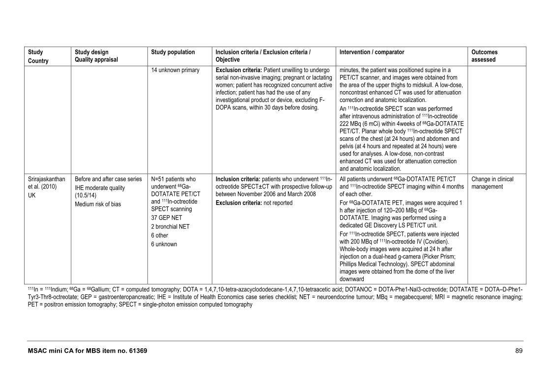
MSAC mini CA for MBS item no. 61369 89
Study
Country
Study design Quality appraisal
Study population Inclusion criteria / Exclusion criteria / Objective
Intervention / comparator Outcomes assessed
14 unknown primary Exclusion criteria: Patient unwilling to undergo serial non-invasive imaging; pregnant or lactating women; patient has recognized concurrent active infection; patient has had the use of any investigational product or device, excluding F-DOPA scans, within 30 days before dosing.
minutes, the patient was positioned supine in a PET/CT scanner, and images were obtained from the area of the upper thighs to midskull. A low-dose, noncontrast enhanced CT was used for attenuation correction and anatomic localization.
An 111In-octreotide SPECT scan was performed after intravenous administration of 111In-octreotide 222 MBq (6 mCi) within 4weeks of 68Ga-DOTATATE PET/CT. Planar whole body 111In-octreotide SPECT scans of the chest (at 24 hours) and abdomen and pelvis (at 4 hours and repeated at 24 hours) were used for analyses. A low-dose, non-contrast enhanced CT was used for attenuation correction and anatomic localization.
Srirajaskanthan et al. (2010)
UK
Before and after case series
IHE moderate quality (10.5/14)
Medium risk of bias
N=51 patients who underwent 68Ga-DOTATATE PET/CT and 111In-octreotide SPECT scanning
37 GEP NET
2 bronchial NET
6 other
6 unknown
Inclusion criteria: patients who underwent 111In-octreotide SPECT±CT with prospective follow-up between November 2006 and March 2008
Exclusion criteria: not reported
All patients underwent 68Ga-DOTATATE PET/CT and 111In-octreotide SPECT imaging within 4 months of each other.
For 68Ga-DOTATATE PET, images were acquired 1 h after injection of 120–200 MBq of 68Ga-DOTATATE. Imaging was performed using a dedicated GE Discovery LS PET/CT unit.
For 111In-octreotide SPECT, patients were injected with 200 MBq of 111In-octreotide IV (Covidien). Whole-body images were acquired at 24 h after injection on a dual-head g-camera (Picker Prism; Phillips Medical Technology). SPECT abdominal images were obtained from the dome of the liver downward
Change in clinical management
111In = 111Indium; 68Ga = 68Gallium; CT = computed tomography; DOTA = 1,4,7,10-tetra-azacyclododecane-1,4,7,10-tetraacetic acid; DOTANOC = DOTA-Phe1-NaI3-octreotide; DOTATATE = DOTA–D-Phe1-Tyr3-Thr8-octreotate; GEP = gastroenteropancreatic; IHE = Institute of Health Economics case series checklist; NET = neuroendocrine tumour; MBq = megabecquerel; MRI = magnetic resonance imaging; PET = positron emission tomography; SPECT = single-photon emission computed tomography
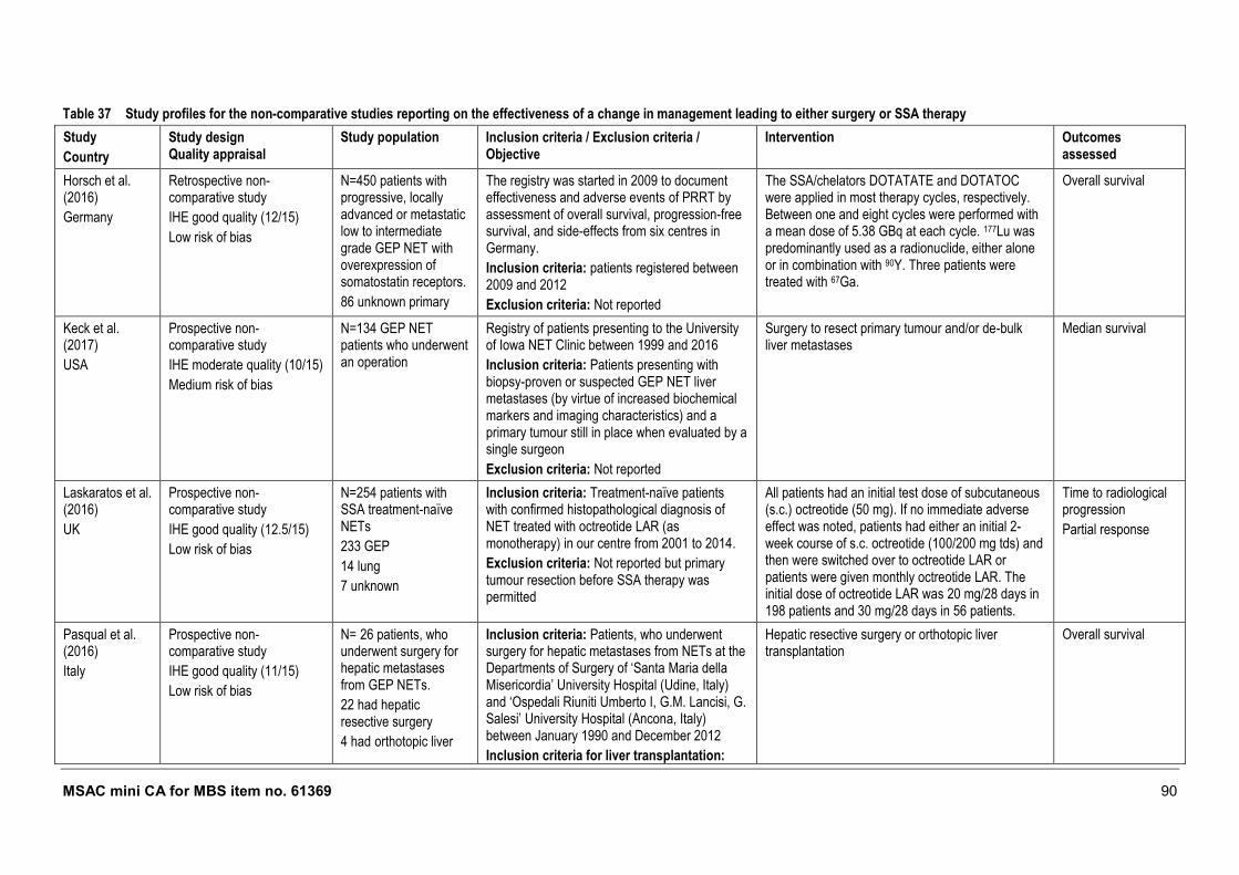
MSAC mini CA for MBS item no. 61369 90
Table 37 Study profiles for the non-comparative studies reporting on the effectiveness of a change in management leading to either surgery or SSA therapy
Study
Country
Study design Quality appraisal
Study population Inclusion criteria / Exclusion criteria / Objective
Intervention Outcomes assessed
Horsch et al. (2016)
Germany
Retrospective non-comparative study
IHE good quality (12/15)
Low risk of bias
N=450 patients with progressive, locally advanced or metastatic low to intermediate grade GEP NET with overexpression of somatostatin receptors.
86 unknown primary
The registry was started in 2009 to document effectiveness and adverse events of PRRT by assessment of overall survival, progression-free survival, and side-effects from six centres in Germany.
Inclusion criteria: patients registered between 2009 and 2012
Exclusion criteria: Not reported
The SSA/chelators DOTATATE and DOTATOC were applied in most therapy cycles, respectively. Between one and eight cycles were performed with a mean dose of 5.38 GBq at each cycle. 177Lu was predominantly used as a radionuclide, either alone or in combination with 90Y. Three patients were treated with 67Ga.
Overall survival
Keck et al. (2017)
USA
Prospective non-comparative study
IHE moderate quality (10/15)
Medium risk of bias
N=134 GEP NET patients who underwent an operation
Registry of patients presenting to the University of Iowa NET Clinic between 1999 and 2016
Inclusion criteria: Patients presenting with biopsy-proven or suspected GEP NET liver metastases (by virtue of increased biochemical markers and imaging characteristics) and a primary tumour still in place when evaluated by a single surgeon
Exclusion criteria: Not reported
Surgery to resect primary tumour and/or de-bulk liver metastases
Median survival
Laskaratos et al. (2016)
UK
Prospective non-comparative study
IHE good quality (12.5/15)
Low risk of bias
N=254 patients with SSA treatment-naïve NETs
233 GEP
14 lung
7 unknown
Inclusion criteria: Treatment-naïve patients with confirmed histopathological diagnosis of NET treated with octreotide LAR (as monotherapy) in our centre from 2001 to 2014.
Exclusion criteria: Not reported but primary tumour resection before SSA therapy was permitted
All patients had an initial test dose of subcutaneous (s.c.) octreotide (50 mg). If no immediate adverse effect was noted, patients had either an initial 2-week course of s.c. octreotide (100/200 mg tds) and then were switched over to octreotide LAR or patients were given monthly octreotide LAR. The initial dose of octreotide LAR was 20 mg/28 days in 198 patients and 30 mg/28 days in 56 patients.
Time to radiological progression
Partial response
Pasqual et al. (2016)
Italy
Prospective non-comparative study
IHE good quality (11/15)
Low risk of bias
N= 26 patients, who underwent surgery for hepatic metastases from GEP NETs.
22 had hepatic resective surgery
4 had orthotopic liver
Inclusion criteria: Patients, who underwent surgery for hepatic metastases from NETs at the Departments of Surgery of ‘Santa Maria della Misericordia’ University Hospital (Udine, Italy) and ‘Ospedali Riuniti Umberto I, G.M. Lancisi, G. Salesi’ University Hospital (Ancona, Italy) between January 1990 and December 2012
Inclusion criteria for liver transplantation:
Hepatic resective surgery or orthotopic liver transplantation
Overall survival
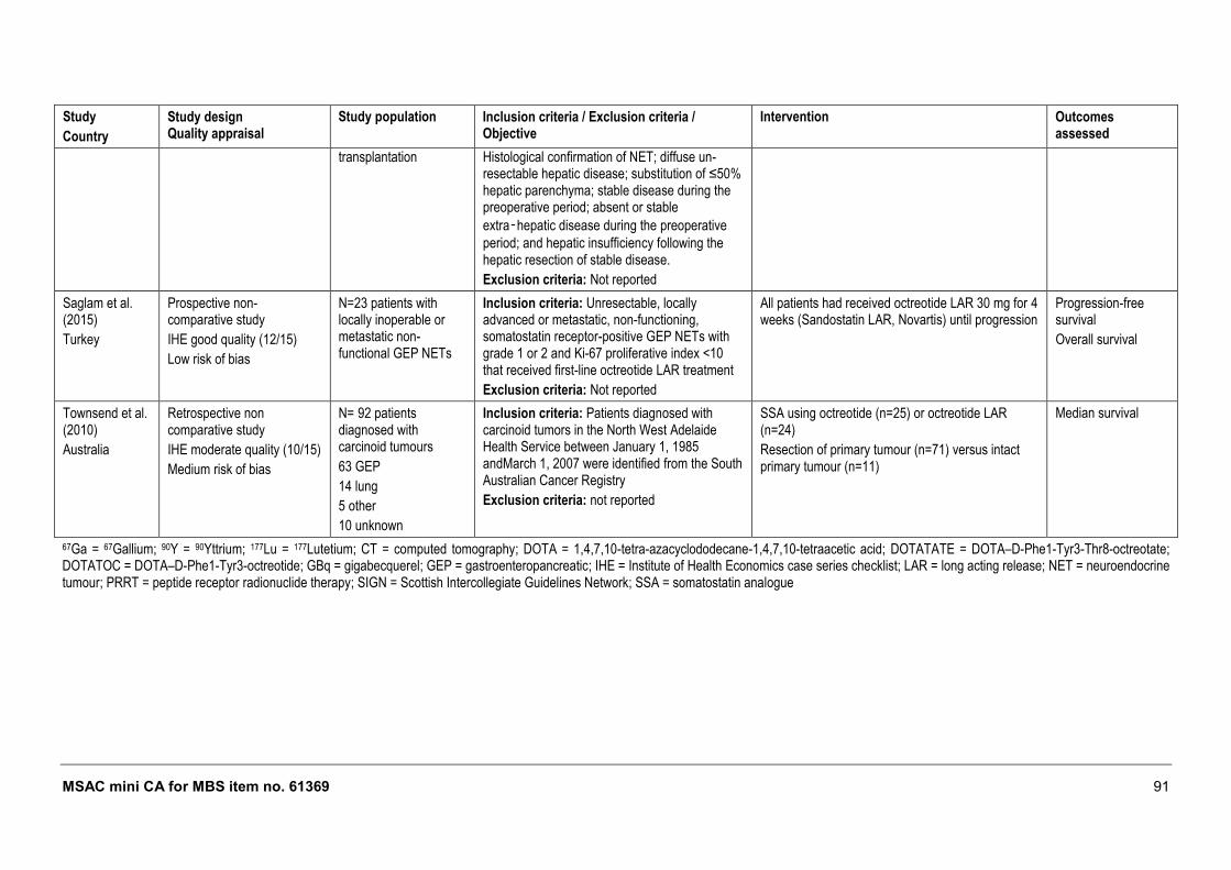
MSAC mini CA for MBS item no. 61369 91
Study
Country
Study design Quality appraisal
Study population Inclusion criteria / Exclusion criteria / Objective
Intervention Outcomes assessed
transplantation Histological confirmation of NET; diffuse un-resectable hepatic disease; substitution of ≤50% hepatic parenchyma; stable disease during the preoperative period; absent or stable
extra‑hepatic disease during the preoperative
period; and hepatic insufficiency following the hepatic resection of stable disease.
Exclusion criteria: Not reported
Saglam et al. (2015)
Turkey
Prospective non-comparative study
IHE good quality (12/15)
Low risk of bias
N=23 patients with locally inoperable or metastatic non-functional GEP NETs
Inclusion criteria: Unresectable, locally advanced or metastatic, non-functioning, somatostatin receptor-positive GEP NETs with grade 1 or 2 and Ki-67 proliferative index <10 that received first-line octreotide LAR treatment
Exclusion criteria: Not reported
All patients had received octreotide LAR 30 mg for 4 weeks (Sandostatin LAR, Novartis) until progression
Progression-free survival
Overall survival
Townsend et al. (2010)
Australia
Retrospective non comparative study
IHE moderate quality (10/15)
Medium risk of bias
N= 92 patients diagnosed with carcinoid tumours
63 GEP
14 lung
5 other
10 unknown
Inclusion criteria: Patients diagnosed with carcinoid tumors in the North West Adelaide Health Service between January 1, 1985 andMarch 1, 2007 were identified from the South Australian Cancer Registry
Exclusion criteria: not reported
SSA using octreotide (n=25) or octreotide LAR (n=24)
Resection of primary tumour (n=71) versus intact primary tumour (n=11)
Median survival
67Ga = 67Gallium; 90Y = 90Yttrium; 177Lu = 177Lutetium; CT = computed tomography; DOTA = 1,4,7,10-tetra-azacyclododecane-1,4,7,10-tetraacetic acid; DOTATATE = DOTA–D-Phe1-Tyr3-Thr8-octreotate; DOTATOC = DOTA–D-Phe1-Tyr3-octreotide; GBq = gigabecquerel; GEP = gastroenteropancreatic; IHE = Institute of Health Economics case series checklist; LAR = long acting release; NET = neuroendocrine tumour; PRRT = peptide receptor radionuclide therapy; SIGN = Scottish Intercollegiate Guidelines Network; SSA = somatostatin analogue
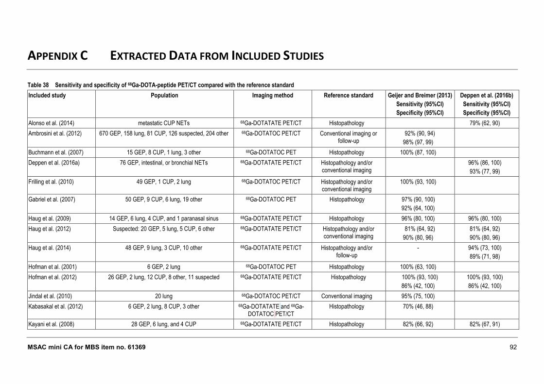
MSAC mini CA for MBS item no. 61369 92
APPENDIX C EXTRACTED DATA FROM INCLUDED STUDIES
Table 38 Sensitivity and specificity of 68Ga-DOTA-peptide PET/CT compared with the reference standard
Included study Population Imaging method Reference standard Geijer and Breimer (2013)
Sensitivity (95%CI)
Specificity (95%CI)
Deppen et al. (2016b)
Sensitivity (95%CI)
Specificity (95%CI)
Alonso et al. (2014) metastatic CUP NETs 68Ga-DOTATATE PET/CT Histopathology 79% (62, 90)
Ambrosini et al. (2012) 670 GEP, 158 lung, 81 CUP, 126 suspected, 204 other 68Ga-DOTATOC PET/CT Conventional imaging or follow-up
92% (90, 94)
98% (97, 99)
Buchmann et al. (2007) 15 GEP, 8 CUP, 1 lung, 3 other 68Ga-DOTATOC PET Histopathology 100% (87, 100)
Deppen et al. (2016a) 76 GEP, intestinal, or bronchial NETs 68Ga-DOTATATE PET/CT Histopathology and/or conventional imaging
96% (86, 100)
93% (77, 99)
Frilling et al. (2010) 49 GEP, 1 CUP, 2 lung 68Ga-DOTATOC PET/CT Histopathology and/or conventional imaging
100% (93, 100)
Gabriel et al. (2007) 50 GEP, 9 CUP, 6 lung, 19 other 68Ga-DOTATOC PET Histopathology 97% (90, 100)
92% (64, 100)
Haug et al. (2009) 14 GEP, 6 lung, 4 CUP, and 1 paranasal sinus 68Ga-DOTATATE PET/CT Histopathology 96% (80, 100) 96% (80, 100)
Haug et al. (2012) Suspected: 20 GEP, 5 lung, 5 CUP, 6 other 68Ga-DOTATATE PET/CT Histopathology and/or conventional imaging
81% (64, 92)
90% (80, 96)
81% (64, 92)
90% (80, 96)
Haug et al. (2014) 48 GEP, 9 lung, 3 CUP, 10 other 68Ga-DOTATATE PET/CT Histopathology and/or follow-up
- 94% (73, 100)
89% (71, 98)
Hofman et al. (2001) 6 GEP, 2 lung 68Ga-DOTATOC PET Histopathology 100% (63, 100)
Hofman et al. (2012) 26 GEP, 2 lung, 12 CUP, 8 other, 11 suspected 68Ga-DOTATATE PET/CT Histopathology 100% (93, 100)
86% (42, 100)
100% (93, 100)
86% (42, 100)
Jindal et al. (2010) 20 lung 68Ga-DOTATOC PET/CT Conventional imaging 95% (75, 100)
Kabasakal et al. (2012) 6 GEP, 2 lung, 8 CUP, 3 other 68Ga-DOTATATE and 68Ga-DOTATOC PET/CT
Histopathology 70% (46, 88)
Kayani et al. (2008) 28 GEP, 6 lung, and 4 CUP 68Ga-DOTATATE PET/CT Histopathology 82% (66, 92) 82% (67, 91)
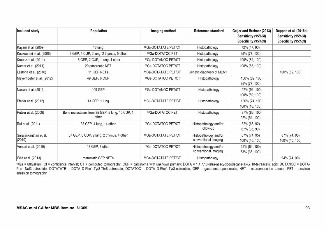
MSAC mini CA for MBS item no. 61369 93
Included study Population Imaging method Reference standard Geijer and Breimer (2013)
Sensitivity (95%CI)
Specificity (95%CI)
Deppen et al. (2016b)
Sensitivity (95%CI)
Specificity (95%CI)
Kayani et al. (2009) 18 lung 68Ga-DOTATATE PET/CT Histopathology 72% (47, 90)
Koukouraki et al. (2006) 9 GEP, 4 CUP, 2 lung, 2 thymus, 5 other 68Ga-DOTATOC PET Histopathology 95% (77, 100)
Krausz et al. (2011) 15 GEP, 2 CUP, 1 lung, 1 other 68Ga-DOTANOC PET/CT Histopathology 100% (82, 100)
Kumar et al. (2011) 20 pancreatic NET 68Ga-DOTATOC PET/CT Histopathology 100% (83, 100)
Lastoria et al. (2016) 11 GEP NETs 68Ga-DOTATATE PET/CT Genetic diagnosis of MEN1 100% (82, 100)
Mayerhoefer et al. (2012) 49 GEP, 6 CUP 68Ga-DOTATOC PET/CT Histopathology 100% (89, 100)
95% (77, 100)
Naswa et al. (2011) 109 GEP 68Ga-DOTANOC PET/CT Histopathology 97% (91, 100)
100% (89, 100)
Pfeifer et al. (2012) 13 GEP, 1 lung 64Cu-DOTATATE PET/CT Histopathology 100% (74, 100)
100% (16, 100)
Putzer et al. (2009) Bone metastases from 35 GEP, 5 lung, 10 CUP, 1 other
68Ga-DOTATOC PET Histopathology 97% (86, 100)
92% (64, 100)
Ruf et al. (2011) 33 GEP, 4 lung, 14 other 68Ga-DOTATOC PET/CT Histopathology and/or follow-up
82% (66, 92)
67% (35, 90)
Srirajaskanthan et al. (2010)
37 GEP, 6 CUP, 2 lung, 2 thymus, 4 other 68Ga-DOTATATE PET/CT Histopathology and/or conventional imaging
87% (74, 95)
100% (40, 100)
87% (74, 95)
100% (40, 100)
Versari et al. (2010) 13 GEP, 6 other 68Ga-DOTATOC PET/CT Histopathology and/or conventional imaging
92% (64, 100)
83% (36, 100)
Wild et al. (2013) metastatic GEP NETs 68Ga-DOTATATE PET/CT Histopathology 94% (74, 99)
68Ga = 68Gallium; CI = confidence interval; CT = computed tomography; CUP = carcinoma with unknown primary; DOTA = 1,4,7,10-tetra-azacyclododecane-1,4,7,10-tetraacetic acid; DOTANOC = DOTA-Phe1-NaI3-octreotide; DOTATATE = DOTA–D-Phe1-Tyr3-Thr8-octreotate; DOTATOC = DOTA–D-Phe1-Tyr3-octreotide; GEP = gastroenteropancreatic; NET = neuroendocrine tumour; PET = positron emission tomography
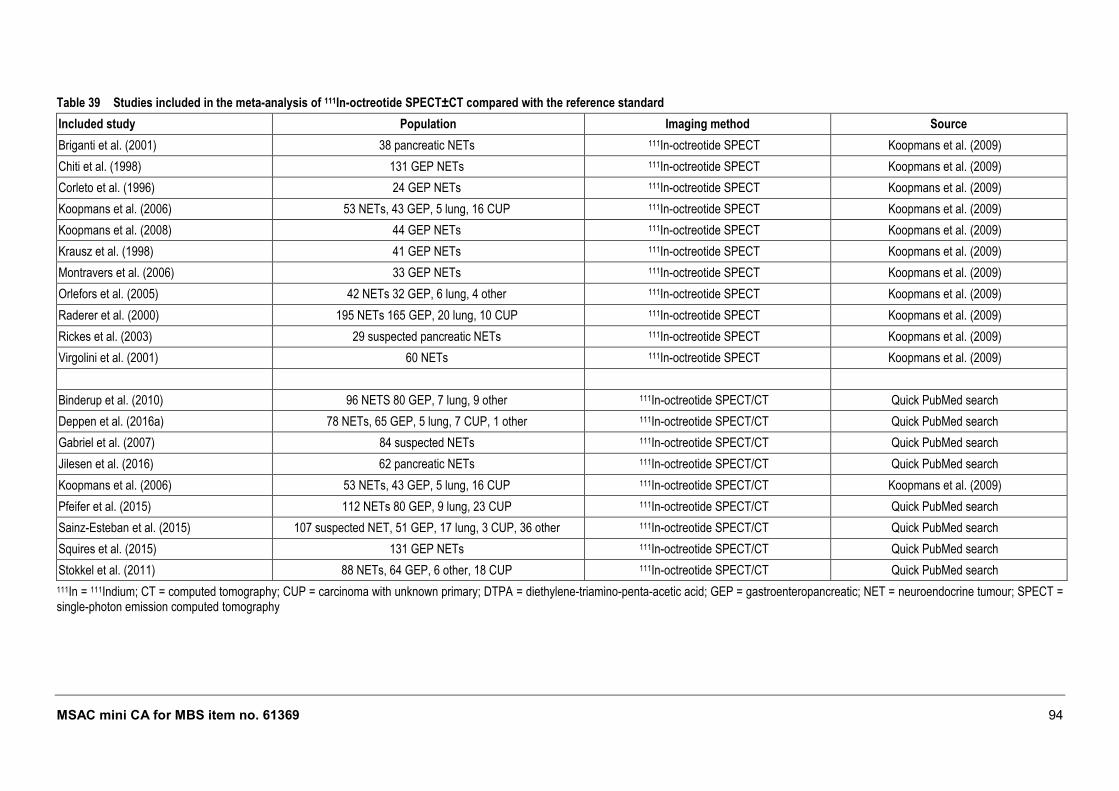
MSAC mini CA for MBS item no. 61369 94
Table 39 Studies included in the meta-analysis of 111In-octreotide SPECT±CT compared with the reference standard
Included study Population Imaging method Source
Briganti et al. (2001) 38 pancreatic NETs 111In-octreotide SPECT Koopmans et al. (2009)
Chiti et al. (1998) 131 GEP NETs 111In-octreotide SPECT Koopmans et al. (2009)
Corleto et al. (1996) 24 GEP NETs 111In-octreotide SPECT Koopmans et al. (2009)
Koopmans et al. (2006) 53 NETs, 43 GEP, 5 lung, 16 CUP 111In-octreotide SPECT Koopmans et al. (2009)
Koopmans et al. (2008) 44 GEP NETs 111In-octreotide SPECT Koopmans et al. (2009)
Krausz et al. (1998) 41 GEP NETs 111In-octreotide SPECT Koopmans et al. (2009)
Montravers et al. (2006) 33 GEP NETs 111In-octreotide SPECT Koopmans et al. (2009)
Orlefors et al. (2005) 42 NETs 32 GEP, 6 lung, 4 other 111In-octreotide SPECT Koopmans et al. (2009)
Raderer et al. (2000) 195 NETs 165 GEP, 20 lung, 10 CUP 111In-octreotide SPECT Koopmans et al. (2009)
Rickes et al. (2003) 29 suspected pancreatic NETs 111In-octreotide SPECT Koopmans et al. (2009)
Virgolini et al. (2001) 60 NETs 111In-octreotide SPECT Koopmans et al. (2009)
Binderup et al. (2010) 96 NETS 80 GEP, 7 lung, 9 other 111In-octreotide SPECT/CT Quick PubMed search
Deppen et al. (2016a) 78 NETs, 65 GEP, 5 lung, 7 CUP, 1 other 111In-octreotide SPECT/CT Quick PubMed search
Gabriel et al. (2007) 84 suspected NETs 111In-octreotide SPECT/CT Quick PubMed search
Jilesen et al. (2016) 62 pancreatic NETs 111In-octreotide SPECT/CT Quick PubMed search
Koopmans et al. (2006) 53 NETs, 43 GEP, 5 lung, 16 CUP 111In-octreotide SPECT/CT Koopmans et al. (2009)
Pfeifer et al. (2015) 112 NETs 80 GEP, 9 lung, 23 CUP 111In-octreotide SPECT/CT Quick PubMed search
Sainz-Esteban et al. (2015) 107 suspected NET, 51 GEP, 17 lung, 3 CUP, 36 other 111In-octreotide SPECT/CT Quick PubMed search
Squires et al. (2015) 131 GEP NETs 111In-octreotide SPECT/CT Quick PubMed search
Stokkel et al. (2011) 88 NETs, 64 GEP, 6 other, 18 CUP 111In-octreotide SPECT/CT Quick PubMed search
111In = 111Indium; CT = computed tomography; CUP = carcinoma with unknown primary; DTPA = diethylene-triamino-penta-acetic acid; GEP = gastroenteropancreatic; NET = neuroendocrine tumour; SPECT = single-photon emission computed tomography
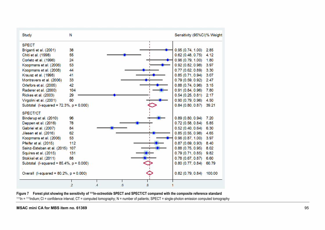
MSAC mini CA for MBS item no. 61369 95
Figure 7 Forest plot showing the sensitivity of 111In-octreotide SPECT and SPECT/CT compared with the composite reference standard 111In = 111Indium; CI = confidence interval; CT = computed tomography; N = number of patients; SPECT = single-photon emission computed tomography
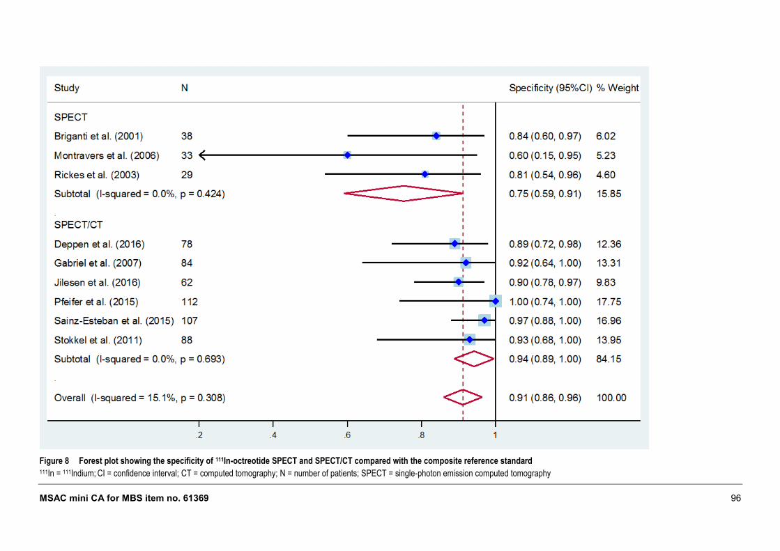
MSAC mini CA for MBS item no. 61369 96
Figure 8 Forest plot showing the specificity of 111In-octreotide SPECT and SPECT/CT compared with the composite reference standard 111In = 111Indium; CI = confidence interval; CT = computed tomography; N = number of patients; SPECT = single-photon emission computed tomography
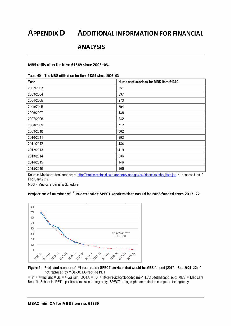
MSAC mini CA for MBS item no. 61369 97
APPENDIX D ADDITIONAL INFORMATION FOR FINANCIAL
ANALYSIS
MBS utilisation for item 61369 since 2002–03.
Table 40 The MBS utilisation for item 61369 since 2002–03
Year Number of services for MBS item 61369
2002/2003 251
2003/2004 237
2004/2005 273
2005/2006 354
2006/2007 436
2007/2008 542
2008/2009 712
2009/2010 802
2010/2011 693
2011/2012 484
2012/2013 419
2013/2014 236
2014/2015 146
2015/2016 106
Source: Medicare item reports; < http://medicarestatistics.humanservices.gov.au/statistics/mbs_item.jsp >, accessed on 2 February 2017.
MBS = Medicare Benefits Schedule
Projection of number of 111In-octreotide SPECT services that would be MBS funded from 2017–22.
Figure 9 Projected number of 111In-octreotide SPECT services that would be MBS funded (2017–18 to 2021–22) if not replaced by 68Ga-DOTA-Peptide PET
111In = 111Indium; 68Ga = 68Gallium; DOTA = 1,4,7,10-tetra-azacyclododecane-1,4,7,10-tetraacetic acid; MBS = Medicare Benefits Schedule; PET = positron emission tomography; SPECT = single-photon emission computed tomography
MSAC mini CA for MBS item no. 61369 98
REFERENCES
ABS 2013, '3222.0 - Population Projections, Australia, 2012 (base) to 2101, Series B', Australian Bureau of Statistics, Commonwealth of Australia, Canberra, viewed 17 December 2015, <http://abs.gov.au/AUSSTATS/[email protected]/DetailsPage/3222.02012%20%28base%29%20to%202101?OpenDocument>.
Alonso, O, Rodriguez-Taroco, M, Savio, E, Bentancourt, C, Gambini, JP & Engler, H 2014, '(68)Ga-DOTATATE PET/CT in the evaluation of patients with neuroendocrine metastatic carcinoma of unknown origin', Ann Nucl Med, vol. 28, no. 7, pp. 638-645.
Ambrosini, V, Campana, D, Tomassetti, P & Fanti, S 2012, '68Ga-labelled peptides for diagnosis of gastroenteropancreatic NET', European Journal of Nuclear Medicine and Molecular Imaging, vol. 39, no. 1, pp. 52-60.
Armitage, P, Berry, G & Matthews, JNS 2002, Statistical methods in medical research, Fourth Edition edn, Blackwell Science, Oxford.
Barrio, M, Czernin, J, Fanti, S, Ambrosini, V, Binse, I, Du, L, Eiber, M, Herrmann, K & Fendler, WP 2017, 'The impact of SSTR-directed PET/CT on the management of patients with neuroendocrine tumor: A systematic review and meta-analysis', J Nucl Med.
Berardi, R, Rinaldi, S, Torniai, M, Morgese, F, Partelli, S, Caramanti, M, Onofri, A, Polenta, V, Pagliaretta, S, Falconi, M & Cascinu, S 2016a, 'Gastrointestinal neuroendocrine tumors: Searching the optimal treatment strategy--A literature review', Crit Rev Oncol Hematol, vol. 98, pp. 264-274.
Binderup, T, Knigge, U, Loft, A, Mortensen, J, Pfeifer, A, Federspiel, B, Hansen, CP, Hojgaard, L & Kjaer, A 2010, 'Functional imaging of neuroendocrine tumors: a head-to-head comparison of somatostatin receptor scintigraphy, 123I-MIBG scintigraphy, and 18F-FDG PET', J Nucl Med, vol. 51, no. 5, pp. 704-712.
Bodei, L, Cremonesi, M, Kidd, M, Grana, CM, Severi, S, Modlin, IM & Paganelli, G 2014, 'Peptide receptor radionuclide therapy for advanced neuroendocrine tumors', Thorac Surg Clin, vol. 24, no. 3, pp. 333-349.
Bodei, L, Ferone, D, Grana, CM, Cremonesi, M, Signore, A, Dierckx, RA & Paganelli, G 2009, 'Peptide receptor therapies in neuroendocrine tumors', J Endocrinol Invest, vol. 32, no. 4, pp. 360-369.
Bodei, L, Kidd, M, Paganelli, G, Grana, CM, Drozdov, I, Cremonesi, M, Lepensky, C, Kwekkeboom, DJ, Baum, RP, Krenning, EP & Modlin, IM 2015, 'Long-term tolerability of PRRT in 807 patients with neuroendocrine tumours: the value and limitations of clinical factors', European Journal of Nuclear Medicine and Molecular Imaging, vol. 42, no. 1, pp. 5-19.
Briganti, V, Matteini, M, Ferri, P, Vaggelli, L, Castagnoli, A & Pieroni, C 2001, 'Octreoscan SPET evaluation in the diagnosis of pancreas neuroendocrine tumors', Cancer Biother Radiopharm, vol. 16, no. 6, pp. 515-524.
Buchmann, I, Henze, M, Engelbrecht, S, Eisenhut, M, Runz, A, Schafer, M, Schilling, T, Haufe, S, Herrmann, T & Haberkorn, U 2007, 'Comparison of 68Ga-DOTATOC PET and 111In-DTPAOC (Octreoscan) SPECT in patients with neuroendocrine tumours', Eur J Nucl Med Mol Imaging, vol. 34, no. 10, pp. 1617-1626.
MSAC mini CA for MBS item no. 61369 99
Chiti, A, Fanti, S, Savelli, G, Romeo, A, Bellanova, B, Rodari, M, van Graafeiland, BJ, Monetti, N & Bombardieri, E 1998, 'Comparison of somatostatin receptor imaging, computed tomography and ultrasound in the clinical management of neuroendocrine gastro-entero-pancreatic tumours', Eur J Nucl Med, vol. 25, no. 10, pp. 1396-1403.
Corleto, VD, Scopinaro, F, Angeletti, S, Materia, A, Basso, N, Polettini, E, Annibale, B, Schillaci, O, D’Ambra, G, Marignani, M, Gualdi, G, Bordi, C, Passaro, EJ & Delle Fave, G 1996, 'Somatostatin Receptor Localization of Pancreatic Endocrine Tumors', World Journal of Surgery, vol. 20, no. 2, pp. 241-244.
COSA 2008, Clinical Oncology Society of Australia NETs guidelines, Cancer Guidelines Wiki, <http://wiki.cancer.org.au/australia/COSA:NETs_guidelines>.
Deeks, JJ 2001, 'Systematic reviews of evaluations of diagnostic and screening tests', British Medical Journal, vol. 323, no. 21 July, pp. 157-162.
Deppen, SA, Blume, J, Bobbey, AJ, Shah, C, Graham, MM, Lee, P, Delbeke, D & Walker, RC 2016b, '68Ga-DOTATATE Compared with 111In-DTPA-Octreotide and Conventional Imaging for Pulmonary and Gastroenteropancreatic Neuroendocrine Tumors: A Systematic Review and Meta-Analysis', J Nucl Med, vol. 57, no. 6, pp. 872-878.
Deppen, SA, Liu, E, Blume, JD, Clanton, J, Shi, C, Jones-Jackson, LB, Lakhani, V, Baum, RP, Berlin, J, Smith, GT, Graham, M, Sandler, MP, Delbeke, D & Walker, RC 2016a, 'Safety and Efficacy of 68Ga-DOTATATE PET/CT for Diagnosis, Staging, and Treatment Management of Neuroendocrine Tumors', Journal of Nuclear Medicine, vol. 57, no. 5, pp. 708-714.
Deroose, CM, Hindie, E, Kebebew, E, Goichot, B, Pacak, K, Taieb, D & Imperiale, A 2016, 'Molecular Imaging of Gastroenteropancreatic Neuroendocrine Tumors: Current Status and Future Directions', J Nucl Med, vol. 57, no. 12, pp. 1949-1956.
di Bartolomeo, M, Bajetta, E, Buzzoni, R, Mariani, L, Carnaghi, C, Somma, L, Zilembo, N & di Leo, A 1996, 'Clinical efficacy of octreotide in the treatment of metastatic neuroendocrine tumors. A study by the Italian Trials in Medical Oncology Group', Cancer, vol. 77, no. 2, pp. 402-408.
Etchebehere, EC, de Oliveira Santos, A, Gumz, B, Vicente, A, Hoff, PG, Corradi, G, Ichiki, WA, de Almeida Filho, JG, Cantoni, S, Camargo, EE & Costa, FP 2014, '68Ga-DOTATATE PET/CT, 99mTc-HYNIC-octreotide SPECT/CT, and whole-body MR imaging in detection of neuroendocrine tumors: a prospective trial', J Nucl Med, vol. 55, no. 10, pp. 1598-1604.
Fraenkel, M, Kim, M, Faggiano, A, de Herder, WW, Valk, GD & Knowledge, N 2014, 'Incidence of gastroenteropancreatic neuroendocrine tumours: a systematic review of the literature', Endocr Relat Cancer, vol. 21, no. 3, pp. R153-163.
Frilling, A, Sotiropoulos, GC, Radtke, A, Malago, M, Bockisch, A, Kuehl, H, Li, J & Broelsch, CE 2010, 'The impact of 68Ga-DOTATOC positron emission tomography/computed tomography on the multimodal management of patients with neuroendocrine tumors', Ann Surg, vol. 252, no. 5, pp. 850-856.
Gabriel, M, Decristoforo, C, Kendler, D, Dobrozemsky, G, Heute, D, Uprimny, C, Kovacs, P, Von Guggenberg, E, Bale, R & Virgolini, IJ 2007, '68Ga-DOTA-Tyr3-octreotide PET in neuroendocrine tumors: comparison with somatostatin receptor scintigraphy and CT', J Nucl Med, vol. 48, no. 4, pp. 508-518.
MSAC mini CA for MBS item no. 61369 100
Geijer, H & Breimer, LH 2013, 'Somatostatin receptor PET/CT in neuroendocrine tumours: update on systematic review and meta-analysis', European Journal of Nuclear Medicine and Molecular Imaging, vol. 40, no. 11, pp. 1770-1780.
Hartmann, H, Zophel, K, Freudenberg, R, Oehme, L, Andreeff, M, Wunderlich, G, Eisenhofer, G & Kotzerke, J 2009, '[Radiation exposure of patients during 68Ga-DOTATOC PET/CT examinations]', Nuklearmedizin, vol. 48, no. 5, pp. 201-207.
Haug, A, Auernhammer, CJ, Wängler, B, Tiling, R, Schmidt, G, Göke, B, Bartenstein, P & Pöpperl, G 2009, 'Intraindividual comparison of 68Ga-DOTA-TATE and 18F-DOPA PET in patients with well-differentiated metastatic neuroendocrine tumours', European Journal of Nuclear Medicine and Molecular Imaging, vol. 36, no. 5, pp. 765-770.
Haug, AR, Cindea-Drimus, R, Auernhammer, CJ, Reincke, M, Beuschlein, F, Wangler, B, Uebleis, C, Schmidt, GP, Spitzweg, C, Bartenstein, P & Hacker, M 2014, 'Neuroendocrine tumor recurrence: diagnosis with 68Ga-DOTATATE PET/CT', Radiology, vol. 270, no. 2, pp. 517-525.
Haug, AR, Cindea-Drimus, R, Auernhammer, CJ, Reincke, M, Wangler, B, Uebleis, C, Schmidt, GP, Goke, B, Bartenstein, P & Hacker, M 2012, 'The role of 68Ga-DOTATATE PET/CT in suspected neuroendocrine tumors', J Nucl Med, vol. 53, no. 11, pp. 1686-1692.
Higgins, JP, Thompson, SG, Deeks, JJ & Altman, DG 2003, 'Measuring inconsistency in meta-analyses', British Medical Journal, vol. 327, no. 7414, pp. 557–560.
Hofman, MS, Kong, G, Neels, OC, Eu, P, Hong, E & Hicks, RJ 2012, 'High management impact of Ga-68 DOTATATE (GaTate) PET/CT for imaging neuroendocrine and other somatostatin expressing tumours', Journal of Medical Imaging and Radiation Oncology, vol. 56, no. 1, pp. 40-47.
Hofmann, M, Maecke, H, Borner, R, Weckesser, E, Schoffski, P, Oei, L, Schumacher, J, Henze, M, Heppeler, A, Meyer, J & Knapp, H 2001, 'Biokinetics and imaging with the somatostatin receptor PET radioligand (68)Ga-DOTATOC: preliminary data', Eur J Nucl Med, vol. 28, no. 12, pp. 1751-1757.
Horsch, D, Ezziddin, S, Haug, A, Gratz, KF, Dunkelmann, S, Miederer, M, Schreckenberger, M, Krause, BJ, Bengel, FM, Bartenstein, P, Biersack, HJ, Popperl, G & Baum, RP 2016, 'Effectiveness and side-effects of peptide receptor radionuclide therapy for neuroendocrine neoplasms in Germany: A multi-institutional registry study with prospective follow-up', Eur J Cancer, vol. 58, pp. 41-51.
IHE 2014, Quality Appraisal of Case Series Studies Checklist, Edmonton (AB).
Imhof, A, Brunner, P, Marincek, N, Briel, M, Schindler, C, Rasch, H, Mäcke, HR, Rochlitz, C, Müller-Brand, J & Walter, MA 2011, 'Response, Survival, and Long-Term Toxicity After Therapy With the Radiolabeled Somatostatin Analogue [90Y-DOTA]-TOC in Metastasized Neuroendocrine Cancers', Journal of Clinical Oncology, vol. 29, no. 17, pp. 2416-2423.
Jilesen, APJ, Hoefnagel, SJM, Busch, ORC, Bennink, RJ, Gouma, DJ & Nieveen van Dijkum, EJM 2016, 'The influence of somatostatin receptor scintigraphy during preoperative staging of non-functioning pancreatic neuroendocrine tumours', Clinical Radiology, vol. 71, no. 6, pp. 537-542.
Jindal, T, Kumar, A, Venkitaraman, B, Dutta, R & Kumar, R 2010, 'Role of (68)Ga-DOTATOC PET/CT in the evaluation of primary pulmonary carcinoids', Korean J Intern Med, vol. 25, no. 4, pp. 386-391.
Kabasakal, L, Demirci, E, Ocak, M, Decristoforo, C, Araman, A, Ozsoy, Y, Uslu, I & Kanmaz, B 2012, 'Comparison of 68Ga-DOTATATE and 68Ga-DOTANOC PET/CT imaging in the same patient group
MSAC mini CA for MBS item no. 61369 101
with neuroendocrine tumours', European Journal of Nuclear Medicine and Molecular Imaging, vol. 39, no. 8, pp. 1271-1277.
Kayani, I, Bomanji, JB, Groves, A, Conway, G, Gacinovic, S, Win, T, Dickson, J, Caplin, M & Ell, PJ 2008, 'Functional imaging of neuroendocrine tumors with combined PET/CT using 68Ga-DOTATATE (DOTA-DPhe1,Tyr3-octreotate) and 18F-FDG', Cancer, vol. 112, no. 11, pp. 2447-2455.
Kayani, I, Conry, BG, Groves, AM, Win, T, Dickson, J, Caplin, M & Bomanji, JB 2009, 'A Comparison of 68Ga-DOTATATE and 18F-FDG PET/CT in Pulmonary Neuroendocrine Tumors', Journal of Nuclear Medicine, vol. 50, no. 12, pp. 1927-1932.
Keck, KJ, Maxwell, JE, Menda, Y, Bellizzi, A, Dillon, J, O'Dorisio, TM & Howe, JR 2017, 'Identification of primary tumors in patients presenting with metastatic gastroenteropancreatic neuroendocrine tumors', Surgery, vol. 161, no. 1, pp. 272-279.
Kizilgul, M & Delibasi, T 2014, 'Gastroenteropancreatic neuroendocrine tumors (GEP-NETs)', Translational Gastrointestinal Cancer, vol. 4, no. 1, pp. 39-56.
Koopmans, KP, de Vries, EG, Kema, IP, Elsinga, PH, Neels, OC, Sluiter, WJ, van der Horst-Schrivers, AN & Jager, PL 2006, 'Staging of carcinoid tumours with 18F-DOPA PET: a prospective, diagnostic accuracy study', Lancet Oncol, vol. 7, no. 9, pp. 728-734.
Koopmans, KP, Neels, OC, Kema, IP, Elsinga, PH, Sluiter, WJ, Vanghillewe, K, Brouwers, AH, Jager, PL & de Vries, EG 2008, 'Improved staging of patients with carcinoid and islet cell tumors with 18F-dihydroxy-phenyl-alanine and 11C-5-hydroxy-tryptophan positron emission tomography', J Clin Oncol, vol. 26, no. 9, pp. 1489-1495.
Koopmans, KP, Neels, ON, Kema, IP, Elsinga, PH, Links, TP, de Vries, EG & Jager, PL 2009, 'Molecular imaging in neuroendocrine tumors: molecular uptake mechanisms and clinical results', Crit Rev Oncol Hematol, vol. 71, no. 3, pp. 199-213.
Koukouraki, S, Strauss, LG, Georgoulias, V, Schuhmacher, J, Haberkorn, U, Karkavitsas, N & Dimitrakopoulou-Strauss, A 2006, 'Evaluation of the pharmacokinetics of 68Ga-DOTATOC in patients with metastatic neuroendocrine tumours scheduled for 90Y-DOTATOC therapy', Eur J Nucl Med Mol Imaging, vol. 33, no. 4, pp. 460-466.
Krausz, Y, Bar-Ziv, J, de Jong, RBJ, Ish-Shalom, S, Chisin, R, Shibley, N & Glaser, B 1998, 'Somatostatin-receptor scintigraphy in the management of gastroenteropancreatic tumors', The American Journal of Gastroenterology, vol. 93, no. 1, pp. 66-70.
Krausz, Y, Freedman, N, Rubinstein, R, Lavie, E, Orevi, M, Tshori, S, Salmon, A, Glaser, B, Chisin, R, Mishani, E & D, JG 2011, '68Ga-DOTA-NOC PET/CT imaging of neuroendocrine tumors: comparison with (1)(1)(1)In-DTPA-octreotide (OctreoScan(R))', Mol Imaging Biol, vol. 13, no. 3, pp. 583-593.
Kumar, R, Sharma, P, Garg, P, Karunanithi, S, Naswa, N, Sharma, R, Thulkar, S, Lata, S & Malhotra, A 2011, 'Role of (68)Ga-DOTATOC PET-CT in the diagnosis and staging of pancreatic neuroendocrine tumours', Eur Radiol, vol. 21, no. 11, pp. 2408-2416.
Kunikowska, J, Pawlak, D, Kolasa, A, Mikolajczak, R & Krolicki, L 2014, 'A frequency and semiquantitative analysis of pathological 68Ga DOTATATE PET/CT uptake by primary site-dependent neuroendocrine tumor metastasis', Clin Nucl Med, vol. 39, no. 10, pp. 855-861.
MSAC mini CA for MBS item no. 61369 102
Laskaratos, FM, Walker, M, Naik, K, Maragkoudakis, E, Oikonomopoulos, N, Grant, L, Meyer, T, Caplin, M & Toumpanakis, C 2016, 'Predictive factors of antiproliferative activity of octreotide LAR as first-line therapy for advanced neuroendocrine tumours', Br J Cancer, vol. 115, no. 11, pp. 1321-1327.
Lastoria, S, Marciello, F, Faggiano, A, Aloj, L, Caraco, C, Aurilio, M, D'Ambrosio, L, Di Gennaro, F, Ramundo, V, Camera, L, De Luca, L, Fonti, R, Napolitano, V & Colao, A 2016, 'Role of (68)Ga-DOTATATE PET/CT in patients with multiple endocrine neoplasia type 1 (MEN1)', Endocrine, vol. 52, no. 3, pp. 488-494.
Luke, C, Price, T, Townsend, A, Karapetis, C, Kotasek, D, Singhal, N, Tracey, E & Roder, D 2010, 'Epidemiology of neuroendocrine cancers in an Australian population', Cancer Causes Control, vol. 21, no. 6, pp. 931-938.
Mayerhoefer, ME, Schuetz, M, Magnaldi, S, Weber, M, Trattnig, S & Karanikas, G 2012, 'Are contrast media required for (68)Ga-DOTATOC PET/CT in patients with neuroendocrine tumours of the abdomen?', Eur Radiol, vol. 22, no. 4, pp. 938-946.
McLean, JR, Blakey, DH, Douglas, GR & Bayley, J 1989, 'The Auger electron dosimetry of indium-111 in mammalian cells in vitro', Radiat Res, vol. 119, no. 2, pp. 205-218.
Modlin, IM, Moss, SF, Oberg, K, Padbury, R, Hicks, RJ, Gustafsson, BI, Wright, NA & Kidd, M 2010, 'Gastrointestinal neuroendocrine (carcinoid) tumours: current diagnosis and management', Med J Aust, vol. 193, no. 1, pp. 46-52.
Mojtahedi, A, Thamake, S, Tworowska, I, Ranganathan, D & Delpassand, ES 2014, 'The value of (68)Ga-DOTATATE PET/CT in diagnosis and management of neuroendocrine tumors compared to current FDA approved imaging modalities: a review of literature', American Journal of Nuclear Medicine and Molecular Imaging, vol. 4, no. 5, pp. 426-434.
Montravers, F, Grahek, D, Kerrou, K, Ruszniewski, P, de Beco, V, Aide, N, Gutman, F, Grange, JD, Lotz, JP & Talbot, JN 2006, 'Can fluorodihydroxyphenylalanine PET replace somatostatin receptor scintigraphy in patients with digestive endocrine tumors?', J Nucl Med, vol. 47, no. 9, pp. 1455-1462.
MSAC 1999, OctreoScan®scintigraphy for gastroentero-pancreatic-neuroendocrine tumours, no. MSAC application 1003, Medicare Services Advisory Committee, Canberra, Australia.
Narayanan, S & Kunz, PL 2016, 'Role of Somatostatin Analogues in the Treatment of Neuroendocrine Tumors', Hematol Oncol Clin North Am, vol. 30, no. 1, pp. 163-177.
Naswa, N, Sharma, P, Kumar, A, Nazar, AH, Kumar, R, Chumber, S & Bal, C 2011, 'Gallium-68-DOTA-NOC PET/CT of patients with gastroenteropancreatic neuroendocrine tumors: a prospective single-center study', AJR Am J Roentgenol, vol. 197, no. 5, pp. 1221-1228.
Neuroendocrine Tumours Working Party 2013, South Australian Gastroenteropancreatic Neuroendocrine Tumour Pathway, Government of South Australia.
Öberg, K, Knigge, U, Kwekkeboom, D, Perren, A & Group, obotEGW 2012, 'Neuroendocrine gastro-entero-pancreatic tumors: ESMO Clinical Practice Guidelines for diagnosis, treatment and follow-up', Annals of Oncology, vol. 23, no. suppl 7, pp. vii124-vii130.
MSAC mini CA for MBS item no. 61369 103
Oberg, K & Lamberts, SW 2016, 'Somatostatin analogues in acromegaly and gastroenteropancreatic neuroendocrine tumours: past, present and future', Endocr Relat Cancer, vol. 23, no. 12, pp. R551-R566.
Oladejo, AO 2009, 'Gastroenteropancreatic neuroendocrine tumors (GEP-NETs) - approach to diagnosis and management', Annals of Ibadan Postgraduate Medicine, vol. 7, no. 2, pp. 29-33.
Orlefors, H, Sundin, A, Garske, U, Juhlin, C, Oberg, K, Skogseid, B, Langstrom, B, Bergstrom, M & Eriksson, B 2005, 'Whole-Body 11C-5-Hydroxytryptophan Positron Emission Tomography as a Universal Imaging Technique for Neuroendocrine Tumors: Comparison with Somatostatin Receptor Scintigraphy and Computed Tomography', The Journal of Clinical Endocrinology & Metabolism, vol. 90, no. 6, pp. 3392-3400.
Pasqual, EM, Bertozzi, S, Londero, AP, Bacchetti, S, Lorenzin, D, Pasqualucci, A, Moccheggiani, F, Federici, A, Vivaverlli, M & Risaliti, A 2016, 'Long term results of hepatic resection or orthotopic liver transplantation in patients with liver metastases from gastrointestinal neuroendocrine tumors', Oncol Lett, vol. 12, no. 5, pp. 3563-3570.
Patel, D, Chan, D, Cehic, G, Pavlakis, N & Price, TJ 2016, 'Systemic therapies for advanced gastroenteropancreatic neuroendocrine tumors', Expert Review of Endocrinology & Metabolism, vol. 11, no. 4, pp. 311-327.
Pfeifer, A, Knigge, U, Binderup, T, Mortensen, J, Oturai, P, Loft, A, Berthelsen, AK, Langer, SW, Rasmussen, P, Elema, D, von Benzon, E, Hojgaard, L & Kjaer, A 2015, '64Cu-DOTATATE PET for Neuroendocrine Tumors: A Prospective Head-to-Head Comparison with 111In-DTPA-Octreotide in 112 Patients', J Nucl Med, vol. 56, no. 6, pp. 847-854.
Pfeifer, A, Knigge, U, Mortensen, J, Oturai, P, Berthelsen, AK, Loft, A, Binderup, T, Rasmussen, P, Elema, D, Klausen, TL, Holm, S, von Benzon, E, Højgaard, L & Kjaer, A 2012, 'Clinical PET of Neuroendocrine Tumors Using 64Cu-DOTATATE: First-in-Humans Study', Journal of Nuclear Medicine, vol. 53, no. 8, pp. 1207-1215.
Prasad, V, Ambrosini, V, Hommann, M, Hoersch, D, Fanti, S & Baum, RP 2010, 'Detection of unknown primary neuroendocrine tumours (CUP-NET) using (68)Ga-DOTA-NOC receptor PET/CT', Eur J Nucl Med Mol Imaging, vol. 37, no. 1, pp. 67-77.
Putzer, D, Gabriel, M, Henninger, B, Kendler, D, Uprimny, C, Dobrozemsky, G, Decristoforo, C, Bale, RJ, Jaschke, W & Virgolini, IJ 2009, 'Bone metastases in patients with neuroendocrine tumor: 68Ga-DOTA-Tyr3-octreotide PET in comparison to CT and bone scintigraphy', J Nucl Med, vol. 50, no. 8, pp. 1214-1221.
Raderer, M, Kurtaran, A, Leimer, M, Angelberger, P, Niederle, B, Vierhapper, H, Vorbeck, F, Hejna, MH, Scheithauer, W, Pidlich, J & Virgolini, I 2000, 'Value of peptide receptor scintigraphy using (123)I-vasoactive intestinal peptide and (111)In-DTPA-D-Phe1-octreotide in 194 carcinoid patients: Vienna University Experience, 1993 to 1998', J Clin Oncol, vol. 18, no. 6, pp. 1331-1336.
Rickes, S, Unkrodt, K, Ocran, K, Neye, H & Wermke, W 2003, 'Differentiation of neuroendocrine tumors from other pancreatic lesions by echo-enhanced power Doppler sonography and somatostatin receptor scintigraphy', Pancreas, vol. 26, no. 1, pp. 76-81.
Ruf, J, Schiefer, J, Furth, C, Kosiek, O, Kropf, S, Heuck, F, Denecke, T, Pavel, M, Pascher, A, Wiedenmann, B & Amthauer, H 2011, '68Ga-DOTATOC PET/CT of neuroendocrine tumors: spotlight on the CT phases of a triple-phase protocol', J Nucl Med, vol. 52, no. 5, pp. 697-704.
MSAC mini CA for MBS item no. 61369 104
Rufini, V, Calcagni, ML & Baum, RP 2006, 'Imaging of Neuroendocrine Tumors', Seminars in Nuclear Medicine, vol. 36, no. 3, pp. 228-247.
Sabet, A, Biersack, HJ & Ezziddin, S 2016, 'Advances in Peptide Receptor Radionuclide Therapy', Semin Nucl Med, vol. 46, no. 1, pp. 40-46.
Sadowski, SM, Neychev, V, Millo, C, Shih, J, Nilubol, N, Herscovitch, P, Pacak, K, Marx, SJ & Kebebew, E 2016, 'Prospective Study of 68Ga-DOTATATE Positron Emission Tomography/Computed Tomography for Detecting Gastro-Entero-Pancreatic Neuroendocrine Tumors and Unknown Primary Sites', J Clin Oncol, vol. 34, no. 6, pp. 588-596.
Saglam, S, Hacisahinogullari, H, Ozturk, N, Kapran, Y, Gulluoglu, M, Turkmen, C, Adalet, I, Orhan Bilge, A & Cem Balci, N 2015, 'Outcomes of first-line long-acting octreotide treatment in non-functional, advanced gastroenteropancreatic neuroendocrine tumors', J BUON, vol. 20, no. 5, pp. 1201-1205.
Sainz-Esteban, A, Olmos, R, Gonzalez-Sagrado, M, Gonzalez, ML, Ruiz, MA, Garcia-Talavera, P, Gamazo, C, Villanueva, JG, Cobo, A & de Luis, D 2015, 'Contribution of (1)(1)(1)In-pentetreotide SPECT/CT imaging to conventional somatostatin receptor scintigraphy in the detection of neuroendocrine tumours', Nucl Med Commun, vol. 36, no. 3, pp. 251-259.
Scoazec, JY, Couvelard, A, Monges, G, Guyetant, S, Bisot-Locard, S, Parot, X & Lepage, C 2016, 'Professional Practices and Diagnostic Issues in Neuroendocrine Tumour Pathology: Results of a Prospective One-Year Survey Among French Pathologists (the PRONET Study)', Neuroendocrinology.
Shea, BJ, Grimshaw, JM, Wells, GA, Boers, M, Andersson, N, Hamel, C, Porter, AC, Tugwell, P, Moher, D & Bouter, LM 2007, 'Development of AMSTAR: a measurement tool to assess the methodological quality of systematic reviews', BMC Med Res Methodol, vol. 7, p. 10.
Sidéris, L, Dubé, P & Rinke, A 2012, 'Antitumor Effects of Somatostatin Analogs in Neuroendocrine Tumors', The Oncologist, vol. 17, no. 6, pp. 747-755.
Singh, S, Asa, SL, Dey, C, Kennecke, H, Laidley, D, Law, C, Asmis, T, Chan, D, Ezzat, S, Goodwin, R, Mete, O, Pasieka, J, Rivera, J, Wong, R, Segelov, E & Rayson, D 2016, 'Diagnosis and management of gastrointestinal neuroendocrine tumors: An evidence-based Canadian consensus', Cancer Treat Rev, vol. 47, pp. 32-45.
Skoura, E, Michopoulou, S, Mohmaduvesh, M, Panagiotidis, E, Al Harbi, M, Toumpanakis, C, Almukhailed, O, Kayani, I, Syed, R, Navalkissoor, S, Ell, PJ, Caplin, ME & Bomanji, J 2016, 'The Impact of 68Ga-DOTATATE PET/CT Imaging on Management of Patients with Neuroendocrine Tumors: Experience from a National Referral Center in the United Kingdom', J Nucl Med, vol. 57, no. 1, pp. 34-40.
Squires, MH, 3rd, Volkan Adsay, N, Schuster, DM, Russell, MC, Cardona, K, Delman, KA, Winer, JH, Altinel, D, Sarmiento, JM, El-Rayes, B, Hawk, N, Staley, CA, 3rd, Maithel, SK & Kooby, DA 2015, 'Octreoscan Versus FDG-PET for Neuroendocrine Tumor Staging: A Biological Approach', Ann Surg Oncol, vol. 22, no. 7, pp. 2295-2301.
Srirajaskanthan, R, Kayani, I, Quigley, AM, Soh, J, Caplin, ME & Bomanji, J 2010, 'The Role of 68Ga-DOTATATE PET in Patients with Neuroendocrine Tumors and Negative or Equivocal Findings on 111In-DTPA-Octreotide Scintigraphy', Journal of Nuclear Medicine, vol. 51, no. 6, pp. 875-882.
MSAC mini CA for MBS item no. 61369 105
StataCorp 2015, Stata: Release 14.1 Statistical Software, [computer program], Stata Corporation LP, College Station, Texas.
Stokkel, MP, Rietbergen, DD, Korse, CM & Taal, BG 2011, 'Somatostatin receptor scintigraphy and chromogranin A assay in staging and follow-up of patients with well-differentiated neuroendocrine tumors', Nucl Med Commun, vol. 32, no. 8, pp. 731-737.
Tamburrino, D, Spoletini, G, Partelli, S, Muffatti, F, Adamenko, O, Crippa, S & Falconi, M 2016, 'Surgical management of neuroendocrine tumors', Best Pract Res Clin Endocrinol Metab, vol. 30, no. 1, pp. 93-102.
Townsend, A, Price, T, Yeend, S, Pittman, K, Patterson, K & Luke, C 2010, 'Metastatic carcinoid tumor: changing patterns of care over two decades', J Clin Gastroenterol, vol. 44, no. 3, pp. 195-199.
Treglia, G, Castaldi, P, Rindi, G, Giordano, A & Rufini, V 2012, 'Diagnostic performance of Gallium-68 somatostatin receptor PET and PET/CT in patients with thoracic and gastroenteropancreatic neuroendocrine tumours: a meta-analysis', Endocrine, vol. 42, no. 1, pp. 80-87.
van Essen, M, Krenning, EP, Kam, BL, de Jong, M, Valkema, R & Kwekkeboom, DJ 2009, 'Peptide-receptor radionuclide therapy for endocrine tumors', Nat Rev Endocrinol, vol. 5, no. 7, pp. 382-393.
Versari, A, Camellini, L, Carlinfante, G, Frasoldati, A, Nicoli, F, Grassi, E, Gallo, C, Giunta, FP, Fraternali, A, Salvo, D, Asti, M, Azzolini, F, Iori, V & Sassatelli, R 2010, 'Ga-68 DOTATOC PET, endoscopic ultrasonography, and multidetector CT in the diagnosis of duodenopancreatic neuroendocrine tumors: a single-centre retrospective study', Clin Nucl Med, vol. 35, no. 5, pp. 321-328.
Virgolini, I, Patri, P, Novotny, C, Traub, T, Leimer, M, Fuger, B, Li, SR, Angelberger, P, Raderer, M, Wogritsch, S, Kurtaran, A, Kletter, K & Dudczak, R 2001, 'Comparative somatostatin receptor scintigraphy using in-111-DOTA-lanreotide and in-111-DOTA-Tyr3-octreotide versus F-18-FDG-PET for evaluation of somatostatin receptor-mediated radionuclide therapy', Ann Oncol, vol. 12 Suppl 2, pp. S41-45.
Walker, RC, Smith, GT, Liu, E, Moore, B, Clanton, J & Stabin, M 2013, 'Measured Human Dosimetry of (68)Ga-DOTATATE', Journal of nuclear medicine : official publication, Society of Nuclear Medicine, vol. 54, no. 6, pp. 855-860.
Wild, D, Bomanji, JB, Benkert, P, Maecke, H, Ell, PJ, Reubi, JC & Caplin, ME 2013, 'Comparison of 68Ga-DOTANOC and 68Ga-DOTATATE PET/CT within patients with gastroenteropancreatic neuroendocrine tumors', J Nucl Med, vol. 54, no. 3, pp. 364-372.
Yang, J, Kan, Y, Ge, BH, Yuan, L, Li, C & Zhao, W 2014, 'Diagnostic role of Gallium-68 DOTATOC and Gallium-68 DOTATATE PET in patients with neuroendocrine tumors: a meta-analysis', Acta Radiol, vol. 55, no. 4, pp. 389-398.
Yao, JC, Hassan, M, Phan, A, Dagohoy, C, Leary, C, Mares, JE, Abdalla, EK, Fleming, JB, Vauthey, JN, Rashid, A & Evans, DB 2008, 'One hundred years after "carcinoid": epidemiology of and prognostic factors for neuroendocrine tumors in 35,825 cases in the United States', J Clin Oncol, vol. 26, no. 18, pp. 3063-3072.