Embed Size (px)
Citation preview
Substrates of Metacognition on Perception andMetacognition on Higher-order Cognition
Relate to Different Subsystems of theMentalizing Network
Sofie L. Valk,1†* Boris C. Bernhardt,1,2† Anne B€ockler,1,3 Philipp Kanske,1 andTania Singer1
1Department of Social Neuroscience, Max Planck Institute for Human Cognitive and BrainSciences, Leipzig, Germany
2Neuroimaging of Epilepsy Lab and McConnell Brain Imaging Center, Department of Neurol-ogy and Neurosurgery, Montreal Neurological Institute and Hospital, McGill University,
Montreal, QC, Canada3Department of Psychology, Julius Maximilians University, W€urzburg, Germany
r r
Abstract: Humans have the ability to reflect upon their perception, thoughts, and actions, known asmetacognition (MC). The brain basis of MC is incompletely understood, and it is debated whether MCon different processes is subserved by common or divergent networks. We combined behavioral phe-notyping with multi-modal neuroimaging to investigate whether structural substrates of individual dif-ferences in MC on higher-order cognition (MC-C) are dissociable from those underlying MC onperceptual accuracy (MC-P). Motivated by conceptual work suggesting a link between MC and cogni-tive perspective taking, we furthermore tested for overlaps between MC substrates and mentalizingnetworks. In a large sample of healthy adults, individual differences in MC-C and MC-P did not corre-late. MRI-based cortical thickness mapping revealed a structural basis of this independence, by show-ing that individual differences in MC-P related to right prefrontal cortical thickness, while MC-Cscores correlated with measures in lateral prefrontal, temporo-parietal, and posterior midline regions.Surface-based superficial white matter diffusivity analysis revealed substrates resembling those seenfor cortical thickness, confirming the divergence of both MC faculties using an independent imagingmarker. Despite their specificity, substrates of MC-C and MC-P fell clearly within networks known toparticipate in mentalizing, confirmed by task-based fMRI in the same subjects, previous meta-analytical findings, and ad-hoc Neurosynth-based meta-analyses. Our integrative multi-methodapproach indicates domain-specific substrates of MC; despite their divergence, these nevertheless likelyrely on component processes mediated by circuits also involved in mentalizing. Hum Brain Mapp00:000–000, 2016. VC 2016 Wiley Periodicals, Inc.
Additional Supporting Information may be found in the onlineversion of this article.
Contract grant sponsor: European Research Council under theEuropean Community’s Seventh Framework Program; Contractgrant number: FP7/2007-2013/ERC Grant agreement number205557 [EMPATHICBRAIN]; Contract grant sponsor: CanadianInstitutes of Health Research†These authors contributed equally to the study*Correspondence to: Sofie Valk, Department of Social Neuro-science, Max Planck Institute for Human Cognitive and Brain Sci-
ences, Stephanstrasse 1a, 04103 Leipzig, Germany. E-mail:[email protected]
Received for publication 8 December 2015; Revised 15 April 2016;Accepted 24 April 2016.
DOI: 10.1002/hbm.23247Published online 00 Month 2016 in Wiley Online Library(wileyonlinelibrary.com).
r Human Brain Mapping 00:00–00 (2016) r
VC 2016 Wiley Periodicals, Inc.
Key words: metacognition; structural MRI; individual differences; cognition; theory of mind; interocep-tive accuracy
r r
INTRODUCTION
Metacognition (MC) is the process through which indi-viduals reflect on and control their perceptions, memories,and actions [Metcalfe, 1994]. A key aspect of MC is theevaluation of one’s own performance, and such metacogni-tive accuracy can be measured based on the fidelity ofsubjects’ trial-by-trial confidence judgments with respect totheir objective task performance [Clarke et al., 1959; Flem-ing et al., 2012a; Galvin et al., 2003; Koriat 2007; Manis-calco and Lau, 2012]. MC has been assessed by studyingconfidence ratings of signal detection task performance forperception [Fleming et al., 2010, 2012a] and with respect tomemory accuracy [Koriat and Bjork, 2006; Oppenheimer,2008]. It has been suggested that MC may be key to execu-tive function [Koriat, 2007], social cognition [Frith, 2012],and mental health [Teasdale et al., 2002].
Despite its important role in guiding behavior and cog-nition, fundamental aspects of MC are still incompletelyunderstood. Particularly, it is debated whether MC is adomain-specific skill (i.e., MC abilities are specific to agiven domain, and are not mandatory to translate toothers) or rather domain-general (i.e., MC is a general abil-ity, independent of the specific domain involved). Behav-ioral studies addressing this question, mostly focusing MCon memory vs MC on perception (MC-P), have been ratherinconclusive, with some work showing correlationsbetween individual abilities across both MC faculties[McCurdy et al., 2013], while others suggested their inde-pendence [Baird et al., 2013; Fleming et al., 2014]. Simi-larly, the brain basis of MC is still unknown. On the onehand, functional and structural neuroimaging studies havefrequently localized substrates of MC in prefrontal regions[Baird et al., 2013, 2013; Fleming et al., 2010, 2012b, 2014;McCaig et al., 2011; Yokoyama et al., 2010], possibly sug-gesting some domain-generality. In an influential theory,MC was related to mentalizing, also called Theory ofMind (ToM) [Frith, 2012], where taking the perspective onone’s own actions may rely on processes and (largely pre-frontal) networks similar to those involved in taking theperspective of others [Lombardo et al., 2010; Frith, 2012].Yet, prefrontal substrates for MC have not always beenreported [McCurdy et al., 2013], and there is overall onlymodest overlap across studies trying to localize MC [Bairdet al., 2013; Cabeza, 2008; Fleck et al., 2006; Henson et al.,2000], possibly challenging domain-general accounts. Fur-thermore, no study to date has compared MC on percep-tion to MC on higher-order cognition, involving high-levelfactual reasoning and mentalizing.
Based on previous work, one can thus test for two com-peting hypotheses. The first is that MC may be a rather
domain-general ability, and that different types of MCmay relate to mentalizing networks. Alternatively, MC ondifferent processes may relate to different networks, possi-bly those involved in the basic process itself. This wouldthen speak for a shared network of only MC on mentaliz-ing/factual reasoning and mentalizing. To address thistopic at a structural-anatomical level, we carried out amultimodal neuroimaging assessment in a large cohort ofhealthy participants and contrasted substrates of individ-ual differences in well-established markers of MC-P with anovel marker of MC on the accuracy in a high-level cogni-tive task (henceforth, MC-C; [Kanske et al., 2015]. Ouradvanced MRI framework assessed cortical morphologyand diffusion anisotropy of the superficial white matter(WM) running immediately below the cortical mantle, forthe evaluation of multimethod consistency. Analyses werespatially unconstrained (i.e., no a-priori ROIs were chosen)to objectively evaluate whether substrates of MC-P andMC-C would overlap or diverge. However, based on pre-vious conceptual suggestions indicating that MC abilitiesmay rely on operations that also play a role in mentalizing[Frith, 2012], we tested specifically whether substrates ofboth might generally fall into subcomponents of mentaliz-ing networks, such as the medial prefrontal, lateral tempo-ral, temporo-parietal, and parietal midline regions [Bzdoket al., 2012; Mar, 2011]. We therefore assessed the overlapbetween our structural MRI findings and a map of task-based fMRI activations obtained during a mentalizing taskin the same subjects from a previously published study[Kanske et al., 2015], meta-analytical data on imaging stud-ies on mentalizing [Bzdok et al., 2012], and ad-hoc forwardand reverse inference obtained from Neurosynth (http://www.neurosynth.org/).
MATERIALS AND METHODS
Participants
Following an extensive advertising campaign for alarge-scale longitudinal study in 2013 (for details, see[Singer et al., in press]), we studied 191 consecutivelyenrolled healthy adults (116 women, age mean 6 SD 5 41 6
10 years, 20–55 years). Participants were recruited as twomatched subsamples from the cities of Berlin (n 5 93; 55women, age mean 6 SD 5 43 6 8.4 years, 26–55 years) andLeipzig (n 5 98; 61 women, age mean 6 SD 5 39.2 6 10.1years, 20–55 years). Participants had normal-to-high IQ(mean 6 SD 115 6 15 years, 78–152 years), an average of18 6 3 years of education, and normal or corrected-to-normal vision. Volunteers gave written and informed
r Valk et al. r
r 2 r
consent prior to participation. The study was approved bythe Research Ethics Committees of the University of Leip-zig and the Humboldt University in Berlin.
Measuring Metacognition
In two separate sessions, participants completed a per-ceptual discrimination task and a high-level mentalizing/factual reasoning task (Figs. 1A and 2A). In both measures,signal-detection-theory [Green and Swets, 1974] was usedto quantify individual differences in metacognitive ability(“type II sensitivity”), here defined as the ability to accu-rately link confidence with performance.
MC-P
A visual discrimination-task [Baird et al., 2013; Fleminget al., 2010; Song et al., 2011] was performed at an individ-ually determined threshold. Each trial (n 5 120) consistedof a display of six Gabor patches, followed by a blankscreen, prior to a second display of six patches. In one ofthe two displays, one randomly selected Gabor patch wasrotated using an adaptive staircase procedure. Participantswere asked to assess whether the shift occurred in the firstor second stimulus display, prior to them rating their con-fidence in the accuracy of their response on a trial-by-trialbasis on a Likert scale [ranging from 0 (not confident) to 6(confident)].
MC-C
The EmpaToM is a newly developed task measuringempathy, compassion, mentalizing, and MC based on na-turalistic video stimuli [Kanske et al., 2015]. In this 30-min-long paradigm, people recount autobiographical epi-sodes that are either emotionally negative (e.g., loss of aloved one) or neutral (e.g., commuting to work). Multiple-choice questions with three response options after eachvideo assessed either mentalizing (24 questions about themental states of people in the video) or factual reasoning(24 questions about the content of the story). After eachtrial (total of 48 trails), participants rated their confidencein performance accuracy after having performed the high-level cognition questions on a trial-by-trial basis on a con-tinuous rating scale (ranging from “not confident” to“confident”). Agreement between confidence ratings andactual performance was then used as MC measure.
We chose the well-established receiver operator charac-teristic, ROC, to quantify meta-cognitive accuracy acrossboth tasks using SPSS (Version 22, IBM, Armonck, NY).ROC analyses have been applied in several neuroimagingstudies assessing MC [Baird et al., 2013; Fleming et al.,2010; Song et al., 2011]. Please note that the set-up of theMC-C task does not allow for separate computation of theso-called meta d-prime [Maniscalco and Lau, 2012] metric,an alternative index of MC. Kolmogorov–Smirnov-testsindicated that MC-P (% errors: mean 6 SD 5 25.8 6 5.3,
range 5 15.9–49.0) and MC-C (% errors: mean 6
SD 5 40.6 6 12.4, range 5 12.5–68.7) were both normallydistributed.
MRI Acquisition
MRI data from all participants, irrespective of recruitmentsite, were acquired on the same 3T Siemens MagnetomVerio (Siemens Healthcare, Erlangen, Germany) using a 32-channel head coil in Leipzig. Structural images wereacquired using a T1-weighted 3D-MPRAGE sequence (176sagittal slices, repetition time [TR] 5 2,300 ms, echo time[TE] 5 2.98 ms, flip angle 5 78, field-of-view [FOV] 5 240 3
256 mm2, matrix 5 240 3 256, voxel size 5 1 3 1 3 1 mm3).Diffusion-weighted images (DWI) were obtained usingtwice-refocused EPI sequence (TR 5 8,900 ms, TE 5 90 ms,flip angle 5 908, FOV 5 210 3 210 mm2, matrix 5 110 3 110,no gap, 63 axial slices, voxel size 5 1.9 3 1.9 3 1.9 mm3, 64diffusion directions with b 5 1,000 s mm22 along with seveninterspersed non-diffusion weighted volumes). Functionalmasks were derived from a task-based fMRI paradigmdescribed in more detail by Kanske et al. [2015].
MRI-based Cortical Thickness Measurements
We used FreeSurfer to generate cortical surface modelsand to measure cortical thickness from T1-weigthed MRI(Version 5.1.0; http://surfer.nmr.mgh.harvard.edu). Previ-ous work has cross-validated FreeSurfer with histologicalanalysis [Cardinale et al., 2014; Rosas et al., 2002] andmanual measurements [Kuperberg et al., 2003]. Processingsteps have been outlined in detail elsewhere [Dale et al.,1999; Fischl et al., 1999; Han et al., 2006]. Following surfaceextraction, sulcal and gyral features of an individual werewarped to an average spherical representation, fsaverage5,which allows for accurate matching of measurement loca-tions across participants. Surfaces were visually inspectedand inaccuracies manually corrected (SLV, BCB). The cur-rent analysis employed a 20-mm full-width-at-half-maxi-mum (FWHM) Gaussian smoothing kernel, followingprevious recommendations [Lerch and Evans, 2005] andprevious studies of our group and others [Bermudez et al.,2009; Bernhardt et al., 2010; Doyle-Thomas et al., 2013;Lerch et al., 2005; Shaw et al., 2006, 2015; Valk et al., 2016].Smoothing was carried out along cortical surface topology,to minimize partial volume effects and to offer high ana-tomical sensitivity and specificity.
Assessment of the Superficial White Matter
DWI preprocessing
Preprocessing, based on FSL (Version 5.0; http://www.fmrib.ox.ac.uk/fsl), involved motion correction, eddy cur-rent correction, and estimation of the diffusion tensor andfractional anisotropy (FA), a measure of directionality ofwater diffusion. A boundary-based registration [Greve and
r Multimodal MRI Studies of Metacognition r
r 3 r
Fischl, 2009] aligned FA images with T1-weighted MRI bymaximizing the intensity gradient across tissue bounda-ries, using the surfaces that separate brain structures andtissue types of the T1-weighted reference image, and thetissue intensity of diffusion image.
White matter surface generation
For each participant, we generated a surface to system-atically sample diffusion anisotropy of the superficial WMimmediately below the cortical mantle, similar to previouswork [Fjell et al., 2008; Kang et al., 2011, 2012]. We esti-mated a Laplacian deformation field running from thecortical interface toward the ventricles, which guided sub-sequent placement of a surface running �2 mm below thecortical interface. A Laplacian field guarantees point-wisecorrespondence between cortical and superficial WMsurfaces, a requirement for meaningful integration of corti-cal thickness and WM diffusion measures. The correspon-dence guarantees implicit between-subject alignment ofWM parameters via the surface-registration estimated atthe level of the neocortex. In agreement with previousfindings [Kang et al., 2011, 2012], high anisotropy-valueswere detected in regions proximal to the corpus callosum,bilateral central cortices, and insula; conversely, low ani-sotropy was observed in occipital regions, temporo-parietal regions, and medial prefrontal cortices. Similar tothickness measures, superficial WM anisotropy data weresurface-smoothed at 20-mm FWHM.
Quality control and case selection
A total of 155/191 (91 women, mean 6 SD age 5 40.1 6
9.5 years; Berlin: 78, Leipzig: 77) participants had completemetacognitive and performance measures (SupportingInformation Fig. 1). Quality controlled thickness measureswere available in all remaining 155 participants, DWI datain 151 (Berlin: 77, Leipzig: 74).
Statistical Analyses
As in previous work [Bernhardt et al., 2014b; Bernhardtet al., 2015; Valk et al., 2015], analyses were performedusing SurfStat [Worsley et al., 2009] for Matlab (Version2013b; The Mathworks, Natick, MA).
Behavioral analysis
Correlational analyses tested for pair-wise associationsbetween individual differences in MC-C, MC-P, perform-ance measures, and IQ.
Cortical substrates: Cortical thickness mapping
Linear models at each cortical surface-point i assessedthe relationship between thickness T and metacognitivecapacities:
Ti5b01b�1 Sex 1b�2 Age 1b�3 Performance 1 b�4MC
Ti is the thickness at surface-point i. In the formula above,we corrected for Sex and Age, given their marked effectson brain structure [Salat et al., 2004; Sowell, et al., 2003,2007]; the MC effects of interest were either MC-P or MC-C scores. In each domain, we controlled for Performance,given a possible relationship between task-performanceand MC [Fleming et al., 2012a; Galvin et al., 2003; Manis-calco and Lau, 2012]. To account for shared variancebetween MC-P and MC-C, we also analyzed models thatcontrolled for MC-C score when calculating brain corre-lates of MC-P, and including MC-P score when calculatingbrain correlates of MC-C. We also analyzed the modelincluding recruitment site (Berlin, Leipzig) as a covariateto rule out possible recruitment effects, and a modelincluding IQ score to rule out confounds of generalintelligence.
Analysis of the superficial WM
We followed the same modeling approach as in (b) toevaluate the relationship between individual differences inMC and diffusion FA sampled from the superficial WM.
Multi-modal overlap analysis
Within each MC domain (i.e., MC-P, MC-C), we inter-sected findings from cortical thickness and superficial WManalysis (i.e., (b) and (c)) to assess common substratesacross MRI modalities. Within intersections, we evaluatedeffects of the other MC domain, as above, to test forspecificity.
Multi-method overlaps with mentalizing network
We overlaid results generated by (d) with task-basedactivations during ToM questions in the same subjectsfrom a previously published study [Kanske et al., 2015], apreviously published meta-analysis on mentalizing [Bzdoket al., 2012], and an automated ad-hoc meta-analysis usingboth forward and reverse inference masks of mentalizingbased on a total of 124 studies at the time of study inNeurosynth (October 2015; http://www.neurosynth.org/analyses/terms/mentalizing/). Furthermore, to test theassociation of the MC-C and MC-P correlates with ROIsbased on the fMRI ToM localizer in the same subjects[Kanske et al., 2015], we performed a post-hoc analysiscorrelating thickness in the 10 largest regions of thisactivation mask (cluster size> 250 mm2) with MC-P andMC-C score.
Correction for multiple comparisons
Surface-based findings were adjusted using randomfield theory for nonisotropic images [Worsley et al., 1999],controlling the probability of reporting a family-wise error
r Valk et al. r
r 4 r
to PFWE< 0.05. Post-hoc analyses within overlaps were cor-rected at a family-wise error level of PFWE< 0.05 usingBonferroni-adjustment.
RESULTS
Behavioral Findings
Neither MC-P nor MC-C showed significant correlationswith task accuracy in their respective domain (r 5 20.11,P> 0.1). Importantly, although task-accuracies marginallycorrelated across both domains (r 5 0.17, P< 0.04), MC-Pand MC-C did not correlate (r 5 20.08, P> 0.1). No corre-
lation was consistently seen when additionally controllingfor age and sex. Power analysis (G*power 3.1; [Faul et al.,2009]) based on the observed effect size indicated that>1600 participants would have been necessary to detect apositive correlation, even at uncorrected thresholds (set 1-beta 5 0.9, alpha 5 0.05, two-tailed). For a single inter-correlation between two behavioral variables, our study ingeneral had a high power to detect small-to-mediumeffects (for an r 5 0.25, we would have achieved a powerof 1-beta 5 0.94 with 155 subjects at alpha 5 0.05). On theother hand, for very small effects, such as the correlationof r 5 20.08 between MC-C and MC-P, our large samplewas not offering sufficient power (1-beta 5 0.25).
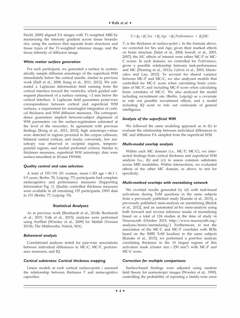
Figure 1.
Substrates of metacognition on higher-order cognition (MC-C).
(A) i Behavioral assessment: all participants underwent the
EmpaTom-task (n 5 48 trials) [Kanske et al., 2015], where they
were asked to answer three-way multiple choice questions con-
cerning details of 15-sec video-stories they just saw, followed by
confidence ratings; ii Distribution of task-performance (left) and
respective MC-C scores (right) (z-scored); (B) Cortical thick-
ness correlates of individual differences in MC-C, where warm/
cold colors indicate cortical thickness increases/decreases in indi-
viduals with higher MC-C scores; (C) Superficial WM anisotropy
(FA) correlates with individual differences in MC-C. To correct
for multiple comparisons in B and C, findings were thresholded
at PFWE< 0.05, using random field theory for non-isotropic
images (black outlines), superimposed on trends (semi-transpar-
ent, no black outlines).
r Multimodal MRI Studies of Metacognition r
r 5 r
Individual difference analysis, thus, suggests independ-ence between the abilities to accurately evaluate one’s ownperformance during perceptual processing as opposed tohigher-level cognitive tasks.
Cortical Substrates of Metacognitive Abilities
Cortical thickness analysis complemented behavioralfindings and revealed specific, non-overlapping structuralsubstrates of individual differences in both MC domains.
Higher MC-C scores correlated positively with thicknessincreases in a large bilateral cluster encompassing lateralfrontal, superior and inferior temporal, temporo-parietal,and posterior midline regions (PFWE< 0.05; Fig. 1). Sepa-rate assessment of WM diffusion parameters revealed
overlapping, yet more restricted and only marginally sig-nificant positive correlations between MC-C and FA ofright middle temporal regions (PFWE< 0.07), please seeSupporting Information Table I for further details on theclusters reported.
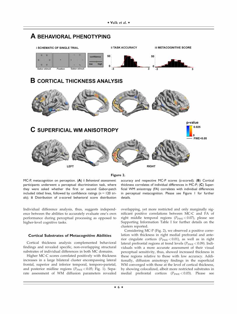
Considering MC-P (Fig. 2), we observed a positive corre-lation with thickness in right medial prefrontal and ante-rior cingulate cortices (PFWE< 0.01), as well as in rightlateral prefrontal regions at trend levels (PFWE< 0.09). Indi-viduals with a more accurate assessment of their visualperceptual sensitivity, thus, showed increased thickness inthese regions relative to those with low accuracy. Addi-tionally, diffusion anisotropy findings in the superficialWM converged with those at the level of cortical thickness,by showing colocalized, albeit more restricted substrates inmedial prefrontal cortices (PFWE< 0.03). Please see
Figure 2.
MC-P, metacognition on perception. (A) i Behavioral assessment:
participants underwent a perceptual discrimination task, where
they were asked whether the first or second Gabor-patch
included titled lines, followed by confidence ratings (n 5 120 tri-
als); ii Distribution of z-scored behavioral score distribution
accuracy and respective MC-P scores (z-scored); (B) Cortical
thickness correlates of individual differences in MC-P; (C) Super-
ficial WM anisotropy (FA) correlates with individual differences
in perceptual metacognition. Please see Figure 1 for further
details.
r Valk et al. r
r 6 r
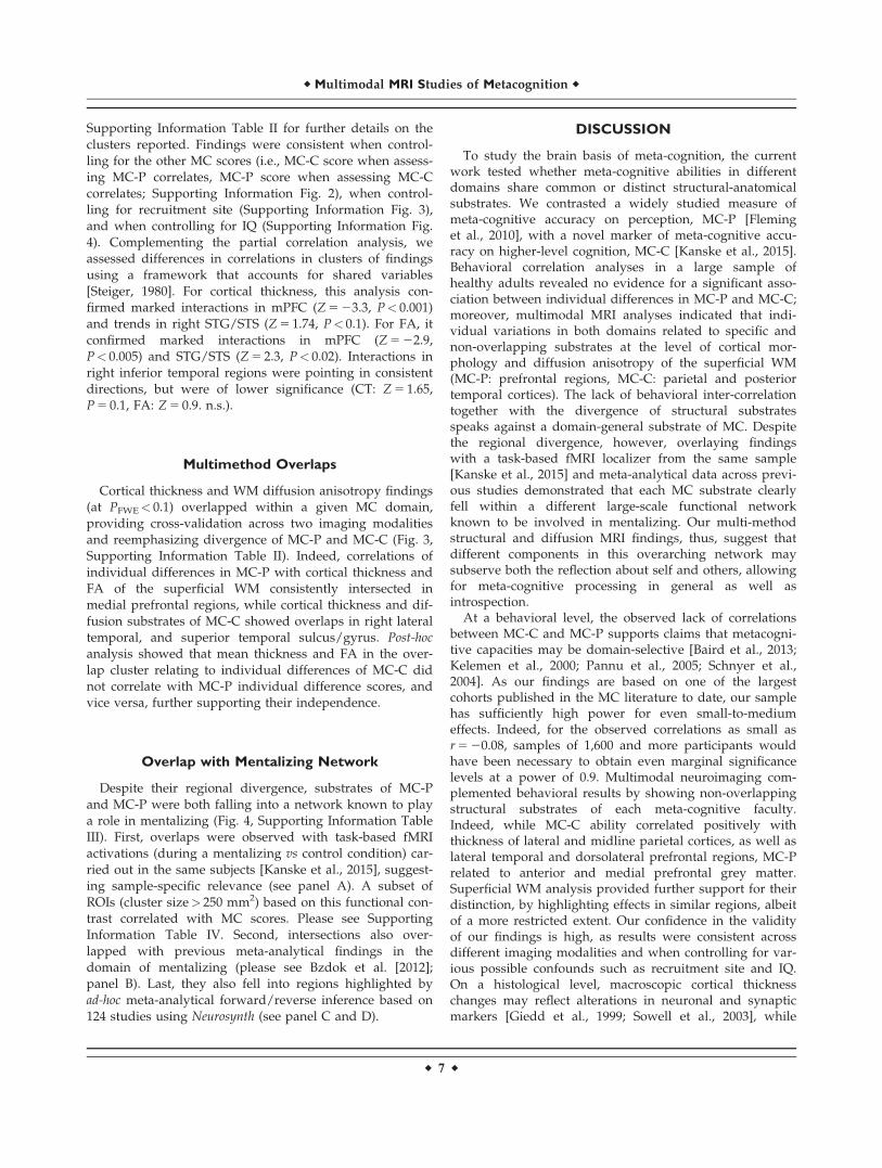
Supporting Information Table II for further details on theclusters reported. Findings were consistent when control-ling for the other MC scores (i.e., MC-C score when assess-ing MC-P correlates, MC-P score when assessing MC-Ccorrelates; Supporting Information Fig. 2), when control-ling for recruitment site (Supporting Information Fig. 3),and when controlling for IQ (Supporting Information Fig.4). Complementing the partial correlation analysis, weassessed differences in correlations in clusters of findingsusing a framework that accounts for shared variables[Steiger, 1980]. For cortical thickness, this analysis con-firmed marked interactions in mPFC (Z 5 23.3, P< 0.001)and trends in right STG/STS (Z 5 1.74, P< 0.1). For FA, itconfirmed marked interactions in mPFC (Z 5 22.9,P< 0.005) and STG/STS (Z 5 2.3, P< 0.02). Interactions inright inferior temporal regions were pointing in consistentdirections, but were of lower significance (CT: Z 5 1.65,P 5 0.1, FA: Z 5 0.9. n.s.).
Multimethod Overlaps
Cortical thickness and WM diffusion anisotropy findings(at PFWE< 0.1) overlapped within a given MC domain,providing cross-validation across two imaging modalitiesand reemphasizing divergence of MC-P and MC-C (Fig. 3,Supporting Information Table II). Indeed, correlations ofindividual differences in MC-P with cortical thickness andFA of the superficial WM consistently intersected inmedial prefrontal regions, while cortical thickness and dif-fusion substrates of MC-C showed overlaps in right lateraltemporal, and superior temporal sulcus/gyrus. Post-hoc
analysis showed that mean thickness and FA in the over-lap cluster relating to individual differences of MC-C didnot correlate with MC-P individual difference scores, andvice versa, further supporting their independence.
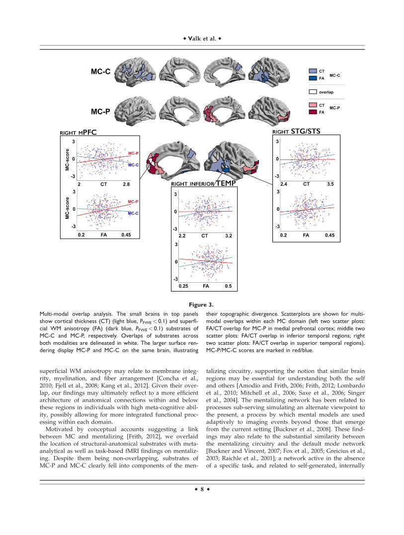
Overlap with Mentalizing Network
Despite their regional divergence, substrates of MC-Pand MC-P were both falling into a network known to playa role in mentalizing (Fig. 4, Supporting Information TableIII). First, overlaps were observed with task-based fMRIactivations (during a mentalizing vs control condition) car-ried out in the same subjects [Kanske et al., 2015], suggest-ing sample-specific relevance (see panel A). A subset ofROIs (cluster size> 250 mm2) based on this functional con-trast correlated with MC scores. Please see SupportingInformation Table IV. Second, intersections also over-lapped with previous meta-analytical findings in thedomain of mentalizing (please see Bzdok et al. [2012];panel B). Last, they also fell into regions highlighted byad-hoc meta-analytical forward/reverse inference based on124 studies using Neurosynth (see panel C and D).
DISCUSSION
To study the brain basis of meta-cognition, the currentwork tested whether meta-cognitive abilities in differentdomains share common or distinct structural-anatomicalsubstrates. We contrasted a widely studied measure ofmeta-cognitive accuracy on perception, MC-P [Fleminget al., 2010], with a novel marker of meta-cognitive accu-racy on higher-level cognition, MC-C [Kanske et al., 2015].Behavioral correlation analyses in a large sample ofhealthy adults revealed no evidence for a significant asso-ciation between individual differences in MC-P and MC-C;moreover, multimodal MRI analyses indicated that indi-vidual variations in both domains related to specific andnon-overlapping substrates at the level of cortical mor-phology and diffusion anisotropy of the superficial WM(MC-P: prefrontal regions, MC-C: parietal and posteriortemporal cortices). The lack of behavioral inter-correlationtogether with the divergence of structural substratesspeaks against a domain-general substrate of MC. Despitethe regional divergence, however, overlaying findingswith a task-based fMRI localizer from the same sample[Kanske et al., 2015] and meta-analytical data across previ-ous studies demonstrated that each MC substrate clearlyfell within a different large-scale functional networkknown to be involved in mentalizing. Our multi-methodstructural and diffusion MRI findings, thus, suggest thatdifferent components in this overarching network maysubserve both the reflection about self and others, allowingfor meta-cognitive processing in general as well asintrospection.
At a behavioral level, the observed lack of correlationsbetween MC-C and MC-P supports claims that metacogni-tive capacities may be domain-selective [Baird et al., 2013;Kelemen et al., 2000; Pannu et al., 2005; Schnyer et al.,2004]. As our findings are based on one of the largestcohorts published in the MC literature to date, our samplehas sufficiently high power for even small-to-mediumeffects. Indeed, for the observed correlations as small asr 5 20.08, samples of 1,600 and more participants wouldhave been necessary to obtain even marginal significancelevels at a power of 0.9. Multimodal neuroimaging com-plemented behavioral results by showing non-overlappingstructural substrates of each meta-cognitive faculty.Indeed, while MC-C ability correlated positively withthickness of lateral and midline parietal cortices, as well aslateral temporal and dorsolateral prefrontal regions, MC-Prelated to anterior and medial prefrontal grey matter.Superficial WM analysis provided further support for theirdistinction, by highlighting effects in similar regions, albeitof a more restricted extent. Our confidence in the validityof our findings is high, as results were consistent acrossdifferent imaging modalities and when controlling for var-ious possible confounds such as recruitment site and IQ.On a histological level, macroscopic cortical thicknesschanges may reflect alterations in neuronal and synapticmarkers [Giedd et al., 1999; Sowell et al., 2003], while
r Multimodal MRI Studies of Metacognition r
r 7 r
superficial WM anisotropy may relate to membrane integ-rity, myelination, and fiber arrangement [Concha et al.,2010; Fjell et al., 2008; Kang et al., 2012]. Given their over-lap, our findings may ultimately reflect to a more efficientarchitecture of anatomical connections within and belowthese regions in individuals with high meta-cognitive abil-ity, possibly allowing for more integrated functional proc-essing within each domain.
Motivated by conceptual accounts suggesting a linkbetween MC and mentalizing [Frith, 2012], we overlaidthe location of structural-anatomical substrates with meta-analytical as well as task-based fMRI findings on mentaliz-ing. Despite them being non-overlapping, substrates ofMC-P and MC-C clearly fell into components of the men-
talizing circuitry, supporting the notion that similar brainregions may be essential for understanding both the selfand others [Amodio and Frith, 2006; Frith, 2012; Lombardoet al., 2010; Mitchell et al., 2006; Saxe et al., 2006; Singeret al., 2004]. The mentalizing network has been related toprocesses sub-serving simulating an alternate viewpoint tothe present, a process by which mental models are usedadaptively to imaging events beyond those that emergefrom the current setting [Buckner et al., 2008]. These find-ings may also relate to the substantial similarity betweenthe mentalizing circuitry and the default mode network[Buckner and Vincent, 2007; Fox et al., 2005; Greicius et al.,2003; Raichle et al., 2001]; a network active in the absenceof a specific task, and related to self-generated, internally
Figure 3.
Multi-modal overlap analysis. The small brains in top panels
show cortical thickness (CT) (light blue, PFWE< 0.1) and superfi-
cial WM anisotropy (FA) (dark blue, PFWE< 0.1) substrates of
MC-C and MC-P, respectively. Overlaps of substrates across
both modalities are delineated in white. The larger surface ren-
dering display MC-P and MC-C on the same brain, illustrating
their topographic divergence. Scatterplots are shown for multi-
modal overlaps within each MC domain (left two scatter plots:
FA/CT overlap for MC-P in medial prefrontal cortex; middle two
scatter plots: FA/CT overlap in inferior temporal regions; right
two scatter plots: FA/CT overlap in superior temporal regions).
MC-P/MC-C scores are marked in red/blue.
r Valk et al. r
r 8 r
focused, and self-referential thought [Amodio and Frith,2006; Bernhardt et al., 2014a; Botvinick et al., 2004; Christ-off et al., 2009; Smallwood and Schooler, 2006]. The defaultmode network has been associated with “decoupling,” aclass of cognitive processes possibly underlying both tak-ing the cognitive perspective of others and by MC[Andrews-Hanna et al., 2010; Buckner et al., 2008; Bucknerand Vincent, 2007; Vincent et al., 2006].
The focus on structural substrates underlying individualdifferences in the current work may not have targeted allbrain regions functionally involved in MC-P and MC-C.Future studies based on fMRI may provide furtherinsights, and also address dynamic and connectional prop-erties of different network components. Nevertheless, thespecific role of prefrontal regions in MC-P may relate totheir involvement in both decoupled self-generatedthought and the evaluation of incoming sensory informa-tion [Gilbert et al., 2005; Sommer et al., 2007], possiblymediated by pathways from and to higher visual areas
[Fleming et al., 2012b; Ramnani and Owen, 2004; Young,1992]. This might, in turn, subserve action monitoring andthe planning of possible future actions [D’Argembeauet al., 2007; Gusnard et al., 2001; Mitchell et al., 2005;Northoff et al., 2006; Vogeley et al., 2004], likely requiringaccurate representations of sensory stimuli and perceptualperformance. Contrary, MC-C substrates fell more specifi-cally into posterior midline and lateral temporal nodes ofthe mentalizing and default mode networks. Posteriornodes are heavily involved in episodic memory process-ing, and strongly interconnected with the medial temporallobe system [Andrews-Hanna et al., 2010; Buckner et al.,2008; Buckner and Carroll, 2007]. Episodic memoryretrieval may indeed have been relevant for MC-C, as theemployed paradigm queried participants’ confidence ongiven answers to questions relating to the content ofothers’ autobiographical narratives. At trend level, wefound that high MC-C ability related to decreases inmPFC. In previous literature not only increases but also
Figure 4.
Relation of MC substrates the mentalizing network, derived
from three different methods. (A) Correspondence to fMRI acti-
vations derived from a mentalizing task in the same subjects
[Kanske et al., 2015]; (B) Correspondence to a previously pub-
lished meta-analysis on mentalizing and social cognition [Bzdok
et al., 2012]; (C) Correspondence with Neurosynth reverse
inference map of the term “mentalizing”; (D) Correspondence
with Neurosynth forward inference map of the term
“mentalizing”. (Masks are depicted in semi-transparent white
with black delineation).
r Multimodal MRI Studies of Metacognition r
r 9 r
decreases in thickness have been related to expert knowl-edge and enhanced behavioral performance [Hyde et al.,2007; Shaw et al., 2006]. Though beyond the scope of thecurrent project, future studies might follow up on thisinteresting pattern of results. Also, it is worth pointing outthat mentalizing relies on multiple inferences (e.g., inferactor’s goals; infer most likely action given a goal), whichindividually will have a certain amount of associateduncertainty, all of which would finally be integrated whenmaking confidence judgments. On this basis, one mighthave expected a more widespread substrate involved inMC for high-level cognition than for MC about perception,as shown by our findings.
To conclude, our findings robustly suggest divergentstructural–anatomical substrates for MC in differentdomains that nevertheless colocalize with different compo-nents of the mentalizing circuitry. Our result could informtraining studies that aim at enhancing MC, ToM, andsocial cognition in general. Specifically, a crucial questionfor future intervention research will be to investigatewhether improvement in MC transfers to mentalizingskills, and vice versa.
ACKNOWLEDGMENTS
The authors are thankful to the Department of Social Neu-rosciences for their support with the ReSource project.
REFERENCES
Amodio DM, Frith CD (2006): Meeting of minds: The medial fron-
tal cortex and social cognition. Nat Rev Neurosci 7:268–277.Andrews-Hanna JR, Reidler JS, Sepulcre J, Poulin R, Buckner RL
(2010): Functional-anatomic fractionation of the brain’s default
network. Neuron 65:550–562.Baird B, Smallwood J, Gorgolewski KJ, Margulies DS (2013):
Medial and lateral networks in anterior prefrontal cortex sup-
port metacognitive ability for memory and perception.
J Neurosci 33:16657–16665.Baird B, Cieslak M, Smallwood J, Grafton ST, Schooler JW (2013):
Regional white matter variation associated with domain-specific
metacognitive accuracy. J Cogn Neurosci 33:16657–16665.Bermudez P, Lerch JP, Evans AC, Zatorre RJ (2009): Neuroana-
tomical correlates of musicianship as revealed by cortical thick-
ness and voxel-based morphometry. Cereb Cortex 19:1583–
1596.Bernhardt BC, Bernasconi N, Concha L, Bernasconi A (2010): Cort-
ical thickness analysis in temporal lobe epilepsy: Reproducibil-
ity and relation to outcome. Neurology 74:1776–1784.Bernhardt BC, Smallwood J, Tusche A, Ruby FJ, Engen HG,
Steinbeis N, Singer T (2014a): Medial prefrontal and anterior
cingulate cortical thickness predicts shared individual differen-
ces in self-generated thought and temporal discounting. Neu-
roImage 90:290–297.Bernhardt BC, Valk SL, Silani G, Bird G, Frith U, Singer T (2014b):
Selective disruption of sociocognitive structural brain networks
in autism and alexithymia. Cereb Cortex 24:3258–3267.
Bernhardt BC, Hong S, Bernasconi A, Bernasconi N (2015): MRI
pattern learning in temporal lobe epilepsy: patient classifica-
tion and prognostics. Annals of Neurology 77:436–446Botvinick MM, Cohen JD, Carter CS (2004): Conflict monitoring
and anterior cingulate cortex: An update. Trends Cogn Sci 8:
539–546.Buckner RL, Carroll DC (2007): Self-projection and the brain.
Trends Cogn Sci 11:49–57.Buckner RL, Vincent JL (2007): Unrest at rest: Default activity and
spontaneous network correlations. NeuroImage 37:1091–1096;discussion 1097–1099.
Buckner RL, Andrews-Hanna JR, Schacter DL (2008): The brain’s
default network: Anatomy, function, and relevance to disease.
Ann N Y Acad Sci 1124:1–38.Bzdok D, Schilbach L, Vogeley K, Schneider K, Laird AR, Langner
R, Eickhoff SB (2012): Parsing the neural correlates of moral
cognition: ALE meta-analysis on morality, theory of mind, and
empathy. Brain Struct Funct 217:783–796.Cabeza R (2008): Role of parietal regions in episodic memory
retrieval: The dual attentional processes hypothesis. Neuropsy-
chologia 46:1813–1827.Cardinale F, Chinnici G, Bramerio M, Mai R, Sartori I, Cossu M,
Lo Russo G, Castana L, Colombo N, Caborni C, De Momi E,Ferrigno G (2014): Validation of FreeSurfer-estimated brain
cortical thickness: Comparison with histologic measurements.
Neuroinformatics 12:535–542.Christoff K, Gordon AM, Smallwood J, Smith R, Schooler JW
(2009): Experience sampling during fMRI reveals default net-
work and executive system contributions to mind wandering.
Proc Natl Acad Sci USA 106:8719–8724.Clarke F, Birdsall T, Tanner W (1959): Two types of ROC curves
and definition of parameters. J Acoust Soc Am 31:629–630.Concha L, Livy DJ, Beaulieu C, Wheatley BM, Gross DW (2010):
In vivo diffusion tensor imaging and histopathology of the
fimbria-fornix in temporal lobe epilepsy. J Neurosci 30:996–
1002.D’Argembeau A, Ruby P, Collette F, Degueldre C, Balteau E, Luxen
A, Maquet P, Salmon E (2007): Distinct regions of the medial pre-
frontal cortex are associated with self-referential processing and
perspective taking. J Cogn Neurosci 19:935–944.Dale AM, Fischl B, Sereno MI (1999): Cortical surface-based analy-
sis. I. Segmentation and surface reconstruction. NeuroImage 9:
179–194.Doyle-Thomas KA, Duerden EG, Taylor MJ, Lerch JP, Soorya LV,
Wang AT, Fan J, Hollander E, Anagnostou E (2013): Effects of
age and symptomatology on cortical thickness in autism spec-
trum disorders. Res Autism Spectr Disord 7:141–150.Faul F, Erdfelder E, Buchner A, Lang AG (2009): Statistical power
analyses using G*Power 3.1: Tests for correlation and regres-
sion analyses. Behav Res Methods 41:1149–1160.Fischl B, Sereno MI, Dale AM (1999): Cortical surface-based analy-
sis. II: Inflation, flattening, and a surface-based coordinate sys-
tem. NeuroImage 9:195–207.Fjell AM, Westlye LT, Greve DN, Fischl B, Benner T, van der
Kouwe AJ, Salat D, Bjornerud A, Due-Tonnessen P, Walhovd
KB (2008): The relationship between diffusion tensor imaging
and volumetry as measures of white matter properties. Neuro-Image 42:1654–1668.
Fleck MS, Daselaar SM, Dobbins IG, Cabeza R (2006): Role of pre-
frontal and anterior cingulate regions in decision-making proc-
esses shared by memory and nonmemory tasks. Cereb Cortex
16:1623–1630.
r Valk et al. r
r 10 r
Fleming SM, Dolan RJ, Frith CD (2012a): Metacognition: Computa-
tion, biology and function. Philos Trans R Soc Lond B Biol Sci
367:1280–1286.Fleming SM, Huijgen J, Dolan RJ (2012b): Prefrontal contributions
to metacognition in perceptual decision making. J Neurosci 32:
6117–6125.Fleming SM, Weil RS, Nagy Z, Dolan RJ, Rees G (2010): Relating
introspective accuracy to individual differences in brain struc-
ture. Science 329:1541–1543.Fleming SM, Ryu J, Golfinos JG, Blackmon KE (2014): Domain-
specific impairment in metacognitive accuracy following ante-
rior prefrontal lesions. Brain 137:2811–2822.Fox MD, Snyder AZ, Vincent JL, Corbetta M, Van Essen DC,
Raichle ME (2005): The human brain is intrinsically organized
into dynamic, anticorrelated functional networks. Proc Natl
Acad Sci USA 102:9673–9678.Frith CD (2012): The role of metacognition in human social inter-
actions. Philos Trans R Soc Lond B Biol Sci 367:2213–2223.Galvin SJ, Podd JV, Drga V, Whitmore J (2003): Type 2 tasks
in the theory of signal detectability: Discrimination between
correct and incorrect decisions. Psychon Bull Rev 10:843–
876.Giedd JN, Blumenthal J, Jeffries NO, Castellanos FX, Liu H,
Zijdenbos A, Paus T, Evans AC, Rapoport JL (1999): Brain
development during childhood and adolescence: A longitudi-
nal MRI study. Nat Neurosci 2:861–863.Gilbert SJ, Frith CD, Burgess PW (2005): Involvement of rostral
prefrontal cortex in selection between stimulus-oriented and
stimulus-independent thought. Eur J Neurosci 21:1423–1431.Green DM, Swets JA (1974): Signal Detection Theory and Psycho-
physics (A Reprint, With Corrections of the Original 1966 ed.).
Huntington, NY: Robert E. Krieger Publishing Co.Greicius MD, Krasnow B, Reiss AL, Menon V (2003): Functional
connectivity in the resting brain: A network analysis of the
default mode hypothesis. Proc Natl Acad Sci USA 100:
253–258.Greve DN, Fischl B (2009): Accurate and robust brain image align-
ment using boundary-based registration. NeuroImage 48:
63–72.Gusnard DA, Akbudak E, Shulman GL, Raichle ME (2001): Medial
prefrontal cortex and self-referential mental activity: Relation
to a default mode of brain function. Proc Natl Acad Sci USA
98:4259–4264.Han X, Jovicich J, Salat D, van der Kouwe A, Quinn B, Czanner S,
Busa E, Pacheco J, Albert M, Killiany R, Maguire P, Rosas D,
Makris N, Dale A, Dickerson B, Fischl B (2006): Reliability of
MRI-derived measurements of human cerebral cortical thick-
ness: The effects of field strength, scanner upgrade and manu-
facturer. NeuroImage 32:180–194.Henson RN, Rugg MD, Shallice T, Dolan RJ (2000): Confidence in
recognition memory for words: Dissociating right prefrontal
roles in episodic retrieval. J Cogn Neurosci 12:913–923.Hyde KL, Lerch JP, Zatorre RJ, Griffiths TD, Evans AC, Peretz I
(2007): Cortical thickness in congenital amusia: When less is
better than more. J Neurosci 27:13028–13032.Kang X, Herron TJ, Woods DL (2011): Regional variation, hemi-
spheric asymmetries and gender differences in pericorticalwhite matter. NeuroImage 56:2011–2023.
Kang X, Herron TJ, Turken AU, Woods DL (2012): Diffusion prop-
erties of cortical and pericortical tissue: Regional variations,
reliability and methodological issues. Magn Reson Imaging 30:
1111–1122.
Kanske P, Bockler A, Trautwein FM, Singer T (2015): Dissecting
the social brain: Introducing the EmpaToM to reveal distinct
neural networks and brain-behavior relations for empathy and
theory of mind. NeuroImage 122:6–19.Kelemen WL, Frost PJ, Weaver CA III (2000): Individual differen-
ces in metacognition: Evidence against a general metacognitive
ability. Mem Cogn 28:92–107.Koriat A (2007): Metacognition and conciousness. In: Zelalazo PD,
Moscovitch M, Thompson E, editors. Cambridge Handbook of
Conciousness. New York, USA: Cambridge University Press.Koriat A, Bjork RA (2006): Mending metacognitive illusions: A
comparison of mnemonic-based and theory-based procedures.
J Exp Psychol Learn Mem Cogn 32:1133–1145.Kuperberg GR, Broome MR, McGuire PK, David AS, Eddy M,
Ozawa F, Goff D, West WC, Williams SC, van der Kouwe AJ,
Salat DH, Dale AM, Fischl B (2003): Regionally localized thin-
ning of the cerebral cortex in schizophrenia. Arch Gen Psychia-
try 60:878–888.Lerch JP, Evans AC (2005): Cortical thickness analysis examined
through power analysis and a population simulation. Neuro-
Image 24:163–173.Lerch JP, Pruessner JC, Zijdenbos A, Hampel H, Teipel SJ, Evans
AC (2005): Focal decline of cortical thickness in Alzheimer’s
disease identified by computational neuroanatomy. Cereb Cor-
tex 15:995–1001.Lombardo MV, Chakrabarti B, Bullmore ET, Wheelwright SJ,
Sadek SA, Suckling J, Consortium MA, Baron-Cohen S (2010):
Shared neural circuits for mentalizing about the self and
others. J Cogn Neurosci 22:1623–1635.Maniscalco B, Lau H (2012): A signal detection theoretic approach
for estimating metacognitive sensitivity from confidence rat-
ings. Conscious Cogn 21:422–430.Mar RA (2011): The neural bases of social cognition and story
comprehension. Annu Rev Psychol 62:103–134.McCaig RG, Dixon M, Keramatian K, Liu I, Christoff K (2011):
Improved modulation of rostrolateral prefrontal cortex using
real-time fMRI training and meta-cognitive awareness. Neuro-
Image 55:1298–1305.McCurdy LY, Maniscalco B, Metcalfe J, Liu KY, de Lange FP, Lau
H (2013): Anatomical coupling between distinct metacognitive
systems for memory and visual perception. J Neurosci 33:
1897–1906.Metcalfe J (1994): Metacognition: Knowing About Knowing. Cam-
bridge, MA: Bradford Books.Mitchell JP, Banaji MR, Macrae CN (2005): The link between social
cognition and self-referential thought in the medial prefrontal
cortex. J Cogn Neurosci 17:1306–1315.Mitchell JP, Macrae CN, Banaji MR (2006): Dissociable medial pre-
frontal contributions to judgments of similar and dissimilar
others. Neuron 50:655–663.Northoff G, Heinzel A, de Greck M, Bermpohl F, Dobrowolny H,
Panksepp J (2006): Self-referential processing in our brain—A
meta-analysis of imaging studies on the self. NeuroImage 31:
440–457.Oppenheimer DM (2008): The secret life of fluency. Trends Cogn
Sci 12:237–241.Pannu JK, Kaszniak AW, Rapcsak SZ (2005): Metamemory for
faces following frontal lobe damage. J Int Neuropsychol Soc
11:668–676.Raichle ME, MacLeod AM, Snyder AZ, Powers WJ, Gusnard DA,
Shulman GL (2001): A default mode of brain function. Proc
Natl Acad Sci USA 98:676–682.
r Multimodal MRI Studies of Metacognition r
r 11 r
Ramnani N, Owen AM (2004): Anterior prefrontal cortex: Insightsinto function from anatomy and neuroimaging. Nat Rev Neu-rosci 5:184–194.
Rosas HD, Liu AK, Hersch S, Glessner M, Ferrante RJ, Salat DH,van der Kouwe A, Jenkins BG, Dale AM, Fischl B (2002):Regional and progressive thinning of the cortical ribbon inHuntington’s disease. Neurology 58:695–701.
Salat DH, Buckner RL, Snyder AZ, Greve DN, Desikan RS, BusaE, Morris JC, Dale AM, Fischl B (2004): Thinning of the cere-bral cortex in aging. Cereb Cortex 14:721–730.
Saxe R, Moran JM, Scholz J, Gabrieli J (2006): Overlapping andnon-overlapping brain regions for theory of mind and selfreflection in individual subjects. Social Cogn Affect Neurosci 1:229–234.
Schnyer DM, Verfaellie M, Alexander MP, LaFleche G, Nicholls L,Kaszniak AW (2004): A role for right medial prefontal cortexin accurate feeling-of-knowing judgements: Evidence frompatients with lesions to frontal cortex. Neuropsychologia 42:957–966.
Shaw P, Greenstein D, Lerch J, Clasen L, Lenroot R, Gogtay N,Evans A, Rapoport J, Giedd J (2006): Intellectual ability andcortical development in children and adolescents. Nature 440:676–679.
Shaw P, Sharp W, Sudre G, Wharton A, Greenstein D, RaznahanA, Evans A, Chakravarty MM, Lerch JP, Rapoport J (2015):Subcortical and cortical morphological anomalies as an endo-phenotype in obsessive-compulsive disorder. Mol Psychiatry20:224–231.
Singer T, Kok BE, Bornemann B, Zurborg S, Bolz M, Bochow CA.(in press) The ReSource Project. Background, Design, Samplesand Measurements, 2nd ed.
Singer T, Seymour B, O’Doherty J, Kaube H, Dolan RJ, Frith CD(2004): Empathy for pain involves the affective but not sensorycomponents of pain. Science 303:1157–1162.
Smallwood J, Schooler JW (2006): The restless mind. Psychol Bull132:946–958.
Sommer M, Dohnel K, Sodian B, Meinhardt J, Thoermer C, HajakG (2007): Neural correlates of true and false belief reasoning.NeuroImage 35:1378–1384.
Song C, Kanai R, Fleming SM, Weil RS, Schwarzkopf DS, Rees G(2011): Relating inter-individual differences in metacognitiveperformance on different perceptual tasks. Conscious Cogn 20:1787–1792.
Sowell ER, Peterson BS, Thompson PM, Welcome SE, HenkeniusAL, Toga AW (2003): Mapping cortical change across thehuman life span. Nat Neurosci 6:309–315.
Sowell ER, Peterson BS, Kan E, Woods RP, Yoshii J, Bansal R, XuD, Zhu H, Thompson PM, Toga AW (2007): Sex differences incortical thickness mapped in 176 healthy individuals between7 and 87 years of age. Cereb Cortex 17:1550–1560.
Steiger JH (1980): Tests for comparing elements of a correlationmatrix. Psychol Bull 87:245–251.
Teasdale JD, Moore RG, Hayhurst H, Pope M, Williams S, SegalZV (2002): Metacognitive awareness and prevention of relapsein depression: Empirical evidence. J Consult Clin Psychol 70:275–287.
Valk SL, Di Martino A, Milham MP, Bernhardt BC (2015): Multi-center mapping of structural network alterations in autism.Hum Brain Mapp 36:2364–2373.
Valk SL, Bernhardt BC, Bockler A, Trautwein FM, Kanske P,Singer T (2016): Socio-cognitive phenotypes differentially mod-ulate large-scale structural covariance networks. Cereb Cortex.doi: 10.1093/cercor/bhv319.
Vincent JL, Snyder AZ, Fox MD, Shannon BJ, Andrews JR,Raichle ME, Buckner RL (2006): Coherent spontaneous activityidentifies a hippocampal-parietal memory network.J Neurophysiol 96:3517–3531.
Vogeley K, May M, Ritzl A, Falkai P, Zilles K, Fink GR (2004):Neural correlates of first-person perspective as one constituentof human self-consciousness. J Cogn Neurosci 16:817–827.
Worsley KJ, Andermann M, Koulis T, MacDonald D, Evans AC(1999): Detecting changes in nonisotropic images. Human brainMapping 8:98–101.
Worsley K, Taylor JE, Carbonell F, Chung MK, Duerden E,Bernhardt BC, Lyttelton OC, Boucher M, Evans A (2009): Surf-Stat: A Matlab toolbox for the statistical analysis of univariateand multivariate surface and volumetric data using linear mixedeffect models and random field theory. NeuroImage S102.
Yokoyama O, Miura N, Watanabe J, Takemoto A, Uchida S,Sugiura M, Horie K, Sato S, Kawashima R, Nakamura K(2010): Right frontopolar cortex activity correlates with reliabil-ity of retrospective rating of confidence in short-term recogni-tion memory performance. Neuroscience Research 68:199–206.
Young MP (1992): Objective analysis of the topological organi-zation of the primate cortical visual system. Nature 358:152–155.
r Valk et al. r
r 12 r


![Domain-Specific Batch Normalization for Unsupervised ...domain-specific information in unsupervised domain adap-tation scenarios. AdaBN [7] proposes a post-processing method to re-estimate](https://img.pdfslide.net/doc/110x75/604975c40b57ad0df931a59b/domain-speciic-batch-normalization-for-unsupervised-domain-speciic-information.jpg)