Embed Size (px)
Citation preview
Cancer Cell, Volume 17
Supplemental Information
Regression of Castrate-Recurrent Prostate Cancer
by a Small-Molecule Inhibitor of the Amino-Terminus
Domain of the Androgen Receptor Raymond J. Andersen, Nasrin R. Mawji, Jun Wang, Gang Wang, Simon Haile, Jae-Kyung Myung, Kate Watt, Teresa Tam, Yu Chi Yang, Carmen A Bañuelos, David E. Williams, Iain J. McEwan, Yuzhou Wang, and Marianne D. Sadar
CONTENTS:
Supplemental Figures
Figure S1. Related to Figure 1.
Figure S2. Related to Figure 2.
Figure S3. Related to Figure 6. Levels of AR protein in harvested xenografts.
Supplemental Experimental Procedures
Supplemental References
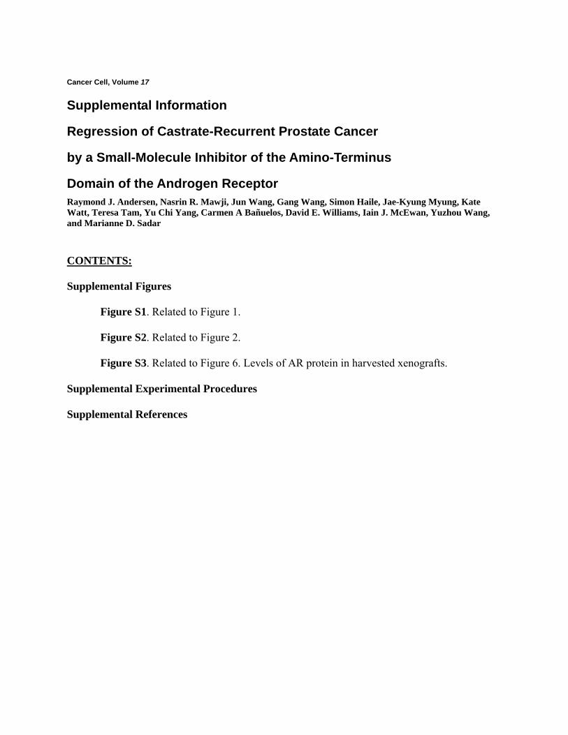
LNCaP ARO AR-13 Untrans-fected
AR
β-Actin
Figure S1. Related to Figure 1
Western blot analysis of levels of AR in LNCaP cells (endogenous levels) compared to Cos-1
cells transfected with expression vectors for wild-type AR (AR0), AR1-653 (AR-13), or
untransfected using an antibody to an epitope in the NTD.
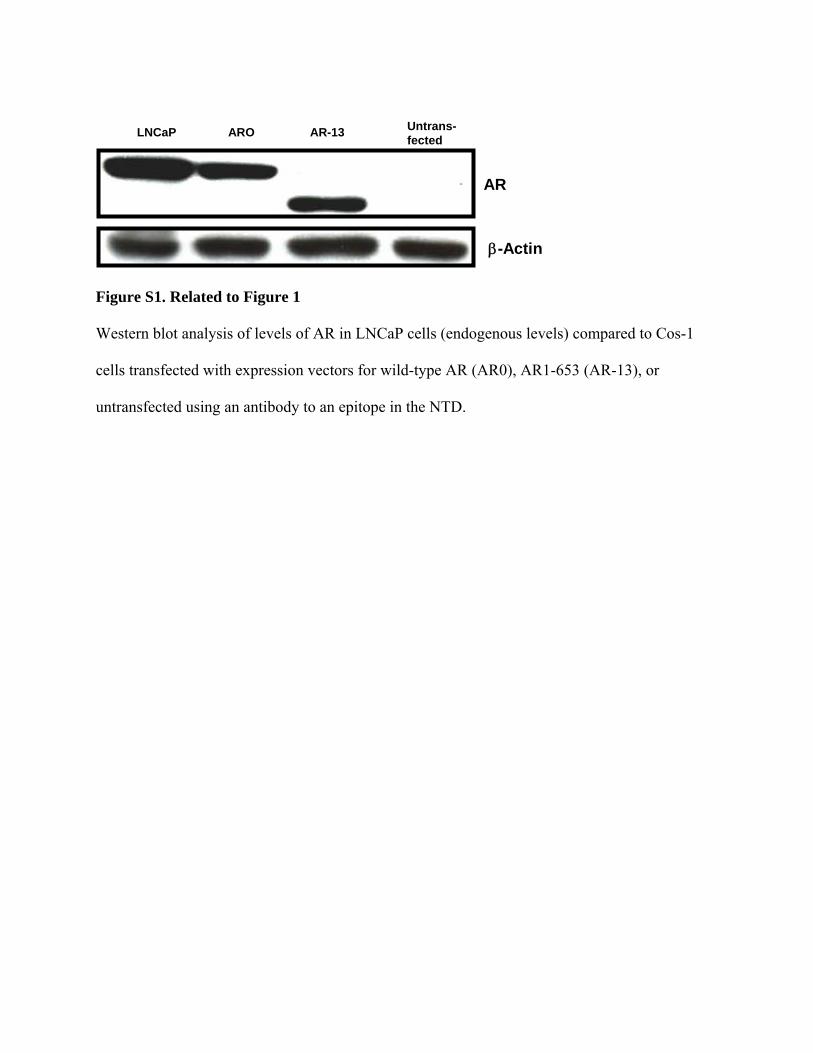
E
Figure S2. Related to Figure 2, EPI-001 does not inhibit AR activity by reducing levels of
protein, serine phosphorylation, or prevent nuclear translocation. A, EPI-001 blocks
androgen-regulated gene expression. LNCaP cells were treated with 1 nM R1881 or vehicle
(ethanol; ETOH) control for 16-24 hrs following a 1 h pre-treatment with 10 µg/ml EPI-001 or
an equivalent amount of vehicle (DMSO). Quantitative RT-PCR was employed to measure the
mRNA levels of known androgen-regulated genes. Levels of expression of genes that were
normalized to levels of GAPDH mRNA and then presented as a ratio to values from control
(DMSO and ETOH-treated cells). Bars represent the mean ± SD. P<0.05 for all mRNAs except
for BLVRB. B, Levels of AR protein in whole cell lysates of LNCaP cells treated for 48h with
EPI-001 (10 µg/ml) plus or minus R1881 (1 nM) measured by Western blot analysis using an
anti-AR antibody (PG21) and β-actin for loading control. Results from three separate
experiments are shown. C, Western blot analysis using antibodies to AR phosphorylated on
serine 81 (S81) or serine 213/210 (S213/210). LNCaP cells in serum-free and phenol red-free
RPMI media were incubated with EPI-001 (10 µg/ml) for 1 h followed by R1881 (1 nM) for 3 h.
Levels of phosphorylated AR and total AR protein in whole cell lysates of LNCaP cells were
analyzed by Western blot analysis using an anti-pAR antibody (pAR-S81, pAR-S213/210) and
anti-AR antibody. D, EPI-001 does not prevent ligand-induced nuclear translocation of the AR.
LNCaP cells were pre-treated for 1 h with EPI-001 (10 µg/ml) before addition of R1881 (1 nM)
or EtOH control for 15, 30, 60, or 120 mins before preparation of cytoplasmic and nuclear
fractions and probed for AR protein by western blot analysis using an antibody to the AR. Levels
of β-actin and lamin were used as loading controls of cytoplasmic and nuclear fractions
respectively. E, Fluorescent microscopy of representative LNCaP cells transfected with AR-YFP
in serum-free and phenol red-free media treated with EPI-001 (10 µg/ml) or DMSO control for 1

h before the addition of R1881 (1 nM) for the indicated times and then stained with DAPI. Scale
bar represents 2 µm.
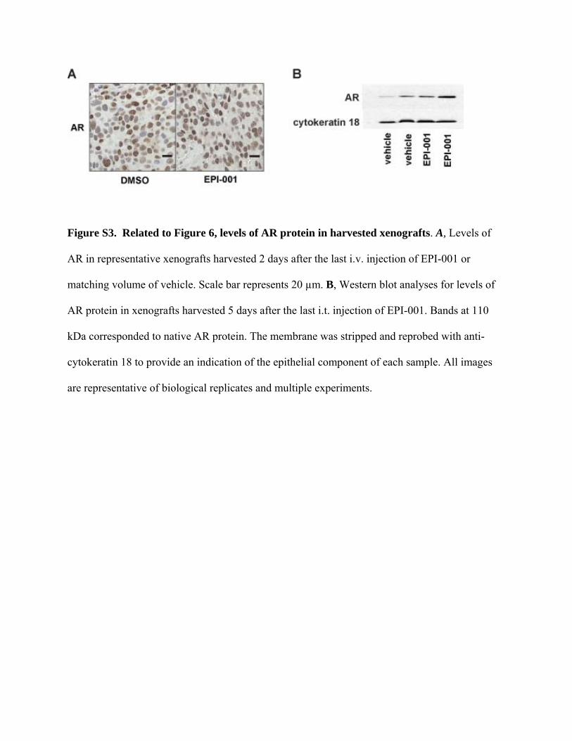
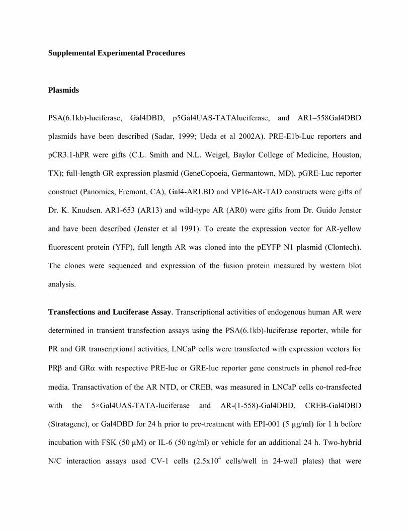
Figure S3. Related to Figure 6, levels of AR protein in harvested xenografts. A, Levels of
AR in representative xenografts harvested 2 days after the last i.v. injection of EPI-001 or
matching volume of vehicle. Scale bar represents 20 µm. B, Western blot analyses for levels of
AR protein in xenografts harvested 5 days after the last i.t. injection of EPI-001. Bands at 110
kDa corresponded to native AR protein. The membrane was stripped and reprobed with anti-
cytokeratin 18 to provide an indication of the epithelial component of each sample. All images
are representative of biological replicates and multiple experiments.
Supplemental Experimental Procedures
Plasmids
PSA(6.1kb)-luciferase, Gal4DBD, p5Gal4UAS-TATAluciferase, and AR1–558Gal4DBD
plasmids have been described (Sadar, 1999; Ueda et al 2002A). PRE-E1b-Luc reporters and
pCR3.1-hPR were gifts (C.L. Smith and N.L. Weigel, Baylor College of Medicine, Houston,
TX); full-length GR expression plasmid (GeneCopoeia, Germantown, MD), pGRE-Luc reporter
construct (Panomics, Fremont, CA), Gal4-ARLBD and VP16-AR-TAD constructs were gifts of
Dr. K. Knudsen. AR1-653 (AR13) and wild-type AR (AR0) were gifts from Dr. Guido Jenster
and have been described (Jenster et al 1991). To create the expression vector for AR-yellow
fluorescent protein (YFP), full length AR was cloned into the pEYFP N1 plasmid (Clontech).
The clones were sequenced and expression of the fusion protein measured by western blot
analysis.
Transfections and Luciferase Assay. Transcriptional activities of endogenous human AR were
determined in transient transfection assays using the PSA(6.1kb)-luciferase reporter, while for
PR and GR transcriptional activities, LNCaP cells were transfected with expression vectors for
PRβ and GRα with respective PRE-luc or GRE-luc reporter gene constructs in phenol red-free
media. Transactivation of the AR NTD, or CREB, was measured in LNCaP cells co-transfected
with the 5×Gal4UAS-TATA-luciferase and AR-(1-558)-Gal4DBD, CREB-Gal4DBD
(Stratagene), or Gal4DBD for 24 h prior to pre-treatment with EPI-001 (5 µg/ml) for 1 h before
incubation with FSK (50 µM) or IL-6 (50 ng/ml) or vehicle for an additional 24 h. Two-hybrid
N/C interaction assays used CV-1 cells (2.5x10⁴ cells/well in 24-well plates) that were
transfected using Fugene6 (Roche) with 0.25 µg/well 5XGAL4Luc reporter vector, VP-AR-(1–
565) (0.25 µg/well) that encodes the VP16 transactivation domain fused to amino residues 1-565
of AR NTD, and GAL4DBD-AR628–919 (0.25 µg/well) encoding the wild-type AR LBD C-
terminus amino acid residues 628-919 fused to the Gal4 DBD. Transfected cells were incubated
for 24 h in the absence and presence of 1 nM R1881 with or without inhibitors. LNCaP cells
stably expressing the ARR3-luciferase reporter were prepared using the ARR3 fragment
containing three androgen response regions -244 to -96 region of the rat probasin gene that was
excised from ARR3-tk-Luc vector with BamH1 and Xho1 and cloned into BglII and Xho1 site of
pGL4.26 and pGL4.27 with minimal TATA promoter vector (Promega Corporation, Madison,
WI). LNCaP cells were transfected with either pGL4.26ARR3luc or pGL4.27ARR3luc vector
using lipofectin overnight. At 24 h the media was replaced with RPMI + 5% FBS and 150 µg/ml
Hygromycin (sigma). 2 weeks later the surviving colonies were maintained in 150 µg/ml
Hygromycin and assayed for induction of luciferase activity using Steady Glo Luciferase assay
buffer (Promega, Madison, WI) with CLIPR (Molecular Devices). Luciferase activities were
normalized to protein concentrations of the samples. Transfection experiments were performed
in 3 separate experiments using triplicate wells.
Endogenous expression of androgen-regulated genes
LNCaP cells were pretreated for 1 h with vehicle, bicalutamide (10 µM), EPI-001 (10 µg/ml), or
185-9-1 (10 µg/ml) before addition of 1 nM R1881 under serum-free and phenol red-free
conditions. PSA, TRMPRSS2 and other transcripts were measured using total RNA (Trizol®
Reagent, Invitrogen) isolated from cells treated for 16 or 24 h with R1881 plus or minus either
EPI-001 or 185-9-1 by quantitative real-time (qRT)-PCR. qRT-PCR was performed separately in
triplicates for each biological sample. Levels of expression were normalized to the
glyceraldehyde-3-phosphate dehydrogenase (GAPDH) housekeeping gene. Primers employed
were as described in (Romanuik et al 2009A; Romanuik et al 2009B; Wang et al 2009).
Co-immunoprecipations
LNCaP cells in serum-free and phenol red-free RMPI were pre-incubated with EPI-001 (10
µg/ml) or DMSO (vehicle control) for 1 h and then subsequently treated for 6 h with IL-6 (50
ng/ml), R1881, or their respective vehicles. Whole cell lysates were prepared from harvested
cells. CBP was immunoprecipitated with an antibody against a C-terminus peptide (C-1)
followed by immunoblot probed with anti-AR antibody (AR441) and anti-CBP antibody (CBP-
A22). All antibodies are from Santa Cruz Biotechnology, CA, USA.
Chromatin immunoprecipitation (ChIP) - LNCaP cells were serum-starved for 48 h and
pretreated with 10 µg/ml EPI-001 or DMSO as control for 1 h before treatment with 1 nM R1881
or vehicle (ethanol) for the indicated times. The proteins were cross-linked with 1%
formaldehyde. Cell extracts were sonicated to yield an average length of 600-800 bp sheared
DNA fragments. The sonicated sample was pelleted and the supernatant diluted 10-fold with
ChIP dilution buffer (0.22 % Triton X- 100, 1.2 mM EDTA, 16.7 mM Tris-HCl [pH 8.1], 167
mM NaCl, 1× protease inhibitor). 100 µl of diluted supernatant was reserved as input. After pre-
clearing with 50 µl nProtein A-Sepharose™ beads (Amersham Biosciences) in TE (1:1) with
sheared salmon sperm DNA, the remaining proteins were incubated with 2 µg of anti-AR (C19),
or anti-CBP (A22) overnight at 4oC. The antibody-protein-DNA complex was precipitated by
incubating with 100 µl of 1:1 nProtein A-Sepharose™ beads. The protein-DNA complex was
eluted from the beads with elution buffer (1 % SDS, 0.1 M NaHCO3). The cross-linking of the
DNA protein complex was reversed by incubating at 65oC for 6 h. The DNA was recovered and
purified. The AR binding region was amplified applying Q-PCR using the following primers:
PSA-EN-ARE-F, 5’-TGGGACAACTTGCAAACCTG-3’; PSA-EN-ARE-R, 5’-
CCAGAGTAGGTCTGTTTTCAATCCA-3’; PSA-PR-ARE-, 5’- CCTAG
ATGAAGTCTCCATGAGCTACA-3’; PSA-PR-ARE-R, 5’- GGGAGGGAGAGCT
AGCACTTG-3’; TMPRSS2-ARE-F, 5’-TGGTCCTGGATGATAAAAAAAGTTT-3’; and
TMPRSS2-ARE-R, 5’-GACATACGCCCCACAACAGA-3’. The thermal cycling conditions
were 50ºC for 2 min, 95ºC for 2 min, followed by 45 cycles of 30 s at 95ºC, 30 s at 55ºC and 30 s
at 72ºC. Percentage input was calculated from dividing the arbitrary Q-PCR numbers obtained
by each sample by that of the input.
Nuclear translocation and levels of AR protein - LNCaP cells in serum-free and phenol red-
free RPMI were pre-treated with DMSO or EPI-001 (10 µg/ml) for 1h before the treatment with
R1881 (1 nM) for 15, 30, 60 and 120 min. Cell extracts were prepared by lysing in hypotonic
buffer and extraction of nuclear extracts in high salt buffer. Proteins were separated by 10%
SDS-PAGE and subjected to Western blot analysis. For long-term exposure to EPI-001, LNCaP
cells in serum-free and phenol red-free RPMI media were pre-treated with EPI-001 (10 µg/ml)
for 1h followed by addition of R1881 (1 nM) for 48 h. Levels of AR protein in cell extracts and
whole cell lysates were analyzed by Western blot analysis using an anti-AR antibody (PG21) and
normalized to levels of β-actin or lamin as loading controls.
Fluorescent microscopy - LNCaP cells were transiently transfected with an expression vector
for AR-YFP for 24 hr using serum-free and phenol red-free RPMI media. Transfected cells were
pre-treated with DMSO or EPI-001 (10 µg/ml) for 1 hr before the addition of R1881 (1 nM).
Cells were fixed with 4% paraformaldehyde at various time points, and mounted onto
FisherBrand microscope slides with VECTASHIELD® fluorescence mounting medium with
DAPI. The slides were examined by using a Zeiss Axioplan-2 Fluorescence Microscope (Zeiss,
Toronto, ON, Canada).
Immunohistochemistry
For immunohistochemical staining, sections were cut from the paraffin blocks. The staining was
carried out using anti–Ki-67 (1:50), anti–caspase-3 (1:50; DAKO), and anti-AR (PG21) as
primary antibodies.
Supplemental References
Romanuik TL, Ueda T, Le N, Haile S, Yong TM, Thomson T, Vessella RL, Sadar MD. (2009A)
Novel biomarkers for prostate cancer including noncoding transcripts. American Journal of
Pathology. 175(6):2264-76.
Romanuik TL, Wang G, Holt RA, Jones SJ, Marra MA, Sadar MD. (2009B) Identification of
novel androgen-responsive genes by sequencing of LongSAGE libraries. BMC Genomics.
15;10:476
Wang Q, Li W, Zhang Y, Yuan X, Xu K, Yu J, Chen Z, Beroukhim R, Wang H, Lupien M, Wu
T, Regan MM, Meyer CA, Carroll JS, Manrai AK, Jänne OA, Balk SP, Mehra R, Han B,
Chinnaiyan AM, Rubin MA, True L, Fiorentino M, Fiore C, Loda M, Kantoff PW, Liu XS,
Brown M. (2009) Androgen receptor regulates a distinct transcription program in androgen-
independent prostate cancer. Cell. 138(2):245-56.