Embed Size (px)
Citation preview
Biological Small Molecules as Receptors
Carolina Godoy-Alcantar1 and Anatoly K. Yatsimirsky2
1Universidad Autonoma del Estado de Morelos, Cuernavaca, Mexico2Universidad Nacional Autonoma de Mexico, Mexico D.F., Mexico
1 Introduction 12 Biological Ionophores 13 Macrocyclic Glycopeptide Antibiotics 34 Bile Acids 75 Rifamycins 86 Aminoglycosides 107 Cyclic Polypeptide Antibiotics 128 Alkaloids 149 Conclusions 17References 17
1 INTRODUCTION
Many natural low-molecular weight compounds that func-tion as antibiotics or have other biological activities,for example, as receptor antagonists, possess structuralelements such as specific arrangements of charged ordonor–acceptor groups, cavities or clefts, or chiral centers,making them promising host molecules for recognition ofboth ionic and neutral guests. Among these compounds,biological ionophores really use the classical host–guestcomplexation mechanism for their action, but in other casesthe ability of natural compounds to bind guests of differ-ent types is not directly related to their biological functions;nevertheless, it can be fairly significant. Since many of thesecompounds are commercially available and inexpensive,
they find practical applications as host molecules in analysisand separation.
Usually small biomolecules have one or more functionalgroups which are not essential for their recognition proper-ties but can be used for their modification or incorporationinto more sophisticated hosts. Such semisynthetic receptorswith improved recognition or sensor properties are moreeasily accessible than purely synthetic receptors, and someof them also are discussed in this review.
Particularly popular biological small molecules withreceptor properties are cyclodextrins, which are covered ina separate chapter in this book (see Cyclodextrins: FromNature to Nanotechnology, Molecular Recognition). Thisreview is focused on structures and physicochemical prop-erties of other small biological molecules, mostly antibi-otics, relevant to their use as receptors. Reported data ontheir recognition properties, as well as analytical applica-tions, are discussed. Natural ionophors, which are exten-sively covered in previous literature, are touched upon onlybriefly.
2 BIOLOGICAL IONOPHORES
Ionophores, which are lipophilic molecules capable oftransporting ions across the lipid bilayer of the cellmembrane, are synthesized by microorganisms and haveantibiotic properties by disrupting transmembrane ion con-centration gradients. Ionophores are not used in humanmedicine because of their potent cardiovascular effects,but they are used in farming for the prevention of coc-cidiodomycosis in poultry and are fed to cattle to improvegrowth. They may be relatively large molecules, such as thepolypeptide gramicidin which forms a transmembrane chan-nel for ion transport, or smaller molecules acting as mobile
Supramolecular Chemistry: From Molecules to Nanomaterials, Online 2012 John Wiley & Sons, Ltd.This article is 2012 John Wiley & Sons, Ltd.This article was published in the Supramolecular Chemistry: From Molecules to Nanomaterials in 2012 by John Wiley & Sons, Ltd.DOI: 10.1002/9780470661345.smc065
2 Molecular recognition
ion carriers. Ionophores of this last type were the subjectof intensive studies during 1960–1980 and have servedin many aspects as reference compounds for syntheticionophores. They also were used for the development ofthe first neutral carrier-based selective electrodes for alkali-metal ions.1 Thermodynamic parameters of cation bindingto natural ionophores, measured mostly in methanol andother organic solvents, can be found in several reviews.2–4
The properties of siderophores, an important group ofionophores specifically designed for transport of iron ions,are covered in several recent reviews5–7 and are not dis-cussed in this chapter.
In biological systems, ionophores selectively transportalkali- and alkaline-earth metal ions: for example, vali-nomycin is a selective carrier for K+ and monensin forNa+, but they also can bind other cations for whichthey have not been designed by nature8 and this is anarea of active current research with natural ionophores.
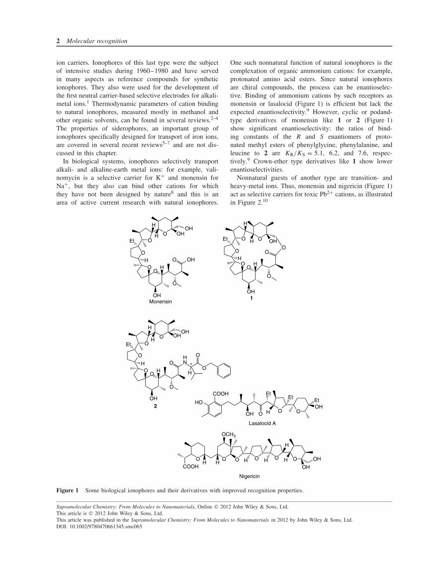
One such nonnatural function of natural ionophores is thecomplexation of organic ammonium cations: for example,protonated amino acid esters. Since natural ionophoresare chiral compounds, the process can be enantioselec-tive. Binding of ammonium cations by such receptors asmonensin or lasalocid (Figure 1) is efficient but lack theexpected enantioselectivity.9 However, cyclic or podand-type derivatives of monensin like 1 or 2 (Figure 1)show significant enantioselectivity: the ratios of bind-ing constants of the R and S enantiomers of proto-nated methyl esters of phenylglycine, phenylalanine, andleucine to 2 are KR/KS = 5.1, 6.2, and 7.6, respec-tively.9 Crown-ether type derivatives like 1 show lowerenantioselectivities.
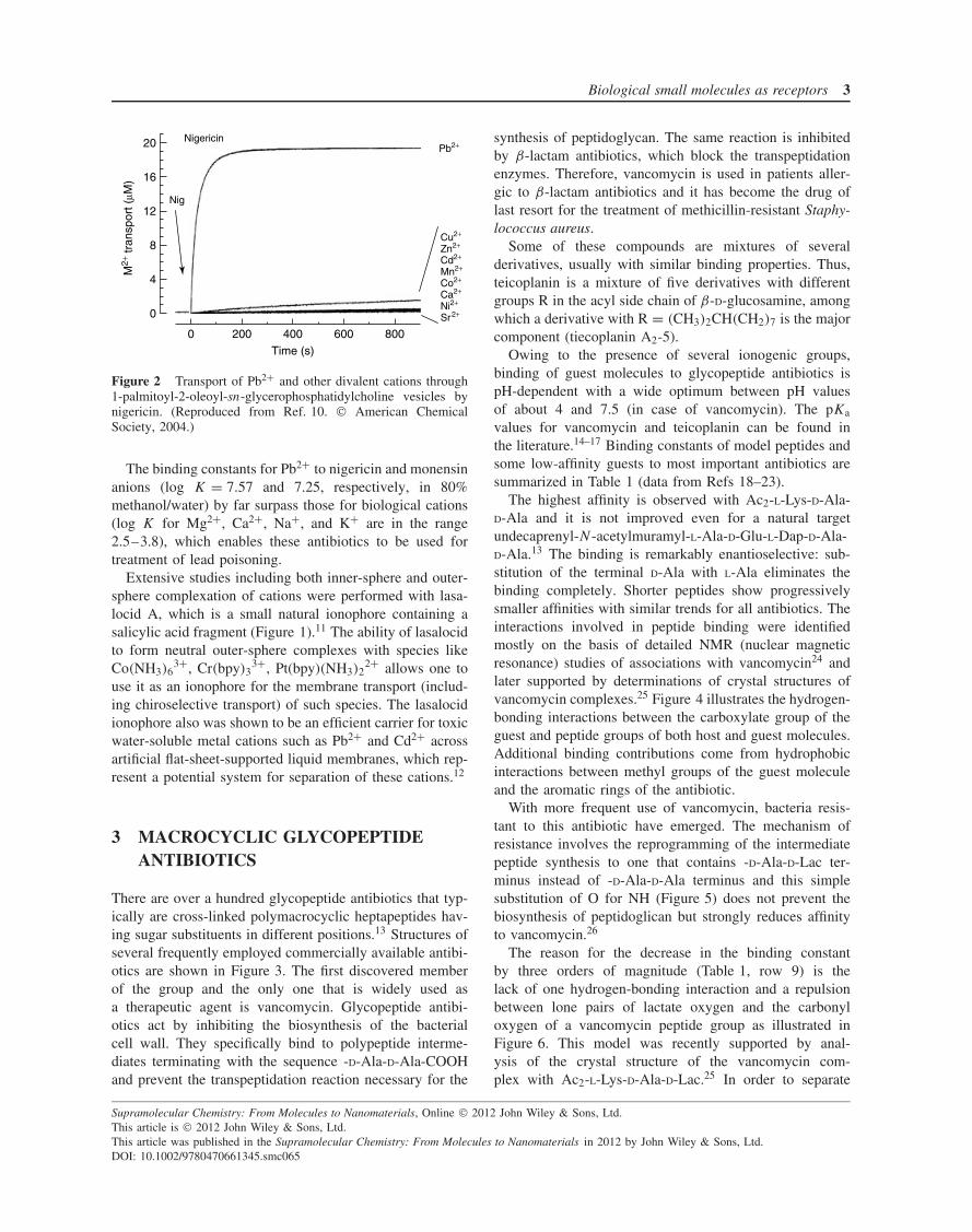
Nonnatural guests of another type are transition- andheavy-metal ions. Thus, monensin and nigericin (Figure 1)act as selective carriers for toxic Pb2+ cations, as illustratedin Figure 2.10
OO
O
OO OHH
OH
Et
H
H
OH
H
O
OHO
OO
O
OO OH
H
OH
Et
H
H
OH
H
O
HNO
H
O
O
OO
O
O O OHHEt
H
H
OH
H
O
OO
Monensin1
2OO
HO
OH O HOH
EtEt Et
COOH
OOOOOO
COOHH H OH
H H
H
HOH
OCH3
Lasalocid A
Nigericin
Figure 1 Some biological ionophores and their derivatives with improved recognition properties.
Supramolecular Chemistry: From Molecules to Nanomaterials, Online 2012 John Wiley & Sons, Ltd.This article is 2012 John Wiley & Sons, Ltd.This article was published in the Supramolecular Chemistry: From Molecules to Nanomaterials in 2012 by John Wiley & Sons, Ltd.DOI: 10.1002/9780470661345.smc065
Biological small molecules as receptors 3
20
16
12
8
4
0
M2+
tran
spor
t (µM
)
0 200 400 600 800
Time (s)
Nig
NigericinPb2+
Cu2+
Zn2+
Cd2+
Mn2+
Co2+
Ca2+
Ni2+
Sr2+
Figure 2 Transport of Pb2+ and other divalent cations through1-palmitoyl-2-oleoyl-sn-glycerophosphatidylcholine vesicles bynigericin. (Reproduced from Ref. 10. American ChemicalSociety, 2004.)
The binding constants for Pb2+ to nigericin and monensinanions (log K = 7.57 and 7.25, respectively, in 80%methanol/water) by far surpass those for biological cations(log K for Mg2+, Ca2+, Na+, and K+ are in the range2.5–3.8), which enables these antibiotics to be used fortreatment of lead poisoning.
Extensive studies including both inner-sphere and outer-sphere complexation of cations were performed with lasa-locid A, which is a small natural ionophore containing asalicylic acid fragment (Figure 1).11 The ability of lasalocidto form neutral outer-sphere complexes with species likeCo(NH3)6
3+, Cr(bpy)33+, Pt(bpy)(NH3)2
2+ allows one touse it as an ionophore for the membrane transport (includ-ing chiroselective transport) of such species. The lasalocidionophore also was shown to be an efficient carrier for toxicwater-soluble metal cations such as Pb2+ and Cd2+ acrossartificial flat-sheet-supported liquid membranes, which rep-resent a potential system for separation of these cations.12
3 MACROCYCLIC GLYCOPEPTIDEANTIBIOTICS
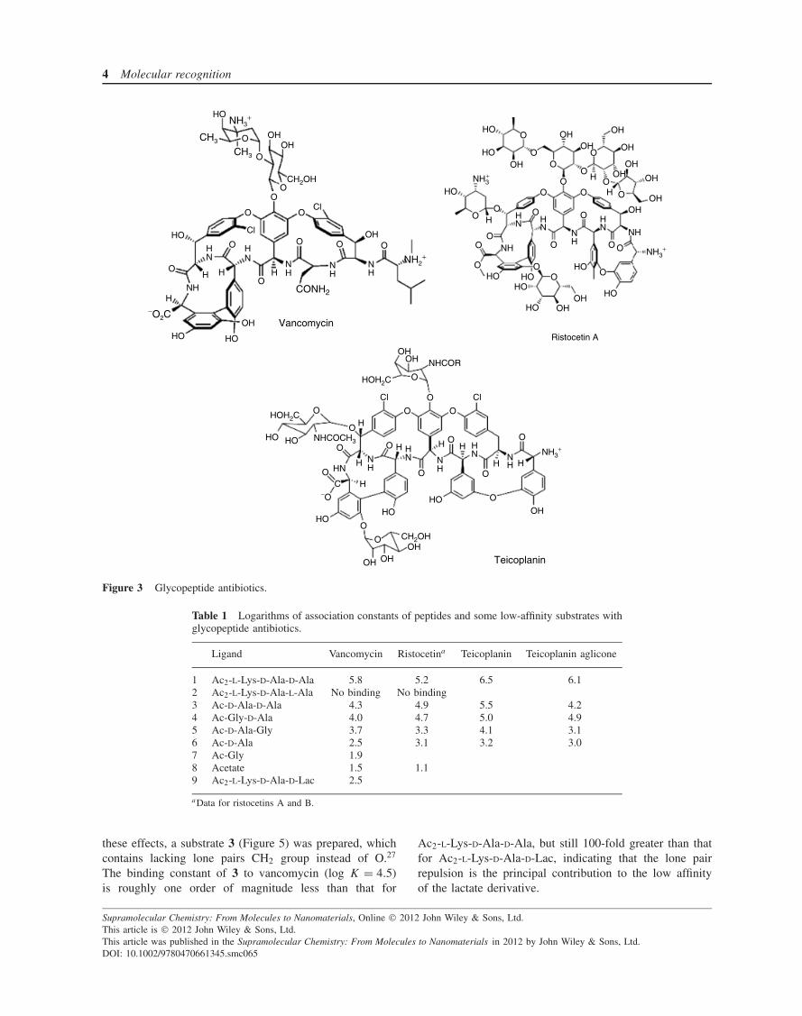
There are over a hundred glycopeptide antibiotics that typ-ically are cross-linked polymacrocyclic heptapeptides hav-ing sugar substituents in different positions.13 Structures ofseveral frequently employed commercially available antibi-otics are shown in Figure 3. The first discovered memberof the group and the only one that is widely used asa therapeutic agent is vancomycin. Glycopeptide antibi-otics act by inhibiting the biosynthesis of the bacterialcell wall. They specifically bind to polypeptide interme-diates terminating with the sequence -D-Ala-D-Ala-COOHand prevent the transpeptidation reaction necessary for the
synthesis of peptidoglycan. The same reaction is inhibitedby β-lactam antibiotics, which block the transpeptidationenzymes. Therefore, vancomycin is used in patients aller-gic to β-lactam antibiotics and it has become the drug oflast resort for the treatment of methicillin-resistant Staphy-lococcus aureus.
Some of these compounds are mixtures of severalderivatives, usually with similar binding properties. Thus,teicoplanin is a mixture of five derivatives with differentgroups R in the acyl side chain of β-D-glucosamine, amongwhich a derivative with R = (CH3)2CH(CH2)7 is the majorcomponent (tiecoplanin A2-5).
Owing to the presence of several ionogenic groups,binding of guest molecules to glycopeptide antibiotics ispH-dependent with a wide optimum between pH valuesof about 4 and 7.5 (in case of vancomycin). The pKa
values for vancomycin and teicoplanin can be found inthe literature.14–17 Binding constants of model peptides andsome low-affinity guests to most important antibiotics aresummarized in Table 1 (data from Refs 18–23).
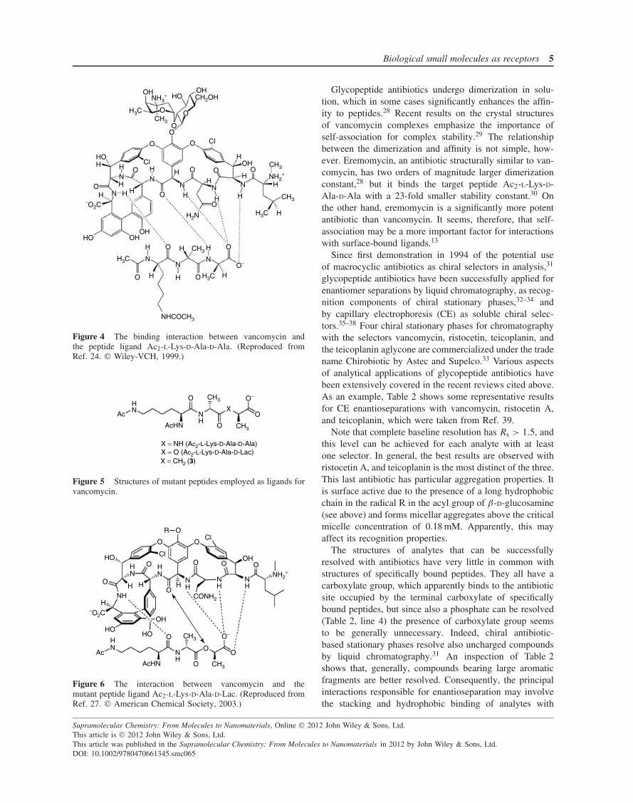
The highest affinity is observed with Ac2-L-Lys-D-Ala-D-Ala and it is not improved even for a natural targetundecaprenyl-N-acetylmuramyl-L-Ala-D-Glu-L-Dap-D-Ala-D-Ala.13 The binding is remarkably enantioselective: sub-stitution of the terminal D-Ala with L-Ala eliminates thebinding completely. Shorter peptides show progressivelysmaller affinities with similar trends for all antibiotics. Theinteractions involved in peptide binding were identifiedmostly on the basis of detailed NMR (nuclear magneticresonance) studies of associations with vancomycin24 andlater supported by determinations of crystal structures ofvancomycin complexes.25 Figure 4 illustrates the hydrogen-bonding interactions between the carboxylate group of theguest and peptide groups of both host and guest molecules.Additional binding contributions come from hydrophobicinteractions between methyl groups of the guest moleculeand the aromatic rings of the antibiotic.
With more frequent use of vancomycin, bacteria resis-tant to this antibiotic have emerged. The mechanism ofresistance involves the reprogramming of the intermediatepeptide synthesis to one that contains -D-Ala-D-Lac ter-minus instead of -D-Ala-D-Ala terminus and this simplesubstitution of O for NH (Figure 5) does not prevent thebiosynthesis of peptidoglican but strongly reduces affinityto vancomycin.26
The reason for the decrease in the binding constantby three orders of magnitude (Table 1, row 9) is thelack of one hydrogen-bonding interaction and a repulsionbetween lone pairs of lactate oxygen and the carbonyloxygen of a vancomycin peptide group as illustrated inFigure 6. This model was recently supported by anal-ysis of the crystal structure of the vancomycin com-plex with Ac2-L-Lys-D-Ala-D-Lac.25 In order to separate
Supramolecular Chemistry: From Molecules to Nanomaterials, Online 2012 John Wiley & Sons, Ltd.This article is 2012 John Wiley & Sons, Ltd.This article was published in the Supramolecular Chemistry: From Molecules to Nanomaterials in 2012 by John Wiley & Sons, Ltd.DOI: 10.1002/9780470661345.smc065
4 Molecular recognition
HO
HO
HO
CH3
CH3
O
O
OO
O
O
OO
O
O O O
OHOH
NH3+
CH2OH
HOCl
Cl
H
H
H
H
NH
HH
NN
HN
HN
NH
OH
NH2+
−O2C
CONH2
HOHO
OH Vancomycin
OH
OHOH
OH
OH
OHOH
O O
O
O
O
OO
O
O
OO O
OO
OO
OO
O
O
OO
O
H
H
H
OH
OHOH
NHH N
H
H
NN NH
NH
HOHO
HO
HO
HO
HO
OHOH
NH3+
NH3+
HO
Ristocetin A
OHOH
O
O
O
O
O
O
O
O
O
O
−O
O O
O
O
O
O
HOH2C
HOH2C
NHCOCH3
CH2OH
NH3+
Cl Cl
HO HO
HN
HO
H NH
N NNNH
H H H
HHH
H
H H
HC
OH OHOH
HOHO
OH
NHCOR
Teicoplanin
Figure 3 Glycopeptide antibiotics.
Table 1 Logarithms of association constants of peptides and some low-affinity substrates withglycopeptide antibiotics.
Ligand Vancomycin Ristocetina Teicoplanin Teicoplanin aglicone
1 Ac2-L-Lys-D-Ala-D-Ala 5.8 5.2 6.5 6.12 Ac2-L-Lys-D-Ala-L-Ala No binding No binding3 Ac-D-Ala-D-Ala 4.3 4.9 5.5 4.24 Ac-Gly-D-Ala 4.0 4.7 5.0 4.95 Ac-D-Ala-Gly 3.7 3.3 4.1 3.16 Ac-D-Ala 2.5 3.1 3.2 3.07 Ac-Gly 1.98 Acetate 1.5 1.19 Ac2-L-Lys-D-Ala-D-Lac 2.5
aData for ristocetins A and B.
these effects, a substrate 3 (Figure 5) was prepared, whichcontains lacking lone pairs CH2 group instead of O.27
The binding constant of 3 to vancomycin (log K = 4.5)is roughly one order of magnitude less than that for
Ac2-L-Lys-D-Ala-D-Ala, but still 100-fold greater than thatfor Ac2-L-Lys-D-Ala-D-Lac, indicating that the lone pairrepulsion is the principal contribution to the low affinityof the lactate derivative.
Supramolecular Chemistry: From Molecules to Nanomaterials, Online 2012 John Wiley & Sons, Ltd.This article is 2012 John Wiley & Sons, Ltd.This article was published in the Supramolecular Chemistry: From Molecules to Nanomaterials in 2012 by John Wiley & Sons, Ltd.DOI: 10.1002/9780470661345.smc065
Biological small molecules as receptors 5
OHHO
H3C
NH3+
OCH3
O
O
O
O
O
O
O
O
O
O
O
OHCH2OH
Cl
HOH H
HHH
H
H
H
H
H
H
HH
H
Cl
NN
N
N
OHO
N N
−O2C
HO OHOH
H
H
H
H
H
NN
N
O
O
O
O−
O
H3C
CH3
HH3C
NHCOCH3
CH3
CH3
NH2+
H2N H3C H
H
Figure 4 The binding interaction between vancomycin andthe peptide ligand Ac2-L-Lys-D-Ala-D-Ala. (Reproduced fromRef. 24. Wiley-VCH, 1999.)
Ac
AcHN
HN
HN
O
O
O
CH3
CH3
X
O−
X = O (Ac2-L-Lys-D-Ala-D-Lac) X = NH (Ac2-L-Lys-D-Ala-D-Ala)
X = CH2 (3)
Figure 5 Structures of mutant peptides employed as ligands forvancomycin.
R O
O
O O O
O
O
O
O
O
OO
O
ClHOH
H
H
H
N
NH
H
HH
NN
HN
HN
HN
HN
Cl
OH
OH−O2C
NH2+
O−HO
HO
Ac
AcHN
CH3
CH3
CONH2
Figure 6 The interaction between vancomycin and themutant peptide ligand Ac2-L-Lys-D-Ala-D-Lac. (Reproduced fromRef. 27. American Chemical Society, 2003.)
Glycopeptide antibiotics undergo dimerization in solu-tion, which in some cases significantly enhances the affin-ity to peptides.28 Recent results on the crystal structuresof vancomycin complexes emphasize the importance ofself-association for complex stability.29 The relationshipbetween the dimerization and affinity is not simple, how-ever. Eremomycin, an antibiotic structurally similar to van-comycin, has two orders of magnitude larger dimerizationconstant,28 but it binds the target peptide Ac2-L-Lys-D-Ala-D-Ala with a 23-fold smaller stability constant.30 Onthe other hand, eremomycin is a significantly more potentantibiotic than vancomycin. It seems, therefore, that self-association may be a more important factor for interactionswith surface-bound ligands.13
Since first demonstration in 1994 of the potential useof macrocyclic antibiotics as chiral selectors in analysis,31
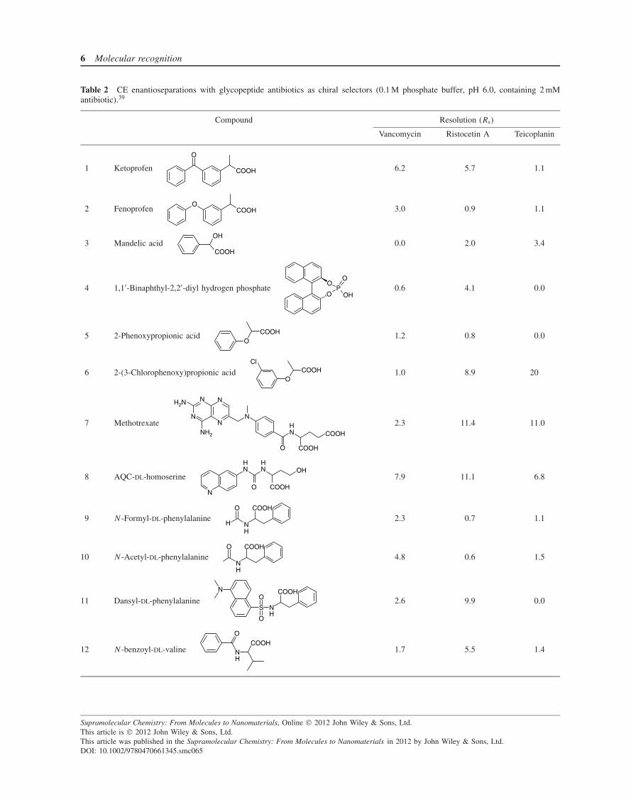
glycopeptide antibiotics have been successfully applied forenantiomer separations by liquid chromatography, as recog-nition components of chiral stationary phases,32–34 andby capillary electrophoresis (CE) as soluble chiral selec-tors.35–38 Four chiral stationary phases for chromatographywith the selectors vancomycin, ristocetin, teicoplanin, andthe teicoplanin aglycone are commercialized under the tradename Chirobiotic by Astec and Supelco.33 Various aspectsof analytical applications of glycopeptide antibiotics havebeen extensively covered in the recent reviews cited above.As an example, Table 2 shows some representative resultsfor CE enantioseparations with vancomycin, ristocetin A,and teicoplanin, which were taken from Ref. 39.
Note that complete baseline resolution has Rs > 1.5, andthis level can be achieved for each analyte with at leastone selector. In general, the best results are observed withristocetin A, and teicoplanin is the most distinct of the three.This last antibiotic has particular aggregation properties. Itis surface active due to the presence of a long hydrophobicchain in the radical R in the acyl group of β-D-glucosamine(see above) and forms micellar aggregates above the criticalmicelle concentration of 0.18 mM. Apparently, this mayaffect its recognition properties.
The structures of analytes that can be successfullyresolved with antibiotics have very little in common withstructures of specifically bound peptides. They all have acarboxylate group, which apparently binds to the antibioticsite occupied by the terminal carboxylate of specificallybound peptides, but since also a phosphate can be resolved(Table 2, line 4) the presence of carboxylate group seemsto be generally unnecessary. Indeed, chiral antibiotic-based stationary phases resolve also uncharged compoundsby liquid chromatography.31 An inspection of Table 2shows that, generally, compounds bearing large aromaticfragments are better resolved. Consequently, the principalinteractions responsible for enantioseparation may involvethe stacking and hydrophobic binding of analytes with
Supramolecular Chemistry: From Molecules to Nanomaterials, Online 2012 John Wiley & Sons, Ltd.This article is 2012 John Wiley & Sons, Ltd.This article was published in the Supramolecular Chemistry: From Molecules to Nanomaterials in 2012 by John Wiley & Sons, Ltd.DOI: 10.1002/9780470661345.smc065
6 Molecular recognition
Table 2 CE enantioseparations with glycopeptide antibiotics as chiral selectors (0.1 M phosphate buffer, pH 6.0, containing 2 mMantibiotic).39
Compound Resolution (Rs)
Vancomycin Ristocetin A Teicoplanin
1 Ketoprofen
O
COOH 6.2 5.7 1.1
2 FenoprofenO
COOH 3.0 0.9 1.1
3 Mandelic acidOH
COOH0.0 2.0 3.4
4 1,1′-Binaphthyl-2,2′-diyl hydrogen phosphateO
PO
O
OH0.6 4.1 0.0
5 2-Phenoxypropionic acidO
COOH 1.2 0.8 0.0
6 2-(3-Chlorophenoxy)propionic acidO
COOHCl
1.0 8.9 20
7 Methotrexate
O
NH
COOH
COOH
N
N
N
N
NH2
N
H2N
2.3 11.4 11.0
8 AQC-DL-homoserine
N
NH
NH
O
OH
COOH
7.9 11.1 6.8
9 N -Formyl-DL-phenylalanineH N
H
O COOH
2.3 0.7 1.1
10 N -Acetyl-DL-phenylalanineNH
O COOH
4.8 0.6 1.5
11 Dansyl-DL-phenylalanineNH
COOHN
S
O
O
2.6 9.9 0.0
12 N -benzoyl-DL-valine
O
NH
COOH1.7 5.5 1.4
Supramolecular Chemistry: From Molecules to Nanomaterials, Online 2012 John Wiley & Sons, Ltd.This article is 2012 John Wiley & Sons, Ltd.This article was published in the Supramolecular Chemistry: From Molecules to Nanomaterials in 2012 by John Wiley & Sons, Ltd.DOI: 10.1002/9780470661345.smc065
Biological small molecules as receptors 7
F
H
COOHH2N COOH
H
Cl
S-flurbiprofen R-baclofen
Figure 7 Structures of chiral drugs determined with electrodesbased on glycopeptide antibiotics.
the aromatic rings of antibiotics. Unfortunately, no studieswith nonpeptide substrates have been reported yet thatcould clarify the nature of enantioselectivity for practicallyimportant analytes.
Further analytical applications of glycopeptide antibi-otics involve the development of enantioselective poten-tiometric membrane electrodes employed as sensors forchiral drugs. A carbon paste electrode impregnated withvancomycin or teicoplanin was used for detection of S-flurbiprofen or R-baclofen (Figure 7) with high sensitivityand selectivity.40, 41
4 BILE ACIDS
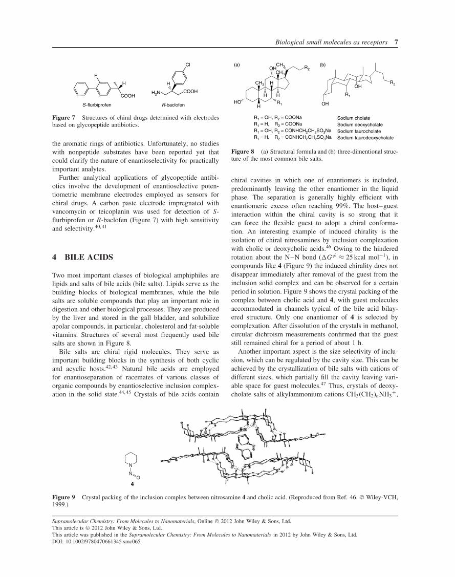
Two most important classes of biological amphiphiles arelipids and salts of bile acids (bile salts). Lipids serve as thebuilding blocks of biological membranes, while the bilesalts are soluble compounds that play an important role indigestion and other biological processes. They are producedby the liver and stored in the gall bladder, and solubilizeapolar compounds, in particular, cholesterol and fat-solublevitamins. Structures of several most frequently used bilesalts are shown in Figure 8.
Bile salts are chiral rigid molecules. They serve asimportant building blocks in the synthesis of both cyclicand acyclic hosts.42, 43 Natural bile acids are employedfor enantioseparation of racemates of various classes oforganic compounds by enantioselective inclusion complex-ation in the solid state.44, 45 Crystals of bile acids contain
CH3
CH3
CH3
H
H
HO R1
H H
R2OH
R1
OHR2
OH
(a) (b)
R1 = OH, R2 = COONa Sodium cholateR1 = H, R2 = COONa Sodium deoxycholateR1 = OH, R2 = CONHCH2CH2SO3Na Sodium taurocholateR1 = H, R2 = CONHCH2CH2SO3Na Sodium taurodeoxycholate
Figure 8 (a) Structural formula and (b) three-dimentional struc-ture of the most common bile salts.
chiral cavities in which one of enantiomers is included,predominantly leaving the other enantiomer in the liquidphase. The separation is generally highly efficient withenantiomeric excess often reaching 99%. The host–guestinteraction within the chiral cavity is so strong that itcan force the flexible guest to adopt a chiral conforma-tion. An interesting example of induced chirality is theisolation of chiral nitrosamines by inclusion complexationwith cholic or deoxycholic acids.46 Owing to the hinderedrotation about the N–N bond (�G �= ≈ 25 kcal mol−1), incompounds like 4 (Figure 9) the induced chirality does notdisappear immediately after removal of the guest from theinclusion solid complex and can be observed for a certainperiod in solution. Figure 9 shows the crystal packing of thecomplex between cholic acid and 4, with guest moleculesaccommodated in channels typical of the bile acid bilay-ered structure. Only one enantiomer of 4 is selected bycomplexation. After dissolution of the crystals in methanol,circular dichroism measurements confirmed that the gueststill remained chiral for a period of about 1 h.
Another important aspect is the size selectivity of inclu-sion, which can be regulated by the cavity size. This can beachieved by the crystallization of bile salts with cations ofdifferent sizes, which partially fill the cavity leaving vari-able space for guest molecules.47 Thus, crystals of deoxy-cholate salts of alkylammonium cations CH3(CH2)nNH3
+,
N
NO
4
Figure 9 Crystal packing of the inclusion complex between nitrosamine 4 and cholic acid. (Reproduced from Ref. 46. Wiley-VCH,1999.)
Supramolecular Chemistry: From Molecules to Nanomaterials, Online 2012 John Wiley & Sons, Ltd.This article is 2012 John Wiley & Sons, Ltd.This article was published in the Supramolecular Chemistry: From Molecules to Nanomaterials in 2012 by John Wiley & Sons, Ltd.DOI: 10.1002/9780470661345.smc065
8 Molecular recognition
Host cavity
Deoxycholate anion Ammonium cation
Hydrophilic layer
Lipophiliclayer
Figure 10 Crystal structure of an inclusion complex of n-propylammonium deoxycholate with 2-propanol (1 : 1). Hydrogenatoms are omitted. Empty, filled, and gray circles representcarbon, nitrogen, and oxygen atoms, respectively. (Reproducedfrom Ref. 47. American Chemical Society, 1998.)
where n = 0–4, contain cavities of progressively reducedsize on increase in number n of methylene groups in theammonium cation. To test the size selectivity, a seriesof aliphatic alcohols from methanol to isomeric butanolswere employed as guests. Figure 10 shows the structureof n-propylammonium salt with 2-propanol as the guest.The host cavity indeed is partially occupied by the cationhydrocarbon chain, which, however, leaves enough space toaccommodate alcohols of different sizes from methanol totert-butanol. Crystals with smaller methylammonium andethylammonium cations possessing larger cavities do notform inclusion complexes with small alcohols (methanoland ethanol), but form 1 : 1 complexes with butanols,which better fit larger cavities. Crystals with the largern-butylammonium cation still bind isomeric butanols, butwith stoichiometry less than 1 : 1, and crystals withn-pentylammonium cation do not form inclusion com-pounds at all since the cavity is completely filled with thelong n-pentyl group.
Natural bile salts are employed for enantiodiscrimina-tion in micellar electrokinetic chromatography (MEKC).37
The principle of separation is the enantioselective inclu-sion of analytes in micellar aggregates of bile salts, whichare formed in the concentration range 1–10 mM. Severalstudies of host properties of bile salt micelles by using spec-troscopic probes were reported, which allowed the charac-terization of sites of guest localizations inside micelles.48–50
Owing to the rigidity of the hydrophobic moieties of bilesalts, their micellar properties are significantly differentfrom those of common surfactants possessing long, flexiblehydrocarbon chains.51 Several discrete types of premicellarand micellar aggregates are formed in solution at increasedbile salt concentration, and the aggregation is driven notonly by hydrophobic interactions but also by hydrogenbonding.
5 RIFAMYCINS
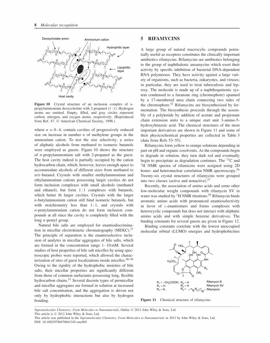
A large group of natural macrocylic compounds poten-tially useful as receptors constitutes the clinically importantantibiotics rifamycins. Rifamycins are antibiotics belongingto the group of naphthalenic ansamycins which exert theiractivity by specific inhibition of bacterial DNA-dependentRNA polymerase. They have activity against a large vari-ety of organisms, such as bacteria, eukaryotes, and viruses;in particular, they are used to treat tuberculosis and lep-rosy. The molecule is made up of a naphthoquinonic sys-tem condensed to a furanone ring (chromophore) spannedby a 17-membered ansa chain connecting two sides ofthe chromophore.52 Rifamycins are biosynthesized by fer-mentation. The biosynthesis proceeds through the assem-bly of a polyketide by addition of acetate and propionatechain extension units to a unique start unit 3-amino-5-hydroxybenzoic acid. The chemical structures of the mostimportant derivatives are shown in Figure 11 and some oftheir physicochemical properties are collected in Table 3(data from Refs 53–55).
Rifamycins form yellow to orange solutions depending inpart on pH and organic cosolvents. As the compounds beginto degrade in solution, they turn dark red and eventuallybegin to precipitate as degradation continues. The 13C and1H NMR spectra of rifamicins were assigned using 2Dhomo- and heteronuclear correlation NMR spectroscopy.56
Twenty-six crystal structures of rifamycins were groupedinto two classes (active and nonactive).57
Recently, the association of amino acids and some otherlow-molecular weight compounds with rifamycin SV inwater was studied by 1H NMR titrations.58 Rifamycin bindsaromatic amino acids with pronounced enantioselectivityin favor of L-enantiomers and forms complexes withheterocyclic compounds but does not interact with aliphaticamino acids and with simple benzene derivatives. Thebinding constants for several guests are given in Figure 12.
Binding constants correlate with the lowest unoccupiedmolecular orbital (LUMO) energies and hydrophobicities
O R2
OR1
OH
NH
OH
O
OOH
HO
O
H3CO
O
O
NN
N
R1 = −CH2COOH, Rifamycin BR1 = H, R2 = H Rifamycin SVR1 = H, R2 = Rifampicin
R2 = H
Figure 11 Chemical structure of rifamycins.
Supramolecular Chemistry: From Molecules to Nanomaterials, Online 2012 John Wiley & Sons, Ltd.This article is 2012 John Wiley & Sons, Ltd.This article was published in the Supramolecular Chemistry: From Molecules to Nanomaterials in 2012 by John Wiley & Sons, Ltd.DOI: 10.1002/9780470661345.smc065
Biological small molecules as receptors 9
Table 3 Physicochemical properties of some rifamycins.
Characteristics Rifamycin B Rifamycin SV Rifampicin
Chemical formula C38H49O14N C37H46NO12Na as sodiumsalt
C43H58N4O12
Formula weight (Da) 755.8 719.75 822.94Solubility Light alcohols and acetone;
slightly soluble in waterEther; water in basic medium CHCl3, water in acid or
basic mediaStereogenic centers 9 9 9Hydroxyl groups 4 5 5Aromatic rings 2 2 2Ionogenic groups and pKa’s
valuespKa1 = 2.8 (COOH),
pKa2 = 6.7 (Ph-OH)pKa = 1.8 pKa1 = 1.7 (Ph-OH),
pKa2 = 7.9 (piperazine)UV–vis spectrum Maxima at approximately
220, 304, 425 nmMaxima at approximately
220, 304, 425 nmMaxima at 237, 255, 334,
475 nm
NH
NH3+
COO−
NN
NH2
O
N
NH2
O
+NH2+
KL = 34, KD = 18 74 M−1 10 M−1 7 M−1 150 M−1
Figure 12 The binding constants (M−1) for several guests to rifamycin SV in water at pH 9.0.
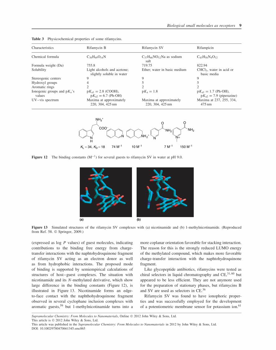
(a) (b)(a) (b)
Figure 13 Simulated structures of the rifamycin SV complexes with (a) nicotinamide and (b) 1-methylnicotinamide. (Reproducedfrom Ref. 58. Springer, 2009.)
(expressed as log P values) of guest molecules, indicatingcontributions to the binding free energy from charge-transfer interactions with the naphtohydroquinone fragmentof rifamycin SV acting as an electron donor as wellas from hydrophobic interactions. The proposed modeof binding is supported by semiempirical calculations ofstructures of host–guest complexes. The situation withnicotinamide and its N-methylated derivative, which showlarge difference in the binding constants (Figure 12), isillustrated in Figure 13. Nicotinamide forms an edge-to-face contact with the naphthohydroquinone fragmentobserved in several cyclophane inclusion complexes witharomatic guests,59 but 1-methylnicotinamide turns into a
more coplanar orientation favorable for stacking interaction.The reason for this is the strongly reduced LUMO energyof the methylated compound, which makes more favorablecharge-transfer interaction with the naphtohydroquinonefragment.
Like glycopeptide antibiotics, rifamycins were tested aschiral selectors in liquid chromatography and CE,31, 60 butappeared to be less efficient. They are not anymore usedfor the preparation of stationary phases, but rifamycins Band SV are used as selectors in CE.36
Rifamycin SV was found to have ionophoric proper-ties and was successfully employed for the developmentof a potentiometric membrane sensor for potassium ion.61
Supramolecular Chemistry: From Molecules to Nanomaterials, Online 2012 John Wiley & Sons, Ltd.This article is 2012 John Wiley & Sons, Ltd.This article was published in the Supramolecular Chemistry: From Molecules to Nanomaterials in 2012 by John Wiley & Sons, Ltd.DOI: 10.1002/9780470661345.smc065
10 Molecular recognition
The potentiometric selectivity to alkali and alkaline-earth cations is in the order K+ > Rb+ > Cs+ > Na+ >
NH4+ > Ba2+ > Mg2+ > Ca2+ > Sr2+ >Li+ and the inter-
ference from transition-metal ions is negligible. No bindingconstants were reported.
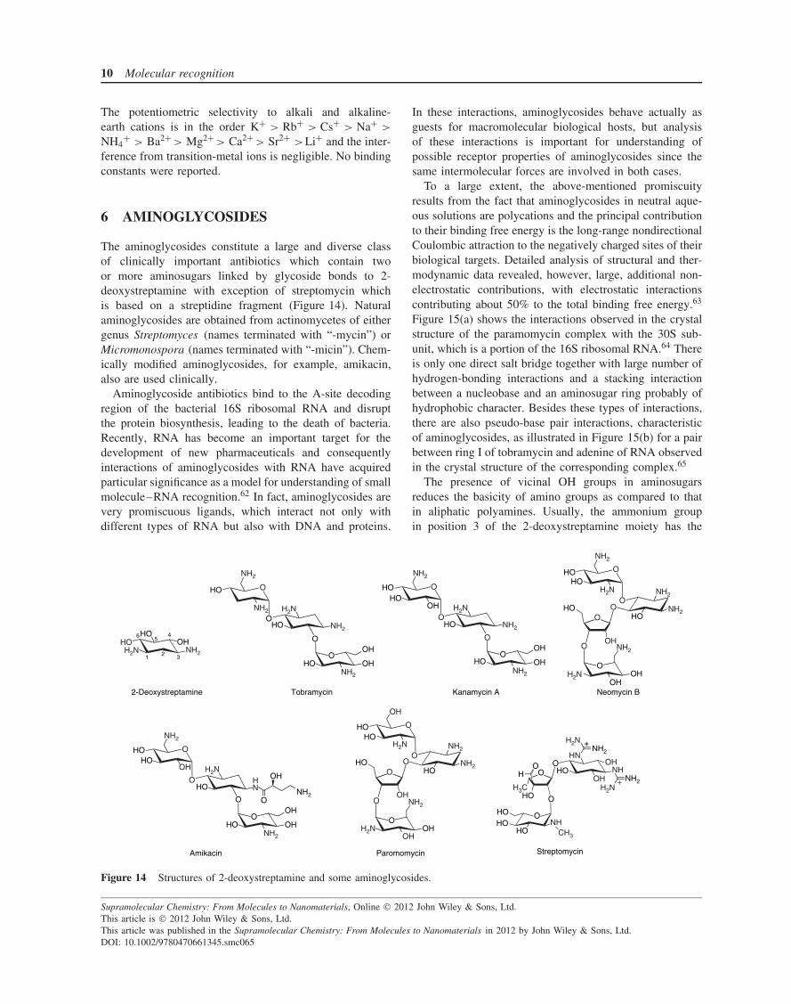
6 AMINOGLYCOSIDES
The aminoglycosides constitute a large and diverse classof clinically important antibiotics which contain twoor more aminosugars linked by glycoside bonds to 2-deoxystreptamine with exception of streptomycin whichis based on a streptidine fragment (Figure 14). Naturalaminoglycosides are obtained from actinomycetes of eithergenus Streptomyces (names terminated with “-mycin”) orMicromonospora (names terminated with “-micin”). Chem-ically modified aminoglycosides, for example, amikacin,also are used clinically.
Aminoglycoside antibiotics bind to the A-site decodingregion of the bacterial 16S ribosomal RNA and disruptthe protein biosynthesis, leading to the death of bacteria.Recently, RNA has become an important target for thedevelopment of new pharmaceuticals and consequentlyinteractions of aminoglycosides with RNA have acquiredparticular significance as a model for understanding of smallmolecule–RNA recognition.62 In fact, aminoglycosides arevery promiscuous ligands, which interact not only withdifferent types of RNA but also with DNA and proteins.
In these interactions, aminoglycosides behave actually asguests for macromolecular biological hosts, but analysisof these interactions is important for understanding ofpossible receptor properties of aminoglycosides since thesame intermolecular forces are involved in both cases.
To a large extent, the above-mentioned promiscuityresults from the fact that aminoglycosides in neutral aque-ous solutions are polycations and the principal contributionto their binding free energy is the long-range nondirectionalCoulombic attraction to the negatively charged sites of theirbiological targets. Detailed analysis of structural and ther-modynamic data revealed, however, large, additional non-electrostatic contributions, with electrostatic interactionscontributing about 50% to the total binding free energy.63
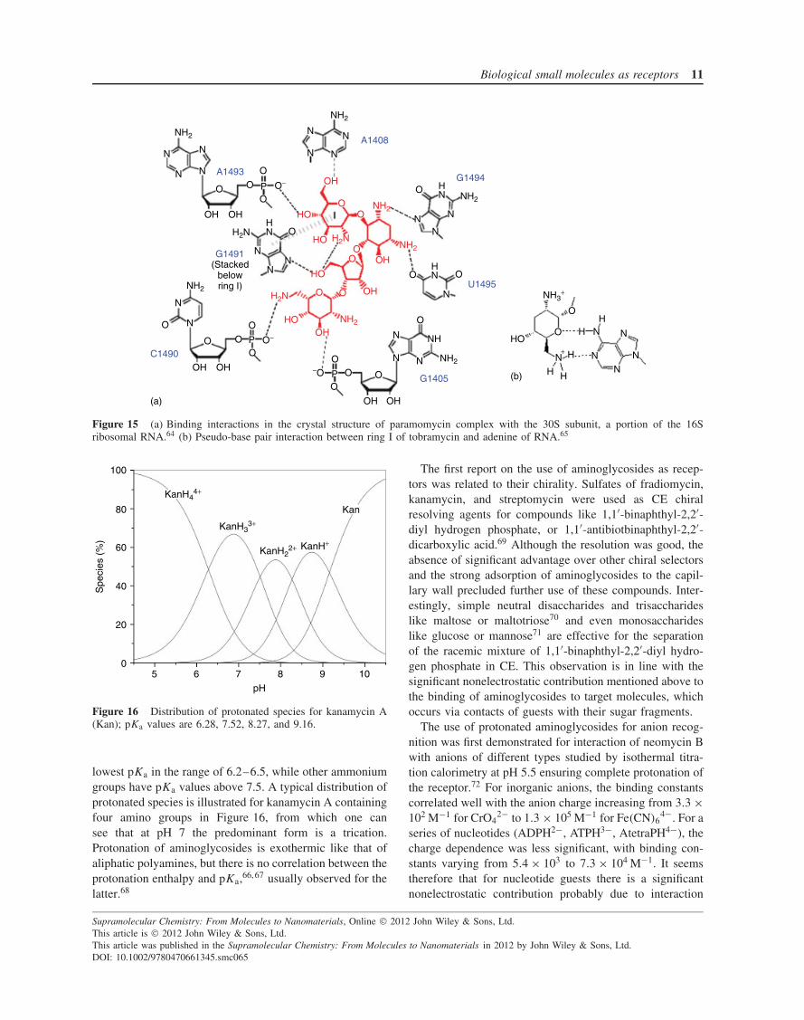
Figure 15(a) shows the interactions observed in the crystalstructure of the paramomycin complex with the 30S sub-unit, which is a portion of the 16S ribosomal RNA.64 Thereis only one direct salt bridge together with large number ofhydrogen-bonding interactions and a stacking interactionbetween a nucleobase and an aminosugar ring probably ofhydrophobic character. Besides these types of interactions,there are also pseudo-base pair interactions, characteristicof aminoglycosides, as illustrated in Figure 15(b) for a pairbetween ring I of tobramycin and adenine of RNA observedin the crystal structure of the corresponding complex.65
The presence of vicinal OH groups in aminosugarsreduces the basicity of amino groups as compared to thatin aliphatic polyamines. Usually, the ammonium groupin position 3 of the 2-deoxystreptamine moiety has the
O
NH2
NH2
NH2
HO
O
O
NH2
OH
HOHO
H2N
H2N
O
O
O
HO
NH2
HO OHHO
HONH2
OH
OH
O
O
HONH2
OH
OH
O
O
O
NH2
NH2
H2N
HOHO
OH
O
OO
NH2
H2N
H3C H2N
H2N
CH3
NH2
NH2
NH2
HOHO
HO
OH
OHHN
NHHOH2N
HO
OHO
NH2 H2N
OH
OH OOH
O
OHOH
6
12 3
45
Kanamycin ATobramycin2-Deoxystreptamine Neomycin B
NH2
H2N
OOH
O
OHOH
O HO
NHHO
HOHO
NH2
NH2
HO
HO
OO
HO
HN NH2
OH
O
NH2
NH2
O
OH2N
HOHO
HO O
O
OOO
H+
+
Amikacin Paromomycin
OH
Streptomycin
Figure 14 Structures of 2-deoxystreptamine and some aminoglycosides.
Supramolecular Chemistry: From Molecules to Nanomaterials, Online 2012 John Wiley & Sons, Ltd.This article is 2012 John Wiley & Sons, Ltd.This article was published in the Supramolecular Chemistry: From Molecules to Nanomaterials in 2012 by John Wiley & Sons, Ltd.DOI: 10.1002/9780470661345.smc065
Biological small molecules as receptors 11
NH2
NH2
NH2
NH2
NH2
H2N
N
N N
N
N
N
N
N
OO
O
OO−
O−
O
OH OH
P
HN
N
O
N
N
N
N
O
O O
O
O
OH OH
P
−O O O
O
O O
O
OP
OH OH
N
N
N
N
NH
HN
NN
N
HN
(Stackedbelowring I)
A1493
A1408
G1494
G1491
C1490
G1405
U1495
OH
HO
HO
HO
HO
OH
OH
OH
OO
OO
OO
H2N
NH2
NH2
NH2
H2N NH3+
O
OHO
H
HH
HH
N
N
N
N
NN+
(a)
(b)
Figure 15 (a) Binding interactions in the crystal structure of paramomycin complex with the 30S subunit, a portion of the 16Sribosomal RNA.64 (b) Pseudo-base pair interaction between ring I of tobramycin and adenine of RNA.65
100
80
60
40
20
05 6 7 8 9 10
pH
Spe
cies
(%
)
KanH44+
KanH33+
KanH22+ KanH+
Kan
Figure 16 Distribution of protonated species for kanamycin A(Kan); pKa values are 6.28, 7.52, 8.27, and 9.16.
lowest pKa in the range of 6.2–6.5, while other ammoniumgroups have pKa values above 7.5. A typical distribution ofprotonated species is illustrated for kanamycin A containingfour amino groups in Figure 16, from which one cansee that at pH 7 the predominant form is a trication.Protonation of aminoglycosides is exothermic like that ofaliphatic polyamines, but there is no correlation between theprotonation enthalpy and pKa,66, 67 usually observed for thelatter.68
The first report on the use of aminoglycosides as recep-tors was related to their chirality. Sulfates of fradiomycin,kanamycin, and streptomycin were used as CE chiralresolving agents for compounds like 1,1′-binaphthyl-2,2′-diyl hydrogen phosphate, or 1,1′-antibiotbinaphthyl-2,2′-dicarboxylic acid.69 Although the resolution was good, theabsence of significant advantage over other chiral selectorsand the strong adsorption of aminoglycosides to the capil-lary wall precluded further use of these compounds. Inter-estingly, simple neutral disaccharides and trisaccharideslike maltose or maltotriose70 and even monosaccharideslike glucose or mannose71 are effective for the separationof the racemic mixture of 1,1′-binaphthyl-2,2′-diyl hydro-gen phosphate in CE. This observation is in line with thesignificant nonelectrostatic contribution mentioned above tothe binding of aminoglycosides to target molecules, whichoccurs via contacts of guests with their sugar fragments.
The use of protonated aminoglycosides for anion recog-nition was first demonstrated for interaction of neomycin Bwith anions of different types studied by isothermal titra-tion calorimetry at pH 5.5 ensuring complete protonation ofthe receptor.72 For inorganic anions, the binding constantscorrelated well with the anion charge increasing from 3.3 ×10² M−1 for CrO4
2− to 1.3 × 10⁵ M−1 for Fe(CN)64−. For a
series of nucleotides (ADPH2−, ATPH3−, AtetraPH4−), thecharge dependence was less significant, with binding con-stants varying from 5.4 × 10³ to 7.3 × 10⁴ M−1. It seemstherefore that for nucleotide guests there is a significantnonelectrostatic contribution probably due to interaction
Supramolecular Chemistry: From Molecules to Nanomaterials, Online 2012 John Wiley & Sons, Ltd.This article is 2012 John Wiley & Sons, Ltd.This article was published in the Supramolecular Chemistry: From Molecules to Nanomaterials in 2012 by John Wiley & Sons, Ltd.DOI: 10.1002/9780470661345.smc065
12 Molecular recognition
Table 4 Logarithms of association constants of nucleotides and nucleosides with differently protonated forms of kanamycin A (Kan)and tetrandrine derivatives at 25 ◦C.
Receptor AMP2− ADP3− ATP4− CTP4− GTP4− Ado Guo References
Kan 2.45 3.12 74Kan H+ 1.75 2.35 1.90 1.86 2.60 2.28 3.04Kan H2
2+ 1.86 2.51 2.44 2.34 3.16 2.37 3.41Kan H3
3+ 2.20 3.02 3.20 3.32 3.76 2.46 3.49Kan H4
4+ 2.47 3.40 4.15 4.26 5.05 2.29 3.3510 1.68 1.60 2.04 7511 2.69 2.93 3.73 About 3 About 3 7612 1.89 2.04 3.86
with the nucleobase. In all cases, the binding is enthalpydriven, indicating that the interaction is not a simple ionpairing which normally is entropy-driven.73
A more systematic study of nucleotide recognition byprotonated forms of kanamycin A, streptomycin, andamikacin demonstrated that guest binding constants aresimilar to those observed with aliphatic macrocyclicpolyamines of similar charge although aminoglycosidesare linear polyamines and that, more importantly, thereis a noticeable selectivity to the type of the nucleobase(Table 4).74 As follows from Table 4, kanamycin bindswith increased affinity AMP2−, ADP2−, and ATP4− dueto the charge effect and in a series of nucleotide triphos-phates binds GTP4− approximately one order of magni-tude stronger than ATP4− and CTP4−. In line with this,kanamycin binds neutral guanosine one order of magnitudestronger than adenosine. Binding of nucleosides is inde-pendent of the receptor charge, which is quite significant,indicating large nonelectrostatic contribution to nucleotiderecognition. The binding of ATP4− to all protonated formsof kanamycin is exothermic.
Aminoglycosides are expected to act as ligands for metalcoordination, but only Cu(II) complexes were studied indetail.77 With kanamycin A as a lignad, a set of monuclearcomplexes with differently protonated/deprotonated formsranging from CuH2L to CuH−2L were found with stabilityconstant log β11 = 8.83 for the neutral form.78 Coppercomplexes of aminoglycosides are active catalysts forseveral hydrolytic and oxidative reactions.77
7 CYCLIC POLYPEPTIDE ANTIBIOTICS
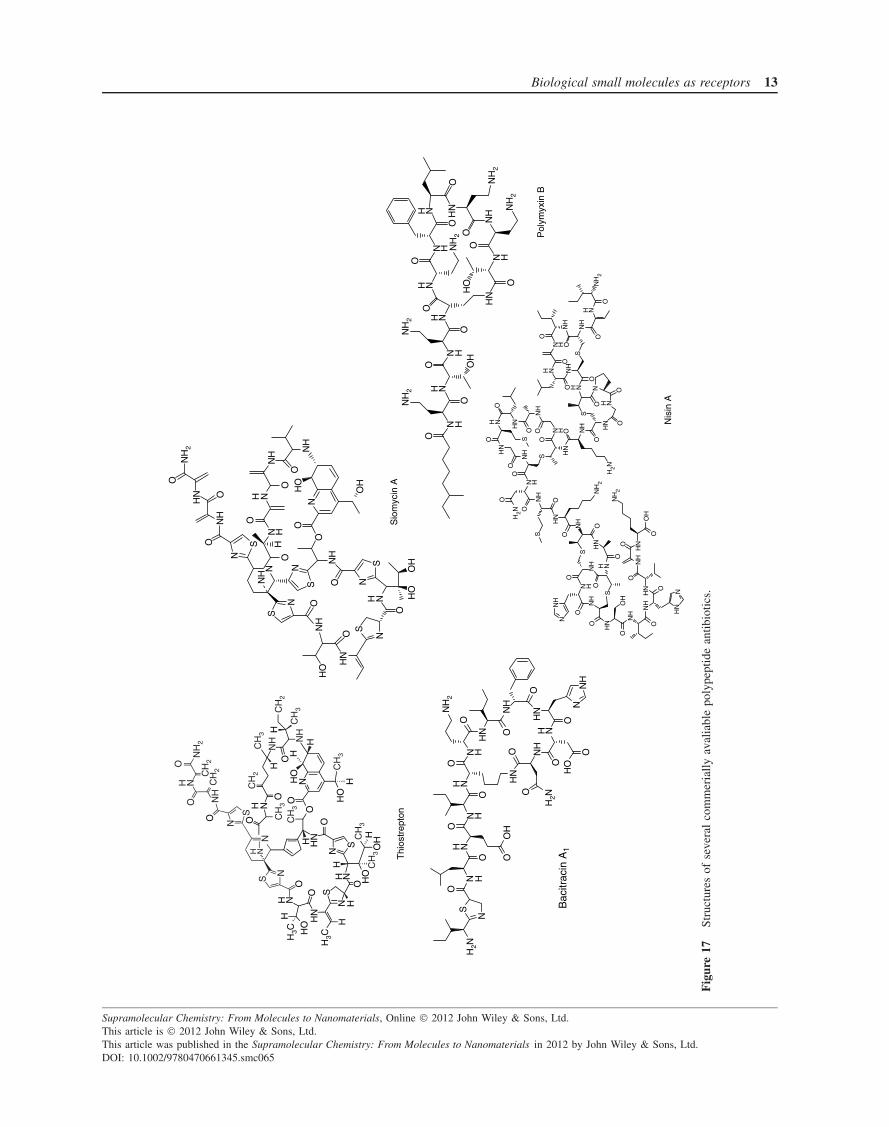
Peptide antibiotics operate through numerous differentmechanisms.79 They are large molecules approaching pro-teins in their structural complexity and often have cyclicstructures (Figure 17), making them promising compoundsas receptors for small guests. Nevertheless, the poten-tial of these compounds as receptors remains practicallyunexplored.
Thiostrepton, a thiopeptide antibiotic (a group of natu-rally occurring, sulfur-containing, highly modified, macro-cyclic peptides),80 was applied for the preparation of chiralstationary phases for liquid chromatography.31 Its perfor-mance was later improved by chemical modification ofthe antibiotic.81 Thiostrepton is insoluble in water, but itwas studied as a hydrogen-bonding receptor for anions inorganic solvents.82 It forms both 1 : 1 and 1 : 2 complexeswith anions with K2 > K1 for F− and K2 � K1 for allother anions studied. Stability constants of 1 : 1 complexesrange from 102 to 5 × 10³ M−1. In DMSO (dimethyl sul-foxide), they follow the order AcO− ≈ F− � Cl−, Br−,HSO4
−, H2PO4−, which roughly correlates with basic-
ities of anions, but in CHCl3 they follow a differentorder: Cl− ≈ HSO4
− > F− ≈ AcO− > Br− > H2PO4−. A
reason for this change is the stronger solvation of morebasic anions by chloroform, which acts as a weak protondonor solvent.
Bacitracin A1 is a lariat-type macrocycle (Figure 17) well“designed” for complexation of metal ions. Indeed, it needsthe presence of a metal ion for the antibiotic activity, withmaximum activity observed with Zn2+.83 Table 5 showsthe pKa values of ionogenic groups of bacitracin andlogarithms of binding constants of some divalent metal ionsto differently protonated forms of the peptide.84 Affinity ofthe neutral form, predominating at pH 7, is not very largeand surprisingly does not follow the Irwing–Williams series(Ni2+ < Cu2+ > Zn2+).
The polymyxins, polymyxin B (Figure 17), and colistin(contains D-Leu instead of D-Phe in the macrocycle), areused as last-line antibiotics to treat infections caused byGram-negative bacteria that are resistant to essentiallyall other currently available antibiotics.85 They bind tothe lipid A (a β-1′-6-linked D-glucosamine disaccharidephosphorylated at the 1- and 4′-positions containing sixlong fatty acyl chains attached to OH and NH2 groupsof the disaccharide) through a combination of electrostaticand hydrophobic interactions disrupting the bacterial outermembrane. Owing to the high total positive charge (5+)
Supramolecular Chemistry: From Molecules to Nanomaterials, Online 2012 John Wiley & Sons, Ltd.This article is 2012 John Wiley & Sons, Ltd.This article was published in the Supramolecular Chemistry: From Molecules to Nanomaterials in 2012 by John Wiley & Sons, Ltd.DOI: 10.1002/9780470661345.smc065
Biological small molecules as receptors 13
OH
Sio
myc
in A
Thi
ostr
epto
n
Pol
ymyx
in B
Nis
in A
NO
O
OO
OO
OO
O
O
SS
O
O
O
O
OO
OO
OO
S
S
N
N
HN
HN
HN
HN
NH
NH
NH
NH
NH
NH
2
H2N
NH
H
N
N
NH
H
N H
N
NH
NH
H
H
O
OO
OH
O
HO
OH
OS
S
S
S
S
OO
OH
O
O
OO
ON
H2
NH
NH
NH
NH
N
NN
H
HN
NH
NH
H
N
NN
HN
H
N
N
HN
CH
2C
H2
CH
2C
H3
CH
2
CH
3
CH
3
CH
3
CH
3
CH
3
CH
3
NH
2
OH N
O NH
N
S
N
NHS
N
O
OO
O
O
NHH HN
HO
H3C
H3C
H
H
NN
H NHH
H
H
H
H
H
OO
HO
HO
O
O
ON
HN
NN
HH
HH
N
S
S OH
HO
N
N
NS
S
S
OO
O
O
O
O
O
O
O
OO
O
OO
O
HN
HN
HN
HNH
2N
NH
2
NH
2
HN
HNO
H
OH
H
NHO
NH NH
NH
NH
NH
NH
NH
NH
O
O OO
HH
N
H2N
NH
NH
NH
OS
NN
H2N
H
H
H
H N
H N
OO
O
O
O
O
N
O
O
O
O
HO
NN
HH
N
HN
N
NH
2
Bac
itrac
in A
1
N H
H NN H
H N
O
NH
2
OO
H
O
NH
2
O
HN
N H
NH
O
HO
O
NH
2
NH
2
HN
O
OH N
N H
H N
O
O
ON
H2
Fig
ure
17St
ruct
ures
ofse
vera
lco
mm
eria
llyav
alia
ble
poly
pept
ide
antib
iotic
s.
Supramolecular Chemistry: From Molecules to Nanomaterials, Online 2012 John Wiley & Sons, Ltd.This article is 2012 John Wiley & Sons, Ltd.This article was published in the Supramolecular Chemistry: From Molecules to Nanomaterials in 2012 by John Wiley & Sons, Ltd.DOI: 10.1002/9780470661345.smc065
14 Molecular recognition
Table 5 Proton dissociation (pKa) and metal binding (log K)constants for bacitracin A1.84
Group pKa Peptide log Kspecies
Ni2+ Cu2+ Zn2+
D-Asp (macrocycle) 3.6 P0 4.36 3.69 3.18D-Glu (lateral chain) 4.4 P1− 7.08 6.30 4.98His (macrocycle) 6.4 P2− 9.66 9.08 <6.9Ile (N-terminal) 7.6D-Orn (macrocycle) 9.7
and specific arrangement of ammonium groups, it may beexpected to act as a receptor for various anionic species.
A lantibiotic nisin (Figure 17) widely used for foodpreservation targets the cell wall biosynthetic intermediatelipid II.86 The structure of the complex of nisin A and a lipidII analog solved by NMR studies in DMSO87 demonstratestrapping of the pyrophosphate moiety of the lipid betweentwo rings (first and second rings from the Ile terminus ofthe molecule) through five H-bonds to amide NH groups. Itseems probable that nisin may act as a hydrogen-bondingreceptor for free pyrophosphate as well as for some otheranions.
8 ALKALOIDS
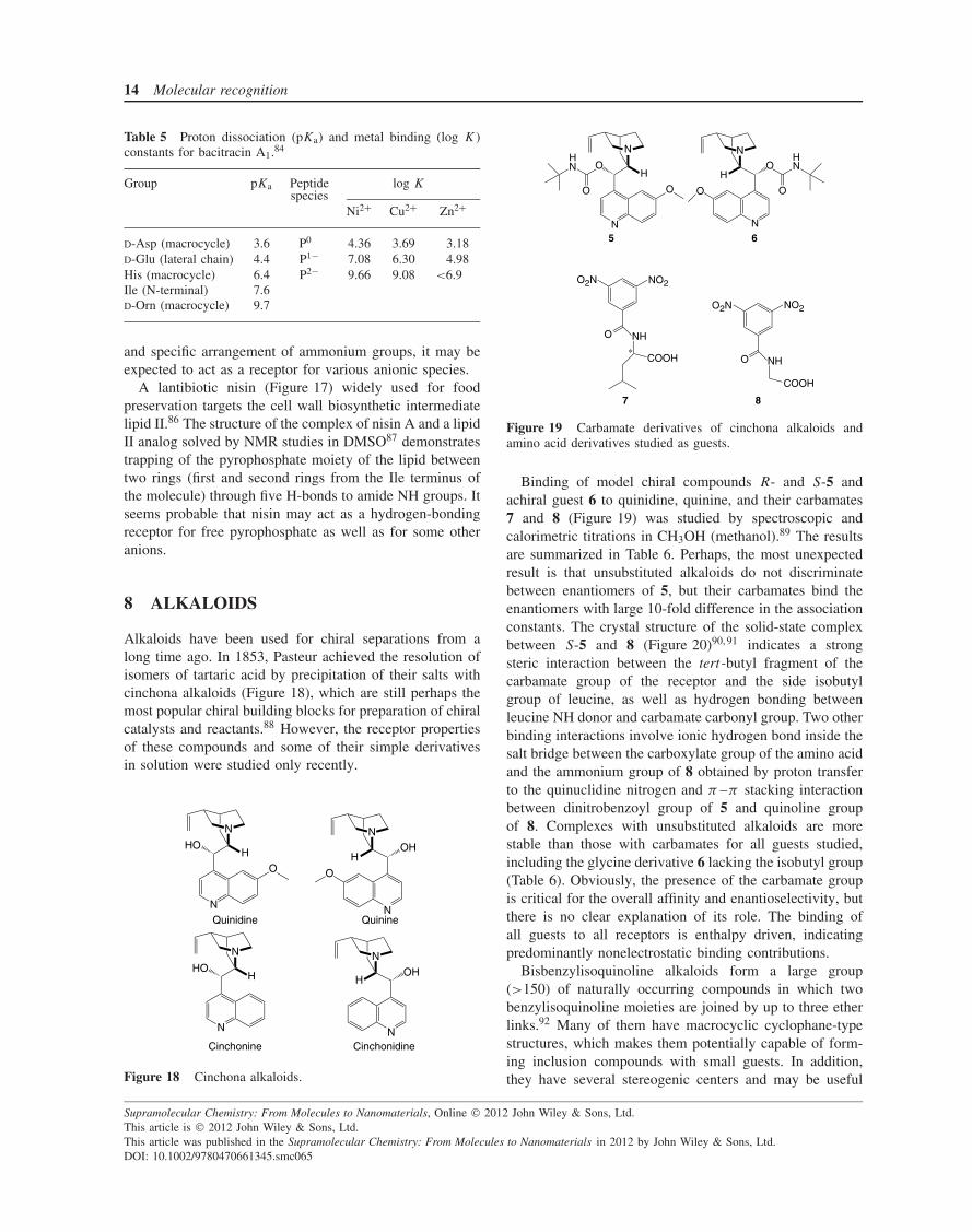
Alkaloids have been used for chiral separations from along time ago. In 1853, Pasteur achieved the resolution ofisomers of tartaric acid by precipitation of their salts withcinchona alkaloids (Figure 18), which are still perhaps themost popular chiral building blocks for preparation of chiralcatalysts and reactants.88 However, the receptor propertiesof these compounds and some of their simple derivativesin solution were studied only recently.
N
O
HO
N
H
QuinidineN
N
Quinine
O
OHH
N
HO
N
H
Cinchonine
N
CinchonidineN
OHH
Figure 18 Cinchona alkaloids.
NO2O2N
NH
COOH
O
NO2O2N
NH
COOH
O
N
O
O
N
H
HN
N
N
O
OH
O
HN
O
5 6
7 8
Figure 19 Carbamate derivatives of cinchona alkaloids andamino acid derivatives studied as guests.
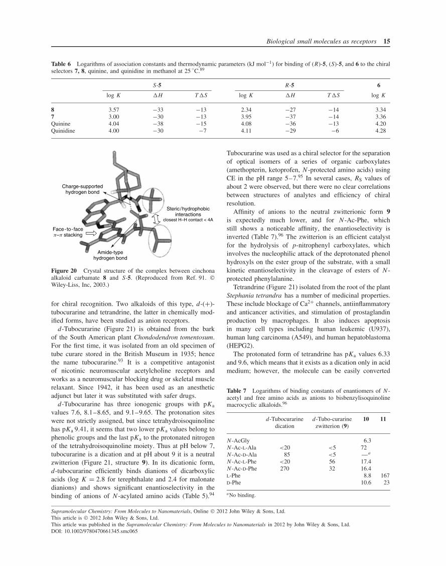
Binding of model chiral compounds R- and S-5 andachiral guest 6 to quinidine, quinine, and their carbamates7 and 8 (Figure 19) was studied by spectroscopic andcalorimetric titrations in CH3OH (methanol).89 The resultsare summarized in Table 6. Perhaps, the most unexpectedresult is that unsubstituted alkaloids do not discriminatebetween enantiomers of 5, but their carbamates bind theenantiomers with large 10-fold difference in the associationconstants. The crystal structure of the solid-state complexbetween S-5 and 8 (Figure 20)90, 91 indicates a strongsteric interaction between the tert-butyl fragment of thecarbamate group of the receptor and the side isobutylgroup of leucine, as well as hydrogen bonding betweenleucine NH donor and carbamate carbonyl group. Two otherbinding interactions involve ionic hydrogen bond inside thesalt bridge between the carboxylate group of the amino acidand the ammonium group of 8 obtained by proton transferto the quinuclidine nitrogen and π –π stacking interactionbetween dinitrobenzoyl group of 5 and quinoline groupof 8. Complexes with unsubstituted alkaloids are morestable than those with carbamates for all guests studied,including the glycine derivative 6 lacking the isobutyl group(Table 6). Obviously, the presence of the carbamate groupis critical for the overall affinity and enantioselectivity, butthere is no clear explanation of its role. The binding ofall guests to all receptors is enthalpy driven, indicatingpredominantly nonelectrostatic binding contributions.
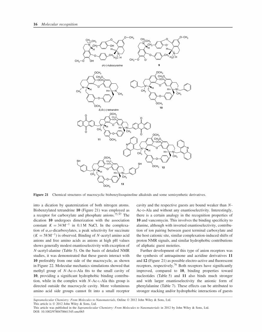
Bisbenzylisoquinoline alkaloids form a large group(>150) of naturally occurring compounds in which twobenzylisoquinoline moieties are joined by up to three etherlinks.92 Many of them have macrocyclic cyclophane-typestructures, which makes them potentially capable of form-ing inclusion compounds with small guests. In addition,they have several stereogenic centers and may be useful
Supramolecular Chemistry: From Molecules to Nanomaterials, Online 2012 John Wiley & Sons, Ltd.This article is 2012 John Wiley & Sons, Ltd.This article was published in the Supramolecular Chemistry: From Molecules to Nanomaterials in 2012 by John Wiley & Sons, Ltd.DOI: 10.1002/9780470661345.smc065
Biological small molecules as receptors 15
Table 6 Logarithms of association constants and thermodynamic parameters (kJ mol−1) for binding of (R)-5, (S)-5, and 6 to the chiralselectors 7, 8, quinine, and quinidine in methanol at 25 ◦C.89
S-5 R-5 6
log K �H T �S log K �H T �S log K
8 3.57 −33 −13 2.34 −27 −14 3.347 3.00 −30 −13 3.95 −37 −14 3.36Quinine 4.04 −38 −15 4.08 −36 −13 4.20Quinidine 4.00 −30 −7 4.11 −29 −6 4.28
Charge-supportedhydrogen bond
Face-to-facep–p stacking
Amide-typehydrogen bond
Steric/hydrophobicinteractions
closest H–H contact < 4A
Figure 20 Crystal structure of the complex between cinchonaalkaloid carbamate 8 and S-5. (Reproduced from Ref. 91. Wiley-Liss, Inc, 2003.)
for chiral recognition. Two alkaloids of this type, d-(+)-tubocurarine and tetrandrine, the latter in chemically mod-ified forms, have been studied as anion receptors.
d-Tubocurarine (Figure 21) is obtained from the barkof the South American plant Chondodendron tomentosum.For the first time, it was isolated from an old specimen oftube curare stored in the British Museum in 1935; hencethe name tubocurarine.93 It is a competitive antagonistof nicotinic neuromuscular acetylcholine receptors andworks as a neuromuscular blocking drug or skeletal musclerelaxant. Since 1942, it has been used as an anestheticadjunct but later it was substituted with safer drugs.
d-Tubocurarine has three ionogenic groups with pKa
values 7.6, 8.1–8.65, and 9.1–9.65. The protonation siteswere not strictly assigned, but since tetrahydroisoquinolinehas pKa 9.41, it seems that two lower pKa values belong tophenolic groups and the last pKa to the protonated nitrogenof the tetrahydroisoquinoline moiety. Thus at pH below 7,tubocurarine is a dication and at pH about 9 it is a neutralzwitterion (Figure 21, structure 9). In its dicationic form,d-tubocurarine efficiently binds dianions of dicarboxylicacids (log K = 2.8 for terephthalate and 2.4 for malonatedianions) and shows significant enantioselectivity in thebinding of anions of N-acylated amino acids (Table 5).94
Tubocurarine was used as a chiral selector for the separationof optical isomers of a series of organic carboxylates(amethopterin, ketoprofen, N-protected amino acids) usingCE in the pH range 5–7.95 In several cases, RS values ofabout 2 were observed, but there were no clear correlationsbetween structures of analytes and efficiency of chiralresolution.
Affinity of anions to the neutral zwitterionic form 9is expectedly much lower, and for N-Ac-Phe, whichstill shows a noticeable affinity, the enantioselectivity isinverted (Table 7).96 The zwitterion is an efficient catalystfor the hydrolysis of p-nitrophenyl carboxylates, whichinvolves the nucleophilic attack of the deprotonated phenolhydroxyls on the ester group of the substrate, with a smallkinetic enantioselectivity in the cleavage of esters of N-protected phenylalanine.
Tetrandrine (Figure 21) isolated from the root of the plantStephania tetrandra has a number of medicinal properties.These include blockage of Ca2+ channels, antiinflammatoryand anticancer activities, and stimulation of prostaglandinproduction by macrophages. It also induces apoptosisin many cell types including human leukemic (U937),human lung carcinoma (A549), and human hepatoblastoma(HEPG2).
The protonated form of tetrandrine has pKa values 6.33and 9.6, which means that it exists as a dication only in acidmedium; however, the molecule can be easily converted
Table 7 Logarithms of binding constants of enantiomers of N-acetyl and free amino acids as anions to bisbenzylisoquinolinemacrocyclic alkaloids.96
d-Tubocurarine d-Tubo-curarine 10 11dication zwitterion (9)
N -AcGly 6.3N -Ac-L-Ala <20 <5 72N -Ac-D-Ala 85 <5 —a
N -Ac-L-Phe <20 56 17.4N -Ac-D-Phe 270 32 16.4L-Phe 8.8 167D-Phe 10.6 23
aNo binding.
Supramolecular Chemistry: From Molecules to Nanomaterials, Online 2012 John Wiley & Sons, Ltd.This article is 2012 John Wiley & Sons, Ltd.This article was published in the Supramolecular Chemistry: From Molecules to Nanomaterials in 2012 by John Wiley & Sons, Ltd.DOI: 10.1002/9780470661345.smc065
16 Molecular recognition
CH3 CH3 CH3
CH2
H
N
O OH
O
OH
CH3 CH3
H
O
N
HH3CCH3
CH2O
+
+
CH3
CH2H
O O−
O
O−
H
O
N
HH3CCH3
CH2O
+
+
N
N
CH3
OCH3
OCH3
O
N
O
CH3
CH3O
OCH3
H H
N+
CH3
OCH3
OCH3
O
N+O
CH3
CH3O
OCH3
H H
N+
CH3
OCH3
OCH3
O
N+O
CH3
CH3O
OCH3
H H
O
O
O
O
N+
CH3
OCH3
OCH3
O
N+O
CH3
CH3O
OCH3
H H
NN
N+
CH3
OCH3
OCH3
O
N+O
CH3
CH3O
OCH3
H H
SHSH
d-(+)-tubocurarine 9
10
11
12
13
S,S-(+)-tetrandrin
Figure 21 Chemical structures of macrocyclic bisbenzylisoquinoline alkaloids and some semisynthetic derivatives.
into a dication by quaternization of both nitrogen atoms.Bisbenzylated tetrandrine 10 (Figure 21) was employed asa receptor for carboxylate and phosphate anions.75, 97 Thedication 10 undergoes dimerization with the associationconstant K = 34 M−1 in 0.1 M NaCl. In the complexa-tion of α,ω-dicarboxylates, a peak selectivity for succinate(K = 58 M−1) is observed. Binding of N-acetyl amino acidanions and free amino acids as anions at high pH valuesshows generally modest enantioselectivity with exception ofN-acetyl-alanine (Table 5). On the basis of detailed NMRstudies, it was demonstrated that these guests interact with10 preferably from one side of the macrocycle, as shownin Figure 22. Molecular mechanics simulations showed thatmethyl group of N-Ac-D-Ala fits to the small cavity of10, providing a significant hydrophobic binding contribu-tion, while in the complex with N-Ac-L-Ala this group isdirected outside the macrocycle cavity. More voluminousamino acid side groups cannot fit into a small receptor
cavity and the respective guests are bound weaker than N-Ac-D-Ala and without any enantioselectivity. Interestingly,there is a certain analogy in the recognition properties of10 and vancomycin. This involves the binding specificity toalanine, although with inverted enantioselectivity, contribu-tion of ion pairing between guest terminal carboxylate andthe host cationic site, similar complexation-induced shifts ofproton NMR signals, and similar hydrophobic contributionsof aliphatic guest moieties.
Further development of this type of anion receptors wasthe synthesis of antraquinone and acridine derivatives 11and 12 (Figure 21) as possible electro-active and fluorescentreceptors, respectively.76 Both receptors have significantlyimproved, compared to 10, binding properties towardnucleotides (Table 5) and 11 also binds much strongerand with larger enantioselectivity the anionic form ofphenylalanine (Table 7). These effects can be attributed tostronger stacking and/or hydrophobic interactions of guests
Supramolecular Chemistry: From Molecules to Nanomaterials, Online 2012 John Wiley & Sons, Ltd.This article is 2012 John Wiley & Sons, Ltd.This article was published in the Supramolecular Chemistry: From Molecules to Nanomaterials in 2012 by John Wiley & Sons, Ltd.DOI: 10.1002/9780470661345.smc065
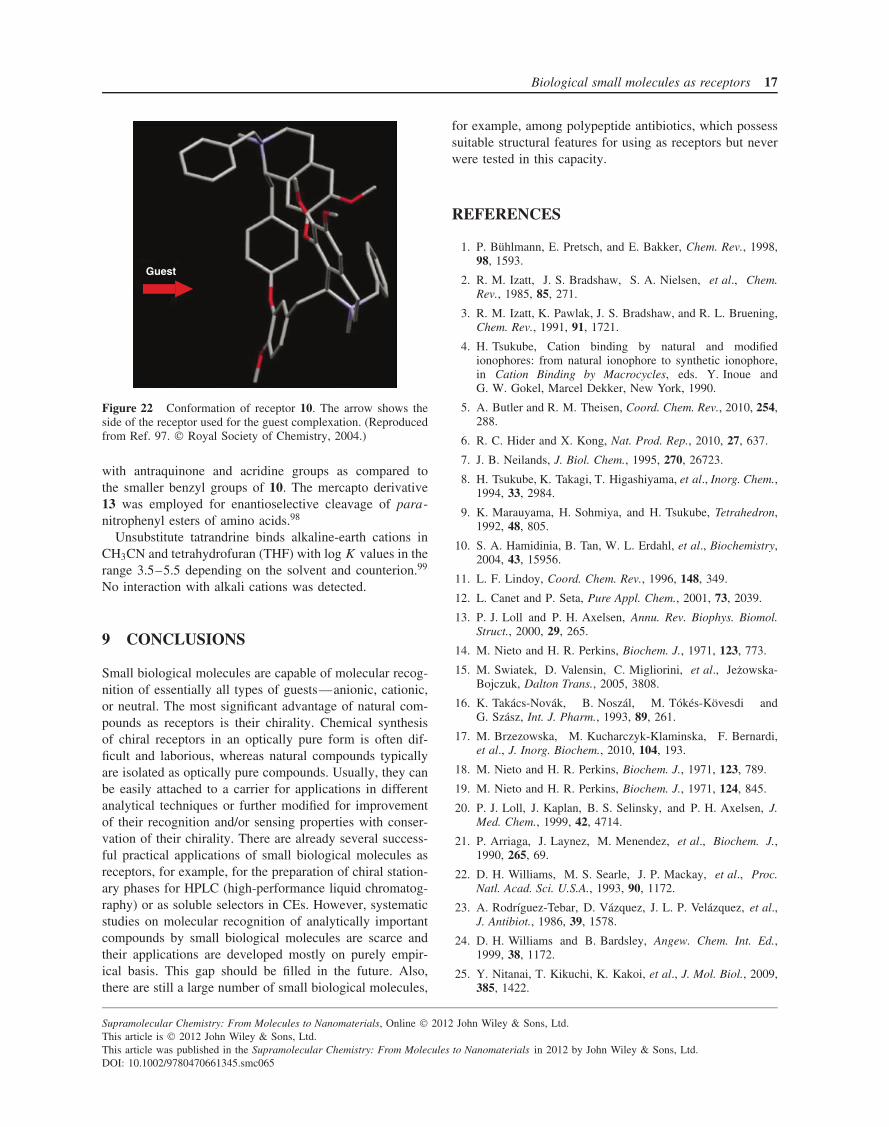
Biological small molecules as receptors 17
Guest
Figure 22 Conformation of receptor 10. The arrow shows theside of the receptor used for the guest complexation. (Reproducedfrom Ref. 97. Royal Society of Chemistry, 2004.)
with antraquinone and acridine groups as compared tothe smaller benzyl groups of 10. The mercapto derivative13 was employed for enantioselective cleavage of para-nitrophenyl esters of amino acids.98
Unsubstitute tatrandrine binds alkaline-earth cations inCH3CN and tetrahydrofuran (THF) with log K values in therange 3.5–5.5 depending on the solvent and counterion.99
No interaction with alkali cations was detected.
9 CONCLUSIONS
Small biological molecules are capable of molecular recog-nition of essentially all types of guests—anionic, cationic,or neutral. The most significant advantage of natural com-pounds as receptors is their chirality. Chemical synthesisof chiral receptors in an optically pure form is often dif-ficult and laborious, whereas natural compounds typicallyare isolated as optically pure compounds. Usually, they canbe easily attached to a carrier for applications in differentanalytical techniques or further modified for improvementof their recognition and/or sensing properties with conser-vation of their chirality. There are already several success-ful practical applications of small biological molecules asreceptors, for example, for the preparation of chiral station-ary phases for HPLC (high-performance liquid chromatog-raphy) or as soluble selectors in CEs. However, systematicstudies on molecular recognition of analytically importantcompounds by small biological molecules are scarce andtheir applications are developed mostly on purely empir-ical basis. This gap should be filled in the future. Also,there are still a large number of small biological molecules,
for example, among polypeptide antibiotics, which possesssuitable structural features for using as receptors but neverwere tested in this capacity.
REFERENCES
1. P. Buhlmann, E. Pretsch, and E. Bakker, Chem. Rev., 1998,98, 1593.
2. R. M. Izatt, J. S. Bradshaw, S. A. Nielsen, et al., Chem.Rev., 1985, 85, 271.
3. R. M. Izatt, K. Pawlak, J. S. Bradshaw, and R. L. Bruening,Chem. Rev., 1991, 91, 1721.
4. H. Tsukube, Cation binding by natural and modifiedionophores: from natural ionophore to synthetic ionophore,in Cation Binding by Macrocycles, eds. Y. Inoue andG. W. Gokel, Marcel Dekker, New York, 1990.
5. A. Butler and R. M. Theisen, Coord. Chem. Rev., 2010, 254,288.
6. R. C. Hider and X. Kong, Nat. Prod. Rep., 2010, 27, 637.
7. J. B. Neilands, J. Biol. Chem., 1995, 270, 26723.
8. H. Tsukube, K. Takagi, T. Higashiyama, et al., Inorg. Chem.,1994, 33, 2984.
9. K. Marauyama, H. Sohmiya, and H. Tsukube, Tetrahedron,1992, 48, 805.
10. S. A. Hamidinia, B. Tan, W. L. Erdahl, et al., Biochemistry,2004, 43, 15956.
11. L. F. Lindoy, Coord. Chem. Rev., 1996, 148, 349.
12. L. Canet and P. Seta, Pure Appl. Chem., 2001, 73, 2039.
13. P. J. Loll and P. H. Axelsen, Annu. Rev. Biophys. Biomol.Struct., 2000, 29, 265.
14. M. Nieto and H. R. Perkins, Biochem. J., 1971, 123, 773.
15. M. Swiatek, D. Valensin, C. Migliorini, et al., Jezowska-Bojczuk, Dalton Trans., 2005, 3808.
16. K. Takacs-Novak, B. Noszal, M. Tokes-Kovesdi andG. Szasz, Int. J. Pharm., 1993, 89, 261.
17. M. Brzezowska, M. Kucharczyk-Klaminska, F. Bernardi,et al., J. Inorg. Biochem., 2010, 104, 193.
18. M. Nieto and H. R. Perkins, Biochem. J., 1971, 123, 789.
19. M. Nieto and H. R. Perkins, Biochem. J., 1971, 124, 845.
20. P. J. Loll, J. Kaplan, B. S. Selinsky, and P. H. Axelsen, J.Med. Chem., 1999, 42, 4714.
21. P. Arriaga, J. Laynez, M. Menendez, et al., Biochem. J.,1990, 265, 69.
22. D. H. Williams, M. S. Searle, J. P. Mackay, et al., Proc.Natl. Acad. Sci. U.S.A., 1993, 90, 1172.
23. A. Rodrıguez-Tebar, D. Vazquez, J. L. P. Velazquez, et al.,J. Antibiot., 1986, 39, 1578.
24. D. H. Williams and B. Bardsley, Angew. Chem. Int. Ed.,1999, 38, 1172.
25. Y. Nitanai, T. Kikuchi, K. Kakoi, et al., J. Mol. Biol., 2009,385, 1422.
Supramolecular Chemistry: From Molecules to Nanomaterials, Online 2012 John Wiley & Sons, Ltd.This article is 2012 John Wiley & Sons, Ltd.This article was published in the Supramolecular Chemistry: From Molecules to Nanomaterials in 2012 by John Wiley & Sons, Ltd.DOI: 10.1002/9780470661345.smc065
18 Molecular recognition
26. C. T. Walsh, S. L. Fisher, I.-S. Park, et al., Chem. Biol.,1996, 3, 21.
27. C. C. McComas, B. M. Crowley, and D. L. Boger, J. Am.Chem. Soc., 2003, 125, 9314.
28. D. McPhail and A Cooper, J. Chem. Soc., Faraday Trans.,1997, 93, 2283.
29. P. J. Loll, A. Derhovanessian, M. V. Shapovalov, et al.,J. Mol. Biol., 2009, 385, 200.
30. V. M. Good, M. N. Gwynn, and D. J. C. Knowles,J. Antibiot., 1990, 43, 550.
31. D. W. Armstrong, Y. Tang, S. Chen, et al., Anal. Chem.,1994, 66, 1473.
32. I. D’Acquarica, F. Gasparrini, D. Misiti, et al., Adv. Chro-matogr., 2008, 46, 109.
33. A. Berthod, Chirality, 2009, 21, 167.
34. M. Lammerhofer, J. Chromatogr. A, 2010, 1217, 814–856.
35. T. J. Ward, Anal. Chem., 2006, 78, 3947.
36. T. J. Ward and A. B. Farris III, J. Chromatogr. A, 2001, 906,73.
37. M. Blanco and I. Valverde, Trends Anal. Chem., 2003, 22,428.
38. F. Hui and M. Caude, Analusis, 1999, 27, 131.
39. M. P. Gasper, A. Berthod, U. B. Nair, and D. W. Arm-strong, Anal. Chem., 1996, 68, 2501.
40. A. A. Rat’ko and R.-I. Stefan, Analyt. Lett., 2004, 37, 3161.
41. R.-I. Stefan-van Staden, J. F. van Staden, and H. Y. Aboul-Enein, Anal. Bioanal. Chem., 2009, 394, 821.
42. J. Tamminen and E. Kolehmainen, Molecules, 2001, 6, 21.
43. A. P. Davis, Molecules, 2007, 12, 2106.
44. M. Miyata, N. Tohnai, and I. Hisaki, Molecules, 2007, 12,1973.
45. O. Bortolini, G. Fantin, and M. Fogagnolo, Chirality, 2005,17, 121.
46. M. Gdaniec, M. J. Milewska, and T. Połonski, Angew.Chem. Int. Ed., 1999, 38, 392.
47. K. Sada, N. Shiomi, and M. Miyata, J. Am. Chem. Soc.,1998, 120, 10543.
48. S.-Z. Zhang, J.-W. Xie, and C.-S. Liu, Anal. Chem., 2003,75, 91.
49. C. M. Hebling, L. E. Thompson, K. W. Eckenroad, et al.,Langmuir, 2008, 24, 13866.
50. L. L. Amundson, R. Li, and C. Bohne, Langmuir, 2008, 24,8491.
51. D. Madenci and S. U. Egelhaaf, Curr. Opin. Colloid In.,2010, 15, 109.
52. A. Bacchi, G. Pelizzi, M. Nebuloni, and P. Ferrari, J. Med.Chem., 1998, 41, 2319.
53. P. Sensi, A. M. Greco, and R. Ballota, Antibiotics Annual(1959–1960), Antibiotics, Inc, New York, 1959.
54. G. G. Gallo, C. R. Pasqualucci, and P. Radaelli, Farmaco(Pavia) Ed. Prat., 1963, 18, 78.
55. C. R. Pasqualucci, A. Vigevani, and P. Radaelli, Farmaco(Pavia) Ed. Prat., 1969, 24, 46.
56. L. Santos, M. A. Medeiros, S. Santos, et al., J. Mol. Struct.,2001, 563–564, 61.
57. A. Bacchi and G. Pelizzi, J. Comput. Aided Mol. Des., 1999,13, 385.
58. C. Godoy-Alcantar, F. Medrano, and A. K. Yatsimirsky,J. Incl. Phenom. Macrocycl. Chem., 2009, 63, 347.
59. E. A. Meyer, R. K. Castellano, and F. Diederich, Angew.Chem. Int. Ed., 2003, 42, 1210.
60. D. W. Armstrong, K. Rundlett, and G. L. Reid III, Anal.Chem., 1994, 66, 1690.
61. S. S. M. Hassan, W. H. Mahmound, and A. H. M. Othman,Talanta, 1997, 44, 1087.
62. M. Chittapragada, S. Roberts, and Y. W. Ham, Perspect.Med. Chem., 2009, 21.
63. J. R. Thomas and P. J. Hergenrother, Chem. Rev., 2008, 108,1171.
64. A. P. Carter, W. M. Clemons, D. E. Brodersen, et al.,Nature, 2000, 407, 340.
65. Q. Vicens and E. Westhof, Biopolymers, 2003, 70, 42.
66. C. M. Barbieri and D. S. Pilch, Biophys. J., 2006, 90, 1338.
67. Y. Fuentes-Martınez, C. Godoy-Alcantar, F. Medrano, et al.,Bioorg. Chem., 2010, 38, 173.
68. A. Bencini, A. Bianchi, E. Garcia-Espana, et al., Coord.Chem. Rev., 1999, 188, 97.
69. H. Nishi, K. Nakamura, H. Nakai, and T. Sato, Chro-matographia, 1996, 43, 426.
70. B. Chankvetadze, M. Saito, E. Yashima, and Y. Okamoto,Chirality, 1998, 10, 134.
71. H. Nakamura, A. Sano, and H. Sumii, Anal. Sci., 1998, 14,375.
72. T. Ohyama, D. Wang, and J. A. Cowan, Chem. Commun.,1998, 467.
73. C. Bazzicalupi, A. Bencini, A. Bianchi, et al., J. Am. Chem.Soc., 1999, 121, 6807.
74. Y. Fuentes-Martınez, C. Godoy-Alcantar, F. Medrano, et al.,Supramol. Chem., 2010, 22, 212.
75. K. O. Lara, C. Godoy-Alcantar, I. L. Rivera, et al., J. Phys.Org. Chem., 2001, 14, 453.
76. R. Moreno-Corral and K. Ochoa Lara, Supramol. Chem.,2008, 20, 427.
77. N. D’Amelio, E. Gaggelli, N. Gaggelli, et al., Dalton Trans.,2004, 363 and references therein.
78. W. Szczepanik, P. Kaczmarek, J. Sobczak, et al., New J.Chem., 2002, 26, 1507.
79. C. J. Dutton, M. A. Haxell, H. A. I. McArthur, and R. G.Wax, eds. Peptide Antibiotics: Discovery, Modes of Actionand Applications, Marcel Dekker, 2002.
80. M. C. Bagley, J. W. Dale, E. A. Merritt, and X. Xiong,Chem. Rev., 2005, 105, 685.
81. Y. L. Hsiao and S. S. Chen, Chromatographia, 2009, 70,1031.
82. C. Godoy-Alcantar, I. Leon-Rivera, and A. K. Yatsimirsky,Bioorg. Med. Chem. Lett., 2001, 11, 651.
Supramolecular Chemistry: From Molecules to Nanomaterials, Online 2012 John Wiley & Sons, Ltd.This article is 2012 John Wiley & Sons, Ltd.This article was published in the Supramolecular Chemistry: From Molecules to Nanomaterials in 2012 by John Wiley & Sons, Ltd.DOI: 10.1002/9780470661345.smc065
Biological small molecules as receptors 19
83. L.-J. Ming and J. D. Epperson, J. Inorg. Biochem., 2002, 91,46.
84. M. Castagnola, D. V. Rossetti, R. Inzitari, et al., Elec-trophoresis, 2004, 25, 846.
85. T. Velkov, P. E. Thompson, R. L. Nation, and J. Li, J. Med.Chem., 2010, 53, 1898.
86. C. Chatterjee, M. Paul, L. Xie, and W. A. van der Donk,Chem. Rev., 2005, 105, 633.
87. S. T. Hsu, E. Breukink, E. Tischenko, et al., Nat. Struct.Mol. Biol., 2004, 11, 963.
88. C. E. Song, ed., Cinchona Alkaloids in Synthesis and Catal-ysis, Wiley-VCH, 2009.
89. J. Lah, N. M. Maier, W. Lindner, and G. Vesnaver, J. Phys.Chem. B, 2001, 105, 1670.
90. N. M. Maier, L. Nicoletti, M. Lammerhofer, and W. Lin-dner, Chirality, 1999, 11, 522.
91. K. H. Krawinkler, N. M. Maier, R. Ungaro, et al., Chirality,2003, 15, S17.
92. M. Shamma, The Isoquinoline Alkaloids, Academic Press,New York, 1972.
93. H. King, J. Chem. Soc., 1935, 1381.
94. C. Godoy-Alcantar, A. V. Eliseev, and A. K. Yatsimirsky,J. Mol. Recognit., 1996, 9, 54.
95. U. B. Nair, D. W. Armstrong, and W. L. Hinze, Anal.Chem., 1998, 70, 1059.
96. C. Godoy-Alcantar, M. I. Nelen, A. V. Eliseev, and A. K.Yatsimirsky, J. Chem. Soc. Perkin Trans. 2, 1999, 353.
97. K. Ochoa Lara, C. Godoy-Alcantar, A. V. Eliseev, andA. K. Yatsimirsky, Org. Biomol. Chem., 2004, 2, 1712.
98. K. Ochoa Lara, C. Godoy-Alcantar, A. V. Eliseev, andA. K. Yatsimirsky, Arkivoc, 2005 (vi), 293.
99. I. Stanculescu, C. Mandravel, F. Delattre, et al., J. Pho-tochem. Photobiol. A, 2003, 161, 79.
Supramolecular Chemistry: From Molecules to Nanomaterials, Online 2012 John Wiley & Sons, Ltd.This article is 2012 John Wiley & Sons, Ltd.This article was published in the Supramolecular Chemistry: From Molecules to Nanomaterials in 2012 by John Wiley & Sons, Ltd.DOI: 10.1002/9780470661345.smc065


![7. Supramolecular structures - Acclab h55.it.helsinki.fiknordlun/nanotiede/nanosc7nc.pdf · 7. Supramolecular structures [Poole-Owens 11.5] Supramolecular structures are large molecules](https://img.pdfslide.net/doc/110x75/5f071ded7e708231d41b63bf/7-supramolecular-structures-acclab-h55it-knordlunnanotiedenanosc7ncpdf.jpg)


![Reversible Manipulation of Supramolecular Chirality using ... · molecular system is based on discovery of serendipity among numerous newly synthesized molecules,[6b,7a] anditisstilla](https://img.pdfslide.net/doc/110x75/5f64e54208025e205533969c/reversible-manipulation-of-supramolecular-chirality-using-molecular-system-is.jpg)