Embed Size (px)
Citation preview
http://jba.sagepub.com/Applications
Journal of Biomaterials
http://jba.sagepub.com/content/24/2/119The online version of this article can be found at:
DOI: 10.1177/0885328208094265
2009 24: 119 originally published online 18 September 2008J Biomater ApplYuan Gao and Jiang Chang
PDLLA/Bioactive Glass Composite FilmsSurface Modification of Bioactive Glasses and Preparation of
Published by:
http://www.sagepublications.com
can be found at:Journal of Biomaterials ApplicationsAdditional services and information for
http://jba.sagepub.com/cgi/alertsEmail Alerts:
http://jba.sagepub.com/subscriptionsSubscriptions:
http://www.sagepub.com/journalsReprints.navReprints:
http://www.sagepub.com/journalsPermissions.navPermissions:
http://jba.sagepub.com/content/24/2/119.refs.htmlCitations:
What is This?
- Sep 18, 2008 OnlineFirst Version of Record
- Sep 2, 2009Version of Record >>
at DREXEL UNIV LIBRARIES on October 9, 2014jba.sagepub.comDownloaded from at DREXEL UNIV LIBRARIES on October 9, 2014jba.sagepub.comDownloaded from
Surface Modification of BioactiveGlasses and Preparation of
PDLLA/Bioactive GlassComposite Films
YUAN GAO1,2
AND JIANG CHANG1,*
1Biomaterials and Tissue Engineering Research Center
Shanghai Institute of Ceramics, Chinese Academy of Sciences
1295 Dingxi Road, Shanghai 200050, People’s Republic of China2Graduate School of the Chinese Academy of Sciences
319 Yueyang Road, Shanghai 200031, People’s Republic of China
ABSTRACT: In order to improve the homogeneous dispersion of particles inthe polymeric matrix, 45S5, mesoporous 58S, and 58S bioactive glasses weresurface modified by esterification reactions with dodecyl alcohol at refluxtemperature of 2608C (named as m-45S5, m-mesoporous 58S, and m-58S,respectively). The modified particles showed better hydrophobicity and longertime of suspension in organic matrix. The PDLLA/bioactive glass compositefilms were fabricated using surface modified bioactive glass particles throughsolvent casting-evaporation method. Surface morphology, mechanical property,and bioactivity were investigated. The results revealed that the inorganicparticle distribution and tensile strength of the composite films with modifiedbioactive glass particles were significantly improved while great bioactiveproperties were maintained. Scanning electron microscopy (SEM) observationillustrated that the modified bioactive glass particles were homogeneouslydispersed in the PDLLA matrix. The maximum tensile strengths of compositefilms with modified bioactive glass particles were higher than that of compositefilms with unmodified bioactive glass particles. The bioactivity of thecomposite films were evaluated by being soaked in the simulated body fluid(SBF) and the SEM observation of the films suggested that the modifiedcomposite films were still bioactive in that they could induce the formation
*Author to whom correspondence should be addressed. E-mail: [email protected] 2, 3 and 6 appear in color online: http://jba.sagepub.com
JOURNAL OF BIOMATERIALS APPLICATIONS Volume 24 — August 2009 119
0885-3282/09/02 0119–20 $10.00/0 DOI: 10.1177/0885328208094265� SAGE Publications 2009
Los Angeles, London, New Delhi and Singapore
at DREXEL UNIV LIBRARIES on October 9, 2014jba.sagepub.comDownloaded from
of HAp on its surface and the distribution of HAp was even more homogeneouson the film. The results mentioned above indicated that the surface modificationof bioactive glasses with dodecyl alcohol was an effective method to preparePDLLA/bioactive glass composites with enhanced properties. By studying thecomparisons of modification effects among the three types of bioactive glasses,we could get the conclusion that the size and morphology of the inorganicparticles would greatly affect the modification effects and the properties ofcomposites.
KEY WORDS: surface modification, composite film, bioactive glass, PDLLA,dodecyl alcohol.
INTRODUCTION
Many kinds of polymers have been widely used as scaffoldbiomaterials in bone tissue engineering research, such as poly
(lactic acid) (PLA) [1], poly (glycolic acid) (PGA) [2], and their copoly-mers [3]. These materials have many advantages, such as excellentbiocompatibility, biodegradability, and feasibility being made intodesired shape. However, these kinds of polymers have a few disadvan-tages such as the lack of bioactivity [4] and poor mechanical property [5],which restrict their practical use in tissue engineering applications.In order to ameliorate these problems, bioactive inorganic fillers, such ashydroxyapatite (HAp) [6], wollastonite [7], or b-tricalcium phosphate [8]were introduced into biodegradable polymers to fabricate filler/polymercomposites. Bioactive glasses with different components, such as 45S5(CaO-P2O5- SiO2-Na2O) [9] and 58S (CaO-P2O5-SiO2) [10], have beenproved highly bioactive since they could induce the formation of HApwhich could create tight bonds between materials and tissue.Mesoporous bioactive glasses have also been studied due to their highspecific surface area and well ordered pores [11]. These bioactive glasseshave already been used to prepare PDLLA/bioactive glass compo-sites [12]. However, one of the most problematic issues for thesepolymer/inorganic composites is the tendency for agglomeration of theinorganic particles in the polymer matrix [7]. Particle aggregation incomposite materials tends to increase susceptibility to chemical andmechanical failure. On the contrary, well-dispersed particles couldincrease the contact area between the polymer matrix and the inorganicreinforcing phases for interfacial bond development. Therefore, it iscritical to improve the dispersion of inorganic particles in preparingPDLLA/bioactive glass composites.
The mostly used methods to improve the dispersion of inorganicparticles in polymeric matrix were mechanical stirring [13] and
120 Y. GAO AND J. CHANG
at DREXEL UNIV LIBRARIES on October 9, 2014jba.sagepub.comDownloaded from
ultrasonic energy [14] which only showed temporary affects since theparticle aggregation would occur due to the colloid stability.
Chemical modification of the surface of inorganic particles may be asolution to this problem. Using esterification which involves thecondensation reaction that an alcohol reacts with an acid to form anester, certain alcohols had been used to react with the active hydroxylgroups of silica [15], hydroxyapatite [16], dicalcium silicate [17], andCaSiO3 [18] particles.
The aim of this study was to improve the homogeneous dispersion ofbioactive glass particles in polymeric matrix by surface modificationthrough esterification and to study its effect on the properties of theprepared PDLLA/bioactive glass composites. In addition, the surfacemodification effects of three types of bioactive glasses which vary inparticle sizes and morphology were compared.
EXPERIMENTAL PROCEDURE
Materials
Melt-derived 45S5 bioglasses with size around 20mm were obtainedfrom US Biomaterials. 58S bioactive glasses with size around 1 mm wereprepared using sol–gel processing according to Zhong’s method [10].Fibrous mesoporous 58S bioactive glasses with length of 1 mm wereprepared using sol–gel and hydrothermal processing according toprevious report [11].
PDLLA with an average molecular weight (MW) of 45 kDa wasobtained from Chengdu Institute of Organic Chemistry (Sichuan, China).
Dodecyl alcohol was purchased from A.R. Shanghai ChemicalReagent Co., LTD (Shanghai, China).
Surface Modification of Bioactive Glasses
The esterification of silanols with alcohols is well known as describedin Equation (1) [19]:
� Si�OHþ R�OH !� Si�OH . . . HO� R !� Si�ORþH2O ð1Þ
The modification of bioactive glass particles was carried out according tothe procedure reported for the modification of hydroxyapatite [16].Five grams of bioactive glass particles were immersed into 20 mL of 95%ethyl alcohol in the flask and put under ultrasonic for 15 min to increase
Bioactive Glass Composite Films 121
at DREXEL UNIV LIBRARIES on October 9, 2014jba.sagepub.comDownloaded from
the dispersion of particles. After the evaporation of ethyl alcohol, 100 mLdodecyl alcohol was put into the flask and 0.1 g para-toluenesulfonatewas added as the catalyzer. The suspension was put under mechanicalstirring for 4 h at 2608C. The modified bioactive glass (m-bioactive glass)particles were separated by centrifugation, washed in anhydrous ethylalcohol for 3 times and then dried in a vacuum oven at 608C for 24 h toremove the remaining solvent.
Preparation of the PDLLA/m-Bioactive Glass Composite Films
Our previous work has shown that the mechanical strength of thecomposites changed with the concentration of wollastonite particles inthe composites [18], and the highest tensile strength was obtained in thecomposite with 15 wt% of the concentration of inorganic particles.Therefore, the concentration of 15 wt% bioactive glass particles wasselected in the present study. PDLLA was dissolved in chloroform toform 10% (w/v) solution, and then m-bioactive glass particles were addedinto the PDLLA solution to form a 15 wt% suspension, followed bystirring in PDLLA solution for 2 h to disperse the bioactive glassparticles homogeneously. The suspension was poured into a glass plateand due to the good rheology, it could spread on the whole plate withuniform thickness. Then it was placed in air ventilation for 12 h toevaporate the chloroform and in a vacuum oven at room temperature for12 h to remove the remaining solvent. Control groups (PDLLA/unmodified bioactive glass) were fabricated by the same procedureas mentioned above.
Characterization of the Modified Particles
Thermo Nicolet nexus-IR spectrometer was used to identify thespecific functional groups on the surface of three types of bioactive glassparticles. Samples were prepared by mixing bioactive glass particleswith KBr. The spectra were collected after 64 scans in 4500–400 cm�1
region at a resolution of 4 cm�1.The dispersion stability of the three kinds of modified bioactive glass
particles in water and hexane was determined by sedimentation studiesaccording to the process reported for the test of hydroxyapatite [16].0.1 g of m-bioactive glass particles were ultrasonically dispersed in10 mL of solvent. The sedimentation property was observed and deter-mined by the time that all of the particles were precipitated out of thesolvent which then became lucid and clear. The tests of unmodifiedbioactive glass particles were also taken as controls.
122 Y. GAO AND J. CHANG
at DREXEL UNIV LIBRARIES on October 9, 2014jba.sagepub.comDownloaded from
The hydrophilicity of the three types of modified bioactive glassparticles was evaluated by measuring the water contact angles of thewafer composed of m-bioactive glass particles using the sessile dropmethod. A total of 0.05 g of m-bioactive glass was put into tablet pressunder the pressure of 6 MPa for 2 min in order to get a wafer withdiameter of 6 mm. One droplet (about 0.5 mL) of distilled water wasadded to the surface of the wafer. The photo of the sample with dropleton it was taken under an optical microscope and the water contact anglewas evaluated by Photoshop Software (Adobe, USA). Each determina-tion was obtained by averaging the result of five measurements.The water contact angles of untreated bioactive glass were alsomeasured as control groups by the same method. All data wereexpressed as means� standard deviation (SD) for n¼ 5.
The amounts of Si-OH groups on the surface of three types ofbioactive glass particles after modification under different temperatureswere measured by the method reported by HAMAWA [20]. Ammoniumchloride solution (4 mol/L and 300 mL) was mixed with zinc chloride(0.4 mol/L and 150 mL) solution and the pH of the mixture was adjustedwith 28% ammonium hydroxide to 6.9. The volume of the mixture wasadjusted to 500 mL with deionized water.
A total of 0.1 g modified bioactive glass particles were immersed in80 mL of the above resultant solution at room temperature and themixture was stirred for 5 min. During the mixing, active hydroxylradicals were mostly exchanged for zinc ions and zinc chelates wereformed. After this, the particles were collected by filtration and wereimmersed in 150 mL of stirring deionized water three times to removechemical species without adsorption. The specimens were dried in adesiccator and then immersed in 60 mL of 2 mol/L nitric acid for 10 minto release zinc ions into the nitric acid solution. The amount of zinc ionsin the nitric acid solution was measured by an inductively coupledplasma atomic emission spectrometer (ICP-AES, Vista AX, Varian). Thezinc concentration corresponds to the amount of Si-OH groups on thesurface of a specimen.
The surface concentration of OH groups (the silanol number) aOH,expressed in OH groups nm�2, is determined by Zhuravlev [21]as Equation (2):
aOH ¼ �ðsÞOH
NA � 10�21S�1 ð2Þ
where �ðsÞOH is the concentration of OH groups on bioactive glass surfacereferred to the unit mass of the sample (m mol of OH/g of bioactive glass)found by the zinc ions-exchange method with ICP, S is the specific
Bioactive Glass Composite Films 123
at DREXEL UNIV LIBRARIES on October 9, 2014jba.sagepub.comDownloaded from
surface area with respect to krypton adsorption (m2 g�l) and is measuredby BET surface area analysis and NA is the Avogadro number.
So the conversion equation between ICP value and Si-OH groups isillustrated as Equation (3):
OH=nm2 ¼ ½ICPðug=mLÞ � 10�3 � 2 � 60 mL=65:39ðg=molÞ=0:1 g�
� 6:02 � 1023 � 10�21 � S�1 ð3Þ
The number of Si-OH groups of unmodified bioactive glass particles wasalso measured by the same method as control.
Characterization of the PDLLA/m-Bioactive GlassComposite Films
Square samples with size of 5� 5 mm were cut from the compositefilms composed of PDLLA and three types of modified bioactive glass,respectively, for optical microscopy and SEM observation. For SEManalysis, surface of the samples were coated with gold and SEMexamination was carried out on an Electron Probe X-ray Microanalyzer(EPMA-8705QH2, Shimadzu. Japan) at an accelerating voltage of 20 kVto observe the particles distribution and agglomeration in the films.Composite films with unmodified bioactive glass particles were alsoobserved as controls.
The quality of dispersion of bioactive glass particles in PDLLA wasevaluated by a semi-quantitative assay. Briefly, five optical images at amagnification of 200 times from different part of each sample weretaken, and the number of the aggregated particles with a size largerthan 5 mm in diameter was counted.
Mechanical properties of the composite films composed of PDLLA andthree types of modified bioactive glass particles with the filler content of15% were evaluated by tensile testing on an AG-1 Shimadzu mechanicalmachine (Shimadzu Co., Japan) at a crosshead speed of 2 mm/min.Dumbbell-shaped tensile specimens (with a size of 30� 8 mm for theeffective area) were cut from the prepared composite films formechanical testing. Pure PDLLA and composite films with unmodifiedparticles were used as controls. Ten samples of each kind of the filmwere tested. The student t-test was used for statistical analysis anda p-value50.05 was considered statistically significant.
The bioactivity of the different composite films was evaluated bythe soaking experiment in SBF. The SBF solution was preparedaccording to the procedure described by Kobubo [22] and pH value
124 Y. GAO AND J. CHANG
at DREXEL UNIV LIBRARIES on October 9, 2014jba.sagepub.comDownloaded from
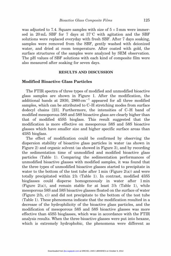
was adjusted to 7.4. Square samples with size of 5� 5 mm were immer-sed in 20 mL SBF for 7 days at 378C with agitation and the SBFsolutions were replaced everyday with fresh SBF. After 7 days soaking,samples were removed from the SBF, gently washed with deionizedwater, and dried at room temperature. After coated with gold, thesurface structures of the samples were analyzed by SEM observation.The pH values of SBF solutions with each kind of composite film werealso measured after soaking for seven days.
RESULTS AND DISCUSSION
Modified Bioactive Glass Particles
The FTIR spectra of three types of modified and unmodified bioactiveglass samples are shown in Figure 1. After the modification, theadditional bands at 2930, 2860 cm�1 appeared for all three modifiedsamples, which can be attributed to C–H stretching modes from surfacedodecyl chains [23]. Furthermore, the intensities of C–H band ofmodified mesoporous 58S and 58S bioactive glass are clearly higher thanthat of modified 45S5 bioglass. This result suggested that themodification is more effective on mesoporous 58S and 58S bioactiveglasses which have smaller size and higher specific surface areas than45S5 bioglass.
The effect of modification could be confirmed by observing thedispersion stability of bioactive glass particles in water (as shown inFigure 2) and organic solvent (as showed in Figure 3), and by recordingthe sedimentation time of unmodified and modified bioactive glassparticles (Table 1). Comparing the sedimentation performances ofunmodified bioactive glasses with modified samples, it was found thatthe three types of unmodified bioactive glasses started to precipitate inwater to the bottom of the test tube after 1 min (Figure 2(a)) and weretotally precipitated within 2 h (Table 1). In contrast, modified 45S5bioglasses could disperse homogeneously in water after 1 min(Figure 2(a)), and remain stable for at least 3 h (Table 1), whilemesoporous 58S and 58S bioactive glasses floated on the surface of water(Figure 2(b, c)) and did not precipitate to the bottom of the test tube(Table 1). These phenomena indicate that the modification resulted in adecrease of the hydrophilicity of the bioactive glass particles, and themodification of mesoporous 58S and 58S bioactive glasses was moreeffective than 45S5 bioglasses, which was in accordance with the FTIRanalysis results. When the three bioactive glasses were put into hexane,which is extremely hydrophobic, the phenomena were different as
Bioactive Glass Composite Films 125
at DREXEL UNIV LIBRARIES on October 9, 2014jba.sagepub.comDownloaded from
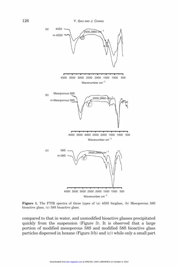
compared to that in water, and unmodified bioactive glasses precipitatedquickly from the suspension (Figure 3). It is observed that a largeportion of modified mesoporous 58S and modified 58S bioactive glassparticles dispersed in hexane (Figure 3(b) and (c)) while only a small part
45S5(a)
(b)
(c)
m-45S5
58S
m-58S
4000 3500 3000 2500 2000
Wavenumber cm−1
Mesoporous 58S
m-Mesoporous 58S
1500 1000 500
4000 3500 3000 2500 2000
Wavenumber cm−1
1500 1000 500
4000 3500 3000 2500 2000
Wavenumber cm−1
1500 1000 500
2930,2860 cm−1
2930,2860 cm−1
2930,2860 cm−1
Figure 1. The FTIR spectra of three types of (a) 45S5 bioglass, (b) Mesoporous 58S
bioactive glass, (c) 58S bioactive glass.
126 Y. GAO AND J. CHANG
at DREXEL UNIV LIBRARIES on October 9, 2014jba.sagepub.comDownloaded from
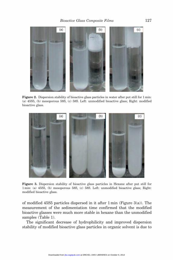
of modified 45S5 particles dispersed in it after 1 min (Figure 3(a)). Themeasurement of the sedimentation time confirmed that the modifiedbioactive glasses were much more stable in hexane than the unmodifiedsamples (Table 1).
The significant decrease of hydrophilicity and improved dispersionstability of modified bioactive glass particles in organic solvent is due to
(a) (b) (c)
Figure 3. Dispersion stability of bioactive glass particles in Hexane after put still for
1 min: (a) 45S5, (b) mesoporous 58S, (c) 58S. Left: unmodified bioactive glass; Right:
modified bioactive glass.
(a) (b) (c)
Figure 2. Dispersion stability of bioactive glass particles in water after put still for 1 min:
(a) 45S5, (b) mesoporous 58S, (c) 58S. Left: unmodified bioactive glass; Right: modifiedbioactive glass.
Bioactive Glass Composite Films 127
at DREXEL UNIV LIBRARIES on October 9, 2014jba.sagepub.comDownloaded from
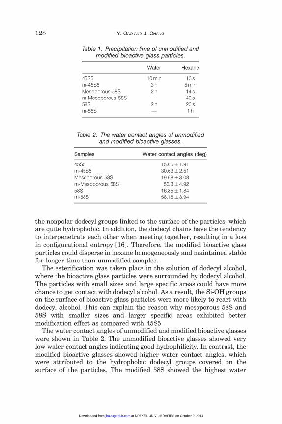
the nonpolar dodecyl groups linked to the surface of the particles, whichare quite hydrophobic. In addition, the dodecyl chains have the tendencyto interpenetrate each other when meeting together, resulting in a lossin configurational entropy [16]. Therefore, the modified bioactive glassparticles could disperse in hexane homogeneously and maintained stablefor longer time than unmodified samples.
The esterification was taken place in the solution of dodecyl alcohol,where the bioactive glass particles were surrounded by dodecyl alcohol.The particles with small sizes and large specific areas could have morechance to get contact with dodecyl alcohol. As a result, the Si-OH groupson the surface of bioactive glass particles were more likely to react withdodecyl alcohol. This can explain the reason why mesoporous 58S and58S with smaller sizes and larger specific areas exhibited bettermodification effect as compared with 45S5.
The water contact angles of unmodified and modified bioactive glasseswere shown in Table 2. The unmodified bioactive glasses showed verylow water contact angles indicating good hydrophilicity. In contrast, themodified bioactive glasses showed higher water contact angles, whichwere attributed to the hydrophobic dodecyl groups covered on thesurface of the particles. The modified 58S showed the highest water
Table 1. Precipitation time of unmodified andmodified bioactive glass particles.
Water Hexane
45S5 10 min 10 sm-45S5 3 h 5 minMesoporous 58S 2 h 14 sm-Mesoporous 58S — 40 s58S 2 h 20 sm-58S — 1 h
Table 2. The water contact angles of unmodifiedand modified bioactive glasses.
Samples Water contact angles (deg)
45S5 15.65� 1.91m-45S5 30.63� 2.51Mesoporous 58S 19.68� 3.08m-Mesoporous 58S 53.3� 4.9258S 16.85� 1.84m-58S 58.15� 3.94
128 Y. GAO AND J. CHANG
at DREXEL UNIV LIBRARIES on October 9, 2014jba.sagepub.comDownloaded from
contact angles, which indicated the most effective modification amongthe three kinds of bioactive glasses.
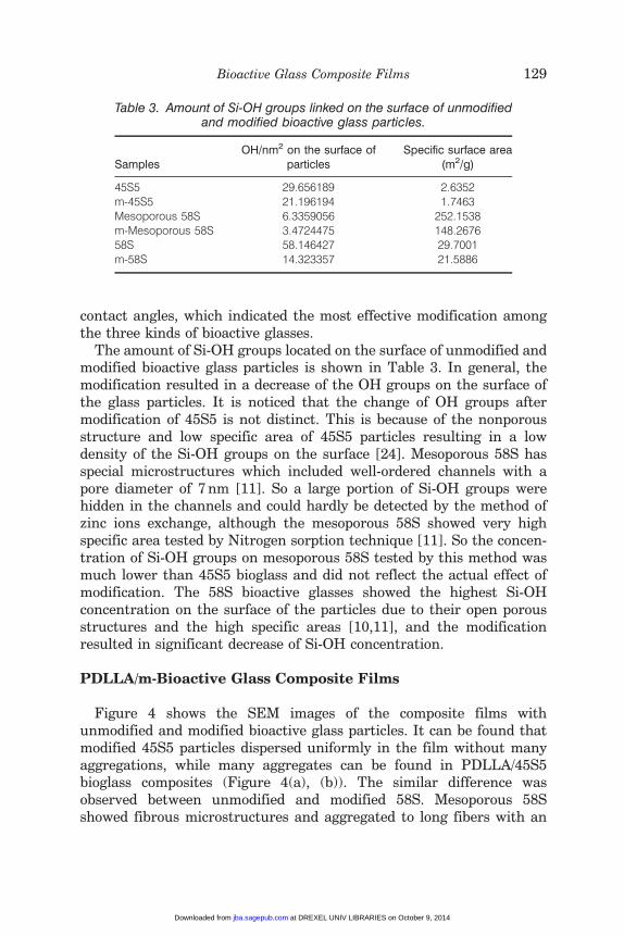
The amount of Si-OH groups located on the surface of unmodified andmodified bioactive glass particles is shown in Table 3. In general, themodification resulted in a decrease of the OH groups on the surface ofthe glass particles. It is noticed that the change of OH groups aftermodification of 45S5 is not distinct. This is because of the nonporousstructure and low specific area of 45S5 particles resulting in a lowdensity of the Si-OH groups on the surface [24]. Mesoporous 58S hasspecial microstructures which included well-ordered channels with apore diameter of 7 nm [11]. So a large portion of Si-OH groups werehidden in the channels and could hardly be detected by the method ofzinc ions exchange, although the mesoporous 58S showed very highspecific area tested by Nitrogen sorption technique [11]. So the concen-tration of Si-OH groups on mesoporous 58S tested by this method wasmuch lower than 45S5 bioglass and did not reflect the actual effect ofmodification. The 58S bioactive glasses showed the highest Si-OHconcentration on the surface of the particles due to their open porousstructures and the high specific areas [10,11], and the modificationresulted in significant decrease of Si-OH concentration.
PDLLA/m-Bioactive Glass Composite Films
Figure 4 shows the SEM images of the composite films withunmodified and modified bioactive glass particles. It can be found thatmodified 45S5 particles dispersed uniformly in the film without manyaggregations, while many aggregates can be found in PDLLA/45S5bioglass composites (Figure 4(a), (b)). The similar difference wasobserved between unmodified and modified 58S. Mesoporous 58Sshowed fibrous microstructures and aggregated to long fibers with an
Table 3. Amount of Si-OH groups linked on the surface of unmodifiedand modified bioactive glass particles.
SamplesOH/nm2 on the surface of
particlesSpecific surface area
(m2/g)
45S5 29.656189 2.6352m-45S5 21.196194 1.7463Mesoporous 58S 6.3359056 252.1538m-Mesoporous 58S 3.4724475 148.267658S 58.146427 29.7001m-58S 14.323357 21.5886
Bioactive Glass Composite Films 129
at DREXEL UNIV LIBRARIES on October 9, 2014jba.sagepub.comDownloaded from
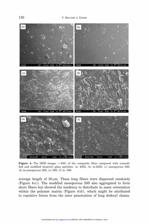
average length of 30 mm. These long fibers were dispersed randomly(Figure 4(c)). The modified mesoporous 58S also aggregated to formshort fibers but showed the tendency to distribute in same orientationwithin the polymer matrix (Figure 4(d)), which might be attributedto repulsive forces from the inter penetration of long dodecyl chains.
(a) (b)
(c) (d)
(f)(e)
NONE SEI 10.0kV X500 10µm WD 8.0mm NONE SEI 10.0kV X500 10µm WD 8.1mm
NONE SEI 10.0kV X500 10 µm WD 7.9 mm NONE SEI 10.0kV X500 10µm WD 9.1mm
SICCA SEI 20.0kV X500 10µm WD 11mmSICCA SEI 20.0kV X500 10µm WD 11mm
Figure 4. The SEM images (�500) of the composite films composed with unmodi-
fied and modified bioactive glass particles: (a) 45S5, (b) m-45S5, (c) mesoporous 58S,
(d) m-mesoporous 58S, (e) 58S, (f) m- 58S.
130 Y. GAO AND J. CHANG
at DREXEL UNIV LIBRARIES on October 9, 2014jba.sagepub.comDownloaded from
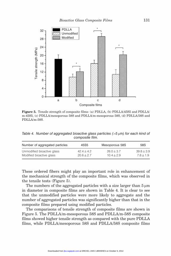
These ordered fibers might play an important role in enhancement ofthe mechanical strength of the composite films, which was observed inthe tensile tests (Figure 5).
The numbers of the aggregated particles with a size larger than 5 mmin diameter in composite films are shown in Table 4. It is clear to seethat the unmodified particles were more likely to aggregate and thenumber of aggregated particles was significantly higher than that in thecomposite films prepared using modified particles.
The comparisons of tensile strength of composite films are shown inFigure 5. The PDLLA/m-mesoporous 58S and PDLLA/m-58S compositefilms showed higher tensile strength as compared with the pure PDLLAfilms, while PDLLA/mesoporous 58S and PDLLA/58S composite films
a b
*
*
*
c
Composite films
PDLLAUnmodifiedModified
Ten
sile
str
engt
h (M
Pa)
d
32
28
24
20
16
12
8
4
0
Figure 5. Tensile strength of composite films: (a) PDLLA, (b) PDLLA/45S5 and PDLLA/
m-45S5, (c) PDLLA/mesoporous 58S and PDLLA/m-mesoporous 58S, (d) PDLLA/58S and
PDLLA/m-58S.
Table 4. Number of aggregated bioactive glass particles (45mm) for each kind ofcomposite film.
Number of aggregated particles 45S5 Mesoporous 58S 58S
Unmodified bioactive glass 42.4� 4.2 26.0� 3.7 39.8� 3.9Modified bioactive glass 20.6� 2.7 10.4� 2.9 7.8� 1.9
Bioactive Glass Composite Films 131
at DREXEL UNIV LIBRARIES on October 9, 2014jba.sagepub.comDownloaded from
exhibited lower tensile strength. During the mixing of bioactive glassparticles in PDLLA solvent, the unmodified bioactive glass particles hadthe tendency to agglomerate, while the surface modified bioactive glassparticles could be easily dispersed in PDLLA matrix to form homo-geneous suspension, which resulted in an increase of the tensilestrength of the composite films. Furthermore, the porous structures ofm-mesoporous 58S and m-58S could also attribute to the increase ofthe tensile strength, since the polymer might be able to penetrate intothe mesopores, which could result in tight integration between thetwo phases and prevent abruption at the interface. In addition,m-mesoporous 58S short fibers were observed to distribute in sameorientation within the polymer matrix, which might also be a factor forenhancing the mechanical property of the composites. Therefore,composite films fabricated with PDLLA and m-mesoporous 58Sexhibited the highest tensile strength among the tested samples. Onthe contrary, the tensile strengths of the composite films withunmodified bioactive glass particles decreased comparing with purePDLLA, and it was found that the mechanical failures always occurredat the interface between the particle agglomerate and the polymericmatrix (Figure 6), which was accord with the conclusion of Ye [18]. Sothe tensile strengths of the composite films with unmodified bioactiveglass particles decreased comparing with pure PDLLA. The abnormalphenomena occurred on composite films with modified 45S5, whichshowed a lower mechanical strength than the pure polymer films.One explanation is that the melt-derived 45S5 bioglass particles weredense without porous structures as compared with sol–gel derivedbioactive glasses [10], and could not integrate with PDLLA firmly.
Figure 7 shows the SEM images of composite films with unmodifiedand modified bioactive glass particles after soaking in SBF for 7 days.The morphology of the precipitates formed on the surface of the films(Figure 7 (b), (d) and (f)) was similar to that of the hydroxyapatiteformed on the surface of bioactive glasses fabricated by otherresearchers [14,25] which proved the good bioactivity of these threetypes of bioactive glasses, even after modification. The HAp was morelikely to be detected on the whole films composed of modified bioactiveglass particles. On the contrary, the HAp was often detected on thesurrounding of the particle agglomerations in the composite filmswith unmodified glass particles.
Bioactivity is one of the most important factors for the applicationof biomaterials in hard tissue regeneration. One reason is that theHAp layer, which is induced to precipitate on the surface of thebioactive materials, could form chemical bond with the surrounding
132 Y. GAO AND J. CHANG
at DREXEL UNIV LIBRARIES on October 9, 2014jba.sagepub.comDownloaded from
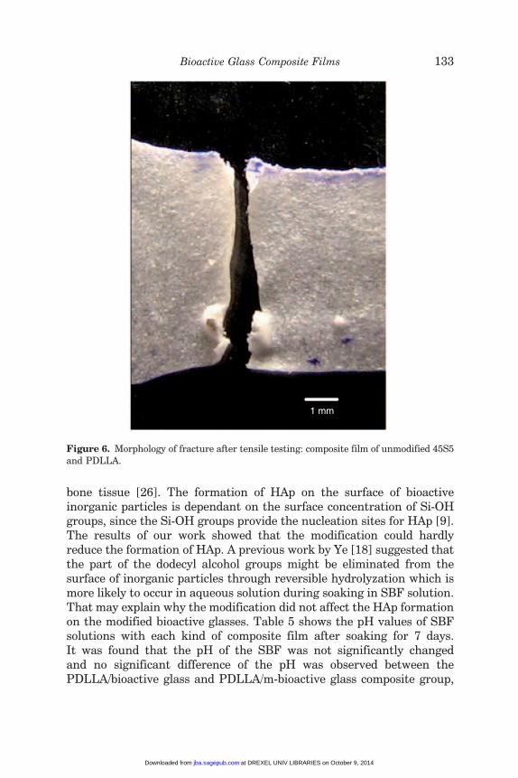
bone tissue [26]. The formation of HAp on the surface of bioactiveinorganic particles is dependant on the surface concentration of Si-OHgroups, since the Si-OH groups provide the nucleation sites for HAp [9].The results of our work showed that the modification could hardlyreduce the formation of HAp. A previous work by Ye [18] suggested thatthe part of the dodecyl alcohol groups might be eliminated from thesurface of inorganic particles through reversible hydrolyzation which ismore likely to occur in aqueous solution during soaking in SBF solution.That may explain why the modification did not affect the HAp formationon the modified bioactive glasses. Table 5 shows the pH values of SBFsolutions with each kind of composite film after soaking for 7 days.It was found that the pH of the SBF was not significantly changedand no significant difference of the pH was observed between thePDLLA/bioactive glass and PDLLA/m-bioactive glass composite group,
1 mm
Figure 6. Morphology of fracture after tensile testing: composite film of unmodified 45S5and PDLLA.
Bioactive Glass Composite Films 133
at DREXEL UNIV LIBRARIES on October 9, 2014jba.sagepub.comDownloaded from
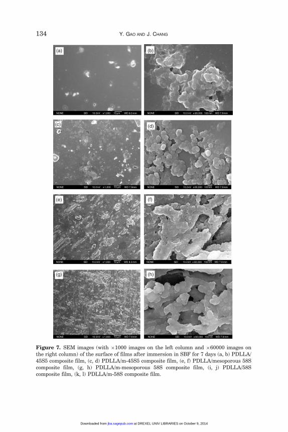
Figure 7. SEM images (with �1000 images on the left column and �60000 images on
the right column) of the surface of films after immersion in SBF for 7 days (a, b) PDLLA/
45S5 composite film, (c, d) PDLLA/m-45S5 composite film, (e, f) PDLLA/mesoporous 58S
composite film, (g, h) PDLLA/m-mesoporous 58S composite film, (i, j) PDLLA/58Scomposite film, (k, l) PDLLA/m-58S composite film.
134 Y. GAO AND J. CHANG
at DREXEL UNIV LIBRARIES on October 9, 2014jba.sagepub.comDownloaded from
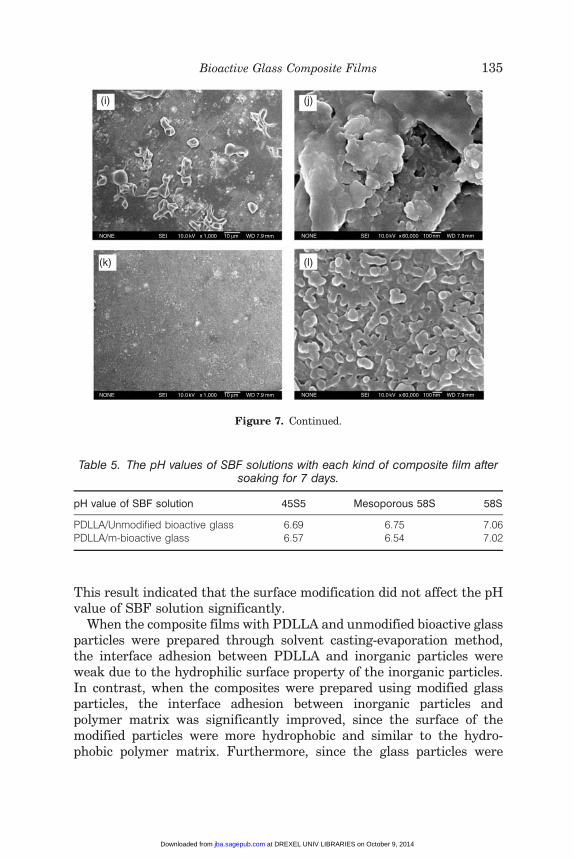
This result indicated that the surface modification did not affect the pHvalue of SBF solution significantly.
When the composite films with PDLLA and unmodified bioactive glassparticles were prepared through solvent casting-evaporation method,the interface adhesion between PDLLA and inorganic particles wereweak due to the hydrophilic surface property of the inorganic particles.In contrast, when the composites were prepared using modified glassparticles, the interface adhesion between inorganic particles andpolymer matrix was significantly improved, since the surface of themodified particles were more hydrophobic and similar to the hydro-phobic polymer matrix. Furthermore, since the glass particles were
(i) (j)
(k) (l)
Figure 7. Continued.
Table 5. The pH values of SBF solutions with each kind of composite film aftersoaking for 7 days.
pH value of SBF solution 45S5 Mesoporous 58S 58S
PDLLA/Unmodified bioactive glass 6.69 6.75 7.06PDLLA/m-bioactive glass 6.57 6.54 7.02
Bioactive Glass Composite Films 135
at DREXEL UNIV LIBRARIES on October 9, 2014jba.sagepub.comDownloaded from
closely surrounded by dodecyl alcohol, it may reduce the possible contactof the glass particles with water phase and reduce the degradation of theglass components of the composites.
CONCLUSIONS
In this study,45S5, mesoporous 58S and 58S bioactive glassparticles weresuccessfully surface modified with dodecyl alcohol and showed improvedhydrophobicity as well as homogeneous dispersion in organic solvent. Themodification effects increased from45S5, mesoporous 58S to58S, which hadsmaller sizes and larger specific areas. The composite films prepared usingmodified bioactive glasses showed more uniform particle distribution andenhanced mechanical strength as compared with unmodified samples. SBFsoaking experiments confirmed that the modification did not affect thebioactivity of the bioactive glasses. All these results indicate that the surfacemodification of bioactive glasses was quite effective in improving the overallproperties of the composite films.
ACKNOWLEDGMENTS
This work was financially supported by the National Basic ScienceResearch Program of China (973 Program) (Grant No: 2005CB522704)and the Natural Science Foundation of China (Grant No.: 30730034).
REFERENCES
1. Giurea, A., Klein, T.J., Chen, A.C., Goomer, R.S., Coutts, R.D. and Akeson,W.H. (2003). Adhesion of Perichondrial Cells to a Polylactic Acid Scaffold,J. Orthop. Res., 21(4): 584–589.
2. Shum, A.W.T. and Mak, A.F.T. (2003). Morphological and BiomechanicalCharacterization of Poly(glycolic acid) Scaffolds after in vitro Degradation,Polym. Degra. Stab., 81(1): 141–149.
3. Uematsu, K., Hattori, K., Ishimoto, Y., Yamauchi, J., Habata, T. andTakakura, Y. (2005). Cartilage Regeneration using Mesenchymal Stem Cellsand a Three-dimensional Poly-lactic-glycolic Acid (PLGA) Scaffold,Biomaterials, 26(20): 4273–4279.
4. Boccaccini, A.R., Notingher, I., Maquet, V. and Jerome, R. (2003).Bioresorbable and Bioactive Composite Materials based on PolylactideFoams Filled with and Coated by Bioglass� Particles for Tissue EngineeringApplications, J. Mater. Sci.: Mater. Med., 14(5): 443–450.
5. Daniels, A.U., Chang, M.K.O., Andriano, K.P. and Heller, J. (1990).Mechanical Properties of Biodegradable Polymers and CompositesProposed for Internal Fixation of Bone, J. Appl. Biomater., 1(1): 57–78.
136 Y. GAO AND J. CHANG
at DREXEL UNIV LIBRARIES on October 9, 2014jba.sagepub.comDownloaded from
6. Kasuga, T., Ota, Y., Nogami, M. and Abe, Y. (2001). Preparation andMechanical Properties of Polylactic Acid Composites ContainingHydroxyapatite Fibers, Biomaterials, 22(1): 19–23.
7. Haiyan, L. and Jiang, C. (2004). Preparation and Characterization ofBioactive and Biodegradable Wollastonite/Poly (D,L-lactic acid) CompositeScaffolds, J. Mater. Sci.: Mater. Med., 15(10): 1089–1095.
8. Peter, S.J., Lu, L., Kim, D.J. and Mikos, A.G. (2000). Marrow StromalOsteoblast Function on a Poly(Propylene Fumarate)/b-tricalciumPhosphate Biodegradable Orthopaedic Composite, Biomaterials, 21(12):1207–1213.
9. Hench, L.L. (1991). Bioceramics: From Concept to Clinic, J. Am. Ceram.Soc., 74(7):1487–1510.
10. Jipin, Z. and Greenspan, D.C. (2000). Processing and Properties of Sol–gelBioactive Glasses, J. Biomed. Mater. Res., 53(6): 694–701.
11. Wei, X. and Jiang, C. (2006). Well-ordered Mesoporous Bioactive Glasses(MBG): A Promising Bioactive Drug Delivery System, J. Contro. Relea.,110(3): 522–530.
12. Verrier, S., Blaker, J.J., Maquet, V., Hench, L.L. and Boccaccini, A.R. (2004).PDLLA/Bioglass� Composites for Soft-tissue and Hard-tissue Engineering:An in vitro Cell Biology Assessment, Biomaterials, 25(15): 3013–3021.
13. Boccaccini, A.R., Gerhardt, L.C., Rebeling, S. and Blaker, J.J. (2005).Fabrication, Characterisation and Assessment of Bioactivity ofPoly(d,l Lactid Acid) (PDLLA)/TiO2 Nanocomposite Films. CompositesPart A, Appl. Sci. Manu., 36(6): 721–727.
14. Maquet, V., Boccaccini, A.R., Pravata, L., Notingher, I. and Jerome, R.(2004). Porous Poly([alpha]-hydroxyacid)/Bioglass� Composite Scaffolds forBone Tissue Engineering. I: Preparation and in vitro Characterisation,Biomaterials, 25(18): 4185–4194.
15. Helden, A.V., Jansen, J. and Vrij, A. (1981). Preparation andCharacterization of Spherical Monodisperse Silica Dispersions inNonaqueous Solvents, J. Coll. Inter. Sci., 81(2): 354–368.
16. Borum-Nicholas, L. and Wilson, O.C. (2003). Surface Modification ofHydroxyapatite. Part I. Dodecyl Alcohol, Biomaterials, 24(21): 3671–3679.
17. Cheng, W. and Chang, J. (2006). Fabrication and Characterization ofPolysulfone-Dicalcium Silicate Composite Films, J. Biomater. Appl., 20(4):361–376.
18. Ye, L., Chang, J., Ning, C. and Lin, K. (2007). Fabrication of Poly-(DL-LacticAcid)-Wollastonite Composite Films with Surface Modified {beta}-CaSiO3
Particles, J. Biomater. Appl., In press
19. Dzhigit, O., Kiselev, A. and Mikos-Avgul, N. (1950). Otravlenie IVozrozhednie Poverkhnosti Silikagelya Pri Adsorbtsii Parov, Dokl AkadNauk SSSR, 70(3): 441–444.
20. HANAWA, T. (1998). Amount of Hydroxyl Radical on Calcium-ion-implanted Titanium and Point of Zero Charge of Constituent Oxide of theSurface-modified Layer, J. Mater. Sci.: Mater. Med., 9(2): 89–92.
21. Zhuravlev, L.T. (1987). Concentration of Hydroxyl Groups on the Surfaceof Amorphous Silicas, Langmuir, 3(3): 316–318.
Bioactive Glass Composite Films 137
at DREXEL UNIV LIBRARIES on October 9, 2014jba.sagepub.comDownloaded from
22. Kokubo, T. (1990). Surface Chemistry of Bioactive Glass-Ceramics,J. Non-Crystal. Solid., 120(1–3): 138–151.
23. Weiss, P., Lapkowski, M., Legeros, R.Z., Bouler, J.M., Jean, A. and Daculsi,G. (1997). Fourier-transform Infrared Spectroscopy Study of an Organic–mineral Composite for Bone and Dental Substitute Materials, J. Mater. Sci.:Mater. Med., 8(10): 621–629.
24. Sepulveda, P., Jones, J.R. and Hench, L.L. (2001). Characterization ofMelt-derived 45S5 and Sol–gel -Derived 58S Bioactive Glasses, J. Biomed.Mater. Res., 58(6): 734–740.
25. Kai, Z., Ma, Y. and Francis, L.F. (2002). Porous Polymer/Bioactive GlassComposites for Soft-to-hard Tissue Interfaces, J. Biomed. Mater. Res., 61(4):551–563.
26. Ohura, K., Nakamura, T., Yamamuro, T., Kokubo, T., Ebisawa, Y. andKotoura, Y. (1991). Bone-bonding Ability of P2O5-Free CaO SiO2 Glasses,J. Biomed. Mater. Res., 25(3): 357–365.
138 Y. GAO AND J. CHANG
at DREXEL UNIV LIBRARIES on October 9, 2014jba.sagepub.comDownloaded from


![INFLUENCE OF MAGNESIA ON THE STRUCTURE AND … · 2015. 12. 14. · Magnesia has often been incorporated into bioactive glasses [18-29] however, despite this work, there have been](https://img.pdfslide.net/doc/110x75/6128165b1b1f8273b11c1bd9/influence-of-magnesia-on-the-structure-and-2015-12-14-magnesia-has-often-been.jpg)